The natural frequency of the foot-surface cushion during the stance phase of running

DEVELOPMENTAL BIOLOGY 188, 64–74 (1997)ARTICLE NO. DB978637
Induction of Endocardial Cushion Tissuein the Avian Heart is Regulated, in Part,by TGFb-3-Mediated Autocrine Signaling
Ann F. Ramsdell1 and Roger R. MarkwaldCardiovascular Developmental Biology Center and Department of Cell Biology and Anatomy,Medical University of South Carolina, Charleston, South Carolina 29425
Valvuloseptal morphogenesis of the primitive heart tube into a four-chambered organ requires the formation of endocardialcushion tissue. The latter is the outcome of an inductive interaction in which endocardial (endothelial) cells are inducedto transform into mesenchyme by paracrine signals secreted by the adjacent myocardium. In this study, we propose thattransforming endothelial/mesenchymal cells themselves secrete a factor—TGFb-3—that functions in an autocrine modeto promote/sustain mesenchyme formation and possibly in a paracrine manner to amplify the original (myocardial) induc-tive event. Cushion mesenchyme-conditioned medium, previously demonstrated to be an endogenous source of autocrine,migration-promoting factors, was found in the present study to contain TGFb-3, as detected by immunoblot analysis.Immunoneutralization of TGFb-3 in preparations of cushion mesenchyme-conditioned medium resulted in a failure oftreated target endocardial cells to migrate as mesenchyme, whereas inclusion of a control antibody did not inhibit themigration-promoting activity of the conditioned medium. Similar to treatment with the conditioned medium, directaddition of TGFb-3 to target endocardial cells also elicited invasive migration but only in cultures which had been activatedin vivo by inductive interaction with the myocardium prior to treatment. Selective inhibition of TGFb-3-mediated autocrinesignaling in continuous cocultures of endocardium plus myocardium resulted in endocardial cells which did not migrate,even though they had expressed early markers associated with endocardial cell activation (e.g., a-smooth muscle actin,ES/130, and TGFb-3). Collectively, these results suggest that (i) two signaling pathways, myocardial and endocardial, arerequired to start and complete epithelial–mesenchymal transformation in cushion-forming regions of the heart and (ii) theendocardial pathway signals through iteration of TGFb-3 and is not functionally redundant to the myocardial pathway.q 1997 Academic Press
INTRODUCTION Similar to epithelial–mesenchymal transformationsoccurring in other systems, formation of endocardialcushion tissue is a multistep process of specific morpho-Epithelial–mesenchymal transformations are fundamen-logical and biochemical modifications of cells as they ac-tal to differentiation and organogenesis in the vertebratequire a mesenchymal phenotype (Hay, 1995). Precedingembryo (Hay, 1996). Such transformations are particularlythe formation of endocardial cushion tissue in the chickprevalent in the morphogenesis of the heart (Markwald et(i.e., HH stages 10–14; Hamburger and Hamilton, 1951),al., 1996). The generation of endocardial cushion tissue, thethe primitive heart tube consists of two concentricallypresumptive valvuloseptal tissue of the embryonic heart,arranged epithelia—the myocardium and the endocar-represents one of the best-characterized systems of epithe-dium—separated by cardiac jelly, an expansive extracel-lial–mesenchymal transformation of cells within a cardiaclular matrix which constitutes the myocardial basementlineage (Eisenberg and Markwald, 1995; Markwald et al.,membrane (MBM)2 (Kitten et al., 1987). Beginning at stage1996).15, endocardial cells positioned specifically within the
2 Abbreviations used: AV, atrioventricular; MCCM, mesen-1 Current address: Cardiovascular Research Center, Massachu-setts General Hospital, Harvard Medical School, Charlestown, MA chyme-conditioned medium; MBM, myocardial basement mem-
brane; OT, outflow tract; TGFb, transforming growth factor beta.02129.
64
0012-1606/97 $25.00Copyright q 1997 by Academic Press
All rights of reproduction in any form reserved.
AID DB 8637 / 6x29$$$$61 07-14-97 07:42:24 dba
65Endocardially Expressed TGFb-3
atrioventricular (AV) and conotruncal regions of the endo- treated cultures and to abolish the ability of endocardial/mesenchymal-conditioned medium to signal new mesen-cardium progressively transform from an epithelial to a
migratory mesenchymal cell phenotype (Kinsella and chyme formation in companion control cultures. Reversal(rescue) of inhibitory effects of the antibodies was possibleFitzharris, 1979; Markwald et al., 1984; Icardo, 1989). The
earliest morphological signs of the transformation pro- if TGFb ligands were added directly to culture media. To-gether, these results suggest that a myocardial (paracrine)cess have been called ‘‘activation’’ (Bolender and Mark-
wald, 1979; Markwald and Funderburg, 1983) and include pathway and an endocardial (autocrine/paracrine) pathwaymay combine to initiate and complete transformation ofhypertrophy and loss of uniform cell shape, detachment
from one another, and extension of elongated cell pro- endocardial cells into cushion mesenchyme.cesses into the adjoining extracellular matrix (Markwaldet al., 1975; Bolender and Markwald, 1979; Icardo, 1989).Subsequently, a subset of activated cells then completely MATERIALS AND METHODSdetaches from the endocardial tube and migrates into theadjacent MBM (Markwald et al., 1975; Wunsch et al., Epithelial–mesenchymal bioassay. Fertilized chicken eggs1994). Endocardially derived cells which invade the MBM were obtained from a local hatchery (PeeDee Hatchery, Hartsville,are otherwise known as cushion mesenchyme and the SC) and incubated at 377C and 60% humidity to achieve desired
stages. Embryos were carefully staged according to criteria of Ham-entire population of such cells constitutes the endocardialburger and Hamilton (1951). Hearts were removed from embryoscushion tissue.under sterile conditions, and AV explants containing myocardiumIt is generally accepted that the process of endocardial celland endocardium were established as described previously (Ber-transformation is mediated by inductive factors secretednanke and Markwald, 1982; Runyan and Markwald, 1983). Forby the myocardium (Krug et al., 1985, 1987). The currentsome experiments, myocardial tissue was removed 4–6 hr follow-paradigm is that epithelial–mesenchymal transformation ising placement of the explants into culture. At this time remaining
initially elicited by regionally restricted, inductive factors endocardial monolayers were extensively rinsed with serum-freesecreted by the AV and conotruncal myocardium before the medium M199 (Gibco) and then treated with freshly harvested mes-endocardium visibly begins its transformation (Krug et al., enchyme-conditioned medium (MCCM, described below); MCCM1987; Mjaatvedt and Markwald, 1989). These paracrine-act- containing 100 mg/ml nonimmune goat IgG (Chemicon) or 100 mg/
ml goat anti-TGFb-3 neutralizing grade antibody (R&D Systems);ing factors are detectable in the stage 14 MBM and in EDTAM199 supplemented with 1% chick serum (Spafas) plus 5 mg/mlextracts of the MBM as adheron-like particulates (Mjaatvedtinsulin, 5 ng/ml selenium, and 5 mg/ml transferrin premix (Sigma)and Markwald, 1989) which contain fibronectin, proteins(complete M199); or complete M199 supplemented with 10 ng/with affinity for soybean agglutin (Sinning et al., 1995),ml recombinant chicken TGFb-3 (R&D Systems). In accord withand antigens recognized by antibodies (ES antibodies) thatprevious use of this assay (Potts and Runyan, 1989) promotion ofblock cushion mesenchyme formation in culture assaysmesenchyme formation was evaluated in endocardial monolayers
(Mjaatvedt et al., 1991; Isokawa et al., 1994; Rezaee et al., following 48 hr of treatment. Data for all experiments were ana-1993). Recently, however, in response to paracrine myocar- lyzed by a x2 test, since it is percentages of cultures which containdial signaling, the transforming endocardial/mesenchymal invasive cells that are being compared.cells themselves were found to secrete a factor(s) into its For inhibition experiments, explants containing AV myocardium
plus endocardium were established from stage 13/ and stage 14growth medium which induced, in turn, invasive mesen-hearts, rinsed following 6–8 hr of incubation, and treated withchyme formation when applied to control, endocardialcomplete medium 199 supplemented with either 100 mg/ml nonim-monolayers which otherwise would not have done so undermune goat IgG or 100 mg/ml goat anti-TGFb-3 neutralizing anti-the given experimental conditions (Ramsdell et al., submit-body or with complete M199 supplemented with both 100 mg/mlted). One likely candidate for this endothelial/mesenchy-goat anti-TGFb-3 neutralizing antibody and 10 ng/ml recombinantmal-derived factor is a TGFb growth factor. Changes inchicken TGFb-3. AV cocultures were evaluated following 24 hr of
TGFb expression occur in endothelial cells and their mesen- treatment, and data for all experiments was analyzed by a x2 test,chymal progeny which correlate with the onset of cushion since it is percentages of cultures which contain invasive cells thatformation (Akhurst et al., 1990; Nakajima et al., 1994). The are being compared.expression of type II TGFb receptors on these cells in vivo Preparation of mesenchyme-conditioned medium. AV cushion
pads (containing endocardially derived mesenchymal cells, sur-(Brown et al., 1996) suggests the secreted growth factorrounding extracellular matrix, plus a single-cell layer of endocardialcould function as an autocrine signaling mechanism acti-sheath) were dissected from Day 4 whole hearts (stages 19–24)vated in response to myocardial induction. Thus, the objec-and carefully isolated from the adjacent myocardial wall. Cushionstive of the present study was to test the hypothesis thatdevoid of myocardial walls were then rinsed with cold Earle’s bal-TGFb is secreted by endothelial cells that are previouslyanced salt solution (EBSS; pH 7.2; Gibco) and briefly dispersed withactivated by myocardial signaling in order to promote, sus-0.25% pancreatic trypsin (Gibco) in EBSS and cultured overnight
tain, or even amplify their transformation into cushion mes- in complete medium. The next day, cultures were extensivelyenchymal cells. Results obtained using both purified TGFb- rinsed with serum-free M199 and incubated in fresh complete3 polypeptide and TGFb-3 antibody indicate that TGFb-3 is M199. The resulting conditioned medium was harvested and clari-an endocardially expressed factor whose selective inhibition fied by centrifugation at 1000g just prior to use.
Immunofluorescent labeling of endocardial cell cultures. Cul-was sufficient to arrest mesenchyme formation within
Copyright q 1997 by Academic Press. All rights of reproduction in any form reserved.
AID DB 8637 / 6x29$$$$62 07-14-97 07:42:24 dba

66 Ramsdell and Markwald
tures were fixed and immunofluorescently stained as described 14, endocardial monolayers cultured without myocardium(Nakajima et al., 1994). Antisera used in this study were a-smooth and treated with standard control medium remained as epi-muscle actin mouse ascites fluid, diluted 1/200 (Sigma), and rabbit thelial-like sheets of cells and did not show evidence ofanti-TGFb-3 used 10 mg/ml (Santa Cruz). For dual labeling, cultures cell migration or invasion into the gel (Figs. 1A and 1B).were processed as described except primary antibodies were incu- In contrast, variable numbers of cultures from either stagebated simultaneously, followed by rinsing and simultaneous addi-
responded to TGFb-3 by forming mesenchyme. However, ation of goat anti-mouse IgG and goat anti-rabbit IgG (Organon Tek-statistically significant difference was noted between thenika Corp.). Specific staining was determined by comparison totwo stages: at stage 13/, 40–45% on average of the endocar-cultures incubated with nonimmune mouse IgG (Organon Teknikadial cultures were able to form mesenchymal cells (aboveCorp.) in place of primary antibody plus rabbit anti-TGFb-3 which
had been preadsorbed with a 10-fold excess (w/w) of TGFb-3 peptide background levels) compared to nearly 75% at stage 14.(Santa Cruz). We chose to report the percentage of cultures from a given
For ES/130 immunostaining, culture were fixed with ice-cold treatment group that had formed mesenchyme rather thanmethanol, rehydrated with PBS, and processed as above with 20 counting the total number of mesenchymal cells in all themg/ml immunogen-affinity purified rabbit anti-ES/130 peptide II explants combined for that particular treatment (Fig. 2). The(Ramsdell et al., submitted). Controls included substitution of non- rationale for this was that for any given explant (control orimmune rabbit IgG (20 mg/ml; Organon Tenika Corp.) for primary
experimental) the number of mesenchymal cells that hadantibody and preadsorption of rabbit anti-ES/130 peptide II with abeen formed could vary from 0 to 100/ owing in part to (i)10-fold excess (w/w) of ES/130 peptide II (Ramsdell et al., submit-technical difficulties in obtaining explants of the exact sameted) prior to incubation with cultures.size and position within the AV canal and (ii) the inherentImmunoblot analysis of MCCM. Clarified conditioned me-
dium (800 ml) was applied to an Ultrafree centrifugal filtration unit differences between the capacity of the superior and inferior(10,000 MW cutoff; Millipore) at 47C. Both the resulting concen- AV cushions to initiate mesenchyme formation (Moreno ettrate and the 10-ng samples of recombinantly expressed chicken al., 1997). Thus, based on relative frequencies of culturesTGFb-3 (R&D Systems) were boiled in sample buffer prior to 12% to form mesenchyme, TGFb-3 was a potent inductive stim-SDS–PAGE and electrophoretic transfer to Immobilon (Schleicher ulus for endocardial cell transformation into mesenchyme.and Schuell) as described (Rezaee et al., 1993). Blots were incubated This was particularly evident in endocardial monolayerswith 2 mg/ml rabbit anti-TGFb-3 (Santa Cruz), followed by biotinyl-
established from older, stage 14 hearts (in which the major-ated goat anti-rabbit IgG (1/4000; Bio-Rad), and immunoreactiveity of endocardial cells had been previously activated inproteins were detected with a streptavidin–horseradish peroxidasevivo by the myocardium—Ramsdell et al., submitted).(HRP) conjugate (Amersham; 1/4000) and an HRP chemilumines-
cent substrate (ECL kit; Amersham) according to manufacturer’sspecifications. Control blots were incubated with rabbit anti-TGFb-3 that had been preadsorbed with a 10-fold excess (w/w) of Detection of TGFb-3-Immunoreactive Protein inTGFb-3 peptide (Santa Cruz) and then processed as described above. Mesenchyme-Conditioned Medium
The increased incidence of mesenchyme formation in re-sponse to TGFb-3 was statistically similar to results re-RESULTScently obtained for similar stages using medium condi-tioned by transforming endocardial/mesenchymal cells andExogenous TGFb-3 Promotes Migration/Invasion ofreferred to as mesenchymal cell conditioned mediumAV Endocardial Cells(Ramsdell et al., submitted). Thus, we next sought to deter-mine whether TGFb-3 was a component of MCCM andPrior immunohistochemical analysis in chick hearts indi-
cated that TGFb-3 was expressed in target endocardial cells therefore a protein secreted by transforming cushion cellsin vitro as suggested by in vivo immunostaining (Nakajimain response to myocardial induction. Accordingly, it was
hypothesized that endocardial cells, upon activation by et al., 1994). Endocardial cushion tissue (endocardium /mesenchyme) was enzymatically dispersed and maintainedmyocardial signaling, secreted TGFb-3 as part of an in vivo
autocrine mechanism for regulating the transformation in culture to produce a mesenchymal cell condition me-dium which was examined for the presence of TGFb-3 pro-from endocardial to mesenchymal phenotype (Nakajima et
al., 1994; Ghosh and Brauer, 1996; Ramsdell et al., submit- tein by immunoblot analysis. As shown in Fig. 3, MCCMcontains TGFb-3-immunoreactive proteins, as evidenced byted). To pursue this hypothesis, recombinant, chick TGFb-
3 polypeptide was tested for its potential to elicit mesen- detection of bands that comigrated with those detected insamples of recombinant chicken TGFb-3 under reducingchyme formation using HH stage 13/ vs HH stage 14 AV
endocardial monolayers growing on collagen gel lattices. conditions (lanes 1 and 2), and which were not detected insamples probed with a TGFb-3 peptide-preadsorbed anti-One significance of selecting these two particular stages is
that previous in vivo studies have shown that while visible body (lanes 3 and 4). The 12.5-kDa bands correspond toa proteolytically processed form of TGFb, and the higherevidence of invasive migration does not occur until stage
15, the majority of stage 14 but not stage 13 endocardial molecular weight bands represent latent forms of TGFb.These findings indicate that TGFb-3 protein is present incells have already received a myocardial activation signal
(Ramsdell et al., submitted). At either stage 13/ or stage MCCM and is therefore available as a potential candidate to
Copyright q 1997 by Academic Press. All rights of reproduction in any form reserved.
AID DB 8637 / 6x29$$$$62 07-14-97 07:42:24 dba

67Endocardially Expressed TGFb-3
FIG. 1. Exogenous TGFb-3 promotes invasive mesenchyme formation in activated AV endocardial cultures. Stage 14 AV endocardialmonolayers cultured in the absence of myocardial tissue and treated with control medium M199 typically do not form invasive mesenchyme[(A) surface view, (B) deep view]. Stage 14 AV endocardial monolayers treated with M199 supplemented with 10 ng/ml recombinantchicken TGFb-3 give rise to formation of invasive mesenchyme (arrowheads) which are detected beneath the plane of the monolayer [(C)surface view, (D) deep view]. Bar, 100 mm.
modulate the mesenchyme-promoting activity associated the majority of cultures treated with MCCM alone (datanot shown) or MCCM plus the control (nonimmune IgG)with the MCCM.antibody (Figs. 4A and 4B). However, inclusion of an anti-body specific for the TGFb-3 isoform antagonized the
TGFb-3 Antibodies Antagonize the Mesenchyme- mesenchyme-promoting activity of the MCCM (Figs. 4CPromoting Activity of Mesenchyme-Conditioned and 4D). As summarized in Fig. 5, about 75% of the stageMedium 14 cultures treated with MCCM plus control antibody
contained invasive mesenchyme, compared to about 10%As indicated above, treatment with TGFb-3 producedof cultures treated with MCCM plus the TGFb-3 anti-results similar to those previously reported for MCCMbody. Stage 14 cultures that had been treated with eitherwhen applied to HH stage 13/ vs HH stage 14 AV endo-the control IgG antibody or anti-TGFb-3 antibodies alsocardial cell monolayers. Thus, to determine whether itwere examined for the expression of in vivo markers ofwas the presence of TGFb-3 within MCCM that was act-the activated endocardial phenotype [ES130 (Rezeae et al.,ing as the bioactive signal for mesenchyme formation,1995), a-smooth muscle actin (Nakajima et al., 1997) orwe treated HH stage 14 AV endocardial monolayers withTGFb-3 itself (Nakajima et al., 1994). ImmunoabsorptionMCCM containing either an antibody specific for theof TGFb from MCCM did not decrease or inhibit expres-TGFb-3 isoform or a nonimmune IgG control antibody
(Fig. 4). Formation of invasive mesenchyme occurred in sion of any of the early activation markers compared to
Copyright q 1997 by Academic Press. All rights of reproduction in any form reserved.
AID DB 8637 / 6x29$$$$62 07-14-97 07:42:24 dba

68 Ramsdell and Markwald
associated myocardium results in mesenchyme formationafter 24 hr in nearly each instance (Bernanke and Markwald,1982; Runyan and Markwald, 1983). Similar results wereobtained in the present study for both HH stages 13/ and14 as evidenced by cells that invaded the collagen gels (Figs.7 and 8). Inclusion of a nonimmune control IgG antibodydid not inhibit this process (Figs. 7A, 7B, and 8). However,addition of a TGFb-3 antibody to continuous cocultures ofendocardium and myocardium from either HH stage 13/or HH stage 14 inhibited both the outgrowth of endocardialcells on the surface of the gels and the invasion of cellsinto the gels (Figs. 7C and 7D). Mesenchyme formation wassignificantly restored in both stage 13/ (data not shown)FIG. 2. Quantitative summary of mesenchyme formation in
TGFb-3-treated AV endocardial cultures. Stages 13/ and 14 target and stage 14 hearts by the simultaneous inclusion of excessAV endocardial monolayers were cultured in the absence of myo- TGFb-3 polypeptide growth factor (Figs. 7E and 7F). Thecardial tissue and treated with control medium M199 or M199 results obtained using either the control or the TGFb-3 anti-supplemented with 10 ng/ml recombinant chicken TGFb-3. Cul- bodies are summarized quantitatively in Fig. 8. As withtures were scored positive for mesenchyme formation based on findings obtained with MCCM, adding anti-TGFb-3 anti-detection of more than three cells beneath the plane of the ex-
bodies to cocultures of endocardium and myocardium in-planted monolayer. The results are expressed as averages {SD andhibited mesenchyme formation but it did not suppress ex-were significantly different as evaluated by a x2 test for proportionspression of markers associated with endocardial activation(x2 Å 19.76, P õ 0.00001 for stage 13/; x2 Å 21.53, P õ 0.00001(Fig. 9). Similar to IgG control antibody-treated cultures (Fig.for stage 14). Stage 13/, n Å 19 (M199), n Å 19 (M199 / TGFb-3).9A), HH stage 13/ endocardial cells present in TGFb-3 anti-Stage 14, n Å 27 (M199), n Å 27 (M199 / TGFb-3).body-treated cultures expressed a-smooth muscle actin andTGFb-3 (Fig. 9B) and ES/130 protein (not shown), indicating,again, that even though the TGFb-3 antibody-treated cellshad failed to migrate, many of them had still undergone theMCCM treated with control antibody (Fig. 6). This indi-
cates that even though endocardial cells were inhibited initial steps of transformation, or activation. Similar resultswere obtained using HH stage 14 AV cocultures (data notfrom forming mesenchyme in response to MCCM by the
addition of anti-TGFb-3 antibodies, expression of early shown).Together, these findings suggest that endocardially-ex-activation markers including TGBb-3 itself was not.
pressed TGFb-3 is required to sustain endocardial cell trans-formation in vitro, and that under conditions in which such
Immunoneutralization of TGFb-3 Is Also Sufficientto Inhibit Transformation of AV Endocardial CellsCocultured with Associated Myocardial Tissue
It was next examined whether the TGFb-3 secreted intoMCCM by transforming endocardium/mesenchyme is actu-ally an autocrine mechanism needed to sustain or completetransformation of AV endocardium cultured in the presenceof the myocardium. In other words, is the autocrine secre-tion of TGF by transforming cushion cells merely redundantto a paracrine myocardial signaling pathway which alonecould support complete transformation? The presence ofTGFb-3 in MCCM indicates that this growth factor is se-creted by cushion-forming cells, whereas no evidence cur-rently exists for the presence of activated TGFb-3 in the
FIG. 3. Detection of TGFb-3-immunoreactive proteins in mesen-acellular myocardial basement membrane (cardiac jelly)chyme-conditioned medium. Western analysis of mesenchyme-that separates endocardium and myocardium prior to mes-conditioned medium (lane 2) reveals immunoreactive bands whichenchyme formation (Nakajima et al., 1994). Thus, assumingcomigrate with bands detected in samples of recombinant chickenthe myocardium does not secrete any immunologically de-TGFb-3 polypeptide under reducing conditions (lane 1). The 12.5-tectable TGFb-3, the use of a TGFb-3 antibody in cocultureskDa bands represent the proteolytically processed form of TGFb-
of myocardial and endocardial tissue would be expected to 3, whereas the higher molecular weight bands correspond to latentprimarily affect the endocardial/mesenchymal signaling forms of TGFb-3. Immunoreactive proteins present in samples ofpathway. recombinant chicken TGFb-3 (lane 3) and in mesenchyme-condi-
As has been historically and repeatedly shown, continu- tioned medium (lane 4) are less detectable with a TGFb-3 peptide-preadsorbed antibody.ous coculture of HH stage 14 AV endocardium with the
Copyright q 1997 by Academic Press. All rights of reproduction in any form reserved.
AID DB 8637 / 6x29$$$$62 07-14-97 07:42:24 dba

69Endocardially Expressed TGFb-3
FIG. 4. Immunoneutralization of TGFb-3 in mesenchyme-conditioned medium. Treatment of stage 14 target AV endocardial monolayerswith MCCM plus polyclonal goat anti-TGFb-3 IgG does not induce formation of invasive mesenchyme [(C) surface view, (D) deep view].In contrast, inclusion of nonimmune goat IgG results in formation of invasive mesenchyme (arrowheads) which are detected beneath theplane of the monolayer [(A) surface view, (B) deep view]. Bar, 100 mm.
autocrine signaling is inhibited, the paracrine myocardial DISCUSSIONpathway by itself is not sufficient to support and completethe transformation process. Runyan and his colleagues (Potts and Runyan, 1989; Potts
et al., 1991; Runyan et al., 1992; Huang et al., 1995) werethe first to recognize that exogenous and endogenous TGFbcould promote the process by which cultured endocardialcells of specific heart regions transformed into cushion mes-enchyme. However, the mechanism by which TGFb func-tions in this system has remained undefined, largely be-cause panspecific antibodies and antisense deoxyoligo-nucleotides directed against specific TGFb isoforms (Pottsand Runyan, 1989; Potts et al., 1991, 1992; Ghosh andBrauer, 1996) or TGFb type II receptor (Brown et al., 1996)were applied to continuous cocultures of AV endocardiumand myocardium. As a result, key important, unresolvedquestions include (1) do exogenously added TGFb ligandspromote mesenchyme formation by directly affecting the
FIG. 5. Quantitative summary of TGFb-3 immunoneutralization endocardium or the myocardium, (2) which TGFb iso-studies. Stage 14 AV endocardial cells were treated with MCCM form(s) is required for regulating cushion formation, andplus either control or TGFb-3 antibody and evaluated for the pres- (3) is endogenously secreted TGFb functioning as a signalence of invasive (seeded) mesenchyme following 48 hr of treatment. derived from the myocardium, the endocardium, or both.Cultures were scored positive for mesenchyme formation based on
The present study demonstrates for the first time thatdetection of at least three cells beneath the plane of the explantedTGFb can directly affect the fate of AV endocardial cells.monolayer. The results are expressed as averages {SD and wereEmbryonic AV endocardial cells responded to TGFb-3 bysignificantly different as evaluated by a x2 test for proportions (x2
migrating (invading) into a collagen gel lattice. However,Å 25.93, P õ 0.000001). Stage 14, n Å 30 (MCCM / nonimmuneIgG), n Å 25 (MCCM / TGFb-3 antibody). we found that the majority of endocardial cells did so only if
Copyright q 1997 by Academic Press. All rights of reproduction in any form reserved.
AID DB 8637 / 6x29$$$$62 07-14-97 07:42:24 dba
70 Ramsdell and Markwald
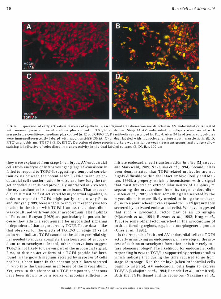
FIG. 6. Expression of early activation markers of epithelial mesenchymal transformation are detected in AV endocardial cells treatedwith mesenchyme-conditioned medium plus control or TGFb-3 antibodies. Stage 14 AV endocardial monolayers were treated withmesenchyme-conditioned medium plus control (A, B) or TGFb-3 (C, D) antibodies as described for Fig. 4. After 24 hr of treatment, cultureswere immunofluorescently labeled with rabbit anti-ES/130 (A, C) or dual labeled with monoclonal anti-a-smooth muscle actin (B, D;FITC) and rabbit anti-TGFb-3 (B, D; RITC). Detection of these protein markers was similar between treatment groups, and orange-yellowstaining is indicative of colocalized immunoreactivity in the dual-labeled cultures (B, D). Bar, 100 mm.
they were explanted from stage 14 embryos. AV endocardial initiate endocardial cell transformation in vitro (Mjaatvedtand Markwald, 1989; Nakajima et al., 1994). Second, it hascells from embryos only 8 hr younger (stage 13) consistently
failed to respond to TGFb-3, suggesting a temporal correla- been demonstrated that TGFb-related molecules are nothighly diffusible within the intact embryo (Reilly and Mel-tion exists between the potential for TGFb-3 to induce en-
docardial cell transformation in vitro and how long the tar- ton, 1996), a property which is inconsistent with a signalthat must traverse an extracellular matrix of 150-plus mmget endothelial cells had previously interacted in vivo with
the myocardium or its basement membrane. That endocar- separating the myocardium from its target endocardium(Kitten et al., 1987). Rather, something else secreted by thedial cells must be first activated by myocardial signaling in
order to respond to TGFb might partly explain why Potts myocardium is more likely needed to bring the endocar-dium to a point where it can respond to TGFb (presumablyand Runyan (1989) were unable to induce mesenchyme for-
mation in the chick with TGFb-1 unless the endocardium secreted by activated endocardial cells). We have suggestedthat such a myocardial factor may be an ES antigenwas cocultured with ventricular myocardium. The findings
of Potts and Runyan (1989) are particularly important be- (Mjaatvedt et al., 1991; Rezeaee et al., 1993; Krug et al.,1995) or some other factor enriched in the myocardium ofcause they affirm a requirement for myocardial signaling
independent of that engendered by TGFb. These data—like cushion-forming regions, e.g., bone morphogenetic protein(Jones et al., 1991).that observed for the effects of TGFb-3 on stage 13 vs 14
cultures—indicate TGFb cannot be the sole myocardial sig- Is the response of cultured AV endocardial cells to TGFbactually mimicking an endogenous, in vivo step in the pro-nal needed to induce complete transformation of endocar-
dium to mesenchyme. Indeed, other observations suggest cess of cushion mesenchyme formation, or is it merely cul-ture phenomenology? The likelihood for endocardial cellsTGFb is not likely to be even part of the myocardial signal.
First, to date no active form of a TGFb peptide has been responding in vivo to TGFb is supported by previous studieswhich indicate that during the time required to go fromfound in the growth medium secreted by myocardial cells
nor has it been found in the adheron particulates secreted stage 13 to stage 15 in the embryo (when endocardial cellsbegin to migrate), AV endocardial cells begin to expressin vivo into the basement membrane of the myocardium.
Yet, even in the absence of a TGF component, adherons TGFb-3 (Nakajima et al., 1994; Ramsdell et al., submitted).Both the TGFb ligand and its receptors (Nakajima et al.,have been shown to be a source of proteins sufficient to
Copyright q 1997 by Academic Press. All rights of reproduction in any form reserved.
AID DB 8637 / 6x29$$$$63 07-14-97 07:42:24 dba

71Endocardially Expressed TGFb-3
FIG. 7. Treatment of target AV endocardial cells with a TGFb-3 antibody in the presence of cocultured myocardial tissue. Stage 14 AVexplants containing myocardium plus endocardium attach to collagen gels [(A) surface view] and give rise to invasive mesenchyme(arrowheads) which are detected beneath the plane of the monolayer [(B) deep view] in the presence of nonimmune goat IgG. Addition ofgoat anti-TGFb-3 IgG results in endocardial cells which attach to the collagen gel [(C) surface view], but which are inhibited in migrationon top of the gel and which form little invasive mesenchyme [(D) deep view]. The inhibitory effect of the TGFb-3 antibody is overriddenby simultaneous addition of 10 ng/ml recombinant chicken TGFb-3 such that formation of invasive mesenchyme (arrowheads) is rescued[(E) surface view, (F) deep view]. Bar, 100 mm.
1994; Brown et al., 1996) continue to be expressed on endo- to this question relates to the processing of TGFb. Whileendocardial cells may begin expressing TGFb at stage 13/cardial cells migrating in vivo as free mesenchyme. Thus,
our present finding of TGFb-3 in MCCM is consistent with or 14, Ghosh and Brauer (1996) have shown that the pro-cessing of latent TGFb into an active form does not occurin vivo evidence for the secretion of this growth factor.
Therefore, if AV endocardial/mesenchymal cells respond in until stage 15, exactly the point at which endocardial forma-tion of mesenchyme becomes fully autonomous (Runyanvivo to TGFb, it is likely to be an autocrine response to
their own secretion. However, if TGFb is truly acting in an and Markwald, 1983). After stage 15, neither myocardialcoculture nor exogenous signals are required to initiate mes-autocrine mode, why must we add exogenous TGFb to stage
14 endocardial monolayers to induce their transformation enchyme formation. Thus, by adding exogenous TGFb tostage 13 and 14 cultures, we most likely bypassed the timeto mesenchyme? Presumably, if some stage 13 and 14 endo-
cardial cells are expressing TGFb in response to prior myo- required for the cells to process their own latent TGFb, anobservation supported by the recent finding of Nakajima etcardial signaling (Figs. 6 and 8), they should form mesen-
chyme if left untreated. Yet, as shown in Fig. 2, only 20% al. (1997b).Importantly, results obtained with MCCM and TGFb-3do so (and even less for stage 13). We suggest the resolution
Copyright q 1997 by Academic Press. All rights of reproduction in any form reserved.
AID DB 8637 / 6x29$$$$63 07-14-97 07:42:24 dba

72 Ramsdell and Markwald
can elicit mesenchyme migration in another appropriatelystaged (stage 14) population of endocardial cells (Figs. 4 and5). This raises the intriguing possibility that in vivo, in addi-tion to an autocrine role to initiate or sustain cell migration,secreted TGFb-3 might also feed back upon neighboringendocardial cells to promote the onset of their migratorybehavior. Feedback of this type could serve to enhance oramplify the process of mesenchyme formation originallytriggered by a myocardial signal. The actual purpose of myo-cardial signaling may be to bring an endocardial cell to adevelopmental status where it becomes competent to se-
FIG. 8. Quantitative summary of AV cocultures containing myo- crete a factor which can act both autocrinely to self-regulatecardium plus endocardium treated with either control or TGFb-3 the final steps of transformation and paracrinely to induceantibody. Stages 13/ and 14 AV cocultures containing myocardium
similar transforming events in neighboring endocardialplus endocardium treated with either nonimmune IgG or a TGFb-cells. Such an inductive interaction where target cells, in3 antibody as described for Fig. 6 were evaluated for the presenceturn, become signaling cells upon induction has been de-of invasive (seeded) mesenchyme following 24 hr of treatment. Thescribed historically as ‘‘homogenetic induction’’ (Spemann,results are expressed as averages {SD and were significantly differ-1938). With respect to cushion formation, such an inductiveent as evaluated by a x2 test for proportions (x2 Å 26.63, P õ
0.000001 for stage 13/; x2 Å 17.14, P õ 0.0001 for stage 14). Stage mechanism could mean that myocardial signaling would13/, n Å 19 (nonimmune IgG), n Å 18 (TGFb-3 antibody); stage not have to be sustained until all mesenchymal cell forma-14, n Å 12 (nonimmune IgG), n Å 12 (TGFb-3 antibody).
antibodies indicate that the TGFb-3 secreted by trans-forming cushion cells, like that of exogenously addedTGFb-3, is sufficient to initiate endocardial cell migration.The ability of TGFb-3 to elicit changes in morphology andprotein expression associated with a motile phenotype isnot unique to AV endocardial cells, but rather has also beenobserved for other cell types (reviewed by Sporn and Rob-erts, 1988; Miyazono et al., 1994). In a recent study, wefound that TGFb-3 increased expression of a-smooth mus-cle actin in the migratory appendages of cultured cushionendocardial cells (Nakajima et al., 1997a). Others have simi-larly predicted or concluded that TGFb affects migratorybehavior required to form cushion mesenchyme (Potts andRunyan, 1991; Brown et al., 1996; Ghosh and Brauer, 1996).To that, we now propose that a relevant source of TGFb isfrom the transforming endocardial/mesenchymal cellsthemselves and that their secretion (and processing) ofTGFb-3 specifically is an inducible (target cell) response toin vivo interaction with the myocardium.
Thus, two signaling pathways, myocardial and endocar-dial/mesenchymal, are seemingly needed to bring endocar-dial cells to a point where they can transform into mesen-chyme. The data would suggest that the myocardial path-way first signals the activation of the endocardial pathway
FIG. 9. Expression of mesenchymal cell protein markers by cul-by some mechanism other than TGFb-3. Second, the endo-tured AV endocardial cells. AV explants containing myocardiumcardial pathway signals invasive mesenchyme formationplus endocardium treated with either control [(A) deep view ofthrough TGFb-3 functioning in an autocrine manner. Fi-invaded mesenchyme] or TGFb-3 antibody [(B) surface view] asnally, there was no evidence that the two signaling path-described for Fig. 7 were immunofluorescently dual labeled for a-
ways are functionally redundant. smooth muscle actin (FITC, green) and TGFb-3 (RITC, red). Or-One further significance of an ‘‘endocardial signaling ange-yellow staining is indicative of colocalized immunoreactivity.
pathway’’ suggested by the results obtained with MCCM The myocardium was removed from the TGFb-3 antibody-treatedand anti-TGFb antibodies is that TGFb-3 secreted by one culture after immunohistochemical labeling to facilitate viewing
of the endocardial monolayer. Bars, 100 mm.population of transforming endocardial/mesenchymal cells
Copyright q 1997 by Academic Press. All rights of reproduction in any form reserved.
AID DB 8637 / 6x29$$$$63 07-14-97 07:42:24 dba

73Endocardially Expressed TGFb-3
for nodal in the formation and maintenance of the primitivetion had been completed nor would each potential targetstreak in the mouse. Development 120, 1919–1928.endothelial cell have to be directly activated by a myocar-
Delannet, M., and Duband, J.-L. (1992). Transforming growth fac-dial signaling molecule to initiate invasive migration.tor-beta control of cell-substratum adhesion during avian neuralTGFb-3 is such a candidate homogenetic inducer: its ex-crest cell emigration in vitro. Development 116, 275–287.pression is markedly enhanced after myocardial signaling
Eisenberg, L. M., and Markwald, R. R. (1995). Molecular regulation(Nakajima et al., 1994) and, as shown in the present study, of atrioventricular valvuloseptal morphogenesis. Circ. Res. 77,TGFb-3 secreted into MCCM promotes invasive migration. 1–6.Identification of TGFb-3 as a candidate autocrine/paracrine- Ghosh, S., and Brauer, P. R. (1996). Latent TGF-beta is present inacting signal involved in endocardial cell migration and in- the extracellular matrix of embryonic hearts in situ. Dev. Dyn.vasion in vitro will facilitate future studies of endocardial 205, 126–134.
Hamburger, V., and Hamilton, H. E. (1951). A series of normalcushion formation in vivo.stages in the development of the chick embryo. J. Morphol. 88,The heart is not the only epithelial–mesenchymal trans-49–92.formation in which a role for TGFb has been proposed.
Hay, E. D. (1995). An overview of epithelio-mesenchymal transfor-TGFb family members have been reported to mediate cellmation. Acta. Anat. 154, 8–20.motility during gastrulation (Conlon et al., 1994) and neural
Huang, J.-X., Potts, J. D., Vincent, E. B., Weeks, D. L., and Runyan,crest cell emigration (Delannet and Duband, 1992; BaslerR. B. (1995). Mechanisms of cell transformation in the embryonic
et al., 1993). In these two systems as well as in the heart, heart. Ann. N. Y. Acad. Sci. 752, 317–330.the TGFb-related proteins important for mesenchyme for- Icardo, J. M. (1989). Changes in endocardial cell morphology duringmation appear to be an autocrine mechanism to complete, development of the endocardial cushions. Anat. Embryol. 179,but not actually activate, the transformation process (Basler 443–448.et al., 1993; Brauer and Yee, 1993). Together, these findings Isokawa, K., Rezaee, M., Wunsch, A. M., Markwald, R. R., and
Krug, E. L. (1994). Identification of transferrin as one of multipleraise the possibility that epithelial–mesenchymal transfor-EDTA-extractable extracellular proteins involved in early chickmations occuring in diverse systems during vertebrate em-heart morphogenesis. J. Cell. Biochem. 55, 1–12.bryogenesis may be regulated through a common, progres-
Jones, C. M., Lyons, K. M., and Hogan, B. L. (1991). Involvement ofsive process involving TGFb-mediated autocrine or para-bone morphogenetic protein-4 (BMP-4) and Vgr-1 in morphogene-crine signaling.sis and neurogenesis in the mouse. Development 111, 531–542.
Kinsella, M. G., and Fitzharris, T. P. (1979). Origin of cushion tissuein the developing chick heart: Cinematographic recordings of in
ACKNOWLEDGMENTS situ formation. Science 207, 1359–1360.Kitten, G. T., Markwald, R. R., and Bolender, D. L. (1987). Distribu-
We thank Joshua Spruill, Dr. Thomas Trusk, and Sue Tjepkema- tion of basement membrane antigens in cryopreserved early em-Burrows for invaluable assistance in preparing the figures; Dr. Phil- bryonic hearts. Anat. Rec. 217, 379–390.lip Rust for performing the statistical analyses; Dr. Philip Brauer Krug, E. L., Mjaatvedt, C. H., and Markwald, R. R. (1987). Extracel-for helpful discussion; and Dr. Robert Gourdie for critically reading lular matrix from embryonic myocardium elicits an early mor-the manuscript. This study was supported by NIH Grants HL 33756 phogenetic event in cardiac endothelial cell differentiation. Dev.and HL 52813. Biol. 120, 348–355.
Krug, E. L., Runyan, R. B., and Markwald, R. R. (1985). Protein ex-tracts from early embryonic heart initiate cardiac endothelialcytodifferentiation. Dev. Biol. 112, 414–426.REFERENCES
Markwald, R. R., and Funderburg, F. M. (1983). Use of 6-diazo-5-oxo-L-norleucine to study interaction between myocardial glyco-Akhurst, R. J., Lehnert, S. A., Faissner, A., and Duffie, E. (1990).conjugate secretion and endothelial activation in the early em-TGF beta in murine morphogenetic processes: The early embryobryonic chick heart. Dev. Biol. 99, 395–407.and cardiogenesis. Development 108, 645–656.
Markwald, R. R., Fitzharris, T. P., and Adams Smith, W. N. (1975).Basler, K., Edlund, T., Jessell, T. M., and Yamada, T. (1993). ControlStructural analysis of endocardial cytodifferentiation. Dev. Biol.of cell pattern in the neural tube: Regulation of cell differentia-42, 160–180.tion by dorsalin-1, a novel TGF beta family member. Cell 73,
Markwald, R. R., Kitten, G. T., Funderburg, F. M., Bernanke, D. H.,687–702.and Brauer, P. R. (1984). Use of collagen gel cultures to studyBernanke, D. H., and Markwald, R. R. (1982). Migratory behaviordevelopment: Proteoglycan and glycoprotein interactions duringof cardiac cushion tissue cells in a collagen lattice culture sys-formation of endocardial cushion tissue. In ‘‘The Role of thetem. Dev. Biol. 91, 235–245.Extracellular Matrix in Development’’ (R. L. Trelstad, Ed.), pp.Bolender, D. L., and Markwald, R. R. (1979). Epithelial–mesenchy-323–350. A. R. Liss, New York.mal transformation in atrioventricular cushion tissue morpho-
Markwald, R., Eisenberg, L., Eisenberg, C., and Sugi, Y. (1996). Epi-genesis. Scanning Electron Microsc. 3, 313–321.thelial–mesenchymal transformations in early avian heart devel-Brown, C. B., Boyer, A. S., Runyan, R. B., and Barnett, J. V. (1996).opment. Acta Anat. 156, 173–186.Antibodies to the type II TGF beta receptor block cell activation
Miyazono, K., Ten Dijke, P., Ichijo, H., and Heldin, C.-H. (1994).and migration during atrioventricular cushion transformation inReceptors for transforming growth factor-b. Adv. Immunol. 55,the heart. Dev. Biol. 174, 248–257.181–221.Conlon, F. L., Lyons, K. M., Takaesu, N., Barth, K. S., Kispert, A.,
Herrmann, B., and Robertson, E. J. (1994). A primary requirement Mjaatvedt, C. H., and Markwald, R. R. (1989). Induction of an epi-
Copyright q 1997 by Academic Press. All rights of reproduction in any form reserved.
AID DB 8637 / 6x29$$$$63 07-14-97 07:42:24 dba

74 Ramsdell and Markwald
thelial–mesenchymal transition by an in vivo adheron-like com- Sense and antisense TGFb3 mRNA levels correlate with cardiacvalve induction. Dev. Dyn. 193, 340–345.plex. Dev. Biol. 136, 118–128.
Ramsdell, A. F., Krug, E. L., and Markwald, R. R. Regulation of val-Mjaatvedt, C. H., Krug, E. L., and Markwald, R. R. (1991). An antise-vuloseptal morphogenesis: Patterning of ES/130 expression sug-rum (ES1) against a particulate form of extracellular matrixgests induction of cushion mesenchyme is a progressive, homoge-blocks the transformation of cardiac endothelium into mesen-netic event. [Submitted for publication]chyme in culture. Dev. Biol. 145, 219–230.
Reilly, K. M., and Melton, D. A. (1996). Short-range signaling byMoreno-Rodriguez, R. A., De la Cruz, M. V., and Krug, E. L. (1997).candidate morphogens of the transforming growth factor betaTemporal and spatial asymmetries in the initial distribution offamily and evidence for a relay mechanism of induction. Cell 86,mesenchyme cells in the atrioventricular canal cushions of the743–754.developing chick heart. Anat. Rec. 248, 84–92.
Rezaee, M., Isokawa, K., Halligan, N., Markwald, R. R., and Krug,Nakajima, Y., Krug, E. L., and Markwald, R. R. (1994). MyocardialE. L. (1993). Identification of an extracellular 130 kDa proteinregulation of transforming growth factor-beta expression by out-involved in early cardiac morphogenesis. J. Biol. Chem. 268,flow tract endothelium in the early embryonic chick heart. Dev.14404–14411.
Biol. 165, 615–626.Runyan, R. B., and Markwald, R. R. (1983). Invasion of mesen-
Nakajima, Y., Mironov, V., Yamagishi, T., Nakamura, H., and chyme into three-dimensional collagen gels: A regional and tem-Markwald, R. R. (1997a). Expression of smooth muscle alpha- poral analysis of interaction in embryonic heart tissue. Dev. Biol.actin in mesenchymal cells during formation of avian endocar- 95, 108–114.dial cushion tissue: A role for transforming growth factor beta- Runyan, R. B., Potts, J. D., and Weeks, D. L. (1992). TGF-beta-3-3. Dev. Dyn. [In press] mediated tissue interaction during embryonic heart develop-
Nakajima, Y., Miyazono, K., Kato, M., Takase, M., Yamagishi, T., ment. Mol. Reprod. Dev. 32, 152–159.and Nakamura, H. (1997b). Extracellular fibrillar structure of la- Sinning, A. R., Hewitt, C. C., and Markwald, R. R. (1995). A subsettent TGF beta binding protein-1: Role in TGF beta-dependent of SBA lectin-binding proteins isolated from myocardial-condi-endothelial–mesenchymal transformation dring endocardial tioned media transforms cardiac endothelium into mesenchyme.cushion tissue formation in mouse embryonic heart. J. Cell Biol. Acta Anat. 154, 111–119.136, 193–204. Spemann, H. (1938). ‘‘Embryonic Development and Induction.’’
Potts, J. D., and Runyan, R. B. (1989). Epithelial–mesenchymal cell Yale Univ. Press, New Haven, CT.Sporn, M. B., and Roberts, A. B. (1988). Peptide growth factors aretransformation in the embryonic heart can be mediated, in part,
multifunctional. Nature 332, 217–219.by transforming growth factor beta. Dev. Biol. 134, 392–401.Wunsch, A. M., Little, C. D., and Markwald, R. R. (1994). CardiacPotts, J. D., Dagle, J. M., Walder, J. A., Weeks, D. L., and Runyan,
endothelial heterogeneity defines valvular development as dem-R. B. (1991). Epithelial–mesenchymal transformation of embry-onstrated by the diverse expression of JB3, an antigen of the endo-onic cardiac endothelial cells is inhibited by a modified antisensecardial cushion tissue. Dev. Biol. 165, 585–601.oligonucleotide to transforming growth factor beta-3. Proc. Natl.
Acd. Sci. USA 88, 1516–1520. Received for publication January 3, 1997Accepted May 27, 1997Potts, J. D., Vincent, E. B., Runyan, R. B., and Weeks, D. L. (1992).
Copyright q 1997 by Academic Press. All rights of reproduction in any form reserved.
AID DB 8637 / 6x29$$$$64 07-14-97 07:42:24 dba
Copyright © 2022 FDOKUMEN