ALK4 functions as a receptor for multiple TGFβ-related ligands to regulate left–right axis...
15
ALK4 functions as a receptor for multiple TGFh-related ligands to regulate left–right axis determination and mesoderm induction in Xenopus Yumei Chen, a Ekaterina Mironova, b Lisha L. Whitaker, b Laura Edwards, b,c H. Joseph Yost, d and Ann F. Ramsdell a,b,c, * a Department of Cell Biology and Anatomy, Medical University of South Carolina, Charleston, SC 29425, USA b Department of Cell and Developmental Biology and Anatomy, School of Medicine, University of South Carolina, Columbia, SC 29208, USA c Program in Women’s Studies, University of South Carolina, Columbia, SC 29208 USA d Huntsman Cancer Institute, Center for Children, University of Utah, Salt Lake City, UT 84112 USA Received for publication 2 January 2003, revised 16 December 2003, accepted 16 December 2003 Abstract In Xenopus, several TGFhs, including nodal-related 1 (Xnr1), derriere, and chimeric forms of Vg1, elicit cardiac and visceral organ left – right (LR) defects when ectopically targeted to right mesendoderm cell lineages, suggesting that LR axis determination may require activity of one or more TGFhs. However, it is not known which, if any, of these ligands is required for LR axis determination, nor is it known which type I TGFh receptor(s) are involved in mediating left-side TGFh signaling. We report here that similar to effects of ectopic TGFhs, right- side expression of constitutively active activin-like kinase (ALK) 4 results in LR organ reversals as well as altered Pitx2 expression in the lateral plate mesoderm. Moreover, left-side expression of a kinase-deficient, dominant-negative ALK4 (DN-ALK4) or an ALK4 antisense morpholino also results in abnormal embryonic body situs, demonstrating a left-side requirement for ALK4 signaling. To determine which TGFh(s) utilize the ALK4 pathway to mediate LR development, biochemical and functional assays were performed using an Activin-Vg1 chimera (AVg), Xnr1, and derriere. Whereas ALK4 can co-immunoprecipitate all of these TGFhs, including endogenous Vg1 protein from embryo homogenates, functional assays demonstrate that not all of these ligands require an intact ALK4 signaling pathway to modulate LR asymmetry. When AVg and DN-ALK4 are co-expressed, LR defects otherwise induced by AVg alone are attenuated by DN-ALK4; however, when functional assays are performed with Xnr1 or derriere, LR defects otherwise elicited by these ligands alone still occur in the presence of DN-ALK4. Intriguingly, when any of these TGFhs is expressed at a higher concentration to elicit primary axis defects, DN-ALK4 blocks gastrulation and dorsoanterior/ventroposterior defects that otherwise occur following ligand-only expression. Together, these results suggest not only that ALK4 interacts with multiple TGFhs to generate embryonic pattern, but also that ALK4 ligands differentially utilize the ALK4 pathway to regulate distinct aspects of axial pattern, with Vg1 as a modulator of ALK4 function in LR axis determination and Vg1, Xnr1, and derriere as modulators of ALK4 function in mesoderm induction during primary axis formation. D 2004 Elsevier Inc. All rights reserved. Keywords: ActRIB; ALK4; Derriere; Heterotaxy; Left – right asymmetry; Xnr1; Nodal; TGFh; Vg1 Introduction Left–right (LR) axis determination is a highly conserved feature of vertebrate embryogenesis by which the heart, brain, and visceral organs develop LR asymmetries that are consistently oriented with respect to the dorsoventral and anteroposterior body axes. Although morphological asym- metry in vertebrates is not apparent until formation of the looped, embryonic heart tube, many genes that are critical to establishing LR pattern are asymmetrically expressed before the onset of organogenesis (Kathiriya and Srivastava, 2000; Mercola and Levin, 2001; Ramsdell and Yost, 2001). Examples include Xnr1/nodal and Pitx2, two genes that are expressed by the left lateral plate mesoderm and that are required for execution of LR organ morphogenesis in all vertebrates thus far investigated (Capdevila et al., 2000; Ramsdell and Yost, 1998; Whitman, 1998). Altered Xnr1/ nodal and Pitx2 expression patterns are associated with defects in LR axis determination in several animal models 0012-1606/$ - see front matter D 2004 Elsevier Inc. All rights reserved. doi:10.1016/j.ydbio.2003.12.035 * Corresponding author. Department of Cell Biology and Anatomy, Medical University of South Carolina, Suite 601, 171 Ashley Avenue, Charleston, SC 29425. Fax: +1-843-792-0664. E-mail address: [email protected] (A.F. Ramsdell). www.elsevier.com/locate/ydbio Developmental Biology 268 (2004) 280 – 294
Transcript of ALK4 functions as a receptor for multiple TGFβ-related ligands to regulate left–right axis...

www.elsevier.com/locate/ydbio
Developmental Biology 268 (2004) 280–294
ALK4 functions as a receptor for multiple TGFh-related ligands to
regulate left–right axis determination and mesoderm induction in Xenopus
Yumei Chen,a Ekaterina Mironova,b Lisha L. Whitaker,b Laura Edwards,b,c
H. Joseph Yost,d and Ann F. Ramsdella,b,c,*
aDepartment of Cell Biology and Anatomy, Medical University of South Carolina, Charleston, SC 29425, USAbDepartment of Cell and Developmental Biology and Anatomy, School of Medicine, University of South Carolina, Columbia, SC 29208, USA
cProgram in Women’s Studies, University of South Carolina, Columbia, SC 29208 USAdHuntsman Cancer Institute, Center for Children, University of Utah, Salt Lake City, UT 84112 USA
Received for publication 2 January 2003, revised 16 December 2003, accepted 16 December 2003
Abstract
In Xenopus, several TGFhs, including nodal-related 1 (Xnr1), derriere, and chimeric forms of Vg1, elicit cardiac and visceral organ left–
right (LR) defects when ectopically targeted to right mesendoderm cell lineages, suggesting that LR axis determination may require activity
of one or more TGFhs. However, it is not known which, if any, of these ligands is required for LR axis determination, nor is it known which
type I TGFh receptor(s) are involved in mediating left-side TGFh signaling. We report here that similar to effects of ectopic TGFhs, right-side expression of constitutively active activin-like kinase (ALK) 4 results in LR organ reversals as well as altered Pitx2 expression in the
lateral plate mesoderm. Moreover, left-side expression of a kinase-deficient, dominant-negative ALK4 (DN-ALK4) or an ALK4 antisense
morpholino also results in abnormal embryonic body situs, demonstrating a left-side requirement for ALK4 signaling. To determine which
TGFh(s) utilize the ALK4 pathway to mediate LR development, biochemical and functional assays were performed using an Activin-Vg1
chimera (AVg), Xnr1, and derriere. Whereas ALK4 can co-immunoprecipitate all of these TGFhs, including endogenous Vg1 protein from
embryo homogenates, functional assays demonstrate that not all of these ligands require an intact ALK4 signaling pathway to modulate LR
asymmetry. When AVg and DN-ALK4 are co-expressed, LR defects otherwise induced by AVg alone are attenuated by DN-ALK4; however,
when functional assays are performed with Xnr1 or derriere, LR defects otherwise elicited by these ligands alone still occur in the presence of
DN-ALK4. Intriguingly, when any of these TGFhs is expressed at a higher concentration to elicit primary axis defects, DN-ALK4 blocks
gastrulation and dorsoanterior/ventroposterior defects that otherwise occur following ligand-only expression. Together, these results suggest
not only that ALK4 interacts with multiple TGFhs to generate embryonic pattern, but also that ALK4 ligands differentially utilize the ALK4
pathway to regulate distinct aspects of axial pattern, with Vg1 as a modulator of ALK4 function in LR axis determination and Vg1, Xnr1, and
derriere as modulators of ALK4 function in mesoderm induction during primary axis formation.
D 2004 Elsevier Inc. All rights reserved.
Keywords: ActRIB; ALK4; Derriere; Heterotaxy; Left– right asymmetry; Xnr1; Nodal; TGFh; Vg1
Introduction metry in vertebrates is not apparent until formation of the
Left–right (LR) axis determination is a highly conserved
feature of vertebrate embryogenesis by which the heart,
brain, and visceral organs develop LR asymmetries that are
consistently oriented with respect to the dorsoventral and
anteroposterior body axes. Although morphological asym-
0012-1606/$ - see front matter D 2004 Elsevier Inc. All rights reserved.
doi:10.1016/j.ydbio.2003.12.035
* Corresponding author. Department of Cell Biology and Anatomy,
Medical University of South Carolina, Suite 601, 171 Ashley Avenue,
Charleston, SC 29425. Fax: +1-843-792-0664.
E-mail address: [email protected] (A.F. Ramsdell).
looped, embryonic heart tube, many genes that are critical to
establishing LR pattern are asymmetrically expressed before
the onset of organogenesis (Kathiriya and Srivastava, 2000;
Mercola and Levin, 2001; Ramsdell and Yost, 2001).
Examples include Xnr1/nodal and Pitx2, two genes that
are expressed by the left lateral plate mesoderm and that are
required for execution of LR organ morphogenesis in all
vertebrates thus far investigated (Capdevila et al., 2000;
Ramsdell and Yost, 1998; Whitman, 1998). Altered Xnr1/
nodal and Pitx2 expression patterns are associated with
defects in LR axis determination in several animal models

Y. Chen et al. / Developmental Biology 268 (2004) 280–294 281
of LR development (Collignon et al., 1996; Hyatt and Yost,
1998; Levin et al., 1997; Lohr et al., 1997; Lowe et al.,
1996; Piedra et al., 1998; Yoshioka et al., 1998), and direct
misexpression of Xnr1/nodal or Pitx2 similarly results in
defects in heart and visceral organ LR asymmetry (Cam-
pione et al., 1999; Levin et al., 1997; Logan et al., 1998;
Sampath et al., 1997).
Whereas much progress has been made in elucidating
the cellular and molecular interactions that are proximal to
Xnr1/nodal and Pitx2 expression in the lateral plate me-
soderm (Capdevila et al., 2000; Whitman and Mercola,
2001), the LR signaling pathways that function much
earlier in development to initiate the LR axis remain less
defined. In Xenopus, several studies have implicated a role
for TGFh signaling in establishing the LR axis (Fig. 1).
Specifically, activation of a Smad-dependent, BMP/ALK2
pathway in right mesendoderm cell lineages is required for
proper LR determination of the heart and visceral organs
(Ramsdell and Yost, 1999). This pathway also regulates
Xnr1 and Pitx2 expression in the lateral plate mesoderm,
indicating that BMP/ALK2 signaling functions upstream of
midline LR specification (Ramsdell and Yost, 1999). With
regard to left cell lineages, other studies have shown that
ectopic TGFh misexpression in right mesendoderm cells of
the early Xenopus embryo also results in laterality defects,
suggesting a role for additional TGFh pathway(s) in LR
axis determination. Specifically, right-side misexpression
of activin (Hyatt and Yost, 1998), Xnr1 (Sampath et al.,
1997), derriere (Hanafusa et al., 2000b), and chimeric
forms of Vg1 (Branford et al., 2000; Hyatt et al., 1996;
Hyatt and Yost, 1998, Ramsdell and Yost, 1999) all result
in significant LR defects, suggesting that one or more of
these factors may be involved in a left-side pathway to
mediate LR axis formation. Expression of a dominant-
Fig. 1. TGFh signaling in Xenopus LR development. Two distinct TGFh pathways
embryo (A), unidentified TGFh(s) bind to a Type I/Type II TGFh receptor comp
lineages. Candidates for the left-side TGFh signal include Xnr1/nodal, derriere, a
side of the embryo (B), BMP binds to a Type I/Type II TGFh receptor complex co
specify right cell lineages. Identification of the Type I ALK receptor and its ligand
aspect of this model is numbered to correspond to references to be seen for furthe
(1998), Hyatt et al. (1996), Ramsdell and Yost (1999), Sampath et al. (1997); 2
candidates based on Ramsdell and Yost (1999); 4—Ramsdell and Yost (1999); 5
negative form of the type II TGFh receptor, ActRIIb, also
results in LR organ reversals when targeted to the left side
of the Xenopus embryo (Hyatt and Yost, 1998), supporting
the interpretation that endogenous TGFh signaling is
required in left cell lineages. However, due to the ability
of this dominant-negative receptor to interfere with activity
of multiple TGFhs (Chang et al., 1997; Frisch and Wright,
1998; Gray et al., 2000; Hemmati-Brivanlou and Melton,
1992; Nagaso et al., 1999; Reissmann et al., 2001; Sakuma
et al., 2002; Schulte-Merker et al., 1994; Wilson and
Hemmati-Brivanlou, 1995), it remains unclear which, if
any, of the abovementioned TGFhs are involved in LR
axis determination.
Beginning with use of constitutively active and domi-
nant-negative forms of type I activin-like kinase (ALK)
receptors, we present evidence that left-side TGFh signaling
is indeed required for LR axis determination in Xenopus.
Results obtained with an in vivo laterality assay demonstrate
that ALK4 (also termed ActRIb) is both necessary and
sufficient to modulate morphological and molecular aspects
of LR development. Analysis of candidate ALK4 ligands
(Vg1, Xnr1, derriere) reveals that although each of these
growth factors is capable of binding to ALK4 and regulating
both primary and LR axial pattern, they differ in whether
they mediate such embryonic patterning events via the
ALK4 pathway. Specifically, our results suggest that where-
as Vg1 acts through the ALK4 pathway to modulate LR axis
determination, Xnr1 and derriere do not. Although it is not
known which other ALK pathway(s) are utilized by Xnr1
and derriere to modulate LR development, our results
suggest that in addition to Vg1, these two TGFhs utilize
the ALK4 pathway to modulate mesoderm induction and
dorsoanterior/ventroposterior development during primary
axis formation.
are required for normal LR development in Xenopus. On the left side of the
lex composed of ActRIIb and an unknown Type I ALK to specify left cell
nd Vg1; ALK4 is a candidate for the left-side Type I receptor. On the right
mposed of ALK2 and an unknown Type II receptor (probably BMPRIIa) to
(s) that function as left-side determinants is the focus of this paper, and each
r details: 1—Branford et al. (2000), Hanafusa et al. (2000a), Hyatt and Yost
—Hyatt and Yost (1998); 3—ALK2 and ALK3 have been eliminated as
—Frisch and Wright (1998); 6—Ramsdell and Yost (1999).
Y. Chen et al. / Developmental Biology 268 (2004) 280–294282
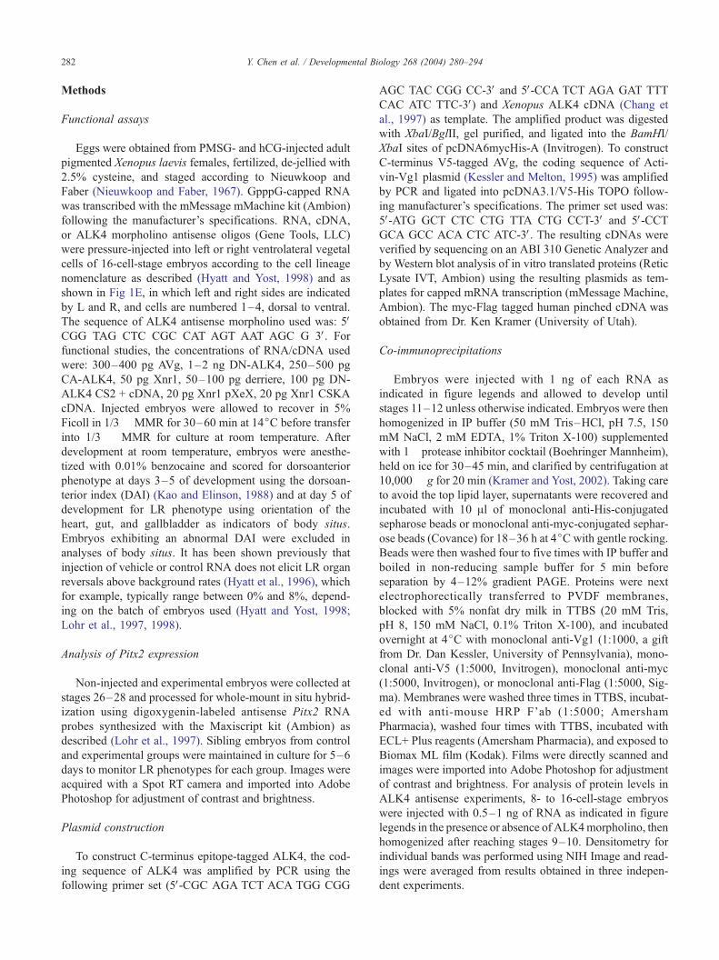
Methods
Functional assays
Eggs were obtained from PMSG- and hCG-injected adult
pigmented Xenopus laevis females, fertilized, de-jellied with
2.5% cysteine, and staged according to Nieuwkoop and
Faber (Nieuwkoop and Faber, 1967). GpppG-capped RNA
was transcribed with the mMessage mMachine kit (Ambion)
following the manufacturer’s specifications. RNA, cDNA,
or ALK4 morpholino antisense oligos (Gene Tools, LLC)
were pressure-injected into left or right ventrolateral vegetal
cells of 16-cell-stage embryos according to the cell lineage
nomenclature as described (Hyatt and Yost, 1998) and as
shown in Fig 1E, in which left and right sides are indicated
by L and R, and cells are numbered 1–4, dorsal to ventral.
The sequence of ALK4 antisense morpholino used was: 5VCGG TAG CTC CGC CAT AGT AAT AGC G 3V. For
functional studies, the concentrations of RNA/cDNA used
were: 300–400 pg AVg, 1–2 ng DN-ALK4, 250–500 pg
CA-ALK4, 50 pg Xnr1, 50–100 pg derriere, 100 pg DN-
ALK4 CS2 + cDNA, 20 pg Xnr1 pXeX, 20 pg Xnr1 CSKA
cDNA. Injected embryos were allowed to recover in 5%
Ficoll in 1/3 �MMR for 30–60 min at 14jC before transfer
into 1/3 � MMR for culture at room temperature. After
development at room temperature, embryos were anesthe-
tized with 0.01% benzocaine and scored for dorsoanterior
phenotype at days 3–5 of development using the dorsoan-
terior index (DAI) (Kao and Elinson, 1988) and at day 5 of
development for LR phenotype using orientation of the
heart, gut, and gallbladder as indicators of body situs.
Embryos exhibiting an abnormal DAI were excluded in
analyses of body situs. It has been shown previously that
injection of vehicle or control RNA does not elicit LR organ
reversals above background rates (Hyatt et al., 1996), which
for example, typically range between 0% and 8%, depend-
ing on the batch of embryos used (Hyatt and Yost, 1998;
Lohr et al., 1997, 1998).
Analysis of Pitx2 expression
Non-injected and experimental embryos were collected at
stages 26–28 and processed for whole-mount in situ hybrid-
ization using digoxygenin-labeled antisense Pitx2 RNA
probes synthesized with the Maxiscript kit (Ambion) as
described (Lohr et al., 1997). Sibling embryos from control
and experimental groups were maintained in culture for 5–6
days to monitor LR phenotypes for each group. Images were
acquired with a Spot RT camera and imported into Adobe
Photoshop for adjustment of contrast and brightness.
Plasmid construction
To construct C-terminus epitope-tagged ALK4, the cod-
ing sequence of ALK4 was amplified by PCR using the
following primer set (5V-CGC AGA TCT ACA TGG CGG
AGC TAC CGG CC-3V and 5V-CCA TCT AGA GAT TTT
CAC ATC TTC-3V) and Xenopus ALK4 cDNA (Chang et
al., 1997) as template. The amplified product was digested
with XbaI/BglII, gel purified, and ligated into the BamHI/
XbaI sites of pcDNA6mycHis-A (Invitrogen). To construct
C-terminus V5-tagged AVg, the coding sequence of Acti-
vin-Vg1 plasmid (Kessler and Melton, 1995) was amplified
by PCR and ligated into pcDNA3.1/V5-His TOPO follow-
ing manufacturer’s specifications. The primer set used was:
5V-ATG GCT CTC CTG TTA CTG CCT-3V and 5V-CCTGCA GCC ACA CTC ATC-3V. The resulting cDNAs were
verified by sequencing on an ABI 310 Genetic Analyzer and
by Western blot analysis of in vitro translated proteins (Retic
Lysate IVT, Ambion) using the resulting plasmids as tem-
plates for capped mRNA transcription (mMessage Machine,
Ambion). The myc-Flag tagged human pinched cDNA was
obtained from Dr. Ken Kramer (University of Utah).
Co-immunoprecipitations
Embryos were injected with 1 ng of each RNA as
indicated in figure legends and allowed to develop until
stages 11–12 unless otherwise indicated. Embryos were then
homogenized in IP buffer (50 mM Tris–HCl, pH 7.5, 150
mM NaCl, 2 mM EDTA, 1% Triton X-100) supplemented
with 1� protease inhibitor cocktail (Boehringer Mannheim),
held on ice for 30–45 min, and clarified by centrifugation at
10,000� g for 20 min (Kramer and Yost, 2002). Taking care
to avoid the top lipid layer, supernatants were recovered and
incubated with 10 Al of monoclonal anti-His-conjugated
sepharose beads or monoclonal anti-myc-conjugated sephar-
ose beads (Covance) for 18–36 h at 4jC with gentle rocking.
Beads were then washed four to five times with IP buffer and
boiled in non-reducing sample buffer for 5 min before
separation by 4–12% gradient PAGE. Proteins were next
electrophorectically transferred to PVDF membranes,
blocked with 5% nonfat dry milk in TTBS (20 mM Tris,
pH 8, 150 mM NaCl, 0.1% Triton X-100), and incubated
overnight at 4jC with monoclonal anti-Vg1 (1:1000, a gift
from Dr. Dan Kessler, University of Pennsylvania), mono-
clonal anti-V5 (1:5000, Invitrogen), monoclonal anti-myc
(1:5000, Invitrogen), or monoclonal anti-Flag (1:5000, Sig-
ma). Membranes were washed three times in TTBS, incubat-
ed with anti-mouse HRP F’ab (1:5000; Amersham
Pharmacia), washed four times with TTBS, incubated with
ECL+ Plus reagents (Amersham Pharmacia), and exposed to
Biomax ML film (Kodak). Films were directly scanned and
images were imported into Adobe Photoshop for adjustment
of contrast and brightness. For analysis of protein levels in
ALK4 antisense experiments, 8- to 16-cell-stage embryos
were injected with 0.5–1 ng of RNA as indicated in figure
legends in the presence or absence of ALK4morpholino, then
homogenized after reaching stages 9–10. Densitometry for
individual bands was performed using NIH Image and read-
ings were averaged from results obtained in three indepen-
dent experiments.

Y. Chen et al. / Developmental Biology 268 (2004) 280–294 283
Results
ALK4 signaling is both necessary and sufficient for Xenopus
LR development
To determine whether ALK4 plays a role in LR devel-
opment, RNA encoding a constitutively active form of
Fig. 2. Left– right axis defects elicited by CA-ALK4 expression. Control and exp
CA-ALK4 RNA. The orientation of embryos shown is a ventral view, with anteri
heart looping (arrowhead) plus counterclockwise coiling and leftward rotation of th
situs inversus, as defined by complete reversal of the heart and gut (B), or situs am
or normal heart looping plus reversed gut orientation (D). The vegetal cell lineage
indicated in an illustration of a 16-cell embryo (E, animal pole view). As a cont
Western blot analysis in embryos injected with epitope-tagged ALK4 in the absen
reducing expression of ectopic ALK4 by approximately 30% when densitometry r
comparison, levels of a control protein, pinched, are not decreased by co-express
ALK4 (CA-ALK4) was microinjected into defined left or
right lateral vegetal cell lineages of the 16-cell Xenopus
blastula and embryos were scored for LR body situs at days
5–6 of development (Fig. 2). CA-ALK4 contains a Thr-to-
Glu mutation near the intracellular GS domain, which
permits constitutive kinase activity in a ligand-independent
manner (Chang et al., 1997). As shown in Table 1 and Fig.
erimental embryos were scored for LR phenotype at day 5 postinjection of
or at the top. Control embryos exhibit situs solitus, as defined by rightward
e gut (A). In contrast, embryos with right-side CA-ALK4 expression exhibit
biguus, as defined by reversed heart looping plus normal gut orientation (C)
s that were targeted for left-side (L3) or right-side (R3) RNA expression are
rol for ALK4 antisense experiments (F), protein levels were examined by
ce and presence of ALK4 morpholino (MO). The ALK4 MO is effective in
eadings are averaged over the course of three independent experiments. By
ed ALK4 MO.

Table 1
Left– right axis defects are induced by ALK4 gain-of-function and ALK4
loss-of-function
RNA Cell n (LR normal) (LR abnormal)
lineagesitus
solitus
(%)
situs
ambiguus
(%)
situs
inversus
(%)
CA-ALK4 L3a 121 75 18 7
CA-ALK4 R3a 210 24 42 34
DN-ALK4 L3b 116 68 23 9
DN-ALK4 R3b 103 88 8 4
ALK4 MO L3c,d 94 28 52 20
ALK4 MO R3c 99 91 9 0
ALK4 MO + CA-ALK4 L3d 97 90 8 2
Left (L3) or right (R3) cell lineages were injected with the RNA or
antisense morpholino oligonucleotide indicated and scored for body situs as
described in Methods. The percentages of embryos with normal (situs
solitus) or abnormal (situs ambiguus, situs inversus) laterality phenotypes
are shown. Results obtained from v2 analysis indicate that the distribution
of normal vs. abnormal laterality phenotypes in embryos injected in L3
lineages is significantly different from that observed in embryos injected in
R3 lineages and that the distribution of normal vs. abnormal laterality
phenotypes in embryos injected with ALK4 morpholino (ALK4 MO) alone
in the L3 lineage is significantly different from that observed in embryos
injected with ALK4 MO plus CA-ALK4 RNA in the L3 lineage. va =
82.93, P < 0.001; vb = 13.01, P < 0.01; vc = 74.89, P < 0.001; vd = 76.15,
P < 0.001.
Y. Chen et al. / Developmental Biology 268 (2004) 280–294284
2, right-side expression of CA-ALK4 results in only 24% of
embryos exhibiting normal LR body situs (situs solitus), as
assessed by the orientation of the heart, gut, and gallbladder.
Of the remaining embryos, 42% exhibit discordant LR
organ asymmetries (situs ambiguus) and 34% exhibit a full
mirror-image reversal of LR asymmetries (situs inversus). In
contrast, expression of CA-ALK4 on the left side of the
embryo results in significantly fewer LR defects, with 75%
of embryos exhibiting situs solitus, 18% exhibiting situs
ambiguus, and 7% exhibiting situs inversus. Together, these
results indicate that ectopic ALK4 signaling not only elicits
defects in the LR body plan, but also that the maximal effect
of ectopic ALK4 signaling on LR development occurs when
right cell lineages are targeted.
The results with CA-ALK4 expression suggest that
endogenous ALK4 signaling in left cell lineages may be
required for Xenopus LR development. To test this hypoth-
esis, RNA encoding a kinase-deficient, dominant-negative
(DN) ALK4 (Chang et al., 1997) was injected into defined
left or right lateral vegetal cell lineages. Expression of DN-
ALK4 elicits the greatest percentage of LR defects when
expressed on the left side of the embryo, consistent with a
role for ALK4 signaling in specification of left cell lineages.
As shown in Table 1, 23% of embryos exhibit situs
ambiguus and 9% exhibit situs inversus following left-side
DN-ALK4 expression. DN-ALK4 expression in right cell
lineages results in significantly fewer LR defects, with only
8% of embryos exhibiting situs ambiguus and 4% exhibiting
situs inversus. By comparison to these results, injection of
DN-ALK4 cDNA, which does not express the DN receptor
until after the mid-blastula transition, fails to elicit LR
defects when targeted to either left or right cell lineages
(data not shown).
Because the effects of DN-ALK4 on LR development
were modest compared to results obtained with CA-ALK4,
a second approach using antisense morpholino ‘‘knock-
down’’ was used to confirm that ALK4 is required for LR
development. As shown in Table 1, left-side expression of
ALK4 morpholino is effective in perturbing LR axis deter-
mination, with 52% of embryos exhibiting situs ambiguus
and 20% exhibiting situs inversus. By contrast, only 9% of
embryos exhibit situs ambiguus and 0% exhibit situs inver-
sus following right-side expression. As also shown in Table
1, LR defects induced by ALK4 morpholino expression can
be rescued by co-expression of CA-ALK4, with 90% of
embryos exhibiting situs solitus. Analysis of ALK4 protein
expression in injected embryos demonstrates that the ALK4
morpholino is effective in reducing expression of ectopic
ALK4 (Fig. 2F). By comparison, levels of an ectopically
expressed control protein, pinched, are not decreased by
ALK4 morpholino expression (Fig. 2F). Together, these
results are consistent with the functional results obtained
with DN-ALK4 and provide additional evidence that ALK4
signaling is required in left but not right-side cell lineages
for LR axis determination in Xenopus.
ALK4 signaling regulates Pitx2 expression in the lateral
plate mesoderm
In all vertebrates thus far investigated, the expression of
the TGFh family member, Xnr1/nodal, in the left lateral
plate mesoderm is required for normal LR organ morpho-
genesis (Capdevila et al., 2000; Ramsdell and Yost, 1998).
Left-side expression of Xnr1/nodal, in turn, is responsible
for activating expression of the bicoid-related transcription
factor Pitx2, another laterality gene whose conserved
expression is also required for LR development of the
heart and visceral organs (Capdevila et al., 2000; Ramsdell
and Yost, 1998). To determine whether ALK4 signaling
affects laterality gene expression in the lateral plate
mesoderm, embryos injected with CA-ALK4 RNA were
examined for Pitx2 expression by whole-mount in situ
hybridization (Fig. 3). Analysis of Pitx2 expression indi-
cates that ectopic ALK4 signaling can alter expression of
this important regulator of LR development (Fig. 3).
Whereas the majority of control embryos or embryos with
left-side CA-ALK4 expression exhibit normal, left-side
Pitx2 expression (94% left, 6% absent; n = 30; 81% left,
19% absent; n = 37, respectively), approximately half of
embryos injected with CA-ALK4 RNA in right cell
lineages show bilateral (26%) or right-side only (30%)
expression (n = 32). The remaining embryos in this group
show either normal, left-side Pitx2 expression (40%), or
fail to express Pitx2 (4%). These results, coupled with the
above analyses of body situs, indicate that ALK4 signaling
is sufficient to modulate both morphological and molecu-
lar aspects of LR asymmetry.

Fig. 3. Perturbation of Pitx2 expression by ALK4. Xenopus blastulae were injected with CA-ALK4 RNA in left (L3) or right (R3) cell lineages and control and
experimental embryos were processed for whole-mount in situ hybridization to detect Pitx2 expression in the lateral plate mesoderm (ventral view,
arrowheads). The percentages of embryos exhibiting normal (left side) or abnormal (bilateral or right side only) expression patterns are indicated.
Y. Chen et al. / Developmental Biology 268 (2004) 280–294 285
Co-immunoprecipitation of Vg1-ALK4 and derriere-ALK4
protein complexes from whole embryo homogenates
To date, Xnr1 is the only TGFh that has been shown to
bind to ALK4 (Reissmann et al., 2001; Yeo and Whitman,
2001), and a more recent report shows that non-native,
chimeric forms of Vg1/Gdf1 also can bind this receptor
(Cheng et al., 2003). Thus, to determine which TGFh(s)other than Xnr1 can potentially function as ALK4 ligands to
modulate LR axis determination in Xenopus, co-immuno-
precipitations were performed using His-tagged ALK4 plus
V5-tagged versions of native Vg1 and derriere. As shown in
Fig. 4, when embryos injected with RNA encoding ALK4-
His plus Vg1-V5 are homogenized at gastrula stages and
immunoprecipitated with His antibody, both the nonpro-
cessed and processed forms of native Vg1 protein are
detected on Western blots probed with a V5 monoclonal
Fig. 4. ALK4 binds to both Vg1 and derriere. Xenopus blastulae were
injected with the RNA(s) indicated and processed for co-immunoprecip-
itation at stages 11–12 with monoclonal anti-His. Immunoprecipitated
proteins were detected by Western blot under nonreducing conditions with
monoclonal anti-V5. The nonprocessed (full length) and the processed
(mature) forms of Vg1 and derriere are indicated.
antibody. Similarly, both the nonprocessed and processed
forms of derriere are found to co-immunoprecipitate with
ALK4-His (Fig. 4). Together, these results provide biochem-
ical evidence of a type I Vg1/derriere receptor, indicating
that ALK4 may function to transduce signaling for TGFhsother than, and in addition to, nodal/Xnr-1.
Vg1, but not Xnr1 or derriere, modulates LR axis
determination via an ALK4 pathway
Because Xnr1 (Reissmann et al., 2001; Yeo and Whit-
man, 2001), Vg1, and derriere are capable of binding
ALK4, this raises the possibility that one or more of these
TGFhs may utilize the ALK4 pathway to modulate LR
development. To test this hypothesis, experiments were
performed in which each individual ligand was targeted to
right mesendoderm cell lineages in the presence and ab-
sence of DN-ALK4. As we reported previously (Hyatt and
Yost, 1998; Ramsdell and Yost, 1999), right-side expression
of AVg, a chimeric form of Vg1 containing the activin
prodomain (Kessler and Melton, 1995), results in a majority
of embryos that exhibit LR situs defects without discernible
effects on dorsoanterior development. In the current study,
right-side expression of AVg alone results in only 12% of
embryos exhibiting situs solitus, with 34% exhibiting situs
ambiguus and 54% exhibiting situs inversus (Table 2). In
the co-expression experiment, DN-ALK4 rescues LR de-
velopment to normal in a majority of embryos. For embryos
co-injected with AVg plus DN-ALK4 RNAs, 72% of
embryos exhibit situs solitus (Table 2). Of the remaining
embryos in this group, only 20% exhibit situs ambiguus and
8% situs inversus (Table 2). Together, these results indicate
that the ability of ectopic Vg1 to mediate LR patterning in
Xenopus requires a functionally intact ALK4 signaling
pathway.
To determine whether the other TGFhs that bind to
ALK4 modulate LR axis determination via the ALK4
pathway, the DN-ALK4 co-expression experiment also
was performed using Xnr1 and derriere. As shown in Fig.
5 and as reported previously (Jones et al., 1995; Sampath et
al., 1997), when Xnr1 RNA is misexpressed in right cell

Table 2
DN-ALK4 blocks laterality defects induced by AVg, but not by Xnr-1 nor
derriere
RNA/cDNA n DAI 5 (LR normal) (LR abnormal)
(%)Situs
solitus
(%)
Situs
ambiguus
(%)
Situs
inversus
(%)
AVg (300 pg RNA)a 84 100 12 34 54
AVg
(300 pg RNA) +
DN-ALK4 (1 ng RNA)a
82 100 72 20 8
Xnr1
(20 pg pXeX cDNA)b113 96 18 28 54
Xnr1
(20 pg pXeX cDNA) +
DN-ALK4 (1 ng RNA)b
112 88 30 40 30
Derriere (50 pg RNA)c 69 91 7 33 60
Derriere (50 pg RNA) +
DN-ALK4 (1 ng RNA)c69 91 20 24 56
Right (R3) cell lineages were injected as indicated and embryos exhibiting
normal dorsoanterior development using the dorsoanterior index (DAI) of
Kao and Elinson (1988) were scored for LR body situs as described in
Methods. The percentages of embryos exhibiting normal DAI (DAI 5) plus
either normal (situs solitus) or abnormal (situs ambiguus, situs inversus)
laterality phenotypes are shown. Results obtained from v2 analysis indicatethat the distribution of normal vs. abnormal laterality phenotypes in
embryos injected with AVg RNA is significantly different from that
observed in embryos injected with AVg plus DN-ALK4 RNAs (va = 66.31,
P < 0.001). However, the distribution of normal vs. abnormal laterality
phenotypes in embryos injected with Xnr1 cDNA alone is not significantly
different than that observed in embryos injected with Xnr1 cDNA plus DN-
ALK4 RNA (vb = 3.61, P > 0.10), nor is the distribution of normal vs.
abnormal laterality phenotypes in embryos injected with derriere RNA
alone significantly different from that observed in embryos injected with
derriere plus DN-ALK4 RNA (vc = 5.43, P > 0.10).
Y. Chen et al. / Developmental Biology 268 (2004) 280–294286
lineages, the potent mesoderm-inducing activity of Xnr1
protein results in extreme dorsoanterior defects, precluding
analysis of LR phenotype (Table 3). To circumvent this
issue, previous analyses of Xnr1 function in LR develop-
ment have relied upon use of Xnr1 cDNA expression
(Cheng et al., 2000; Sampath et al., 1997), which can be
used to elicit heart and visceral organ reversals in the
absence of significant defects in primary axis formation.
Consistent with a previous report of Xnr1 pXeX cDNA
activity (Sampath et al., 1997), we find that right-side
expression of Xnr1 pXeX alone results in a majority of
embryos that have heart and visceral organ reversals, with
28% of embryos exhibiting situs ambiguus and 54% of
embryos exhibiting situs inversus (Table 2). Surprisingly,
the effects of Xnr-1 on LR development cannot be reduced
by DN-ALK4 co-expression, which results in a majority of
embryos with LR situs defects (Table 2). The inability of
DN-ALK4 to block Xnr1-mediated LR defects is not likely
due to issues of ligand diffusibility or inefficient receptor
expression compared to ligand, because as discussed below,
we found that DN-ALK4 is able to rescue other types of
defects elicited by either Xnr1 RNA or cDNA expression.
Thus, unlike results obtained with AVg + DN-ALK4 co-
injections, these results suggest that although Xnr1 is
capable of modulating early aspects of LR development in
Xenopus, Xnr1 differs from Vg1 in that it appears to do so
via a non-ALK4 pathway. Precisely which ALK pathway is
utilized by Xnr1 in this process is not yet known; our work
thus far has eliminated ALK7 as a candidate based on the
inability of DN-ALK7 to attenuate LR defects elicited by
Xnr1 in the in vivo laterality assay. Specifically, we find that
following Xnr1 pXeX expression, 31% of embryos exhibit
situs solitus, 19% situs ambiguus, and 50% situs inversus (n
= 42). Co-expression of DN-ALK7 does not substantially
alter the distribution of LR phenotypes (24% situs solitus,
25% situs ambiguus, and 49% situs inversus; n = 50). As a
control for RNA activity, DN-ALK7 RNA was targeted to
both dorsal blastomeres of a 4-cell-stage embryo, which in
agreement with previous work (Reissmann et al., 2001),
results in extreme ventralization (100% DAI > 5; n = 25).
Derriere is 79% identical to Vg1 in the mature domain
and elicits cardiac and visceral organ reversals when ectop-
ically expressed in right cell lineages in Xenopus (Sun et al.,
1999). Consistent with this report, we find that right-side
ectopic expression of derriere results in 60% of embryos
exhibiting situs inversus and 33% exhibiting situs ambiguus
(Table 2). Co-expression of DN-ALK4 with derriere does
not significantly alter the distribution of LR phenotypes;
56% of embryos exhibit situs inversus and 24% exhibit situs
ambiguus (Table 2). Thus, in contrast to results obtained
with AVg + DN-ALK4 co-expression, DN-ALK4 does not
block LR axis defects induced by ectopic derriere, although
as discussed below, DN-ALK4 is capable of blocking
ventroposterior defects that are otherwise elicited by derriere
expression alone. Together, these data suggest that although
ALK4 is capable of functioning as a derriere receptor, an
intact ALK4 pathway is not required by derriere to mediate
its effects specifically on LR development.
Multiple ligands can modulate primary axis development
via an ALK4 pathway
In the functional laterality assays presented above, RNA
encoding each TGFh was titrated to a concentration that
causes LR axis defects without significant effects on other
early developmental events, for example, dorsoanterior
development. Because Vg1, Xnr1, and derriere can bind
to ALK4, and because it is known that each of these ligands
can play multiple roles in embryonic patterning events, we
next addressed whether they mediate other aspects of early
development via the ALK4 pathway. As shown in Table 3
and Fig. 5, when the concentrations of each TGFh are
increased over those used in the laterality assay, primary
axis formation is perturbed in a majority of embryos
expressing ligand alone. Specifically, overexpression of
AVg RNA results in two types of defects, with 36% of
embryos showing reduced dorsoanterior development plus
enhanced endoderm formation and 52% of embryos show-
ing secondary axis formation (Fig. 5A). DN-ALK4 co-
expression rescues both types of defects in nearly all

Fig. 5. Rescue of Xnr1-induced dorsoanterior defects by DN-ALK4. Right cell lineages of Xenopus blastulae were injected with RNA or cDNA encoding
TGFhs in the absence or presence of DN-ALK4 RNA as indicated in Methods. Embryos were imaged at 3–5 days following injections and incubation at
16jC. Expression of AVg RNA alone (A) results in some embryos that are ventralized with excess endoderm formation and others that exhibit secondary axis
formation (arrows). DN-ALK4 co-expression (B) blocks both types of defects. Expression of Xnr1 pCSKA alone (C) results in embryos that are ventralized as
characterized by shortened primary axes and absent eye development on the injected side of the embryo. In addition, many embryos exhibit tail defects
resulting from incomplete blastopore closure (arrowheads). DN-ALK4 co-expression blocks the ventralizing properties of Xnr1 pCSKA (D), although minor
tail defects still occur in some embryos (arrowheads). Expression of Xnr1 pXeX in the absence (E) or presence (F) of DN-ALK4 results in embryos that have
normal primary axis formation and no discernible tail defects. Expression of Xnr1 RNA alone (G) results in embryos that are hyperdorsalized and DN-ALK4
co-expression (H) blocks this effect, although some embryos still exhibit posterior tail truncation (arrowheads). Expression of derriere RNA alone (I) results in
embryos that are extremely ventralized and DN-ALK4 co-expression (J) blocks this effect. See Table 3 and text for details.
Y. Chen et al. / Developmental Biology 268 (2004) 280–294 287
embryos examined (91% normal vs. 12% following inclu-
sion of DN-ALK4; Fig. 5B). When Xnr1 cDNA is overex-
pressed from the CSKA plasmid, 91% of embryos are
ventralized (note shortened axes and impaired eye develop-
ment on injected side of embryos in Fig. 5C), with many
embryos also exhibiting incomplete blastopore closure that
later results in tail defects. Following DN-ALK4 co-expres-
sion, minor defects in blastopore closure are still observed in
some embryos, although dorsoanterior development is res-
cued in nearly all (Table 3 and Fig. 5D), leading to a
majority of embryos with normally formed primary axes
(85%) and subtle tail defects (19%). In contrast to results
obtained with the Xnr1 CSKA plasmid, overexpression of
Xnr1 from the pXeX plasmid has no significant effect on
Table 3
DN-ALK4 rescues dorsoanterior defects induced by AVg, Xnr1, and derriere
RNA/cDNA n DAI 5 (%)
AVg (400 pg RNA)a 96 12
AVg (400 pg RNA) + DN-ALK4 (1 ng RNA)a 98 91
Xnr1 (20 pg pCSKA cDNA)b 95 9
Xnr1 (20 pg pCSKA cDNA) + DN-ALK4 (1 ng RNA)b 85 66
Xnr1 (50 pg RNA)c 182 1
Xnr1 (50 pg RNA) + DN-ALK4 (1 ng RNA)c 182 54
Derriere (100 pg RNA)d 127 57
Derriere (100 pg RNA) + DN-ALK4 (1 ng RNA)d 148 89
Right (R3) cell lineages were injected with the RNA(s) and/or cDNA indicated and
Kao and Elinson (1988). The percentages of embryos with normal dorsoanterior
blastopore closure (DAI 5 + posterior truncation), and abnormal dorsoanterior deve
exhibiting primary axis duplication. Results obtained from v2 analysis indicate that thXnr1, or derriere alone is significantly different from the distribution of DAI pheno
126.56, P < 0.001; vb = 123.97, P < 0.001; vc = 298.2, P < 0.001; vd = 42.85, P
primary axis formation (Figs. 5E, F), a result that may be
related to the temporal differences in transcription due to the
EF1a promoter in pXeX driving RNA expression slightly
earlier than does the cytoskeletal actin promoter in the
pCSKA construct (Sampath et al., 1997). When Xnr1
RNA is tested in this assay, 96% of embryos are hyper-
sdorsalized (Table 3 and Fig. 5G), an effect that can be
reversed in 81% of embryos receiving co-expressed DN-
ALK4, with 27% of embryos exhibiting slight posterior
truncation due to incomplete blastopore closure (Table 3 and
Fig. 5H). Finally, we find that overexpression of derriere
alone results in 34% of embryos with decreased dorsoante-
rior development (Fig. 5I), an effect that also can be rescued
by co-expressed DN-ALK4 (Table 3 and Fig. 5J). Together,
DAI 5 + posterior
truncation (%)
DAI > 5 (%) DAI < 5 (%) Duplicated
axis (%)
0 0 36 52
0 0 6 3
0 0 91 0
19 0 15 0
0 96 3 0
27 6 13 0
9 0 34 0
6 0 5 0
scored for dorsoanterior development using the dorsoanterior index (DAI) of
development (DAI 5), normal dorsoanterior development plus incomplete
lopment (DAI > 5 or DAI < 5) are shown as are the percentages of embryos
e distribution of DAI phenotypes in embryoswith ectopic expression of AVg,
types in embryos expressing these ligands in the presence of DN-ALK4. va =< 0.001.

Y. Chen et al. / Developmental Biology 268 (2004) 280–294288
results obtained in this set of experiments demonstrate that
whereas DN-ALK4 can block primary axis defects elicited
by any of its ligands, it cannot block LR axis defects elicited
by all. It is important to emphasize that the ability of DN-
ALK4 to attenuate primary axis defects occurs at concen-
trations of TGFhs that are higher than those used to induce
LR axis defects, suggesting that ALK4 ligands differentially
utilize the ALK4 pathway to regulate distinct aspects of
embryonic pattern.
Detection of endogenously processed Vg1 protein by ALK4
co-immunoprecipitation
Although our findings strongly suggest that Vg1 is a
ligand for ALK4, the above functional assays nevertheless
used the AVg chimera to demonstrate a role for Vg1 in
ALK4-mediated LR and primary axis development. To
determine whether native Vg1 may function as an ALK4
ligand, we next addressed whether endogenous Vg1 pro-
tein(s) can be co-immunoprecipitated with ALK4-His. As
shown in Fig. 6A, we chose to perform this experiment using
blastula-stage embryos (st 8–9) because during this study,
we discovered that antibodies directed against the processed
(mature) form of Vg1 also cross-react with derriere, a finding
that is not surprising given that these two growth factors are
79% identical in their mature domains. But because derriere
is not maternally expressed (Sun et al., 1999), it is possible to
detect Vg1 without concern for derriere cross-reactivity
when embryos are homogenized before the onset of zygotic
transcription (e.g. st 8–9). As shown in Fig. 6B, when
protein complexes immunoprecipitated from st 8–9 embryos
previously injected with ALK4-His RNA are analyzed by
Western blots probed with the Vg1 monoclonal antibody,
Fig. 6. Native Vg1 protein is processed and binds to ALK4 in vivo. A
monoclonal antibody (D5) that was generated against the processed
(mature) form of Xenopus Vg1 was tested for specificity by Western blot
using in vitro translated proteins. As shown, the Vg1 antibody cross-reacts
with derriere (A), a TGFh that is zygotically expressed and that is 79%
identical to Vg1 in the processed (mature) domain. To circumvent derriere
cross-reactivity with the Vg1 antibody so that endogenous Vg1 protein
could be detected, Xenopus blastulae were injected with ALK4-His RNA
and processed for immunoprecipitation at stages 8–9 with monoclonal anti-
His. Immunoprecipitated proteins were detected under nonreducing
conditions with monoclonal anti-Vg1 (B). The nonprocessed (full length)
and the processed (mature) forms of Vg1 are indicated.
two bands are detected that migrate at approximately 50 and
25 kDa. These relative molecular weights correspond with
both the nonprocessed and processed forms of Vg1. Thus,
although the level of endogenous mature Vg1 expression in
the gastrula-stage Xenopus embryo is low (Tannahill and
Melton, 1989), our results demonstrate not only that native
Vg1 is an ALK4 ligand, but also that the processed, mature
form nonetheless is present in sufficient quantity to be
detected in the ALK4 co-immunoprecipitation assay.
Discussion
Although TGFh family members are widely implicated
as important modulators of vertebrate LR patterning (Whit-
man and Mercola, 2001), little information is available
regarding the identity of the type I receptors and other
upstream effectors of TGFh signaling that are involved in
regulating LR development. In fact, to date, the only other
type I TGFh receptor that has been identified as a modulator
of LR development is ALK2; however, ALK2 functions on
the right, but not left, side of the Xenopus embryo to
establish the LR body axis (Ramsdell and Yost, 1999). We
report here several lines of evidence to suggest that ALK4
(also termed ActRIb) functions as a left-side modulator of
LR axis determination during Xenopus embryogenesis.
First, laterality defects resulting from cell lineage-specific
targeting of CA-ALK4, DN-ALK4, and ALK4 antisense
morpholino in the early blastula indicate that ALK4 signal-
ing is both necessary and sufficient to establish the embry-
onic LR body plan. Second, misregulation of ALK4
signaling additionally results in aberrant Pitx2 expression
in the neurula-stage embryo, demonstrating that ALK4
functions upstream of other previously defined and highly
conserved genes in the LR developmental pathway. Third,
the ability of ALK4 to mediate both morphological and
molecular aspects of LR asymmetry in Xenopus cannot be
replicated by other closely related ALKs (Ramsdell and
Yost, 1999), suggesting that there is at least some degree of
specificity in ALK signaling during LR axis determination.
And fourth, systematic investigation of ligands that may
potentially activate the ALK4 LR pathway demonstrates
that although Vg1, derriere, and Xnr1 are each capable of
binding to ALK4 and regulating LR development, only Vg1
appears to modulate LR axis determination via the ALK4
pathway. As discussed below, our data moreover suggest
that ALK4 functions as a receptor for derriere and Xnr1 to
modulate ventroposterior and dorosanterior mesodermal
pattern, respectively, during primary axis formation.
A Vg1-ALK4 pathway modulates LR axis determination
Although we find that ALK4 can bind Vg1 and derriere,
in addition to Xnr1 (Reissmann et al., 2001; Yeo and
Whitman, 2001), our functional studies indicate that DN-
ALK4 blocks only AVg-mediated LR defects, but not LR

Y. Chen et al. / Developmental Biology 268 (2004) 280–294 289
defects elicited by ectopic derriere or Xnr1. Because co-
immunoprecipitation results confirm that ectopic ALK4-
TGFh protein complexes are detectable at least through late
gastrulation stages, the ability of DN-ALK4 to block AVg
but not derriere nor Xnr1 is unlikely due to failure of DN-
ALK4 protein to persist long enough to block activity of
these ligands. In addition, despite the ability of Xnr1 to
function as both a short-range and long-range signal, others
have also shown that Xnr1 activity can be attenuated by
cell-autonomous inhibitors in co-expression studies in Xen-
opus (Reissmann et al., 2001), suggesting that the failure of
DN-ALK4 to block ectopic Xnr1 is not due to nonautono-
mous effects of Xnr1. It should be emphasized that we do
not interpret our findings to exclude roles for derriere or
Xnr1 in LR development in Xenopus, but rather, we propose
that if these TGFhs mediate LR pattern during gastrulation
stages, that they do so via another pathway. Although it is
beyond the scope of the current study to determine which
pathway(s) other than ALK4 may be utilized by these
growth factors to modulate LR pattern, it is possible that
Xnr1 and derriere modulate LR development either via
another ALK pathway, or alternatively, via interference with
right-side BMP activity, which previously has been shown
to be required for LR axis determination in Xenopus
(Ramsdell and Yost, 1999). With regard to the first possi-
bility, we have eliminated a previously characterized Xnr1/
nodal receptor, ALK7 (Reissmann et al., 2001), from
consideration. In Xenopus, ALK7 is expressed in the Orga-
nizer of the gastrula-stage embryo where it overlaps with
expression patterns of Xnr1 and other nodals (Reissmann et
al., 2001; Yeo and Whitman, 2001), and studies have shown
that ALK7 transduces Xnr1 signaling (Reissmann et al.,
2001; Yeo and Whitman, 2001). Specifically, ALK7 gain-
of-function mimics activity of ectopic Xnr1 and DN-ALK7
specifically blocks Xnr1 activity but not activities of other
Xenopus nodals when tested in animal cap and Xenopus
whole embryo dorsoanterior development assays (Reiss-
mann et al., 2001; Yeo and Whitman, 2001). However,
despite these data, we find that Xnr1 still can elicit LR axis
defects in the presence of DN-ALK7, indicating that a
functionally intact ALK7 signaling pathway is not required
for Xnr1-mediated LR development. Whether ALK7 might
function as a derriere receptor has not been tested, although
the ability of both CA-ALK7 and derriere to elicit expres-
sion of mesodermal markers in animal cap assays does raise
this possibility.
A second and equally plausible means by which Xnr1
and/or derriere might mediate LR development includes
antagonism of BMP function via a non-ALK pathway, for
example, heterodimer formation. We have shown previously
that BMP/ALK2 signaling in mesendoderm cell lineages
modulates Xenopus LR development and that inhibition of
this right-side BMP pathway, via expression of DN-ALK2,
is sufficient to elicit LR defects (Ramsdell and Yost, 1999).
Because it more recently has been shown that Xnr1 and
BMP proteins can function as mutual antagonists via
formation of inactive heterodimers (Yeo and Whitman,
2001), it is possible that right-side misexpression of Xnr1
interferes with endogenous, right-side BMP signaling. Be-
cause derriere also is capable of forming heterodimers with
BMP proteins (Eimon and Harland, 2002), a similar mech-
anism might also account for the ability of this TGFh to
modulate LR development via a pathway other than ALK4.
Whereas DN-ALK4 does not block LR defects that are
otherwise obtained with derriere and Xnr1 alone, DN-ALK4
does effectively attenuate LR organ reversals elicited by
AVg. Consistent with this finding, ALK4 also was found to
co-immunoprecipitate Vg1 proteins generated via ectopic
expression of native Vg1. Although these results strongly
suggest that Vg1 functions as a ligand to activate ALK4-
mediated LR patterning, one caveat to this interpretation is
that the functional studies used a chimeric form of Vg1 to
investigate Vg1 activity. Since the first discovery and
characterization of Vg1 expression in the Xenopus, the
processed form of endogenous Vg1 has not been detected
in the embryo (Dale et al., 1989; Dale et al., 1993; Kessler
and Melton, 1995; Kramer and Yost, 2002; Weeks and
Melton, 1987), with only one exception (Kramer and Yost,
2002). The extreme difficulty of detecting endogenous Vg1
processing, coupled with inactivity of overexpressed wild-
type Vg1 in many assays, has led investigators to repeatedly
conclude that Vg1 proteolytic processing is tightly regulated
during development (Dale et al., 1989; Dale et al., 1993;
Dohrmann et al., 1996; Hyatt et al., 1996; Joseph and
Douglas, 1998; Kessler and Melton, 1995; Seleiro et al.,
1996; Shah et al., 1997; Weeks and Melton, 1987). To
circumvent this strict regulatory control, functional studies
are typically performed with chimeric versions of Vg1 in
which the prodomain of Vg1 is substituted with a prodo-
main from another TGFh (e.g. the activin prodomain is
present in AVg). Prodomain swapping results in readily
detectable levels of processed Vg1 protein as well as in
discernible Vg1 function (Dale et al., 1993; Kessler and
Melton, 1995; Thomsen and Melton, 1993).
We have proposed previously that processing of Vg1
occurs in left, but not right, cell lineages in Xenopus (Hyatt
and Yost, 1998; Hyatt et al., 1996; Ramsdell and Yost,
1999). In this model, overexpression of native Vg1 on the
right side of the embryo would not be predicted to elicit LR
defects because of a failure of the exogenous protein to
become processed. However, by targeting AVg to right cell
lineages, sufficient amounts of processed Vg1 become
available to participate in patterning due to an active activin
protein processing pathway. Thus, to substantiate this model
of Vg1-mediated LR axis determination, it was necessary in
the present study to demonstrate not only that ALK4 can
bind endogenous Vg1 protein but also that the Xenopus
embryo is capable of generating the mature form of Vg1
during early stages encompassing LR axis determination.
Results obtained in co-immunoprecipitation studies using
either epitope-tagged native Vg1 or detection of endogenous
Vg1 proteins with a Vg1 monoclonal antibody indicate that

Y. Chen et al. / Developmental Biology 268 (2004) 280–294290
processed Vg1 can be detected in either case. Somewhat
surprisingly, ALK4 also was found to bind the nonprocessed
form of Vg1; the significance of this is not yet known. There
is precedence that other TGFhs can bind and activate
signaling of their receptors in the absence of proteolytic
processing (Murphy-Ullrich and Poczatek, 2000; Ulloa et
al., 2001). Therefore, as discussed below, an alternative, but
not mutually exclusive, model for Vg1 signaling via the
ALK4 pathway is that Vg1 activity may also become
restricted to left cell lineages due to the presence of an
asymmetrically activated cofactor, for example, syndecan2
(Kramer and Yost, 2002).
Multiple TGFbs modulate primary axis formation via the
ALK4 pathway
Several previous studies have suggested that Xnr1/nodal
functions as an upstream activator of the ALK4 signaling
pathway. First, both Xnr1 and CA-ALK4 exhibit similar
mesoderm-inducing activity when tested in Xenopus animal
cap explants, providing functional evidence that Xnr1/nodal
may act as an ALK4 ligand (Schier and Shen, 2000). In
agreement with these findings, DN-ALK4 can block Xnr1
activity when both RNAs are expressed in Nieuwkoop
animal–vegetal conjugates (Agius et al., 2000). Second,
the mesoderm-deficient phenotype of the one-eyed pinhead
zebrafish mutant, which is defective in nodal signaling, can
be rescued by ectopic expression of CA-ALK4 (Gritsman et
al., 1999). Third, the gastrulation defects observed in nodal
null mice are similar (though not identical) to those in ALK4
null mice (Conlon et al., 1994; Gu et al., 1998). Fourth,
consistent with results obtained in functional studies, more
recent biochemical evidence also indicates that nodal can
bind to ALK4 in the presence of CFC-cripto (Reissmann et
al., 2001; Yeo and Whitman, 2001), the latter of which is the
same co-receptor that is encoded by one-eyed pinhead in
zebrafish (Zhang et al., 1998).
Consistent with the role of a Xnr1/nodal-ALK4 pathway
in mediating mesoderm induction, our results in the present
study both confirm and extend these findings by demon-
strating that mesoderm induction in Xenopus also occurs via
a Xnr1-ALK4 pathway in vivo. Through use of DN-ALK4,
we show that ectopic Xnr1 cannot elicit dorsoanterior
mesoderm formation when the ALK4 pathway is inhibited.
However, in the absence of DN-ALK4, embryos develop
extreme dorsoanterior defects in response to ectopic Xnr1.
Comparable results also were obtained in assays using AVg
and derriere, which elicit secondary axis formation and/or
ventroposterior mesoderm formation, respectively, when
ectopically expressed in the embryo (Sun et al., 1999).
Similar to studies with Xnr1/nodal, several studies have
suggested that AVg and derriere function as ALK4 ligands.
First, ectopic AVg or derriere expression in Xenopus em-
bryos or explants can induce expression of mesodermal
markers similar to effects seen with CA-ALK4 (Agius et al.,
2000; Armes and Smith, 1997; Chang et al., 1997; Reiss-
mann et al., 2001; Sun et al., 1999). Second, the activity of
ectopic AVg or derriere in Xenopus animal cap assays can
be blocked by DN-ALK4 (Agius et al., 2000). And third, a
very recent report indicates that AVg is capable of signaling
via ALK4 as assessed in a reconstituted receptor assay that
measured luciferase activity as a signaling endpoint (Cheng
et al., 2003). In the present study, we demonstrate not only a
direct interaction of Vg1 and derriere with ALK4, but also a
DN-ALK4-mediated attenuation of primary axis defects that
are otherwise elicited by either ligand alone. Taken together,
our findings conclusively show that ALK4 also may func-
tion as Vg1 and derriere receptor, in addition to its role as an
Xnr1/nodal receptor.
How do multiple ligands elicit distinct biological responses
in the ALK4 pathway?
That ALK4 signaling can modulate several different
embryonic processes is not surprising given that the
ALK4 pathway can be activated by multiple TGFhs. Yet,the basis for how the ALK4 pathway becomes activated by
specific TGFhs in different developmental contexts is
unclear, and we propose that the specificity of ALK4
activation and ensuing biological responses may be regu-
lated at more than one level within the pathway. For
example, since the identification and elucidation of the role
that CFC-cripto plays in Xnr1/nodal signaling (Gritsman et
al., 1999; Schier and Shen, 2000), it has become apparent
that at least some (if not all) TGFhs require cofactors that
interact with the type I/type II receptor complex to facilitate
the signaling process. Members of the CFC-cripto family
are structurally and functionally conserved genes that en-
code extracellular proteins containing a divergent EGF-like
motif and a novel cysteine-rich CFC motif (Shen and Schier,
2000). Zebrafish embryos that are null for maternal and
zygotic CFC fail to undergo normal germ layer formation,
shield (Organizer) formation, or anteroposterior body pat-
terning (Gritsman et al., 1999). Because these defects are
phenocopied by nodal null mutants, these results and others
led to the discovery that CFC-cripto functions as an essen-
tial cofactor in nodal signaling. Subsequent analyses have
indicated that CFC-cripto binds ALK4 and ALK7 (Reiss-
mann et al., 2001; Yeo and Whitman, 2001), although CFC-
cripto may also be involved in signaling via non-ALK-
related pathways (Reissmann et al., 2001; Yan et al., 2002;
Yeo and Whitman, 2001). In addition, other studies have
shown that CFC-cripto is not required for all nodal-mediat-
ed processes, suggesting that Xnr1/nodal can signal via non-
ALK mechanisms (Yeo and Whitman, 2001). Intriguingly, a
very recent study shows that Vg1/Gdf1 requires CFC-cripto
for dorsal mesoderm induction (Cheng et al., 2003); how-
ever, with regard to LR development, another cofactor
unrelated to CFC-cripto, syndecan2, appears to be necessary
for Vg1-mediated effects on LR axis determination (Kramer
and Yost, 2002). Similar to Vg1/Gdf1, derriere requires
CFC-cripto for some, but not all, effects on mesoderm

Y. Chen et al. / Developmental Biology 268 (2004) 280–294 291
induction and patterning (Cheng et al., 2003), and activin,
the other TGFh that signals via ALK4, does not require
CFC-cripto nor syndecan2 for activity, implying that there
are yet other cofactors that function to promote ALK4
signaling. Thus, we propose that depending on the particular
combination of cofactors and type II co-receptors available
to interact with ALK4, the ALK4 pathway may become
activated by different ligands, each of which could in turn
promote a distinct biological effect. With regard to LR axis
determination, our working model is that the Vg1-ALK4
pathway is facilitated not only by asymmetric Vg1 protein
processing, but also by the asymmetric activity of synde-
can2, which is differentially phosphorylated in left vs. right
cell lineages of the gastrula-stage Xenopus embryo (Kramer
and Yost, 2002; Kramer et al., 2002). In addition to binding
to Vg1 protein, syndecan2 function, as assessed using
dominant-negative syndecans, is required for normal LR
development (Kramer and Yost, 2002; Kramer et al., 2002).
Rescue experiments furthermore demonstrate that co-
expressed Vg1 or CA-ALK4 can restore normal LR asym-
metry in embryos exposed to dominant-negative versions of
syndecan2 (Kramer and Yost, 2002; Kramer et al., 2002).
In addition to upstream regulators of ALKs, dynamic
expression patterns of activated Smad2, an intracellular
transducer of TGFh signals, also affect the ability of ALKs
to modulate embryonic processes (Whitman and Mercola,
2001). Ectopic expression studies in Xenopus have shown
that activated Smad2 transduces ‘‘activin-like’’ signals,
including not only activin, but also Xnr1/nodal, Vg1,
derriere, and TGFh (Attisano and Wrana, 2002; Massague,
1998; Whitman, 1998). Similar to these TGFhs, CA-ALK4also can elicit phosphorylation of Smad2 (Schier and Shen,
2000). However, of these TGFhs, only activin can cause
phosphorylation of Smad2 before gastrulation (Lee et al.,
2001), which coincides with the initiation of zygotic tran-
scription in Xenopus at the mid-blastula transition (MBT).
In contrast, Xnr1/nodal, Vg1, and derriere only elicit Smad2
phosphorylation (and hence presumably transduce ALK4
signal) after the MBT, suggesting that there are other
necessary and yet unidentified signal transduction compo-
nents of these pathways that only become functional begin-
ning at gastrulation (Lee et al., 2001). Because Smads are
known to associate with other cytoplasmic factors to regu-
late transcription of target genes (Attisano and Wrana, 2002;
Massague, 1998; Whitman, 1998), it is quite likely that the
divergent responses elicited by the Xnr1/nodal, Vg1, and
derriere signaling pathways also could be explained by
differences in signal transduction proteins that complex with
Smad2 and that are differentially expressed or active post-
MBT. Thus, although precisely when ALK4 signaling is
active in the Xenopus embryo to modulate axial patterning is
not known, it is probable that ALK4 pathways are func-
tional just before or during gastrulation. Consistent with this
possibility, we find that misregulation of ALK4 signaling
via RNA expression can result in axial defects; yet, ALK4
cDNA expression constructs fail to elicit LR or other
defects. Second, with regard to LR axis determination,
ALK4 signaling regulates Pitx2 expression, placing this
pathway upstream of neurulation stages of development.
And third, syndecan2 function as a necessary Vg1 cofactor
in regulating LR development is known to be required
specifically during early gastrulation stages in Xenopus
(Kramer and Yost, 2002; Kramer et al., 2002).
Is the Vg1-ALK4 pathway conserved in vertebrate LR
development?
The identification of ALK4 as a left-side regulator of
Xenopus LR development raises the question of whether
the Vg1-ALK4 pathway plays a conserved role in LR axis
determination in other vertebrates. Although loss-of-func-
tion studies for ALK4 have been conducted in mouse, the
early embryonic lethality of the ALK4 null mutant pre-
cludes analysis of LR phenotype (Gu et al., 1998).
Nevertheless, other studies suggest that left-side activin-
like signaling is critical to proper establishment of the
vertebrate LR body plan. Mice homozygous null for a type
II activin co-receptor, ActRIIb (Oh and Li, 1997), as well
as mice homozygous null for Smad2 (Nomura and Li,
1998), survive long enough to assess organogenesis, and
both of these mutants exhibit pronounced LR axis defects
(predominately situs ambiguus). As in mouse and Xeno-
pus, activin-like signaling also is required on the left side
of the node in the chick embryo to subsequently establish
normal patterns of nodal and Pitx2 expression in the lateral
plate mesoderm (Levin, 1998). Interestingly, the ligand that
initiates left-side activin-like signaling in any of these
vertebrates does not appear to be activin. In Xenopus,
left-side expression of follistatin, an extracellular inhibitor
of activin, has no effect on LR axis determination (Hyatt
and Yost, 1998), nor does ectopic, right-side expression of
activin in the blastula result in high frequencies of later-
ality defects that are comparable to results obtained with
other activin-like TGFh family members (Hyatt and Yost,
1998). In chick, activin is expressed on the right, but not
left side, of the node (Levin et al., 1997), and functional
studies suggest that if activin plays a role in LR axis
determination in chick, that it does so as a right-side
determinant (Levin et al., 1997). Similar to chick, loss-
of-function studies in mouse indicate that activin is not
required for LR axis determination. Mice that are homo-
zygous null for activin bB or activin bA do not display
laterality defects (Matzuk et al., 1995), suggesting that the
left-side activin-like pathway is mediated by a signal other
than activin itself. In chick, expression patterns and ectopic
expression experiments implicate cVg1 (Seleiro et al.,
1996; Shah et al., 1997) and/or nodal (Levin et al.,
1995; Levin et al., 1997) as candidate initiators of the
left-side activin-like pathway. In mouse, the leading can-
didate for the left-side signal is another TGFh family
member, Gdf1, which bears high functional and structural
homology to Xenopus Vg1 (Wall et al., 2000).

Y. Chen et al. / Developmental Biology 268 (2004) 280–294292
When the mature form of mouse Gdf1 is tested in a
variety of assays, Gdf1 exhibits identical activity to that
observed for processed Xenopus Vg1. As previously
reported for chimeric forms of Vg1 (Kessler and Melton,
1995; Thomsen and Melton, 1993), overexpression of Gdf1
derived from a BMP-Gdf1 chimera (B-Gdf1) is sufficient to
elicit mesendoderm formation in Xenopus animal caps, and
in whole embryos, B-Gdf1 elicits axis duplication (Wall et
al., 2000). When tested in the Xenopus laterality assay, B-
Gdf1 causes altered Xnr1 and Pitx2 expression as well as
morphological LR organ reversals, nearly identical to the
effects that chimeric Vg1 has when targeted to the same
right-side mesendoderm cell lineages (Wall et al., 2000).
Moreover, B-Gdf1 signaling can be inhibited by a dominant-
negative form of the type II receptor ActRIIb, in addition to
attenuation by other inhibitors of Vg1 activity (Wall et al.,
2000). The high conservation in functional activity that is
shared by Vg1 and Gdf1 is also reflected at the structural
level of these two TGFhs; protein analysis indicates that
mouse Gdf1 is 70% similar to Vg1 and that mouse Gdf1 can
cross-react with a Vg1 antibody in Western blots (Wall et al.,
2000). The antigenic relationship between these two TGFhs,coupled with their nearly identical activity profiles in Xen-
opus, indicate that mouse Gdf1 is an orthologue of Xenopus
Vg1. Consistent with this, Gdf1 null mice develop severe LR
axis defects that are similar to the range of LR defects that
occur when Vg1 inhibitors are targeted to left mesendoderm
cell lineages in the Xenopus blastula (Hyatt and Yost, 1998;
Ramsdell and Yost, 1999), including both situs ambiguus
and situs inversus (Rankin et al., 2000). Thus, together, the
findings in both Xenopus and mouse indicate that left-side
Vg1-like signaling is necessary for vertebrate LR axis
determination, thereby implicating a role for Vg1/Gdf1 as
components of the ALK4-mediated LR pathway.
Finally, with regard to our findings on the Vg1-ALK4
pathway in Xenopus LR development, we wish to empha-
size that we do not interpret our results to exclude a role for
other TGFhs in left–right axis determination, mesoderm
induction, or primary axis formation. Abundant evidence
suggests that many TGFhs are involved in these patterning
processes, and we propose not only that multiple TGFhligands are involved in establishing the vertebrate body
plan, but also that multiple ALK receptors, type II receptors,
and other cofactors are required in concert to execute normal
axial pattern. Thus, given the complexity associated with
TGFh pathways, it is not surprising that the same ligand
and/or receptor may exhibit different biological activities
when tested in more than one context, depending on the
particular mix of pathway components that may (or may
not) be functional in specific cell lineages and assays used.
Acknowledgments
We thank D. Kessler for the gift of the Vg1 monoclonal
antibody and helpful discussion, K. Kramer and C. Knaak
for technical advice, C. Brandon for technical assistance,
and A. Hemmati-Brivanlou, C.F. Ibanez, K. Kramer, D.A.
Melton, H. Sive, G.H. Thomsen, and C.V.E. Wright for
providing cDNAs. Pilot experiments for this study were
conducted in the lab of H.J.Y. and supported by a grant from
the NIH/HLBI to H.J.Y and an AHA Western States
Affiliate fellowship to A.F.R. Subsequent support was
provided by a University of South Carolina Research and
Productive Scholarship Award and grants from the National
AHA and NIH/HLBI to A.F.R.
References
Agius, E., Oelgeschlager, M., Wessely, O., Kemp, C., De Robertis, E.M.,
2000. Endodermal Nodal-related signals and mesoderm induction in
Xenopus. Development 127, 1173–1183.
Armes, N.A., Smith, J.C., 1997. The ALK-2 and ALK-4 activin receptors
transduce distinct mesoderm-inducing signals during early Xenopus
development but do not co-operate to establish thresholds. Develop-
ment 124, 3797–3804.
Attisano, L., Wrana, J.L., 2002. Signal transduction by the TGF-beta su-
perfamily. Science 296, 1646–1647.
Branford, W.W., Essner, J.J., Yost, H.J., 2000. Regulation of gut and heart
left – right asymmetry by context-dependent interactions between Xen-
opus lefty and BMP4 signaling. Dev. Biol. 223, 291–306.
Campione, M., Steinbeisser, H., Schweickert, A., Deissler, K., van Bebber,
F., Lowe, L.A., Nowotschin, S., Viebahn, C., Haffter, P., Kuehn, M.R.,
Blum, M., 1999. The homeobox gene Pitx2: mediator of asymmetric
left – right signaling in vertebrate heart and gut looping. Development
126, 1225–1234.
Capdevila, J., Vogan, K.J., Tabin, C.J., Izpisua Belmonte, J.C., 2000.
Mechanisms of left – right determination in vertebrates. Cell 101, 9–21.
Chang, C., Wilson, P.A., Mathews, L.S., Hemmati-Brivanlou, A., 1997.
A Xenopus type I activin receptor mediates mesodermal but not
neural specification during embryogenesis. Development 124,
827–837.
Cheng, A.M., Thisse, B., Thisse, C., Wright, C.V., 2000. The lefty-related
factor Xatv acts as a feedback inhibitor of nodal signaling in mesoderm
induction and L–R axis development in Xenopus. Development 127,
1049–1061.
Cheng, S.K., Olale, F., Bennett, J.T., Brivanlou, A.H., Schier, A.F., 2003.
EGF-CFC proteins are essential coreceptors for the TGF-beta signals
Vg1 and GDF1. Genes Dev. 17, 31–36.
Collignon, J., Varlet, I., Robertson, E.J., 1996. Relationship between asym-
metric nodal expression and the direction of embryonic turning. Nature
381, 155–158.
Conlon, F.L., Lyons, K.M., Takaesu, N., Barth, K.S., Kispert, A., Herr-
mann, B., Robertson, E.J., 1994. A primary requirement for nodal in the
formation and maintenance of the primitive streak in the mouse. De-
velopment 120, 1919–1928.
Dale, L., Matthews, G., Tabe, L., Colman, A., 1989. Developmental ex-
pression of the protein product of Vg1, a localized maternal mRNA in
the frog Xenopus laevis. EMBO J. 8, 1057–1065.
Dale, L., Matthews, G., Colman, A., 1993. Secretion and mesoderm-induc-
ing activity of the TGF-beta-related domain of Xenopus Vg1. EMBO J.
12, 4471–4480.
Dohrmann, C.E., Kessler, D.S., Melton, D.A., 1996. Induction of axial
mesoderm by zdvr-1, the zebrafish orthologue of Xenopus Vg1. Dev.
Biol. 175, 108–117.
Eimon, P.M., Harland, R.M., 2002. Effects of heterodimerization and pro-
teolytic processing on Derriere and Nodal activity: implications for
mesoderm induction in Xenopus. Development 129, 3089–3103.
Frisch, A., Wright, C., 1998. XBMPRII, a novel Xenopus type II re-
Y. Chen et al. / Developmental Biology 268 (2004) 280–294 293
ceptor mediating BMP signaling in embryonic tissues. Dev. Suppl.
125, 431–442.
Gray, P.C., Greenwald, J., Blount, A.L., Kunitake, K.S., Donaldson, C.J.,
Choe, S., Vale, W., 2000. Identification of a binding site on the type II
activin receptor for activin and inhibin. J. Biol. Chem. 275, 3206–3212.
Gritsman, K., Zhang, J., Cheng, S., Heckscher, E., Talbot, W.S., Schier,
A.F., 1999. The EGF-CFC protein one-eyed pinhead is essential for
nodal signaling. Cell 97, 121–132.
Gu, Z.Y., Nomura, M., Simpson, B.B., Lei, H., Feijen, A., van den Eijnden-
van Raaij, J., Donahoe, P.K., Li, E., 1998. The type I activin receptor
ActRIB is required for egg cylinder organization and gastrulation in the
mouse. Genes Dev. 12, 844–857.
Hanafusa, H., Masuyama, N., Kusakabe, M., Shibuya, H., Nishida, E.,
2000a. The TGF-beta family member derriere is involved in regulation
of the establishment of left – right asymmetry. EMBO Rep. 1, 32–39.
Hanafusa, H., Masuyama, N., Kusakabe, M., Shibuya, H., Nishida, E.,
2000b. The TGF-beta family member derriere is involved in regulation
of the establishment of left – right asymmetry. EMBO Rep. 1, 32–39.
Hemmati-Brivanlou, A., Melton, D.A., 1992. A truncated activin receptor
inhibits mesoderm induction and formation of axial structures in Xen-
opus embryos. Nature 359, 609–614.
Hyatt, B.A., Yost, H.J., 1998. The left – right coordinator: the role of Vg1 in
organizing left – right axis formation. Cell 93, 37–46.
Hyatt, B.A., Lohr, J.L., Yost, H.J., 1996. Initiation of vertebrate left – right
axis formation by maternal vg1. Nature 384, 62–65.
Jones, C.M., Kuehn, M.R., Hogan, B., Smith, J.C., Wright, C.V.E., 1995.
Nodal-related signals induce axial mesoderm and dorsalize mesoderm
during gastrulation. Development 121, 3651–3662.
Joseph, E.M., Douglas, A.M., 1998. Mutant Vg1 ligands disrupt endoderm
and mesoderm formation in Xenopus embryos. Development 125,
2677–2685.
Kao, K.R., Elinson, R.P., 1988. The entire mesodermal mantle behaves as
Spemann’s organizer in dorsoanterior enhanced Xenopus laevis embry-
os. Dev. Biol. 127, 64–77.
Kathiriya, I.S., Srivastava, D., 2000. Left – right asymmetry and cardiac
looping: implications for cardiac development and congenital heart dis-
ease. Am. J. Med. Genet. 97, 271–279.
Kessler, D.S., Melton, D.A., 1995. Induction of dorsal mesoderm by sol-
uble, mature vg1 protein. Development 121, 2155–2164.
Kramer, K.L., Yost, H.J., 2002. Ectodermal syndecan-2 mediates left – right
axis formation in migrating mesoderm as a cell-nonautonomous Vg1
cofactor. Dev. Cell 2, 115–124.
Kramer, K.L., Barnette, J.E., Yost, H.J., 2002. PKCgamma regulates syn-
decan-2 inside-out signaling during Xenopus left – right development.
Cell 111, 981–990.
Lee, M.A., Heasman, J., Whitman, M., 2001. Timing of endogenous acti-
vin-like signals and regional specification of the Xenopus embryo. De-
velopment 126, 2939–2952.
Levin, M., 1998. Left– right asymmetry and the chick embryo. Semin. Cell
Dev. Biol. 9, 67–76.
Levin, M., Johnson, R.L., Stern, C.D., Kuehn, M., Tabin, C., 1995. A
molecular pathway determining left – right asymmetry in chick embryo-
genesis. Cell 82, 803–814.
Levin, M., Pagan, S., Roberts, D.J., Cooke, J., Kuehn, M.R., Tabin,
C.J., 1997. Left/right patterning signals and the independent regula-
tion of different aspects of situs in the chick embryo. Dev. Biol.
189, 57–67.
Logan, M., Pagan-Westphal, S.M., Smith, D.M., Paganessi, L., Tabin, C.J.,
1998. The transcription factor Pitx2 mediates situs-specific morphogen-
esis in response to left – right asymmetric signals. Cell 94, 307–317.
Lohr, J.L., Danos, M.C., Yost, H.J., 1997. Left – right asymmetry of a
nodal-related gene is regulated by dorsoanterior midline structures dur-
ing Xenopus development. Development 124, 1465–1472.
Lohr, J.L., Danos, M.C., Groth, T.W., Yost, H.J., 1998. Maintenance of
asymmetric nodal expression in Xenopus laevis. Dev. Genet. 23,
194–202.
Lowe, L.A., Supp, D.M., Sampath, K., Yokoyama, T., Wright, C.V.E.,
Potter, S.S., Overbeek, P., Kuehn, M.R., 1996. Conserved left – right
asymmetry of nodal expression and alterations in murine situs inversus.
Nature 381, 158–161.
Massague, J., 1998. TGF-beta signal transduction. Annu. Rev. Biochem.
67, 753–791.
Matzuk, M.M., Kumar, T.R., Vassalli, A., Bickenbach, J.R., Roop, D.R.,
Jaenisch, R., Bradley, A., 1995. Functional analysis of activins during
mammalian development. Nature 374, 354–356.
Mercola, M., Levin, M., 2001. Left – right asymmetry determination in
vertebrates. Annu. Rev. Cell Dev. Biol. 17, 779–805.
Murphy-Ullrich, J.E., Poczatek, M., 2000. Activation of latent TGF-beta by
thrombospondin-1: mechanisms and physiology. Cytokine Growth Fac-
tor Rev. 11, 59–69.
Nagaso, H., Suzuki, A., Tada, M., Ueno, N., 1999. Dual specificity of
activin type II receptor ActRIIb in dorso-ventral patterning during
zebrafish embryogenesis. Dev. Growth Differ. 41, 119–133.
Nieuwkoop, P.D., Faber, J., 1967. Normal Table of Xenopus laevis (Dau-
din). North-Holland, Amsterdam.
Nomura, M., Li, E., 1998. Smad2 role in mesoderm formation, left – right
patterning and craniofacial development. Nature 393, 786–790.
Oh, S.P., Li, E., 1997. The signaling pathway mediated by the type IIB
activin receptor controls axial patterning and lateral asymmetry in the
mouse. Genes Dev. 11, 1812–1826.
Piedra, M.E., Icardo, J.M., Albajar, M., Rodriguez-Rey, J.C., Ros, M.A.,
1998. Pitx2 participates in the late phase of the pathway controlling
left – right asymmetry. Cell 94, 319–324.
Ramsdell, A.F., Yost, H.J., 1998. Molecular mechanisms of vertebrate left –
right development. Trends Genet. 14, 459–465.
Ramsdell, A.F., Yost, H.J., 1999. Cardiac looping and the left – right axis:
antagonism of left-sided Vg1 activity by a right-sided ALK2-dependent
BMP pathway. Development 126, 5195–5205.
Ramsdell, A.F., Yost, H.J., 2001. Cardiac looping and the left – right axis:
integrating morphological, molecular, and genetic analyses of verte-
brate left – right asymmetry. In: Tomanek, R.J., Runyan, R.B. (Eds.),
Formation of the Heart and its Regulation. Birkhauser, Boston, MA,
pp. 45–73.
Rankin, C.T., Bunton, T., Lawler, A.M., Lee, S.J., 2000. Regulation of left –
left – right patterning in mice by growth/differentiation factor-1. Nat.
Genet. 24, 262–265.
Reissmann, E., Jornvall, H., Blokzijl, A., Andersson, O., Chang, C.,
MInchiotti, G., Persico, M.G., Ibanez, C.F., Brivanlou, A.H., 2001.
The orphan receptor ALK7 and the activin receptor ALK4 mediate
signaling by nodal proteins during vertebrate development. Genes
Dev. 15, 1–14.
Sakuma, R., Ohnishi Yi, Y., Meno, C., Fujii, H., Juan, H., Takeuchi, J.,
Ogura, T., Li, E., Miyazono, K., Hamada, H., 2002. Inhibition of Nodal
signalling by Lefty mediated through interaction with common recep-
tors and efficient diffusion. Genes Cells 7, 401–412.
Sampath, K., Cheng, A.M., Frisch, A., Wright, C.V., 1997. Functional
differences among Xenopus nodal-related genes in left – right axis de-
termination. Development 124, 3293–3302.
Schier, A.F., Shen, M.M., 2000. Nodal signalling in vertebrate develop-
ment. Nature 403, 385–389.
Schulte-Merker, S., Smith, J.C., Dale, L., 1994. Effects of truncated activin
and FGF receptors and of follistatin on the inducing activities of BVg1
and activin: does activin play a role in mesoderm induction? EMBO J.
13, 3533–3541.
Seleiro, E.A., Connolly, D.J., Cooke, J., 1996. Early developmental expres-
sion and experimental axis determination by the chicken Vg1 gene.
Curr. Biol. 6, 1476–1486.
Shah, S.B., Skromne, I., Hume, C.R., Kessler, D.S., Lee, K.J., Stern, C.D.,
Dodd, J., 1997. Misexpression of chick Vg1 in the marginal zone
induces primitive streak formation. Development 124, 5127–5138.
Shen, M.M., Schier, A.F., 2000. The EGF-CFC gene family in vertebrate
development. Trends Genet. 16, 303–309.
Sun, B.I., Bush, S.M., Collins-Racie, L.A., LaVallie, E.R., DiBlasio-
Smith, E.A., Wolfman, N.M., McCoy, J.M., Sive, H.L., 1999. Derri-

Y. Chen et al. / Developmental Biology 268 (2004) 280–294294
ere: a TGFh family member required for posterior development in
Xenopus. Development 126, 1467–1482.
Tannahill, D., Melton, D.A., 1989. Localized synthesis of the Vg1 protein
during early Xenopus development. Development 106, 775–785.
Thomsen, G.H., Melton, D.A., 1993. Processed Vg1 protein is an axial
mesoderm inducer in Xenopus. Cell 74, 433–441.
Ulloa, L., Creemers, J.W., Roy, S., Liu, S., Mason, J., Tabibzadeh, S., 2001.
Lefty proteins exhibit unique processing and activate the MAPK path-
way. J. Biol. Chem. 276, 21387–21396.
Wall, N.A., Craig, E.J., Labosky, P.A., Kessler, D.S., 2000. Mesendoderm
induction and reversal of left – right pattern by mouse Gdf1, a Vg1-
related gene. Dev. Biol. 227, 495–509.
Weeks, D.L., Melton, D.A., 1987. A maternal mRNA localized to the
vegetal hemisphere in Xenopus eggs codes for a growth factor related
to TGF-beta. Cell 51, 861–867.
Whitman, M., 1998. Smads and early developmental signaling by the
TGFbeta superfamily. Genes Dev. 12, 2445–2462.
Whitman, M., Mercola, M., 2001. TGF-beta superfamily signaling and
left – right asymmetry. Sci. STKE 2001. Jan 9(64):RE1.
Wilson, P.A., Hemmati-Brivanlou, A., 1995. Induction of epidermis and
inhibition of neural fate by Bmp-4. Nature 376, 331–333.
Yan, Y.T., Liu, J.J., Luo, Y.E.C., Haltiwanger, R.S., Abate-Shen, C., Shen,
M.M., 2002. Dual roles of Cripto as a ligand and coreceptor in the nodal
signaling pathway. Mol. Cell. Biol. 22, 4439–4449.
Yeo, C., Whitman, M., 2001. Nodal signals to Smads through Cripto-de-
pendent and Cripto-independent mechanisms. Mol. Cell 7, 949–957.
Yoshioka, H., Meno, C., Koshiba, K., Sugihara, M., Itoh, H., Ishimaru, Y.,
Inoue, T., Ohuchi, H., Semina, E.V., Murray, J.C., Hamada, H., Noji, S.,
1998. Pitx2, a bicoid-type homeobox gene, is involved in a lefty-sig-
naling pathway in determination of left – right asymmetry. Cell 94,
299–305.
Zhang, J., Talbot, W.S., Schier, A.F., 1998. Positional cloning identifies
zebrafish one-eyed pinhead as a permissive EGF-related ligand required
during gastrulation. Cell 92, 241–251.