Notes and communications: The Tail-Fatness of FX Returns Reconsidered
Upload
independentCategory
view
2download
0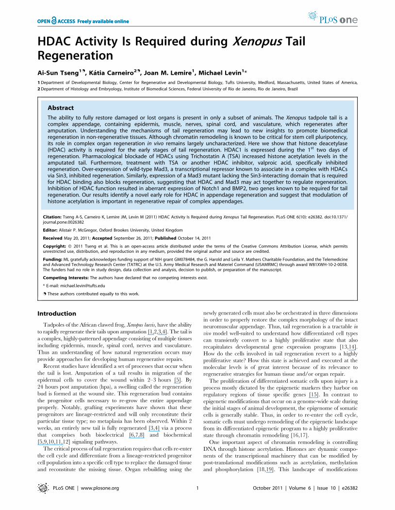
HDAC Activity Is Required during Xenopus TailRegenerationAi-Sun Tseng1., Katia Carneiro2., Joan M. Lemire1, Michael Levin1*
1 Department of Developmental Biology, Center for Regenerative and Developmental Biology, Tufts University, Medford, Massachusetts, United States of America,
2 Department of Histology and Embryology, Institute of Biomedical Sciences, Federal University of Rio de Janeiro, Rio de Janeiro, Brazil
Abstract
The ability to fully restore damaged or lost organs is present in only a subset of animals. The Xenopus tadpole tail is acomplex appendage, containing epidermis, muscle, nerves, spinal cord, and vasculature, which regenerates afteramputation. Understanding the mechanisms of tail regeneration may lead to new insights to promote biomedicalregeneration in non-regenerative tissues. Although chromatin remodeling is known to be critical for stem cell pluripotency,its role in complex organ regeneration in vivo remains largely uncharacterized. Here we show that histone deacetylase(HDAC) activity is required for the early stages of tail regeneration. HDAC1 is expressed during the 1st two days ofregeneration. Pharmacological blockade of HDACs using Trichostatin A (TSA) increased histone acetylation levels in theamputated tail. Furthermore, treatment with TSA or another HDAC inhibitor, valproic acid, specifically inhibitedregeneration. Over-expression of wild-type Mad3, a transcriptional repressor known to associate in a complex with HDACsvia Sin3, inhibited regeneration. Similarly, expression of a Mad3 mutant lacking the Sin3-interacting domain that is requiredfor HDAC binding also blocks regeneration, suggesting that HDAC and Mad3 may act together to regulate regeneration.Inhibition of HDAC function resulted in aberrant expression of Notch1 and BMP2, two genes known to be required for tailregeneration. Our results identify a novel early role for HDAC in appendage regeneration and suggest that modulation ofhistone acetylation is important in regenerative repair of complex appendages.
Citation: Tseng A-S, Carneiro K, Lemire JM, Levin M (2011) HDAC Activity Is Required during Xenopus Tail Regeneration. PLoS ONE 6(10): e26382. doi:10.1371/journal.pone.0026382
Editor: Alistair P. McGregor, Oxford Brookes University, United Kingdom
Received May 20, 2011; Accepted September 26, 2011; Published October 14, 2011
Copyright: � 2011 Tseng et al. This is an open-access article distributed under the terms of the Creative Commons Attribution License, which permitsunrestricted use, distribution, and reproduction in any medium, provided the original author and source are credited.
Funding: ML gratefully acknowledges funding support of NIH grant GM078484, the G. Harold and Leila Y. Mathers Charitable Foundation, and the Telemedicineand Advanced Technology Research Center (TATRC) at the U.S. Army Medical Research and Materiel Command (USAMRMC) through award W81XWH-10-2-0058.The funders had no role in study design, data collection and analysis, decision to publish, or preparation of the manuscript.
Competing Interests: The authors have declared that no competing interests exist.
* E-mail: [email protected]
. These authors contributed equally to this work.
Introduction
Tadpoles of the African clawed frog, Xenopus laevis, have the ability
to rapidly regenerate their tails upon amputation [1,2,3,4]. The tail is
a complex, highly-patterned appendage consisting of multiple tissues
including epidermis, muscle, spinal cord, nerves and vasculature.
Thus an understanding of how natural regeneration occurs may
provide approaches for developing human regenerative repairs.
Recent studies have identified a set of processes that occur when
the tail is lost. Amputation of a tail results in migration of the
epidermal cells to cover the wound within 2–3 hours [5]. By
24 hours post amputation (hpa), a swelling called the regeneration
bud is formed at the wound site. This regeneration bud contains
the progenitor cells necessary to re-grow the entire appendage
properly. Notably, grafting experiments have shown that these
progenitors are lineage-restricted and will only reconstitute their
particular tissue type; no metaplasia has been observed. Within 2
weeks, an entirely new tail is fully regenerated [3,4] via a process
that comprises both bioelectrical [6,7,8] and biochemical
[5,9,10,11,12] signaling pathways.
The critical process of tail regeneration requires that cells re-enter
the cell cycle and differentiate from a lineage-restricted progenitor
cell population into a specific cell type to replace the damaged tissue
and reconstitute the missing tissue. Organ rebuilding using the
newly generated cells must also be orchestrated in three dimensions
in order to properly restore the complex morphology of the intact
neuromuscular appendage. Thus, tail regeneration is a tractable in
vivo model well-suited to understand how differentiated cell types
can transiently convert to a highly proliferative state that also
recapitulates developmental gene expression programs [13,14].
How do the cells involved in tail regeneration revert to a highly
proliferative state? How this state is achieved and executed at the
molecular levels is of great interest because of its relevance to
regenerative strategies for human tissue and/or organ repair.
The proliferation of differentiated somatic cells upon injury is a
process mostly dictated by the epigenetic markers they harbor on
regulatory regions of tissue specific genes [15]. In contrast to
epigenetic modifications that occur on a genome-wide scale during
the initial stages of animal development, the epigenome of somatic
cells is generally stable. Thus, in order to re-enter the cell cycle,
somatic cells must undergo remodeling of the epigenetic landscape
from its differentiated epigenetic program to a highly proliferative
state through chromatin remodeling [16,17].
One important aspect of chromatin remodeling is controlling
DNA through histone acetylation. Histones are dynamic compo-
nents of the transcriptional machinery that can be modified by
post-translational modifications such as acetylation, methylation
and phosphorylation [18,19]. This landscape of modifications
PLoS ONE | www.plosone.org 1 October 2011 | Volume 6 | Issue 10 | e26382

plays a dynamic role in chromatin structure, as they may influence
histone-DNA interactions that regulate genetic activities [20]. In
addition, it has been shown that acetylation of the chromatin is a
crucial scaffold for histone methyl transferases to amplify the
complex milieu of epigenetic markers found in the cell [21].
In particular, the acetylation of the e-amino group of lysines
residues on the histone tail by Histones acetyltranferases (HAT) is
tightly correlated to gene transcription during development [22]
and conditions such as cancer, inflammatory lung diseases and
viral infections [23]. Conversely, histone deacetylases (HDACs)
reverse the modifications made on histone tails and this correlates
with a repressive state of the chromatin that is linked to terminal
differentiation and cell cycle exit [24].
HDACs are highly conserved enzymes with homologues in
yeast, humans, Xenopus, and zebrafish [25]. HDACs are classified
based on their homology with yeast HDACs. Class I HDACs (1–3,
and 8, homologous to yeast RPD3) are nuclear, expressed widely,
and play an important role in cell proliferation and survival. Class
II HDACs (4–7, and 9–10) shuttle between the nucleus and
cytoplasm and have tissue-specific functions. Furthermore, HDAC
activity has been shown to be important during multiple aspects of
animal development including stem cell differentiation [26] and
heart [27] and skull [28] morphogenesis.
Because HDACs are transcriptional repressors that lack DNA
binding domains, their specificity is mediated by direct interaction
with transcriptional repressors in large multi-protein complexes
containing components such as NuRD, CoREST, or Sin3 proteins
[29]. In particular, Class I HDAC complexes containing Sin3 also
interacts with Mad proteins to act as a repressor of gene
transcription [30]. Mad is a repressor of gene expression belonging
to the basic-region-helix-loop-helix-leucine zipper (bHLH-Zip)
family of transcription factors that binds to E-box sequences on the
DNA [31]. Thus, Mad proteins are important modulators of
HDAC action, targeting HDAC activity to specific regions on the
chromatin.
As epigenetic regulation of DNA has been shown to be
important for modulating stem cell states, such regulation may also
play an important role in complex organ regeneration in vivo.
Previous studies have identified methylation states and histone
demethylases as regulatory components of regeneration in Xenopus
tadpole limbs [32] and zebrafish fins [33]. However, the role of
acetylation and histone acetylases is unknown. In order to
determine whether modulation of histone acetylation can regulate
regeneration, we took advantage of the tractable Xenopus tail model
and used molecular and pharmacological tools to show that
HDAC activity is required for regeneration. Inhibition of HDAC
function blocks regeneration by altering the histone acetylation
state and results in aberrant expression of the downstream genes
involved in driving regenerative outgrowth. Furthermore, HDACs
likely associate with the transcriptional repressor Mad3 to regulate
histone acetylation.
Results
HDAC1 and Mad3 are expressed during tail regenerationTo determine whether HDACs play a role in regeneration, we
first examined the endogenous expression patterns of Xenopus
HDACs during regeneration. We identified full-length clones of
Xenopus HDAC1 [34], and HDAC6 [35] using Xenbase [36]. We
then examined their expression at several timepoints after tail
amputation. RNA in situ hybridization with gene-specific probes
showed that HDAC1 (also known as Rpd3) was strongly expressed
at 24, 48, and 72 hpa (hours post amputation) in the mesenchymal
cells of the regeneration bud (Fig. 1A–C, black arrowheads and
Fig. S1) as compared to base levels of expression in flank (proximal
tail) tissues. In contrast, HDAC6 expression was absent at both 24
and 48 hpa (Fig. 1G–H, open arrowheads). The difference in
expression of HDAC1 and HDAC6 during regeneration suggests
that, like their mammalian counterparts [22], HDAC function is
likely not redundant and that subsets of HDACs play roles in
different biological processes. While our expression data suggests
specific HDACs as likely participants in regeneration, our studies
do not rule out involvement of additional HDACs.
HDACs are known to associate with Mad3 to form a complex
that regulates transcription; thus we also examined the expression
pattern of Mad3 after tail amputation. Both HDAC1 and Mad3
RNAs were detected at low levels throughout the un-amputated
tail, consistent with potential roles during primary tail develop-
ment (Fig. S1). As expected, Mad3 RNA also becomes expressed
in the regeneration bud at 24, 48, and 72 hpa (Fig. 1C, F).
Together, our data indicate that HDAC1 and Mad3 are present in
the correct spatiotemporal pattern to participate in appendage
regeneration.
HDAC function is required during early stages of tailregeneration
To determine whether HDAC function is required for tail
regeneration, we assessed the effect of pharmacologically ablating
HDAC activity. To effectively block HDAC function, we used
Trichostatin A (TSA), a well-known specific and potent chemical
inhibitor of both Class I and Class II HDACs [37]. Tadpoles
(whether control or amputated) that were treated with 25 nM TSA
grew similarly to their untreated control siblings, and were
indistinguishable with respect to developmental stage, axial
proportions, gross organ morphology, and size (data not shown).
After amputation at st. 40, tails of control larvae regenerated fully
(Fig. 2A). In contrast, treatment with 25 nM TSA after tail
amputation specifically inhibited regeneration (a decrease of 62%
as determined by the Regeneration Index (RI), Fig. 2B). Similarly,
treatment of st. 40 tadpoles after tail amputation with 500 mM of
Valproic Acid (VPA), another well-characterized HDAC inhibitor
[38], also significantly blocked regeneration (Fig. 2C). Together,
these results demonstrated that HDAC activity is required for
regeneration.
Our RNA expression data showed that HDAC1 is present
during the events occurring right after tail amputation. Thus, we
tested the hypothesis that HDAC activity is required during the
early stages of regeneration by determining the temporal
requirement for HDAC function. Tadpole tails were amputated
and incubated for specific durations with TSA and assayed for
their regenerative ability at 7 dpa (days post amputation) (Fig. 2D).
Treatment through the entire length of the assay was sufficient to
inhibit regeneration in 78% of tails (regenerates scored as weak or
none) when compared to control siblings (1%) with no effects on
overall development. Our RNA expression data showed that
HDAC1 is expressed during the first two days of regeneration.
Consistent with this observation, TSA treatment for the first 2 days
after amputation caused 89% inhibition of tail regeneration. This
result fully recapitulates the phenotype seen when the blocker was
present for the entire duration of the assay. Further supporting an
early role for HDACs, addition of TSA after 2 dpa had no effect
on tail regeneration (1%), similar to control siblings. Together, our
results demonstrate that HDAC activity is required specifically
during the first 2 days of regeneration.
Mad3 is required during regenerationClass I HDACs are widely expressed transcriptional repressors
that lack DNA binding domains. Thus their specificity is due to
HDAC and Tadpole Tail Regeneration
PLoS ONE | www.plosone.org 2 October 2011 | Volume 6 | Issue 10 | e26382

direct interaction with transcriptional repressors in large multi-
protein complexes harboring NuRD, CoREST, or Sin3 proteins
[29]. In particular, the HDAC-Sin3 complex also associates with
the transcriptional repressor Mad proteins to regulate gene
transcription [30].
Our previous work showed that Mad3 and the maternal HDAC
functionally interact in the establishment of left-right asymmetry
during early Xenopus development [39]. As Mad3 is expressed
following tail amputation (Fig. 1D–F), it is a likely candidate for
participation in the regenerative response. To further characterize
the role of HDAC in regeneration, we looked to determine
whether its partner Mad3 also participates in this process.
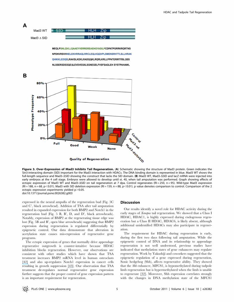
First, we characterized the potential role of Mad3 in
regeneration. The over-expression of Mad3 has been shown to
exert a dominant-negative effect on the HDAC1-Mad3 complex
[39] and thus decreases its activity (likely via a titration
mechanism). To determine whether Mad3 function affects
regeneration, wild-type Mad3 RNA was injected into each
blastomere of embryos at the 4-cell stage and expressed
ubiquitously. As predicted, ectopic expression of Mad3 decreased
tail regenerative ability by 25% when compared to control siblings
(Fig. 3B), indicating that Mad3 participates in this process.
The ability of Mad3 to repress gene expression is dependent on
its Sin3-Interacting Domain (SID), which enables Mad3 to interact
with Sin3 co-factors. Sin3 in turn, recruits HDAC1 multi-protein
complexes containing Mad, leading to transcriptional repression
[40]. Indeed, the repressive activity is of Mad3 is dependent on the
presence of HDAC and is fully blocked by HDAC inhibitors [30].
Because we showed that tail regeneration is sensitive to a HDAC
blockade by TSA and VPA (Fig. 2B–C), we hypothesized that the
requirement for Mad3 in regeneration is dependent upon HDAC
function. To test this hypothesis, we generated a mutant Mad3
construct carrying a deletion of SID (Fig. 3A). This mutant Mad3
can not interact with Sin3-HDAC and should thus block HDAC-
dependent functions during embryogenesis. This was indeed
observed: ectopic expression of Mad3-DSID RNA reduced
regenerative ability by 36% when compared to control siblings
(Fig. 3B). This result shows that the function of Mad3 in
Figure 1. HDAC1 and Mad3 are Expressed During Xenopus Tail Regeneration. (A–C) RNA in situ hybridization to detect gene expression intail regenerates at 24, 48 and 72 hpa for HDAC1, (D–F) Mad3, and (G and H) HDAC6. Probe targets are shown at the left. Black arrowheads indicatepresence of RNA whereas open arrowheads indicate absence of expression. Anterior is to the left.doi:10.1371/journal.pone.0026382.g001
HDAC and Tadpole Tail Regeneration
PLoS ONE | www.plosone.org 3 October 2011 | Volume 6 | Issue 10 | e26382

regeneration requires SID and likely its interaction with HDAC
via Sin3.
Inhibition of HDAC Function Alters Regenerative GeneExpression
Histone Deacetylases (HDAC) act to remove acetyl groups from
the lysine amino acid on histones. To better understand the
mechanism by which inhibition of HDAC activity blocks
regeneration, we examined the effect of the HDAC inhibitor,
TSA, on the acetylation state of the tail regenerate. Using an
antibody that specifically detects acetylation on Histone H4, we
observed a weak signal on control 24 hpa regeneration buds,
consistent with our data that a low acetylation level of histone H4
is necessary for regeneration (Fig. 4A). In contrast, treatment with
TSA immediately after tail amputation greatly increased the level
of acetylation as seen by the strong acetylated Histone H4 signal in
the regeneration bud (Fig. 4D compared to 4A). This result
demonstrates that TSA acts to inhibit HDAC activity by altering
the acetylation state of histones in the tail regeneration bud during
regeneration.
The acetylation state of histones modulates genes expression.
The removal of acetyl groups on histones by HDACs acts to
repress transcription whereas the presence of acetyl groups enables
transcription [41]. Our data indicated that HDAC inhibition
abrogates tail regeneration by altering the acetylation state of
histones. A likely reason for the regeneration defect is altered
transcription of genes necessary for tail regeneration in regener-
ation bud. To assess the consequence of HDAC inhibition on key
regenerative gene transcription, we examined the RNA expression
pattern of Notch1 [13] and BMP2 [12], two genes that are
required for promoting tail outgrowth after amputation. Individ-
ually activating either pathway promotes tail regeneration whereas
inhibition prevents regeneration [13].
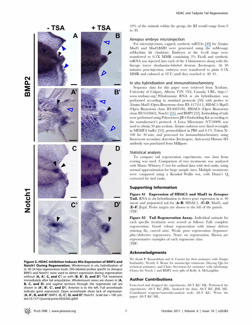
At 24 hpa, Xenopus BMP2 is up-regulated in the regeneration
bud (Fig. 5A and A’, black arrowhead) and at the regenerating fin
edge of the amputation site (Fig. 5A, blue arrowhead). Notch1 is
Figure 2. Pharmacological HDAC Blockade using TSA or VPA Inhibits Tail Regeneration. (A) After st. 40 tail amputation, tadpoles wereassayed for tail regeneration at 7 days post amputation (dpa). Yellow arrowheads demarcate amputation site. (B) Control tadpoles (RI = 290, n = 69).25 nM TSA treatment (RI = 109, n = 69), * denotes p,0.001. (C) Control tadpoles (RI = 283, n = 72). 500 mM Valproic Acid treatment (RI = 126, n = 53),* denotes p,0.001. (D) Temporal requirement for HDAC activity during regeneration. Percentage of regeneration shows total number of tailregenerates scored as full or good. TSA treatment as follows: Control/untreated (98.6%, RI = 290, n = 69), 0–2 dpa (10.8%, RI = 105, n = 65), 0–7 dpa(22.1%, RI = 118, n = 77), and 2–7 dpa (1.5%, RI = 272, n = 65). * denotes p,0.01 as compared to either Control or 2–7 dpa treatment.doi:10.1371/journal.pone.0026382.g002
HDAC and Tadpole Tail Regeneration
PLoS ONE | www.plosone.org 4 October 2011 | Volume 6 | Issue 10 | e26382
expressed in the neural ampulla of the regeneration bud (Fig. 5C
and C’, black arrowhead). Addition of TSA after tail amputation
resulted in expanded expression for both BMP2 and Notch1 in the
regeneration bud (Fig. 5 B, B’, D, and D’, black arrowheads).
Notably, expression of BMP2 at the regenerating tissue edge was
lost (Fig. 5B and B’, open blue arrowheads) suggesting that BMP2
expression during regeneration is regulated differentially by
epigenetic control. Our data demonstrate that alteration in
acetylation state causes mis-expression of regenerative gene
programs.
The ectopic expression of genes that normally drive appendage
regenerative outgrowth is counter-intuitive because HDAC
inhibition blocks regeneration. However, our observations are
consistent with other studies which have shown that TSA
treatment increases BMP2 mRNA level in human osteoclasts
[42] and also up-regulates Notch1 expression in cancer cells
resulting in growth suppression [43]. Our observation that TSA
treatment de-regulates normal regenerative gene expression
further suggests that the proper control of gene expression pattern
is an important requirement for regeneration.
Discussion
Our results identify a novel role for HDAC activity during the
early stages of Xenopus tail regeneration. We showed that a Class I
HDAC, HDAC1, is highly expressed during endogenous regen-
eration but a Class II HDAC, HDAC6, is likely absent, although
additional unidentified HDACs may also participate in regener-
ation.
The requirement for HDAC during regeneration is early,
during the first two days following tail amputation. While the
epigenetic control of DNA and its relationship to appendage
regeneration is not well understood, previous studies have
indicated that methylation states of gene enhancers may regulate
regeneration. Work by Yakushiji and coworkers suggested that the
epigenetic regulation of a gene expressed during regeneration,
Sonic hedgehog (Shh), affects regenerative ability. They showed
that the Shh enhancer, MFCS1, is hypomethylated during tadpole
limb regeneration but is hypermethylated when the limb is unable
to regenerate [32]. Moreover, Shh expression correlates strongly
with the changes in DNA methylation state of the MFCS1
Figure 3. Over-Expression of Mad3 Inhibits Tail Regeneration. (A) Schematic showing the structure of Mad3 protein. Green indicates theSin3-interacting domain (SID) important for the Mad3 interaction with HDACs. The DNA binding domain is represented in blue. Mad3 WT shows thefull-length sequence and Mad3-DSID showing the construct that lacks the SID domain. (B) Mad3 WT, Mad3-DSID and lacZ mRNA were injected intoearly embryos at the 4 cell stage. Embryos were allowed to develop until st. 40, when tail amputation was performed. Graph showing effects ofectopic expression of Mad3 WT and Mad3-DSID on tail regeneration at 7 dpa. Control regenerates (RI = 250, n = 95). Wild-type Mad3 expression(RI = 188, n = 68, p,0.01). Mad3 with SID deletion expression (RI = 159, n = 88, p,0.01). p value denotes comparison to control. Comparison of the 2ectopic expression experiments yielded p.0.05.doi:10.1371/journal.pone.0026382.g003
HDAC and Tadpole Tail Regeneration
PLoS ONE | www.plosone.org 5 October 2011 | Volume 6 | Issue 10 | e26382

enhancer. Additionally, it has also been shown that a histone
demeythlase is required for zebrafish fin regeneration [33].
Analogous to DNA methylation control, our study indicates that
the establishment of a regenerative DNA acetylation state is
important for enabling the correct spatial expression of genes that
promote regeneration. A correct balance between DNA methyl-
ation and acetylation may be required to properly control
regeneration.
Numerous studies support the hypothesis that HDACs can act as
promoters of growth and proliferation [44]. For this reason, HDAC
inhibitors have generated great interest and been pursued for their
potential as anti-cancer therapies [45]. Our work is consistent with
these previous studies in that treatment with TSA blocked tail
regeneration. Surprisingly, we observed that inhibition of HDAC
function caused aberrant expression of genes in pathways (BMP and
Notch) that drive regenerative outgrowth (Fig. 4). This result was
not unexpected since BMP2 mRNA has been demonstrated to
increase in the presence of TSA treatment [42], and HDAC
inhibitor treatment results in the up-regulation of Notch and
suppressed cellular growth [43,46]. Previous Xenopus work used
constitutively-active forms of either the BMP receptor Alk3 or the
intracellular active Notch domain (NICD) [13] to promote tail
regeneration. Although the expression of BMP2 and Notch1
correlates with regenerative ability [8], it is not known whether
the specific over-expression of BMP2 or Notch1 acts similarly.
Notably, our data show that HDAC function is critical for
properly regulating the expression patterns of regenerative genes as
an essential component of this process. Importantly, the direct
regulation of Notch1 and BMP2 by histone acetylation is unlikely to
account for the regenerative failure due to HDAC inhibition. It has
been shown that HDAC inhibitor treatment in myeloma cells can
modulate the mRNA levels of approximately 2% of expressed genes
[47]. The identification of the affected genes during regeneration is
of great interest, as they are likely to regulate the response to
regeneration and coordinately act to re-grow the tail. It will be
necessary to undertake global studies of acetylation states and
corresponding microarray studies during the regenerative and non-
regenerative states to identify these genes, as well as understand the
interplay between genetic, epigenetic, and bioelectrical programs
that are known to drive the regenerative response. A comprehensive
understanding of this process will enable exciting new biomedical
therapies for promoting regenerative repair of complex structures.
Materials and Methods
Tail regeneration assayXenopus laevis larvae were cultured via approved protocols
(Institutional Animal Care and Use Committee, #M2008-08).
Tails at stages (st.) 40–41 [48] were amputated at the midpoint
between the anus and the tip. Tadpoles were separated into
control or treated groups, to which 25 nM of Trichostatin A (TSA,
Calbiochem) was added in 0.1X MMR at 22uC for 7 days and
scored for tail regeneration. To quantify and compare regener-
ation in groups of tadpoles treated with or without TSA, a
composite regeneration index (RI), ranging from 0 (no regener-
ation) to 300 (complete regeneration) was calculated as described
previously [8]. Tail regenerates are scored into 4 phenotype
categories (full, good, weak, none) (see Fig. S2). For example, a
group of tails in which .80% were fully regenerated would have
an RI ranging from 240 to 300; if full regeneration occurred in
Figure 4. HDAC Inhibition Increases Histone Acetylation During Regeneration. The acetylation state of tail regenerates were examined at24 hpa using an anti-acetylated Histone H4 antibody. Yellow arrowheads denote regeneration bud. Top row shows untreated controls. Bottom rowshows tadpoles treated with 25 nM TSA after tail amputation. (A, D) Acetylated Histone H4. (B, E) Hoechst DNA stain. (C) merge of A and B. (F)merge of D and E.doi:10.1371/journal.pone.0026382.g004
HDAC and Tadpole Tail Regeneration
PLoS ONE | www.plosone.org 6 October 2011 | Volume 6 | Issue 10 | e26382
10% of the animals within the group, the RI would range from 0
to 30.
Xenopus embryo microinjectionFor microinjections, capped, synthetic mRNAs [49] for Xenopus
Mad3 and Mad3DSID were generated using the mMessage
mMachine kit (Ambion). Embryos at the 4-cell stage were
transferred to 0.1X MMR containing 3% Ficoll and synthetic
mRNA was injected into each of the 4 blastomeres along with the
lineage tracer rhodamine-labeled dextran (Invitrogen). At 30
minutes post-injection, embryos were transferred to plain 0.1X
MMR and cultured at 18uC until they reached st. 40–41.
In situ hybridization and immunohistochemistrySequence data for this paper were retrieved from Xenbase,
University of Calgary, Alberta T2N 1N4, Canada; URL: http://
www.xenbase.org/.Wholemount RNA in situ hybridization was
performed according to standard protocols [50] with probes to
Xenopus Mad3 (Open Biosystems clone ID: 4175511), HDAC1/Rpd3
(Open Biosystems clone ID:4683530), HDAC6 (Open Biosystems
clone ID:5542663), Notch1 [51], and BMP2 [52]. Embedding of tails
were performed using Polysciences JB-4 Embedding Kit according to
the manufacturer’s protocol. A Leica Microtome (VT1000S) was
used to obtain 50 mm sections. Xenopus embryos were fixed overnight
in MEMFA buffer [53], permeabilized in PBS and 0.1% Triton X-
100 for 30 min, and processed for immunohistochemistry using
fluorescent secondary detection (Invitrogen). Anti-acetyl Histone H4
antibody was purchased from Millipore.
Statistical analysisTo compare tail regeneration experiments, raw data from
scoring was used. Comparison of two treatments was analyzed
with Mann–Whitney U test for ordinal data with tied ranks, using
normal approximation for large sample sizes. Multiple treatments
were compared using a Kruskal–Wallis test, with Dunn’s Q
corrected for tied ranks.
Supporting Information
Figure S1 Expression of HDAC1 and Mad3 in XenopusTail. RNA in situ hybridization to detect gene expression in st. 40
uncut and amputated tail for (A–B) HDAC1, (C–D) Mad3, and
(E–F) b-gal. Probe targets are shown to the left of the panels.
(TIF)
Figure S2 Tail Regeneration Assay. Individual animals for
each specific treatment were scored as follows: Full: complete
regeneration. Good: robust regeneration with minor defects
(missing fin, curved axis). Weak: poor regeneration (hypomor-
phic/defective regenerates). None: no regeneration. Shown are
representative examples of each regenerate class.
(TIF)
Acknowledgments
We thank P. Koustubhan and A. Currier for their assistance with Xenopus
husbandry, Wendy S. Beane for manuscript comments, Dayong Qiu for
technical assistance, and Claire Stevenson for assistance with subcloning.
Clones for Notch 1 and BMP2 were gifts of Kelly A. McLaughlin.
Author Contributions
Conceived and designed the experiments: AS-T KC ML. Performed the
experiments: AS-T KC JML. Analyzed the data: AS-T KC JML ML.
Contributed reagents/materials/analysis tools: AS-T KC. Wrote the
paper: AS-T KC ML.
Figure 5. HDAC Inhibition Induces Mis-Expression of BMP2 andNotch1 During Regeneration. Wholemount in situ hybridization ofst. 40 24 hpa regeneration buds. DIG-labeled probes specfic to XenopusBMP2 and Notch1 were used to detect expression during regenerationwithout (A, A’, C, and C’) or with (B, B’, D, and D’) TSA treatmentimmediately after tail amputation. Wholemount views are shown in (A,B, C, and D) and sagittal sections through the regenerate tail areshown in (A’, B’, C’, and D’). Anterior is to the left. Full arrowheadsindicate gene expression. Open arrowheads show lack of expression.(A, A’, B, and B’) BMP2. (C, C’, D, and D’) Notch1. Scale bar = 100 mm.doi:10.1371/journal.pone.0026382.g005
HDAC and Tadpole Tail Regeneration
PLoS ONE | www.plosone.org 7 October 2011 | Volume 6 | Issue 10 | e26382
References
1. Mochii M, Taniguchi Y, Shikata I (2007) Tail regeneration in the Xenopus
tadpole. Dev Growth Differ 49: 155–161.
2. Beck CW, Izpisua Belmonte JC, Christen B (2009) Beyond early development:
Xenopus as an emerging model for the study of regenerative mechanisms. Dev
Dyn 238: 1226–1248.
3. Slack JM, Beck CW, Gargioli C, Christen B (2004) Cellular and molecular
mechanisms of regeneration in Xenopus. Philos Trans R Soc Lond B Biol Sci
359: 745–751.
4. Tseng AS, Levin M (2008) Tail regeneration in Xenopus laevis as a model for
understanding tissue repair. J Dent Res 87: 806–816.
5. Ho DM, Whitman M (2008) TGF-beta signaling is required for multiple
processes during Xenopus tail regeneration. Dev Biol 315: 203–216.
6. Reid B, Song B, Zhao M (2009) Electric currents in Xenopus tadpole tail
regeneration. Dev Biol 335: 198–207.
7. Adams DS, Masi A, Levin M (2007) H+ pump-dependent changes in membrane
voltage are an early mechanism necessary and sufficient to induce Xenopus tail
regeneration. Development 134: 1323–1335.
8. Tseng AS, Beane WS, Lemire JM, Masi A, Levin M (2010) Induction of
vertebrate regeneration by a transient sodium current. J Neurosci 30:
13192–13200.
9. Contreras EG, Gaete M, Sanchez N, Carrasco H, Larrain J (2009) Early
requirement of Hyaluronan for tail regeneration in Xenopus tadpoles.
Development 136: 2987–2996.
10. Fukazawa T, Naora Y, Kunieda T, Kubo T (2009) Suppression of the immune
response potentiates tadpole tail regeneration during the refractory period.
Development 136: 2323–2327.
11. Sugiura T, Tazaki A, Ueno N, Watanabe K, Mochii M (2009) Xenopus Wnt-5a
induces an ectopic larval tail at injured site, suggesting a crucial role for
noncanonical Wnt signal in tail regeneration. Mech Dev 126: 56–67.
12. Beck CW, Christen B, Barker D, Slack JM (2006) Temporal requirement for
bone morphogenetic proteins in regeneration of the tail and limb of Xenopus
tadpoles. Mech Dev 123: 674–688.
13. Beck CW, Christen B, Slack JM (2003) Molecular pathways needed for
regeneration of spinal cord and muscle in a vertebrate. Dev Cell 5: 429–439.
14. Beck CW, Slack JM (1999) A developmental pathway controlling outgrowth of
the Xenopus tail bud. Development 126: 1611–1620.
15. Shaw T, Martin P (2009) Epigenetic reprogramming during wound healing: loss
of polycomb-mediated silencing may enable upregulation of repair genes.
EMBO Rep 10: 881–886.
16. Riau AK, Wong TT, Finger SN, Chaurasia SS, Hou AH, et al. (2011) Aberrant
DNA Methylation of Matrix Remodeling and Cell Adhesion Related Genes in
Pterygium. PLoS One 6: -.
17. Xiong L, Darwanto A, Sharma S, Herring J, Hu S, et al. (2011) Mass
Spectrometric Studies on Epigenetic Interaction Networks in Cell Differentia-
tion. J Biol Chem 286: 13657–13668.
18. Sterner DE, Berger SL (2000) Acetylation of histones and transcription-related
factors. Microbiol Mol Biol Rev 64: 435–459.
19. Nowak SJ, Corces VG (2004) Phosphorylation of histone H3: a balancing act
between chromosome condensation and transcriptional activation. Trends
Genet 20: 214–220.
20. Wolffe AP, Hayes JJ (1999) Chromatin disruption and modification. Nucleic
Acids Res 27: 711–720.
21. Nightingale KP, Gendreizig S, White DA, Bradbury C, Hollfelder F, et al.
(2007) Cross-talk between histone modifications in response to histone
deacetylase inhibitors: MLL4 links histone H3 acetylation and histone H3K4
methylation. J Biol Chem 282: 4408–4416.
22. Haberland M, Montgomery RL, Olson EN (2009) The many roles of histone
deacetylases in development and physiology: implications for disease and
therapy. Nat Rev Genet 10: 32–42.
23. Dekker FJ, Haisma HJ (2009) Histone acetyl transferases as emerging drug
targets. Drug Discov Today 14: 942–948.
24. Eberharter A, Becker PB (2002) Histone acetylation: a switch between repressive
and permissive chromatin. Second in review series on chromatin dynamics.
EMBO Rep 3: 224–229.
25. Leipe DD, Landsman D (1997) Histone deacetylases, acetoin utilization proteins
and acetylpolyamine amidohydrolases are members of an ancient protein
superfamily. Nucleic Acids Res 25: 3693–3697.
26. Dovey OM, Foster CT, Cowley SM (2010) Histone deacetylase 1 (HDAC1), but
not HDAC2, controls embryonic stem cell differentiation. Proc Natl Acad
Sci U S A 107: 8242–8247.
27. Montgomery RL, Davis CA, Potthoff MJ, Haberland M, Fielitz J, et al. (2007)Histone deacetylases 1 and 2 redundantly regulate cardiac morphogenesis,
growth, and contractility. Genes Dev 21: 1790–1802.28. Haberland M, Mokalled MH, Montgomery RL, Olson EN (2009) Epigenetic
control of skull morphogenesis by histone deacetylase 8. Genes Dev 23:1625–1630.
29. Heinzel T, Lavinsky RM, Mullen TM, Soderstrom M, Laherty CD, et al. (1997)
A complex containing N-CoR, mSin3 and histone deacetylase mediatestranscriptional repression. Nature 387: 43–48.
30. Laherty CD, Yang WM, Sun JM, Davie JR, Seto E, et al. (1997) Histonedeacetylases associated with the mSin3 corepressor mediate mad transcriptional
repression. Cell 89: 349–356.
31. Ayer DE, Kretzner L, Eisenman RN (1993) Mad: a heterodimeric partner forMax that antagonizes Myc transcriptional activity. Cell 72: 211–222.
32. Yakushiji N, Suzuki M, Satoh A, Sagai T, Shiroishi T, et al. (2007) Correlationbetween Shh expression and DNA methylation status of the limb-specific Shh
enhancer region during limb regeneration in amphibians. Dev Biol 312:
171–182.33. Stewart S, Tsun ZY, Izpisua Belmonte JC (2009) A histone demethylase is
necessary for regeneration in zebrafish. Proc Natl Acad Sci U S A 106:19889–19894.
34. Damjanovski S, Sachs LM, Shi YB (2000) Multiple stage-dependent roles forhistone deacetylases during amphibian embryogenesis: implications for the
involvement of extracellular matrix remodeling. Int J Dev Biol 44: 769–776.
35. Hageman J, Rujano MA, van Waarde MA, Kakkar V, Dirks RP, et al. ADNAJB chaperone subfamily with HDAC-dependent activities suppresses toxic
protein aggregation. Mol Cell 37: 355–369.36. Bowes JB, Snyder KA, Segerdell E, Jarabek CJ, Azam K, et al. (2009) Xenbase:
gene expression and improved integration. Nucleic Acids Res 38: D607–612.
37. Yoshida M, Kijima M, Akita M, Beppu T (1990) Potent and specific inhibitionof mammalian histone deacetylase both in vivo and in vitro by trichostatin A. J
Biol Chem 265: 17174–17179.38. Phiel CJ, Zhang F, Huang EY, Guenther MG, Lazar MA, et al. (2001) Histone
deacetylase is a direct target of valproic acid, a potent anticonvulsant, moodstabilizer, and teratogen. J Biol Chem 276: 36734–36741.
39. Carneiro K, Donnet C, Rejtar T, Karger BL, Barisone GA, et al. (2011) Histone
deacetylase activity is necessary for left-right patterning during vertebratedevelopment. BMC Dev Biol 11: 29.
40. Hurlin PJ, Queva C, Eisenman RN (1997) Mnt, a novel Max-interacting proteinis coexpressed with Myc in proliferating cells and mediates repression at Myc
binding sites. Genes Dev 11: 44–58.
41. Shahbazian MD, Grunstein M (2007) Functions of site-specific histoneacetylation and deacetylation. Annu Rev Biochem 76: 75–100.
42. Li X, Bai XZ (2008) [NF-kappaB modulates activation of the BMP-2 gene bytrichostatin A]. Mol Biol (Mosk) 42: 990–996.
43. Adler JT, Hottinger DG, Kunnimalaiyaan M, Chen H (2008) Histonedeacetylase inhibitors upregulate Notch-1 and inhibit growth in pheochromo-
cytoma cells. Surgery 144: 956–961; discussion 961–952.
44. Glozak MA, Seto E (2007) Histone deacetylases and cancer. Oncogene 26:5420–5432.
45. Marks PA, Xu WS (2009) Histone deacetylase inhibitors: Potential in cancertherapy. J Cell Biochem 107: 600–608.
46. Xiao X, Ning L, Chen H (2009) Notch1 mediates growth suppression of
papillary and follicular thyroid cancer cells by histone deacetylase inhibitors. MolCancer Ther 8: 350–356.
47. Mitsiades CS, Mitsiades NS, McMullan CJ, Poulaki V, Shringarpure R, et al.(2004) Transcriptional signature of histone deacetylase inhibition in multiple
myeloma: biological and clinical implications. Proc Natl Acad Sci U S A 101:540–545.
48. Nieukoop PD, Faber J (1967) Normal Table of Xenopus laevis. Amersterdam:
North-Holland Publishing Company.49. Sive HL, Grainger RM, Harland RM (2000) Early Development of Xenopus
Laevis. New York: Cold Spring Harbor Laboratory Press.50. Harland RM (1991) In situ hybridization: an improved whole-mount method for
Xenopus embryos. Methods Cell Biol 36: 685–695.
51. Coffman C, Harris W, Kintner C (1990) Xotch, the Xenopus homolog ofDrosophila notch. Science 249: 1438–1441.
52. Feledy JA, Beanan MJ, Sandoval JJ, Goodrich JS, Lim JH, et al. (1999)Inhibitory patterning of the anterior neural plate in Xenopus by homeodomain
factors Dlx3 and Msx1. Dev Biol 212: 455–464.
53. Sive HL GR, Harland RM (2000) Early Development of Xenopus Laevis: ALaboratory Manual. New York: Cold Spring Harbor Laboratory Press.
HDAC and Tadpole Tail Regeneration
PLoS ONE | www.plosone.org 8 October 2011 | Volume 6 | Issue 10 | e26382
Copyright © 2022 FDOKUMEN