A systems biology approach to identify molecular pathways altered by HDAC inhibition in osteosarcoma
11
A Systems Biology Approach to Identify Molecular Pathways Altered by HDAC Inhibition in Osteosarcoma Luke A. Wittenburg, 1,2 * Andrey A. Ptitsyn, 3 and Douglas H. Thamm 1,2 1 Animal Cancer Center, Department of Clinical Sciences, Colorado State University Animal Cancer Center, 300 W. Drake Rd., Fort Collins, Colorado 80523-1620 2 Cell and Molecular Biology Graduate Program, Colorado State University Animal Cancer Center, 300 W. Drake Rd., Fort Collins, Colarado 80523-1620 3 Center for Bioinformatics, Department of Microbiology, Immunology, and Pathology, Colorado State University Animal Cancer Center, 300 W. Drake Rd., Fort Collins, Colorado 80523-1620 ABSTRACT Osteosarcoma (OS) is the most common primary tumor in humans and dogs affecting the skeleton, and spontaneously occurring OS in dogs serves as an extremely useful model. Unacceptable toxicities using current treatment protocols prevent further dose-intensification from being a viable option to improve patient survival and thus, novel treatment strategies must be developed. Histone deacetylase inhibitors (HDACi) have recently emerged as a promising class of therapeutics demonstrating an ability to enhance the anti-tumor activity of traditional chemotherapeutics. To date, gene expression analysis of OS cell lines treated with HDACi has not been reported, and evaluation of the resultant gene expression changes may provide insight into the mechanisms that lead to success of HDACi. Canine OS cells, treated with a clinically relevant concentration of the HDACi valproic acid (VPA), were used for expression analysis on the Affymetrix canine v2.0 genechip. Differentially expressed genes were grouped into pathways based upon functional annotation; pathway analysis was performed with MetaCore and Ingenuity Pathways Analysis software. Validation of microarray results was performed by a combination of qRT-PCR and functional/biochemical assays revealing oxidative phosphorylation, cytoskeleton remodeling, cell cycle, and ubiquitin-proteasome among those pathways most affected by HDACi. The mitomycin C-bioactivating enzyme NQ01 also demonstrated upregulation following VPA treatment, leading to synergistic reductions in cell viability. These results provide a better understanding of the mechanisms by which HDACi exert their effect in OS, and have the potential to identify biomarkers that may serve as novel targets and/or predictors of response to HDACi- containing combination therapies in OS. J. Cell. Biochem. 113: 773–783, 2012. ß 2011 Wiley Periodicals, Inc. KEY WORDS: VALPROATE; OSTEOSARCOMA; PATHWAY ANALYSIS; EXPRESSION PROFILING O steosarcoma (OS) is a high-grade primary bone neoplasm of mesenchymal origin and represents the most common malignant tumor of the bone in both humans and dogs [Dernell et al., 2001; Kansara and Thomas, 2007]. There are remarkable similarities between human and canine OS, which include a bimodal peak incidence, a slight male predilection, similar anatomic site predilections (metaphyses of long bones), aggressive biological behavior, hematogenous metastasis to the lungs early in the course of disease, similar sensitivity to chemotherapeutics, and a nearly identical reported incidence of occult metastasis at the time of diagnosis [Dernell et al., 2001; Kansara and Thomas, 2007; Mueller et al., 2007; Cleton-Jansen et al., 2009]. In both cases, the addition of adjuvant chemotherapy results in an improved overall survival, although this does not hold true for patients presenting with metastatic disease at the time of diagnosis, and in humans the 5-year survival for these patients remains around 30% [Rosen et al., 1983; Link et al., 1986; Dernell et al., 2001]. Unacceptable toxicity prevents further dose intensification of current multimodal Journal of Cellular Biochemistry ARTICLE Journal of Cellular Biochemistry 113:773–783 (2012) 773 Additional Supporting Information may be found in the online version of this article. Grant sponsor: Morris Animal Foundation (CSU Fund); Grant number: 5-354500; Grant sponsor: National Institutes of Health; Grant number: NCRR T32-RR-007072-06; Grant sponsor: American Cancer Society; Grant number: RSG-04- 219-01. Andrey A. Ptitsyn’s present address is University of FloridaWhitney Laboratory for Marine Biosciences9505 Ocean Shore Blvd. St. Augustine FL 32080. *Correspondence to: Dr. Luke A. Wittenburg, DVM, PhD, Animal Cancer Center, Department of Clinical Sciences, Colorado State University Animal Cancer Center, 300 W. Drake Rd., Fort Collins, CO 80523-1620. E-mail: [email protected] Received 2 September 2010; Accepted 30 September 2011 DOI 10.1002/jcb.23403 ß 2011 Wiley Periodicals, Inc. Published online 4 October 2011 in Wiley Online Library (wileyonlinelibrary.com).
Transcript of A systems biology approach to identify molecular pathways altered by HDAC inhibition in osteosarcoma

A Systems Biology Approach to Identify MolecularPathways Altered by HDAC Inhibition in Osteosarcoma
Luke A. Wittenburg,1,2* Andrey A. Ptitsyn,3 and Douglas H. Thamm1,2
1Animal Cancer Center, Department of Clinical Sciences, Colorado State University Animal Cancer Center,300 W. Drake Rd., Fort Collins, Colorado 80523-1620
2Cell and Molecular Biology Graduate Program, Colorado State University Animal Cancer Center,300 W. Drake Rd., Fort Collins, Colarado 80523-1620
3Center for Bioinformatics, Department of Microbiology, Immunology, and Pathology,Colorado State University Animal Cancer Center, 300 W. Drake Rd., Fort Collins, Colorado 80523-1620
ABSTRACTOsteosarcoma (OS) is the most common primary tumor in humans and dogs affecting the skeleton, and spontaneously occurring OS in dogs
serves as an extremely useful model. Unacceptable toxicities using current treatment protocols prevent further dose-intensification from
being a viable option to improve patient survival and thus, novel treatment strategies must be developed. Histone deacetylase inhibitors
(HDACi) have recently emerged as a promising class of therapeutics demonstrating an ability to enhance the anti-tumor activity of traditional
chemotherapeutics. To date, gene expression analysis of OS cell lines treated with HDACi has not been reported, and evaluation of the
resultant gene expression changes may provide insight into the mechanisms that lead to success of HDACi. Canine OS cells, treated with a
clinically relevant concentration of the HDACi valproic acid (VPA), were used for expression analysis on the Affymetrix canine v2.0 genechip.
Differentially expressed genes were grouped into pathways based upon functional annotation; pathway analysis was performed with
MetaCore and Ingenuity Pathways Analysis software. Validation of microarray results was performed by a combination of qRT-PCR and
functional/biochemical assays revealing oxidative phosphorylation, cytoskeleton remodeling, cell cycle, and ubiquitin-proteasome among
those pathways most affected by HDACi. The mitomycin C-bioactivating enzyme NQ01 also demonstrated upregulation following VPA
treatment, leading to synergistic reductions in cell viability. These results provide a better understanding of the mechanisms by which HDACi
exert their effect in OS, and have the potential to identify biomarkers that may serve as novel targets and/or predictors of response to HDACi-
containing combination therapies in OS. J. Cell. Biochem. 113: 773–783, 2012. � 2011 Wiley Periodicals, Inc.
KEY WORDS: VALPROATE; OSTEOSARCOMA; PATHWAY ANALYSIS; EXPRESSION PROFILING
O steosarcoma (OS) is a high-grade primary bone neoplasm of
mesenchymal origin and represents the most common
malignant tumor of the bone in both humans and dogs [Dernell
et al., 2001; Kansara and Thomas, 2007]. There are remarkable
similarities between human and canine OS, which include a bimodal
peak incidence, a slight male predilection, similar anatomic site
predilections (metaphyses of long bones), aggressive biological
behavior, hematogenous metastasis to the lungs early in the course
of disease, similar sensitivity to chemotherapeutics, and a nearly
identical reported incidence of occult metastasis at the time of
diagnosis [Dernell et al., 2001; Kansara and Thomas, 2007; Mueller
et al., 2007; Cleton-Jansen et al., 2009]. In both cases, the addition of
adjuvant chemotherapy results in an improved overall survival,
although this does not hold true for patients presenting with
metastatic disease at the time of diagnosis, and in humans the 5-year
survival for these patients remains around 30% [Rosen et al., 1983;
Link et al., 1986; Dernell et al., 2001]. Unacceptable toxicity
prevents further dose intensification of current multimodal
Journal of CellularBiochemistry
ARTICLEJournal of Cellular Biochemistry 113:773–783 (2012)
773
Additional Supporting Information may be found in the online version of this article.
Grant sponsor: Morris Animal Foundation (CSU Fund); Grant number: 5-354500; Grant sponsor: National Institutes ofHealth; Grant number: NCRR T32-RR-007072-06; Grant sponsor: American Cancer Society; Grant number: RSG-04-219-01.
Andrey A. Ptitsyn’s present address is University of FloridaWhitney Laboratory for Marine Biosciences9505 OceanShore Blvd. St. Augustine FL 32080.
*Correspondence to: Dr. Luke A. Wittenburg, DVM, PhD, Animal Cancer Center, Department of Clinical Sciences,Colorado State University Animal Cancer Center, 300 W. Drake Rd., Fort Collins, CO 80523-1620.E-mail: [email protected]
Received 2 September 2010; Accepted 30 September 2011 � DOI 10.1002/jcb.23403 �� 2011 Wiley Periodicals, Inc.
Published online 4 October 2011 in Wiley Online Library (wileyonlinelibrary.com).

chemotherapy protocols from being a viable option to further
improve survival, and thus there is a need to develop novel
therapeutics for this disease. Generating meaningful data from
clinical studies in humans with OS can be difficult because of the
relatively low incidence; approximately 900 new cases are reported
per year in the U.S. [Kansara and Thomas, 2007]. In contrast, the
high incidence of OS in dogs (approximately 8–12,000 cases/year)
[Dernell et al., 2001] presents a unique opportunity to not only study
the biology of OS but also rigorously evaluate novel therapeutics.
Histone deacetylase inhibitors (HDACi) have emerged as a very
promising novel class of therapeutics in cancer treatment. There are
currently a number of HDACi in clinical evaluation for hematologic
and solid tumors, and two (vorinostat and romidepsin) have received
FDA approval as single agents in the treatment of relapsed or
refractory T-cell lymphoma [Mann et al., 2007; Marsoni et al., 2008;
Prince et al., 2009]. The exact mechanisms by which these drugs act
in cancer therapy is not fully understood, but they appear to have
pleiotropic anti-tumor effects including induction of differentiation,
growth arrest, initiation of senescence, enhanced apoptosis,
decreased angiogenesis, immunomodulatory activities, and an
ability to synergize with traditional cytotoxic chemotherapies
and radiation [Deroanne et al., 2002; Kim et al., 2003; Bolden et al.,
2006; Minucci and Pelicci, 2006; Tomasi et al., 2006; Xu et al.,
2007]. The clinical utility of agents that target HDAC enzymes is, in
part, based upon the aberrant expression and/or function of HDACs
in cancer initiation and progression, with overexpression of some
HDACs correlating with poor prognosis in a number of tumor types
including gastric, prostate, breast, pancreatic, lung, and hepatocel-
lular cancers [Rikimaru et al., 2007; Miyake et al., 2008; Weichert
et al., 2008abc].
The specific role of HDAC enzymes in the pathogenesis of OS has
not been elucidated; however, HDAC enzymes play crucial roles in
the normal development of bone cells including osteoblasts and
osteoclasts [Schroeder et al., 2004; Schroeder andWestendorf, 2005;
Westendorf, 2007]. It has been shown that the treatment of normal
osteoblasts by HDACi results in accelerated maturation and
differentiation, in part due to the inhibition of the interaction
between HDAC3 and the transcription factor Runx2 which controls
expression of osteocalcin, a marker of osteoblast differentiation
[Schroeder et al., 2004]. In addition, the HDACi-induced effect on
osteoblast differentiation has been shown to act through the
regulation of Wnt/b catenin pathway [Schroeder et al., 2007]. It
has been reported that the majority of high-grade OS have a
demonstrable inactivity of theWnt pathway, determined by a lack of
nuclear b-catenin staining in biopsies [Kansara and Thomas, 2007;
Cai et al., 2009; Cleton-Jansen et al., 2009]. However, there are
conflicting reports on the activity of the Wnt pathway in OS, and in
some cases inhibition of the pathway in OS has been shown to
reduce tumorigenesis and metastatic potential of some cell
lines [Rubin et al., 2010]. In fact, one decoy receptor for the Wnt
pathway, secreted frizzled-related protein 3 (SFRP3), has a potential
tumor suppressor function in OS, showing an ability to reduce
osteoblast proliferation and increase osteoblast differentiation.
Furthermore, high-grade OS tumors appear to lack or have
downregulated expression of SFRP3 [Chung et al., 2004; Mandal
et al., 2007].
In addition to the effects on differentiation processes of
osteoblasts, HDAC inhibition also results in alterations of chromatin
structure, a property that has been utilized to sensitize cells to
chemotherapeutic agents that target DNA. The chromatin decon-
densation resulting from HDAC inhibition has been shown to
increase sensitivity to topoisomerase I and II inhibitors such as
doxorubicin, epirubicin, and keranotecin in a variety of models of
human and canine solid tumors, including OS [Marchion et al., 2004,
2005; Munster et al., 2007; Wittenburg et al., 2010].
Although microarray data exist on the effect of HDACi in
osteoblasts, to our knowledge there are no reports of microarray data
utilizing pathway analysis in HDACi-treated OS cells. Here, we
attempt to further elucidate the molecular sequelae of HDAC
inhibition in OS cells through pathway analysis of a canine OS cell
line treated with the HDACi valproic acid (VPA), and use a
combination of quantitative PCR and biochemical/functional assays
in canine OS cells to validate the results of our microarray analysis.
MATERIALS AND METHODS
CELL LINES AND CONDITIONS
The D17 canine OS cell line was purchased from the American Type
Culture Collection (Rockville, MD) The Abrams canine OS cell line
was kindly provided by Dr. William Dernell (Colorado State
University, Fort Collins, CO). Cells were cultured in minimum
essential medium Eagle (Lonza, Walkersville, MD) supplemented
with 10% fetal bovine serum (Atlas, Fort Collins, CO) and penicillin/
streptomycin (Invitrogen, Carlsbad, CA). For experimental proce-
dures, cells were incubated in a humidified atmosphere with 5% CO2
at 378C.
CHEMICALS AND ANTIBODIES
Valproic acid was purchased from Sigma Chemical, Co. (St. Louis,
MO). Bovine serum albumin, 2,6-dichlorophenolindophenol
(DCPIP), dicumarol, and NADH were purchased from Sigma–Aldrich
(St. Louis, MO). Suc-Leu-Tyr-AMC fluorogenic substrate was
purchased from AnaSpec (San Jose, CA). Bicinchoninic acid protein
assay reagents and SuperSignal chemiluminescent substrate were
purchased from Pierce Chemical Co. (Rockford, IL). The proteasome
inhibitor ONX-912 was provided by Onyx Pharmaceuticals (South
San Francisco, CA). Alamar BlueTM fluorogenic substrate was
obtained from AbD Serotec (Oxford, UK).
GENE EXPRESSION MICROARRAY
Valproic acid treated (1mM) and untreated D17 canine OS cells were
used for analysis of differential gene expression using the
AffymetrixTM Canine v2.0 gene chip, which was performed by
the Microarray Core at the University of Colorado Health Sciences
Center (Denver, CO). Briefly, cells in log-phase growth were treated
in triplicate with 0 or 1mM VPA for 48 h, after which cells were
harvested and stored in RNAlater for transfer to the core. RNA was
extracted using the Qiashredder and RNeasy Kit from Qiagen
(Valencia, CA). RNA quantity and integrity were assessed using the
Nanodrop 1000 (ThermoScientific, Waltham, MA) and Bioanalyzer
(Agilent Technologies, Foster City, CA) respectively. Resulting RNA
was then used for first- and second-strand cDNA synthesis followed
774 MOLECULAR PATHWAY ANALYSIS OF HDACi-TREATED OS CELLS JOURNAL OF CELLULAR BIOCHEMISTRY

by cDNA cleanup and overnight in vitro transcription. Following
cRNA cleanup and fragmentation steps, samples were then
hybridized to the Affymetrix chip and scanned.
MICROARRAY DATA ANALYSIS
Data normalization and selection of differentially expressed genes
was performed as previously described by Ptitsyn et al. [2008].
Briefly, normalized data were used to select a preliminary set of
differentially expressed genes using the University of Pittsburgh
Gene Expression Data Analysis suite (GEDA, http://bioinformatics.
upmc.edu/GE2/GEDA.html). To reduce the number of false-positive
differential genes and provide a shortlist of genes deviating from the
expected average value and enriched with differential genes, a
standard J5 metric with threshold 4 and optional 4 iteration of
jackknife procedure was applied. Then we prepared a proxy set of
human genes based on homology of canine probe targets to human
genes determined by BLAST and BLAT search tools. This curated set
of nearest homologs was submitted to pathways analysis. The reason
for using the proxy set is that gene interaction databases and
analysis software support only human, mouse, and rat data input.
FUNCTIONAL ANNOTATION AND PATHWAY ANALYSIS
Analysis of biological pathways was performed using MetaCore
software version 6.4 (GeneGo Inc., Division of Thomson Reuters),
Ingenuity Pathways Analysis version 8.8 (IPA, Ingenuity Systems
Inc.). Significance of a particular pathway represented in a given list
of genes is estimated by Fisher’s exact test with adjustments to
current database contents. The GeneGo and IPA databases are
accessed online and the contents (including Canonic Maps,
Molecular Functions, Gene Interactions, etc.) are frequently updated
by the corps of curators reading research publications and
extracting information related to all forms of interaction among
genes and chemical compounds. Consequently, the results of
pathway analyses performed at different times may differ in details.
The maps of canonic signaling and metabolic pathways (groups of
interacting genes and other factors such as metabolites and chemical
compounds drawn by Ingenuity and Genego developers) are
convoluted, but tested for significance independently. As a result,
the list of significant pathways often enriched with redundant
pathways overlapping by a majority of components. The interpre-
tation of the meaning of statistically significant pathways relies on
the knowledge of the biological function underlying the pathway
maps and cross-comparison between two independent pathway
databases (IPA and Metacore). We also applied DAVID web-based
tools to provide preliminary functional annotation of all potentially
differential genes [Huang da et al., 2007].
QUANTITATIVE REAL-TIME RT-PCR
For validation of microarray results, a subset of VPA-altered genes
was chosen for qRT-PCR in canine OS cell lines treated with VPA.
RNA was extracted using the Qiashredder and RNeasy Kit (Qiagen)
according to manufacturer’s recommendations. Reverse transcrip-
tion of RNA was performed using the Omniscript1 RT Kit (Qiagen)
with a no-RT control for each sample. Each PCR reaction contained
iQTM SYBR1 Green Supermix (Bio-Rad, Hercules, CA), 100 nM
forward primer, 300 nM reverse primer, 50 nM reference dye (ROX),
and 25 ng template cDNA. Primers for canine gene sequences used
for microarray validation are listed in Table I. PCR reactions were
carried out in duplicate on an Mx3000PTM thermal cycler
(Stratagene, La Jolla, CA) programmed to run at 958C for 10min,
608C for 1min with fluorescence monitoring (42 cycles), then ramp
back up to 958C with continuous fluorescence monitoring for
dissociation curves. Average threshold values (Ct) were then used to
evaluate changes in gene expression using freely available Relative
Expression Software Tool (REST) v2.0.13 (Qiagen).
LACTATE MEASUREMENT
Lactate generation by OS cells, measured by concentration in cell
culture media, was evaluated as a surrogate for the overall activity of
the oxidative phosphorylation pathway. Cells were plated in
triplicate in 24-well plates and allowed to adhere overnight. VPA
was added to the wells and incubated for 48 h at 378C, after whichmedia was aspirated and spun down to remove any remaining cells.
Relative lactate concentrations were evaluated using a commercially
available D-Lactate colorimetric assay (BioVision, Mountain View,
CA) according to the manufacturer’s recommendations. Results were
integrated from a standard curve generated at the same time
experimental values were measured and normalized to cell number.
NQO1 ACTIVITY MEASUREMENT
The NQO1 activity in VPA treated and untreated cells was measured
as previously described [Gustafson et al., 2003]. Briefly, canine OS
cell lines were grown in the presence of 0 or 1mM VPA, and,
following treatment, were washed twice with PBS and pelleted by
centrifugation. Pellets were resuspended in 100ml of 25mM Tris (pH
7.4) and disrupted by sonication using three 2-s bursts at 30%
power; the resulting sonicates were centrifuged at 15,000g and the
supernatant was collected and assayed for NQO1 activity and total
protein content. For NQO1 activity measurement in lysates, a
reaction mix containing 25mM Tris (pH 7.4), 0.07% bovine serum
albumin (w/v), 200mM NADH, and 40mM DCPIP was used and
reactions were carried out in the presence and absence of 20mM
dicumarol. The NQO1 activity is described as the dicumarol
inhibitable decrease in absorbance at 600 nm with DCPIP as a
substrate and is expressed in nanomoles of DCPIP reduced per
minute per milligram of protein. Total protein in cell lysates was
determined by the bicinchoninic acid assay using BSA as a standard.
20S PROTEOSOME CHYMOTRYPSIN-LIKE ACTIVITY
Canine OS cells were plated in six-well plates and allowed to adhere
overnight prior to treatment with 0 or 1mM VPA for 48 h. After
incubation, media was aspirated and cells were washed twice with
PBS. Plates were then placed on ice and 90–100ml of lysis buffer
(20mM Tris–HCl pH 8.0; 5mM EDTA) was added to wells. Cells were
scraped into the lysis buffer and placed into 1.5ml Eppendorf tubes
prior to freezing at �208C. Lysates were thawed on ice and spun
down at 3,000g for 15min at 48C and the supernatants were
transferred to fresh ice-cold tubes. Twenty microliters of lysate was
added, in duplicate, to wells of a black-walled 384-well plate
followed by 20ml of Assay Buffer (20mMTris pH 8.0; 0.5mMEDTA)
containing 120mM Suc-Leu-Tyr-AMC substrate. Fluorescence was
determined on a SynergyTM HTmicroplate reader (BioTek, Winooski,
JOURNAL OF CELLULAR BIOCHEMISTRY MOLECULAR PATHWAY ANALYSIS OF HDACi-TREATED OS CELLS 775

VT) using an excitation wavelength of 340 nm and an emission
wavelength of 465 nm. The fluorescence reader was programmed to
read relative fluorescence units (RFU) every 3.5min for 12 cycles.
Mean slopes of the linear portion of the curves were calculated as
RFU/min. Protein content of the lysates was determined with the
BCA assay using bovine serum albumin as a standard. Calpain
activity was then determined as the RFU/min/mg of protein loaded.
Results were then normalized to the untreated controls and
expressed as a fold change in calpain activity.
ENDOTHELIN-1 MEASUREMENT
Canine cells were incubated in 0, 0.5, or 1.0mMVPA for 48 h and the
levels of endothelin in cell culture supernatants were evaluated
using the ELISA-based Endothelin-1 Assay Kit (Immuno-Biological
Laboratories Co., Ltd, Gunma, Japan) according to manufacturer’s
recommendations. Briefly, cell supernatants were added to a
precoated ELISA plate provided in the kit and allowed to incubate
overnight at 48C. Then labeled antibody solution was added to each
well and incubated at 378C for 30min, followed by addition of a
chromogenic substrate and measurement of absorbance at 450 nm.
Endothelin levels were calculated by comparing to a standard curve
prepared simultaneously with the measurement of test samples.
CELL GROWTH INHIBITION
For the endothelin measurement assays, parallel proliferation assays
were set up to determine if changes in the levels of measured Et-1
were a result of decreased cell viability. Cells were plated in triplicate
in 96-well plates and allowed to adhere overnight at 378C. Cells werethen incubated in 0, 0.25, 0.5, or 1.0mM VPA for 48 h, after which
the relative viable cell numbers were determined by fluorometric
bioreductive assay (Alamar blue), normalized to untreated controls.
For proliferation assays involving proteasome inhibitor therapy,
cells were plated in drug-free media in triplicate into 96-well plates
and allowed to adhere overnight at 378C. Cells were then exposed to
VPA and increasing concentrations of proteasome inhibitor and
incubated for 48 h at 378C. Following incubation, relative viable cellnumbers were determined by Alamar blue assay, normalized to
untreated controls. For combination treatments involving mitomy-
cin C, cells were plated in triplicate in 96-well plates and incubated
in 0, 0.5, or 1.0mM VPA for 48 h, then media was removed and
replaced with mitomycin C-containing media for an additional 24-h
incubation. Relative viable cell number was determined by Alamar
blue assay.
STATISTICAL ANALYSIS
For qRT-PCR experiments, results of three independent runs
performed in duplicate were used for analysis of relative expression
changes by entering average threshold values (Ct) into the REST
v2.0.13 software program. Changes in expression for all bars
represented graphically were considered statistically significant
(P< 0.05). For calpain and NQ01 activity, results were generated
from three independent experiments and represent mean� SD. Fold
change differences were compared by one-sample, two-tailed t-test
and a P-value <0.05 was considered significant. Results of
proliferation experiments are reported as mean� SD of three
replicates.
In order to determine whether the addition of VPA to proteasome
inhibitor synergistically reduced viable cell number, the Bliss
independence model was utilized. Briefly, the Bliss criterion is
described by the following equation:
Eðx; yÞ ¼ EðxÞ þ EðyÞ � EðxÞ � EðyÞ
where E(x) is the fractional inhibition of concentration x of VPA
(between 0 and 1), E(y) is the fractional inhibition of concentration y
of chemotherapy, and E(x,y) is the combined effect. Theoretical
growth inhibition curves were constructed using this equation, and
standard deviations were estimated by error propagation of
experimental SD. Differences between treatment groups (Bliss
theoretical vs. experimental) were assessed using two-way ANOVA
and a Bonferroni post-test. Using this model, if the experimental
fractional inhibition across concentration was significantly higher
than the theoretical value, the interaction was considered
synergistic.
RESULTS
SELECTION OF GENES AND PATHWAY ANALYSIS
Analysis of our dataset from the microarray resulted in the selection
of 1,570 genes that were differentially expressed (674 upregulated,
894 downregulated) between VPA-treated and control cells. As
shown in Figure 1, the genes that were selected were a balanced
group between those that were moderately and highly expressed,
categories that are most appropriate for selection of potential
biomarkers [Ptitsyn et al., 2008]. After selection of the differentially
expressed genes, a proxy set of human genes was generated using
DAVID web-based tools to provide functional annotation based on
sequence homology to probe targets using BLAST and BLAT web-
TABLE I. Primer Sequences of Canine Genes Selected From Pathway Analysis for Validation Using Quantitative Real-Time RT-PCR
Gene Forward Reverse
Ezrin AGC CAA TCA ACG TCC GAG TTA CCA ACT GGA GGC CAA AGT ACC ACA CTTMoesin ATT GGC CAA GGA ACG TCA AGA AGC AGA TTC GAG CTG TCA ACT CTG CCAPSMD6 AAG GAT GGT GCT CTG ACA GCC TTT TTA GGC GGT TTC TCC TGT CCC AATHSP90 TGT AAT TGC TGA CCC ACG AGG GAA TTC TTC CAT GGG CTC CTC AAC AGTNCAPH ATG TGG AGC TTG CTG ACA AAG TGC AGA CAG GCA AAG GCC AGA GGT ATTMCM4 TCC CAG CTG ATT CCA GAG ATG CAA AAA CAG AAG GCT CAG CAA TGC GACAURKA TTG GGT GGT CAG TAC ATG CTC CAT AGG TCT CTT GGT ATG TGC TTG CCTCCNA2 AGC ACT CTA CAC AGT CAC AGG ACA TCT GGT GGG TTG AGG AGA GAA ACATHBS1 CAA TGC CAA CCA AGC TGA CCA TGA ACA AGT CTG CAG TTG TCC CTG TCAHPRT ACT TTG CTT TCC TTG GTC AGG CAG GGC TTA TAT CCA ACA CTT CGT GGG
776 MOLECULAR PATHWAY ANALYSIS OF HDACi-TREATED OS CELLS JOURNAL OF CELLULAR BIOCHEMISTRY

based tools (Supplementary Data 1), and this proxy set of human
genes was then used for subsequent pathway analysis.
Analysis of gene expression to identify altered pathways revealed
43 that were statistically overrepresented (P< 0.05) in the list of
genes; the top 15 are represented in Figure 2. The pathway
demonstrating the highest gene enrichment in this dataset involves
oxidative phosphorylation and, as shown in Supplementary Data 2,
the overall result of VPA treatment is an upregulation of a number of
genes involved in oxidative phosphorylation, resulting in an overall
upregulation of respiratory complex I, respiratory complex II,
cytochrome C oxidase, and ATP synthase. We also indirectly
demonstrate the effect of VPA on oxidative phosphorylation by the
reduction in the amount of lactate produced by the cells and secreted
into the media (Fig. 3), implying a functional shift away from
glycolysis. The regulation of oxidative phosphorylation may have
important implications in the progression of metastasis as well as
susceptibility to apoptosis in OS cells [Dey and Moraes, 2000; Harris
et al., 2000; Ptitsyn et al., 2008]. Additional pathways with potential
importance to OS initiation, progression, and response to therapy
include development through the hedgehog pathway, cytoskeleton
remodeling, antigen presentation by MHC class I, cell adhesion, cell
cycle, and proteolysis (Supplementary Data 3 and 4). Interestingly,
our results demonstrate similarities to a study that evaluated the
effect of HDACi on cultured osteoblasts, as both studies have
discovered that treatment with HDACi results in modulation of
differentiation-associated genes involving theWnt pathway, among
others [Schroeder et al., 2007].
VALIDATION BY QUANTITATIVE REAL-TIME RT-PCR
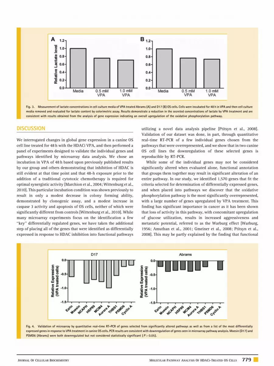
In order to validate the results of the microarray, a short list of genes
chosen from the pathway maps as well as from a list of the most
differentially expressed genes was generated for quantitative real-
time PCR. These genes were chosen from the pathway maps because
of their putative role in tumor initiation, progression, or response to
therapy. These genes include the metastasis-related genes ezrin and
moesin, the S-phase replication-associated protein MCM4, heat-
shock protein HSP90, cell cycle-associated proteins cyclin A and
aurora A, proteosome regulatory subunit PSMD6, and thrombos-
pondin 1. The results of PCR analysis are shown in Figure 4 and serve
to validate the gene expression changes and the selection criteria for
these genes.
PROTEASOME INHIBITION BY VALPROIC ACID
In addition to PCR analysis of the proteasomal regulatory subunit
PSMD6, we also evaluated the inhibition of the proteasome pathway
as a whole through evaluation of chymotrypsin-like activity in
VPA-treated cells. As shown in Figure 5a, canine OS cells treated for
48 h with VPA exhibit an overall decrease in the chymotrypsin-like
activity compared to controls, providing further validation of the
pathway analysis.
In addition, based upon the finding that VPA treatment appears to
lead to proteasome inhibition, we evaluated the potential of a
combination therapy utilizing VPA and the proteasome inhibitor
ONX0912. The addition of VPA led to a synergistic reduction in
viable cell numbers compared to treatment with ONX0912 alone
(Fig. 5b).
ENHANCEMENT OF NQO1 ACTIVITY BY VALPROIC ACID
One of the pathways that was statistically overrepresented in our
microarray analysis is the ubiquinone metabolism pathway, and
NAD(P)H:quinone oxidoreductase 1 (NQO1) was also found to be
amongst the most highly upregulated genes in response to VPA
treatment. To validate this finding, we evaluated NQO1 activity in
VPA-treated cells. As shown in Figure 6A, the specific activity of
NQO1 was enhanced by treatment with VPA, which is consistent
with upregulation of NQO1 gene transcription observed by
microarray.
In addition to increased NQO1 activity, we also assessed the
ability of VPA to sensitize OS cells to the antiproliferative effects of
the NQO1 substrate drug mitomycin C. As shown in Figure 6, canine
Fig. 1. MA plot of genes selected from microarray analysis of VPA-treated canine OS cells; 1,570 genes were selected (light gray) based on those that were differentially
expressed in response to VPA treatment and represent a balanced group of those that are moderately and highly expressed.
JOURNAL OF CELLULAR BIOCHEMISTRY MOLECULAR PATHWAY ANALYSIS OF HDACi-TREATED OS CELLS 777

OS cells treated for 48 h with VPA and mitomycin C show an
enhanced anti-proliferative effect compared to mitomycin C alone.
DOWNREGULATION OF ENDOTHELIN-1 BY VPA
The pro-tumorigenic protein endothelin 1 (Et-1), which plays a role
in promoting OS metastasis through production of matrix-
degrading matrix metalloproteins, and is found at elevated levels
in patients with metastatic disease [Grant et al., 2003; Felx et al.,
2006], was found to be highly downregulated by VPA via gene
expression microarray. We used a commercially available ELISA-
based assay to evaluate the levels of Et-1 in supernatants of cells
treated with a clinically achievable concentration of VPA for 48 h.
As shown in Figure 7, the concentrations of Et-1 secreted by OS cells
is reduced in the presence of 1mMVPA, and this reduction is not the
result of reduced cell viability as there is negligible anti-proliferative
effect at that concentration of VPA (Fig. 7).
Fig. 2. Top 15 biological pathways significantly overrepresented in the list of genes differentially expressed between VPA-treated and control OS cells.[Color figure can be
seen in the online version of this article, available at http://wileyonlinelibrary.com/journal/jcb]
778 MOLECULAR PATHWAY ANALYSIS OF HDACi-TREATED OS CELLS JOURNAL OF CELLULAR BIOCHEMISTRY
DISCUSSION
We interrogated changes in global gene expression in a canine OS
cell line treated for 48 h with the HDACi VPA, and then performed a
panel of experiments designed to validate the individual genes and
pathways identified by microarray data analysis. We chose an
incubation in VPA of 48 h based upon previously published results
by our group and others demonstrating that inhibition of HDAC is
still evident at that time point and that 48-h exposure prior to the
addition of a traditional cytotoxic chemotherapy is required for
optimal synergistic activity [Marchion et al., 2004;Wittenburg et al.,
2010]. This particular incubation condition was shown previously to
result in only a modest decrease in colony forming ability,
demonstrated by clonogenic assay, and a modest increase in
caspase 3 activity and apoptosis of OS cells, neither of which were
significantly different from controls [Wittenburg et al., 2010]. While
many microarray experiments focus on the identification a few
‘‘key’’ differentially regulated genes, we have taken the additional
step of placing all of the genes that were identified as differentially
expressed in response to HDAC inhibition into functional pathways
utilizing a novel data analysis pipeline [Ptitsyn et al., 2008].
Validation of our dataset was done, in part, through quantitative
real-time RT-PCR of a few individual genes chosen from the
pathways that were overrepresented, and we show that in two canine
OS cell lines the downregulation of these selected genes is
reproducible by RT-PCR.
While some of the individual genes may not be considered
significantly altered when evaluated alone, functional annotation
that groups them together may result in significant alteration of an
entire pathway. In our study, we identified 1,570 genes that fit the
criteria selected for determination of differentially expressed genes,
and when placed into pathways we discover that the oxidative
phosphorylation pathway is the most significantly overrepresented,
with a large number of genes upregulated by VPA treatment. This
finding has significant importance in cancer as it has been shown
that loss of activity in this pathway, with concomitant upregulation
of glucose utilization, results in increased aggressiveness and
metastatic potential, referred to as the Warburg effect [Warburg,
1956; Amuthan et al., 2001; Gmeiner et al., 2008; Ptitsyn et al.,
2008]. This may be partly explained by the finding that functional
Fig. 3. Measurement of lactate concentrations in cell culture media of VPA treated Abrams (A) and D17 (B) OS cells. Cells were incubated for 48 h in VPA and then cell culture
media removed and evaluated for lactate content by colorimetric assay. Results demonstrate a reduction in the secreted concentrations of lactate by VPA treatment and are
consistent with results obtained from the analysis of gene expression indicating an overall upregulation of the oxidative phosphorylation pathway.
Fig. 4. Validation of microarray by quantitative real-time RT-PCR of genes selected from significantly altered pathways as well as from a list of the most differentially
expressed genes in response to VPA treatment in canine OS cells. PCR results are consistent with downregulation of genes seen in microarray pathway analysis. Moesin (D17) and
PSMD6 (Abrams) were both downregulated but not considered statistically significant (P> 0.05).
JOURNAL OF CELLULAR BIOCHEMISTRY MOLECULAR PATHWAY ANALYSIS OF HDACi-TREATED OS CELLS 779

oxidative phosphorylation is required for apoptosis induced by the
pro-apoptotic protein Bax in OS, providing evidence that evasion of
apoptosis can be obtained by downregulation of oxidative
phosphorylation [Dey and Moraes, 2000; Harris et al., 2000]. Our
results suggest that one potential mechanism by which HDACi, and
particularly VPA, exert an anti-tumor effect may be through re-
sensitization of intrinsic apoptotic pathways via upregulation of
oxidative phosphorylation.
An additional pathway that was overrepresented in our
microarray analysis involves protein degradation via the ubiquitin-
proteasome pathway. While evaluation of mRNA levels by
quantitative real-time RT-PCR did show a reduction in the level
of one regulatory subunit of the 20S proteasome (PSMD6), we also
performed a functional assay that evaluated proteosomal activity in
VPA-treated cells to determine the biological relevance. VPA-
treated OS cells clearly demonstrate an inhibition of chymotrypsin-
like activity, and this result is in agreement with another study
describing a siRNA-mediated loss-of-function screen that identified
the proteasome as playing an important role in HDACi-induced
apoptosis [Fotheringham et al., 2009]. In fact, this same study
Fig. 5. (A) Evaluation of proteasome inhibition by VPA. Human and canine OS cells were treated with 0 or 1.0mM VPA for 48 h and then proteasome inhibition was measured
by evaluation of calpain activity in cell lysates, �P< 0.05. Proliferation evaluation of Abrams (B) and D17 (C) OS cells co-treated with 0 or 0.5mM VPA and increasing
concentrations of the proteasome inhibitor ONX0912 demonstrate sensitization to proteasome inhibition by VPA. Bliss analysis comparing fractional inhibition of theoretical
additivity to experimental values reveals synergistic antiproliferative activity of the 0.5mM VPA/ONX0912 combination in Abrams (D) and D17 (E) OS cells.
Fig. 6. (A) NQO1 activity measured in lysates of VPA-treated (1mM) and untreated OS cells. Both cell lines demonstrate increased NQO1 activity in response to VPA
treatment, consistent with upregulation of NQO1 gene activity; �P< 0.05. Proliferation evaluation of Abrams (B) and D17 (C) OS cells co-treated with 0, 0.5, or 1.0mM VPA
and increasing concentrations of mitomycin C demonstrate sensitization by VPA. Bliss analysis comparing fractional inhibition of theoretical additivity to experimental values
reveals synergistic anti-proliferative activity of the 0.5mM VPA/mitomycin C combination in Abrams (D) and additive activity in D17 (E) OS cells.
780 MOLECULAR PATHWAY ANALYSIS OF HDACi-TREATED OS CELLS JOURNAL OF CELLULAR BIOCHEMISTRY

suggests that one particular protein involved in shuttling of
ubiquitinated cargo proteins may govern tumor sensitivity to drugs
that target the proteasome, and found this particular protein
overexpressed in cutaneous T-cell lymphoma, a cancer that appears
particularly sensitive to HDACi. Proteasome inhibitors have been
reported to be effective in the treatment of OS in vitro in a number of
studies [Lauricella et al., 2003; Lim et al., 2004; Yan et al., 2007],
possibly through regulation of cell cycle-associated proteins such as
the cyclin-dependent kinases. This led us to test whether a
combination of HDACi with VPA and proteasome inhibition with
a novel compound, ONX0912, would provide an enhanced anti-
tumor effect. We show that the combination of VPA and ONX0912
results in a synergistic reduction in viability of canine OS cells
compared to cells treated with the proteasome inhibitor alone. It is
important to note that, while these results suggest that enhanced
inhibition of the proteasome results in improved reduction of cell
viability, it is possible that this is not the only mechanism by which
this effect occurs with the combination of these two drugs.
We also found ubiquinone metabolism to be one of the pathways
altered by VPA treatment and NQO1 was additionally found to be
one of the most significantly upregulated genes by the microarray.
Upregulation of NQO1 could have important implications for the
treatment of cancer as this particular enzyme plays a role in the bio-
activation of anti-tumor quinines such as mitomycin C [Gustafson
et al., 2003]. We proceeded to validate this finding through
evaluation of NQO1-specific activity in canine OS cell lysates
following 48-h treatment with VPA. We demonstrate that the ability
of NQO1 to reduce DCPIP is enhanced following VPA treatment,
consistent with increased levels of the enzyme. To further test the
significance of this, we evaluated the ability of VPA to sensitize OS
cells to the anti-proliferative effects of mitomycin C and show a
sensitizing effect on Abrams and D17 canine OS cell lines. This is
consistent with in vitro reports of synergy between VPA and
mitomycin C in colon carcinoma cells [Friedmann et al., 2006].
However, as mitomycin C is also a DNA-binding drug [Verweij and
Pinedo, 1990], the effect of VPA-induced chromatin decondensation
and increased access to DNA bymitomycin C, as observed with other
DNA-intercalating agents [Marchion et al., 2005; Wittenburg et al.,
2010], cannot be ruled out as a contributing factor. Given that a
number of identified pathways play important roles in DNA damage
response, including protein turnover, cytoskeleton remodeling, and
the electron transport chain, another possibility for the enhanced
effect observed when VPA is combined with mitomycin C,
doxorubicin, or other DNA damaging agents is an inhibition of
the cellular ability to repair DNA damage. Indeed, the coordinated
cellular response to DNA damage involves multiple levels of
regulation involving protein–DNA interactions, protein–protein
interactions, and multiple interconnected cellular pathways [Begley
and Samson, 2004]. The discovery that DNA damage results in an
extensive change in transcription factor promoter binding with
some transcription factors binding significantly more genes in the
presence of a DNA damaging agent [Workman et al., 2006] provides
another potential explanation for increased sensitivity to DNA
damage following HDACi therapy, as alterations in chromatin
structure will change availability of promoter sites to transcription
factors that bind specific regions in response to DNA damage. While
the sensitization of OS cells to mitomycin C by VPA may result, in
part, from the upregulation of NQO1, the effect of HDACi with VPA
on the DNA damage response as a mechanism of sensitization to
mitomycin C and other DNA-targeting anti-cancer agents certainly
warrants future investigation.
An additional gene identified by microarray analysis as one
significantly downregulated by VPA treatment is Et-1. Et-1 is
reported to have a variety of pro-tumorigenic properties that include
acting as a mitogen for numerous human cancer cell lines as well as
endothelial cells and vascular smooth muscle cells, protecting cells
from Fas ligand-mediated apoptosis, and promoting metastasis
through upregulation of stroma-degrading matrix metalloprotei-
nases in OS [Grant et al., 2003; Felx et al., 2006]. In addition,
elevated plasma levels of Et-1 have been found in tumor-bearing
patients with the highest levels found in those with metastatic
disease [Grant et al., 2003]. This provides rationale for targeting Et-1
in cancer therapy. We found that incubation of OS cells with
clinically achievable levels of VPA results in decreased Et-1
Fig. 7. Measurement of endothelin-1 in supernatants of VPA-treated canine OS cells by ELISA. A marked reduction in endothelin-1 (gray lines) is observed in the D17 canine
OS cell line upon 48-h exposure to 1mM VPA. This reduction in endothelin is not the result of reduced cell viability as the percent viable cells remains relatively constant across
the range of VPA doses (black lines).
JOURNAL OF CELLULAR BIOCHEMISTRY MOLECULAR PATHWAY ANALYSIS OF HDACi-TREATED OS CELLS 781

secretion into culture media, consistent with the downregulation
observed on microarray. This may provide additional support to the
use of combinations of VPA and proteasome inhibitors for the
treatment of OS, as the induction of pro-metastatic genes such as the
MMPs by Et-1 appears to be mediated through NF-kB, a well-
documented target of proteasome inhibitors [Felx et al., 2006;
Orlowski and Kuhn, 2008].
One potential drawback of this study is the lack of secondary
evaluation of HDAC inhibition, which might be performed through
use of additional inhibitors or RNA interference studies, providing
more detailed and mechanistic information of the effect HDACi as a
whole. However, our main goal was to utilize a novel method of
analyzing the gene expression information resulting from a 48-h
exposure to VPA specifically, and to validate this method of
pathway analysis. Additionally, future studies should focus on
examining the interrelationships between the pathways that were
identified and the potential mechanisms by which HDACi controls
these pathways. While our results focused on changes in gene
expression, they do not provide mechanistic details necessary to
determine if altered gene transcription is due simply to altered
chromatin structure or is secondary to an ‘‘off-target’’ effect such as
alterations in acetylation and activity of other non-histone proteins,
resulting in secondary changes in gene expression.
The results of this study shed further light onto the potential
mechanisms by which HDACi with VPA exerts its antitumor effect in
OS. We have identified oxidative phosphorylation as the most
significantly altered metabolic pathway in response to VPA. In
addition, we show that the proteasome pathway is altered through
decreased chymotrypsin-like activity, leading us to test a
combination of VPA and a proteasome inhibitor in OS cells. Our
results suggest that this particular combination may prove
beneficial in the treatment of OS. We show that VPA is capable
of upregulating the oxidoreductase NQO1, enhancing its enzymatic
activity leading to increased sensitivity of OS cells to the NQO1
substrate drug mitomycin C. In addition, we show that treatment of
OS cells with VPA results in decreased secretion of Et-1, which has
been shown to correlate with poor outcome in cancer when elevated
serum levels are detected in patients. Taken together, the results of
our pathway analysis in OS cells treated with an HDACi reveal a
number of pathways that have already been identified as potential
anti-tumor targets and provide further support for the design of
VPA-containing combination protocols in the treatment of OS.
REFERENCES
Amuthan G, Biswas G, Zhang SY, Klein-Szanto A, Vijayasarathy C, AvadhaniNG. 2001. Mitochondria-to-nucleus stress signaling induces pheno-typic changes, tumor progression and cell invasion. EMBO J 20:1910–1920.
Begley TJ, Samson LD. 2004. Network responses to DNA damaging agents.DNA Repair (Amst) 3:1123–1132.
Bolden JE, Peart MJ, Johnstone RW. 2006. Anticancer activities of histonedeacetylase inhibitors. Nat Rev Drug Discov 5:769–784.
Cai Y, Mohseny AB, Karperien M, Hogendoorn PC, Zhou G, Cleton-JansenAM. 2010. Inactive Wnt/Beta-catenin pathway in conventional high-gradeosteosarcoma. J Pathol 220(1):24–33.
Chung YS, Baylink DJ, Srivastava AK, Amaar Y, Tapia B, Kasukawa Y, MohanS. 2004. Effects of secreted frizzled-related protein 3 on osteoblasts in vitro.J Bone Miner Res 19:1395–1402.
Cleton-Jansen AM, Anninga JK, Briaire-de Bruijn IH, Romeo S, Oosting J,Egeler RM, Gelderblom H, Taminiau AH, Hogendoorn PC. 2009. Profiling ofhigh-grade central osteosarcoma and its putative progenitor cells identifiestumourigenic pathways. Br J Cancer 101:2064.
Dernell WS, Straw RC, Withrow SJ. 2001. Tumors of the skeletal system In:Withrow SJ, MacEwen EG, editors. Small animal clinical oncology.Philadelphia: W.B. Saunders Company. pp 378–417.
Deroanne CF, Bonjean K, Servotte S, Devy L, Colige A, Clausse N, Blacher S,Verdin E, Foidart JM, Nusgens BV, Castronovo V. 2002. Histone deacetylasesinhibitors as anti-angiogenic agents altering vascular endothelial growthfactor signaling. Oncogene 21:427–436.
Dey R, Moraes CT. 2000. Lack of oxidative phosphorylation and lowmitochondrial membrane potential decrease susceptibility to apoptosisand do not modulate the protective effect of Bcl-x(L) in osteosarcoma cells.J Biol Chem 275:7087–7094.
Felx M, Guyot M-C, Isler M, Turcotte RE, Doyon J, Khatib A-M, Leclerc S,Moreau A, Moldovan F. 2006. Endothelin-1 (ET-1) promotes MMP-2 andMMP-9 induction involving the transcription factor NF-kB in humanosteosarcoma. Clin Sci 110:645–654.
Fotheringham S, Epping MT, Stimson L, Khan O, Wood V, Pezzella F,Bernards R, La Thangue NB. 2009. Genome-wide loss-of-function screenreveals an important role for the proteasome in HDAC inhibitor-inducedapoptosis. Cancer Cell 15:57–66.
Friedmann I, Atmaca A, Chow KU, Jager E, Weidmann E. 2006. Synergisticeffects of valproic acid and mitomycin C in adenocarcinoma cell lines andfresh tumor cells of patients with colon cancer. J Chemother 18:415–420.
Gmeiner WH, Hellmann GM, Shen P. 2008. Tissue-dependent and -indepen-dent gene expression changes in metastatic colon cancer. Oncol Rep 19:245–251.
Grant K, Loizidou M, Taylor I. 2003. Endothelin-1: A multifunctionalmolecule in cancer. Br J Cancer 88:163–166.
Gustafson DL, Siegel D, Rastatter JC, Merz AL, Parpal JC, Kepa JK, Ross D,Long ME. 2003. Kinetics of NAD(P)H:quinone oxidoreductase I (NQO1)inhibition by mitomycin C in vitro and in vivo. J Pharmacol Exp Ther305:1079–1086.
Harris MH, Vander Heiden MG, Kron SJ, Thompson CB. 2000. Role ofoxidative phosphorylation in Bax toxicity. Mol Cell Biol 20:3590–3596.
Huang daW, Sherman BT, Tan Q, Collins JR, AlvordWG, Roayaei J, StephensR, Baseler MW, Lane HC, Lempicki RA. 2007. The DAVID Gene FunctionalClassification Tool: A novel biological module-centric algorithm to func-tionally analyze large gene lists. Genome Biol 8:R183.
Kansara M, Thomas DM. 2007. Molecular pathogenesis of osteosarcoma.DNA Cell Biol 26:1–18.
Kim MS, Blake M, Baek JH, Kohlhagen G, Pommier Y, Carrier F. 2003.Inhibition of histone deacetylase increases cytotoxicity to anticancer drugstargeting DNA. Cancer Res 63:7291–7300.
Lauricella M, D’Anneo A, Giuliano M, Calvaruso G, Emanuele S, Vento R,Tesoriere G. 2003. Induction of apoptosis in human osteosarcoma Saos-2cells by the proteasome inhibitor MG132 and the protective effect of pRb. CellDeath Differ 10:930–932.
Lim JH, Chang YC, Park YB, Park JW, Kwon TK. 2004. Transcriptionalrepression of E2F gene by proteasome inhibitors in human osteosarcomacells. Biochem Biophys Res Commun 318:868–872.
Link MP, Goorin AM, Miser AW, Green AA, Pratt CB, Belasco JB, Pritchard J,Malpas JS, Baker AR, Kirkpatrick JA, Ayala AG, Shuster JJ, Abelson HT,Simone JV, Vietti TJ. 1986. The effect of adjuvant chemotherapy on relapse-free survival in patients with osteosarcoma of the extremity. N Engl J Med314:1600–1606.
782 MOLECULAR PATHWAY ANALYSIS OF HDACi-TREATED OS CELLS JOURNAL OF CELLULAR BIOCHEMISTRY

Mandal D, Srivastava A, Mahlum E, Desai D, Maran A, Yaszemski M, JalalSM, Gitelis S, Bertoni F, Damron T, Irwin R, O’Connor M, Schwartz H,BolanderME, Sarkar G. 2007. Severe suppression of Frzb/sFRP3 transcriptionin osteogenic sarcoma. Gene 386:131–138.
Mann BS, Johnson JR, He K, Sridhara R, Abraham S, Booth BP, Verbois L,Morse DE, Jee JM, Pope S, Harapanhalli RS, Dagher R, Farrell A, Justice R,Pazdur R. 2007. Vorinostat for treatment of cutaneous manifestations ofadvanced primary cutaneous T-cell lymphoma. Clin Cancer Res 13:2318–2322.
Marchion DC, Bicaku E, Daud AI, Richon V, Sullivan DM, Munster PN. 2004.Sequence-specific potentiation of topoisomerase II inhibitors by the histonedeacetylase inhibitor suberoylanilide hydroxamic acid. J Cell Biochem92:223–237.
Marchion DC, Bicaku E, Daud AI, Sullivan DM, Munster PN. 2005. Valproicacid alters chromatin structure by regulation of chromatin modulationproteins. Cancer Res 65:3815–3822.
Marsoni S, Damia G, Camboni G. 2008. A work in progress: The clinicaldevelopment of histone deacetylase inhibitors. Epigenetics 3:164–171.
Minucci S, Pelicci PG. 2006. Histone deacetylase inhibitors and the promiseof epigenetic (and more) treatments for cancer. Nat Rev Cancer 6:38–51.
Miyake K, Yoshizumi T, Imura S, Sugimoto K, Batmunkh E, Kanemura H,Morine Y, ShimadaM. 2008. Expression of hypoxia-inducible factor-1 alpha,histone deacetylase 1, and metastasis-associated protein 1 in pancreaticcarcinoma: Correlation with poor prognosis with possible regulation. Pan-creas 36:e1–e9.
Mueller F, Fuchs B, Kaser-Hotz B. 2007. Comparative biology of human andcanine osteosarcoma. Anticancer Res 27:155–164.
Munster P, Marchion D, Bicaku E, Schmitt M, Lee JH, DeConti R, Simon G,Fishman M, Minton S, Garrett C, Chiappori A, Lush R, Sullivan D, Daud A.2007. Phase I trial of histone deacetylase inhibition by valproic acid followedby the topoisomerase II inhibitor epirubicin in advanced solid tumors:A clinical and translational study. J Clin Oncol 25:1979–1985.
Orlowski RZ, Kuhn DJ. 2008. Proteasome inhibitors in cancer therapy:Lessons from the first decade. Clin Cancer Res 14:1649–1657.
Prince HM, Bishton MJ, Harrison SJ. 2009. Clinical studies of histonedeacetylase inhibitors. Clin Cancer Res 15:3958–3969.
Ptitsyn AA, Weil MM, Thamm DH. 2008. Systems biology approach toidentification of biomarkers for metastatic progression in cancer. BMCBioinformatics 9(Suppl 9):S8.
Rikimaru T, Taketomi A, Yamashita Y, Shirabe K, Hamatsu T, Shimada M,Maehara Y. 2007. Clinical significance of histone deacetylase 1 expression inpatients with hepatocellular carcinoma. Oncology 72:69–74.
Rosen G, Marcove RC, Huvos AG, Caparros BI, Lane JM, Nirenberg A, CacavioA, Groshen S. 1983. Primary osteogenic sarcoma: Eight-year experience withadjuvant chemotherapy. J Cancer Res Clin Oncol 106(Suppl):55–67.
Rubin EM, Guo Y, Tu K, Xie J, Zi X, Hoang BH. 2010. Wnt inhibitory factor 1decreases tumorigenesis and metastasis in osteosarcoma. Mol Cancer Ther9:731–741.
Schroeder TM, Westendorf JJ. 2005. Histone deacetylase inhibitors promoteosteoblast maturation. J Bone Miner Res 20:2254–2263.
Schroeder TM, Kahler RA, Li X, Westendorf JJ. 2004. Histone deacetylase 3interacts with runx2 to repress the osteocalcin promoter and regulateosteoblast differentiation. J Biol Chem 279:41998–42007.
Schroeder TM, Nair AK, Staggs R, Lamblin AF, Westendorf JJ. 2007. Geneprofile analysis of osteoblast genes differentially regulated by histonedeacetylase inhibitors. BMC Genomics 8:362.
Tomasi TB, Magner WJ, Nazmul A, Khan H. 2006. Epigenetic regulation ofimmune escape genes in cancer. Cancer Immunol Immunother 55:1159–1184.
Verweij J, Pinedo HM. 1990. Mitomycin C: Mechanism of action, usefulnessand limitations. Anticancer Drugs 1:5–13.
Warburg O. 1956. On respiratory impairment in cancer cells. Science 124:269–270.
Weichert W, Roske A, Gekeler V, Beckers T, Ebert MP, Pross M, Dietel M,Denkert C, Rocken C. 2008a. Association of patterns of class I histonedeacetylase expression with patient prognosis in gastric cancer: A retrospec-tive analysis. Lancet Oncol 9:139–148.
Weichert W, Roske A, Gekeler V, Beckers T, Stephan C, Jung K, Fritzsche FR,Niesporek S, Denkert C, Dietel M, Kristiansen G. 2008b. Histone deacetylases1, 2 and 3 are highly expressed in prostate cancer and HDAC2 expression isassociated with shorter PSA relapse time after radical prostatectomy. Br JCancer 98:604–610.
Weichert W, Roske A, Niesporek S, Noske A, Buckendahl AC, Dietel M,Gekeler V, BoehmM, Beckers T, Denkert C. 2008c. Class I histone deacetylaseexpression has independent prognostic impact in human colorectal cancer:Specific role of class I histone deacetylases in vitro and in vivo. Clin CancerRes 14:1669–1677.
Westendorf JJ. 2007. Histone deacetylases in control of skeletogenesis. J CellBiochem 102:332–340.
Wittenburg L, Bisson L, Rose B, Thamm DH. 2011. The histone deacetylaseinhibitor valproic acid sensitizes human and canine osteosarcoma to doxo-rubicin. Cancer Chemother Pharmacol 67(1):83–92.
Workman CT, Mak HC, McCuine S, Tagne J-B, Agarwal M, Ozier O, Begley TJ,Samson LD, Ideker T. 2006. A systems approach to mapping DNA damageresponse pathways. Science 312:1054–1059.
Xu WS, Parmigiani RB, Marks PA. 2007. Histone deacetylase inhibitors:Molecular mechanisms of action. Oncogene 26:5541–5552.
Yan XB, Yang DS, Gao X, Feng J, Shi ZL, Ye Z. 2007. Caspase-8 dependentosteosarcoma cell apoptosis induced by proteasome inhibitor MG132. CellBiol Int 31:1136–1143.
JOURNAL OF CELLULAR BIOCHEMISTRY MOLECULAR PATHWAY ANALYSIS OF HDACi-TREATED OS CELLS 783