Morphogenesis of theCaenorhabditis elegansMale Tail Tip
21
Morphogenesis of the Caenorhabditis elegans Male Tail Tip Can Q. Nguyen,* ,1 David H. Hall,² ,1 Ying Yang,* and David H. A. Fitch* ,2 *Department of Biology, New York University, New York, New York 10003; and ²Center for C. elegans Anatomy, Department of Neuroscience, Kennedy Center, Albert Einstein College of Medicine, Bronx, New York 10461 Using electron microscopy and immunofluorescent labeling of adherens junctions, we have reconstructed the changes in cell architecture and intercellular associations that occur during morphogenesis of the nematode male tail tip. During late postembryonic development, the Caenorhabditis elegans male tail is reshaped to form a copulatory structure. The most posterior hypodermal cells in the tail define a specialized, sexually dimorphic compartment in which cells fuse and retract in the male, changing their shape from a tapered cone to a blunt dome. Developmental profiles using electron microscopy and immunofluorescent staining suggest that cell fusions are initiated at or adjacent to adherens junctions. Anterior portions of the tail tip cells show the first evidence of retractions and fusions, consistent with our hypothesis that an anterior event triggers these morphogenetic events. Available mutations that interfere with morphogenesis implicate particular regulatory pathways and suggest loci at which evolutionary changes could have produced morphological diversity. © 1999 Academic Press Key Words: morphogenesis; C. elegans; adherens junctions; gap junctions; cell fusion; male tail; evolution. INTRODUCTION How cells are coordinated to change their form remains a fundamental question in developmental biology. To under- stand the mechanisms and components underlying multi- cellular morphogenesis, we have begun a comprehensive study of the four-celled male tail tip of Caenorhabditis elegans. Cell fusion, changes in cell shape and position, fluid displacement, nuclear migration, and changes in in- tercellular associations are all important features of male tail tip morphogenesis, as they are in other systems. Cell fusion plays an important role in many steps of develop- ment in C. elegans (Podbilewicz and White, 1994; Newman et al., 1996), in other invertebrates (Doberstein et al., 1997; Tanaka-Matakatsu et al., 1996), and in vertebrates (see Podbilewicz and White, 1994). In vertebrates and inverte- brates, cell shape changes are important during embryogen- esis in events like gastrulation or elongation and in forming an array of epithelial structures via folds or placodes (Priess and Hirsh, 1986; Kam et al., 1991). Morphogenesis occurs rapidly during the last larval (L4) stage to dramatically change the shape of the tail from a simple tapered cone to the complex morphology of the copulatory bursa or “male tail” (Figs. 1A–1F). General male tail morphogenesis and patterning of the male-specific sensilla (“ray” papillae) also involve a complex series of developmental events, including anteroposterior identity specification by the Hox gene complex, intercellular signal- ing, and hypodermal cell fusions (reviewed in Emmons and Sternberg, 1997). The first visible sign of male tail morpho- genesis occurs when the cells in the tail tip become rounded and retract anteriorly (Fig. 1B). Here we focus on the dynamic cellular and subcellular changes in the architec- ture of the tail tip during this first step of male tail morphogenesis. The lineages of the tail tip cells (hyp8 – hyp11) are known and are the same for both sexes (Sulston and Horvitz, 1977; Sulston et al., 1980, 1983). These hypodermal cells origi- nate early in embryogenesis and adopt their characteristic tapered shapes when other hypodermal cells elongate dur- 1 These two authors contributed equally. 2 To whom correspondence should be addressed at Department of Biology, New York University, Room 1009 Main Building, 100 Washington Square East, New York, NY 10003. E-mail: david.fi[email protected]. Developmental Biology 207, 86 –106 (1999) Article ID dbio.1998.9173, available online at http://www.idealibrary.com on 0012-1606/99 $30.00 Copyright © 1999 by Academic Press All rights of reproduction in any form reserved. 86
Transcript of Morphogenesis of theCaenorhabditis elegansMale Tail Tip

a
eC
Ucppiapapd
Developmental Biology 207, 86–106 (1999)Article ID dbio.1998.9173, available online at http://www.idealibrary.com on
Morphogenesis of the Caenorhabditis elegansMale Tail Tip
Can Q. Nguyen,*,1 David H. Hall,†,1 Ying Yang,*nd David H. A. Fitch*,2
*Department of Biology, New York University, New York, New York 10003; and †Center for C.legans Anatomy, Department of Neuroscience, Kennedy Center, Albert Einsteinollege of Medicine, Bronx, New York 10461
sing electron microscopy and immunofluorescent labeling of adherens junctions, we have reconstructed the changes inell architecture and intercellular associations that occur during morphogenesis of the nematode male tail tip. During lateostembryonic development, the Caenorhabditis elegans male tail is reshaped to form a copulatory structure. The mostosterior hypodermal cells in the tail define a specialized, sexually dimorphic compartment in which cells fuse and retractn the male, changing their shape from a tapered cone to a blunt dome. Developmental profiles using electron microscopynd immunofluorescent staining suggest that cell fusions are initiated at or adjacent to adherens junctions. Anteriorortions of the tail tip cells show the first evidence of retractions and fusions, consistent with our hypothesis that annterior event triggers these morphogenetic events. Available mutations that interfere with morphogenesis implicatearticular regulatory pathways and suggest loci at which evolutionary changes could have produced morphologicaliversity. © 1999 Academic Press
Key Words: morphogenesis; C. elegans; adherens junctions; gap junctions; cell fusion; male tail; evolution.
eaa
ssctsdsiSgadtm
aS
INTRODUCTION
How cells are coordinated to change their form remains afundamental question in developmental biology. To under-stand the mechanisms and components underlying multi-cellular morphogenesis, we have begun a comprehensivestudy of the four-celled male tail tip of Caenorhabditiselegans. Cell fusion, changes in cell shape and position,fluid displacement, nuclear migration, and changes in in-tercellular associations are all important features of maletail tip morphogenesis, as they are in other systems. Cellfusion plays an important role in many steps of develop-ment in C. elegans (Podbilewicz and White, 1994; Newmanet al., 1996), in other invertebrates (Doberstein et al., 1997;Tanaka-Matakatsu et al., 1996), and in vertebrates (seePodbilewicz and White, 1994). In vertebrates and inverte-brates, cell shape changes are important during embryogen-
1 These two authors contributed equally.2 To whom correspondence should be addressed at Department
of Biology, New York University, Room 1009 Main Building, 100
Washington Square East, New York, NY 10003. E-mail:[email protected].86
sis in events like gastrulation or elongation and in formingn array of epithelial structures via folds or placodes (Priessnd Hirsh, 1986; Kam et al., 1991).Morphogenesis occurs rapidly during the last larval (L4)
tage to dramatically change the shape of the tail from aimple tapered cone to the complex morphology of theopulatory bursa or “male tail” (Figs. 1A–1F). General maleail morphogenesis and patterning of the male-specificensilla (“ray” papillae) also involve a complex series ofevelopmental events, including anteroposterior identitypecification by the Hox gene complex, intercellular signal-ng, and hypodermal cell fusions (reviewed in Emmons andternberg, 1997). The first visible sign of male tail morpho-enesis occurs when the cells in the tail tip become roundednd retract anteriorly (Fig. 1B). Here we focus on theynamic cellular and subcellular changes in the architec-ure of the tail tip during this first step of male tailorphogenesis.The lineages of the tail tip cells (hyp8–hyp11) are known
nd are the same for both sexes (Sulston and Horvitz, 1977;ulston et al., 1980, 1983). These hypodermal cells origi-
nate early in embryogenesis and adopt their characteristictapered shapes when other hypodermal cells elongate dur-
0012-1606/99 $30.00Copyright © 1999 by Academic Press
All rights of reproduction in any form reserved.

h
l
87Morphogenesis of the C. elegans Male Tail Tip
FIG. 1. Male tail morphogenesis in two species (anterior at left, lateral views, except ventral views in F and L). (A–F) Wild-type C. elegansmale from late L3 (A) through L4 morphogenesis (B–D) to adulthood (E, F). (B) The arrow marks the extracellular space left in the wake ofanteriorward retraction of the tail tip. (E) Side view of the “peloderan” adult male tail with rays (numbered). (G) The C. elegans
ermaphrodite retains the pointed shape of the larval tail. Failure in tail tip retraction during male morphogenesis in O. myriophila (H–J)
also results in a leptoderan adult tail (arrows in K and L). In O. myriophila, fluid still accumulates in the extracellular space while aeptoderan tail tip is maintained (J). (M) Adult hermaphrodite of O. myriophila.Copyright © 1999 by Academic Press. All rights of reproduction in any form reserved.

gtfrui
ioanfsmrsastaisfiss
gop
afi
Aab1r(aC
88 Nguyen et al.
ing midembryonic morphogenesis (Priess and Hirsh, 1986).The sexual dimorphism of the tail tip cells of adults(compare Figs. 1E and 1G) thus results not from differencesin cell lineage, but from late postembryonic morphogeneticevents specific to males.
Because C. elegans hermaphrodites are self-fertilizing andmales are nonessential, genetic analysis of the male tail tipis greatly facilitated. Some mutations that disrupt tail tipmorphogenesis are already available and are likely to leadus to the components and regulatory mechanisms underly-ing this process. However, a complete delineation of wild-type morphogenesis—undertaken here—is first required toprovide a context for a meaningful analysis of mutantphenotypes.
Tail tip morphogenesis has changed during evolution.Within the nematode family Rhabditidae, which includesC. elegans, there is a surprising amount of morphologicaldiversity for a structure with so few cells (Chitwood andChitwood, 1950; Sudhaus, 1976). Tail tip morphology hasclassically been used to diagnose taxonomic levels as highas subfamilies (Andrassy, 1983). For example, the tail tip ofOscheius myriophila fails to retract during male morpho-enesis (Figs. 1H–1J), resulting in a pointed (“leptoderan”)ail tip that protrudes beyond the posterior edge of the adultan (Figs. 1K and 1L). To reconstruct evolutionary changesesulting in this morphological diversity, a comprehensivenderstanding of the architecture of tail tip morphogenesiss first required.
To provide a detailed cellular foundation for understand-ng morphogenesis, we present an ultrastructural analysisf changes in cell–cell relationships during morphogenesisnd identify key features that could underlie morphoge-etic mechanisms: e.g., a posteriorward progression of cellusions which are initiated at adherens junctions, sex-pecific cell–cell contacts, gap junctions between hypoder-al cells, and a system of vacuoles that could have been
ecruited for fluid transport. We have used serial thin-ection reconstruction and immunofluorescence labeling ofdherens junctions to identify structural bases for possibleites of cell fusions and cell–cell communication as well ashe nature of the changes in cell shape and position thatccompany tail tip morphogenesis. We have also usedmmunofluorescence to characterize a mutant and otherpecies in which male tail tip morphogenesis specificallyails. These comparisons suggest that heterochronic delaysn fusions and retraction can be effected by changes at aingle locus and could have produced morphological diver-ity during evolution.
MATERIALS AND METHODS
Nematode Strains and Culture
Mixed stages of wild-type strains of C. elegans, N2 and CB4088(Hodgkin et al., 1979), were maintained separately at 20°C on
NGM plates seeded with Escherichia coli OP50 (Brenner, 1974).CB4088 bears the mutation him-5(e1490)V, which confers a highCopyright © 1999 by Academic Press. All right
incidence of males with normal tails (Hodgkin et al., 1979). Thebx37 and bx42 alleles of lep-1 (strains EM101 and EM106) wereenerously provided by Dr. Scott Emmons (Albert Einstein Collegef Medicine). The EM435 strain of O. myriophila was describedreviously by Fitch and Emmons (1995) as “Rhabditis sp. br.”
Details about the strains of Rhabditella axei (DF5006) and othersre available from our web site (http://www.nyu.edu/projects/tch/WSRN/). Another unidentified Rhabditis species was iso-
lated from an earthworm in the Bronx, New York, and used forimmunofluorescent labeling experiments; unfortunately, thisstrain has since been lost.
Mixed-stage populations containing mostly late (L3 and L4)larvae and adults were obtained by two alternative methods. (1) Forimmunohistochemistry, gravid adults were washed with M9 buffer(Brenner, 1974) and treated with alkaline hypochlorite as describedby Sulston and Hodgkin (1988). (2) For electron microscopy (EM),gravid adults were placed on freshly seeded plates for several hoursand allowed to lay eggs; adults were subsequently washed off. Bothmethods left synchronous populations of fertilized eggs to developon plates at room temperature (20–22°C) until they reached latestages.
MH27 Immunofluorescence and Image ProcessingMixed-stage populations containing mostly late larvae were
washed twice in M9 and fixed in 1% paraformaldehyde and MRWB(Finney and Ruvkun, 1990) during four to five freeze–thaw cyclesover 30 min. Fixed worms were permeabilized by the “redox”method of Finney and Ruvkun (1990), incubated for 4–12 h at 37°Cwith a 1:100 dilution of monoclonal MH27 antibody (generouslyprovided by R. Waterston, Washington University, St. Louis, MO),and incubated with a 1:50 dilution of goat serum and a 1:50 dilutionof rhodamine-conjugated goat anti-mouse antibody (Boehringer-Mannheim Biochemicals) for 4–12 h at 37°C. Specimens wereobserved on a Zeiss Axioskop equipped for epifluorescence andphotographed at several focal planes using Kodak TMax-400 filmexposed for 15 s and push-processed two stops. Negatives weredigitally scanned with a Nikon scanner. Using Adobe Photoshop,digital images of different focal planes for the same animal werelayered together and contrast was enhanced.
Electron MicroscopyMixed-stage populations of late larvae were washed with M9
buffer and fixed without anesthesia in 2.5% glutaraldehyde, 1%formaldehyde in 0.05 M Hepes at room temperature for 150 min(general methods have been reviewed by Hall, 1995). The tails werecut open immediately in the primary fixative to ensure good accessto the tissue. After several buffer rinses, tails were fixed again in1% osmium tetroxide in 0.1 M Hepes for 2 h at room temperature.
fter further rinses, tails were embedded in 2.5% SeaPlaquegarose (FMC Bioproducts) and stored overnight at 4°C underuffer. Postembedded tails were stained in 1% uranyl acetate forh at room temperature, dehydrated, and embedded in Medcast
esin (Ted Pella) for thin sectioning. Serial transverse thin sections1000 per tail) were collected and poststained with alcoholic uranylcetate and aqueous lead citrate before viewing with a PhilipsM10 electron microscope. Selected sections were photographed.
Serial Section Reconstruction
The three-dimensional shapes and sizes of individual cells andorganelles were reconstructed by tracing their outlines on a digi-
s of reproduction in any form reserved.
apal(abboc
lrarn
wa(loTaTcrbotlaaawFmmtnec(cch(arstasdp
MtmfOc[cthto“hv
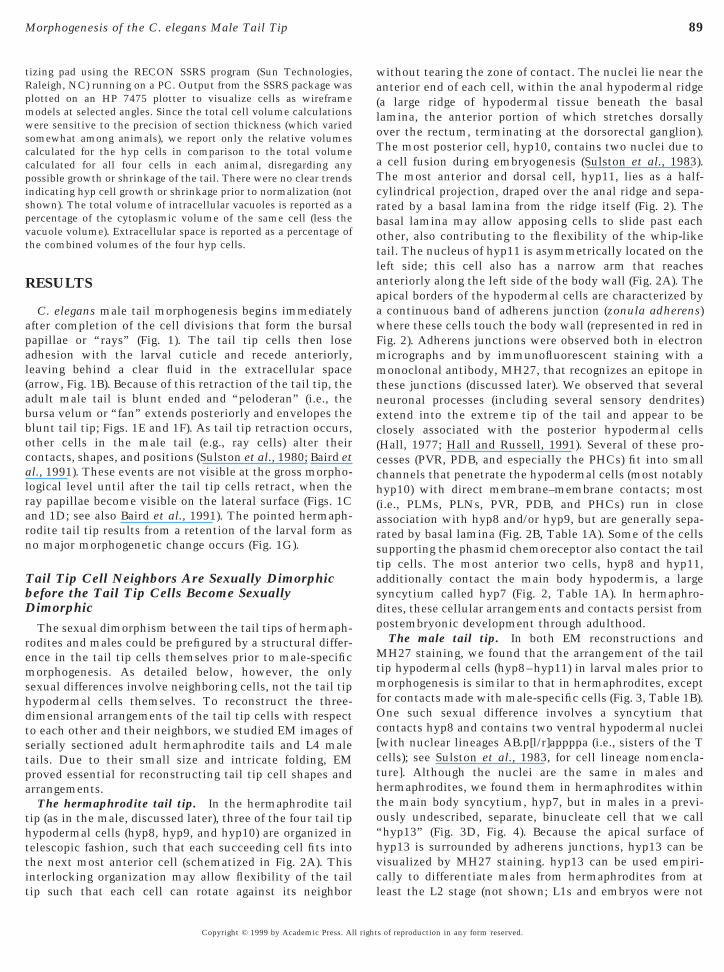
89Morphogenesis of the C. elegans Male Tail Tip
tizing pad using the RECON SSRS program (Sun Technologies,Raleigh, NC) running on a PC. Output from the SSRS package wasplotted on an HP 7475 plotter to visualize cells as wireframemodels at selected angles. Since the total cell volume calculationswere sensitive to the precision of section thickness (which variedsomewhat among animals), we report only the relative volumescalculated for the hyp cells in comparison to the total volumecalculated for all four cells in each animal, disregarding anypossible growth or shrinkage of the tail. There were no clear trendsindicating hyp cell growth or shrinkage prior to normalization (notshown). The total volume of intracellular vacuoles is reported as apercentage of the cytoplasmic volume of the same cell (less thevacuole volume). Extracellular space is reported as a percentage ofthe combined volumes of the four hyp cells.
RESULTS
C. elegans male tail morphogenesis begins immediatelyfter completion of the cell divisions that form the bursalapillae or “rays” (Fig. 1). The tail tip cells then losedhesion with the larval cuticle and recede anteriorly,eaving behind a clear fluid in the extracellular spacearrow, Fig. 1B). Because of this retraction of the tail tip, thedult male tail is blunt ended and “peloderan” (i.e., theursa velum or “fan” extends posteriorly and envelopes thelunt tail tip; Figs. 1E and 1F). As tail tip retraction occurs,ther cells in the male tail (e.g., ray cells) alter theirontacts, shapes, and positions (Sulston et al., 1980; Baird et
al., 1991). These events are not visible at the gross morpho-ogical level until after the tail tip cells retract, when theay papillae become visible on the lateral surface (Figs. 1Cnd 1D; see also Baird et al., 1991). The pointed hermaph-odite tail tip results from a retention of the larval form aso major morphogenetic change occurs (Fig. 1G).
Tail Tip Cell Neighbors Are Sexually Dimorphicbefore the Tail Tip Cells Become SexuallyDimorphic
The sexual dimorphism between the tail tips of hermaph-rodites and males could be prefigured by a structural differ-ence in the tail tip cells themselves prior to male-specificmorphogenesis. As detailed below, however, the onlysexual differences involve neighboring cells, not the tail tiphypodermal cells themselves. To reconstruct the three-dimensional arrangements of the tail tip cells with respectto each other and their neighbors, we studied EM images ofserially sectioned adult hermaphrodite tails and L4 maletails. Due to their small size and intricate folding, EMproved essential for reconstructing tail tip cell shapes andarrangements.
The hermaphrodite tail tip. In the hermaphrodite tailtip (as in the male, discussed later), three of the four tail tiphypodermal cells (hyp8, hyp9, and hyp10) are organized intelescopic fashion, such that each succeeding cell fits intothe next most anterior cell (schematized in Fig. 2A). This
interlocking organization may allow flexibility of the tailtip such that each cell can rotate against its neighborcl
Copyright © 1999 by Academic Press. All right
ithout tearing the zone of contact. The nuclei lie near thenterior end of each cell, within the anal hypodermal ridgea large ridge of hypodermal tissue beneath the basalamina, the anterior portion of which stretches dorsallyver the rectum, terminating at the dorsorectal ganglion).he most posterior cell, hyp10, contains two nuclei due tocell fusion during embryogenesis (Sulston et al., 1983).he most anterior and dorsal cell, hyp11, lies as a half-ylindrical projection, draped over the anal ridge and sepa-ated by a basal lamina from the ridge itself (Fig. 2). Theasal lamina may allow apposing cells to slide past eachther, also contributing to the flexibility of the whip-likeail. The nucleus of hyp11 is asymmetrically located on theeft side; this cell also has a narrow arm that reachesnteriorly along the left side of the body wall (Fig. 2A). Thepical borders of the hypodermal cells are characterized bycontinuous band of adherens junction (zonula adherens)here these cells touch the body wall (represented in red inig. 2). Adherens junctions were observed both in electronicrographs and by immunofluorescent staining with aonoclonal antibody, MH27, that recognizes an epitope in
hese junctions (discussed later). We observed that severaleuronal processes (including several sensory dendrites)xtend into the extreme tip of the tail and appear to belosely associated with the posterior hypodermal cellsHall, 1977; Hall and Russell, 1991). Several of these pro-esses (PVR, PDB, and especially the PHCs) fit into smallhannels that penetrate the hypodermal cells (most notablyyp10) with direct membrane–membrane contacts; most
i.e., PLMs, PLNs, PVR, PDB, and PHCs) run in closessociation with hyp8 and/or hyp9, but are generally sepa-ated by basal lamina (Fig. 2B, Table 1A). Some of the cellsupporting the phasmid chemoreceptor also contact the tailip cells. The most anterior two cells, hyp8 and hyp11,dditionally contact the main body hypodermis, a largeyncytium called hyp7 (Fig. 2, Table 1A). In hermaphro-ites, these cellular arrangements and contacts persist fromostembryonic development through adulthood.The male tail tip. In both EM reconstructions andH27 staining, we found that the arrangement of the tail
ip hypodermal cells (hyp8–hyp11) in larval males prior toorphogenesis is similar to that in hermaphrodites, except
or contacts made with male-specific cells (Fig. 3, Table 1B).ne such sexual difference involves a syncytium that
ontacts hyp8 and contains two ventral hypodermal nucleiwith nuclear lineages AB.p[l/r]appppa (i.e., sisters of the Tells); see Sulston et al., 1983, for cell lineage nomencla-ure]. Although the nuclei are the same in males andermaphrodites, we found them in hermaphrodites withinhe main body syncytium, hyp7, but in males in a previ-usly undescribed, separate, binucleate cell that we callhyp13” (Fig. 3D, Fig. 4). Because the apical surface ofyp13 is surrounded by adherens junctions, hyp13 can beisualized by MH27 staining. hyp13 can be used empiri-
ally to differentiate males from hermaphrodites from ateast the L2 stage (not shown; L1s and embryos were nots of reproduction in any form reserved.

c
nterihypo(nams bac
90 Nguyen et al.
tested). hyp13 fuses with hyp7 only at the very end of male
FIG. 2. Three-dimensional arrangement of the tail tip hypodermdeformed (made shorter along the anteroposterior axis) to fit the phypodermal cells are shown separated in an “exploded” view and t(cyan) fits into hyp9 (gold), which penetrates hyp8 (purple); hyp11 (adherens junctions which contiguously mark the cell borders at thseam (se), and phasmid socket (Ph). Eight transverse sections fromSchematized sections 1–8 are abstracted from thin sections (from a1231 of 1600 total) from an adult hermaphrodite series (B116). Theorange, basal lamina is beige, neuronal dendrites are brown dotsrectangles represent adherens junctions. The dendrite of PDB loop
morphogenesis.We observed another sexual difference that involves the
Ae
Copyright © 1999 by Academic Press. All right
ells contacting the anteriorly projecting left arm of hyp11.
ells in the C. elegans hermaphrodite. This schematic model isactual tails are quite elongated (cf. Fig. 1G). The four color-codeder as they would normally appear in the conical tail tip (A); hyp10) fits on top. Anterior is left, ventral down. Red lines indicate they wall. Also depicted are the main body syncytium (hyp7), lateraloints indicated by the numbered arrows are also schematized (B).or to posterior, respectively, 172, 259, 346, 376, 481, 595, 900, anddermal cells are color coded as in A: body wall muscle (m) is lighted as in White, 1988; see Table 1), and other cells are gray. Redk at section 600.
al cage;
ogethgreene bodthe p
s in hermaphrodites, this arm was consistently found inight reconstructed male tails (e.g., Figs. 3C and 3D). In one
s of reproduction in any form reserved.
91Morphogenesis of the C. elegans Male Tail Tip
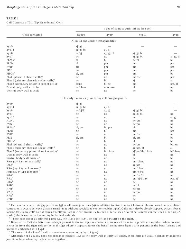
TABLE 1Cell Contacts of Tail Tip Hypodermal Cells
Cells contacted
Type of contact with tail tip hyp cella
hyp10 hyp9 hyp11 hyp8
A. In L4 and adult hermaphrodites
hyp9 aj, gj — — —hyp11 aj, gj, bl aj, bl — —hyp8 nc/gj aj, gj, bl aj, gj, bl —hyp7 nc nc aj, gj, bl aj, gj, blPLMsb bl bl nc/bl blPLNsb bl pm pm pmPVRc pm pm pm pmPDB pm pm pm pmPHCsb bl, pm pm pm blPhsh (phasmid sheath cells)b nc nc pm/nc pmPhso1 (primary phasmid socket cells)b nc bl aj ajPhso2 (secondary phasmid socket cells)b bl/nc bl/nc pm pm/blDorsal body wall muscles nc/close nc/close bl ncVentral body wall muscle nc nc nc bl
B. In early L4 males prior to ray cell morphogenesis
hyp9 aj, gj — — —hyp11 aj, gj, bl aj, bl — —hyp8 nc/gj/bl aj, gj aj, gj, bl —hyp7 nc nc aj, gj, bl nchyp13 nc nc nc aj, gjALNL nc nc nc/pm ncPVNL nc nc nc/pm ncPLMsb bl, pm bl, pm bl blPLNsb nc bl pm pmPVRc nc nc pm/nc ncPDB bl, pm bl bl, pm blPHCsb bl bl bl blPhsh (phasmid sheath cells)b nc nc nc/pm bl, pmPhso1 (primary phasmid socket cells)b nc bl aj, pm/bl ajPhso2 (secondary phasmid socket cells)b nc bl/close aj/pmd/bl/close bl/closeDorsal body wall muscle nc nc bl ncventral body wall muscleb nc nc nc blR9st (ray 9 structural cell)b nc nc pm/bl/nc ncR9.pb nc nc aj, pm ncR9A (ray 9 type A neuron)b nc nc pm/bl/nc ncR9B (ray 9 type B neuron)b nc nc pm/nc/bl ncR8stb nc nc pm/nc/bl ncR8.pb nc nc pm/aj/bl/nc pm/aje
R8Ab nc nc nc ncR8Bb nc nc nc/bl ncR7stb nc nc nc/pm ncR7.pb nc nc nc/pm ncR7Ab nc nc nc ncR7Bb nc nc nc nc
a Cell contacts occur via gap junctions (gj) or adherens junctions (aj) in addition to direct contact between plasma membranes or directcontact only occurs between plasma membranes without specialized contacts evident (pm). Cells may also be closely apposed across a basallamina (bl). Some cells do not touch directly but are in close proximity to each other (close). Several cells never contact each other (nc). Aslash (/) indicates variation among individual animals.
b These cells occur as bilateral pairs; e.g., the PLMs are PLML on the left and PLMR on the right.c Because the PVR dendrite is not always present in the tail tip, the contacts it makes with the tail tip cells are variable. When present,
it crosses from the right side to the anal ridge where it appears across the basal lamina from hyp11 or it penetrates the basal lamina andbecomes embedded into hyp11.
d The soma of the Phso2L cell is sometimes contacted by hyp11 (pm).
e Although hyp8 usually does not appear to contact R8.p at the body wall at early L4 stages, these cells are usually joined by adherensjunctions later when ray cells cluster together.
tti
vide
92 Nguyen et al.
reconstructed male animal the nucleus and the anteriorarm of hyp11 were on the right side, apparently due to amirror reversal of the body plan (not shown; cf. Wood,1991). Although in hermaphrodites hyp7 is the only hypo-dermal cell that is contacted by this hyp11 arm, in malesthis arm contacts several male-specific cells (Fig. 3D; Table1B), of which R9.p and sometimes R8.p contact hyp11 atadherens junctions (Figs. 4A and 4B; Table 1B). At the apicalsurfaces of these cells, these contacts are mediated byadherens junctions and are easily visualized by MH27staining (Table 1B; Fig. 4A, represented by red lines in Fig.4B). R9.p and R8.p are posterior sisters of cells that areprogenitors of the ray neurons and support cells. Themale-specific lineages that produce these cells begin during
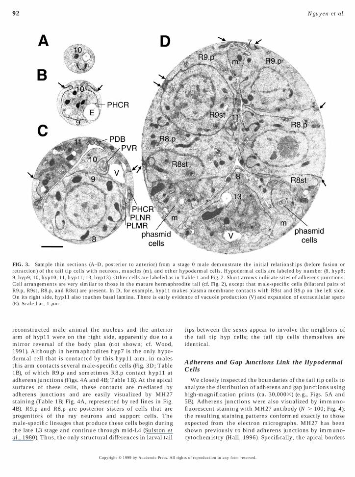
FIG. 3. Sample thin sections (A–D, posterior to anterior) fromretraction) of the tail tip cells with neurons, muscles (m), and oth9, hyp9; 10, hyp10; 11, hyp11; 13, hyp13). Other cells are labeled asCell arrangements are very similar to those in the mature hermapR9.p, R9st, R8.p, and R8st) are present. In D, for example, hyp11 mOn its right side, hyp11 also touches basal lamina. There is early e(E). Scale bar, 1 mm.
the late L3 stage and continue through mid-L4 (Sulston etal., 1980). Thus, the only structural differences in larval tail
Copyright © 1999 by Academic Press. All right
ips between the sexes appear to involve the neighbors ofhe tail tip hyp cells; the tail tip cells themselves aredentical.
Adherens and Gap Junctions Link the HypodermalCells
We closely inspected the boundaries of the tail tip cells toanalyze the distribution of adherens and gap junctions usinghigh-magnification prints (ca. 30,0003) (e.g., Figs. 5A and5B). Adherens junctions were also visualized by immuno-fluorescent staining with MH27 antibody (N . 100; Fig. 4);the resulting staining patterns conformed exactly to thoseexpected from the electron micrographs. MH27 has been
ge 0 male demonstrate the initial relationships (before fusion orpodermal cells. Hypodermal cells are labeled by number (8, hyp8;able 1 and Fig. 2. Short arrows indicate sites of adherens junctions.te tail (cf. Fig. 2), except that male-specific cells (bilateral pairs ofs plasma membrane contacts with R9st and R9.p on the left side.nce of vacuole production (V) and expansion of extracellular space
a staer hyin T
hrodiake
shown previously to bind adherens junctions by immuno-cytochemistry (Hall, 1996). Specifically, the apical borders
s of reproduction in any form reserved.

uj5h1ctSio
long
93Morphogenesis of the C. elegans Male Tail Tip
of all hypodermal cells are bordered by adherens junctionswhere these cells touch the body wall (Fig. 5A).
Many adherens junctions are distributed similarly inhermaphrodites and larval males; e.g., around the tail tipcells hyp8–hyp11, hyp7, the lateral seam (se) cells, and theprimary phasmid socket (Phsol) cells (see Table 1). How-ever, the male-specific hyp13 compartment and cells de-rived from the ray lineages form adherens-mediated con-tacts that are not present in hermaphrodites (compare partsA and B of Table 1 and Figs. 4A and 4B with Figs. 4G and4H). In males, adherens junctions remain intact until the
FIG. 4. Early stages in wild-type male tail development, prior to tby MH27 immunofluorescent staining. Scale bar, 10 mm. (A, C,successive stages during L4 male development. (B, D, F, H) TracingsUntil the mid-L4 stage, the tail tip hyp cells (hyp8–hyp11), phasmidthe sexes. Male-specific cells include the ray cell clusters (numberin hermaphrodites before L2 or earlier, but only at very late L4 in(D, F). The tail tip cells do not fuse in hermaphrodites; arrangementH). Positions of the hyp cell nuclei are shown as blue ovals (determto form a ray is numbered (1–9). During male development, the tslightly away from the body wall, leaving a small circle of MH27numbered 1–9 in F), while the exterior face of each Rn.p hypodermwith the anterior cells, to form the tail seam (“set”) syncytium abetween the ray cell and the tail tip cell morphogenetic events.
time of hyp cell fusions (discussed later).By closely examining the tail tip hyp cells, we also
ct
Copyright © 1999 by Academic Press. All right
ncovered an extensive series of electrical synapses, or gapunctions, connecting these cells to their neighbors (Fig.B). In hermaphrodites, gap junctions are common betweenypodermal cells in the tail and elsewhere (Table 1A; Hall,987 and unpublished; White, 1988) and permit the inter-ellular transfer of small molecules from embryogenesishrough adulthood (Bossinger and Schierenberg, 1992;tarich et al., 1996). A similar distribution of gap junctionss found between female and male tail tips before the onsetf tail tip morphogenesis (Table 1B).We did not find these specialized junctions at all areas of
p retraction (A–F), and a late L4 hermaphrodite tail (G, H), viewedmunofluorescence images (negatives) of MH27 staining at threee MH27 staining (red lines) shown in A, C, E, and G, respectively.
, and lateral “seam” hypodermis (se) are arranged similarly betweend the hyp13 compartment (which fuses with the hyp7 syncytium
s). Cell borders show fragmented staining as cell fusion progressesells in the adult hermaphrodite tail are similar to those shown (G,from DAPI costaining). Each cluster of cells that will differentiate
ells from each ray cell cluster that will become ray neurons sinkning at the surface corresponding to the ray structural cell (Rnst;ll enlarges. Some of these Rn.p cells fuse (dashed lines), beginningthe lateral margin (F). There is little variation in relative timing
ail tiE) Im
of th(Ph)
ed) anmales of cined
wo cstai
al ce
ell–cell contact. For example, hypodermal cells may con-act other cells over extensive distances where there are no
s of reproduction in any form reserved.
t
e larvore
mall
94 Nguyen et al.
adherens junctions (e.g., between hyp10 and hyp9 in Fig.
FIG. 5. Ultrastructural features of cell junctions, cell fusions, and tmale tails. Hypodermal cells are labeled by number as in Fig. 3. (A)portion of the tail tip. (B) Gap junctions (gjs) between hyp9 and hythe plasma membranes that tend to be closer to the body wall in posanteriorly and “zippered” posteriorward (scale bar, 1 mm for C–E aremnants. Long arrows indicate remaining “broken” edges of membhyp cells (open arrow, D). (E) In one stage 3 male where fusions werformer boundary between hyp8 and hyp9. (F) During retraction, th(ex) at the tail tip with only a remnant of hyp10 tissue (arrow). (G) Mand new cuticle layers, whereas the space between cells remains s
3C; between hyp8 and hyp13 in Fig. 3D). Also, gap junctionshave not yet been found between hyp9 and hyp11. Some tail
ec
Copyright © 1999 by Academic Press. All right
ip hyp cells may not always maintain direct contact. For
p retraction in sample electron micrographs from serially sectioneddherens junction (aj) that seals hyp9 to itself in the very posteriorScale bar, 10 nm for A and B. Fusions are initiated as “breaks” inr (C) than in anterior (D) sections, suggesting that the fusions began, but 0.5 mm for F). Short arrows mark curled membrane/adherensnear presumptive sites of fusion. A mitochondrion spans the fusedpleted, a collection of membranous vesicles accumulated near theal cuticle in a stage 3 animal surrounds much extracellular space
anteriorly, increased extracellular space primarily lies between old.
ail tiAn a
p10.teriond Grane
e com
xample, hyp8 and hyp10 often do not touch but sometimesontact each other via gap junctions or are apposed across
s of reproduction in any form reserved.

chAhccWtmw(mtcw
t1tgcsaafo2
dttwtscstwEa
nlcatar
tawc
as(
ercmosdaotfmtdsj
95Morphogenesis of the C. elegans Male Tail Tip
basal lamina. Although each tail tip hyp cell may not bedirectly linked to every other hyp cell (“in parallel”), thefour cells in this “tail tip compartment” are all linkedtogether through at least one other hyp cell (“in series”).
Temporal Analysis of Male Tail TipMorphogenesis
Using MH27 staining of adherens junctions (N . 100) inonjunction with serial thin section analysis (N 5 9), weave delineated stages of male tail tip morphogenesis.lthough MH27 staining cannot display all cell contacts, itas been extremely useful in monitoring changes in relativeell positions and cell fusions during morphogenesis (Fran-is and Waterston, 1985; Baird et al., 1991; Podbiliwicz andhite, 1994; Fitch and Emmons, 1995) and allows observa-
ions of large numbers of fixed animals. To provide land-arks for predicting the relative timing of tail tip events,e used states of differentiation of the ray cell groups
coincident with tail tip morphogenesis within a smallargin of variation) as follows [note that these “stages” of
ail tip morphogenesis do not correspond to the stages of rayell morphogenesis defined by Fitch and Emmons (1995)hich cover a broader temporal range].At a time point that we call “stage 0” (Figs. 4A and 4B),
he tail tip remains completely unretracted (see also Fig.A). Early ray cell progenitors occupy large territories alonghe body wall. Several of the more posterior ray cell pro-enitors form adherens junctions directly with tail tip hypells (“aj” contacts in Table 1B). Note also that the hyp13yncytium contacts hyp8 via adherens junctions. The samerrangements of apical cell surfaces and adherens junctionst this stage were noted by EM (N 5 1; Fig. 3). Progressionrom stage 0 through the following stages (to “stage 3”)ccurs from mid-L4 to late L4 and takes about 2.5 h at 20 6°C.By “stage 1” (Figs. 4C and 4D), the territories of the
eveloping ray cells along the body wall begin to constricto very small patches as viewed by MH27 staining, whereashe territories of the Rn.p hypodermal cells expand some-hat on the body wall. R1.p and R2.p have often fused by
his time. By EM (N 5 2), however, the ray cell precursorshow little differentiation at this stage. In the tail tip, hypell fusions begin to appear as punctate losses of MH27taining at the hyp cell borders and progress from anterioro posterior: hyp8 and hyp11 fuse first, followed by fusionsith hyp9 and finally hyp10. Fusions were also observed inM serial sections as small breaks in the plasma membranet the adherens junctions (Figs. 5C and 5D; described later).By “stage 2” (Figs. 4E and 4F) the dendrite of each ray
euron begins to narrow and its soma sinks away from theateral surface and is encircled by the tubular structuralell, which remains attached to the body wall by robustdherens junctions. R1.p–R5.p are in the process of fusing athis time to form the “tail seam” (“set”; Fig. 4F). As
dditionally observed by EM (N 5 3), distal portions of theay cells begin to form rudimentary dendritic specializa-ni
Copyright © 1999 by Academic Press. All right
ions. Adherens junctions between the four tail tip hyp cellsre almost completely gone by this stage, correspondingith EM observations. Positions of the nuclei have not
hanged significantly from their original positions.By stage 3, each neuronal ray cell soma continues to sink
way from the body wall, while its apical dendrite forms amall cilium attached to the body wall. As observed by EMN 5 2), fusions between the tail tip hyp cells are complete.The posterior hyp cells lose adherence with the L4 cuticleand begin to retract, leaving the characteristic fluid-filledextracellular space at the tail tip (Figs. 1B and 5F). By thisstage, the nuclei of the hyp cells have begun their anterior-ward migration. Retraction then proceeds rapidly to pullthe rest of the cells anteriorward as the tail changes shapedramatically. In one animal, partial retraction of the tail tipwas initiated prior to fluid expulsion, causing the distalportion of the tail spike to shorten and warp (not shown).
During retraction, some of the caudal dendrites thatextend into the larval tail tip are gradually withdrawnanteriorly, although they are still present at stage 3. Even-tually, some tail sense cells (PLM, PHC, and others) mustresorb all or most of their posterior dendrites. The PLMcells retain only their anterior dendrite in the adult male.After retraction of the tail tip, many of these somata lie socaudal that there is no room to extend a posterior process.The caudal processes of the phasmid neurons are not lost,but are deflected to ventrolateral positions, still posterior totheir somata.
Cell Fusions in Male Tail Tip MorphogenesisInitiate at or near Adherens Junctions andProgress from Anterior to Posterior
During stages 1 and 2, the fusions between the tail tip hypcells occur at or adjacent to the adherens junctions. Noadditional specialized cytoplasmic structures are alignedwith any of these junctions that would prefigure fusionevents, in contrast to the prefusion vesicles required forDrosophila myotube fusion (Geiger et al., 1995; Dobersteint al., 1997). Fusions in the male tail result in a topologicalecombination of the plasma membranes between adjacentells. One product of this fusion is a conjoined apicalembrane bilayer (which thus surrounds both cells); the
ther product is a conjoined double bilayer at the formerurfaces of contact between the cells. This latter conjoinedouble-bilayer product is detached from the body wall (longrrows in Figs. 5C and 5D) and internalized. Tiny segmentsf membrane remaining at the body wall (apparently con-aining vestiges of darkly staining adherens material) oftenorm short curls (short arrows in Figs. 5C and 5D) which
ay drift away from the former anchorage site. Thereafter,he adherens junctions themselves (and the MH27 epitope)isappear completely (Figs. 4C–4F). Thus, we find a strongpatial correlation between sites of fusion and the adherensunctions. Although gap junctions are sometimes found
ear fusion sites (i.e., are near the conjoined “edges” of thenternalized plasma membranes), they are also distributeds of reproduction in any form reserved.
eca
ahaw(fih(wmmd
96 Nguyen et al.
throughout these membranes where breaks have not oc-curred. Perhaps because of these gap junctions, the inter-nalized membranes remain “pressed together” for a while.
Fusions appear to occur in a wave from anterior toposterior. By both EM and MH27 staining, we find thatmost early fusions occur between hyp9 and hyp11 (Figs. 5Cand 5D) and between hyp8 and hyp11 (Fig. 5D); hyp10 isinvariably the last cell to fuse (Fig. 4D). The internalizedplasma membranes in the anterior portions of the cells havegenerally retreated further from the body wall (open arrowin Fig. 5D) than those in the posterior portions, which canstill be attached to adherens junctions (or are at least closerto the body wall where adherens junctions have disap-peared) (the internalized double bilayer may retreat fromthe body wall as a result of its elasticity). This topologysuggests that fusions may occur by “zippering” in a poste-rior direction along the adherens junctions.
Fusions are essentially complete by stage 3, althoughexcess membrane products persist for a short time. In atleast one case, the aftermath of fusion was marked by alocalized accumulation of many membranous vesicles, pre-sumably preliminary products of membrane turnover(Fig. 5E).
Cell Shapes and Nuclear Positions Are GenerallyStable until Fusions Occur
By MH27 staining (N . 100), the beginning of tail tipretraction does not appear to occur until after fusion initia-tion. However, details of individual cell shapes are noteasily distinguished by this method. By tracing the outlinesof cells in serial sections of staged male tails (as in Fig. 6),we have confirmed that the shapes of at least hyp9 andhyp10 are practically invariant among animals in stages 0–2when fusions are initiated and completed (cells of a stage 0male are shown in Figs. 6A, 6C, 6E, and 6G). Positions of allthe nuclei remain stable throughout stages 0–2 (Table 2).
The earliest evidence for change in cell shapes (butwithout substantial change in nuclear position) was ob-served in one stage 1 male (Figs. 6B and 6D). In this animal,the anterior arm of hyp11 is enlarged, suggesting that thecell’s volume is in the process of translocating rostrallyalong the arm itself (compare Fig. 6B to 6A). Similarly, thevolume of hyp8 in this animal is subtly more rostral (Fig.6D) than in an animal at an earlier stage (Fig. 6C). Nofusions are observed in this particular animal, suggestingthat initiation of retraction does not depend on fusion.
Evidence for retraction of the tail tip hyp tissues is alwaysnoticeable in stage 3, when fusions are essentially com-plete. In the wake of the anteriorward retraction of the hyptissue, the extracellular space within the tail spike enlargesdue to the accumulation of fluid (Fig. 1B and Figs. 5F and5G). Because of the extensive fusion of hypodermal cellborders during stage 3, it was not usually possible toreconstruct their individual outlines as retraction pro-
gressed. However, in one stage 3 animal with hyp10 andhyp9 cell borders that could still be reconstructed byCopyright © 1999 by Academic Press. All right
xtrapolation of fusing membranes, there was substantialhange in cell shape (compare Figs. 6F and 6H to Figs. 6End 6G).The nuclei of all four tail tip cells begin to move rapidly
nteriorward in stage 3, concomitant with the hyp9 andyp10 retractions (Table 2). These nuclear translocationsre also observed by staining a population of fixed animalsith DAPI (not shown) and have been observed previously
Sulston et al., 1980), though are not documented at thisne a level of resolution. Interestingly, tail tip nuclei inermaphrodites may also translocate somewhat anteriorly
Table 2). However, these translocations are not associatedith cell shape changes and are not as extensive as inales. The nuclei in L4 males continue their anteriorigrations far beyond where they stop in adult hermaphro-
ites.
Vacuoles May Shuttle Fluid through the Tail Tipto the Extracellular Space during Retraction
Although there is little change in the volumes of indi-vidual tail tip hypodermal cells relative to each other duringmale morphogenesis, there is a local swelling of intracellu-lar vacuoles (denoted “V” in Figs. 3C and 3D). Vacuolarswellings are transitory and somewhat asynchronous, soone hypodermal cell may show pronounced vacuolar swell-ing in one specimen while a different hypodermal cell willbe swollen with vacuoles in another individual (Table 3). Atstage 0, the male tail hypodermal cells show a few cytoplas-mic vacuoles (Figs. 3C and 3D), similar to those in an L4hermaphrodite (not shown). During stages 1–2 of male tailmorphogenesis, these vacuoles show a transient increase involume, but by stage 3 they are greatly diminished. Therelative amount of extracellular space is initially small (Fig.3, Table 3), but suddenly increases at stage 3, particularly inthe extreme tail tip (Figs. 5F and 5G; cf. Figs. 1A and 1B).Concomitantly, the degree of vacuolation dramatically de-creases in hyp8, hyp9, and hyp11. We hypothesize that thehyp cells extrude the contents of these vacuoles around thetime retraction is initiated.
Genetic Changes Can Alter the Relative Timing ofMorphogenetic Events
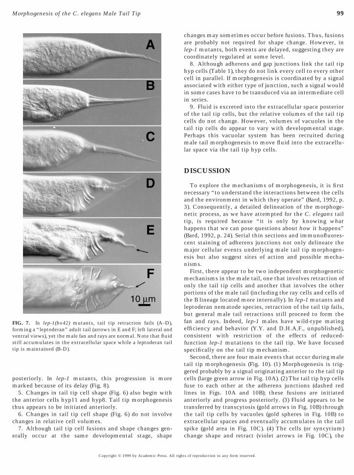
We have characterized the progression of hyp cell fusionsin males bearing mutations (bx37 and bx42) in the locuslep-1. These mutations were originally isolated in thelaboratory of Dr. Scott Emmons (Albert Einstein College ofMedicine) in an F2 screen for male tail morphologicaldefects using Nomarski microscopy. Tail tip retraction failsduring L4 male morphogenesis (Figs. 7A–7D) in nearly allmales that bear either of the hypomorphic mutations at thelep-1 locus (Y.Y. and D.H.A.F., unpublished), resulting inleptoderan tail tips of variable size in adult males (Figs. 7Eand 7F). However, the lep-1 mutations do not affect mor-
phogenesis of the fan and rays, suggesting that tail tipretraction is independent of general male tail retraction.s of reproduction in any form reserved.

sti
ttptdTtn
97Morphogenesis of the C. elegans Male Tail Tip
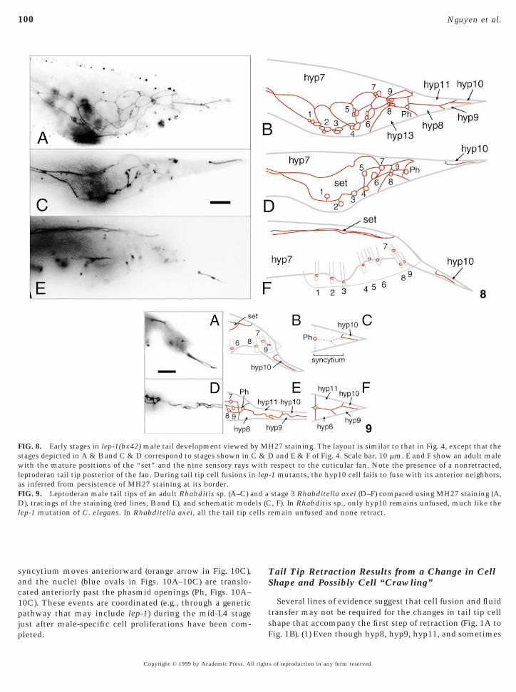
Staining lep-1 mutants with the MH27 antibody showsthat fusions of the tail tip cells are delayed with respectto other morphogenetic events (in all 38 L4 and adultmales that were closely observed), suggesting that theprimary defect in these mutants may be in a regulatory ortriggering event common to both the fusion and retrac-tion pathways. Whereas fusions of at least hyp11 andhyp8 have begun by the time ray cells cluster at stage 1 inwild-type males (Figs. 5C and 5D), tail tip cells in mostlep-1 specimens at this stage have not yet fused (Figs. 8Aand 8B). In many cases (5/10 of the stage 2 males and 3/9of the adult males closely observed), hyp10 fusion with
FIG. 6. Changes in cell shape begin with anterior cells and do noip cells before (A, C, E, G) and during retraction (B, D, F, H) (cf. Fihe nuclei are noted (n). Cell boundaries were traced for several serecocious retraction in a stage 1 male (B, D). Small arrows (in A andoward the central core. Large arrowheads indicate the main directoes not extend much further rostrally, while the semicircular cell bhe soma of hyp8 becomes rostrally enlarged and extended, while
han at stage 0. Tracings from hyp10 and hyp9 in a stage 0 male (E,ot volume during the retraction of these cells.
the other tail tip cells is greatly delayed or fails alto-gether, as indicated by persistence of adherens junctions
hu
Copyright © 1999 by Academic Press. All right
urrounding hyp10 at stage 2 (Figs. 8C and 8D) or even inhe adult (Figs. 8E and 8F). That hyp10 fusions arenvariably the most delayed in lep-1 mutants is also
consistent with a posteriorward progression of cell fu-sions in wild-type animals.
Leptoderan tail tips occur naturally in many species that arefairly closely related to C. elegans (Sudhaus, 1976). As in lep-1mutants of C. elegans, these leptoderan tail tips result whenthe tail tip cells fail to retract (e.g., as in O. myriophila; Figs.1H–1L). In all leptoderan species observed so far, the tail tipcells either fail to fuse at all (as in Rhabditella axei, N . 50,Figs. 9D–9F; and O. myriophila, N . 50, not shown) or only
lve changes in cell volume. Wireframe reconstructions of the tailand 3). Scale bar, 10 mm. Anterior is to the upper left. Positions ofs of hyp11 and hyp8 in a stage 0 male (A, C) and in an example ofndicate portions of the cells initially near the body wall that retractf retraction. The anterior left arm of hyp11 becomes enlarged, butr apposed to the dorsolateral cuticle in the posterior half is reduced.emicircular border with the ventrolateral cuticle is less extensivend a stage 3 male (F, H) demonstrate marked changes in shape but
t invogs. 2ction
C) iion oordeits sG) a
yp10 fails to fuse with the other tail tip cells (as in anndescribed Rhabditis species, N . 10, Figs. 9A–9C).
s of reproduction in any form reserved.
ppm
ishhdcRa
98 Nguyen et al.
Summary of Results
Our data define the following highlights of male tail tipmorphogenesis (schematized in Fig. 10).
1. All fusions of the tail tip hypodermal cells (hyp8–hyp11) occur at sites corresponding spatially to the adher-ens junctions. These data are consistent with a localizationof fusogenic proteins to adherens junctions linking the tailtip cells.
2. However, not all hypodermal adherens junctions in C.elegans correspond to fusion sites. For example, the male-specific hyp13 compartment we discovered does not fusewhen the adjacent tail tip cells fuse. This suggests that only
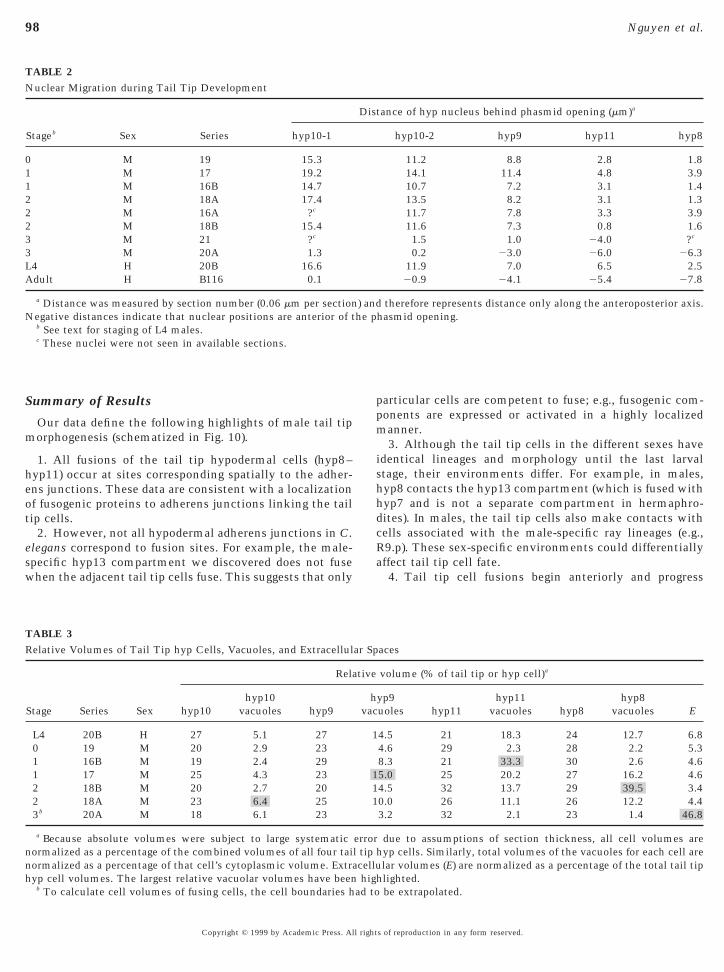
TABLE 2Nuclear Migration during Tail Tip Development
Stageb Sex Series hyp10-1
0 M 19 15.31 M 17 19.21 M 16B 14.72 M 18A 17.42 M 16A ?c
2 M 18B 15.43 M 21 ?c
3 M 20A 1.3L4 H 20B 16.6Adult H B116 0.1
a Distance was measured by section number (0.06 mm per sectionNegative distances indicate that nuclear positions are anterior of t
b See text for staging of L4 males.c These nuclei were not seen in available sections.
TABLE 3Relative Volumes of Tail Tip hyp Cells, Vacuoles, and Extracellul
Stage Series Sex
Rela
hyp10hyp10
vacuoles hyp9
L4 20B H 27 5.1 270 19 M 20 2.9 231 16B M 19 2.4 291 17 M 25 4.3 232 18B M 20 2.7 202 18A M 23 6.4 253b 20A M 18 6.1 23
a Because absolute volumes were subject to large systematicnormalized as a percentage of the combined volumes of all four tainormalized as a percentage of that cell’s cytoplasmic volume. Extra
hyp cell volumes. The largest relative vacuolar volumes have been highb To calculate cell volumes of fusing cells, the cell boundaries had to
Copyright © 1999 by Academic Press. All right
articular cells are competent to fuse; e.g., fusogenic com-onents are expressed or activated in a highly localizedanner.3. Although the tail tip cells in the different sexes have
dentical lineages and morphology until the last larvaltage, their environments differ. For example, in males,yp8 contacts the hyp13 compartment (which is fused withyp7 and is not a separate compartment in hermaphro-ites). In males, the tail tip cells also make contacts withells associated with the male-specific ray lineages (e.g.,9.p). These sex-specific environments could differentiallyffect tail tip cell fate.4. Tail tip cell fusions begin anteriorly and progress
tance of hyp nucleus behind phasmid opening (mm)a
hyp10-2 hyp9 hyp11 hyp8
11.2 8.8 2.8 1.814.1 11.4 4.8 3.910.7 7.2 3.1 1.413.5 8.2 3.1 1.311.7 7.8 3.3 3.911.6 7.3 0.8 1.61.5 1.0 24.0 ?c
0.2 23.0 26.0 26.311.9 7.0 6.5 2.520.9 24.1 25.4 27.8
therefore represents distance only along the anteroposterior axis.hasmid opening.
aces
volume (% of tail tip or hyp cell)a
yp9uoles hyp11
hyp11vacuoles hyp8
hyp8vacuoles E
4.5 21 18.3 24 12.7 6.84.6 29 2.3 28 2.2 5.38.3 21 33.3 30 2.6 4.65.0 25 20.2 27 16.2 4.64.5 32 13.7 29 39.5 3.40.0 26 11.1 26 12.2 4.43.2 32 2.1 23 1.4 46.8
due to assumptions of section thickness, all cell volumes arehyp cells. Similarly, total volumes of the vacuoles for each cell arelar volumes (E) are normalized as a percentage of the total tail tip
Dis
) andhe p
ar Sp
tive
hvac
1
111
errorl tipcellu
lighted.be extrapolated.
s of reproduction in any form reserved.
calc
hcaii
octPml
(cmen
moptlbfecfs
tgcflatte
fvst
99Morphogenesis of the C. elegans Male Tail Tip
posteriorly. In lep-1 mutants, this progression is moremarked because of its delay (Fig. 8).
5. Changes in tail tip cell shape (Fig. 6) also begin withthe anterior cells hyp11 and hyp8. Tail tip morphogenesisthus appears to be initiated anteriorly.
6. Changes in tail tip cell shape (Fig. 6) do not involvechanges in relative cell volumes.
FIG. 7. In lep-1(bx42) mutants, tail tip retraction fails (A–D),orming a “leptoderan” adult tail (arrows in E and F; left lateral andentral views), yet the male fan and rays are normal. Note that fluidtill accumulates in the extracellular space while a leptoderan tailip is maintained (B–D).
7. Although tail tip cell fusions and shape changes gen-erally occur at the same developmental stage, shape
sc
Copyright © 1999 by Academic Press. All right
hanges may sometimes occur before fusions. Thus, fusionsre probably not required for shape change. However, inep-1 mutants, both events are delayed, suggesting they areoordinately regulated at some level.8. Although adherens and gap junctions link the tail tip
yp cells (Table 1), they do not link every cell to every otherell in parallel. If morphogenesis is coordinated by a signalssociated with either type of junction, such a signal wouldn some cases have to be transduced via an intermediate celln series.
9. Fluid is excreted into the extracellular space posteriorf the tail tip cells, but the relative volumes of the tail tipells do not change. However, volumes of vacuoles in theail tip cells do appear to vary with developmental stage.erhaps this vacuolar system has been recruited duringale tail morphogenesis to move fluid into the extracellu-
ar space via the tail tip hyp cells.
DISCUSSION
To explore the mechanisms of morphogenesis, it is firstnecessary “to understand the interactions between the cellsand the environment in which they operate” (Bard, 1992, p.3). Consequently, a detailed delineation of the morphoge-netic process, as we have attempted for the C. elegans tailtip, is required because “it is only by knowing whathappens that we can pose questions about how it happens”Bard, 1992, p. 24). Serial thin sections and immunofluores-ent staining of adherens junctions not only delineate theajor cellular events underlying male tail tip morphogen-
sis but also suggest sites of action and possible mecha-isms.First, there appear to be two independent morphogeneticechanisms in the male tail, one that involves retraction of
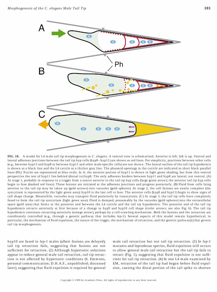
nly the tail tip cells and another that involves the otherortions of the male tail (including the ray cells and cells ofhe B lineage located more internally). In lep-1 mutants andeptoderan nematode species, retraction of the tail tip fails,ut general male tail retractions still proceed to form thean and rays. Indeed, lep-1 males have wild-type matingfficiency and behavior (Y.Y. and D.H.A.F., unpublished),onsistent with restriction of the effects of reduced-unction lep-1 mutations to the tail tip. We have focusedpecifically on the tail tip mechanism.Second, there are four main events that occur during male
ail tip morphogenesis (Fig. 10). (1) Morphogenesis is trig-ered probably by a signal originating anterior to the tail tipells (large green arrow in Fig. 10A). (2) The tail tip hyp cellsuse to each other at the adherens junctions (dashed redines in Figs. 10A and 10B); these fusions are initiatednteriorly and progress posteriorly. (3) Fluid appears to beransferred by transcytosis (gold arrows in Fig. 10B) throughhe tail tip cells by vacuoles (gold spheres in Fig. 10B) toxtracellular spaces and eventually accumulates in the tail
pike (gold area in Fig. 10C). (4) The cells (or syncytium)hange shape and retract (violet arrows in Fig. 10C), thes of reproduction in any form reserved.
ells r
100 Nguyen et al.
syncytium moves anteriorward (orange arrow in Fig. 10C),and the nuclei (blue ovals in Figs. 10A–10C) are translo-cated anteriorly past the phasmid openings (Ph, Figs. 10A–10C). These events are coordinated (e.g., through a geneticpathway that may include lep-1) during the mid-L4 stage
FIG. 8. Early stages in lep-1(bx42) male tail development viewed bstages depicted in A & B and C & D correspond to stages shown inwith the mature positions of the “set” and the nine sensory raysleptoderan tail tip posterior of the fan. During tail tip cell fusions ias inferred from persistence of MH27 staining at its border.FIG. 9. Leptoderan male tail tips of an adult Rhabditis sp. (A–C) aD), tracings of the staining (red lines, B and E), and schematic modlep-1 mutation of C. elegans. In Rhabditella axei, all the tail tip c
just after male-specific cell proliferations have been com-pleted.
Copyright © 1999 by Academic Press. All right
Tail Tip Retraction Results from a Change in CellShape and Possibly Cell “Crawling”
Several lines of evidence suggest that cell fusion and fluidtransfer may not be required for the changes in tail tip cell
27 staining. The layout is similar to that in Fig. 4, except that theand E & F of Fig. 4. Scale bar, 10 mm. E and F show an adult male
respect to the cuticular fan. Note the presence of a nonretracted,-1 mutants, the hyp10 cell fails to fuse with its anterior neighbors,
stage 3 Rhabditella axei (D–F) compared using MH27 staining (A,, F). In Rhabditis sp., only hyp10 remains unfused, much like theemain unfused and none retract.
y MHC & Dwithn lep
nd aels (C
shape that accompany the first step of retraction (Fig. 1A toFig. 1B). (1) Even though hyp8, hyp9, hyp11, and sometimes
s of reproduction in any form reserved.
mmtrc
er, th
101Morphogenesis of the C. elegans Male Tail Tip
hyp10 are fused in lep-1 males (albeit fusions are delayed),tail tip retraction fails, suggesting that fusions are notsufficient for retraction. (2) Although hypertonic solutionsappear to reduce general male tail retraction, tail tip retrac-tion is not affected by hypertonic conditions (S. Emmons,
FIG. 10. A model for L4 male tail tip morphogenesis in C. eleganlateral adherens junctions between the tail tip hyp cells (hyp8–hyp(e.g., between hyp13 and hyp8 or between hyp11 and other male-speis shown as a black line and the L4 cuticle as a thicker gray line. Tlines (Ph). Nuclei are represented as blue ovals. In A, the anterior pperspective the rest of hyp11 lies behind (dorsal to) hyp8. The onlyAt stage 1, probably in response to a trigger from a source anterior tbegin to fuse (dashed red lines). These fusions are initiated at theanterior to the tail tip may be taken up (gold arrows) into vacuolsyncytium is represented by the light green area); hyp10 is the lastcell shape change. Meanwhile, vacuoles may transport fluid postefused to form the tail tip syncytium (light green area). Fluid is dumspace (gold area) that forms at the posterior end between the L4hypodermis retracts anteriorly at first because of a change in hyhypodermis continues retracting anteriorly (orange arrow), perhapscoordinately controlled (e.g., through a genetic pathway that inparticular, the mechanism of fluid expulsion, the nature of the triggtail tip morphogenesis.
personal communication; D.H.A.F., unpublished; discussedlater), suggesting that fluid expulsion is required for general
Es
Copyright © 1999 by Academic Press. All right
ale tail retraction but not tail tip retraction. (3) In lep-1utants and leptoderan species, fluid expulsion still occurs
o allow general male tail retraction but the tail tip fails toetract (Fig. 1), suggesting that fluid expulsion is not suffi-ient for tail tip retraction. (4) In one L4 male examined by
entral view is schematized. Anterior is left, left is up. Ventral andre shown as red lines. For simplicity, junctions between other cellscells) are not shown. The lateral outline of the tail tip hypodermisasmid openings in the cuticle are indicated as short black paralleln of hyp11 is shown in light green shading, but from this ventral
erens borders between hyp11 and hyp8 are lateral, not ventral. (A)tail tip hyp cells (large green arrow), the anterior tail tip hyp cells
rens junctions and progress posteriorly. (B) Fluid from cells lyingld spheres). At stage 2, the cell fusions are nearly complete (the
to fuse. The anterior cells (hyp8 and hyp11) begin to show signs ofby transcytosis. (C) At stage 3, the tail tip cells have completely
, presumably by the vacuoles (gold spheres) into the extracellularle and the tail tip hypodermis. The posterior end of the tail tip
nd hyp10 cell shape (violet arrows; see also Fig. 6). The tail tipcell-crawling mechanism. Both the fusions and the retraction ares lep-1). Several aspects of this model remain hypothetical; ine mechanism of retraction, and the genetic pathway(s) that govern
s. A v11) acific
he phortioadh
o theadhees (gocell
riorlyped
cuticp9 aby a
clude
M, retraction of the tail tip had begun before fluid expul-ion, causing the distal portion of the tail spike to shorten
s of reproduction in any form reserved.

spgSsb(si
ffta
iefc
sgimptoeamfg(rbsbbtflmerdfibcnvftDt
svicpvKaCsebbs
102 Nguyen et al.
and warp. One possible mechanism for the observedchanges in cell shapes (as well as the nuclear migrations) isan active change in the architecture of the cytoskeleton inthese cells, although any such change in the organization ofcytoskeletal actin microfilaments was not observable in ourEM studies.
We speculate that the observed changes in cell shape (aswell as the nuclear migrations) may also result from theparticipation of at least hyp11 and hyp8 in a crawling phase,similar to the known migrations of some neurons, muscleprecursors, muscle arms, and other mesodermal processes(Hedgecock et al., 1987). In particular, the shape of themigrating hypodermal tissue is remarkably similar to theshape of the distal tip cell (DTC) of the gonad (D.H.H. andE. Hedgecock, unpublished). As with the DTC, the hypo-dermal migration involves a broad, blunt leading edge oftissue juxtaposed closely to the basal lamina, with the cellnucleus pressed close to the lead and with thinner processestrailing behind the cells. An extrinsic signal expressed inthe basal lamina could guide or stimulate this migration,much as netrin is known to guide the motion of manyclasses of cells in earlier stages of development (Wadsworthet al., 1996). Although no changes in the actin cytoskeleton(as might be expected in a crawling cell) were observable inour study, morphogenesis fails in treatments with cytoskel-etal inhibitors (K. L. Chow and Y. Tse, personal communi-cation). Alternatively, continued anterior migration of thetail tip cells is also compatible with these cells beingpassively drawn forward by their connections with anteriorcells undergoing active morphogenetic movements.
Tail Tip Cell Fusions Probably Initiate atAdherens Junctions
Particular membrane proteins are generally required tohold cells in close proximity before fusogenic proteins canact. In some mutants that block fusion in Drosophila, densemembrane plaques between unfused myoblasts (rollingstone) or membrane-bound vesicles lining the cell border(blown fuse) accumulate to abnormally high levels; in othermutants, all such membrane-associated structures are ab-sent (myoblast city) (Rushton et al., 1995; Doberstein et al.,1997; Paululat et al., 1995). These symmetrical junctionaltructures are postulated to be part of the prefusion com-lex in fly myoblasts, perhaps delivering or holding fuso-enic moieties in place on both membranes prior to fusion.imilarly, gap junctions have been reported to be a neces-ary precursor to a fusion event in some vertebrate myo-lasts, and specific channel blockers can inhibit cell fusionsProulx et al., 1997). Adherens junctions are required atome sites of cell fusion, apparently to help localize signal-ng or fusogenic molecules (Doberstein et al., 1997).
We speculate that adherens junctions provide such aunction in the tail tip cells. Unlike Drosophila myoblastusions, no obvious prefusion complexes or vesicles par-
icipate in initiating tail tip cell fusions. Perhaps otherdjoining cells (hyp13, hyp7) are protected from fusioni1
Copyright © 1999 by Academic Press. All right
nto the same compartment during tail tip morphogen-sis either by failure to express, activate, or localize theusogenic moiety or by expressing an inhibitory mole-ule.While this paper was being reviewed, Mohler et al. (1998)
howed that hypodermal fusions during C. elegans embryo-enesis also originate at or near adherens junctions. That is,n both the tail tip and the embryonic fusions, the fusing
embranes are first held closely together by an adherenslaque, with fusion pore formation occurring at or very nearhe plaque. However, two details of the tail tip and embry-nic mechanisms look slightly different. (1) During thembryonic fusions, the adherens junctions do not immedi-tely disappear, but retreat away from the body wall towardore basal portions of the cell border, leading a front of
usions between adjacent cells beginning apically and pro-ressing basally. Using multiphoton imaging, Mohler et al.1998) detected internalization of the MH27 epitope (adhe-ens junction material) into more basal portions of the cellorder. They also suggested a mechanism where the firsttep is the formation of a small fusion pore at the apicalorder, followed by rapid radial destruction of the cellorder. In the tail tip, we did not observe any basallyranslocated MH27 epitope or adherens plaque by immuno-uorescence or EM. The resolution of our epifluorescenceay not have allowed us to visualize this basal migration of
pitope; also, a low concentration of MH27 epitope in theetreating membrane may not have been morphologicallyistinguishable by EM. (2) By EM, Mohler et al. detected ane vesiculation of the internalized, dispersing membranesefore they were fully digested. These fine vesicles mayorrespond to the membrane fragments and larger membra-ous circles that we detect at former cell borders; minorariations in EM fixation methods could also be responsibleor such differences. There is no evidence from either studyhat “prefusion complexes” such as those required forrosophila myoblast fusions (Doberstein et al., 1997) par-
icipate in these C. elegans fusions.What fusogenic molecules might be involved? In other
ystems, the lipid bilayer surrounding a fusing virus,esicle, or cell carries a membrane-spanning protein whichs complementary to another membrane-spanning proteinarried on the surface of the fusion partner; e.g., viral fusionroteins (hemagglutanins), v-SNAREs and T-SNARES foresicle release, or ADAMs for cell fusion (White, 1992;emble et al., 1994; Lindau and Almers, 1995; Huovila etl., 1996; Myles and Primakoff, 1997; Weber et al., 1998). In. elegans, ADM-1, an ADAM protein with sequence
imilarity to metalloprotease-like fusogenic proteins, isxpressed in hypodermis at times of cell fusion (Pod-ilewicz, 1996). However, no male tail phenotypes haveeen described. Male phenotypes are observed in mutants ofup-17, which encodes another ADAM protein known to
nteract with the LIN-12/NOTCH receptor (Wen et al.,997), but not known to be involved in cell fusions.s of reproduction in any form reserved.
tn
tpsmt
gsittmdw(aofspbo(
hbScbdWsssmtRsiwht
dts
mitecpsdhhtaf(amdHRcwdLt1fdpiila1tS
103Morphogenesis of the C. elegans Male Tail Tip
How Is Tail Tip Morphogenesis Triggered?
The competence of tail tip cells to undergo male-specifictail tip morphogenesis could be determined either autono-mously or nonautonomously. For example, male-specific fateof the tail tip cells could be determined by the somaticsex-determination pathway in which the gene tra-1 is requiredand sufficient for cell-autonomous specification of hermaph-rodite fate (except in the vulval precursor cells, which arenonautonomously specified) (Hunter and Wood, 1990). How-ever, lep-1 tra-1 double mutants still make leptoderan tail tips,suggesting that tra-1 function is not required to make pointyail tips (Y.Y. and D.H.A.F., unpublished). Also, mosaics haveot yet been reported in which tra-1(1) tail tip hyp cells in an
otherwise tra-1(2) male tail fail to retract. It is possible thathe tail tip cells in both hermaphrodites and males are com-etent to undergo morphogenesis, but that external male-pecific cues (such as a hypothetical signal from adjacentale-specific cells) determine the adult male-specific fate of
he tail tip hyp cells.Once competence is determined, male tail tip morpho-
enesis itself could be triggered either autonomously byome internal mechanism or nonautonomously by signal-ng from the environment around the tail tip cells. We favorhe latter hypothesis because of the following. (1) The tailip cells arise very early from embryonic lineages, yet theirorphogenesis only occurs very late during postembryonic
evelopment. (2) Tail tip retractions often fail in mutantsith normal tail tip lineages but disrupted ray cell lineages
discussed later). Whatever triggers morphogenesis, our datare also consistent with a trigger originating at the anteriorf the tail tip compartment: tail tip hyp cell fusions progressrom anterior to posterior (Fig. 4) and changes in tail tip cellhape begin anteriorly (Fig. 6). Interestingly, an anterior–osterior directionality of cell fusions in hypodermis haseen reported in several other regions during both embry-nic and postembryonic development of the hermaphroditePodbilewicz and White, 1994).
Where might an external trigger signal originate? Oneypothetical candidate is a hormonal signal (such as mighte involved in the heterochronic pathway, discussed later;lack and Ruvkun, 1997). Presumably, a hormonal signalould originate anteriorly and move posteriorly down theody. That other postembryonic fusions in the hermaphro-ite and the male have the same polarity (Podbilewicz andhite, 1994; Fitch and Emmons, 1995) is consistent with
uch a notion. The microanatomical work we have pre-ented also allows the identification of other candidateources for a hypothetical triggering signal. For example,ale-specific cells lie adjacent to and share adherens junc-
ions with the tail tip cells, namely the bilateral pairs of9.p and R8.p cells. A priori, the male-specific hyp13yncytium might not seem to be a good candidate becauset is also present very early during larval male developmenthen tail tip morphogenesis does not occur. However,
yp13 itself could be triggered by other male-specific cellso transduce a signal secondarily to the tail tip cells. TheCopyright © 1999 by Academic Press. All right
istinctive, forward-reaching arm of hyp11 suggests thathis cell has the best opportunity to receive signals fromeveral different anterior cells.If morphogenesis is triggered nonautonomously, weight expect hypomorphic mutations in particular signal-
ng pathways to disrupt morphogenesis and result in lep-oderan tail tips. Although our genetic survey has not beenxhaustive, we have not yet identified a good candidateell–cell signaling pathway that triggers male tail tip mor-hogenesis. For example, cadherins may be coupled toignal transduction pathways or might themselves trans-uce signals (Gumbiner, 1996). Although the CDH-3 cad-erin is required for proper embryonic morphogenesis ofyp10, a presumptive null mutation does not affect maleail morphogenesis (Pettitt et al., 1996). lin-44 hypomorphsre the only signal pathway mutants we have observed soar to produce leptoderan tail tips, albeit at low penetranceabout 10%). We interpret this phenotype, however, not as
direct effect of reduced LIN-44 induction of tail tiporphogenesis, but as a secondary effect of the variably
isrupted lineages from the T blast cell (see Herman andorvitz, 1994). That is, the posterior three rays (7–9),7.p–R9.p cells, and phasmid cells are derived from the Tell. If a non-LIN-44 signal inducing tail tip morphogenesisere normally produced by any of these cells, inverting theivision asymmetries of their progenitors by reducingIN-44 function might interpose nonsignaling cells be-ween the tail tip and the signaling cell(s). The low (ca.0%) penetrance of the leptoderan phenotype might resultrom redundancy of the signal, variability in the lineageefects, or variable effects on the fates of cells producing theutative signal. Other mutations in genes not directlynvolved in signaling pathways are consistent with thisnterpretation. For example, leptoderan tail tips occur atow penetrance (ca. 10%) in mab-19 mutants which vari-bly affect the T lineages in males (Sutherlin and Emmons,994). Another mutation, sy68, produces leptoderan tailips but also affects the ray lineages (H. Chamberlin and P.ternberg, personal communication; our observations).
Tail Tip Morphogenesis Is Delayed in lep-1Mutants
The phenotype of the lep-1 mutants is informative inseveral ways about the morphogenetic mechanism. First,because the fan and rays are always normal in these mutants,not all of the components required for tail tip retraction arenecessary for fan and ray morphogenesis. Second, both tail tipcell fusions and retraction appear to be affected, suggestingthat these two processes are not only contemporaneous, butmay also be regulated coordinately at some point in thepathway. Third, the delayed fusions are most pronounced forthe most posterior cell, consistent with a signaling wave thatmight originate anteriorly and progress posteriorly. Finally,these mutations point to a component, encoded by the lep-1
locus, that regulates or participates in the morphogeneticmechanism. We are currently isolating this gene and haves of reproduction in any form reserved.

lh3mca
mtcimclrTp(seamltstihptc
104 Nguyen et al.
initiated screens for more leptoderan mutants to identifyadditional components.
Additional Regulatory Pathways Govern Tail TipMorphogenesis
Other mutations point to the possible involvement of thesex-determination and heterochronic regulatory pathways.For example, leptoderan tail tips are produced infrequentlyby mutations at the mab-3 locus, which may be required forresponding to the sex-determination pathway (Shen andHodgkin, 1988). The leptoderan phenotype has been inter-preted as a sexual transformation to the hermaphrodite fate(Shen and Hodgkin, 1988; Hodgkin, 1997, p. 979). Alterna-tively, the appearance of leptoderan tail tips could becorrelated with morphological defects in cells derived fromT and adjacent to the tail tip (Shen and Hodgkin, 1988).That is, the possibility remains that the tail tip cells are notintrinsically sex transformed in mab-3 mutants, but that aputative signal either triggering or determining retraction issometimes missing due to variable defects in the lineages ordifferentiation of cells that would normally produce thissignal. Perhaps sexual dimorphism in this structure primar-ily results from cues extrinsic to the tail tip itself.
Leptoderan tails also result (at high penetrance) frommutations in some genes (e.g., let-7) of the heterochronicpathway (M. Basson, H. R. Horvitz, and F. Slack, personalcommunication), which regulates the stage specificity ofdevelopmental events (reviewed by Ambros, 1997; Slackand Ruvkun, 1997). The leptoderan phenotype in thesecases could be interpreted as resulting from a heterochronicdelay in the adult program. Perhaps the trigger and/or thecompetence of the tail tip cells for morphogenesis requiresthe “larval-to-adult” switch governed by the heterochronicpathway.
The Tail Tip May Transport Fluid during MaleTail Morphogenesis
The large intracellular vacuoles that appear and disappearduring male tail morphogenesis may underlie a mechanismfor the translocation of fluid from anterior regions intoextracellular spaces during morphogenesis. During morpho-genesis, the cytoplasmic volumes of the tail tip cells them-selves remain surprisingly constant instead of decreasing inproportion to the increase in extracellular volume (althoughtheir shapes change; see Fig. 6). This suggests that theintracellular vacuoles swell by uptake of fluid not from thetail tip cells themselves, but from another source, presum-ably anterior to the tail tip cells. One hypothesis is thatfluid originating anteriorly from the pseudocoelom is trans-ported into the tail tip cells by transcytosis. In this model,the tail tip cells act as the most posterior members in achain of transporters. If so, one might also expect mutationsin genes affecting endo- or exocytosis to affect general male
morphogenesis. Indeed, male tail defects are associatedwith reduced-function mutations of unc-101, a gene encod-Copyright © 1999 by Academic Press. All right
ing a clathrin adaptor (Lee et al., 1994). Interestingly, thearge intracellular vacuoles are not themselves sex specific;ermaphrodite larvae and adults have them as well (Table). We speculate that during the evolution of male tailorphogenesis, this vacuolar system may have been re-
ruited as a mechanism to remove fluid from the tail tollow retraction in the formation of the fan and rays.One role for exocytosis might be to provide a hydrostaticechanism to push the tail tip cells forward during retrac-
ion. When a laser microbeam is used to puncture the L4uticle just at the time of tail tip retraction, or L4 worms aremmersed in hypertonic solution, severe defects in adult
ale tail morphology can result (S. Emmons, personalommunication; D.H.A.F., unpublished). However, pointyeptoderan tail tips are not formed, arguing against a majorole of exocytosis in driving tail tip morphogenesis per se.he entire worm body is maintained under hydrostaticressure that is controlled by an osmoregulatory systemWood, 1988). The gross male tail defects could be producedimply by releasing this pressure posteriorly as opposed toliminating a hydrostatic mechanism specifically drivingnterior migration. Also, leptoderan tail tips in C. elegansutants or other species are formed even as fluid accumu-
ates in the extracellular space. Thus, removal of fluid fromhe male tail by exocytosis and the change in tail tip cellhape may be independent processes. On the other hand,he possibility that cytoskeletal components are involvedn male tail morphogenesis (e.g., as might be predicted if theypodermal cells crawl forward) is supported by the mor-hogenetic failures that result from treatments with cy-oskeletal inhibitors (K. L. Chow and Y. Tse, personalommunication).
Syncytial Compartments Help Organize the BodyPlan and Allow Mosaic Evolution
Epidermal compartmentalization created by cell fusionmay allow a certain degree of mosaic evolution, wherebydifferent compartments evolve somewhat independently.Fusion permits cells within a compartment to be regulatedcoordinately. In the case of the male tail, the four tail tiphyp cells constitute a posterior compartment ofhypodermis—independent of the hypodermal compart-ments in which the fan and rays form—which can beremodeled to form the blunt tip of the male bursa. Forexample, some mutations produce leptoderan tails withoutdisrupting morphogenesis of the fan and rays. Similarvariations have been fixed during evolution. In Rhabditidae,fan and tail tip compartments have evolved fairly indepen-dently (Sudhaus and Fitch, in press). Changes in thesefeatures have arisen independently in different lineages,producing morphologically diverse broad-fanned peloderanspecies (e.g., in Caenorhabditis), leptoderan species withwell-developed fans (e.g., in Oscheius), or leptoderan spe-cies with rudimentary fans (e.g., in Rhabditella) (Sudhaus,
1976). However, we have not yet found peloderan specieswith rudimentary fans in Rhabditidae. This may results of reproduction in any form reserved.
B
B
B
C
D
E
F
F
F
F
F
G
G
H
H
H
H
H
H
105Morphogenesis of the C. elegans Male Tail Tip
more from selection than from developmental nonindepen-dence between fans and tail tips. For example, fans may berequired as “suction cups” for stabilizing mating in caseswhere long leptoderan tail tips are absent; long leptoderantail tips appear to stabilize mating in a different way—bycoiling around the female.
The male tail tip also provides an excellent opportunityto discover how morphological diversity results from evo-lutionary changes. In the leptoderan species R. axei and O.myriophila, tail tip cells fail to fuse and retract; in aRhabditis species, the anterior tail tip cells fuse, but thehyp10 homolog fails to fuse and retraction fails. The latterexample looks strikingly similar in these minute cellulardetails to the lep-1 mutants of C. elegans. Differencesbetween peloderan and leptoderan species might be due tochange(s) at some step in the genetic pathway in whichlep-1 is involved, at the lep-1 locus itself, or in a parallelpathway (see Fitch, 1997).
Our reconstruction of male tail tip morphogenesis showsthat several cellular processes such as fusion, change in cellshape, and fluid displacement are coordinated and preciselytimed to occur at the mid-L4 stage. These data provide abackdrop for future studies to uncover the complete devel-opmental programs and genetic regulatory networks thatproduce this simple multicellular structure, determine itssexual dimorphism, and change through evolution.
ACKNOWLEDGMENTS
We are indebted to M. Basson, H. Chamberlin, K. L. Chow, S.Emmons, H. R. Horvitz, B. Podbilewicz, F. Slack, and Y. Tse forcommunication of results before publication. We gratefully ac-knowledge S. Emmons, G. Kao, A. Rougvie, P. Sternberg, and W.Wadsworth for sending strains; R. Barstead and R. Waterston forMH27 antibody; and L. Hall for help in illustrations. We thank S.Emmons, J. Hubbard, and S. Small for enlightening discussions andtwo anonymous reviewers for their insightful and constructivecomments on the manuscript. The Caenorhabditis Genetics Cen-ter, which is supported by the NIH Center for Research Resources,provided some of the strains. This work was funded by NSF GrantIBN-9506844 and an NYU Whitehead Fellowship to D.H.A.F. andNIH Grant RR12596 (NCRR) to D.H.H. for the Center for C.elegans Anatomy.
REFERENCES
Ambros, V. (1997). Heterochronic genes. In “C. elegans II” (D. L.Riddle, T. Blumenthal, B. J. Meyer, and J. R. Priess, Eds.), Chap.18, pp. 501–518. Cold Spring Harbor Laboratory Press, ColdSpring Harbor, NY.
Andrassy, I. (1983). A taxonomic review of the suborder Rhabditina(Nematoda: Secernentia). Office de la Recherche Scientifique etTechnique Outre-Mer, Paris.
Baird, S. E., Fitch, D. H. A., Kassem, I., and Emmons, S. W. (1991).Pattern formation in the nematode epidermis: Determination of
the spatial arrangement of peripheral sense organs in the C.elegans male tail. Development 113, 515–526.Copyright © 1999 by Academic Press. All right
ard, J. (1992). “Morphogenesis: The Cellular and Molecular Pro-cesses of Developmental Anatomy.” Cambridge Univ. Press,Cambridge.
ossinger, O., and Schierenberg, E. (1992). Cell–cell communica-tion in the embryo of Caenorhabditis elegans. Dev. Biol. 151,401–409.
renner, S. (1974). The genetics of Caenorhabditis elegans. Genet-ics 77, 71–94.hitwood, B. G., and Chitwood, M. B. (1950, 1974). “Introductionto Nematology.” Univ. Park Press, Baltimore.oberstein, S. K., Fetter, R. D., Mehta, A. Y., and Goodman, C. S.(1997). Genetic analysis of myoblast fusion: blown fuse is re-quired for progression beyond the prefusion complex. J. Cell Biol.136, 1249–1261.
mmons, S. W., and Sternberg, P. W. (1997). Male development andmating behavior. In “C. elegans II” (D. L. Riddle, T. Blumenthal,B. J. Meyer, and J. R. Priess, Eds.), Chap. 12, pp. 295–334. ColdSpring Harbor Laboratory Press, Cold Spring Harbor, NY.
inney, M., and Ruvkun, G. (1990). The unc-86 gene productcouples cell lineage and cell identity in C. elegans. Cell 63,895–905.
itch, D. H. A. (1997). Evolution of male tail development inrhabditid nematodes related to Caenorhabditits elegans. Syst.Biol. 46, 145–179.
itch, D. H. A., and Emmons, S. W. (1995). Variable cell positionsand cell contacts underlie morphological evolution of the rays inthe male tails of nematodes related to Caenorhabditis. Dev. Biol.170, 564–582.
itch, D. H. A., Bugaj-Gaweda, B., and Emmons, S. W. (1995). 18Sribosomal RNA gene phylogeny for some Rhabditidae related toCaenorhabditis. Mol. Biol. Evol. 12, 346–358.
rancis, G. R., and Waterston, R. H. (1985). Muscle organization inC. elegans: Localization of proteins implicated in thin filamentattachment and I-band organization. J. Cell Biol. 101, 1532–1549.eiger, B., Yehuda-Levenberg, S., and Bershadsky, A. D. (1995).Molecular interactions in the submembrane plaque of cell–celland cell–matrix adhesions. Acta Anat. 154, 46–62.umbiner, B. M. (1996). Cell adhesion: The molecular basis oftissue architecture and morphogenesis. Cell 84, 345–357.all, D. H. (1977). The posterior nervous system of the nematodeCaenorhabditis elegans. Ph.D. Thesis. California Institute ofTechnology, Pasadena.all, D. H. (1987). Freeze fracture and freeze etch studies of thenematode, Caenorhabditis elegans. N.Y. Acad. Sci. 494, 215–217.all, D. H. (1995). Electron microscopy and three-dimensionalimage reconstruction. In “Methods in Cell Biology (L. Wilsonand P. Matsudaira, Ser. Eds.), Vol. 48, Caenorhabditis elegans:Modern Biological Analysis of an Organism” (H. R. Epstein andD. C. Shakes, Eds.), Chap. 17, pp. 395–436. Academic Press, SanDiego.all, D. H. (1996). MH27 binds to several types of epithelialjunctions by EM-immunocytochemistry in the nematode C.elegans. Microsc. Microanal. 26–27.all, D. H., and Russell, R. L. (1991). The posterior nervous systemof the nematode Caenorhabditis elegans: Serial reconstructionof identified neurons and complete pattern of synaptic interac-tions. J. Neurosci. 11, 1–22.edgecock, E., Culotti, J., Hall, D., and Stern, B. (1987). Genetics of
cell and axon migration in Caenorhabditis elegans. Develop-ment 100, 365–382.s of reproduction in any form reserved.

K
L
L
M
M
N
P
P
P
P
P
P
R
S
S
S
S
S
S
S
S
S
S
T
W
W
W
W
WW
W
106 Nguyen et al.
Herman, M. A., and Horvitz, H. R. (1994). The Caenorhabditiselegans gene lin-44 controls the polarity of asymmetric celldivisions. Development 120, 1035–1047.
Hodgkin, J. (1988). Sexual dimorphism and sex determination. In“The Nematode Caenorhabditis elegans” (W. B. Wood, Ed.), pp.243–279. Cold Spring Harbor Laboratory Press, Cold SpringHarbor, NY.
Hodgkin, J. (1997). Genetics. In “C. elegans II” (D. Riddle, T.Blumenthal, B. J. Meyer, and J. R. Priess, Eds.), Appendix 1, pp.881–1047. Cold Spring Harbor Laboratory Press, Cold SpringHarbor, NY.
Hodgkin, J., Horvitz, H. R., and Brenner, S. (1979). Nondisjunctionmutants of the nematode Caneorhabditis elegans. Genetics 91,67–94.
Hunter, C. P., and Wood, W. B. (1990). The tra-1 gene determinessexual phenotype cell-autonomously in C. elegans. Cell 63,1193–1204.
Huovila, A-P. J., Almeida, E. A. C., and White, J. M. (1996). ADAMsand cell fusion. Curr. Opin. Cell Biol. 8, 692–699.
Kam, Z., Minden, J. S., Agard, D. A., Sedat, J. W., and Leptin, A.(1991). Drosophila gastrulation: Analysis of cell shape changes inliving embryos by three-dimensional fluorescence microscopy.Development 112, 365–370.
emble, G. W., Danieli, T., and White, J. M. (1994). Lipid-anchoredinfluenza hemagglutinin promotes hemifusion, not completefusion. Cell 76, 383–391.
ee, J., Jongeward, G., and Sternberg, P. W. (1994). unc-101, a generequired for many aspects of C. elegans development and behav-ior, encodes a clathrin-associated protein. Genes Dev. 8, 60–73.
indau, M., and Almers, W. (1995). Structure and function of fusionpores in exocytosis and ectoplasmic membrane fusion. Curr.Opin. Cell Biol. 7, 509–517.ohler, W. A., Simske, J. S., Williams-Masson, E. M., Hardin, J. D.,and White, J. G. (1998). Dynamics and ultrastructure of develop-mental cell fusions in the Caenorhabditis elegans hypodermis.Curr. Biol. 8, 1087–1090.yles, D. G., and Primakoff, P. (1997). Why did the sperm cross thecumulus? To get to the oocyte. Functions of the sperm surfaceproteins PH-20 and fertilin in arriving at, and fusing with, theegg. Biol. Reprod. 56, 320–327.ewman, A. P., White, J. G., and Sternberg, P. W. (1996). Morpho-genesis of the C. elegans uterus. Development 122, 3617–3626.
aululat, A., Burchard, S., and Renkawitz-Pohl, R. (1995). Fusionfrom myoblasts to myotubes is dependent on the rolling stonegene (rost) of Drosophila. Development 121, 2611–2620.
ettitt, J., Woold, W. B., and Plasterk, R. H. A. (1996). cdh-3, a geneencoding a member of the cadherin superfamily, functions inepithelial cell morphogenesis in Caenorhabditis elegans. Devel-opment 122, 4149–4157.
odbilewicz, B. (1996). ADM-1, a protein with metalloprotease-and disintegrin-like domains, is expressed in syncytial organs,sperm, and sheath cells of sensory organs in Caenorhabditiselegans. Mol. Biol. Cell 7, 1877–1893.
odbilewicz, B., and White, J. G. (1994). Cell fusions in thedeveloping epithelia of C. elegans. Dev. Biol. 161, 408–424.
riess, J. R., and Hirsh, D. I. (1986). Caenorhabditis elegansmorphogenesis: The role of cytoskeleton in elongation of theembryo. Dev. Biol. 117, 156–173.
roulx, A., Merrifield, P. A., and Naus, C. C. (1997). Blocking gap
junctional intercellular communication in myoblasts inhibitsmyogenin and MRF4 expression. Dev. Genet. 20, 133–144.Copyright © 1999 by Academic Press. All right
ushton, E., Drysdale, R., Abmayr, S. M., Michaelson, A. M., andBate, M. (1995). Mutations in a novel gene, myoblast city,provide evidence in support of the founder cell hypothesis forDrosophila muscle development. Development 121, 1979–1988.
hen, M. M., and Hodgkin, J. (1988). mab-3, a gene required forsex-specific yolk protein expression and a male-specific lineagein C. elegans. Cell 54, 1019–1031.
lack, F., and Ruvkun, G. (1997). Temporal pattern formation byheterochronic genes. Annu. Rev. Genet. 31, 611–634.
tarich, T. A., Lee, R. Y. N., Panzarella, C., Avery, L., and Shaw, J.(1996). eat-5 and unc-7 represent a multigene family in Caeno-rhabditis elegans involved in cell–cell coupling. J. Cell Biol. 134,537–548.
udhaus, W. (1976). Vergleichende Untersuchungen zur Phylog-enie, Systematik, Okologie, Biologie und Ethologie der Rhabditi-dae (Nematoda). Zoologica 43, 1–229.
udhaus, W., and Fitch, D. (in press). Comparative studies on thephylogeny and systematics of the Rhabditidae (Nematoda). Soci-ety of Nematologists, Lawrence, KS.
ulston, J., and Hodgkin, J. (1988). Methods. In “The NematodeCaenorhabditis elegans” (W. B. Wood, Ed.), pp. 587–606. ColdSpring Harbor Laboratory Press, Cold Spring Harbor, NY.
ulston, J. E., and Horvitz, H. R. (1977). Postembryonic cell lineageof the nematode Caenorhabditis elegans. Dev. Biol. 56, 110–156.
ulston, J. E., Albertson, D. J., and Thomson, J. N. (1980). TheCaenorhabditis elegans male: Postembryonic development ofnongonadal structures. Dev. Biol. 78, 542–576.
ulston, J. E., Schierenberg, E., White, J. G., and Thomson, J. N.(1983). The embryonic cell lineage of the nematode Caenorhab-ditis elegans. Dev. Biol. 100, 64–119.
utherlin, M. E., and Emmons, S. W. (1994). Selective lineagespecification by mab-19 during Caenorhabditis elegans maleperipheral sense organ development. Genetics 138, 675–688.
anaka-Matakatsu, M., Uemura, T., Oda, H., Takeichi, M., andHayashi, S. (1996). Cadherin-mediated cell adhesion and cellmotility in Drosophila trachea regulated by the transcriptionfactor Escargot. Development 122, 3697–3705.adsworth, W. G., Bhatt, H., and Hedgecock, E. (1996). Neuroglia andpioneer neurons express UNC-6 to provide global and local netrincues for guiding migrations in C. elegans. Neuron 16, 35–46.eber, T., Zemelman, B. V., McNew, J. A., Westermann, B.,Gmachl, M., Parlati, F., Sollner, T. H., and Rothman, J. E. (1998).SNAREpins: Minimal machinery for membrane fusion. Cell 92,759–772.en, C., Metzstein, M. M., and Greenwald, I. (1997). SUP-17, aCaenorhabditis elegans ADAM protein related to DrosophilaKUZBANIAN, and its role in LIN-12/NOTCH signalling. Devel-opment 124, 4759–4767.hite, J. (1988). The anatomy. In “The Nematode Caenorhabditiselegans” (W. B. Wood, Ed.), Chap. 4, pp. 81–122. Cold SpringHarbor Laboratory Press, Cold Spring Harbor, NY.hite, J. M. (1992). Membrane fusion. Science 258, 917–923.ood, W. B. (1988). Introduction to C. elegans biology. In “TheNematode Caenorhabditis elegans” (W. B. Wood, Ed.), Chap. 1,pp. 1–16. Cold Spring Harbor Laboratory Press, Cold SpringHarbor, NY.ood, W. B. (1991). Evidence from reversal of handedness in C.elegans embryos for early cell interactions determining cell fates.Nature 349, 536–538.
Received for publication September 23, 1998Accepted November 17, 1998
s of reproduction in any form reserved.