Nonthermal Atmospheric Pressure Plasma Enhances Mouse Limb Bud Survival, Growth, and Elongation
Upload
independentCategory
view
0download
0
Središnja medicinska knjižnica
Žižić Mitrečić M., Mitrečić D., Pochet R., Kostović-Knežević L., Gajović
S. (2010) The mouse gene Noto is expressed in the tail bud and
essential for its morphogenesis. Cells Tissues Organs, [Epub ahead of
print]. ISSN 1422-6405
http://www.karger.com/CTO http://dx.doi.org/10.1159/000291015 http://medlib.mef.hr/744
University of Zagreb Medical School Repository
http://medlib.mef.hr/

2
The mouse gene Noto is expressed in the tail bud and essential for its morphogenesis
Marica Zizic Mitrecic1,2, Dinko Mitrecic1,3, Roland Pochet3,
Ljiljana Kostovic-Knezevic1, Srecko Gajovic1
1Laboratory for Neurogenetics and Genetics of Development, Croatian Institute for Brain
Research, School of Medicine, University of Zagreb, Zagreb, Croatia
2 Department of Otorhinolaryngology, Head and Neck Surgery, University Hospital Center
Zagreb, Zagreb, Croatia
3Laboratoire d'histologie générale, de neuroanatomie et de neuropathologie, Faculté de
Médecine, Université Libre de Bruxelles, Bruxelles, Belgium
Corresponding author:
Srecko Gajovic
Croatian Institute for Brain Research, School of Medicine,
University of Zagreb, Šalata 12, HR-10000 Zagreb, Croatia
phone: +385 1 4596 829
fax: +385 1 4596 942
e-mail: [email protected]
Running title: Noto in the tail bud
3
ABSTRACT
The mouse transcription factor Noto is expressed in the notochord and involved in its
development. Noto mouse mutants, Nototc/tc (truncate) and NotoGFP/GFP (Noto null-mutant),
exhibit a segmental lack of the notochord in the caudal part of the embryo, and subsequent tail
truncation in the adult animals. In order to address the relationship between the tail bud (the
undifferentiated mesenchymal cells in the tip of the embryo tail) and the caudal notochord,
NotoGFP/GFP a loss of function mutant was analyzed. Taking advantage of the NotoGFP/+
heterozygotes we could track Noto-GFP expressing cells from the tail bud, over the tail cord,
to the caudal notochord, and confirm a morphological continuum from the tail bud
mesenchyme to the caudal notochord. Loss of Noto disturbed the tail bud morphogenesis:
Noto-GFP expressing cells were scattered in the tail bud mesenchyme and instead at the tail
cord they segregated in the notochord-like structure within the medullary cord, which
subsequently disappeared. In the tail cord, instead of the notochord, additional lumen of the
tail gut was formed. These findings suggest that Noto is involved in both rearrangement and
morphogenesis of the tail bud during notochord formation.
Key words: Noto, tail bud, notochord, tail, mouse

4
INTRODUCTION
The vertebrate body plan is established during gastrulation when the three germ layers are
formed. Nevertheless, after gastrulation is completed and the primitive streak has disappeared,
considerable axial elongation of the embryo is taking place giving rise to the caudal part of
the embryo. However, the origin of nascent cells in the caudal region, and their differentiation
potential remain unclear. One of the suggested sources for the caudal structures is the group
of mesenchymal cells in the caudal tip of the embryo (the tail bud). Development of the
caudal structures from the tail bud is referred to as secondary body formation [Holmdahl,
1925; Catala et al., 1995; Davis and Kirschner, 2000; Mitrecic et al., 2004; McGrew et al.,
2008]. Another concept proposes that the caudal structures are extensions of the
corresponding cell populations predetermined during the gastrulation process (referred to as
primary body formation). Although pluripotent potential of the tail bud was suggested [Tam,
1984; Hall, 2000; Cambray and Wilson, 2002; McGrew et al., 2008], it was shown that the
tail bud is not a uniform blastema, but it is regionalized by distinct domains of gene
expression [Gofflot et al., 1997; Cambray and Wilson, 2007].
During rodent morphogenesis, development of the posterior part of the body includes
formation of the three axial tail structures: the neural tube, the notochord, and the tail gut,
while differentiation of the paraxial mesenchyme leads to the formation of the somites.
Morphological analyses showed that the mesenchymal cells of the tail bud aggregate in the
medullary and the tail cord. The medullary cord gives rise to the secondary neural tube by
rearrangement of cells which exhibit mesenchymal – epithelial transformation (a process
referred to as secondary neurulation) [Schoenwolf, 1984]. Within the tail cord, the notochord
and the tail gut formation takes place [Gajovic et al., 1989, 1993; Mitrecic et al., 2004].

5
Despite the morphological continuum of three tail axial structures (the neural tube, the
notochord, and the tail gut) and the tail bud, their origin from the tail bud remains
questionable.
To address the question whether in the mouse the tail notochord originates from the tail bud,
we have analysed the expression of Noto-GFP and the consequences of Noto loss of function
during tail bud development. Noto is a gene responsible for truncate (Nototc/tc) mutation,
characterized by segmental loss of the notochord in the caudal part of the mouse embryo,
which subsequently leads to the tail truncation in the adult mice [Abdelkhalek et al., 2004;
Mitrecic et al., 2004]. During gastrulation, Noto is involved in node morphogenesis and
migration of nodal and notochordal precursors [Beckers et al., 2007; Yamanaka et al., 2007].
As morphological analyses of truncate (Nototc/tc) hypomorphic mutant indicated that the
notochord malformations were related to the changes in the tail bud [Mitrecic et al., 2004],
newly developed NotoGFP/GFP mouse null-mutant [Abdelkhalek et al., 2004] was used to
address the given question. Its advantage was that Noto activity is completely abolished and
that Noto expression could be traced using in frame GFP expression.
The analyses showed that the loss of Noto disturbed tail bud morphogenesis, suggesting the
function of Noto in the notochord formation from the tail bud cells.
6
MATERIALS AND METHODS
Isolation of embryos and genotyping
NotoGFP/GFP homozygous and heterozygous embryos aged 11.5 days were used. (morning of
plug discovery = 0.5 days). Embryos were isolated from the uterus and the extra-embryonic
membranes were removed. Embryo genotyping was performed according to Abdelkhalek
[Abdelkhalek et al., 2004]. For the purpose of this work 30 homozygous, 30 heterozygous,
and 10 wild type embryos from 10 different litters were used.
Analyses of semithin sections
The posterior parts of the embryos were separated and immersed in a mixture of 1%
paraformaldehyde and 1% glutaraldehyde in 0.1M phosphate buffer. After fixation for 2 h the
specimens were washed in the buffer and postfixed for 1 h in 1% osmium tetroxide. The
specimens were dehydrated in ascending concentrations of ethanol and embedded in
Durcopan (Fluka). Serial semithin sections (perpendicular to the longitudinal tail axis) were
obtained on a Reichert-Jung UltracutE ultramicrotome. They were stained with toluidine blue
and analyzed by light microscopy.
Confocal microscopy
Embryos used for direct confocal visualization of GFP flourescence were isolated in
phosphate buffer. Caudal region of the body was carefully transferred to the slide and attached

7
with Aquatex (Merck). Zeiss LSM 510 Meta confocal microscope and Zeiss software Axio
Vision 4.7.1 for photo analyses were used.
Imunoflourescence against GFP
Immunohistochemical reaction on semithin sections was performed using direct
imunflouresecence visualizations. As immunohistochemistry on plastic sections is burdened
by nonpenetrable material, two approaches from literature were tested. First approach was
based on etching, bleaching and proteolysis [D'Alessandro et al., 2004] and another one on
etching and citrate buffer antigen retrieval [Groos et al., 2001]. As the second protocol yielded
satisfactory results, data presented in this publication were obtained using this approach.
Briefly, sections were immersed in 10-50% NaOH in absolute alcohol for 30 – 60 minutes.
After rinsing in phosphate buffer, sections were transferred to 0.01 M citrate buffer pH 6.0
and boiled in microwave oven around 15 minutes. After cooling in phosphate buffer, routine
immunohistochemitry protocol using primary antibody against GFP (polyclonal rabbit,
A6455, Invitrogen), and Alexa 594 (A11012, Invitrogen) secondary antibody, both in 1:100
dilution, was applied. Obtained immunohistochemical signals were analyzed and
photographed using Zeiss Axiovert 200M fluorescent microscope.
8
RESULTS
Expression of Noto-GFP is present in the tail bud
In order to get insight into the relation between notochord and the tail bud, the expression of
notochordal marker Noto was addressed. As the tail bud region is rather small and a very
precise expression localization analyses was needed, three levels of its visualization were used
with increasing spatial resolution: whole mount embryos, sections of the whole mounts, and
the immunochemistry of the GFP protein on serial semi-thin sections embedded in epoxy
resin. These three levels of analyses clearly revealed that Noto-GFP is expressed in the caudal
notochord and that its expression extends to the tail bud region (Figs. 1, 2 and 3). In the tail
bud region, the expression is wider when compared to those in the narrow notochord.
Analyses of serial sections using both direct confocal visualization of GFP fluorescence and
immunohistochemistry against GFP revealed that Noto-GFP expressing cells reside in the tail
bud, in the dorsal portion of the tail cord (Fig. 4) and in the notochord (Fig. 5). In addition,
some single Noto-GFP expressing cells were found in the surrounding mesenchyme (arrow in
Fig. 1B).
Noto mutation affects the morphology of the tail bud and its relation to the notochord
The important function of Noto in the development of the caudal notochord is revealed in the
mutant phenotype, i.e. notochord is missing specifically in the tail region [Abdelkhalek et al.,
2004; Mitrecic et al., 2004]. Our hypothesis was that if the notochord develops from the tail
bud, a mutant embryo should exhibit changes in the tail bud phenotype. Moreover, we
expected to find more pronounced phenotype in the null-mutants NotoGFP/GFP compared to tc
9
hypomorphs. To test this hypothesis, tail bud phenotype in NotoGFP/GFP mutants was analyzed,
combining the ability to identify Noto-GFP expressing cells by GFP activity and the insight in
detailed morphology using 1 µm thick serial semi-thin epoxy resin embedded sections.
Lack of Noto caused disturbances of notochord development in the caudal part of NotoGFP/GFP
strain (Figs 7, 8). Although in homozygotes the morphology of undifferentiated cells of the
tail bud appeared comparable to the wild type control embryos (Figs. 7A and 8A vs. 6A), the
rearrangement and differentiation of the tail bud cells was different in the mutants (Figs. 7B,
8B). The additional group of cells was found in the ventral part of the medullary cord (marked
by X in the lower third of the structure marked by M in Fig. 7B). In the dorsal part of the tail
cord another lumen of the tail gut was formed (Figs. 7C,D; 8C,D). The group of cells situated
in the ventral region of the medullary cord formed a small group of cells toward the base of
the tail resembling the notochord (arrow in Figs. 7C,D; 8C,D). This structure disappeared in
the more cranial sections (Figs. 7E, 8E). The ventral group of cells which formed the
additional lumen of the tail gut fused with the principal tail gut lumen and/or disappeared
(Figs. 7E, 8E). In 3 analyzed embryos (10% of homozygous), two medullary cords forming
two secondary neural tubes were found (arrowheads in Figs. 8C,D,E). In comparison to
Nototc/tc hypomorphs, NotoGFP/GFP null-mutants exhibited more pronounced phenotype: 1) the
additional group of cells found in the ventral region of the medullary cord was much bigger in
NotoGFP/GFP than in Nototc/tc mutants, 2) the number of notochord fragments in Nototc/tc
mutants ranged from 2 to 5, while in NotoGFP/GFP scattered Noto expressing cells did not form
additional fragments, and 3) two medullary cords/neural tubes were found only in NotoGFP/GFP
mutants.
10
In Noto null-mutant Noto-GFP expressing cells are irregularly distributed within the tail bud
In order to reveal the consequences of Noto loss of function on the Noto expressing cells, they
were traced in homozygous embryos with the help of GFP. Whole mount analysis revealed
that Noto-GFP expressing cells can be found in the tail bud of the homozygous embryo (Figs.
9 and 10). However the cells which express Noto-GFP were not confined to strictly
demarcated region but rather irregularly distributed within the tail bud mesenchyme (Fig. 10).
In the heterozygotes during tail bud morphogenesis the expression of Noto was present in the
dorsal part of the tail cord, but this was not the case in the homozygotes. Noto-GFP
expression was confined only to the small group of cells which on the toluidine stained semi-
thin sections resembled notochord (arrow in Fig. 11). This notochord-like structure, which
readily disappeared in the cranial direction, developed not within the tail cord, but within the
medullary cord. In addition, Noto-GFP positive cells were found in the paraxial mesenchyme
adjacent to the notochord (arrowheads in Figs. 9B, 11, 12). Compared to the heterozygotes, in
homozygotes clearly more Noto-GFP positive cells were found outside the notochord region.

11
DISCUSSION
This work intended to analyze the function of Noto during the tail bud development and to
address the question of the potential of the tail bud mesenchyme. As previous analyses of tc
hypomorph suggested that the disturbed tail bud morphogenesis is responsible for disturbed
development of the notochord [Mitrecic et al., 2004], NotoGFP/GFP offered the possibility to
further analyse the observed phenotype using the null-mutant. The continuity of Noto-GFP
expressing cells from the undifferentiated tail bud mesenchyme to the notochord supported
our hypothesis that mesenchymal cells in the tip of the embryo could represent a source of the
caudal notochord: Noto-GFP expressing cells from the tail bud mesenchyme in the more
cranial sections segregated in the dorsal portion of the tail cord, and consequently formed
Noto-expressing-notochord parallel with the tail gut formation in the ventral part of the tail
cord. Thus, the notochord identity in the caudal part of the embryo seemed to correlate with
the expression of Noto. A similar finding was obtained regarding expression of T gene: there
is continuity from the notochord to the tail bud [Dietrich et al., 1993].
The Noto function during differentiation of the tail bud mesenchyme toward notochord was
revealed in NotoGFP/GFP homozygous. There were three main findings in NotoGFP/GFP
homozygotes: (1) irregularly distributed Noto-GFP expressing cells in the tail bud
mesenchyme, (2) Noto-GFP expressing cells were located in the notochord-like structure
originating from the medullary cord, and (3) these cells were missing in the tail cord, where
instead of the notochord the additional lumen of the tail gut appeared. The morphology of the
notochord development in NotoGFP/GFP was similar, but the phenotype was more severe,

12
compared to the one caused by truncate (Nototc/tc) mutation [Mitrecic et al., 2004]. Lack of
Noto on the tail bud morphology was accompanied by the improper location of Noto-GFP
expressing cells during the tail bud morphogenesis, hence Noto function in the tail bud seems
to be clearly related to the formation of the tail notochord. We could speculate that the tiny
notochord-like structure which indeed expressed Noto as a notochordal marker represents a
result of Noto independent developmental mechanism, which is more similar to those in the
trunk of the embryo, where notochord and the neural plate develop in a close relationship [Le
Douarin and Halpern, 2000]. Nevertheless, these notochord-like cells represent not a complete
substitute for the notochord, they were discontinuous, and as a consequence, the loss of
notochord and the tail truncation developed in these animals [Abdelkhalek et al., 2004;
Mitrecic et al., 2004]. This could be compared to the switch from the axial toward paraaxial
fate reported in flh, zebrafish counterpart of Noto. Flh is expressed in the notochord and floor
plate of early zebrafish embryos [Talbot et al., 1995] its mutants lack notochord, and the cells
expressing flh in the flh mutants contribute to the differentiation of the muscle [Halpern et al.,
1995].
The idea of the secondary body development as an alternative mechanism of body formation
was previously questioned as well at the level of universal applications of biology principles
[Handrigan, 2003; McGrew et al., 2008]. The opponents claimed the existence of the unique
mechanism, i.e. gastrulation, extending into the embryo tail [Catala et al., 1995; Gofflot et al.,
1997]. However, the uniqueness of the mechanism clearly does not exist in the case of
notochord – not only at the tail vs. trunk level, but as well not in other regions of the embryo
body. The analysis of gene expression patterns suggested that at least 3 parts of the notochord
corresponding to the 3 ways of notochord formation can be distinguished: anterior head
process, trunk notochord, and the tail notochord [Yamanaka et al., 2007]. The fourth way of

13
the notochord formation was actually suggested long ago and it corresponds to secondary
notochord development from the tail bud mesenchyme [Holmdahl, 1925; Gajovic et al., 1989;
Mitrecic et al., 2004]. Our findings are in line with the fourth way of the notochord formation,
although the presented descriptive data need further experimental support. The secondary
notochord formation would include differentiation of the tail bud mesenchyme, formation of
the tail cord, and subsequent separation of the notochord and the tail gut. The notochord in the
tail would develop by two different mechanisms related to the switch from the primary (i.e.
gastrulation) toward secondary body development. While the primary tail notochord develops
from the node derived cells which migrate to the caudal part of the embryo before the tail bud
stage, the secondary tail notochord would be derived from the tail bud mesenchyme.
Analogous to the secondary neurulation, the secondary (tail bud derived) notochord formation
would denote one more tail specific developmental mechanism. The existence of the tail-
specific (i.e. secondary) morphogenetic mechanisms is indeed supported by the tail phenotype
of the NotoGFP/GFP mutants and the corresponding tail bud-specific function of Noto.
ACKNOWLEDGMENTS
This work was supported by grants 108-1081870-1902 from Ministry of Science, Education
and Sports of the Republic of Croatia, CRP/CRO06-02 from International Center for Genetic
Engineering and Biotechnology and FNRS grants to R.P. D.M. is supported by Belgian FNRS
postdoctoral research funds (grant n°3.4.545.05 F). M.Z.M. was supported by short term
scientific mission funds awarded by European Cooperation in the field of Scientific and
Technical Research (COST B30 action, Neural Regeneration and Plasticity, grant COST-
STSM-B30-03695). We thank Achim Gossler and Anja Beckers for NotoGFP/GFP animals and
14
critical comments on the manuscript, Sandra Mavric for genotyping of the embryos, and
Dawn Rutland for suggestions regarding English language.
REFERENCES
Abdelkhalek, H.B., A. Beckers, K. Schuster-Gossler, M.N. Pavlova, H. Burkhardt, H. Lickert,
J. Rossant, R. Reinhardt, L.C. Schalkwyk, I. Muller, B.G. Herrmann, M. Ceolin, R. Rivera-
Pomar, A. Gossler (2004) The mouse homeobox gene Not is required for caudal notochord
development and affected by the truncate mutation. Genes Dev 18(14): 1725-1736.
Beckers, A., L. Alten, C. Viebahn, P. Andre, A. Gossler (2007) The mouse homeobox gene
Noto regulates node morphogenesis, notochordal ciliogenesis, and left right patterning. Proc
Natl Acad Sci U S A 104(40): 15765-15770.
Cambray, N., V. Wilson (2002) Axial progenitors with extensive potency are localised to the
mouse chordoneural hinge. Development 129(20): 4855-4866.
Cambray, N., V. Wilson (2007) Two distinct sources for a population of maturing axial
progenitors. Development 134(15): 2829-2840.
Catala, M., M.A. Teillet, N.M. Le Douarin (1995) Organization and development of the tail
bud analyzed with the quail-chick chimaera system. Mech Dev 51(1): 51-65.
D'Alessandro, D., L. Mattii, S. Moscato, N. Bernardini, C. Segnani, A. Dolfi, F. Bianchi
(2004) Immunohistochemical demonstration of the small GTPase RhoA on epoxy-resin
embedded sections. Micron 35(4): 287-296.
Davis, R.L., M.W. Kirschner (2000) The fate of cells in the tailbud of Xenopus laevis.
Development 127(2): 255-267.
15
Dietrich, S., F.R. Schubert, P. Gruss (1993) Altered Pax gene expression in murine notochord
mutants: the notochord is required to initiate and maintain ventral identity in the somite. Mech
Dev 44(2-3): 189-207.
Gajovic, S., L. Kostovic-Knezevic, A. Svajger (1989) Origin of the notochord in the rat
embryo tail. Anat Embryol (Berl) 179(3): 305-310.
Gajovic, S., L. Kostovic-Knezevic, A. Svajger (1993) Morphological evidence for secondary
formation of the tail gut in the rat embryo. Anat Embryol (Berl) 187(3): 291-297.
Gofflot, F., M. Hall, G.M. Morriss-Kay (1997) Genetic patterning of the developing mouse
tail at the time of posterior neuropore closure. Dev Dyn 210(4): 431-445.
Groos, S., E. Reale, L. Luciano (2001) Re-evaluation of epoxy resin sections for light and
electron microscopic immunostaining. J Histochem Cytochem 49(3): 397-406.
Hall, B.K. (2000) A role for epithelial-mesenchymal interactions in tail
growth/morphogenesis and chondrogenesis in embryonic mice. Cells Tissues Organs 166(1):
6-14.
Halpern, M.E., C. Thisse, R.K. Ho, B. Thisse, B. Riggleman, B. Trevarrow, E.S. Weinberg,
J.H. Postlethwait, C.B. Kimmel (1995) Cell-autonomous shift from axial to paraxial
mesodermal development in zebrafish floating head mutants. Development 121(12): 4257-
4264.
Handrigan, G.R. (2003) Concordia discors: duality in the origin of the vertebrate tail. J Anat
202(Pt 3): 255-267.
Holmdahl, D.E. (1925) Experimentelle Untersuchungen über die Lage der Grenze zwischen
primärer und sekundärer Körperentwicklung beim Huhn. Anat Anz 59: 393-396.
Le Douarin, N.M., M.E. Halpern (2000) Discussion point. Origin and specification of the
neural tube floor plate: insights from the chick and zebrafish. Curr Opin Neurobiol 10(1): 23-
30.
16
McGrew, M.J., A. Sherman, S.G. Lillico, F.M. Ellard, P.A. Radcliffe, H.J. Gilhooley, K.A.
Mitrophanous, N. Cambray, V. Wilson, H. Sang (2008) Localised axial progenitor cell
populations in the avian tail bud are not committed to a posterior Hox identity. Development
135(13): 2289-2299.
Mitrecic, D., L. Kostovic-Knezevic, S. Gajovic (2004) Morphological features of tail bud
development in truncate mouse mutants. Cells Tissues Organs 178(1): 23-32.
Schoenwolf, G.C. (1984) Histological and ultrastructural studies of secondary neurulation in
mouse embryos. Am J Anat 169(4): 361-376.
Talbot, W.S., B. Trevarrow, M.E. Halpern, A.E. Melby, G. Farr, J.H. Postlethwait, T. Jowett,
C.B. Kimmel, D. Kimelman (1995) A homeobox gene essential for zebrafish notochord
development. Nature 378(6553): 150-157.
Tam, P.P. (1984) The histogenetic capacity of tissues in the caudal end of the embryonic axis
of the mouse. J Embryol Exp Morphol 82: 253-266.
Yamanaka, Y., O.J. Tamplin, A. Beckers, A. Gossler, J. Rossant (2007) Live imaging and
genetic analysis of mouse notochord formation reveals regional morphogenetic mechanisms.
Dev Cell 13(6): 884-896.

17
FIGURE LEGENDS
Figures 1-5.
Noto expression in the caudal part of 11.5 days old NotoGFP/+ heterozygous embryo.
1A-C. Longitudinal whole mount confocal photos reveal that Noto-GFP reaches the tip of the
embryo (tail bud region). In addition, some Noto-GFP expressing cells not belonging to the
notochord were found (arrow in 1B).
2A-C. Higher magnification of longitudinal whole mount confocal photos visualized Noto-
GFP cells. The red signal corresponds to red blood cells within blood vessels.
3-5. GFP immunohistochemistry performed on semithin sections of the tail bud region at
progressively more cranial levels.
3A-C. Noto-GFP positive cells in the tip of the embryo are clearly visible.
4A-C. In more cranial sections, Noto-GFP expressing cells were present in the dorsal portion
of the tail cord (broad arrow in 4B).
5A-C. Even further cranial, Noto-GFP expressing cells were present in the notochord (broad
arrow in 5B).
B – tail bud, T – tail cord, NT – neural tube, N – notochord, TG – tail gut.

18
19
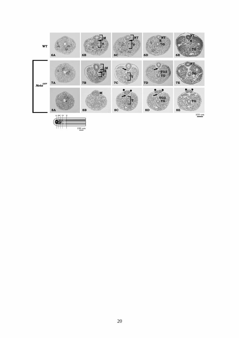
Figures 6-8.
Selected serial cross sections through the tip of the tail arranged in direction toward the base
of the tail.
6A-E. In the tip of the 11.5 days old wild type mouse embryo (Fig. 6A), the tail bud
mesenchyme (B) is visible. In the more cranial sections, the tail bud is continuous via the tail
cord (T) and the medullary cord (M) to the notochord (N) and the tail gut (TG). In paraaxial
mesenchyme, somites are formed (S).
7A-E and 8A-E. In NotoGFP/GFP embryos (Figs. 7A-E, 8A-E), differentiation of the tail bud is
disturbed: notochord-like cells are found in the region of the ventral medullary cord (region X
in Fig. 7B). In the region where in the wild type embryo notochord is found, small irregularly
shaped group of cells resembling notochord are present (arrows in Figs. 7C,D and 8C,D).
These cells disappear in the more cranial sections (Figs. 7E, 8E). Instead of the notochord,
additional lumen of the tail gut is formed (TG2 in Figs. 7D and 8D). In some embryos, two
medullary cords/neural tubes were found (arrowheads in Figs. 8C-8E).
B – tail bud, M – medullary cord, T – tail cord, NT – neural tube, TG – tail gut, TG2 –
additional tail gut, S – somites.
20

21
Figures 9-12.
Expression of Noto in the caudal part of 11.5 days old NotoGFP/GFP homozygous embryo.
9A-C. Longitudinal whole mount confocal photo reveals that Noto reaches the tip of the
embryo (tail bud region) (Figs. 9B,C).
10A-C. Immunohistochemistry performed on semithin sections reveals that the
morphogenesis of the tail bud region is disturbed.
11. Immunohistochemistry performed on semithin sections reveals that instead of the
notochord, additional lumen of the tail cord is formed, and Noto-GFP positive cells were
present only in the small portion of this group of cells (arrow in Fig. 11). Noto-GFP positive
cells were also found in adjacent paraaxial mesenchyme (wide arrows).
12. Longitudinal whole mount confocal photo reveals Noto-GFP expressing cells within
differentiating somites. The same can be seen marked by wide arrows in Figs. 9B and 11.
Copyright © 2022 FDOKUMEN