Effect of pruning strategy on 'Syrah' bud necrosis ... - Hal Inrae
Upload
independentCategory
view
1download
0
Nonthermal Atmospheric Pressure Plasma EnhancesMouse Limb Bud Survival, Growth, and Elongation
Natalie Chernets, PhD,1 Jun Zhang, MD,2,3 Marla J. Steinbeck, PhD,4,5 Deepa S. Kurpad, MS,2
Eiki Koyama, PhD,6 Gary Friedman, PhD,1 and Theresa A. Freeman, PhD2
The enhanced differentiation of mesenchymal cells into chondrocytes or osteoblasts is of paramount importancein tissue engineering and regenerative therapies. A newly emerging body of evidence demonstrates that ap-pendage regeneration is dependent on reactive oxygen species (ROS) production and signaling. Thus, wehypothesized that mesenchymal cell stimulation by nonthermal (NT)-plasma, which produces and induces ROS,would (1) promote skeletal cell differentiation and (2) limb autopod development. Stimulation with a singletreatment of NT-plasma enhanced survival, growth, and elongation of mouse limb autopods in an in vitro organculture system. Noticeable changes included enhanced development of digit length and definition of digitseparation. These changes were coordinated with enhanced Wnt signaling in the distal apical epidermal ridge(AER) and presumptive joint regions. Autopod development continued to advance for approximately 144 h inculture, seemingly overcoming the negative culture environment usually observed in this in vitro system. Real-time quantitative polymerase chain reaction analysis confirmed the up-regulation of chondrogenic transcripts.Mechanistically, NT-plasma increased the number of ROS positive cells in the dorsal epithelium, mesenchyme,and the distal tip of each phalange behind the AER, determined using dihydrorhodamine. The importance ofROS production/signaling during development was further demonstrated by the stunting of digital outgrowthwhen anti-oxidants were applied. Results of this study show NT-plasma initiated and amplified ROS intra-cellular signaling to enhance development of the autopod. Parallels between development and regenerationsuggest that the potential use of NT-plasma could extend to both tissue engineering and clinical applications toenhance fracture healing, trauma repair, and bone fusion.
Introduction
T issue engineering and regenerative medicine employsa variety of strategies to promote cell proliferation,
differentiation, and tissue development of both in vivo andin vitro synthetic constructs. The application of externalstimuli for the purpose of modifying cellular function is akey approach in tissue engineering. Recently, we reportedthat nonthermal (NT) dielectric barrier discharge (DBD)plasma treatment stimulated reactive oxygen species (ROS)–associated cell signaling to enhance both osteoblast andchondrocyte differentiation.1 Results from this study led us toinvestigate the effects of NT-plasma treatment on the morecomplex in vitro tissue model of mouse limb autopod de-velopment. Enhancement of ROS signaling during develop-ment is supported by studies showing tail regeneration in
Xenopus, and zebrafish is dependent on ROS production.2,3
Parallels are known to exist between the blastema in regen-eration and functionality of the apical epidermal ridge (AER)in autopod development. Thus, we hypothesized that engi-neering NT-plasma to produce the appropriate extracellularROS would activate intracellular ROS-sensitive signalingpathways in the AER and enhance digit growth. In the courseof these studies, insights into which pathways are necessarywould be determined and could be applied to further enhancetissue regeneration.
Vertebrate limbs grow through the coordinated activity ofnumerous growth factor signaling pathways, including the Wnt,fibroblast growth factor (FGF), bone morphogenetic protein(BMP), Notch, and transforming growth factor-b pathways.2–6
ROS play a primary role in mediating the activity of thesefactors and/or are integrally involved in regulating downstream
1Electrical and Computer Engineering Department, Drexel University, Philadelphia, Pennsylvania.2Department of Orthopedic Surgery, Thomas Jefferson University, Philadelphia, Pennsylvania.3Department of Orthopedics, The Second Hospital of Jilin University, Chang Chun, Jilin, China.4School of Biomedical Engineering, Science & Health Systems, Drexel University, Philadelphia, Pennsylvania.5Department of Orthopedic Surgery, Drexel University College of Medicine, Philadelphia, Pennsylvania.6Division of Orthopedic Surgery, The Children’s Hospital of Philadelphia, Philadelphia, Pennsylvania.
TISSUE ENGINEERING: Part AVolume 21, Numbers 1 and 2, 2015ª Mary Ann Liebert, Inc.DOI: 10.1089/ten.tea.2014.0039
300

signaling pathways.7 For example, a sustained increase in ROSlevels is required for Wnt b-catenin signaling and the activationof one of its main downstream targets, fgf20.8,9 In addition,naturally occurring oxidative spikes are observed in most of thetransition stages throughout developmental and regenerativeprocesses.10,11 Furthermore, there is precedence for ROS pro-duction to both direct and enhance mesenchymal cell differen-tiation into either chondrocyte or osteoblast lineages.12–14
The roles of ROS in stem cell differentiation and regen-eration described earlier are also directly applicable to de-velopment. For example, ROS involvement has beenexamined in transformation of the autopod during digit in-dividualization. FGF, BMP, and ROS regulate interdigitalcell death/apoptosis within the autopod, thereby restrictinginterdigit growth and promoting regression of interdigitaltissue.15 The ROS-induced oxidative stress associated withapoptosis of these cells is due to mitochondrial productionof ROS, the production by nicotinamide adenine dinucleo-tide phosphate (NADPH) oxidase in response to growthfactors, and a decrease in the expression of the antioxidantenzymes superoxide dismutase 1–3, catalase, and glutathi-one peroxidase 1–7.16 Studies have shown that the additionof antioxidants prevents interdigit cell death.17,18 In contrastwithin the digit, high levels of antioxidants are expressed tocontrol oxidative stress, thereby promoting cell prolifera-tion, growth, and differentiation.
In this study, we are employing an atmospheric pressureplasma known as DBD plasma.19 This type of NT-plasma isproduced when a high-voltage, time-varying waveform is ap-plied between two electrodes in air separated by an insulator.20
The insulator prevents current build-up, thus creating electri-cally safe plasma without a substantial temperature rise. Sincethe NT-plasma discharge occurs in air, it is a highly complexmix of reactive chemical species (i.e., nitric oxide [NO], ROS,oxygen singles, and ozone), free ions, visible, ultraviolet, andnear-infrared light, electric fields, and electromagnetic (EM)radiation.21 Therefore, determination of which single or com-bination of plasma components is responsible for the observedoutcome is difficult to assess. In the development of limbautopods, one study showed that a 2 h treatment with EM fieldstimulated limb growth through increased proliferation andchondrocyte differentiation.22 In clinical orthopedics, bothpulsed EM fields and ultrasound have been shown to enhancebone formation in defects, fractures, and bone lengthening.23,24
The goal of this study was to advance our previous workin a cell culture system to the tissue level, using the limbautopod model, to determine whether NT-plasma couldstimulate ROS-sensitive signaling pathways to enhance limbdevelopment. Here, we show that exposure to NT-plasmapromotes mouse limb autopod survival, growth, and elon-gation. In addition, the changes induced by NT-plasmastimulation are mediated through ROS-associated signaling.Finally, this work provides a fundamental understanding ofNT-plasma effects on developing tissues and its potentialuse as a treatment modality to enhance the development ofbiological substitutes both in vitro and in vivo.
Materials and Methods
Animals
Timed pregnant mice were ordered from Jackson laboratoryto ensure that the embryos would be at gestational age E12 to
E14. The mice either had a CD-1 genetic background or wereb-catenin/TCF/LEF reporter transgenic mice [BAT-GALtransgenic mice—Tg(BAT-lacZ)] mice containing a lacZgene under the control of seven consensus LEF/TCF-bindingmotifs upstream of a minimal promoter of the Xenopus sia-mois gene.25 BAT-lacZ experiments were performed on alitter of nine pups with three pups collected at each time pointat day 1, 2, and 5, after which staining for b-galactosidase wasperformed according to the standardized protocol by Lobeet al.26 All animals were euthanized according to NationalInstitutes of Health (NIH) guidelines for the care and use oflaboratory animals, and all animal protocols were approvedby the Institutional Animal Care and Use Committee (IA-CUC) at Thomas Jefferson University.
Explant cultures
Limb autopods from embryos of gestational ages E12.5and E14.5 were isolated and cultured in Dulbecco’s modifiedEagle’s medium (DMEM; Cellgro, ThermoFisher Scientific)using the method previously described.3 The limbs wereplaced on a nylon membrane in 12-well plates and moistenedwith 220mL of DMEM with no serum.27 Approximately 1 hafter isolation, NT-plasma or sham (limb positioned underelectrode with no discharge) treatment was applied, to the left(NT-plasma) and right limb (sham) autopod from the sameanimal. Immediately after treatment, the limb was transferredto fresh DMEM. Eighteen hours later, the culture mediumwas changed to DMEM with 2% fetal bovine serum (FBS)(Invitrogen, Life Science Technologies) and supplementedwith 100 U/mL penicillin,100mg/mL streptomycin (Cellgro,ThermoFisher Scientific). This medium was changed dailythereafter for the 4–6 days of culture.
At the conclusion of the experiment, the limbs were fixedwith 4% paraformaldehyde in phosphate-buffered saline andimaged to evaluate the length of the digits, the interdigitalclefting, and the appearance of the cartilage. To moreclearly visualize the cartilagenous elements, the limbs werestained with alcian blue.28 Imaging was performed using anOlympus MVX10 microscope with an Olympus DP70 colorcamera (Olympus Corp.).
NT-plasma treatment
Limb autopods were treated with DBD plasma similar tothe procedure described by Fridman et al.,29 where theschematics, voltage, and current curves of the power supply(Plasma Power, LLC) are detailed. The treatment voltage,frequency, pulse width, and time were based on our previouspublication.1 Briefly, NT-plasma was generated using amicrosecond pulsed voltage of 20 kV (peak to peak) with apulse width of 10 ms, a typical rise rate of 5 V/ns, and a pulserepetition rate of 1000 Hz applied through a high-voltageelectrode for 10 s. The high-voltage electrode was composedof an inner copper core surrounded by an outer insulatingshell of acrylic, also detailed by Fridman et al.29 (Fig. 1A).Before NT-plasma and sham treatments, each limb on thenylon membrane was transferred out of the DMEM from the12-well plate to a new 35 mm plastic dish and rewetted with220mL of phenol red-free DMEM. The 35 mm dish wasplaced on a grounded metal plate, and the high voltageelectrode was positioned 2 mm above the limb. The blueplasma discharge (5 mm) surrounded the autopod (2–3 mm)
NT-PLASMA ENHANCES SKELETAL GROWTH 301

as shown in Figure 1B. Of note, plasma produced by thiselectrode is not uniform and filaments are formed inter-mittently during the plasma treatment, visible in Figure 1Bas a brighter intensity in the central region of plasma. Fi-laments represent a more concentrated stream of plasma.Immediately after treatment, the limb on the nylon mem-brane was transferred to a new 12-well plate containingfresh DMEM. Sham treatment group was treated the same,but the high voltage was not applied.
Antioxidant treatments
The following inhibitors dissolved in dimethyl sulfoxidewere used as anti-oxidant treatments: 2-(4-Carboxyphenyl)-4,4,5,5-tetramethylimidazoline-1-oxyl-3-oxide (CPTIO), 300mM (Enzo Life Sciences); n-acetyl cysteine (NAC), 200mM(Sigma-Aldrich); and 4-hydroxy-2,2,6,6-tetramethylpiperidin-1-oxyl (Tempol), 6 mM (Sigma-Aldrich). After isolation, theautopods were placed in DMEM with each inhibitor for 1 hbefore NT-plasma treatment. Just before NT-plasma treatment,limb medium was changed to 220mL phenol red-free DMEMwith no FBS supplemented with each inhibitor. Immediatelyafter NT-plasma treatment, the limb was transferred to 220mLof fresh DMEM with phenol red and no FBS, supplementedwith inhibitors. Eighteen hours later, the culture medium wasremoved and 220mL of DMEM and 2% FBS supplementedwith inhibitors was added. This media with inhibitors wasrefreshed with each daily media change. The sham treatmentgroup was treated the same but received no plasma treatment.
Assessment of limb autopod development
Limb autopod survival, growth, and development were as-sessed using a 1–4 grading system (Fig. 1C). A score of 1indicates stunted growth (lack of survival) with no definedcartilage segments. A score of 2 indicates the autopod hassurvived, developed the first two distinct cartilagenous jointsegments. A score of 3 indicates the autopod has undergonegrowth, developed three joint segments, and the metatarsal ormetacarpal is approximately equal in length to the proximalphalange. A score of 4 indicates the autopod has undergonegrowth and elongation, has three or more joint segments, and
the metatarsal or metacarpal is longer than the proximal pha-lange. The scores between contralateral pairs were calculated todetermine the effectiveness of NT-plasma versus sham treat-ment. A pie chart was created to display the results betweeneach pair of contralateral limbs by tallying which treatmentreceived the higher score. Then, the scoring results1–4 from allcontralateral experiments were tabulated to provide anoverall assessment of growth and development independentof the contralateral limb. A histogram was generated by tal-lying the scores for all limbs for each treatment NT-plasma orsham. Validation that contralateral limbs in organ cultureperform equivalently was determined by evaluating 10 con-tralateral autopods cultured for 6 days without treatment.These results indicated no significant difference in the normalgrowth of contralateral limbs ( p = 1.00).
Histology
Tissue samples were fixed overnight in 4% paraformalde-hyde, dehydrated in a graded series of alcohols, and infiltratedand embedded in paraffin. Sections were cut at 6mm andmounted on slides (Superfrost/Plus; Thermo Fisher Scientific).Cell and tissue morphology between treatments was comparedusing histological staining of limb sections with hematoxylin,eosin, and alcian blue (proteoglycan stain for cartilage).
Real-time polymerase chain reaction for differentiationmarkers and comparative analyses
Limb autopods were harvested at 24 or 144 h after sham orNT-plasma treatment. For each analysis, two autopods ofeach treatment group were pooled to create a sample inwhich RNA was isolated, and data from an n of 3 of thesepooled samples were used to perform polymerase chain re-action (PCR) analysis. All harvested limbs were washed withdiethylpyrocarbonate water before total RNA was isolatedusing the QiagenRNeasy� Micro kit (Qiagen). RNA yieldwas determined spectrophotometrically, and integrity wasconfirmed by gel electrophoresis. RNA was reverse tran-scribed and then amplified using the Superscript� One-StepRT-PCR with Platinum Taq� (Invitrogen) kit. PCR prod-ucts were analyzed by 1.0% agarose gel electrophoresis.
FIG. 1. Microsecondpulsed dielectric barrier dis-charge plasma treatmentconditions. (A) Schematicsof the nonthermal (NT)-plasma electrode applied tolimb buds. (B) Picture of theNT-plasma electrode. Left:bottom view; Right: sideview. (C) Assessment of di-git development and growthof an alcian blue-stainedE12.5 autopod using a de-fined Grading Scale: 1, stun-ted; 2–4, surviving; 3–4,growth; 4, elongation. Colorimages available online atwww.liebertpub.com/tea
302 CHERNETS ET AL.

Template cDNA and gene specific primers were added toFast SYBR Green master mix (Applied Biosystems), andmRNA expression was quantified using the MyIQ Real-TimePCR System (BioRad). The expression level of the house-keeping gene, b-actin was used to normalize the data pre-sented. Melting curves were analyzed to verify the specificityof the reverse transcription (RT)-PCR and the absence ofprimer dimer formation. Each sample was analyzed in du-plicate and included a template-free control. All the primers(Supplementary Table S1; Supplementary Data are availableonline at www.liebertpub.com/ten) used were synthesized byIntegrated DNA Technologies, Inc.
Statistical analysis
Statistical analysis between groups was performed usinga one-way ANOVA for normality and Student’s t-testfor continuous variables. A level of significance (a), or ap-value of less than 0.05, with a 95% confidence intervalwas determined. Representative data are presented as themean – standard error of the mean of individual samplesfrom 2 to 10 independent analyses. For nonparametric data,Mann–Whitney U tests were used to evaluate differences.
Results
NT-plasma enhances limb autopod growthand development
To ascertain whether NT-plasma promotes growth anddevelopment of embryonic tissue, E12.5 mouse autopodswere treated with a single NT-plasma treatment and thenmaintained in culture for approximately 6 days. Figure 2Ashows representative, consecutive images of NT-plasma-enhanced digit development in an E12.5 autopod comparedwith its contralateral sham control (left treated with NT-plasma vs. right sham-treated control forelimbs or hind limbs).In developing embryos, hind limb maturation is delayed ascompared with forelimb, by *1 day. For example, cartilag-inous development evaluated using alcian blue staining toshow digit definition, length, and joint space illustrates thatthe initial digit segmentation occurs at 24 h in the forelimbas compared with 48 h of the hind limb (Fig. 2B). At thesetime points, NT-plasma-treated autopods showed increasedlengthening of the distal segments, signifying accelerateddigit growth of the fore and hind limbs when compared withsham controls. At 48 h (forelimb) and 72 h (hind limb), alldigit segments were noticeably more defined, longer, andwider in response to NT-plasma treatment. At 72 h (fore-limb) and 96 h (hind limb), NT-plasma-treated autopodsdisplayed an additional distal segment with the appearanceof the final joint space. In addition, at 144 h, both the NT-plasma-treated fore and hind limbs were noticeably larger,while the sham-treated autopods had stopped growing.Quantification of contralateral autopod growth shown in thepie chart in Figure 2C indicates that NT-plasma treatmentpromoted and enhanced growth in 76% of the autopods, ascompared with only 5% increase after sham treatment; (n =21; p = 0.0003). The remaining 19% of the limbs showed nodifference between sham and NT-plasma treatments. Todetermine whether growth enhancement could be achievedwith hydrogen peroxide (H2O2), a 30 s treatment with200 mM and 1 mM H2O2 in DMEM was performed in place
of NT-plasma treatment and no effect on limb growth wasobserved (n = 9, results not shown).
In addition, evaluation of autopod development wasperformed using a numerical scoring system of 1–4 and thegrowth status of each limb from every experiment was as-sessed independent of the contralateral limb. Briefly, in thisscoring system, each individual limb received one of thefollowing scores based on morphology and alcian bluestaining: stunted, nonsurvival (1), survival ( > 1), growth(3,4), and elongation (4) (Fig. 1C). Results of E12.5 au-topods (n = 61) limbs from all experimental groups treatedwith NT-plasma indicated that NT-plasma significantly im-proved the outcome of limb survival ( p = 0.01), and in-creased limb elongation ( p = 0.001), as compared with shamcontrol (Fig. 2D; n = 73).
When the same experiments were repeated with E14.5embryo limbs, a less dramatic but still significant increase inlimb development was observed after NT-plasma treatment.Contralateral limb growth was enhanced for 60% of the NT-plasma treated autopods as compared with 23% after shamtreatment (Fig. 2E; n = 30; p = 0.03). Seventeen percent ofthe time, there was no difference between the two treat-ments. Grading analysis of E14.5 autopods showed that NT-plasma treatment (n = 39) resulted in significantly increasedelongation as compared with sham control (n = 135) (Fig.2F; p = 0.001).
Increased chondrogenesis after NT-plasma treatment
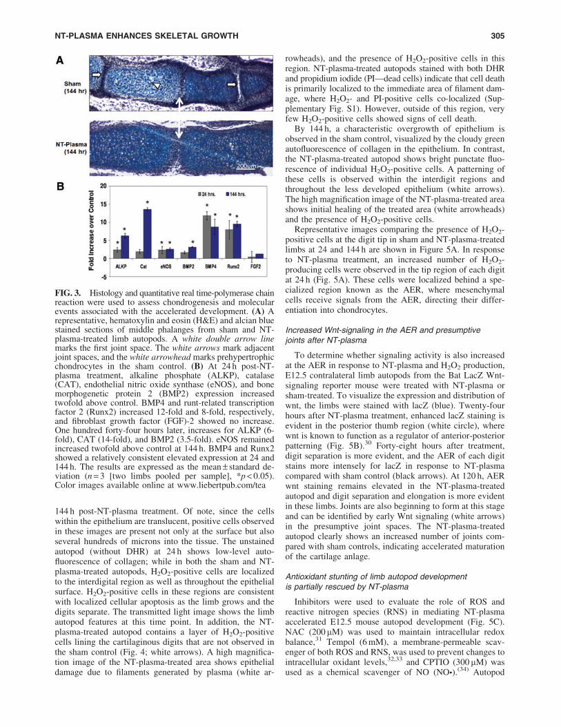
To ascertain whether the enhanced growth observed wasdue to normal or aberrant chondrogenesis, we stained sec-tions of the limb autopods treated with both NT-plasma andsham with H&E and alcian blue. Morphological formationof the joint region and alcian blue staining of proteoglycanappeared normal in sham and NT-plasma-treated digits (Fig.3A; n = 9). Chondrocytes in the proximal segment of thesham-treated digit were entering the prehypertrophic stage,based on the larger size, greater cellular spacing, and de-crease in proteoglycan (white arrowhead). This typicallyprecedes the formation of the primary center of ossificationin the diaphysis. In contrast, chondrocytes from the NT-plasma-treated digit were immature and contained largeamounts of proteoglycan. Even more striking was the muchlarger cartilage phalangeal segments in the NT-plasma-treated digit. A white line with double arrows marks thejoint between the first and second phalanges in both sec-tions. In the sham-treated digit, the adjacent joint spaces(white arrow) can be clearly observed; whereas in the NT-plasma-treated digits, there is no evidence of the next jointspace, due to the increased segment length.
Quantitative RT-PCR was used to assess chondrogenesisand other molecular events associated with the accelerateddevelopment in response to NT-plasma (Fig. 3). In E12.5autopods, at 24 h post-NT-plasma treatment, alkaline phos-phate (ALKP), catalase (CAT), endothelial nitric oxidesynthase (eNOS), and BMP2 expression were increasedtwofold above control ( p < 0.05). BMP4 and runt-relatedtranscription factor 2 (Runx2) were increased 12-fold and8-fold, respectively ( p < 0.05), whereas FGF-2 showed noincrease. Even more significant increases were observed at144 h for ALKP (6-fold), CAT (14-fold), and BMP2 (3.5-fold) ( p < 0.05). eNOS remained increased twofold above
NT-PLASMA ENHANCES SKELETAL GROWTH 303

control at 144 h, and both BMP4 and Runx2 evidencedconsistent elevated expression at 24 and 144 h. Up-regulationof chondrogenic transcripts confirmed the morphologic chan-ges associated with chondrocyte differentiation in the au-topod. The increased expression of CAT emphasized theimportance of antioxidant expression within the digit for cellproliferation, growth, and differentiation.15
NT-plasma treatment increases H2O2 productionby cells within the limb autopod
To directly assess the production of ROS by limb cells,E12.5 autopods were stained with dihydrorhodamine(DHR), a dye that becomes fluorescent after a reaction withH2O2. Figure 4 shows representative images after 24 or
FIG. 2. NT-plasma treatment enhances thriving, growth, and elongation in mouse limb autopod development. (A)Consecutive images of NT-plasma enhanced digit development in an E12.5 autopod compared with its contralateral shamcontrol. (B) Alcian blue (cartilaginous elements) stained fore and hindlimb autopods after NT-plasma or sham treatment.(C) Contralateral limb growth enhancement comparisons for sham and NT-plasma-treated E12.5 autopods. (D) Gradingscale results for all limb experiments showing NT-plasma enhanced survival, growth, and elongation compared with sham-treated E12.5 autopods. (E) Contralateral limb growth enhancement comparisons for sham and NT-plasma-treated E14.5autopods. (F) Grading scale results for all limb experiments showing NT-plasma enhanced elongation compared with shamtreatment of E14.5 autopod. **p < 0.01, ***p < 0.001. Color images available online at www.liebertpub.com/tea
304 CHERNETS ET AL.
144 h post-NT-plasma treatment. Of note, since the cellswithin the epithelium are translucent, positive cells observedin these images are present not only at the surface but alsoseveral hundreds of microns into the tissue. The unstainedautopod (without DHR) at 24 h shows low-level auto-fluorescence of collagen; while in both the sham and NT-plasma-treated autopods, H2O2-positive cells are localizedto the interdigital region as well as throughout the epithelialsurface. H2O2-positive cells in these regions are consistentwith localized cellular apoptosis as the limb grows and thedigits separate. The transmitted light image shows the limbautopod features at this time point. In addition, the NT-plasma-treated autopod contains a layer of H2O2-positivecells lining the cartilaginous digits that are not observed inthe sham control (Fig. 4; white arrows). A high magnifica-tion image of the NT-plasma-treated area shows epithelialdamage due to filaments generated by plasma (white ar-
rowheads), and the presence of H2O2-positive cells in thisregion. NT-plasma-treated autopods stained with both DHRand propidium iodide (PI—dead cells) indicate that cell deathis primarily localized to the immediate area of filament dam-age, where H2O2- and PI-positive cells co-localized (Sup-plementary Fig. S1). However, outside of this region, veryfew H2O2-positive cells showed signs of cell death.
By 144 h, a characteristic overgrowth of epithelium isobserved in the sham control, visualized by the cloudy greenautofluorescence of collagen in the epithelium. In contrast,the NT-plasma-treated autopod shows bright punctate fluo-rescence of individual H2O2-positive cells. A patterning ofthese cells is observed within the interdigit regions andthroughout the less developed epithelium (white arrows).The high magnification image of the NT-plasma-treated areashows initial healing of the treated area (white arrowheads)and the presence of H2O2-positive cells.
Representative images comparing the presence of H2O2-positive cells at the digit tip in sham and NT-plasma-treatedlimbs at 24 and 144 h are shown in Figure 5A. In responseto NT-plasma treatment, an increased number of H2O2-producing cells were observed in the tip region of each digitat 24 h (Fig. 5A). These cells were localized behind a spe-cialized region known as the AER, where mesenchymalcells receive signals from the AER, directing their differ-entiation into chondrocytes.
Increased Wnt-signaling in the AER and presumptivejoints after NT-plasma
To determine whether signaling activity is also increasedat the AER in response to NT-plasma and H2O2 production,E12.5 contralateral limb autopods from the Bat LacZ Wnt-signaling reporter mouse were treated with NT-plasma orsham-treated. To visualize the expression and distribution ofwnt, the limbs were stained with lacZ (blue). Twenty-fourhours after NT-plasma treatment, enhanced lacZ staining isevident in the posterior thumb region (white circle), wherewnt is known to function as a regulator of anterior-posteriorpatterning (Fig. 5B).30 Forty-eight hours after treatment,digit separation is more evident, and the AER of each digitstains more intensely for lacZ in response to NT-plasmacompared with sham control (black arrows). At 120 h, AERwnt staining remains elevated in the NT-plasma-treatedautopod and digit separation and elongation is more evidentin these limbs. Joints are also beginning to form at this stageand can be identified by early Wnt signaling (white arrows)in the presumptive joint spaces. The NT-plasma-treatedautopod clearly shows an increased number of joints com-pared with sham controls, indicating accelerated maturationof the cartilage anlage.
Antioxidant stunting of limb autopod developmentis partially rescued by NT-plasma
Inhibitors were used to evaluate the role of ROS andreactive nitrogen species (RNS) in mediating NT-plasmaaccelerated E12.5 mouse autopod development (Fig. 5C).NAC (200mM) was used to maintain intracellular redoxbalance,31 Tempol (6 mM), a membrane-permeable scav-enger of both ROS and RNS, was used to prevent changes tointracellular oxidant levels,32,33 and CPTIO (300 mM) wasused as a chemical scavenger of NO (NO�).(34) Autopod
FIG. 3. Histology and quantitative real time-polymerase chainreaction were used to assess chondrogenesis and molecularevents associated with the accelerated development. (A) Arepresentative, hematoxylin and eosin (H&E) and alcian bluestained sections of middle phalanges from sham and NT-plasma-treated limb autopods. A white double arrow linemarks the first joint space. The white arrows mark adjacentjoint spaces, and the white arrowhead marks prehypertrophicchondrocytes in the sham control. (B) At 24 h post-NT-plasma treatment, alkaline phosphate (ALKP), catalase(CAT), endothelial nitric oxide synthase (eNOS), and bonemorphogenetic protein 2 (BMP2) expression increasedtwofold above control. BMP4 and runt-related transcriptionfactor 2 (Runx2) increased 12-fold and 8-fold, respectively,and fibroblast growth factor (FGF)-2 showed no increase.One hundred forty-four hours later, increases for ALKP (6-fold), CAT (14-fold), and BMP2 (3.5-fold). eNOS remainedincreased twofold above control at 144 h. BMP4 and Runx2showed a relatively consistent elevated expression at 24 and144 h. The results are expressed as the mean – standard de-viation (n = 3 [two limbs pooled per sample], *p < 0.05).Color images available online at www.liebertpub.com/tea
NT-PLASMA ENHANCES SKELETAL GROWTH 305

morphogenesis was stunted by the pretreatment of the limbswith each antioxidant, confirming the importance of ROSand RNS in development. Inhibition of both ROS andRNS by Tempol completely stunted autopod development.However, removal of RNS by CPTIO had the least effectand mediation of ROS by NAC showed an intermediateeffect. Interestingly, NT-plasma treatment partially rescueddevelopment, moderately enhancing digit morphogenesiseven in the presence of these anti-oxidants.
Discussion
This work follows up on our previous report that NT-plasma treatment increased intracellular ROS productionand promoted the expression of differentiation specificgenes, synergistically enhancing both osteogenic andchondrogenic differentiation.1 In this more complex tissuemodel, we found that NT-plasma treatment promoted bothgrowth and differentiation of cartilaginous elements withinthe embryonic mouse autopod. Based on the lack of limbgrowth after NT-plasma treatment in an antioxidant envi-ronment, and the increase in H2O2-positive cells in andunder the epithelia throughout the 6 day culture period, wepropose that NT-plasma induction of intracellular ROS also
plays a role in the growth and differentiation observed in theautopod. In addition, H2O2-positive cells were observed inthe distal digit tips behind the AER, concurrent with en-hanced Wnt signaling. These findings are consistent with theROS dependence of signaling factors (e.g., Wnt, BMP, andFGF) required for autopod development.
The use of antioxidant treatment confirms that cellularproduction of ROS is required for normal growth and de-velopment of the limb autopod. However, very little hasbeen reported on the direct contribution of ROS in autopoddevelopmental examples of how ROS affects differentiationand development exists. In vivo, stem cells and adult MSCshave been reported to exist in a quiescent, glycolytic statewith immature mitochondria.35 Disruption of this environ-ment by vascularization, growth factor binding, increasedmitochondrial biogenesis, or external stresses induces ele-vations in ROS levels, leading to the induction of differ-entiation.10 In addition, the requirement of ROS in theanalogous process of regeneration was recently elucidatedafter tadpole tail amputation using fluorescent monitoring ofH2O2.36 In addition, regeneration in the nonmammaliansystems, of both the axyotl salamander and Xenopus froglimbs, reports the most upregulated protein at 24 h afteramputation is neuronal NOS.37 A similar result to what we
FIG. 4. NT-plasma treatment increases intracellular hydrogen peroxide (H2O2) in cells of the epithelium. DHR fluorescence1 h after NT-plasma treatment; limb unstained and transmitted light images are shown to verify specificity of DHR andanatomical localization. Twenty-four hours later, control limb shows prominent H2O2 production in the interdigital spaces andNT-plasma-treated limbs show more defined H2O2 production next to the cartilage as the digits separate (white arrows).Additional staining is observed at the edges of the wrist and scattered throughout the epithelium. One hundred forty-four hourslater, control autofluorescence of collagen within the forming epithelial skin overgrowth, and NT-plasma-treated limb withH2O2-positive cells throughout the epithelia and interdigital spaces (white arrows). Higher magnification images at 24 and144 h show that the cells immediately surrounding the site of plasma filament damage (white arrowheads) continue to generateH2O2 after a single 10 s NT-plasma treatment. Color images available online at www.liebertpub.com/tea
306 CHERNETS ET AL.

observed at 24 h post-NT-plasma treatment was observedwhen eNOS expression was increased twofold above controlin the mouse autopod. NO is known to trigger enhancedinduction of vascular endothelial growth factor expressionin cultured keratinocytes (HaCaT) and during cutaneouswound repair.38,39 As stated earlier, increases in angiogen-esis are known to enhance differentiation and undoubtedlyplay a role in enhancing autopod growth and development.
However, the fact that NT-plasma treatment showedsome growth effect even in the presence of antioxidantsindicates that ROS production may not be the sole mecha-nism driving enhanced autopod growth. A second possiblereason that NT-plasma enhanced autopod growth was itsinhibition of epithelial overgrowth. Epithelial overgrowth iscommonly observed in organ culture systems, where directcontact with the nutrient-rich media favors cells on the outerlayers of the tissue.40,41 In limb regeneration, a thickening ofthe epidermis has been shown to inhibit signaling to theblastema, halting the regeneration process.42–44 The ob-served decrease in epithelial thickening and increased limbgrowth after NT-plasma treatment supports this mechanism.One explanation for our findings is the presence and dis-tribution of H2O2-positive cells throughout the culture pe-riod which indicates that the NT-plasma treatment hasaltered signaling and development within the epithelium.
Our study does not rule out the importance of otherelectrically induced changes, including changes in mem-brane permeability, bioelectric signaling, voltage-sensitivedomains of transporter molecules, and/or the movement of
calcium and other ions. Thus, it is possible that a combi-nation of effects is at play in NT-plasma treatment. In ad-dition to ROS and RNS, NT-plasma also generates chargedions, ozone, a pulsed electric field, and some UV andtransmitted light. Pulsed electric fields have been reported toincrease cell permeability, activate voltage-sensitive mem-brane channels, and mobilize calcium.45,46 Mechanical andsonic forces are also generated by DBD plasmas.21 Each ofthese modalities, applied in one way or another, have beenreported to enhance differentiation of skeletal tissues47 andcannot be ruled out as causative factors. Further study isneeded to determine any role these other effects play in limbbud development.
Results of this study showing NT-plasma effects on limbautopod development suggest that this technology could beused to enhance tissue regeneration. This report, coupledwith our previous work, solidifies the use of NT-plasma as ameans to affect cellular differentiation and enhance tissueregeneration. Similar to other physical technologies cur-rently in clinical use, EM fields and ultrasound, develop-ment of NT-plasma technology could provide enhancedoutcomes for tissue engineering applications and regenera-tive medicine. How high-energy electrons produced by aNT-plasma discharge generate short-lived ROS and RNS todirectly activate redox-sensitive signaling pathways therebyinfluencing cell signaling and precursor cell differentiationis only partially understood. Further exploration into bio-physical interactions of NT-plasma with cells and tissuesalso enhances our understanding of cell behaviors to
FIG. 5. NT-plasma treatment of E12.5 mouse autopods increases expression of chondrogenic and anti-oxidant genes andpartially rescues antioxidant stunting. (A) H2O2-positive cells at the digit tip in sham (left) and NT-plasma (right)-treatedlimbs at 24 h (top) and 144 h (bottom). (B) Enhanced Wnt signaling is seen in the AER (black arrows) and presumptivejoints (white arrows) of the NT-plasma-treated E12.5 autopod from the Wnt-signaling reporter mouse. (C) Pretreatment ofE12.5 autopods with n-acetyl cysteine (NAC), 4-hydroxy-2,2,6,6-tetramethylpiperidin-1-oxyl (Tempol) or 2-(4-carbox-yphenyl)-4,4,5,5-tetramethylimidazoline-1-oxyl-3-oxide (CPTIO) inhibitors stunts autopod morphogenesis. Treatment withNT-plasma partially rescues development, moderately enhancing digit morphogenesis compared with sham control. Colorimages available online at www.liebertpub.com/tea
NT-PLASMA ENHANCES SKELETAL GROWTH 307

promote the development of novel treatment modalitiesapplied across many fields of study. NT-plasma is a newmodality in biology and medicine, and its potential impact isyet to be determined.
Acknowledgments
Special thanks are due to Drs. Irving Shapiro, MaurizioPacifici, Motomi Enomoto-Iwamoto, and Masahiro Iwamotofor their valuable discussions during the study. Specialthanks are also due to Carol Diallo for laboratory supportand Alex Fridman from the Drexel Plasma Institute forhelpful insights on plasma conditions. This work was sup-ported by NIH Grants 1 R01 EB 013011-01 (Freeman) and 5R03 DE020840-03 (Freeman).
Disclosure Statement
No competing financial interests exist.
References
1. Steinbeck, M.J., Chernets, N., Zhang, J., Kurpad, D.S.,Fridman, G., Fridman, A., et al. Skeletal cell differentiationis enhanced by atmospheric dielectric barrier dischargeplasma treatment. PLoS One 8, e82143, 2013.
2. Enomoto-Iwamoto, M., Kitagaki, J., Koyama, E., Tama-mura, Y., Wu, C., Kanatani, N., et al. The Wnt antagonistFrzb-1 regulates chondrocyte maturation and long bonedevelopment during limb skeletogenesis. Dev Biol 251,142, 2002.
3. Koyama, E., Shibukawa, Y., Nagayama, M., Sugito, H.,Young, B., Yuasa, T., et al. A distinct cohort of progenitorcells participates in synovial joint and articular cartilageformation during mouse limb skeletogenesis. Dev Biol 316,62, 2008.
4. Benazet, J.D., and Zeller, R. Dual requirement of ectoder-mal Smad4 during AER formation and termination offeedback signaling in mouse limb buds. Genesis 51, 660,2013.
5. Vasiliauskas, D., Laufer, E., and Stern, C.D. A role forhairy1 in regulating chick limb bud growth. Dev Biol 262,94, 2003.
6. Lee, Y., Grill, S., Sanchez, A., Murphy-Ryan, M., and Poss,K.D. Fgf signaling instructs position-dependent growth rateduring zebrafish fin regeneration. Development 132, 5173,2005.
7. Oktyabrsky, O.N., and Smirnova, G.V. Redox regulation ofcellular functions. Biochemistry (Mosc) 72, 132, 2007.
8. Chamorro, M.N., Schwartz, D.R., Vonica, A., Brivanlou,A.H., Cho, K.R., and Varmus, H.E. FGF-20 and DKK1 aretranscriptional targets of beta-catenin and FGF-20 is im-plicated in cancer and development. EMBO J 24, 73, 2005.
9. Funato, Y., Michiue, T., Asashima, M., and Miki, H. Thethioredoxin-related redox-regulating protein nucleoredoxininhibits Wnt-beta-catenin signalling through dishevelled.Nat Cell Biol 8, 501, 2006.
10. Chen, C., Liu, Y., Liu, R., Ikenoue, T., Guan, K.L., andZheng, P. TSC-mTOR maintains quiescence and functionof hematopoietic stem cells by repressing mitochondrialbiogenesis and reactive oxygen species. J Exp Med 205,2397, 2008.
11. Chen, C.-T., Hsu, S.-H., and Wei, Y.-H. Upregulation ofmitochondrial function and antioxidant defense in the dif-
ferentiation of stem cells. Biochim Biophys Acta 1800,257, 2010.
12. Chen, C.-T., Shih, Y.-R.V., Kuo, T.K., Lee, O.K., and Wei,Y.-H. Coordinated changes of mitochondrial biogenesisand antioxidant enzymes during osteogenic differentiationof human mesenchymal stem cells. Stem Cells 26, 960,2008.
13. Ji, A.-R., Ku, S.-Y., Cho, M.S., Kim, Y.Y., Kim, Y.J., Oh,S.K., et al. Reactive oxygen species enhance differentiationof human embryonic stem cells into mesendodermal line-age. Exp Mol Med 42, 175, 2010.
14. Yanes, O., Clark, J., Wong, D.M., Patti, G.J., Sanchez-Ruiz, A., Benton, H.P., et al. Metabolic oxidation regulatesembryonic stem cell differentiation. Nat Chem Biol 6, 411,2010.
15. Schnabel, D., Salas-Vidal, E., Narvaez, V., Sanchez-Carbente Mdel, R., Hernandez-Garcia, D., Cuervo, R., et al.Expression and regulation of antioxidant enzymes in thedeveloping limb support a function of ROS in interdigitalcell death. Dev Biol 291, 291, 2006.
16. Salas-Vidal, E., Lomeli, H., Castro-Obregon, S., Cuervo,R., Escalante-Alcalde, D., and Covarrubias, L. Reactiveoxygen species participate in the control of mouse embry-onic cell death. Exp Cell Res 238, 136, 1998.
17. Rong, Y., Doctrow, S.R., Tocco, G., and Baudry, M. EUK-134, a synthetic superoxide dismutase and catalase mimetic,prevents oxidative stress and attenuates kainate-inducedneuropathology. Proc Natl Acad Sci U S A 96, 9897,1999.
18. Jung, C., Rong, Y., Doctrow, S., Baudry, M., Malfroy, B.,and Xu, Z. Synthetic superoxide dismutase/catalase mi-metics reduce oxidative stress and prolong survival in amouse amyotrophic lateral sclerosis model. Neurosci Lett304, 157, 2001.
19. Siemens, C.W. On the electrical tests employed during theconstruction of the malta and alexandria telegraph, and oninsulating and protecting submarine cables. J Franklin Inst74, 166, 1862.
20. Eliasson, B., Egli, W., and Kogelschatz, U. Modelling ofdielectric barrier discharge chemistry. Pure Appl Chem 66,1275, 1994.
21. Fridman, A., and Friedman, G. Plasma Medicine. 2 ed.West Sussex, UK: Wiley & Sons, Incorporated, 2013.
22. Parivar, K., Kouchesfehani, M.H., Boojar, M.M., andHayati, R.N. Organ culture studies on the development ofmouse embryo limb buds under EMF influence. Int J RadiatBiol 82, 455, 2006.
23. Freeman, T.A., Patel, P., Parvizi, J., Antoci, V., Jr., andShapiro, I.M. Micro-CT analysis with multiple thresholdsallows detection of bone formation and resorption duringultrasound-treated fracture healing. J Orthop Res 27, 673,2009.
24. Griffin, M., and Bayat, A. Electrical stimulation in bonehealing: critical analysis by evaluating levels of evidence.Eplasty 11, e34, 2011.
25. DasGupta, R., and Fuchs, E. Multiple roles for activatedLEF/TCF transcription complexes during hair follicle de-velopment and differentiation. Development 126, 4557,1999.
26. Lobe, C.G., Koop, K.E., Kreppner, W., Lomeli, H., Gert-senstein, M., and Nagy, A. Z/AP, a double reporter for cre-mediated recombination. Dev Biol 208, 281, 1999.
27. Iwamoto, M., Tamamura, Y., Koyama, E., Komori, T., Ta-keshita, N., Williams, J.A., et al. Transcription factor ERG
308 CHERNETS ET AL.

and joint and articular cartilage formation during mouse limband spine skeletogenesis. Dev Biol 305, 40, 2007.
28. Ueta, C., Iwamoto, M., Kanatani, N., Yoshida, C., Liu, Y.,Enomoto-Iwamoto, M., et al. Skeletal malformationscaused by overexpression of Cbfa1 or its dominant negativeform in chondrocytes. J Cell Biol 153, 87, 2001.
29. Fridman, G., Peddinghaus, M., Ayan, H., Fridman, A.,Balasubramanian, M., Gutsol, A., et al. Blood coagulationand living tissue sterilization by floating-electrode dielec-tric barrier discharge in air. Plasma Chem Plasma Proc 26,425, 2006.
30. Parr, B.A., and McMahon, A.P. Dorsalizing signal Wnt-7arequired for normal polarity of D-V and A-P axes of mouselimb. Nature 374, 350, 1995.
31. Parasassi, T., Brunelli, R., Costa, G., De Spirito, M.,Krasnowska, E.K., Lundeberg, T., et al. Thiol redox tran-sitions in cell signaling: a lesson from N-acetylcysteine. SciWorld J 10, 1192, 2010.
32. Castagna, R., Davis, P.A., Vasu, V.T., Soucek, K., Cross,C.E., Greci, L., et al. Nitroxide radical TEMPO reducesozone-induced chemokine IL-8 production in lung epithe-lial cells. Toxicol In Vitro 23, 365, 2009.
33. Suy, S., Mitchell, J.B., Ehleiter, D., Haimovitz-Friedman,A., and Kasid, U. Nitroxides tempol and tempo inducedivergent signal transduction pathways in MDA-MB 231breast cancer cells. J Biol Chem 273, 17871, 1998.
34. Pieper, G.M., and Siebeneich, W. Use of a nitronyl nitr-oxide to discriminate the contribution of nitric oxide radicalin endothelium-dependent relaxation of control and dia-betic blood vessels. J Pharmacol Exp Ther 283, 138, 1997.
35. Fischer, L.J., McIlhenny, S., Tulenko, T., Golesorkhi, N.,Zhang, P., Larson, R., et al. Endothelial differentiation ofadipose-derived stem cells: effects of endothelial cell growthsupplement and shear force. J Surg Res 152, 157, 2009.
36. Love, N.R., Chen, Y., Ishibashi, S., Kritsiligkou, P., Lea,R., Koh, Y., et al. Amputation-induced reactive oxygenspecies are required for successful Xenopus tadpole tailregeneration. Nat Cell Biol 15, 222, 2013.
37. Grow, M., Neff, A.W., Mescher, A.L., and King, M.W.Global analysis of gene expression in Xenopus hindlimbsduring stage-dependent complete and incomplete regener-ation. Dev Dyn 235, 2667, 2006.
38. Frank, S., Stallmeyer, B., Kampfer, H., Kolb, N., andPfeilschifter, J. Nitric oxide triggers enhanced induction ofvascular endothelial growth factor expression in culturedkeratinocytes (HaCaT) and during cutaneous wound repair.FASEB J 13, 2002, 1999.
39. Stallmeyer, B., Kampfer, H., Kolb, N., Pfeilschifter, J., andFrank, S. The function of nitric oxide in wound repair:inhibition of inducible nitric oxide-synthase severely im-pairs wound reepithelialization. J Invest Dermatol 113,1090, 1999.
40. Browning, T.H., and Trier, J.S. Organ culture of mucosalbiopsies of human small intestine. J Clin Invest 48, 1423,1969.
41. Hoffman, D.S., Bringas, P., Jr., and Slavkin, H.C. Co-culture of contiguous developmental fields in a serumless,chemically-defined medium: an in vitro model permissivefor coordinate development of the mouse ear. Int J DevBiol 40, 953, 1996.
42. Mescher, A.L. Effects on adult newt limb regeneration ofpartial and complete skin flaps over the amputation surface.J Exp Zool 195, 117, 1976.
43. Neufeld, D.A., and Day, F.A. Perspective: a suggested rolefor basement membrane structures during newt limb re-generation. Anat Rec 246, 155, 1996.
44. Neufeld, D.A., Day, F.A., and Settles, H.E. Stabilizing roleof the basement membrane and dermal fibers during newtlimb regeneration. Anat Rec 245, 122, 1996.
45. Scarlett, S.S., White, J.A., Blackmore, P.F., Schoenbach,K.H., and Kolb, J.F. Regulation of intracellular calciumconcentration by nanosecond pulsed electric fields. Bio-chim Biophys Acta 1788, 1168, 2009.
46. Pall, M.L. Electromagnetic fields act via activation ofvoltage-gated calcium channels to produce beneficial oradverse effects. J Cell Mol Med 17, 958, 2013.
47. Goodman, R., Lin-Ye, A., Geddis, M.S., Wickramaratne,P.J., Hodge, S.E., Pantazatos, S.P., et al. Extremely lowfrequency electromagnetic fields activate the ERK cascade,increase hsp70 protein levels and promote regeneration inPlanaria. Int J Radiat Biol 85, 851, 2009.
Address correspondence to:Theresa A. Freeman, PhD
Department of Orthopaedic SurgeryThomas Jefferson University
501 Curtis Bldg., 1015 Walnut StreetPhiladelphia, PA 19107
E-mail: [email protected]
Received: January 15, 2014Accepted: July 23, 2014
Online Publication Date: September 16, 2014
NT-PLASMA ENHANCES SKELETAL GROWTH 309
Copyright © 2022 FDOKUMEN