Cloning, sequence analysis and expression of mammalian mitochondrial protein synthesis elongation...
10
' ),7 ELSEVIER Biochimica et Biophysica Acta 1264 (1995) 347- 356 BB Biochi~ic~a et Biophysica Acta Cloning, sequence analysis and expression of mammalian mitochondrial protein synthesis elongation factor Tu 1 Velinda L. Woriax a, Will Burkhart b, Linda L. Spremulli a.c.~ a Department ofChemisto' CB #3290, UniL~ersiO ~ of North Carolina, Chapel Hill, NC 27599-3290, USA h Department of Bioanalytical and Structural Chemistry, Glaxo Research Instituw, Research Triangle Park, NC 27709, USA ~ Lineberger Comprehensil,e Cancer Research Center, University of North Carolina, Chapel Hill, NC 27599-3290, USA Received 31 May 1995; accepted 3 August 1995 Abstract The bovine liver mitochondrial protein synthesis elongation factor Tu. Ts complex (EF-Tu. Tsmt) has been purified and partial peptide sequence information has been obtained for EF-Tumt. A complete cDNA has been obtained encoding bovine EF-Tumt and a nearly complete cDNA has been obtained for human EF-TUmt. The bovine cDNA has a 5' untranslated leader, an open reading frame of 1356 nucleotides and a 3' untranslated region of 189 base pairs. NH2-terminal sequencing of the mature protein indicates that the transit peptide for the mitochondrial localization of this protein is 43 amino acids in length. The human and bovine factors are 95% identical. The deduced protein sequences show considerable identity to bacterial and organellar EF-Tu sequences. At least two genes for EF-TUmt are present in the bovine system. Northern analysis indicates that EF-Tumt is synthesized in all tissues but that the level of expression varies over a wide range. EF-TUmt has been expressed in E. coli as a His-tagged protein and purified to near homogeneity. The expressed form of the factor is active in the poly(U)-directed polymerization of phenylalanine although it is less active than the native EF-Tu • TSmt complex. Keywords: Mitochondrion; Elongation factor; Protein synthesis; Molecular evolution 1. Introduction Prokaryotic elongation factor Tu (EF-Tu) plays a cen- tral role in the elongation cycle of protein synthesis by promoting the binding of aminoacyl-tRNA to the A-site of the ribosome. Escherichia coli EF-Tu forms a ternary complex (EF-Tu:GTP:aminoacyl-tRNA) prior to ribosome binding [1]. Following GTP hydrolysis E. coli EF-Tu is released from the ribosome as an EF-Tu:GDP complex. A second factor, elongation factor Ts (EF-Ts), displaces GDP and is, in turn, displaced by GTP. EF-Ts, thus, promotes guanine nucleotide exchange and the catalytic use of EF-Tu [2,3]. The sequences of the EF-Tu's from numerous prokaryotes are now known [4-7]. In addition, the nuclear Corresponding author. Fax: + 1 (919) 9622388; e-mail: linda_spre- mulli@unc,edu. i The sequence data reported in this paper have been submitted to the EMBL/GenBank Data Libraries under the accession numbers L38995 and L38996. 0167-4781/95/$09.50 © 1995 Elsevier Science B.V. All rights reserved SSDI 01 67-478 1(95)001 76-X gene encoding EF-Tumt from Saccharomyces cerevisiae has been cloned and sequenced as have the genes for several chloroplast EF-Tu's and cytoplasmic EF- 1 a [8-10]. Comparison of primary sequences indicates that there is a high degree of conservation of these sequences between different species. Mitochondria contain a translational system that is dis- tinct from that of the cell cytoplasm and that is more closely related to the prokaryotic system than to the eu- karyotic cytoplasmic translational system [11]. A tight complex consisting of EF-Tu • Ts m~ has been purified from bovine liver mitochondria [12]. EF-Tu'Tsmt differs in a number of ways from its prokaryotic homologues. In vitro assays with EF-Tu • Tsmt indicate that, like E. coli EF-Tu, this factor promotes the binding of aminoacyl-tRNA to the A-site of the ribosome. However, the EF-Tu-Tsmt com- plex cannot be dissociated by guanine nucleotides nor can a complex of EF-Tumt with GTP or GDP be detected [13]. In contrast to the animal EF-Tu'Tsmt complex, yeast EF-Tum~ appears to function without the need for a gua- nine nucleotide recycling factor [14]. These observations
-
Upload
independent -
Category
Documents
-
view
5 -
download
0
Transcript of Cloning, sequence analysis and expression of mammalian mitochondrial protein synthesis elongation...

' ) ,7
ELSEVIER Biochimica et Biophysica Acta 1264 (1995) 347- 356
BB Biochi~ic~a et Biophysica Acta
Cloning, sequence analysis and expression of mammalian mitochondrial protein synthesis elongation factor Tu 1
Velinda L. Woriax a, Will Burkhart b, Linda L. Spremulli a.c.~ a Department ofChemisto' CB #3290, UniL~ersiO ~ of North Carolina, Chapel Hill, NC 27599-3290, USA
h Department of Bioanalytical and Structural Chemistry, Glaxo Research Instituw, Research Triangle Park, NC 27709, USA ~ Lineberger Comprehensil,e Cancer Research Center, University of North Carolina, Chapel Hill, NC 27599-3290, USA
Received 31 May 1995; accepted 3 August 1995
Abstract
The bovine liver mitochondrial protein synthesis elongation factor Tu. Ts complex (EF-Tu. Tsmt) has been purified and partial peptide sequence information has been obtained for EF-Tumt. A complete cDNA has been obtained encoding bovine EF-Tumt and a nearly complete cDNA has been obtained for human EF-TUmt. The bovine cDNA has a 5' untranslated leader, an open reading frame of 1356 nucleotides and a 3' untranslated region of 189 base pairs. NH2-terminal sequencing of the mature protein indicates that the transit peptide for the mitochondrial localization of this protein is 43 amino acids in length. The human and bovine factors are 95% identical. The deduced protein sequences show considerable identity to bacterial and organellar EF-Tu sequences. At least two genes for EF-TUmt are present in the bovine system. Northern analysis indicates that EF-Tumt is synthesized in all tissues but that the level of expression varies over a wide range. EF-TUmt has been expressed in E. coli as a His-tagged protein and purified to near homogeneity. The expressed form of the factor is active in the poly(U)-directed polymerization of phenylalanine although it is less active than the native EF-Tu • TSmt
complex.
Keywords: Mitochondrion; Elongation factor; Protein synthesis; Molecular evolution
1. Introduct ion
Prokaryotic elongation factor Tu (EF-Tu) plays a cen- tral role in the elongation cycle of protein synthesis by promoting the binding of aminoacyl- tRNA to the A-site of the ribosome. Escherichia coli EF-Tu forms a ternary complex (EF-Tu:GTP:aminoacyl- tRNA) prior to r ibosome binding [1]. Fol lowing GTP hydrolysis E. coli EF-Tu is released from the r ibosome as an EF-Tu:GDP complex. A second factor, elongation factor Ts (EF-Ts), displaces GDP and is, in turn, displaced by GTP. EF-Ts, thus, promotes guanine nucleotide exchange and the catalytic use of EF-Tu [2,3]. The sequences of the EF-Tu ' s from numerous prokaryotes are now known [4-7]. In addition, the nuclear
Corresponding author. Fax: + 1 (919) 9622388; e-mail: linda_spre- mulli@unc,edu.
i The sequence data reported in this paper have been submitted to the EMBL/GenBank Data Libraries under the accession numbers L38995 and L38996.
0167-4781/95/$09.50 © 1995 Elsevier Science B.V. All rights reserved SSDI 01 67-478 1(95)001 76-X
gene encoding EF-Tumt from Saccharomyces cerevisiae
has been cloned and sequenced as have the genes for several chloroplast EF-Tu ' s and cytoplasmic EF- 1 a [8-10]. Comparison of primary sequences indicates that there is a high degree of conservation of these sequences between different species.
Mitochondria contain a translational system that is dis- tinct from that of the cell cytoplasm and that is more closely related to the prokaryotic system than to the eu- karyotic cytoplasmic translational system [11]. A tight complex consisting of EF-Tu • Ts m~ has been purified from bovine liver mitochondria [12]. E F-T u 'T smt differs in a number of ways from its prokaryotic homologues. In vitro assays with EF-Tu • Tsmt indicate that, like E. coli EF-Tu, this factor promotes the binding of aminoacyl- tRNA to the A-site of the ribosome. However, the EF-Tu-Tsmt com- plex cannot be dissociated by guanine nucleotides nor can a complex of EF-Tumt with GTP or GDP be detected [13]. In contrast to the animal EF-Tu 'Tsmt complex, yeast EF-Tum~ appears to function without the need for a gua- nine nucleotide recycling factor [14]. These observations

348 V.L. Woriax et al. / Biochimica et Biophysica Acta 1264 (1995) 347-356
suggest that there may be fundamental differences in the elongation cycles in the mitochondria of higher and lower eukaryotes and between mitochondria and prokaryotes.
In the present report, we have sought to gain insight into the structure and mechanism of action of animal EF-Tumt by obtaining the sequence of the cDNAs for bovine and human EF-Tumt. Recently, the cDNA for an antigen, designated p43, which is highly expressed in human and animal tumors, was cloned and sequenced [15]. Analysis of this sequence has lead to the suggestion that this antigen corresponds to the human EF-Tumt. The re- sults presented here confirm this idea and show that the sequence of this factor is highly conserved among mam- malian species.
ponents of the complex were separated by reverse-phase HPLC using a RP300 column (2.1 X 100 ram; Brownlee) with a linear gradient of acetonitrile/0.1% trifluoroacetic acid (20-64%) over 60 rain. The fraction identified as EF-Tu,, t was dried, dissolved in 8 M urea, and incubated at 50°C for 30 rain. The solution was diluted to 4 M urea containing 0.1 M Tris-HCl, pH 8.5. Sequence grade endo- proteinase LysC (Wako, Dallas, TX) (5 /~g) was added, and the sample was incubated at 37°C for 20 b. The resultant peptides were separated on a RP300 column (1.0 X 250 ram; Brownlee) with a linear gradient of aceto- nitrile/0.1% trifluoracetic acid (8-64%) over 90 rain. The prominent peaks were then sequenced on an Applied Biosystems 477A liquid-pulse sequencer connected to an Applied Biosystems 120A PTH analyzer.
2. Experimental procedures 2.4. Primers
2.1. Materials
[o~-32p]dCTP (3000 Ci/mmol) was obtained from DuPont-New England Nuclear. Primers were synthesized in the Lineberger Comprehensive Cancer Research Center or in the Department of Pathology at the University of North Carolina at Chapel Hill. Pure nitrocellulose mem- branes were from Schleicher&Schuell, and Zeta-Probe membranes were obtained from Bio-Rad. A mini-oligo(dT) cellulose spin column kit was purchased from 5 Prime ~ 3 Prime, Inc. (Boulder, CO). Restriction enzymes were ob- tained from Promega while Moloney murine leukemia virus reverse transcriptase and Sequenase Version 2.0 were obtained from United States Biochemical. A Multiple Tis- sue Northern Blot was purchased from Clontech. DNA and protein sequence analysis was done with the Wisconsin Genetics Computer Group (GCG) software analysis pro- grams running on the University of North Carolina VAX computer.
2.2. Buffers
Buffer I was composed of 50 mM Tris-HCl, pH 7.6, 60 mM KCI and 7 mM MgC12. Buffer II contained 50 mM Tris-HC1, pH 7.6, 60 mM KCI, 7 mM MgCI 2, 7 mM /3-mercaptoethanol, 0.1 mM phenylmethylsulfonyl fluoride and 10% glycerol. Buffer III was 50 mM Tris-HC1, pH 7.6, 5 mM /3-mercaptoethanol, 1 M NH4CI, 10 mM imidazole and 10% glycerol. Buffer IV contained 50 mM Tris-HC1, pH 7.6, 40 mM KC1, 5 mM /3-mercaptoethanol, 150 mM imidazole and 10% glycerol. Buffer V consisted of 20 mM Hepes-KOH, pH 7.0, 40 mM KC1, 1 mM MgC12, 0.1 mM EDTA and 10% glycerol.
Three degenerate oligonucleotide primers were synthe- sized based on amino acid sequence information obtained from EF-Tu mr.:
P r i m e r 1: 5 ' - C G G G A T C C C G A T Y G G X - CATGTXGAYCAYGG for IGHVDHG Primer 2: 5 '-GGAATTCCTCIACCATYTCX(C/G) (A/T)RTCYTG for QDSEMVE Primer 3: 5'-GGAATTCCTCXAGTTCXACXAGT- TCXACCATYTC for EMVELVELE Primer 4: 5 '-CGGGATCCCCGGGATCCCGAAG- TAYGAGGAGATTGAYAAYGC for KYEEIDNA where I--inosine; X is I /C; R is A /G; Y is C /T . Primers 2 and 3 were antisense primers. Restriction sites (underlined) were designed into each primer to facilitate cloning.
2.5. RNA isolation and preparation o f EF-Tu,,~-specific cDNA
Liver from a freshly slaughtered male cow was obtained from a local meat processing plant, cut into small pieces, and immediately frozen on dry ice. Total cellular RNA was extracted from the frozen liver by the guanidine isothiocyanate method [16]. Poly(A) + RNA was isolated using a mini-oligo(dT) cellulose spin column kit according to the manufacturer's protocol. A first strand cDNA was prepared by priming 8 /zg of poly(A) + RNA with 2.5 /zg of Primer 3 using Moloney murine leukemia virus reverse transcriptase as described [17]. Samples corresponding to 1/10 of the cDNA synthesis reaction were used in the PCR reactions described below.
2.6. PCR amplification
2.3. Peptide isolation and sequencing
EF-Tu • TSmt was purified as described previously [12] with minor modifications. The EF-Tum~ and EF-TSmt com-
Amplification reactions were performed in a Perkin- Elmer DNA Thermal Cycler. Reaction mixtures (50 /zl) contained 0.5 /zg of each primer, 2.5 units of Taq DNA polymerase, 0.2 mM dNTPs, the buffer system recom-
V.L. Woriax et al. / Biochimica et Biophysica Acta 1264 (1995) 347-356 349
mended by Perkin-Elmer, and either an aliquot of the specifically-primed cDNA prepared as described above or 1 /zg of genomic DNA. The reaction mixtures were over- laid with a drop of light mineral oil. The template DNA was denatured by incubation at 94°C for 5 min followed by 85°C for 5 min (hot start) prior to the addition of MgC12. PCR (94°C for 40 s, 55°C for 1 1 /2 min, 72°C for 1 min) was carried out for 35 cycles using Primers 1 and 2. After the last cycle, the reaction time at 72°C was extended to 5 min to allow completion of chains. Reaction mixtures were analyzed on 3:1 NuSieve R GTG-Agarose gels (FMC) or 1.5% agarose gels. Specific products were detected by ethidium bromide staining and eluted from the gel using the GeneClean kit from Biol01. Products of interest were cloned into pTZ19R (Pharmacia) and amplified in E. coli DH5 c~ using standard techniques [18].
2.7. Screening cDNA libraries and DNA sequencing
Hybridization probes were labelled using the random primer method [19]. Approx. 1 - 1 0 6 bacteriophage from a bovine liver AMaxl cDNA library (Ciontech) were screened by plaque hybridization using a 400 base pair (bp) PCR product encoding an internal portion of the EF-TUmt cDNA as a probe according to standard proce- dures [18]. Plaques carrying putative cDNAs of interest were purified. Positive cDNA inserts were in vivo excised according to the manufacturer's protocol producing pBlue- script S K ( - ) vectors carrying inserts of interest. Plasmid DNA was prepared using the Qiagen DNA plasmid kit and sequenced using the dideoxynucleotide chain termination method with Sequenase Version 2.0 using vector and custom synthesized internal primers [20]. A partial cDNA clone obtained from the initial library screen was then used to screen a AZAP cDNA library from bovine liver and a AZAPII human heart cDNA library (Stratagene). The three largest clones from each library were selected for full sequencing on both strands. Nucleotide sequences were confirmed by automatic microsequencing in the Auto- mated DNA Sequencing Facility (University of North Car- olina).
2.8. Northern and Southern blots
Approx. 30 /zg of bovine chromosomal DNA, provided by Hong Xin (Department of Chemistry, University of North Carolina), was digested with 120 units of the indi- cated restriction enzyme and the fragments separated by electrophoresis on 1.5% agarose gels at 65 V for 18 h. DNA was denatured and transferred to a Zeta-Probe mem- brane filter as described by Southern [21]. A Multiple Tissue Northern blot was probed using the labelled full- length bovine cDNA under the conditions outlined by the manufacturer. The blot contained 2 /zg each of poly(A) + RNA from various human tissues.
2.9. Expression of boL:ine EF-Tu,,, in E. coli
A primer corresponding to the NH 2-terminal end of the mature form of bovine EF-TUmt (CGGGATCCATATG- GCTGTGGAGGCCAAGAAG) was synthesized with BamHI and NdeI restriction sites at the 5' end. A primer for the COOH-terminal end of EF-Tum~ (CCGCTCGA- GACTCCACTTGATGTTCTT) was synthesized carrying a Xhol cutting site. PCR amplification using these primers generated a product containing the mature form of bovine EF-TUm~ flanked by BamHI, NdeI, and XhoI sites. The PCR product was digested with NdeI and Xhol, and cloned into pET24c( + ) (Novagen). This vector provides a sequence encoding 6 His residues (His-tag). The resulting construct was transformed into E. coli DH5~ and subse- quently transferred into E. coli BL21(DE3) for expression. Expression of the mature form of EF-Tumt was monitored under denaturing conditions following IPTG induction as described by Qiagen, Inc.
2.10. Purification of the mature form of bouine EF-Tu,, r (EF- Tu,,,t M)
For large scale preparations, two 1 L cultures were grown to mid-log phase and induced with 0.5 mM IPTG for 1 1/2 h. Cells were harvested by centrifugation at 4000 rpm in a SA600 rotor for 15-20 min at 4°C. Cellular pellets were washed with Buffer I, fast frozen in a dry ice/isopropyl alcohol bath and stored at -70°C. Cellular pellets were placed in a room temperature water bath until the cells had just begun to thaw, then transferred into a cold mortar sitting on ice. The frozen cells were disrupted by grinding with 2 times the cell weight of Alumina Type A-5 (Sigma) for a total of 20 rain. The paste was resus- pended in Buffer II, and debris was removed by centrifuga- tion at 10000 rpm in a SA600 rotor at 4°C for 15 min. DNase I was added to the supernatant at a final concentra- tion of 5 /xg/ml. After a brief incubation on ice, the solution was subjected to centrifugation at 15 000 rpm in a SA600 rotor for 30 min at 4°C. The resultant supernatant was mixed with 0.6 ml of a 50% slurry of Ni-NTA resin equilibrated in Buffer II, then incubated for 60 rain on a test tube rocker at 4°C. The slurry was subjected to centrifugation at 14000 rpm in a SS34 rotor for 10 min at 4°C. The resin was resuspended in Buffer Ill. then allowed to settle into a column. After four washes with 20 ml Buffer III, a 2 ml aliquot of Buffer IV was added to the column and allowed to sit for 30 min. The protein was then eluted with Buffer IV, pooled, dialyzed into Buffer V, fast-frozen and stored at -70°C.
Protein concentrations were determined using the mi- cro-Bradford method (Bio-Rad) according to the manufac- turer's directions using bovine serum albumin as a stan- dard. The activity of EF-Tum~M was determined by its ability to promote the poly(U)~directed polymerization of

350 V.L. Woriax etal./Biochimica et Biophysica Acta 1264 (1995) 347-356
phenylalanine on E. coli ribosomes as described [12]. Samples (7/xg) were analyzed by sodium dodecyl sulfate polyacrylamide gel electrophoresis (SDS-PAGE) and stained with Coomassie Blue [22]. Western analysis was carried out using the enhanced chemiluminescent (ECL) detection system of Amersham.
3. Results and discussion
3.1. Cloning bovine and human EF-Tum, cDNAs
The bovine liver EF-Tu • TSmt complex was purified to near homogeneity and separated into EF-TUmt and EF-TSmt using reverse phase HPLC. EF-Tumt was then subjected to proteolytic cleavage using LysC endopeptidase, and pep- tide sequences were obtained by automated Edman degra- dation. N-terminal and internal amino acid sequences pro- viding seven peptides ranging from 9-22 amino acids in length were obtained (Table 1). The EF-TUmt-derived pep- tide sequences have 40-90% identity to most of the EF-Tu sequences available in GenBank. Together, the peptides comprise 104 amino acids of sequence information, which
Table 1
EF-Tumt peptide sequences derived by Edman degradation
Peptide * Homology to E. coli EF-Tu
% identity position
I = N H 2 - A V E A K K T X V 38 2 0 - 2 7 ~
2 = P H V N V G T I G X V D X G K 81 1 0 - 2 5
3 = ( G ) Q R F r L R D G N R T I G T G L V T X T P 47 3 7 1 - 3 9 1
4 = K Y E E I D N A P E E R A ( H ) G ( I ) 67 4 6 - 6 0
5 = A D A V Q D S E M V E L V E L E I R E L L T 52 1 3 7 - 1 5 9
6 = V E A Q V Y I L T K 50 3 0 4 - 3 1 3
7 = T T L T A A I T K 89 2 5 - 3 3
* An additional peptide was obtained that did not match the deduced
amino acid sequence secured from cDNA sequence. It is believed that
this peptide was from a minor contaminant in the EF-TUmt preparation. a Position is not reliable. X is used to designate an amino acid that could not be determined.
is approx. 25% of the estimated size of EF-Tumt. The peptide sequences were used to design degenerate oligo- nucleotide primers and a specific cDNA was prepared using a primer derived from sequences located near the end of the G-domain. PCR amplification was then carried out and yielded a 400 bp product encompassing a portion
TCT CGTAAAAGATTATCCAAAGCAGCTGGAGAAGGAATTTGGGAGCC CTGATGGGG TCTGCCTTCATT CTG TGGATG CAG CCTTTAC CTG - 151 TCCTGGATCTTCTCAGCTCTACGTCACGGCTGTCGTAATGGCTACCTCATCACTCTTGCACACGCTAACTGTCAGTTTCAACAGG CCCTT - 60 TTCCTGCTGCACCGGCCAGCGCCGGTGCTCGGTACACTCTACGGCGCCAGTACGACCACAATGGCGG CCGCTACCCTGCTGCGCGCGACG ÷ 30
M A A A T L L R A T
CCC~-FF~-TCAGCGGTCTCGGCGCCGGC CCGGCACCACTATTG CAGGGTC-~-tT~GCGGC CGCTGAAGG CT CAGG CACTG CC CGT CTTGTG C 120 P L F S G L G A G P A P L L Q G L L R P L K A Q A L P V L C
CGTGK3C CTGGCTGTGGAGGCCAAGAAGACCTATGTG CGTGACAAG CCCCATGTGAACGTGGGTACCATCGGCCATGTAGAC CATGGCAAG 210 R G L A V E A K K T Y V R D K P H V N V G T I G H V D H G
ACCACCCTGACCGCGGCCATCACG~TTTTAGCGGAGC~TGC-GGCCA~TTTAAGAAGTACGAAGAGATTGACAACGC CC CAGAG 300 T T L T A A r T K I L A E G G G A K F K K Y E E I D N A P E
GAGCGAGCCCGAGGTATCACAATCAATGCGGCTCATGTGGAGTATAG CACAG CCGC CCGCCACTATGCCCACACTGACTGCC CGGGT CAT 390 E R A R G I T I N A A H V E Y S T A A R H Y A H T D C P G H
GCAGATTACG TTAAGAATATGATCACAGGCACTGCC CC CCTCGACGGCTGCAT C CTGGTGGTGGCAG C CAATGATC-G C CCCATGCC CCAG 480 A D Y V K N M I T G T A P L D G C I L V V A A N D G P M P Q
ACCCGAGAACACTTACTGCTAG C CACJACACsATTGGCGTCGAGCATGTGGTGGTGTATGTGAACAAGGCGGACG CTG~GGA~ C~AG 570 T R E H L L L A R Q I G V E H V V V Y V N K A D A V O D S E
ATGGTGGAGCTGGTGGAG CTGGAGATCAGGGAACTG CTGACTGAGTTTGGCTA~AGGAGACT CCGATCATTGTCGGTT CTG CC 660 M V E L V E L E I R E L L T E F G Y K G E E T P I I V G S A
CTCTGTGCCCTTGAGCAACGTGACCCTGAGCTAGGCCTGAAGTCTGTG GAA CTACTGGATGCTGTGGACACATACATCCCGGTGCCC 750 L C A L E Q R D P E L G L K S V Q K L L D A V D T Y I P V P
ACC CGGGACCTGGAGAAGCCTTTTCTG CTGCCCGTGGAG TCAGTGTACTCGATCCCCGG CCGGGGCACAGTGGTGACAGG CACGCTCGAA 840 T R D L E K P F L L P V E S V Y S I P G R G T V V T G T L E
CGTGGCATTTTGAAGAAAGGAGACGAGTGTGAATTC CTGGGACACAGCAAGAATATTCG CACTG TGGTGACAGGCATTGAGATGTTCCAC 930 R G I L K K G D E C E F L G H S K N I R T V V T G I E M F H
AAGAGCCTGGATAGGGCAGAGGCGGGCGACAACCTTGGGGC CCTGGT C CGAGGCTTGAAGCGGGAGGACCTGAGACGTGGCCTGGT CATG 1020 K S L D R A E A G D N L G A L V R G L K R E D L R R G L V M
GCCAAGCCAGGTTC CATCCAGCCTCACCAGAAGGTGGAGGCACATGTGTACAT CCT CACCAAGGAAGAGGGTGGC CGCCACAAGCCTTTT 1110 A K P G S I Q P H Q K V E A H ° V Y I L T K E E G G R H K P F
GTAT CC CACTTCATG CCTGT CATGTT CTCCCTG CTTGGGACATGG CCTGTCGGAT CAT CTTGC CTCCA GAAGGAGCTGGCCATGCCC 1200 V S H F M P V M F S L T W D M A C R I I L P P G K E L A M P
GGGGAGGACCTGAAACT CACTCTGATCTTGCGGCAGCCAATGATCTTAGA~ CCAGCGTTTCACCCTGCGGGATGGCAACCGGACC 1290 G E D L K L T L I r. R Q P M I L E K G O R F T L R D G ~/ R T
ATCGGCACCGGCCTCGTCACTGACACG CCGGCCATGACCGAGGAGGACA~CATCAAGTGGAGTTGAGT CT~ACCT ~G CT~CT 1380 I G T G L V T D T P A M T E E D K N I K W S
CTCTTGTGCTCAGGGCTGCACTGGCCAG CGTT CT CTCCTGCCTCTG eGG CCCTCTTTGCAGGGCAGAGGCTGTGG CC CAGC CGAATTG ~ 1470 ACTAGACTGATACTTCCCCTGGCCCAAGGGCCGTGTGGCCAGCCAGGTGAGGTAGGTCAATAAACCGTTCTGTGATTXG (A) xoo
Fig. 1. Nucleotide and deduced amino acid sequence for bovine EF-TUm~. Nucleotide numbering begins with the proposed translational start codon designated as + 1 and is indicated at the end of each line. The deduced amino acid sequence is shown using the one-letter code directly beneath the first base of each codon. The sequences of the LysC peptides determined experimentally are underlined. The use of * denotes a difference in the residue in this
clone compared to the sequence obtained by peptide sequencing.

V.L. Woriax et al. / Biochimica et Biophysica Acta 1264 (1995) 347-356 351
of the internal coding sequence of EF-TUmt. The deduced amino acid sequence of this DNA matched the protein sequence obtained, indicating that it was derived from an EF-Tumt cDNA. Bovine liver cDNA libraries were screened with this probe and gave 12 positive plaques. The plasmid inserts were excised in vivo and the two largest clones (BovTul and BovTu2) were sequenced on both strands. The human heart library was also screened and produced twenty-two potential clones, with inserts ranging in size from 0.6-2.2 kilobases (kb). Two of the largest clones, HumTul and HumTu2, were selected and se- quenced in both directions.
3.2. Analysis of the bo~'ine and human EF-Tumt cDNAs
The longest bovine EF-Tu~t cDNA clone (BovTul) has an insert of 1892 bp including a 240 bp 5' untranslated leader (UTL) and a single, uninterrupted open reading frame (ORF) of 1356 nucleotides starting from the first in-frame ATG initiation codon at position + 1 (Fig. 1). The TGA termination codon at position + 1357 is fol- lowed by a 3' untranslated region (UTR) of 189 bp with a poly(A) tail. The classical polyadenylation signal (AATAAA) is located beginning at position + 1529. The ORF encodes a precursor protein of 452 amino acids with a theoretical molecular mass of 49 720 (Fig. 1). Since the N-terminal amino acid sequence for the mature protein is known from Edman degradation (Table 1), the transit peptide must be 43 amino acids in length. The mature protein is 409 amino acids long and has a molecular mass of 45 090 which is consistent with the reported mass of 46 000 for the purified protein as observed by SDS-PAGE [12]. BovTu2 is slightly smaller, with an insert of 1726 bp. The 5' UTL is 131 bp in length and the 3' UTR is 187 bp with a poly(A) tail. Mature bovine EF-TUmt has a p l of 6.68 and a net charge of - 6 at neutral pH. Both sequences contain all of the peptide information obtained from LysC digestion of the purified EF-TUmt (Table 1), confirming that the cDNA clones obtained encode bovine liver EF- TUmt. Amino acid residues denoted by X in the peptide
sequences are identified as Tyr in peptide 1, His in peptide 2, and Asp in peptide 3.
The two bovine EF-TUmt clones have identical 5' UTL sequences from positions - 1 to position - 6 6 (Fig. 2). However, the 5' UTLs differ upstream of position - 6 6 . The different 5' ends of these cDNAs most likely arise from the transcription of different genes (see below). PCR amplification using chromosomal DNA as the template indicates that at least one of the genes encoding bovine EF-TUmt has a 5' end corresponding to BovTul. BovTul has three possible AUG start sites upstream of the long open reading frame encoding EF-TUmt. The fourth AUG labelled as position + 1 is believed to be the actual initia- tion site since it is in-frame with the reading frame encod- ing the known amino acid sequence. This AUG is flanked by the consensus mammalian translational start site pro- posed by Kozak, ACAATGGC [23]. Multiple AUG codons located in the 5' leader are believed to play a role in translational control [24].
A comparison of the nucleotide sequence of BovTul and BovTu2 suggests that these cDNA clones are derived from different genes. Differences in the two sequences are located primarily in the 5' UTL (Fig. 2). However, in the coding region, BovTul differs from BovTu2 in two amino acid residues (L versus P at residue 27 and H versus Q at residue 355). Four clones containing L27, one clone with P27, two clones with Q355, and one clone containing H355 were obtained.
The sequences differences observed in several of the clones for EF-Tumt suggest that there is more than one gene encoding this protein. To determine the copy number of the EF-TUmt gene, Southern hybridization of genomic DNA from bovine liver digested with different restriction enzymes was performed (results not shown). A PCR prod- uct amplified between residues 554 and 999 (Fig. 1) was used as a probe. This region was chosen because PCR amplification between these sequences using genomic DNA as a template suggested that no introns were present. Two bands could be detected for Bgll and HindlIl digests while three bands were present in the PstI digest, suggest-
-241 - 191 B o v T u 1: TCTCGTAAAA GATTATCCAA AGCAGCTGGA GAAGGAATTT GGGAGCCCTG B o v T u 2 : . . . . . . . . . . . . . . . . . . . . . . . . . . . . . . . . . . . . . . . . . . . . . . . . . .
-1go -141 ATGGGGTCTG C C T T C A T T C T GTGGATGCAG CCTTTACCTG TCCTGGATCT
-140 -gl TCTCAGCTCT ACGTCACGGC TGTCGTAATG GCTACCTCAT CACTCTTGCA . . . . . . . . . T T C T C T C T G T T T T A A T A C T A T CAAGTAAAAA TTGGTGACCC
-9o -41 CACGCTAACT GTCAGT' f 'TCA A C A ( ~ C C T T TTCCTGCTGC ACCGGCCAGC
/
TGCAACACTG CCAGATTCTC A A T T ~ C C C T T TTCCTGCTGC ACGCGCCAGC
-4g -1 I GCCGGTGCTC GGTACACTCT ACGGCGCCAG TACGACCACA 1
GCCGGTGCTC GGTACACTCT ACGGCGCCAG TACGACCACA I
Fig. 2. 5' Untranslated leaders of two bovine cDNA clones. The boxed area demonstrates the region conserved between the two clones. The start ATG codon would begin at posit ion + 1. The order of the nucleotides at positions - 47 and - 48 in BovTul is unclear.
352 V.L. Woriax et al. / Biochimica et Biophysica Acta 1264 (1995) 347-356
AGIGTTTGGGAAGCCAGTCTGGAGAGCAGGGCCTGGC
TGAAGGGTTGGCCTTACCCCGGCAGATCCCTTGTGCCT
TCCCTCTGTCTCACATTCGCCTTCCTTCTCCCTGCTCCT
TTCCCTTTCCGTCCTAGTGC
Fig. 3. Intron sequence ~und in one of the human eDNA clones ~ r EF-TUmt. The proposed intron/exon junctions am noted by arrows.
ing that bovine liver has at least two copies of the gene for EF-TUmt. Chromosomal mapping of the human EF-TUmt gene [15] indicates that two copies of this gene are also present in the human genome. In comparison, S. cerevisiae has one copy of EF-TUmt gene [8]. In general, gram-nega- tive bacteria contain two copies of the tuf gene [25] while gram-positive bacteria possess only one gene. In E. coli, the two genes encoding this factor, tufA and tufB, differ only in the carboxyl terminal amino acid [26]. There are two nuclear genes for EF-TUch 1 in Nicotiana sylvestris [27] and EF-Tuch j belongs to a multiple gene family in Bras- sica [28].
One of the cDNAs obtained from the human heart library, HumTul, has an insert of 1594 bp (data not shown). The 5' UTL is 7 bp in length, the ORF is 1356 bp, and the 3' UTR is 190 bp with a poly(A) tail. A polyadenylation signal of AATAAA is not found in the 3' UTR; however, there is an AGTAAA at position + 1525 which is 22 bp upstream of the poly(A) tail. Assuming that the N-terminus of the human protein is comparable to that of the bovine factor, the mature form of human EF-TUmt has a pI of 6.88, a net charge of - 4 and a calculated molecular mass of 45050. HumTul is presumably not a complete eDNA clone since the 5' UTL is relatively short and the sequence for this eDNA obtained by Wells et al. [15] has a 5' UTL of 91 residues. The second human clone, HumTu2, has an insert that is 1683 bp; it has a 7 bp 5' UTL followed by a coding region of 1485 bp and a 3' UTR of 191 bp. Although it has the same putative polyadenylation signal at position +1654 as HumTul, HumTu2 lacks a poly(A) tail. HumTu2 has an additional
129 nucleotides (position +914 to + 1042) in the coding region not found in HumTul (Fig. 3). This region probably corresponds to an intron that was not removed. This idea is supported by the observations that the boundaries of the inserted region conform to the GT-AG rule for intron-exon junctions in nuclear genes for many eukaryotes and by the presence of a pyrimidine-rich tract upstream of the pro- posed 3' splice site [29].
3.3. Analysis of the amino acid sequence of bovine and human EF-Tu,, t
Transit peptides (presequences) specifying mitochon- drial import of proteins have several general structural features. They are rich in positively charged and hydroxyl- ated residues. They lack acidic residues and the amino- terminus can generally form a positively charged am- phiphilic a-helix or /3-sheet [30]. Some mitochondrial targeting sequences are cleaved in a single step by a general mitochondrial processing proteinase. Other precur- sor peptides use a two-step peptide cleavage process.These precursors generally have an R at - 10, F (or L, I, or V) at - 8 and S (or T or G) at the - 5 position relative to the final cleavage site [31].
Since the amino-terminus of the bovine EF-TUmt pep- tide has been determined experimentally, the transit pep- tide can accurately be assigned as 43 amino acids in length. The cleavage site for the mitochondrial transit peptide is known to follow the L residue in the sequence RGLAVEAK. The N-terminal sequence of the mature human EF-TUmt is probably identical to that of the bovine EF-Tumt, and the cleavage site is presumably analogous between the two proteins. Bovine and human EF-TUmt presequences have some unusual properties. Neither of these mitochondrial targeting sequences forms a good am- phiphilic helix. The hydrophilicity and surface probability is predicted to be quite low for these targeting sequences. These sequences lack either of the proposed cleavage
Bovine: Human:
MAAATLLR~TP LF SGLGAGPAP LLQGLLRP LKAQALPVLCRGLAVEAKKTYVRDKP HVNV 60 B A RTF L P L
GT IGHVDHGKTT LTAAITKILAEGGGAKFKK3~E IDNAPEERARG I T INAAHVEYSTAAR 120
HYAHTDCPGHADYVKNMITGTAPLDGC i LVVAANDGPMPQTREHLLLARQIGVEHVVVYV 180
NKADAVQDSEMVE LVELE IRELLTEFGYKGEETP I IVGSALCALEQRDPELGLKSVQKLL 240 V G
DAVDTYIPVPTRDLEKPFLLPVE SVYS IPGRGTVVTGTLERGI LKKGDECEF LGHSKN I R 300 A A V L
TVVTGIEMFHKS LDRAEAGDNLGALVRGLKRED LRRGLVMAKPG S IQP HQKVEAHVY I LT 360 E V K Q S
KEEGGP.HKPFVSHFMPVMF SLTWDMACRI I LPPGKELAMPGEDLKLTLILR~PMI LEKGQ 420 N E FN
RFTLRDGNRT IGTGLVTDTPAMTEEDENIKWS 452 N L E G
Fig. 4. Comparison of the deduced amino acid sequences Ibr bovine and human EF-TUmt. Open spaces indicate identical residues between the two sequences. Residues in the human sequence that differ are noted beneath the bovine sequence at the position of difference. The mature proteins begin with the AVE sequence at position 44.

V,L. Woriax et al. / Biochimica et Biophysica Acta 1264 (1995) 347-356 353
motifs described above and it is possible that these prese- quences are cleaved by a peptidase that has not yet been identified.
The bovine and human cDNAs for EF-TUmt differ in 143 nucleotide positions, and the gene products differ in 27 amino acid residues (Fig. 4). Eight of these changes are found in the transit peptide. The presequence would be expected to be less highly conserved than the sequence of the mature protein. Eight of the 19 changes found in the mature form of EF-TUmt involve conservative amino acid substitutions. The predicted primary structures of bovine and human EF-Tu mr display extensive sequence identity to the EF-Tu species of prokaryotic [4-7], mitochondrial [8] and chloroplast [9] origins, indicating that EF-Tu is a highly conserved protein (Table 2). The primary sequences of human and bovine EF-TUmt are about 60% identical to the yeast mitochondrial factor. This degree of identity is about the same as that observed with the corresponding factors from prokaryotes and chloroplasts. EF-1 o~, the eukaryotic cytoplasmic homologue of EF-Tu, displays only about 30% identity (Table 2) to any of the EF-Tu se- quences represented [10]. The primary sequences of the mammalian mitochondrial factors are longer than those from other sources, having an eleven amino acid extension at the C-terminus that is paralleled only by EF-I a. This additional sequence is quite hydrophilic and has a corre- spondingly high surface probability. Secondary structure analysis suggests that this extension is probably helical. The importance of this extension in the mammalian factors remains to be examined.
X-ray structural analysis of bacterial EF-Tu [32-35] indicates that this protein is folded into three domains. The GDP/GTP binding site is located in a crevice in Domain I defined by four loops which connect /3-strands with a- helices. Sequence comparisons with other GTP-binding proteins have led to consensus sequences for G-proteins in three of the four loop regions. The consensus amino acid
sequences known to be involved in GTP interactions are GXXXXGK, DXXG, NKXD, and SALK which interact with the phosphates, the magnesium ion which is coordi- nated to the /3-phosphate group, and the guanine-ring [36,37]. In both bovine and human EF-Tumt, these se- quences are represented as GHVDHGK (amino acid residues 64-70, numbering from the N-terminus of the precursor protein); DCPG (residues 126-129); NKAD (re- sidues 181-184); and SALC (residues 219-222). The presence of these guanine nucleotide binding motifs indi- cates that EF-TUmt will bind guanine nucleotides. Despite the typical GTP/GDP-binding motifs, guanine nucleotide binding to EF-Turn t cannot be detected in the absence of aminoacyl-tRNA [13]. This observation suggests that the strong interaction between EF-TUmt and EF-TSmt regulates the accessibility and conformation of the guanine nu- cleotide binding site.
The interactions of EF-Tu with EF-Ts and aminoacyl- tRNA are less well understood than are the interactions of this factor with guanine nucleotides. In E. coli EF-Tu, amino acid residues 74-118 have been proposed as the active site for aminoacyl-tRNA binding [38]. In this re- gion, both bovine and human sequences have 39 of 44 amino acid residues identical to E. coli EF-Tu. The yeast mitochondrial factor shares 41 of these 44 residues with E. coli EF-Tu.
The function of EF-Tu is affected by the antibiotic kirromycin which binds to EF-Tu and interferes with its action in protein synthesis. In E. coli, two kirromycin-re- sistant mutants have been identified: EF-Tu B o which is G222D and EF-Tu A R, in which A375 is replaced by T or V. It has been reported that bovine EF-TUmt is resistant to kirromycin when assayed on either E. coti or mitochon- drial ribosomes [12]. Analysis of the deduced amino acid sequence reveals the presence of T at position 380 (residue 375 in E. coli EF-Tu), which may account for the resis- tance of this factor to kirromycin. It should also be pointed
Table 2 % Identity of selected EF-Tu protein sequences
E. coli Ttherm M.lut Chlam Yeast EF- 1 Human Bovine
E. coli 100 Ttherm 73 100 M.lut 70 69 100 Chlam 68 72 63 Yeast 65 67 63 EF-I ~ 33 34 36 Human 56 58 57 Bovine 56 59 57
100 63 100 35 34 100 55 61 35 100 56 61 35 95 I O0
E. coli, E. coli. Ttherm, Thermus thermophilus. M.lut, Micrococcus luteus. Chlam, Chlamydomous reinhardtii. Yeast, Saccharomyces cereeisiae mitochondria. EF-I o~, Homo sapiens EF-1 or. Human, Homo sapiens heart mitochondria. Bovine, bovine liver mitochondria.

354 V.L. Woriax et al. / Biochimica et Biophysica Acta 1264 (1995) 347-356
(,9
c.-- 03 >. ,
",~ 69 ¢- ¢. _ _ - ~ . > = ~ ~
9 , 5 -
7 . 5 -
4 , 4 -
2 , 4 -
1 . 3 5 -
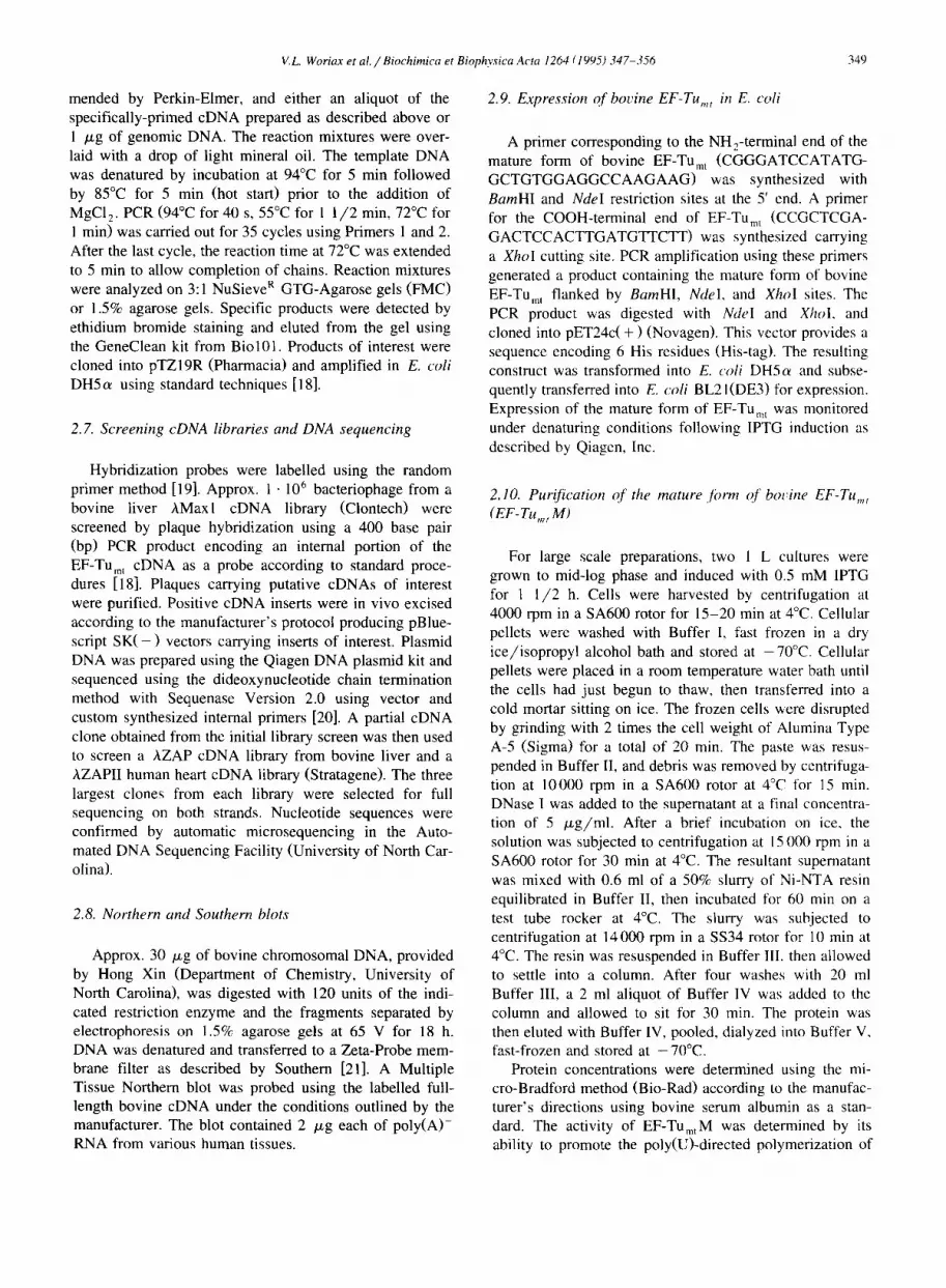
Fig. 5. Northern blot analysis of poly(A) + RNA various human tissues. The Multiple Tissue Northern Blot (Clontech) contains 2 /zg/lane of poly(A) + RNA from various human tissues. The blots were probed with the full-length bovine cDNA clone labelled by the random priming method. The relative positions of size markers are indicated on the left.
out that kirromycin and EF-Ts are mutually exclusive on E. coli EF-Tu. Since EF-Tumt and EF-TSmt remain associ- ated in the mitochondrial system, a potential kirromycin binding site on EF-TUmt may not be accessible. In human EF-Tumt residue 380 is also T, leading one to speculate that this factor may also be kirromycin resistant.
3.4. Tissue specific expression of the EF-TUm, gene
Northern hybridization analysis of poly(A) + mRNA obtained from bovine liver was carried out using the excised full length EF-TUmt cDNA as a probe. This blot
gave a single signal at approx. 1.6 kb (results not shown) which is consistent with the sizes of the cloned cDNAs. In order to assess the distribution of the EF-Tumt transcript in various human tissues, Northern blot analysis using a Multiple Tissue Northern blot was performed. A hybridiza- tion signal with a size of 1.7 to 1.8 kb is evident in all tissues (Fig. 5) suggesting that transcripts in human and bovine tissues are about the same size. This size is slightly smaller than the 2.2 kb detected by Wells et al. [15] in normal and tumor tissues. The relative abundance of the EF-TUmt mRNA is as follows: skeletal muscle > heart > pancreas > kidney > liver > brain > placenta > lung. The signal from skeletal muscle is at least one order of magni- tude greater than that from lung tissue. This result suggests that the EF-TUmt gene is widely expressed in human tissues at a level that is consistent with the rate of oxida- tive metabolism in the various tissues.
An additional signal of 5 kb in the human Multiple Tissue Northern blot has not been characterized. This transcript most likely arises from the use of an alternative polyadenylat ion site since multiple transcripts quite com- monly occur as a result of the use of such sites. This signal might also arise from the presence of an unprocessed precursor of the EF-TUmt mRNA or may represent a spurious hybridization signal. It should be noted that Wells et al. [15] did not observe the presence of the larger mRNA in their Northern analysis. This difference may be due to the fact that they prepared mRNA from cultured cells lines while the Northern probed here was made from mRNA prepared from an alcoholic human male. As indicated above, a single transcript of 1.6 kb was observed with the Northern prepared from bovine mRNA.
3.5. Expression and purification of bovine EF-Tu~, (EF- TumtM) in E. coli
Sequences corresponding to the mature form of bovine EF-Tumt (amino acid residues 44 to 452) have been cloned into the pET expression vector. The construct made has one extra residue (Met) at the N-terminus and a C-terminal
A B C
Fig. 6. Effect of IPTG concentration and time of induction on the yield of EF-Tu,,tM. Cells were grown and induced with IPTG as described in Experimental Procedures. For each lane, 0.8 ml of cells were removed at the indicated times, collected and lysed as indicated in the Qiagen manual. Aliquots (15 /LI) were analyzed by SDS-PAGE. (A) No IPTG added; (B) 0.1 mM IPTG; (C) 0.5 mM IPTG.

A.
V.L. Woriax et al. / Biochimica et Biophysica Acta 1264 (1995) 347-356
B.
355
- 86.8
- 47.8
- 33.3
EF-Tu~, -
EF-TSmt -
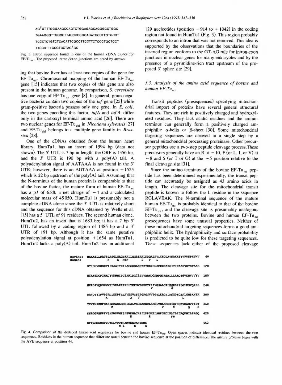
Fig. 7. SDS-PAGE and Western analysis of the purified EF-TumtM following expression. (A) The over-expressed form of EF-Tu,,tM was purified by Ni-NTA affinity chromatography and an aliquot (7/xg) was analyzed by SDS-PAGE. Protein bands were visualized by staining with Coomassie Blue. The numbers on the right indicate the molecular masses in kDa of standard markers. (B) Immunological identification of the expressed factor as EF-TUmt M. Lane 1, native EF-Tu • Tstn t complex (0. /xg); lane 2, EF-TumtM (0,4/xg); lane 3, E. coli EF-Tu (0.9 /xg).
tag consisting of LE(H) 6. This construct should permit the expression in E. coli of a protein with a mass of 45 kDa designated EF-TUmtM. The effects of the induction of EF-TUrn t M on the growth of the host E. coli strain were examined at different IPTG concentrations ranging from 0.1 to 1 raM. The growth of the cells was drastically reduced within 1 h of induction at all of the IPTG concen- trations tested indicating that this protein has some toxic effects on the cell (data not shown). The level of expres- sion of EF-TUmtM was examined following purification on a Ni-NTA affinity column from cell extracts prepared under denaturing conditions. As indicated in Fig. 6, opti- mal levels of expression were observed with a 4 -5 h induction in the presence of 0.1 mM IPTG.
Bovine EF-TumtM was purified from E. coli cells extracts under native conditions as described in Section 2. After chromatography on Ni-NTA resin, the over-ex- pressed EF-TumtM was approx. 90% pure (Fig. 7A). The yield from 1 1 of cell culture was about 2 mg. In order to confirm that the band observed following Ni-NTA affinity
0 E 20
"ID ¢D N
"=- 15 0 E
0 " 10
< z
I 5 0
EF-Tu:T~ t E F - T u . ~
I
50 100 150 200
F a c t o r added , ng
Fig. 8. Activity of the expressed EF-Tu,ntM in poly(U)-directed poly- merization. The indicated amount of EF-TUmt M was tested for the ability to synthesize polyphenylalanine in the assay routinely used to detect the native EF-Tu.Tsmt complex [12].
chromatography was indeed bovine EF-TUmt, Western blot analysis was carried out using antisera raised against the EF-Tu'Tsmt complex. As indicated in Fig. 7B (lane 2), the expressed protein reacted strongly with the antibody indicating that the protein produced was indeed EF-Tu mt M. The expressed product is slightly larger than EF-Tumt obtained from bovine liver (Fig. 7B, lane l). This differ- ence is probably the result of the presence of the His-tag at the C-terminus of the expressed protein. EF-TUmtM is also slightly larger than E. coli EF-Tu which cross-reacts weakly with the antibodies (Fig. 7B, lane 3).
The biological activity of EF-TUmt is normally deter- mined in vitro by measuring the ability of this complex to replace E. coli EF-Tu and EF-Ts in the poly(U)-directed polymerization of phenylalanine. The EF-Tu .Ts , , com- plex purified from bovine mitochondria has a specific activity of about 500000 units/rag in this assay [12]. As indicated in Fig. 8, EF-TUmtM alone is capable of catalyz- ing the poly(U)-directed polymerization of phenylalanine under comparable conditions. The specific activity of EF- TumtM is approx. 43000 units/mg. This lower level of activity is most likely due to the absence of EF-Tsmt which is required for the normal functioning of this protein. Interestingly, the addition of E. coli EF-Ts to assays of EF-TumtM does not increase the catalytic activity of the mitochondrial factor in poly(U)-directed polymerization. These observations suggest that there may be significant differences in the interactions between bacterial and mito- chondrial EF-Tu and EF-Ts. An examination of these differences should provide interesting insights into the sequence of events during chain elongation in mammalian mitochondria.
A c k n o w l e d g e m e n t s
This work has been supported in part by funds provided by the National Institutes of Health (Grant GM32734).

356 V.L. Woriax et a l . / Biochimica et Biophysica Acta 1264 (1995) 347-356
R e f e r e n c e s
[1] Ravel, J., Shorey, R., Froehner, S. and Shive, W. (1968) Arch. Biochem. Biophys. 125, 514-526.
[2] Kaziro, Y. (1978) Biochim. Biophys. Acta 505, 95-127. [3] Sprinzl. M. (1994) Trends Biochem. Sci. 19, 245-250. [4] An, G. and Friesen, J. (1980) Gene 12, 33-39. [5] Ohama, T., Yamao, F., Muto, A. and Osawa, S. (1987) J. Bacteriol.
169, 4770-4777. [6] Seidler, L., Peter, M., Meissner, F. and Sprinzl, M. (1987) Nucleic
Acids Res. 15, 9263-9277. [7] Kushiro, M., Shimizu, M. and Tomita, K. (1987) Eur. J. Biochem.
170, 93-98. [8] Nagata, S., Tsunetsugu-Yokota, Y., Naito, A. and Kaziro, Y. (1983)
Proc. Natl. Acad. Sci. USA 80, 6192-6196. [9] Baldauf, S.L. and Palmer, J. (1990) Nature 344, 262-265.
[10] Brands, J.H.G., Maassen, J.A., van Hemert, F.J., Amons, R. and MSller, W. (1986) Eur. J. Biochem. 155, 167-171.
[l 1] Dubin, D.T., and Baer, R.J. (1980) in The Organization and Expres- sion of the Mitochondrial Genome (Kroon, A.M. and Saccone, C., eds.), pp. 231-240, Elsevier/North-Holland Biomedical Press, Am- sterdam.
[12] Schwartzbach, C. and Spremulli, L.L. (1989) J. Biol. Chem. 264, 19125-19131.
[13] Schwartzbach, C. and Spremulli, L,L. (1990) J. Biol. Chem. 266, 16324-16330.
[14] Rosenthal, L.P., and Bodley, J.W, (1987) J. Biol. Chem. 262, 10955-10959.
[15] Wells, J., Henkler, F., Leversha, M. and Koshy, R. (1995) FEBS Lett. 358, 119-125,
[16] Chirgwin, J.M., Przybyla, A.E., MacDonald, R.J. and Rutter, W. (1979) Biochemistry 18, 5294-5299.
[17] Roth, M.J., Tanese, N. and Goff, S.P. (1985) J. Biol. Chem. 260, 9326-9335.
[18] Sambrook, J., Fritsch, E.F. and Maniatis, T. (1989) Molecular
Cloning, A Laboratory Manual, Cold Spring Harbor, Cold Spring Harbor.
[19] Feinberg, A.P. and Vogelstein, B. (1983) Anal. Biochem. 132, 6-13.
[20] Sanger, F.. Nicklen, S. and Coulson, A. (1977) Proc. Natl. Acad. Sci. USA, 74, 5463-5467.
[21] Southern, E.M. (1975) J. Mol. Biol. 98, 503-517. [22] Laemmli, U.K. (1970) Nature 227, 680-685. [23] Kozak, M. (1987) Nucleic Acids Res. 15, 8125-8148. [24] Geballe, A.P. and Morris, D.R. (1994) Trends Biochem. Sci. 19,
159-164. [25] Filer, D. and Furano, A.V. (1981) J. Bacteriol. 148, 1006-1011. [26] Anborgh, P., Swart, G. and Parmeggiani, A. (1991) FEBS Lett. 292,
232-2. [27] Murayama, Y., Matsubayashi, T., Sugita, M. and Sugiura, M. (1993)
Plant Mol. Biol. 22, 767-774. [28] Baldauf, S.L., Manhart, J.R. and Palmer, J.D. (1990) Proc. Natl.
Acad. Sci. USA 87, 5317-5321. [29] Padgett, R.A., Graboswki, P.J., Konarska, M.M., Seiler, S.S. and
Sharp, P.A. (1986) Annu. Rev. Biochem. 55, 1119-1150. [30] von Heijne, G. (1986) EMBO J. 5, 1335-1342. [31] Hendrick, J.P., Hodges, P.E. and Rosenberg, L.E. (1989) Proc. Natl.
Acad. Sci. USA 86, 4056-4060. [32] la Cour, T.F.M., Nyborg, J., Thirup, S., and Clark, B.F. (1985)
EMBO J. 4, 2385-2388, [33] Jurnak, F. (1985) Science 230, 32-36. [34] Kjeldgaard, M. and Nyborg, J. (1992) J. Mol. Biol. 223, 721-742. [35] Berchtold, H., Reshetnikova, L., Reiser, C.O.A., Schirmer, N,K.,
Sprinzl, M. and Hilgenfeld, R. (1993) Nature 5, 126-132. [36] Wiborg, O., Andersen, C., Knudsen, C., Kristensen, T. and Clark. B.
(1994) Biotechnol. Appl. Biochem. 19, 3-15. [37] Saraste, M., Sibbald, P.R. and Wittinghofer, A. (1990) Trends
Biochem. Sci. 15, 430-434. [38] Hwang, Y-W., Carter, M. and Miller, D. (1992) J. Biol. Chem. 267,
22198-22205.