Misguided Transcriptional Elongation Causes Mixed Lineage Leukemia
14
Misguided Transcriptional Elongation Causes Mixed Lineage Leukemia Dorothee Mueller, Marı ´a-Paz Garcı´a-Cue ´ llar, Christian Bach, Sebastian Buhl, Emanuel Maethner, Robert K. Slany* Department of Genetics, University Erlangen, Erlangen, Germany Abstract Fusion proteins composed of the histone methyltransferase mixed-lineage leukemia (MLL) and a variety of unrelated fusion partners are highly leukemogenic. Despite their prevalence, particularly in pediatric acute leukemia, many molecular details of their transforming mechanism are unknown. Here, we provide mechanistic insight into the function of MLL fusions, demonstrating that they capture a transcriptional elongation complex that has been previously found associated with the eleven-nineteen leukemia protein (ENL). We show that this complex consists of a tight core stabilized by recursive protein– protein interactions. This central part integrates histone H3 lysine 79 methylation, RNA Polymerase II (RNA Pol II) phosphorylation, and MLL fusion partners to stimulate transcriptional elongation as evidenced by RNA tethering assays. Coimmunoprecipitations indicated that MLL fusions are incorporated into this complex, causing a constitutive recruitment of elongation activity to MLL target loci. Chromatin immunoprecipitations (ChIP) of the homeobox gene A cluster confirmed a close relationship between binding of MLL fusions and transcript levels. A time-resolved ChIP utilizing a conditional MLL fusion singled out H3K79 methylation as the primary parameter correlated with target expression. The presence of MLL fusion proteins also kept RNA Pol II in an actively elongating state and prevented accumulation of inhibitory histone methylation on target chromatin. Hox loci remained open and productive in the presence of MLL fusion activity even under conditions of forced differentiation. Finally, MLL-transformed cells were particularly sensitive to pharmacological inhibition of RNA Pol II phosphorylation, pointing to a potential treatment for MLL. In summary, we show aberrant transcriptional elongation as a novel mechanism for oncogenic transformation. Citation: Mueller D, Garcı ´a-Cue ´ llar M-P, Bach C, Buhl S, Maethner E, et al. (2009) Misguided Transcriptional Elongation Causes Mixed Lineage Leukemia. PLoS Biol 7(11): e1000249. doi:10.1371/journal.pbio.1000249 Academic Editor: Nancy Zeleznik-Le, Loyola University, United States of America Received August 24, 2009; Accepted October 15, 2009; Published November 24, 2009 Copyright: ß 2009 Mueller et al. This is an open-access article distributed under the terms of the Creative Commons Attribution License, which permits unrestricted use, distribution, and reproduction in any medium, provided the original author and source are credited. Funding: RKS was supported by Deutschen Forschungsgemeinschaft (DFG) (www.dfg.de)grant SL27/6-2 and by SFB473/D2. Equipment funding came from Jose- Carreras-Stiftung and Curt-Bohnewald-Fond. The funders had no role in study design, data collection and analysis, decision to publish, or preparation of the manuscript Competing Interests: The authors have declared that no competing interests exist. Abbreviations: ChIP, chromatin immunoprecipitation; MLL, mixed-lineage leukemia; qRT-PCR, quantitative reverse-transcriptase PCR * E-mail: [email protected] Introduction Mixed-lineage leukemia (MLL) is a particularly aggressive subtype of acute leukemia with a very dismal prognosis. This disease is caused by chromosomal aberrations, mostly translocations, affecting Chromosome 11 at band q23. This chromosomal locus contains the gene for the histone H3 lysine 4–specific methyltransferase MLL [1–4]. As a corollary of these genomic rearrangements the 59 portion of MLL is fused in frame to a variety of different and mostly unrelated partner genes. The translation of the chimeric RNAs transcribed from the altered locus results in the production of fusion proteins. In these fusions, the original MLL methyltransferase activity is replaced by biological properties provided by the fusion partner. This creates novel oncoproteins that are potently transforming hematopoietic cells (for reviews, see [5–7]). MLL fusions are aberrant transcription factors that induce ectopic expression of their respective target genes, and as a consequence, they block hematopoietic differentiation. Critical targets for MLL-induced transformation are the clustered HOXA homeobox genes and the gene for the HOX-dimerization partner MEIS1 [8]. Accordingly, a relative overexpression of HOXA and MEIS1 transcripts is the characteristic hallmark of the MLL- specific gene expression profile [9,10]. Despite this predominance of HOX expression, however, it has been shown by genome-wide chromatin precipitations that MLL fusion proteins occupy several thousand binding sites [11–13]. As it has been noted some time ago, transcriptional activation by MLL fusions is accompanied by a conspicuous and dramatic increase of histone H3 lysine 79 dimethylation across the HOXA locus [14], and this phenomenon has been confirmed also for many of the other MLL fusion target loci [12]. The only known histone methyltransferase that is capable of introducing the H3K79 mark is DOT1L, a protein conserved from yeast to man [15,16]. Indeed, it could be shown for the MLL fusion partner AF10 that a direct interaction with DOT1L was instrumental for the oncogenic function of the respective fusion protein [17]. First hints for a shared function of several MLL fusion partners came from studies performed by Bitoun et al. [18]. These authors conducted overexpression studies and published data to support a model of multiple MLL fusion partners being involved in a transcriptional elongation complex that includes the MLL partner proteins AF4, AF9, ENL, and AF10, as well as DOT1L, and positive transcription elongation factor b (pTEFb). A direct interaction between proteins of the AF4 and AF9/ENL families PLoS Biology | www.plosbiology.org 1 November 2009 | Volume 7 | Issue 11 | e1000249
-
Upload
independent -
Category
Documents
-
view
4 -
download
0
Transcript of Misguided Transcriptional Elongation Causes Mixed Lineage Leukemia
Misguided Transcriptional Elongation Causes MixedLineage LeukemiaDorothee Mueller, Marıa-Paz Garcıa-Cuellar, Christian Bach, Sebastian Buhl, Emanuel Maethner,
Robert K. Slany*
Department of Genetics, University Erlangen, Erlangen, Germany
Abstract
Fusion proteins composed of the histone methyltransferase mixed-lineage leukemia (MLL) and a variety of unrelated fusionpartners are highly leukemogenic. Despite their prevalence, particularly in pediatric acute leukemia, many molecular detailsof their transforming mechanism are unknown. Here, we provide mechanistic insight into the function of MLL fusions,demonstrating that they capture a transcriptional elongation complex that has been previously found associated with theeleven-nineteen leukemia protein (ENL). We show that this complex consists of a tight core stabilized by recursive protein–protein interactions. This central part integrates histone H3 lysine 79 methylation, RNA Polymerase II (RNA Pol II)phosphorylation, and MLL fusion partners to stimulate transcriptional elongation as evidenced by RNA tethering assays.Coimmunoprecipitations indicated that MLL fusions are incorporated into this complex, causing a constitutive recruitmentof elongation activity to MLL target loci. Chromatin immunoprecipitations (ChIP) of the homeobox gene A cluster confirmeda close relationship between binding of MLL fusions and transcript levels. A time-resolved ChIP utilizing a conditional MLLfusion singled out H3K79 methylation as the primary parameter correlated with target expression. The presence of MLLfusion proteins also kept RNA Pol II in an actively elongating state and prevented accumulation of inhibitory histonemethylation on target chromatin. Hox loci remained open and productive in the presence of MLL fusion activity even underconditions of forced differentiation. Finally, MLL-transformed cells were particularly sensitive to pharmacological inhibitionof RNA Pol II phosphorylation, pointing to a potential treatment for MLL. In summary, we show aberrant transcriptionalelongation as a novel mechanism for oncogenic transformation.
Citation: Mueller D, Garcıa-Cuellar M-P, Bach C, Buhl S, Maethner E, et al. (2009) Misguided Transcriptional Elongation Causes Mixed Lineage Leukemia. PLoSBiol 7(11): e1000249. doi:10.1371/journal.pbio.1000249
Academic Editor: Nancy Zeleznik-Le, Loyola University, United States of America
Received August 24, 2009; Accepted October 15, 2009; Published November 24, 2009
Copyright: � 2009 Mueller et al. This is an open-access article distributed under the terms of the Creative Commons Attribution License, which permitsunrestricted use, distribution, and reproduction in any medium, provided the original author and source are credited.
Funding: RKS was supported by Deutschen Forschungsgemeinschaft (DFG) (www.dfg.de)grant SL27/6-2 and by SFB473/D2. Equipment funding came from Jose-Carreras-Stiftung and Curt-Bohnewald-Fond. The funders had no role in study design, data collection and analysis, decision to publish, or preparation of themanuscript
Competing Interests: The authors have declared that no competing interests exist.
Abbreviations: ChIP, chromatin immunoprecipitation; MLL, mixed-lineage leukemia; qRT-PCR, quantitative reverse-transcriptase PCR
* E-mail: [email protected]
Introduction
Mixed-lineage leukemia (MLL) is a particularly aggressive subtype
of acute leukemia with a very dismal prognosis. This disease is caused
by chromosomal aberrations, mostly translocations, affecting
Chromosome 11 at band q23. This chromosomal locus contains
the gene for the histone H3 lysine 4–specific methyltransferase MLL
[1–4]. As a corollary of these genomic rearrangements the 59 portion
of MLL is fused in frame to a variety of different and mostly unrelated
partner genes. The translation of the chimeric RNAs transcribed
from the altered locus results in the production of fusion proteins. In
these fusions, the original MLL methyltransferase activity is replaced
by biological properties provided by the fusion partner. This creates
novel oncoproteins that are potently transforming hematopoietic
cells (for reviews, see [5–7]).
MLL fusions are aberrant transcription factors that induce
ectopic expression of their respective target genes, and as a
consequence, they block hematopoietic differentiation. Critical
targets for MLL-induced transformation are the clustered HOXA
homeobox genes and the gene for the HOX-dimerization partner
MEIS1 [8]. Accordingly, a relative overexpression of HOXA and
MEIS1 transcripts is the characteristic hallmark of the MLL-
specific gene expression profile [9,10]. Despite this predominance
of HOX expression, however, it has been shown by genome-wide
chromatin precipitations that MLL fusion proteins occupy several
thousand binding sites [11–13]. As it has been noted some time
ago, transcriptional activation by MLL fusions is accompanied by
a conspicuous and dramatic increase of histone H3 lysine 79
dimethylation across the HOXA locus [14], and this phenomenon
has been confirmed also for many of the other MLL fusion target
loci [12]. The only known histone methyltransferase that is
capable of introducing the H3K79 mark is DOT1L, a protein
conserved from yeast to man [15,16]. Indeed, it could be shown
for the MLL fusion partner AF10 that a direct interaction with
DOT1L was instrumental for the oncogenic function of the
respective fusion protein [17].
First hints for a shared function of several MLL fusion partners
came from studies performed by Bitoun et al. [18]. These authors
conducted overexpression studies and published data to support a
model of multiple MLL fusion partners being involved in a
transcriptional elongation complex that includes the MLL partner
proteins AF4, AF9, ENL, and AF10, as well as DOT1L, and
positive transcription elongation factor b (pTEFb). A direct
interaction between proteins of the AF4 and AF9/ENL families
PLoS Biology | www.plosbiology.org 1 November 2009 | Volume 7 | Issue 11 | e1000249
had been noted before by Erfurth et al. [19], as well as our own
group [20]. A somewhat contradictory interaction of AF9 and
DOT1L has also been described to be necessary for aldosterone-
induced gene silencing [21]. To elucidate the function of normal
ENL, we recently purified wild-type ENL from mammalian nuclei
[22]. It could be shown that endogenous ENL was also present in a
large macromolecular protein complex similar to the one
described by Bitoun et al [18]. Although the complex was initially
termed ENL associated proteins (EAP), we now propose to
redefine EAP as ‘‘elongation assisting proteins’’ to better reflect the
function of this protein complex. In addition to DOT1L, EAP
contained pTEFb, a cyclin-dependent kinase 9/cyclin T dimer
that phosphorylates the C-terminal repeat domain (CTD) of RNA
Polymerase II (RNA Pol II) [23]. CTD phosphorylation is
necessary to ensure productive transcriptional elongation. Next
to AF4, the AF4 homologs AF5 and LAF4 were also present in
EAP, confirming the results of Estable et al. [24], who had
copurified AF5 with pTEFb. AF4 itself is the most frequently
encountered MLL fusion partner, and in a recent survey,
approximately 50% of all MLL cases in infants and adults carried
a MLL-AF4 translocation [25]. EAP was ubiquitously expressed,
and interference with EAP assembly affected transcriptional
elongation of many genes. However, it was not clear whether
EAP activity was important for the respective MLL fusion
proteins. In the fusion, a bulky 180-kDa MLL moiety is added
to an ENL protein of approximately 70 kDa. This type of
modification might substantially alter or even destroy the EAP
complex.
Here, we present a comprehensive picture of MLL fusion
biology, demonstrating that EAP has a very stable core that is
capable of also accommodating MLL fusion proteins. The
constitutive recruitment of EAP to MLL target loci is responsible
for persistent target transcription through stimulation of transcrip-
tional elongation. This mechanism resists differentiation stimuli
and therefore causes a maturation arrest. Finally, MLL fusion
transformed cells were sensitive to EAP inhibition, pointing to a
potential lead for pharmaceutical intervention.
Results
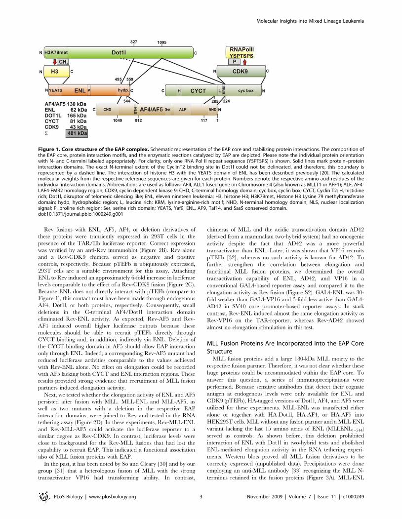
MLL Fusion Partners Are Constituents of an EAP CoreComplex That Is Stabilized by Recursive Protein–ProteinInteractions
In previous studies, the total molecular weight of all proteins
coprecipitating with ENL amounted to more than 1 MDa,
whereas the bulk of ENL eluted on sizing columns with an
apparent molecular weight of approximately 400 kDa to 500 kDa
[22]. To explain this discrepancy and to further elucidate the
molecular architecture of the EAP assembly, we performed two-
hybrid assays to test for mutual protein–protein interactions. A
large deletion series of existing [20,22,26,27] and newly construct-
ed two-hybrid bait clones for ENL, AF4, CYCT2A—the cyclin
component of pTEFb—and Dot1l was probed for interaction with
full-length versions of the same proteins. As reported previously
[22], only the mouse homolog of DOT1L was available in cDNA
repositories, and therefore, mouse Dot1l was used throughout this
study. A total of 78 potential interaction pairs were interrogated
(Figure S1). These experiments showed that EAP contained a tight
core stabilized by a recursive set of direct protein–protein
interactions (Figure 1). Each protein tested was able to interact
with two other proteins, thus linking ENL, AF4, Dot1l, and
CYCT2/CDK9 (pTEFb) in a tight ‘‘circular’’ network. In this
way, histone H3 methylation catalyzed by Dot1l can be
coordinated with RNA Pol II phosphorylation introduced by
pTEFb. The total calculated molecular weight of the EAP core
components was 481 kDa, and this number was very close to the
previously determined [22] value for the EAP complex eluting
from gel filtration. Of note, ENL, Dot1l, and CYCT2 utilized a
single domain to interact with both of their binding partners,
whereas the two binding interfaces were separated in AF4. The
AF4 N-terminal homology domain provided contact to CYCT2,
whereas sequences further C-terminal formed the interface with
ENL. It is important to note that the respective interaction
domains are highly conserved between the homologous MLL
fusion partners ENL and AF9 and between AF4 and the related
AF5, LAF4, and FMR2 proteins as well. In two-hybrid assays,
AF4 sequences could be replaced with the corresponding AF5
regions, yielding identical results (unpublished data). In the cellular
environment, EAP, therefore, is likely to exist in different
configurations, explaining the large number of proteins that have
been identified in ENL precipitates.
MLL Fusion Partners Promote Transcriptional Elongationthrough Interaction with EAP Core Components
Next, we wanted to know whether recruitment of MLL fusion
partners to specific genes would promote transcriptional elonga-
tion. For this purpose, we used an RNA tethering assay to detect
elongation activity. This test places a luciferase reporter gene
downstream of a modified HIV-1 LTR promoter that grafts the
stem loop IIb from the HIV-1 Rev response element (RRE) onto
the TAR (transactivation response RNA) double-stranded RNA
(Figure 2A). RNA Pol II stalls after the TAR element. LTR
regulation is achieved by binding of the transactivator TAT that
regulates promoter output via recruitment of pTEFb to stimulate
elongation [28]. The hybrid IIb/TAR loop allows tethering of any
protein of interest to the LTR RNA by fusing it to the RNA
binding protein Rev. Luciferase levels, therefore, will reflect the
ability to recruit pTEFb elongation activity [29].
Author Summary
The expression level of a gene needs to be preciselyadjusted to ensure proper function. Adjustments can beimposed at different stages during the overall process ofgene expression, including transcription initiation, tran-script elongation, and transcript processing. If control ofone of these mechanisms fails, aberrant gene expressioncan occur, which may have severe consequences such ascellular transformation and the development of cancer.Here, we show that a class of aberrant fusion proteins thatare causal in mixed-lineage leukemia (MLL) hijacks atranscriptional elongation complex. We analyze thearchitecture of this transcriptional elongation complexand demonstrate that the complex is targeted by MLLfusion proteins to genes that should normally be silencedto allow maturation of hematopoietic cells. We show thatthis mistargeting causes constitutive expression of therespective genes, which likely leads to inhibition of bloodcell differentiation at a precursor cell stage in which thecells are highly proliferative. Such abnormal precursor cellshave been shown previously to be resistant to normaldifferentiation signals and to form the leukemia-initiatingpopulation. We further show here that cells carrying MLLfusion proteins are more sensitive to chemical inhibition oftranscriptional elongation than leukemic cells of differentetiology. Our results propose transcriptional elongation asa new oncogenic mechanism and point to a potentialspecific therapy for this hard-to-cure leukemia.
Molecular Insights into Mixed Lineage Leukemia
PLoS Biology | www.plosbiology.org 2 November 2009 | Volume 7 | Issue 11 | e1000249
Rev fusions with ENL, AF5, AF4, or deletion derivatives of
these proteins were transiently expressed in 293T cells in the
presence of the TAR/IIb luciferase reporter. Correct expression
was verified by an anti-Rev immunoblot (Figure 2B). Rev alone
and a Rev-CDK9 chimera served as negative and positive
controls, respectively. Because pTEFb is ubiquitously expressed,
293T cells are a suitable environment for this assay. Attaching
ENL to Rev induced an approximately 6-fold increase in luciferase
levels comparable to the effect of a Rev-CDK9 fusion (Figure 2C).
Because ENL does not directly interact with pTEFb (compare to
Figure 1), this contact must have been made through endogenous
AF4, Dot1l, or both proteins, respectively. Consequently, small
deletions in the C-terminal AF4/Dot1l interaction domain
eliminated Rev-ENL activity. As expected, Rev-AF5 and Rev-
AF4 induced overall higher luciferase outputs because these
molecules should be able to recruit pTEFb directly through
CYCT binding and, in addition, indirectly via ENL. Deletion of
the CYCT binding domain in AF5 should allow EAP interaction
only through ENL. Indeed, a corresponding Rev-AF5 mutant had
reduced luciferase activities comparable to the values achieved
with Rev-ENL alone. No effect on elongation could be recorded
with AF5 lacking both CYCT and ENL interaction regions. These
results provided strong evidence that recruitment of MLL fusion
partners induced elongation activity.
Next, we tested whether the elongation activity of ENL and AF5
persisted after fusion with MLL. MLL-ENL and MLL-AF5, as
well as two mutants with a deletion in the respective EAP
interaction domains, were joined to Rev and tested in the RNA
tethering assay (Figure 2D). In these experiments, Rev-MLL-ENL
and Rev-MLL-AF5 could activate the luciferase reporter to a
similar degree as Rev-CDK9. In contrast, luciferase levels were
close to background for the Rev-MLL fusions that had lost the
capability to recruit EAP. This indicated a functional association
also of MLL fusion proteins with EAP.
In the past, it has been noted by So and Cleary [30] and by our
group [31] that a heterologous fusion of MLL with the strong
transactivator VP16 had transforming ability. In contrast,
chimeras of MLL and the acidic transactivation domain AD42
(derived from a mammalian two-hybrid system) had no oncogenic
activity despite the fact that AD42 was a more powerful
transactivator than ENL. Later, it was shown that VP16 recruits
pTEFb [32], whereas no such activity is known for AD42. To
further strengthen the correlation between elongation and
functional MLL fusion proteins, we determined the overall
transactivation capability of ENL, AD42, and VP16 in a
conventional GAL4-based reporter assay and compared it to the
elongation activity as Rev fusion (Figure S2). GAL4-ENL was 30-
fold weaker than GAL4-VP16 and 5-fold less active than GAL4-
AD42 in SV40 core promoter-based reporter assays. In stark
contrast, Rev-ENL induced almost the same elongation activity as
Rev-VP16 on the TAR-reporter, whereas Rev-AD42 showed
almost no elongation stimulation in this test.
MLL Fusion Proteins Are Incorporated into the EAP CoreStructure
MLL fusion proteins add a large 180-kDa MLL moiety to the
respective fusion partner. Therefore, it was not clear whether these
huge proteins could be accommodated within the EAP core. To
answer this question, a series of immunoprecipitations were
performed. Because sensitive antibodies that detect their cognate
antigen at endogenous levels were only available for ENL and
CDK9 (pTEFb), HA-tagged versions of Dot1l, AF4, and AF5 were
utilized for these experiments. MLL-ENL was transfected either
alone or together with HA-Dot1l, HA-AF4, or HA-AF5 into
HEK293T cells. MLL without any fusion partner and a MLL-ENL
variant lacking the last 15 amino acids of ENL (MLLENL1–544)
served as controls. As shown before, this deletion prohibited
interaction of ENL with Dot1l in two-hybrid tests and abolished
ENL-mediated elongation activity in the RNA tethering experi-
ments. Western blots proved all MLL fusion derivatives to be
correctly expressed (unpublished data). Precipitations were done
employing an anti-MLL antibody [33] recognizing the MLL N-
terminus retained in the fusion proteins (Figure 3A). MLL-ENL
Figure 1. Core structure of the EAP complex. Schematic representation of the EAP core and stabilizing protein interactions. The composition ofthe EAP core, protein interaction motifs, and the enzymatic reactions catalyzed by EAP are depicted. Please note the individual protein orientationwith N- and C-termini labeled appropriately. For clarity, only one RNA Pol II repeat sequence (YSPTSPS) is shown. Solid lines mark protein–proteininteraction domains. The exact N-terminal extent of the CyclinT2 binding site in Dot1l could not be delineated, and therefore, this boundary isrepresented by a dashed line. The interaction of histone H3 with the YEATS domain of ENL has been described previously [20]. The calculatedmolecular weights from the respective reference sequences are given for each protein. Numbers denote the respective amino acid residues of theindividual interaction domains. Abbreviations are used as follows: AF4, ALL1 fused gene on Chromosome 4 (also known as MLLT1 or AFF1); ALF, AF4-LAF4-FMR2 homology region; CDK9, cyclin dependent kinase 9; CHD, C-terminal homology domain; cyc box, cyclin box; CYCT, Cyclin T2; H, histidinerich; Dot1l, disruptor of telomeric silencing like; ENL, eleven nineteen leukemia; H3, histone H3; H3K79met, Histone H3 Lysine 79 methyltransferasedomain; hydp, hydrophobic region; L, leucine rich; KRM, lysine-arginine-rich motif; NHD, N-terminal homology domain; NLS, nuclear localizationsignal; P, proline rich region; Ser, serine rich domain; YEATS, Yaf9, ENL, AF9, Taf14, and Sas5 conserved domain.doi:10.1371/journal.pbio.1000249.g001
Molecular Insights into Mixed Lineage Leukemia
PLoS Biology | www.plosbiology.org 3 November 2009 | Volume 7 | Issue 11 | e1000249

Figure 2. Elongation stimulation by EAP. (A) Mechanism of RNA tethering assay. A luciferase reporter driven by a modified HIV LTR promoter isused to detect elongation stimulation. The Rev RNA recognition site (IIb stem-loop) is grafted onto the TAR Tat-recognition loop. Proteins tethered toRNA via Rev will release the stalled RNA Pol II and create luciferase activity only if they can recruit active pTEFb (dimer of cyclin T1 or T2 and CDK9). (B)Expression of Rev fusion proteins. ENL, AF4, AF5, and derivatives thereof were fused to Rev and expressed in 293T cells. Cell lysates were probed withan antibody specific for Rev. For detection of Rev-MLL-fusion proteins, an anti-MLL antibody was employed. (C) Results of RNA tethering assays withMLL fusion partners. Rev or Rev fusions as indicated were expressed together with the TAR-IIb reporter and luciferase activity was determined. Boxesinside the graphical representation correspond to the protein–protein interaction domains from Figure 1. Values represent average and standarddeviation of triplicate experiments and are expressed relative to background with Rev alone. (D) Elongation activity of MLL fusion proteins. Chimerasof Rev with MLL fusion proteins as depicted were tested in RNA tethering assays as described for (C).doi:10.1371/journal.pbio.1000249.g002
Molecular Insights into Mixed Lineage Leukemia
PLoS Biology | www.plosbiology.org 4 November 2009 | Volume 7 | Issue 11 | e1000249
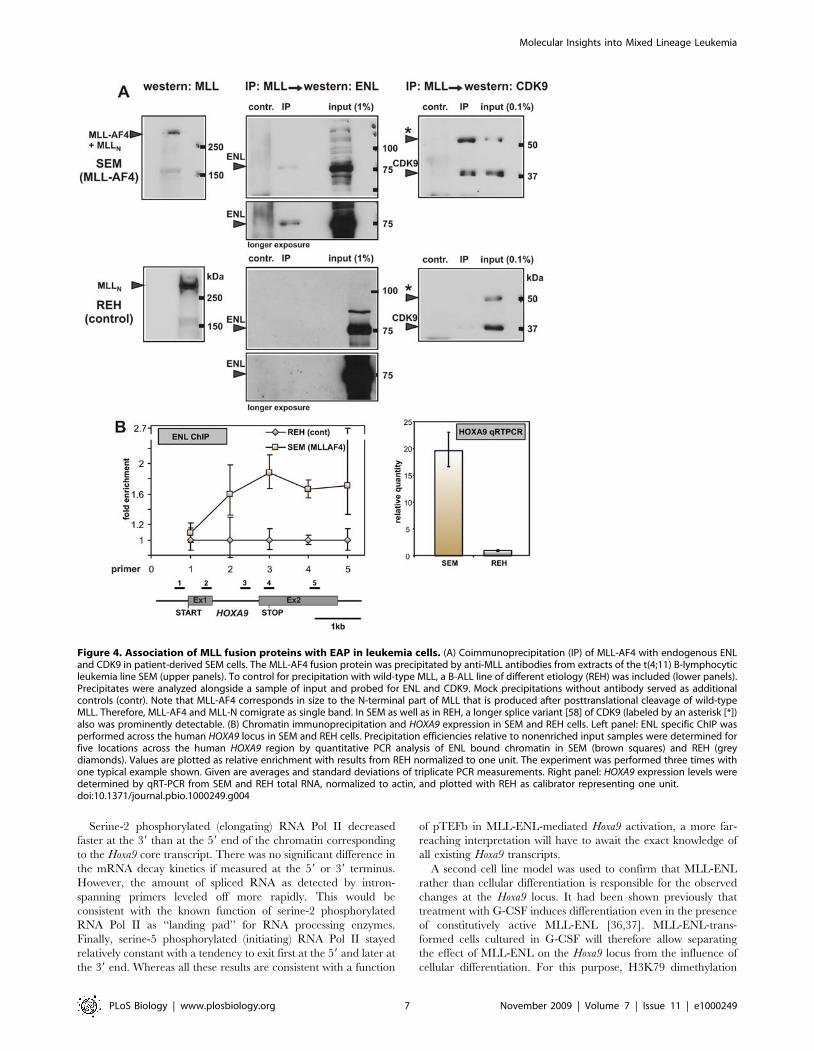
Figure 3. Incorporation of MLL fusion proteins into EAP. (A) Coimmunoprecipitation of MLL-ENL with EAP components. MLL-ENL (MLL-ENL), aMLL-ENL mutant lacking the last 15 amino acids of ENL (MLL-ENL1–544), or MLL without fusion partner (MLL) were expressed either alone or togetherwith HA-tagged proteins AF4, AF5, or Dot1l. A schematic overview of the EAP core structure in the presence of MLL-ENL including the expectedprotein–protein interactions (double-headed arrows) is depicted in the upper-left panel. The presence of HA-tagged proteins and endogenous CDK9in anti-MLL precipitates was probed alongside with a sample of the input (inp, 5%) by immunoblots as indicated. As a control, the successfulprecipitation of the MLL-ENL derivatives was confirmed by an anti-MLL blot. (B) Interaction of MLL-AF4 and MLL-AF5 with endogenous (endo) ENLand CDK9. MLL-AF4 (MLLAF4758–1210) and MLL-AF5 (MLLAF5731–1163) proteins analogous to patient-derived fusions were expressed in 293T cells. Notethat the N-terminal CYCT interaction domain is missing in leukemogenic MLL-AF4/5 fusions as depicted in the upper-left and -right panels. MLL-AF4/5derivatives deleting the ENL binding domain (MLLAF41023–1210, MLLAF5991–1163) served as controls. The coprecipitation of endogenous ENL and CDK9was detected by immunoblot as indicated next to a sample of 5% input (inp). The presence of MLL-AF4/AF5 in the lysates was controlled as above.doi:10.1371/journal.pbio.1000249.g003
Molecular Insights into Mixed Lineage Leukemia
PLoS Biology | www.plosbiology.org 5 November 2009 | Volume 7 | Issue 11 | e1000249
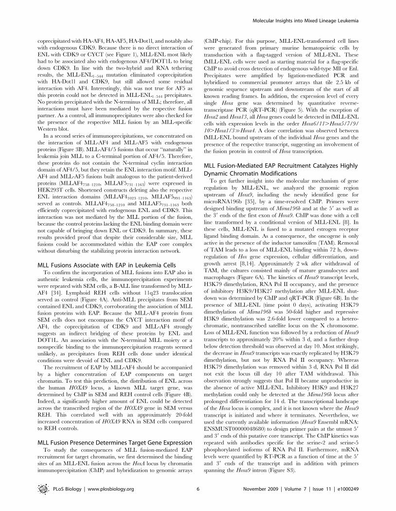
coprecipitated with HA-AF4, HA-AF5, HA-Dot1l, and notably also
with endogenous CDK9. Because there is no direct interaction of
ENL with CDK9 or CYCT (see Figure 1), MLL-ENL most likely
had to be associated also with endogenous AF4/DOT1L to bring
down CDK9. In line with the two-hybrid and RNA tethering
results, the MLL-ENL1–544 mutation eliminated coprecipitation
with HA-Dot1l and CDK9, but still allowed some residual
interaction with AF4. Interestingly, this was not true for AF5 as
this protein could not be detected in MLL-ENL1–544 precipitates.
No protein precipitated with the N-terminus of MLL; therefore, all
interactions must have been mediated by the respective fusion
partner. As a control, all immunoprecipitates were also checked for
the presence of the respective MLL fusion by an MLL-specific
Western blot.
In a second series of immunoprecipitations, we concentrated on
the interaction of MLL-AF4 and MLL-AF5 with endogenous
proteins (Figure 3B). MLL-AF4/5 fusions that occur ‘‘naturally’’ in
leukemia join MLL to a C-terminal portion of AF4/5. Therefore,
these proteins do not contain the N-terminal cyclin interaction
domain of AF4/5, but they retain the ENL interaction motif. MLL-
AF4 and MLL-AF5 fusions built analogous to the patient-derived
proteins (MLLAF4758–1210, MLLAF5731–1163) were expressed in
HEK293T cells. Shortened constructs deleting also the respective
ENL interaction domains (MLLAF41023–1210, MLLAF5991–1163)
served as controls. MLLAF4758–1210 and MLLAF5731–1163 both
efficiently coprecipitated with endogenous ENL and CDK9. This
interaction was not mediated by the MLL portion of the fusion,
because the control proteins lacking the ENL binding domain were
not capable of bringing down ENL or CDK9. In summary, these
results provided proof that despite their considerable size, MLL
fusions could be accommodated within the EAP core complex
without disturbing the stabilizing protein interaction network.
MLL Fusions Associate with EAP in Leukemia CellsTo confirm the incorporation of MLL fusions into EAP also in
authentic leukemia cells, the immunoprecipitation experiments
were repeated with SEM cells, a B-ALL line transformed by MLL-
AF4 [34]. Lymphoid REH cells without 11q23 translocation
served as control (Figure 4A). Anti-MLL precipitates from SEM
contained ENL and CDK9, corroborating the association of MLL
fusion proteins with EAP. Because the MLL-AF4 protein from
SEM cells does not encompass the CYCT interaction motif of
AF4, the coprecipitation of CDK9 and MLL-AF4 strongly
suggests an indirect bridging of these proteins by ENL and
DOT1L. An association with the N-terminal MLL moiety or a
nonspecific binding to the immunoprecipitation reagents seemed
unlikely, as precipitates from REH cells done under identical
conditions were devoid of ENL and CDK9.
The recruitment of EAP by MLL-AF4 should be accompanied
by a higher concentration of EAP components on target
chromatin. To test this prediction, the distribution of ENL across
the human HOXA9 locus, a known MLL target gene, was
determined by ChIP in SEM and REH control cells (Figure 4B).
Indeed, a significantly higher amount of ENL could be detected
across the transcribed region of the HOXA9 gene in SEM versus
REH. This correlated well with an approximately 20-fold
increased concentration of HOXA9 RNA in SEM cells compared
to REH controls.
MLL Fusion Presence Determines Target Gene ExpressionTo study the consequences of MLL fusion-mediated EAP
recruitment for target chromatin, we first determined the binding
sites of an MLL-ENL fusion across the HoxA locus by chromatin
immunoprecipitation (ChIP) and hybridization to genomic arrays
(ChIP-chip). For this purpose, MLL-ENL-transformed cell lines
were generated from primary murine hematopoietic cells by
transduction with a flag-tagged version of MLL-ENL. These
fMLL-ENL cells were used as starting material for a flag-specific
ChIP to avoid cross detection of endogenous wild-type Mll or Enl.
Precipitates were amplified by ligation-mediated PCR and
hybridized to commercial promoter arrays that tile 2.5 kb of
genomic sequence upstream and downstream of the start of all
known reading frames. In addition, the expression level of every
single Hoxa gene was determined by quantitative reverse-
transcriptase PCR (qRT-PCR) (Figure 5). With the exception of
Hoxa2 and Hoxa13, all Hoxa genes could be detected in fMLL-ENL
cells with expression levels in the order Hoxa6/11.Hoxa5/7/9/
10.Hoxa1/3&Hoxa4. A close correlation was observed between
fMLL-ENL bound upstream of the individual Hoxa genes and the
presence of the respective transcript, suggesting an involvement of
the fusion protein in control of Hoxa transcription.
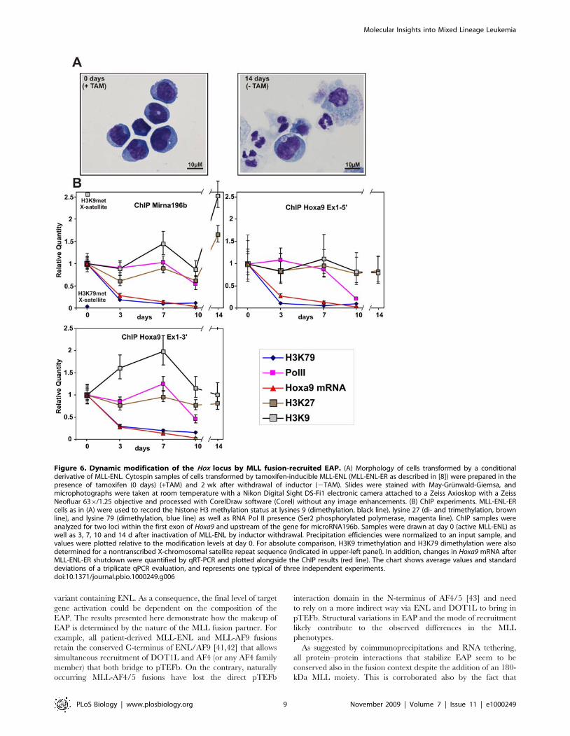
MLL Fusion-Mediated EAP Recruitment Catalyzes HighlyDynamic Chromatin Modifications
To get further insight into the molecular mechanism of gene
regulation by MLL-ENL, we analyzed the genomic region
upstream of Hoxa9, including the newly identified gene for
microRNA196b [35], by a time-resolved ChIP. Primers were
designed binding upstream of Mirna196b and at the 59 as well as
the 39 ends of the first exon of Hoxa9. ChIP was done with a cell
line transformed by a conditional version of MLL-ENL [8]. In
these cells, MLL-ENL is fused to a mutated estrogen receptor
ligand binding domain. As a consequence, the oncogene is only
active in the presence of the inductor tamoxifen (TAM). Removal
of TAM leads to a loss of MLL-ENL binding within 72 h, down-
regulation of Hox gene expression, cellular differentiation, and
growth arrest [8,14]. Approximately 2 wk after withdrawal of
TAM, the cultures consisted mainly of mature granulocytes and
macrophages (Figure 6A). The kinetics of Hoxa9 transcript levels,
H3K79 dimethylation, RNA Pol II occupancy, and the presence
of inhibitory H3K9/H3K27 methylation after MLL-ENL shut-
down was determined by ChIP and qRT-PCR (Figure 6B). In the
presence of MLL-ENL (time point 0 days), activating H3K79
dimethylation of Mirna196b was 50-fold higher and repressive
H3K9 dimethylation was 2.6-fold lower compared to a hetero-
chromatic, nontranscribed satellite locus on the X chromosome.
Loss of MLL-ENL function was followed by a reduction of Hoxa9
transcripts to approximately 20% within 3 d, and a further drop
below detection threshold was observed at day 10. Most strikingly,
the decrease in Hoxa9 transcripts was exactly replicated by H3K79
dimethylation, but not by RNA Pol II occupancy. Whereas
H3K79 dimethylation was removed within 3 d, RNA Pol II did
not exit the locus till day 10 after TAM withdrawal. This
observation strongly suggests that Pol II became unproductive in
the absence of active MLL-ENL. Inhibitory H3K9 and H3K27
methylation could only be detected at the Mirna196b locus after
prolonged differentiation for 14 d. The transcriptional landscape
of the Hoxa locus is complex, and it is not known where the Hoxa9
transcript is initiated and where it terminates. Nevertheless, we
used the currently available information (Hoxa9 Ensembl mRNA:
ENSMUST00000048680) to design primer pairs at the utmost 59
and 39 ends of this putative core transcript. The ChIP kinetics was
repeated with antibodies specific for the serine-2 and serine-5
phosphorylated isoforms of RNA Pol II. Furthermore, mRNA
levels were quantified by RT-PCR as a function of time at the 59
and 39 ends of the transcript and in addition with primers
spanning the Hoxa9 intron (Figure S3).
Molecular Insights into Mixed Lineage Leukemia
PLoS Biology | www.plosbiology.org 6 November 2009 | Volume 7 | Issue 11 | e1000249
Serine-2 phosphorylated (elongating) RNA Pol II decreased
faster at the 39 than at the 59 end of the chromatin corresponding
to the Hoxa9 core transcript. There was no significant difference in
the mRNA decay kinetics if measured at the 59 or 39 terminus.
However, the amount of spliced RNA as detected by intron-
spanning primers leveled off more rapidly. This would be
consistent with the known function of serine-2 phosphorylated
RNA Pol II as ‘‘landing pad’’ for RNA processing enzymes.
Finally, serine-5 phosphorylated (initiating) RNA Pol II stayed
relatively constant with a tendency to exit first at the 59 and later at
the 39 end. Whereas all these results are consistent with a function
of pTEFb in MLL-ENL-mediated Hoxa9 activation, a more far-
reaching interpretation will have to await the exact knowledge of
all existing Hoxa9 transcripts.
A second cell line model was used to confirm that MLL-ENL
rather than cellular differentiation is responsible for the observed
changes at the Hoxa9 locus. It had been shown previously that
treatment with G-CSF induces differentiation even in the presence
of constitutively active MLL-ENL [36,37]. MLL-ENL-trans-
formed cells cultured in G-CSF will therefore allow separating
the effect of MLL-ENL on the Hoxa9 locus from the influence of
cellular differentiation. For this purpose, H3K79 dimethylation
Figure 4. Association of MLL fusion proteins with EAP in leukemia cells. (A) Coimmunoprecipitation (IP) of MLL-AF4 with endogenous ENLand CDK9 in patient-derived SEM cells. The MLL-AF4 fusion protein was precipitated by anti-MLL antibodies from extracts of the t(4;11) B-lymphocyticleukemia line SEM (upper panels). To control for precipitation with wild-type MLL, a B-ALL line of different etiology (REH) was included (lower panels).Precipitates were analyzed alongside a sample of input and probed for ENL and CDK9. Mock precipitations without antibody served as additionalcontrols (contr). Note that MLL-AF4 corresponds in size to the N-terminal part of MLL that is produced after posttranslational cleavage of wild-typeMLL. Therefore, MLL-AF4 and MLL-N comigrate as single band. In SEM as well as in REH, a longer splice variant [58] of CDK9 (labeled by an asterisk [*])also was prominently detectable. (B) Chromatin immunoprecipitation and HOXA9 expression in SEM and REH cells. Left panel: ENL specific ChIP wasperformed across the human HOXA9 locus in SEM and REH cells. Precipitation efficiencies relative to nonenriched input samples were determined forfive locations across the human HOXA9 region by quantitative PCR analysis of ENL bound chromatin in SEM (brown squares) and REH (greydiamonds). Values are plotted as relative enrichment with results from REH normalized to one unit. The experiment was performed three times withone typical example shown. Given are averages and standard deviations of triplicate PCR measurements. Right panel: HOXA9 expression levels weredetermined by qRT-PCR from SEM and REH total RNA, normalized to actin, and plotted with REH as calibrator representing one unit.doi:10.1371/journal.pbio.1000249.g004
Molecular Insights into Mixed Lineage Leukemia
PLoS Biology | www.plosbiology.org 7 November 2009 | Volume 7 | Issue 11 | e1000249

and Hoxa9 expression were determined in primary cells transduced
by MLL-ENL subjected to G-CSF treatment. These data were
compared to those measured in MLL-ENL-ER cells after MLL-
ENL shutdown. Cells transformed by constitutive MLL-ENL
stopped proliferation and induced gr-1 lineage marker expression
after 7 d of G-CSF treatment to a level comparable to conditional
MLL-ENL-ER cells 3 d after TAM withdrawal (Figure 7A and
unpublished data). Despite these clear signs of differentiation,
Hoxa9 levels in G-CSF-cultured cells remained almost stable, and
H3K79 dimethylation even increased slightly (Figure 7B), proving
that MLL-ENL is directly responsible for these effects and that this
molecule is able to override differentiation stimuli.
MLL Fusion-Transformed Cells Are Sensitive to CDK9Inhibition
All results obtained so far indicated that MLL fusion proteins
transform through recruitment of the EAP-associated enzymatic
activities. Therefore, MLL cells might be sensitive to a
pharmacologic inhibition of EAP. To test this prediction, the
proliferation of six MLL cell lines and four controls of different
etiology was recorded in the presence of increasing concentrations
of flavopiridol and alsterpaullone, two substances with known
CDK inhibitory activity [38,39] (Figure 8). The study was
restricted to CDK inhibition as currently, there is no H3K79
methyltransferase inhibitor available. A murine cell line experi-
mentally transformed by MLL-ENL and the corresponding
parental primary cells were also included in the assay because
patient lines might have accumulated unknown additional
mutations that render the cells more resistant to EAP inhibition.
Plotting proliferation against inhibitor concentrations clearly
separated the cells into a sensitive and a more resistant class with
a cutoff value for the two groups at 50% inhibitory concentrations
(IC50s) of approximately 80 nM for flavopiridol and 1 mM for
alsterpaullone. Although two MLL lines fell within the more
resistant group (RS4;11 and SEM for flavopiridol; HB11;19 and
SEM for alsterpaullone), the majority of MLL fusion-transformed
cells reacted significantly more sensitively than the controls. MLL-
ENL-transformed primary cells had anIC50 of 50 nM for
flavopiridol and 0.3 mM for alsterpaullone, whereas nontrans-
duced primary bone marrow cells grown in liquid culture had
significantly higher ID50 values of approximately 100 nM for
flavopiridol and 1 mM for alsterpaullone (Figure 8C). This
confirmed that MLL-transformed cells are particularly sensitive
to these substances.
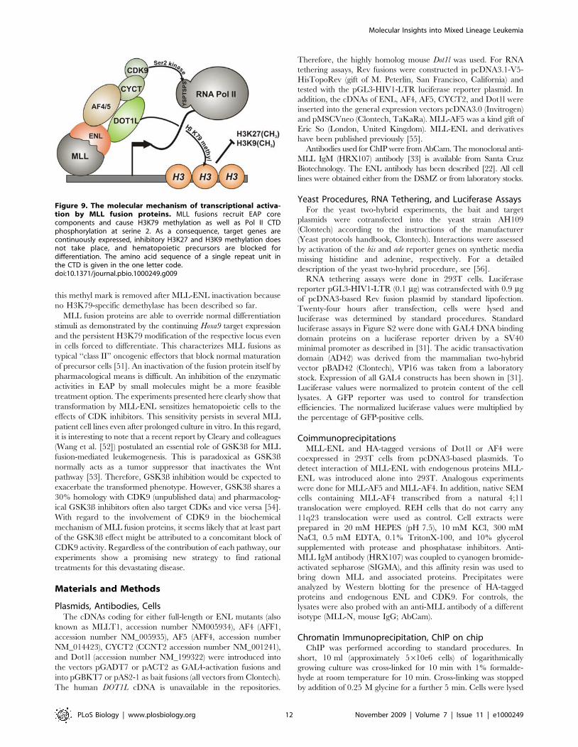
Discussion
In this report, we present evidence that the most frequently
occurring MLL fusion proteins exploit molecular control mech-
anisms of transcriptional elongation to transform hematopoietic
cells. MLL fusions become incorporated into an ‘‘elongation
assisting protein’’ complex, recruit it to their respective target
genes, and enforce ectopic transcription. This is accompanied by
DOT1L-mediated H3K79 methylation and Pol II phosphoryla-
tion through the pTEFb kinase (Figure 9). This mechanism
explains and reconciles seemingly contradictory observations that
have been made previously with respect to MLL fusion proteins. It
has been noted that particular MLL fusion partners are almost
exclusively encountered in MLL with more lymphoid character-
istics, whereas others occur preferentially in the myeloid subtype.
For example, MLL-AF4-transformed cells are very often of
lymphatic nature. In contrast, MLL-AF9 leukemia cells are
myeloid, and MLL-ENL is found in ALL, AML, and also in T-
cell acute leukemia [25,40]. These divergent phenotypes have
been used as an argument against a common function for MLL
fusion partners. However, the particular core structure of EAP
that is stabilized by protein–protein contacts of conserved
interaction domains, allows a high degree of flexibility. There
are four members of the AF4 family (AF5, LAF4, and FMR2).
ENL is closely related to AF9 and two CyclinT molecules (CYCT1
and CYCT2) exist in the cell. Incorporation of different homologs
into the same framework might create variations of EAP that
provide for cell-type or target gene specificity. Although it is
generally assumed that all MLL fusions occupy identical targets,
the preexisting protein environments will vary at different loci. A
co-recruited EAP complex incorporating AF9 might engage in
protein interactions different from those established by an EAP
Figure 5. Correlation of MLL-ENL binding and Hox gene expression. Murine cell lines derived from primary hematopoietic cells bytransformation with flag-MLL-ENL were subjected to a flag-specific ChIP reaction. Precipitates were amplified and hybridized (ChIP-chip) to acommercial promoter array that tiles 2 kb upstream and 0.5 kb downstream of known transcription start sites. In addition, the expression level ofevery Hoxa gene was determined by qRT-PCR with calibrated primers. Values were normalized to actin. The results are included as a heat map in theupper line (array set-up/expression level) of the figure. The heat map legend is shown below the figure. Each color-coded rectangle corresponds to2.5 kb of genomic sequence represented on the promoter array. The second line shows known transcripts within the Hoxa locus. All Hoxa transcripts(green arrows drawn to scale) are transcribed in centromeric direction. The blue arrow denotes an annotated cDNA (5730596B20RIK-201 in Ensembl)transcribed oppositely to the Hox genes. The complete locus as shown encompasses 95 kb of genomic DNA. The third line represents a diagram offlag-MLL-ENL binding. The bar height indicates the log2 ratio of relative enrichment compared to a control precipitation. The bar color indicates afalse discovery probability of ,0.05 (red), ,0.1 (orange), and ,0.2 (yellow). The graph is a composite image compiled from two independentexperiments. The genomic environment between Hoxa9 and the gene for microRNA196b is shown enlarged. Arrows represent known transcript startsites, and double arrows indicate the location of the primer pairs used for the kinetic analysis described in Figure 6. n.d. = not detected.doi:10.1371/journal.pbio.1000249.g005
Molecular Insights into Mixed Lineage Leukemia
PLoS Biology | www.plosbiology.org 8 November 2009 | Volume 7 | Issue 11 | e1000249
variant containing ENL. As a consequence, the final level of target
gene activation could be dependent on the composition of the
EAP. The results presented here demonstrate how the makeup of
EAP is determined by the nature of the MLL fusion partner. For
example, all patient-derived MLL-ENL and MLL-AF9 fusions
retain the conserved C-terminus of ENL/AF9 [41,42] that allows
simultaneous recruitment of DOT1L and AF4 (or any AF4 family
member) that both bridge to pTEFb. On the contrary, naturally
occurring MLL-AF4/5 fusions have lost the direct pTEFb
interaction domain in the N-terminus of AF4/5 [43] and need
to rely on a more indirect way via ENL and DOT1L to bring in
pTEFb. Structural variations in EAP and the mode of recruitment
likely contribute to the observed differences in the MLL
phenotypes.
As suggested by coimmunoprecipitations and RNA tethering,
all protein–protein interactions that stabilize EAP seem to be
conserved also in the fusion context despite the addition of an 180-
kDa MLL moiety. This is corroborated also by the fact that
Figure 6. Dynamic modification of the Hox locus by MLL fusion-recruited EAP. (A) Morphology of cells transformed by a conditionalderivative of MLL-ENL. Cytospin samples of cells transformed by tamoxifen-inducible MLL-ENL (MLL-ENL-ER as described in [8]) were prepared in thepresence of tamoxifen (0 days) (+TAM) and 2 wk after withdrawal of inductor (2TAM). Slides were stained with May-Grunwald-Giemsa, andmicrophotographs were taken at room temperature with a Nikon Digital Sight DS-Fi1 electronic camera attached to a Zeiss Axioskop with a ZeissNeofluar 636/1.25 objective and processed with CorelDraw software (Corel) without any image enhancements. (B) ChIP experiments. MLL-ENL-ERcells as in (A) were used to record the histone H3 methylation status at lysines 9 (dimethylation, black line), lysine 27 (di- and trimethylation, brownline), and lysine 79 (dimethylation, blue line) as well as RNA Pol II presence (Ser2 phosphorylated polymerase, magenta line). ChIP samples wereanalyzed for two loci within the first exon of Hoxa9 and upstream of the gene for microRNA196b. Samples were drawn at day 0 (active MLL-ENL) aswell as 3, 7, 10 and 14 d after inactivation of MLL-ENL by inductor withdrawal. Precipitation efficiencies were normalized to an input sample, andvalues were plotted relative to the modification levels at day 0. For absolute comparison, H3K9 trimethylation and H3K79 dimethylation were alsodetermined for a nontranscribed X-chromosomal satellite repeat sequence (indicated in upper-left panel). In addition, changes in Hoxa9 mRNA afterMLL-ENL-ER shutdown were quantified by qRT-PCR and plotted alongside the ChIP results (red line). The chart shows average values and standarddeviations of a triplicate qPCR evaluation, and represents one typical of three independent experiments.doi:10.1371/journal.pbio.1000249.g006
Molecular Insights into Mixed Lineage Leukemia
PLoS Biology | www.plosbiology.org 9 November 2009 | Volume 7 | Issue 11 | e1000249

introduction of small peptides blocking the AF4–AF9 interface has
been found to be specifically toxic for MLL-AF4 cells but much
less so for leukemic blasts of different etiology [44,45]. MLL-AF4
requires AF9 (or potentially ENL) as a mediator to recruit DOT1L
and pTEFb, and this pathway is blocked by binding site mimetics.
Both the positive readout in RNA tethering assays and the ChIP
results indicate that MLL fusion proteins affect transcription
through stimulation of elongation. In this regard, it is interesting to
note that the ELL protein, the first MLL fusion partner with a
known biochemical function, also is an elongation factor [46].
Later, elongation was dismissed as biochemical basis for MLL-
ELL-mediated transformation because motifs in ELL important
for elongation activity could be deleted in MLL-ELL with no effect
for the transforming function of the protein [47]. However, it was
never thoroughly tested whether domains in ELL that are essential
for transformation might recruit other elongation stimulating
proteins. In this regard, it will be interesting to see whether protein
interaction partners of ELL [48,49] will provide a link to
elongation control. Strikingly, these ELL-associated factors
(EAF1 and EAF2) have a limited but significant homology to
domains in AF4 [49]. Traces of ELL have been detected in ENL
precipitates [22], a possible lead that should be further explored.
At present, it is hard to predict whether the more rare fusion
partners will be connected to elongation control, too. This seems
rather unlikely, because these proteins are mostly cytoplasmatic.
However, it has been shown that the MLL fusion partner ABI1,
normally also in the cytoplasm, is imported into the nucleus as
MLL-ABI1 fusion due to the strong nuclear localization signals of
MLL. There, ABI1 can directly interact with ENL [27], pointing
to a mechanism for how cytoplasmatic fusion partners might also
link to EAP and elongation control.
After initial reports to the contrary [50], it is well established
that methylation of H3K79 by DOT1L is tightly associated with
actively transcribed chromatin [15]. Until now, DOT1L had been
implicated only in the transforming mechanism of MLL-AF10
where it could be demonstrated that interaction with DOT1L was
essential for oncogenic activity of the MLL-AF10 fusion protein
[17]. Here, we demonstrate a participation of DOT1L in a much
Figure 7. MLL-ENL-introduced chromatin modifications resist differentiation stimuli. (A) Left panel: cells transformed by constitutive MLL-ENL. FACS analysis of gr-1 differentiation marker in cells grown in IL-3 and in G-CSF for 7 d. Right panel: cells transformed by conditional MLL-ENL-ER.Gr-1 levels before and 3 d after inactivation of MLL-ENL-ER by removal of tamoxifen (TAM). (B) Left panel: qRT-PCR analysis of Hoxa9 expression incells transformed by constitutive MLL-ENL cultured in IL-3 or G-CSF for 7 d (light columns) and in cells transformed by conditional MLL-ENL-ER beforeand 3 d after tamoxifen removal (dark columns, labeled ‘‘on’’ and ‘‘off’’). Right panel: comparison of H3K79 dimethylation status of the Hoxa9 locus incells treated as before. Values are averages and standard deviations of a triplicate, and the experiment was done twice with comparable results.doi:10.1371/journal.pbio.1000249.g007
Molecular Insights into Mixed Lineage Leukemia
PLoS Biology | www.plosbiology.org 10 November 2009 | Volume 7 | Issue 11 | e1000249

wider range of MLL abnormalities encompassing the majority of
all clinically observed cases. The incorporation of DOT1L in EAP
also provides a molecular explanation for the genome-wide
correlation of MLL-AF4 binding and a drastic increase of
H3K79 methylation at the corresponding loci, a fact that has
raised much interest recently [11,12]. In addition, we show that
H3K79 methylation is highly dynamic and that it is correlated
with target RNA abundance. It will be interesting to know how
Figure 8. Sensitivity of MLL cells to CDK inhibition. (A) Effect of flavopiridol. Top panel: seven MLL cell lines (red lines) and four controlleukemia lines of different etiology (black lines) were treated with increasing concentrations of flavopiridol as indicated. Proliferation wasphotometrically assessed in triplicate samples by MTT reduction. For better comparison, all optical density at 550 nm (OD550) readings werenormalized to yield one unit in the absence of inhibitor. Mean values are plotted semilogarithmically against flavopiridol concentrations. To avoidclutter, standard deviations are indicated only for drug concentrations where MLL and control cell lines diverge in response to treatment. (B). Effect ofalsterpaullone. Alsterpaullone was tested like described for (A). (C) Effect of flavopiridol and alsterpaullone on primary hematopoietic cells. Triplicatesamples of mouse bone marrow cells enriched in early precursors by 5-fluorouracil treatment of the donors were cultured in liquid mediumsupplemented with IL-3, GM-CSF, IL-6, SCF, and the indicated amount of flavopiridol or alsterpaullone (black line). Proliferation was measured by MTTreduction. For comparison, precursor cells were retrovirally transduced with MLL-ENL, and the resulting lines were tested again for drug sensitivityunder identical culture conditions (red line). Given are mean values and standard deviations of triplicates.doi:10.1371/journal.pbio.1000249.g008
Molecular Insights into Mixed Lineage Leukemia
PLoS Biology | www.plosbiology.org 11 November 2009 | Volume 7 | Issue 11 | e1000249
this methyl mark is removed after MLL-ENL inactivation because
no H3K79-specific demethylase has been described so far.
MLL fusion proteins are able to override normal differentiation
stimuli as demonstrated by the continuing Hoxa9 target expression
and the persistent H3K79 modification of the respective locus even
in cells forced to differentiate. This characterizes MLL fusions as
typical ‘‘class II’’ oncogenic effectors that block normal maturation
of precursor cells [51]. An inactivation of the fusion protein itself by
pharmacological means is difficult. An inhibition of the enzymatic
activities in EAP by small molecules might be a more feasible
treatment option. The experiments presented here clearly show that
transformation by MLL-ENL sensitizes hematopoietic cells to the
effects of CDK inhibitors. This sensitivity persists in several MLL
patient cell lines even after prolonged culture in vitro. In this regard,
it is interesting to note that a recent report by Cleary and colleagues
(Wang et al. [52]) postulated an essential role of GSK3ß for MLL
fusion-mediated leukemogenesis. This is paradoxical as GSK3ß
normally acts as a tumor suppressor that inactivates the Wnt
pathway [53]. Therefore, GSK3ß inhibition would be expected to
exacerbate the transformed phenotype. However, GSK3ß shares a
30% homology with CDK9 (unpublished data) and pharmacolog-
ical GSK3ß inhibitors often also target CDKs and vice versa [54].
With regard to the involvement of CDK9 in the biochemical
mechanism of MLL fusion proteins, it seems likely that at least part
of the GSK3ß effect might be attributed to a concomitant block of
CDK9 activity. Regardless of the contribution of each pathway, our
experiments show a promising new strategy to find rational
treatments for this devastating disease.
Materials and Methods
Plasmids, Antibodies, CellsThe cDNAs coding for either full-length or ENL mutants (also
known as MLLT1, accession number NM005934), AF4 (AFF1,
accession number NM_005935), AF5 (AFF4, accession number
NM_014423), CYCT2 (CCNT2 accession number NM_001241),
and Dot1l (accession number NM_199322) were introduced into
the vectors pGADT7 or pACT2 as GAL4-activation fusions and
into pGBKT7 or pAS2-1 as bait fusions (all vectors from Clontech).
The human DOT1L cDNA is unavailable in the repositories.
Therefore, the highly homolog mouse Dot1l was used. For RNA
tethering assays, Rev fusions were constructed in pcDNA3.1-V5-
HisTopoRev (gift of M. Peterlin, San Francisco, California) and
tested with the pGL3-HIV1-LTR luciferase reporter plasmid. In
addition, the cDNAs of ENL, AF4, AF5, CYCT2, and Dot1l were
inserted into the general expression vectors pcDNA3.0 (Invitrogen)
and pMSCVneo (Clontech, TaKaRa). MLL-AF5 was a kind gift of
Eric So (London, United Kingdom). MLL-ENL and derivatives
have been published previously [55].
Antibodies used for ChIP were from AbCam. The monoclonal anti-
MLL IgM (HRX107) antibody [33] is available from Santa Cruz
Biotechnology. The ENL antibody has been described [22]. All cell
lines were obtained either from the DSMZ or from laboratory stocks.
Yeast Procedures, RNA Tethering, and Luciferase AssaysFor the yeast two-hybrid experiments, the bait and target
plasmids were cotransfected into the yeast strain AH109
(Clontech) according to the instructions of the manufacturer
(Yeast protocols handbook, Clontech). Interactions were assessed
by activation of the his and ade reporter genes on synthetic media
missing histidine and adenine, respectively. For a detailed
description of the yeast two-hybrid procedure, see [56].
RNA tethering assays were done in 293T cells. Luciferase
reporter pGL3-HIV1-LTR (0.1 mg) was cotransfected with 0.9 mg
of pcDNA3-based Rev fusion plasmid by standard lipofection.
Twenty-four hours after transfection, cells were lysed and
luciferase was determined by standard procedures. Standard
luciferase assays in Figure S2 were done with GAL4 DNA binding
domain proteins on a luciferase reporter driven by a SV40
minimal promoter as described in [31]. The acidic transactivation
domain (AD42) was derived from the mammalian two-hybrid
vector pBAD42 (Clontech), VP16 was taken from a laboratory
stock. Expression of all GAL4 constructs has been shown in [31].
Luciferase values were normalized to protein content of the cell
lysates. A GFP reporter was used to control for transfection
efficiencies. The normalized luciferase values were multiplied by
the percentage of GFP-positive cells.
CoimmunoprecipitationsMLL-ENL and HA-tagged versions of Dot1l or AF4 were
coexpressed in 293T cells from pcDNA3-based plasmids. To
detect interaction of MLL-ENL with endogenous proteins MLL-
ENL was introduced alone into 293T. Analogous experiments
were done for MLL-AF5 and MLL-AF4. In addition, native SEM
cells containing MLL-AF4 transcribed from a natural 4;11
translocation were employed. REH cells that do not carry any
11q23 translocation were used as control. Cell extracts were
prepared in 20 mM HEPES (pH 7.5), 10 mM KCl, 300 mM
NaCl, 0.5 mM EDTA, 0.1% TritonX-100, and 10% glycerol
supplemented with protease and phosphatase inhibitors. Anti-
MLL IgM antibody (HRX107) was coupled to cyanogen bromide-
activated sepharose (SIGMA), and this affinity resin was used to
bring down MLL and associated proteins. Precipitates were
analyzed by Western blotting for the presence of HA-tagged
proteins and endogenous ENL and CDK9. For controls, the
lysates were also probed with an anti-MLL antibody of a different
isotype (MLL-N, mouse IgG; AbCam).
Chromatin Immunoprecipitation, ChIP on chipChIP was performed according to standard procedures. In
short, 10 ml (approximately 5610e6 cells) of logarithmically
growing culture was cross-linked for 10 min with 1% formalde-
hyde at room temperature for 10 min. Cross-linking was stopped
by addition of 0.25 M glycine for a further 5 min. Cells were lysed
Figure 9. The molecular mechanism of transcriptional activa-tion by MLL fusion proteins. MLL fusions recruit EAP corecomponents and cause H3K79 methylation as well as Pol II CTDphosphorylation at serine 2. As a consequence, target genes arecontinuously expressed, inhibitory H3K27 and H3K9 methylation doesnot take place, and hematopoietic precursors are blocked fordifferentiation. The amino acid sequence of a single repeat unit inthe CTD is given in the one letter code.doi:10.1371/journal.pbio.1000249.g009
Molecular Insights into Mixed Lineage Leukemia
PLoS Biology | www.plosbiology.org 12 November 2009 | Volume 7 | Issue 11 | e1000249
in 50 mM Tris/HCl (pH 8.0), 10 mM EDTA, and 1% SDS, and
chromatin was prepared by sonication. Precipitations were done
with 2 mg of antibody and 30 ml of protein A/G agarose slurry
(Santa Cruz Biotechnology) for 4 h. Precipitates were washed
twice with buffer A (20 mM HEPES [pH 7.5], 10 mM KCl,
0.5 mM EDTA, 0.1% TritonX-100, and 10% glycerol) followed
by two washes each in buffer A supplemented with 500 mM LiCl
and buffer A + 500 mM LiCl + 0.1% SDS. After the final wash in
10 mM Tris/HCl [pH 8.0], 0.5 mM EDTA, the precipitates were
eluted and cross-links were removed by incubation overnight in
50 mM Tris/HCl (pH 8.0), 10 mM EDTA, 1% SDS supple-
mented with 66 mg/ml RNAse A (for input samples only) and
0.5 mg of proteinase K. The treated supernatants were purified by
a QIAquick Spin column (Qiagen) according to the instructions of
the manufacturer. Precipitated DNA was quantified by qPCR with
SYBR based premixes from Stratagene and compared to none-
nriched DNA from input samples. Primer sequences used to
amplify precipitated material are available on request.
For ChIP-chip experiments displayed in Figure 5, two cell lines
were derived from primary mouse hematopoietic cells by transduc-
tion with flag-tagged MLL-ENL as described [57]. Chromatin IP was
done as above with anti-flag agarose (M2 flag agarose, Sigma) and as
a control with flag-agarose preblocked with flag peptide (Sigma).
Precipitates were amplified by ligation-mediated PCR exactly as
described by the manufacturer (NimbleGen-Roche) and hybridized
to NimbleGen promoter MM8 RefSeqArrays. This array design tiles
genomic DNA 500 bp downstream and 2 kb upstream of all known
RefSeq transcripts. Data analysis was done by NimbleGen, and
results were visualized by SignalMap software. The software detects
potential fMLL-ENL binding sites by searching in a 500-bp sliding
window for four or more peaks with a log2 signal-to-noise ratio above
25% of a calculated maximum that equals the average of all peaks
plus six standard deviations. A false-positive discovery rate (FDR) is
calculated by a 20-fold randomization of the ratio data, and a
probability of ‘‘randomness’’ is assigned to each peak. Peaks with a
probability of ,0.2 are indicative for binding. In total, two
independent experiments were performed.
Inhibitor Tests with Primary CellsSix- to 8-wk-old Balb/C mice were treated by intraperitoneal
administration of 150 mg/kg 5-fluorouracil. Five days after
injection, bone marrow enriched in precursor cells was harvested.
Half of the cells were cultivated for 3 to 4 d in RPMI medium
supplemented with 10 ng/ml IL-3, IL-6, and GM-CSF, as well as
with 100 ng/ml SCF (all recombinant cytokines were obtained from
PeproTech) and with increasing amounts of alsterpaullone or
flavopiridol (Sigma). Proliferation was assessed by a standard MTT
assay. The second batch of cells was retrovirally infected with MLL-
ENL as described in [57], and after selection for transduced cells,
the resulting MLL-ENL-transformed lines were checked for
sensitivity towards alsterpaullone and flavopiridol under identical
cytokine conditions as used for nontransformed cells.
Supporting Information
Figure S1 Two-hybrid experiments. (A) Two-hybrid pair-
ings. A series of deletion mutants derived from ENL, AF4,
CYCT2A, and Dot1l was tested in two-hybrid assays with full-
length proteins as interaction partner. Numbers correspond to
amino acid residues. Abbreviations are as in Figure 1. Two-hybrid
outcome is listed either as + = strong interaction, (+) = weak
interaction, growth only after prolonged incubation, or 2 = no
interaction. (B) Expression of two-hybrid clones. Extracts of
transformed yeast cells were blotted and probed with a GAL4-
DNA binding domain-specific antibody. Expression from plasmids
not listed here has been shown previously [20,26,27].
Found at: doi:10.1371/journal.pbio.1000249.s001 (3.48 MB TIF)
Figure S2 Transactivation potential and elongationstimulation by ENL, AD42, and VP16. ENL as well as the
generic transactivation domains AD42 (acidic transactivation
domain derived from a mammalian two-hybrid vector) and
VP16 (a transactivator domain from H. simplex) were fused to
the GAL4 DNA-binding domain and to Rev. (A) General
transactivation potential of GAL4 recruited proteins. GAL4
fusions were tested on a SV40 minimal promoter-based luciferase
reporter as described in [31]. Depicted are average values and
standard deviations of triplicate transfections. The expression of
the corresponding GAL4-proteins has been shown in Zeisig et al.
[31]. The green bar highlights results obtained for the fusion of
GAL4 with ENL. (B) Rev fusions of the same proteins were
examined for their elongation stimulation activity in the TAR-loop
RNA tethering assay. The expression of the respective Rev fusions
is demonstrated by a Rev-specific Western blot. Values are
charted as described for (A).
Found at: doi:10.1371/journal.pbio.1000249.s002 (1.42 MB TIF)
Figure S3 Kinetics of RNA Pol II activity on Hoxa9chromatin. (A) Schematic depiction of the putative Hoxa9 core
transcript as annotated in Ensembl (ENSMUST00000048680).
Primers used for ChIP and for qRT-PCR are indicated. (B) ChIP
and RNA decay kinetics. ChIP was performed on cells
transformed by inducible MLL-ENL as described for Figure 6.
Samples were taken in the presence of tamoxifen (active MLL-
ENL) and at the indicated time points after withdrawal of the
inductor. ChIP was performed with antibodies specific for the
serine-2 and the serine-5 phosphorylated isoforms of RNA Pol II
and RNA was extracted, digested with DNAseI, and reverse
transcribed into cDNA. ChIP precipitates were quantified in
relation to input samples by qPCR with the primers indicated in
(A). Data are plotted as relative values compared to day 0. cDNA
was analyzed by qPCR, and data were normalized to ß-actin. In
addition to the 59 and 39 primers that would detect unspliced and
spliced RNA, the intron-spanning primer is specific for spliced
Hoxa9 transcripts.
Found at: doi:10.1371/journal.pbio.1000249.s003 (0.87 MB TIF)
Acknowledgments
We thank Renate Zimmermann for technical support and Matija Peterlin
for the gift of the Rev plasmids as well as for the introduction to the RNA
tethering system. Eric So is gratefully acknowledged for the MLL-AF5
construct.
Author Contributions
The author(s) have made the following declarations about their
contributions: Conceived and designed the experiments: RKS. Performed
the experiments: DM MPGC CB SB EM RKS. Analyzed the data: RKS.
Wrote the paper: RKS.
References
1. Djabali M, Selleri L, Parry P, Bower M, Young B, et al. (1992) A trithorax-like
gene is interrupted by chromosome 11q23 translocations in acute leukaemias.
Nat Genet 2: 111–118.
2. Gu Y, Nakamura T, Alder H, Prasad R, Canaani O, et al. (1992) The t(4;11)
chromosome translocation of human acute leukemias fuses the ALL-1 gene,
related to Drosophila trithorax, to the AF-4 gene. Cell 71: 701–708.
Molecular Insights into Mixed Lineage Leukemia
PLoS Biology | www.plosbiology.org 13 November 2009 | Volume 7 | Issue 11 | e1000249
3. Tkachuk DC, Kohler S, Cleary ML (1992) Involvement of a homolog of
Drosophila trithorax by 11q23 chromosomal translocations in acute leukemias.Cell 71: 691–700.
4. Ziemin-van der Poel S, McCabe NR, Gill HJ, Espinosa R III, Patel Y, et al.
(1991) Identification of a gene, MLL, that spans the breakpoint in 11q23translocations associated with human leukemias. Proc Natl Acad Sci U S A 88:
10735–10739.5. Harper DP, Aplan PD (2008) Chromosomal rearrangements leading to MLL
gene fusions: clinical and biological aspects. Cancer Res 68: 10024–10027.
6. Hess JL (2004) MLL: a histone methyltransferase disrupted in leukemia. TrendsMol Med 10: 500–507.
7. Slany RK (2005) When epigenetics kills: MLL fusion proteins in leukemia.Hematol Oncol 23: 1–9.
8. Zeisig BB, Milne T, Garcia-Cuellar MP, Schreiner S, Martin ME, et al. (2004)Hoxa9 and Meis1 are key targets for MLL-ENL-mediated cellular immortal-
ization. Mol Cell Biol 24: 617–628.
9. Ferrando AA, Armstrong SA, Neuberg DS, Sallan SE, Silverman LB, et al.(2003) Gene expression signatures in MLL-rearranged T-lineage and B-
precursor acute leukemias: dominance of HOX dysregulation. Blood 102:262–268.
10. Armstrong SA, Staunton JE, Silverman LB, Pieters R, den Boer ML, et al.
(2002) MLL translocations specify a distinct gene expression profile thatdistinguishes a unique leukemia. Nat Genet 30: 41–47.
11. Guenther MG, Lawton LN, Rozovskaia T, Frampton GM, Levine SS, et al.(2008) Aberrant chromatin at genes encoding stem cell regulators in human
mixed-lineage leukemia. Genes Dev 22: 3403–3408.12. Krivtsov AV, Feng Z, Lemieux ME, Faber J, Vempati S, et al. (2008) H3K79
methylation profiles define murine and human MLL-AF4 leukemias. Cancer
Cell 14: 355–368.13. Scacheri PC, Davis S, Odom DT, Crawford GE, Perkins S, et al. (2006)
Genome-wide analysis of menin binding provides insights into MEN1tumorigenesis. PLoS Genet 2: e51. doi:10.1371/journal.pgen.0020051.
14. Milne TA, Martin ME, Brock HW, Slany RK, Hess JL (2005) Leukemogenic
MLL fusion proteins bind across a broad region of the Hox a9 locus, promotingtranscription and multiple histone modifications. Cancer Res 65: 11367–11374.
15. Steger DJ, Lefterova MI, Ying L, Stonestrom AJ, Schupp M, et al. (2008)DOT1L/KMT4 recruitment and H3K79 methylation are ubiquitously coupled
with gene transcription in mammalian cells. Mol Cell Biol 28: 2825–2839.16. Feng Q, Wang H, Ng HH, Erdjument-Bromage H, Tempst P, et al. (2002)
Methylation of H3-lysine 79 is mediated by a new family of HMTases without a
SET domain. Curr Biol 12: 1052–1058.17. Okada Y, Feng Q, Lin Y, Jiang Q, Li Y, et al. (2005) hDOT1L links histone
methylation to leukemogenesis. Cell 121: 167–178.18. Bitoun E, Oliver PL, Davies KE (2007) The mixed-lineage leukemia fusion
partner AF4 stimulates RNA polymerase II transcriptional elongation and
mediates coordinated chromatin remodeling. Hum Mol Genet 16: 92–106.19. Erfurth F, Hemenway CS, de Erkenez AC, Domer PH (2004) MLL fusion
partners AF4 and AF9 interact at subnuclear foci. Leukemia 18: 92–102.20. Zeisig DT, Bittner CB, Zeisig BB, Garcia-Cuellar MP, Hess JL, et al. (2005) The
eleven-nineteen-leukemia protein ENL connects nuclear MLL fusion partnerswith chromatin. Oncogene 24: 5525–5532.
21. Zhang W, Xia X, Reisenauer MR, Hemenway CS, Kone BC (2006) Dot1a-AF9
complex mediates histone H3 Lys-79 hypermethylation and repression ofENaCalpha in an aldosterone-sensitive manner. J Biol Chem 281: 18059–18068.
22. Mueller D, Bach C, Zeisig D, Garcia-Cuellar MP, Monroe S, et al. (2007) A rolefor the MLL fusion partner ENL in transcriptional elongation and chromatin
modification. Blood 110: 4445–4454.
23. Peterlin BM, Price DH (2006) Controlling the elongation phase of transcriptionwith P-TEFb. Mol Cell 23: 297–305.
24. Estable MC, Naghavi MH, Kato H, Xiao H, Qin J, et al. (2002) MCEF, thenewest member of the AF4 family of transcription factors involved in leukemia,
is a positive transcription elongation factor-b-associated protein. J Biomed Sci 9:
234–245.25. Meyer C, Schneider B, Jakob S, Strehl S, Attarbaschi A, et al. (2006) The MLL
recombinome of acute leukemias. Leukemia 20: 777–784.26. Garcia-Cuellar MP, Zilles O, Schreiner SA, Birke M, Winkler TH, et al. (2001)
The ENL moiety of the childhood leukemia-associated MLL-ENL oncoproteinrecruits human Polycomb 3. Oncogene 20: 411–419.
27. Garcia-Cuellar MP, Schreiner SA, Birke M, Hamacher M, Fey GH, et al. (2000)
ENL, the MLL fusion partner in t(11;19), binds to the c-Abl interactor protein 1(ABI1) that is fused to MLL in t(10;11)+. Oncogene 19: 1744–1751.
28. Zhu Y, Pe’ery T, Peng J, Ramanathan Y, Marshall N, et al. (1997) Transcriptionelongation factor P-TEFb is required for HIV-1 tat transactivation in vitro.
Genes Dev 11: 2622–2632.
29. Gold MO, Rice AP (1998) Targeting of CDK8 to a promoter-proximal RNAelement demonstrates catalysis-dependent activation of gene expression. Nucleic
Acids Res 26: 3784–3788.30. So CW, Cleary ML (2003) Common mechanism for oncogenic activation of
MLL by forkhead family proteins. Blood 101: 633–639.31. Zeisig BB, Schreiner S, Garcia-Cuellar MP, Slany RK (2003) Transcriptional
activation is a key function encoded by MLL fusion partners. Leukemia 17:
359–365.
32. Kurosu T, Peterlin BM (2004) VP16 and ubiquitin; binding of P-TEFb via its
activation domain and ubiquitin facilitates elongation of transcription of target
genes. Curr Biol 14: 1112–1116.
33. Butler LH, Slany R, Cui X, Cleary ML, Mason DY (1997) The HRX proto-
oncogene product is widely expressed in human tissues and localizes to nuclear
structures. Blood 89: 3361–3370.
34. Greil J, Gramatzki M, Burger R, Marschalek R, Peltner M, et al. (1994) The
acute lymphoblastic leukaemia cell line SEM with t(4;11) chromosomal
rearrangement is biphenotypic and responsive to interleukin-7. Br J Haematol
86: 275–283.
35. Popovic R, Riesbeck LE, Velu CS, Chaubey A, Zhang J, et al. (2009) Regulation
of mir-196b by MLL and its overexpression by MLL fusions contributes to
immortalization. Blood 113: 3314–3322.
36. Horton SJ, Grier DG, McGonigle GJ, Thompson A, Morrow M, et al. (2005)
Continuous MLL-ENL expression is necessary to establish a ‘‘Hox Code’’ and
maintain immortalization of hematopoietic progenitor cells. Cancer Res 65:
9245–9252.
37. Schreiner S, Birke M, Garcia-Cuellar MP, Zilles O, Greil J, et al. (2001) MLL-
ENL causes a reversible and myc-dependent block of myelomonocytic cell
differentiation. Cancer Res 61: 6480–6486.
38. Chao SH, Fujinaga K, Marion JE, Taube R, Sausville EA, et al. (2000)
Flavopiridol inhibits P-TEFb and blocks HIV-1 replication. J Biol Chem 275:
28345–28348.
39. Schultz C, Link A, Leost M, Zaharevitz DW, Gussio R, et al. (1999) Paullones, a
series of cyclin-dependent kinase inhibitors: synthesis, evaluation of CDK1/
cyclin B inhibition, and in vitro antitumor activity. J Med Chem 42: 2909–2919.
40. Drexler HG, Quentmeier H, MacLeod RA (2004) Malignant hematopoietic cell
lines: in vitro models for the study of MLL gene alterations. Leukemia 18:
227–232.
41. Rubnitz JE, Morrissey J, Savage PA, Cleary ML (1994) ENL, the gene fused
with HRX in t(11;19) leukemias, encodes a nuclear protein with transcriptional
activation potential in lymphoid and myeloid cells. Blood 84: 1747–1752.
42. Yamamoto K, Seto M, Komatsu H, Iida S, Akao Y, et al. (1993) Two distinct
portions of LTG19/ENL at 19p13 are involved in t(11;19) leukemia. Oncogene
8: 2617–2625.
43. Borkhardt A, Repp R, Haupt E, Brettreich S, Buchen U, et al. (1994) Molecular
analysis of MLL-1/AF4 recombination in infant acute lymphoblastic leukemia.
Leukemia 8: 549–553.
44. Palermo CM, Bennett CA, Winters AC, Hemenway CS (2008) The AF4-
mimetic peptide, PFWT, induces necrotic cell death in MV4-11 leukemia cells.
Leuk Res 32: 633–642.
45. Srinivasan RS, Nesbit JB, Marrero L, Erfurth F, LaRussa VF, et al. (2004) The
synthetic peptide PFWT disrupts AF4–AF9 protein complexes and induces
apoptosis in t(4;11) leukemia cells. Leukemia 18: 1364–1372.
46. Shilatifard A, Lane WS, Jackson KW, Conaway RC, Conaway JW (1996) An
RNA polymerase II elongation factor encoded by the human ELL gene. Science
271: 1873–1876.
47. DiMartino JF, Miller T, Ayton PM, Landewe T, Hess JL, et al. (2000) A
carboxy-terminal domain of ELL is required and sufficient for immortalization
of myeloid progenitors by MLL-ELL. Blood 96: 3887–3893.
48. Simone F, Luo RT, Polak PE, Kaberlein JJ, Thirman MJ (2003) ELL-associated
factor 2 (EAF2), a functional homolog of EAF1 with alternative ELL binding
properties. Blood 101: 2355–2362.
49. Simone F, Polak PE, Kaberlein JJ, Luo RT, Levitan DA, et al. (2001) EAF1, a
novel ELL-associated factor that is delocalized by expression of the MLL-ELL
fusion protein. Blood 98: 201–209.
50. Barski A, Cuddapah S, Cui K, Roh TY, Schones DE, et al. (2007) High-
resolution profiling of histone methylations in the human genome. Cell 129:
823–837.
51. Gilliland DG (2002) Molecular genetics of human leukemias: new insights into
therapy. Semin Hematol 39: 6–11.
52. Wang Z, Smith KS, Murphy M, Piloto O, Somervaille TC, et al. (2008)
Glycogen synthase kinase 3 in MLL leukaemia maintenance and targeted
therapy. Nature 455: 1205–1209.
53. Jope RS, Johnson GV (2004) The glamour and gloom of glycogen synthase
kinase-3. Trends Biochem Sci 29: 95–102.
54. Bain J, Plater L, Elliott M, Shpiro N, Hastie CJ, et al. (2007) The selectivity of
protein kinase inhibitors: a further update. Biochem J 408: 297–315.
55. Slany RK, Lavau C, Cleary ML (1998) The oncogenic capacity of HRX-ENL
requires the transcriptional transactivation activity of ENL and the DNA binding
motifs of HRX. Mol Cell Biol 18: 122–129.
56. Garcia-Cuellar MP, Mederer D, Slany RK (2009) Identification of protein
interaction partners by the yeast two-hybrid system. Methods Mol Biol 538:
347–367.
57. Lavau C, Szilvassy SJ, Slany R, Cleary ML (1997) Immortalization and leukemic
transformation of a myelomonocytic precursor by retrovirally transduced HRX-
ENL. EMBO J 16: 4226–4237.
58. Shore SM, Byers SA, Maury W, Price DH (2003) Identification of a novel
isoform of Cdk9. Gene 307: 175–182.
Molecular Insights into Mixed Lineage Leukemia
PLoS Biology | www.plosbiology.org 14 November 2009 | Volume 7 | Issue 11 | e1000249