Overview of the Marine Roseobacter Lineage
13
APPLIED AND ENVIRONMENTAL MICROBIOLOGY, Oct. 2005, p. 5665–5677 Vol. 71, No. 10 0099-2240/05/$08.000 doi:10.1128/AEM.71.10.5665–5677.2005 Copyright © 2005, American Society for Microbiology. All Rights Reserved. MINIREVIEW Overview of the Marine Roseobacter Lineage† Alison Buchan, 1 * Jose ´ M. Gonza ´lez, 2 and Mary Ann Moran 3 Department of Microbiology, University of Tennessee, Knoxville, Tennessee 1 ; Department of Microbiology and Cell Biology, University of La Laguna, La Laguna, Spain 2 ; and Department of Marine Sciences, University of Georgia, Athens, Georgia 3 Despite the overwhelming bacterial diversity present in the world’s oceans, the majority of recognized marine bacteria fall into as few as nine major clades (36), many of which have yet to be cultivated in the laboratory. Molecular-based approaches targeting 16S rRNA genes demonstrate that the Roseobacter clade is one of these major marine groups, typically comprising upwards of 20% of coastal and 15% of mixed-layer ocean bacterioplankton communities (see, e.g., references 36, 37, 42, 98, and 109). Roseobacters are well represented across diverse marine habitats, from coastal to open oceans and from sea ice to sea floor (see, e.g., references 16, 28, 37, 42, 52, and 98). Members have been found to be free living, particle associated, or in commensal relationships with marine phytoplankton, in- vertebrates, and vertebrates (see, e.g., references 4, 6, 7, 44, 49, 115, and 119). Furthermore, representatives of the clade stand out as representing one of the most readily cultivated of the major marine lineages (36). These isolated representatives are serving as the foundation for an improved understanding of marine bacterial ecology and physiology. DESCRIPTION OF THE GROUP The Roseobacter clade falls within the -3 subclass of the class Proteobacteria, with members sharing 89% identity of the 16S rRNA gene. The first strain descriptions appeared in 1991, about the time that 16S rRNA-based approaches for cataloging microbial diversity were revealing the immensity of prokaryotic diversity in the world’s oceans. Interest in the clade has risen steadily since the initial discovery of these strains; at present the clade contains 36 described species, representing 17 genera, and literally hundreds of uncharacterized isolates and clone sequences. The first described members were Roseobacter litoralis and Roseobacter denitrificans, both pink- pigmented bacteriochlorophyll a-producing strains isolated from marine algae (99). Subsequent cultivation of clade mem- bers, however, revealed that many strains are neither pink nor bacteriochlorophyll a producers (see, e.g., references 20, 41, 43, and 61). With the exceptions of the described strains of the genus Ketogulonicigenium (113) and several clones from a South African gold mine (GenBank accession numbers AF546906, -13, -17, -22 to -24, and -26), the Roseobacter clade is exclusively marine or hypersaline, with characterized isolates demonstrating either a salt requirement or tolerance (see, e.g., references 60 and 62). The described strains demonstrate a diverse range of physio- logical and morphological features (e.g., gas vacuoles [43], holdfasts [41], poly--hydroxybutyrate granules [20, 118], ro- sette formation [60, 86], toga-like morphologies [39], sulfur metabolism [39, 104], secondary metabolite production [61], methylotrophy [51], and mixotrophy [73]) that suggest unique adaptations to various marine environments. However, few of these traits are representative of the entire clade. ABUNDANCE AND DISTRIBUTION IN MARINE ENVIRONMENTS Based on culture collections, 16S rRNA clone libraries, and single-cell analyses, roseobacters have been identified in most marine environments sampled. The group is prevalent in 16S rRNA gene inventories of seawater (Table 1) and marine sed- iments (Table 2) and is noticeably absent from analogous in- ventories of freshwater and terrestrial soil environments. Flu- orescent in situ hybridization (FISH) studies quantifying Roseobacter populations in coastal waters of the southeastern United States and the North Sea indicate different relative population sizes (20% of all bacterial cells versus 8%), but similar seasonal trends (populations highest in summer months and dropping off during winter) (28, 82). Quantitative 16S rRNA gene inventories (Table 1) show that 20 to 30% Roseobacter representation is not uncommon in bacterial com- munities in the upper mixed layer of the ocean, but depth profiles suggest that populations fall off with depth (Fig. 1) (1, 42, 107). Roseobacters are often most abundant in bacterial communities associated with marine algae, including natural phytoplankton blooms and algal cultures (see, e.g., references 4, 42, 78, 90, and 125). Roseobacter sequences are also abun- dant in communities associated with polar sea ice (16, 17), diseased corals (21, 80), sponges (111, 119), hypersaline mi- crobial mats (54), cephalopods (cuttlefish and squid) (9, 46), scallop larvae (93), sea grasses (120), and coastal biofilms (24, 25) (Table 2). ARE ISOLATES REPRESENTATIVE OF NATURAL POPULATIONS? While the culturability of roseobacters is well established, an unresolved question is whether these isolates are truly repre- * Corresponding author. Mailing address: Department of Microbi- ology, University of Tennessee, Knoxville, TN 37996-0845. Phone: (865) 974-3441. Fax: (865) 974-4007. E-mail: [email protected]. † Supplemental material for this article may be found at http: //aem.asm.org/. 5665
-
Upload
independent -
Category
Documents
-
view
0 -
download
0
Transcript of Overview of the Marine Roseobacter Lineage

APPLIED AND ENVIRONMENTAL MICROBIOLOGY, Oct. 2005, p. 5665–5677 Vol. 71, No. 100099-2240/05/$08.00�0 doi:10.1128/AEM.71.10.5665–5677.2005Copyright © 2005, American Society for Microbiology. All Rights Reserved.
MINIREVIEW
Overview of the Marine Roseobacter Lineage†Alison Buchan,1* Jose M. Gonzalez,2 and Mary Ann Moran3
Department of Microbiology, University of Tennessee, Knoxville, Tennessee1; Department of Microbiologyand Cell Biology, University of La Laguna, La Laguna, Spain2; and Department of Marine Sciences,
University of Georgia, Athens, Georgia3
Despite the overwhelming bacterial diversity present in theworld’s oceans, the majority of recognized marine bacteria fallinto as few as nine major clades (36), many of which have yetto be cultivated in the laboratory. Molecular-based approachestargeting 16S rRNA genes demonstrate that the Roseobacterclade is one of these major marine groups, typically comprisingupwards of 20% of coastal and 15% of mixed-layer oceanbacterioplankton communities (see, e.g., references 36, 37, 42,98, and 109). Roseobacters are well represented across diversemarine habitats, from coastal to open oceans and from sea iceto sea floor (see, e.g., references 16, 28, 37, 42, 52, and 98).Members have been found to be free living, particle associated,or in commensal relationships with marine phytoplankton, in-vertebrates, and vertebrates (see, e.g., references 4, 6, 7, 44, 49,115, and 119). Furthermore, representatives of the clade standout as representing one of the most readily cultivated of themajor marine lineages (36). These isolated representatives areserving as the foundation for an improved understanding ofmarine bacterial ecology and physiology.
DESCRIPTION OF THE GROUP
The Roseobacter clade falls within the �-3 subclass of theclass Proteobacteria, with members sharing �89% identity ofthe 16S rRNA gene. The first strain descriptions appeared in1991, about the time that 16S rRNA-based approaches forcataloging microbial diversity were revealing the immensity ofprokaryotic diversity in the world’s oceans. Interest in the cladehas risen steadily since the initial discovery of these strains; atpresent the clade contains 36 described species, representing17 genera, and literally hundreds of uncharacterized isolatesand clone sequences. The first described members wereRoseobacter litoralis and Roseobacter denitrificans, both pink-pigmented bacteriochlorophyll a-producing strains isolatedfrom marine algae (99). Subsequent cultivation of clade mem-bers, however, revealed that many strains are neither pink norbacteriochlorophyll a producers (see, e.g., references 20, 41, 43,and 61). With the exceptions of the described strains of the genusKetogulonicigenium (113) and several clones from a South Africangold mine (GenBank accession numbers AF546906, -13, -17, -22
to -24, and -26), the Roseobacter clade is exclusively marine orhypersaline, with characterized isolates demonstrating either asalt requirement or tolerance (see, e.g., references 60 and 62).The described strains demonstrate a diverse range of physio-logical and morphological features (e.g., gas vacuoles [43],holdfasts [41], poly-�-hydroxybutyrate granules [20, 118], ro-sette formation [60, 86], toga-like morphologies [39], sulfurmetabolism [39, 104], secondary metabolite production [61],methylotrophy [51], and mixotrophy [73]) that suggest uniqueadaptations to various marine environments. However, few ofthese traits are representative of the entire clade.
ABUNDANCE AND DISTRIBUTION INMARINE ENVIRONMENTS
Based on culture collections, 16S rRNA clone libraries, andsingle-cell analyses, roseobacters have been identified in mostmarine environments sampled. The group is prevalent in 16SrRNA gene inventories of seawater (Table 1) and marine sed-iments (Table 2) and is noticeably absent from analogous in-ventories of freshwater and terrestrial soil environments. Flu-orescent in situ hybridization (FISH) studies quantifyingRoseobacter populations in coastal waters of the southeasternUnited States and the North Sea indicate different relativepopulation sizes (20% of all bacterial cells versus 8%), butsimilar seasonal trends (populations highest in summer monthsand dropping off during winter) (28, 82). Quantitative 16SrRNA gene inventories (Table 1) show that 20 to 30%Roseobacter representation is not uncommon in bacterial com-munities in the upper mixed layer of the ocean, but depthprofiles suggest that populations fall off with depth (Fig. 1) (1,42, 107). Roseobacters are often most abundant in bacterialcommunities associated with marine algae, including naturalphytoplankton blooms and algal cultures (see, e.g., references4, 42, 78, 90, and 125). Roseobacter sequences are also abun-dant in communities associated with polar sea ice (16, 17),diseased corals (21, 80), sponges (111, 119), hypersaline mi-crobial mats (54), cephalopods (cuttlefish and squid) (9, 46),scallop larvae (93), sea grasses (120), and coastal biofilms (24,25) (Table 2).
ARE ISOLATES REPRESENTATIVE OFNATURAL POPULATIONS?
While the culturability of roseobacters is well established, anunresolved question is whether these isolates are truly repre-
* Corresponding author. Mailing address: Department of Microbi-ology, University of Tennessee, Knoxville, TN 37996-0845. Phone:(865) 974-3441. Fax: (865) 974-4007. E-mail: [email protected].
† Supplemental material for this article may be found at http://aem.asm.org/.
5665

sentative of the populations that are abundant in the environ-ment. Representative strains have been isolated by Brinkmeyeret al. (16), who cultured an Octadecabacter-like strain thatcomprised �20% of an Arctic sea ice bacterial community; byPinhassi et al. (83), who cultured two Roseobacter strains thatby whole genome hybridization contributed 7 and 20% ofNorth Sea and Baltic Sea bacterial communities; and byFuhrman et al. (31), who cultivated the type strain of Roseo-varius nubinhibens, which by whole genome hybridization con-tributed 20% of a Caribbean Sea bacterial community. In someof these cases, the criterion used to assess taxonomic similaritywas not stringent, which is an important issue in light of find-ings that bacterioplankton with as much as 97% identity of the16S rRNA gene can be functionally and genetically divergent(72, 91). Other studies have concluded that cultured membersof the Roseobacter group are not representative of their envi-ronmentally abundant relatives; these studies include those ofEilers et al. (28), who found that specific Roseobacter strainsconstituted �1% of the bacterial community in the GermanBight (even though the group as a whole comprised �10%of the community), and of Selje et al. (98), who found aRoseobacter phylotype with wide geographic distribution in theArctic and Southern Oceans that is not well represented inculture. Thus, there is evidence for both sides of the debate onthe ecological relevance of Roseobacter group members thathave been gathered into culture collections, with methodologyand environment as two potentially important variables.
An alternative approach to addressing the question ofecological relevance of isolates is to determine whetherRoseobacter 16S rRNA gene sequences fall into phylogenetic
clusters that contain only cultured members, only unculturedmembers, or both cultured and uncultured members. This ap-proach uses the single criterion of 16S rRNA similarity todetermine relatedness and integrates sequences across sitesand dates into a single analysis. To this end, we compiled adata set of Roseobacter 16S rRNA gene sequences and identi-fied clusters of sequences with �99% identity.
CLUSTERS WITHIN THE CLADE
The Roseobacter 16S rRNA gene data set was establishedwith 565 sequences from the Ribosomal Database Project IIrelease 9.22 (RDP) that were assigned to any of the fiveRoseobacter genera used by the RDP Classifier (Antarctobacter,Roseivivax, Roseobacter, Roseovarius, and Sulfitobacter). Anadditional 1,251 RDP sequences that were listed as unclassi-fied Rhodobacteraceae family members were screened forRoseobacter clade members based on �97% identity in pair-wise Smith-Waterman alignments (103) to a reference set of391 Roseobacter sequences (89 clones and 302 isolates). Thisreference set included all described strains and all clone andisolate sequences of �1,000 bp in length from the RDP-rec-ognized Roseobacter genera. This screen identified 772 addi-tional Roseobacter sequences (61% of the unclassifiedRhodobacteraceae sequences). The 1,337 Roseobacter se-quences obtained from the RDP accounted for 1% of all bac-terial sequences and 9.5% of all �-proteobacterial sequences inRDP release 9.22. An additional 160 Roseobacter sequenceswere added to the data set because they represented describedgenera not in the RDP 9.22 release (n � 3), were identified in
TABLE 1. Representation of Roseobacter sequences in quantitative 16S rRNA gene clone libraries from seawater
Location Depth (m) No. of clonesequences
% Roseobactersequences Reference
Pacific Ocean �10 16 6 96Sargasso Sea 10 14 0 32Coastal Pacific Ocean (California) 10 71 0 26Sargasso Sea 2 42 24 77Coastal Pacific Ocean (Oregon) 10 58 7 108Coastal North Atlantic Ocean (France) �10 51 18 11Mediterranean Sea (France) �10 50 12 11Coastal North Atlantic Ocean (North Carolina) 10 112 21 88Coastal North Atlantic Ocean (Long Island Sound) �10 17 18 33Coastal Pacific Ocean (California) �10 16 25 33Western Mediterranean Sea; free living Various 120 2 1Coastal Pacific Ocean and Estuary (Washington) 10 189 0.4 23Various Various 660 16 36Coastal North Sea (German Bay) 1 54 1.9 27Coastal Pacific Ocean (Oregon) 10 51 10 89Coastal North Atlantic Ocean (Long Island Sound) �1 126 0 56Antarctic Polar Front 3,000 15 0 65Changjiang Estuary (China) Various 241 19 97Coastal Caribbean Sea; coral reef tracts �10 65 6 30Black Sea Various 38 11 116Coastal North Atlantic Ocean (Massachusetts) �10 2,040 4 2Coastal North Atlantic (Portugal) 0 198 25 48Coastal Pacific Ocean (California)a Various 27,840 27 107Sargasso Sea; seven librariesb �10 2,328 1.7 115Coastal North Atlantic Ocean (Georgia) �10 794 17 W. B. Whitman et al.,
unpublished datac
a BAC libraries of coastal Pacific Ocean assemblages.b Shotgun clone libraries of the Sargasso Sea.c Sequences are available at http://simo.marsci.uga.edu.
5666 MINIREVIEW APPL. ENVIRON. MICROBIOL.

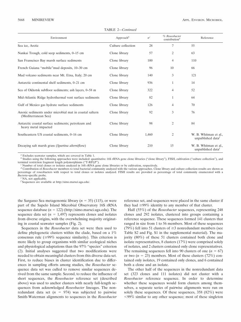
TABLE 2. Representation of Roseobacter sequences in bacterial communities from diverse marine environmentsa
Environment Approachb nc % Roseobactercontributiond Reference
North Sea coastal biofilms Culture collection 463 1 6
Southern U.S. salt marsh biofilms Clone library 43 67 24FISH NA �45 25
Dinoflagellate (Alexandrium sp. and Prorocentrum lima)cultures
Culture collection 44 30 6
Dinoflagellate (Alexandrium sp. and Scrippsiellatrochoidea) cultures
Culture collection 76 55 50
Dinoflagellate (Gymnodinium catenatum) cultures Culture collection 61 31 45
Dinoflagellate (Alexandrium sp.) cultures Culture collection 31 87 3
Dinoflagellate (Gymnodinium catenatum) cultures Culture collection 61 30 45
Marine red algae (Prionitis filiformis) Clone library 3 100 8FISH NAe 100 8
Marine green algae (Laminaria sp.) cultures Culture collection 17 35 81
Green algae (Enteromorpha) Culture collection 99 1 81
Diatom (Thalassiosira sp.) cultures Culture collection 12 8 6
North Atlantic algal (Emiliania huxleyi) bloom FISH NA 26 42T-RFLP NA 32 42Clone library 300 5 42
North Sea alga (Emiliania huxleyi) bloom FISH NA 29.5 125Clone library 50 24 125
Phytoplankton bloom off Plymouth, United Kingdom Clone library 160 9 78
Halophila stipulacea (sea grass) Clone library 59 8 120
North Sea bryozoan (Flustra foliacea) Culture collection 82 10 87
Scleractinian corals with black band disease Clone library 200 24 21
Diseased Caribbean coral (Montastrea annularis) Clone library 41 17 80
Loligo pealei (squid) accessory nidamental gland Clone library 7 100 9Culture collection 12 17 9
Loligo pealei (squid) egg capsules Clone library 12 17 9Culture collection 6 0 9
Hong Kong soft corals (Dendronephthya sp.) Culture collection 11 27 47
Diseased Eastern oyster (Crassotrea virginica) Culture collection 2 100 13
Sepia officinalis (cuttlefish); accessory nidamental glands Clone library 33 64 46
Antarctic hypersaline microbial mats Culture collection 746 3 114
Hypersaline microbial mat Culture collection 3 100 54
Sea ice, Arctic Clone library 192 11.5 16FISH NA 27 16Culture collection 115 32 16
Sea ice, Antarctic Clone library 198 5.5 16FISH NA 11.5 16Culture collection 87 24 16
Continued on following page
VOL. 71, 2005 MINIREVIEW 5667
the Sargasso Sea metagenomic library (n � 35) (115), or werepart of the Sapelo Island Microbial Observatory 16S rRNAsequence database (n � 122) (http://simo.marsci.uga.edu). Thesequence data set (n � 1,497) represents clones and isolatesfrom diverse origins, with the overwhelming majority originat-ing in coastal seawater samples (Fig. 2).
Sequences in the Roseobacter data set were then used todefine phylogenetic clusters within the clade, based on a 1%consensus rule (�99% sequence similarity). This criterion ismore likely to group organisms with similar ecological nichesand physiological adaptations than the 97% “species” criterion(2). Initial analyses suggested that two modifications wereneeded to obtain meaningful clusters from this diverse data set.First, to reduce biases in cluster identification due to differ-ences in sampling efforts among studies, the Roseobacter se-quence data set was culled to remove similar sequences de-rived from the same sample. Second, to reduce the influence ofshort sequences, the Roseobacter reference set (describedabove) was used to anchor clusters with nearly full-length se-quences from acknowledged Roseobacter lineages. The non-redundant data set (n � 974) was subjected to pairwiseSmith-Waterman alignments to sequences in the Roseobacter
reference set, and sequences were placed in the same cluster ifthey had �99% identity to any member of that cluster.
Half (55%) of the Roseobacter sequences, representing 248clones and 292 isolates, clustered into groups containing areference sequence. These sequences formed 141 clusters thatranged in size from 1 to 56 members. Most of these sequences(79%) fell into 51 clusters of �3 nonredundant members (seeTable S2 and Fig. S1 in the supplemental material). The ma-jority (80%) of these 51 clusters contained both clone andisolate representatives, 8 clusters (17%) were comprised solelyof isolates, and 2 clusters contained only clone representatives.The remaining sequences fell into 90 clusters of one (n � 67)or two (n � 23) members. Most of these clusters (72%) con-tained only isolates, 19 contained only clones, and 6 containedboth a clone and an isolate.
The other half of the sequences in the nonredundant dataset (323 clones and 111 isolates) did not cluster with aRoseobacter reference sequence. In order to determinewhether these sequences would form clusters among them-selves, a separate series of pairwise alignments were run onsolely these sequences. Of these sequences, 358 (82%) were�99% similar to any other sequence; most of these singleton
TABLE 2—Continued
Environment Approachb nc % Roseobactercontributiond Reference
Sea ice, Arctic Culture collection 28 7 55
Nankai Trough, cold seep sediments, 0–15 cm Clone library 57 2 63
San Franscisco Bay marsh surface sediments Clone library 100 4 110
French Guiana “mobile”mud deposits, 10–30 cm Clone library 96 10 66
Mud volcano sediments near Mt. Etna, Italy; 20 cm Clone library 140 3 121
Antarctic continental shelf sediments, 0–21 cm Clone library 936 1 14
Sea of Okhotsk subfloor sediments; ash layers, 0–58 m Clone library 322 4 52
Mid-Atlantic Ridge hydrothermal vent surface sediments Clone library 82 1 64
Gulf of Mexico gas hydrate surface sediments Clone library 126 4 70
Anoxic sediments under microbial mat in coastal saltern(Mediterranean Sea)
Clone library 92 3 76
Antarctic coastal surface sediments; petroleum andheavy metal impacted
Clone library 98 2 84
Southeastern US coastal sediments, 0–16 cm Clone library 1,460 2 W. B. Whitman et al.,unpublished dataf
Decaying salt marsh grass (Spartina alterniflora) Clone library 210 15 W. B. Whitman et al.,unpublished dataf
a Excludes seawater samples, which are covered in Table 1.b Studies using the following approaches were included: quantitative 16S rRNA gene clone libraries (“clone library”), FISH, cultivation (“culture collection”), and
terminal restriction fragment length polymorphisms (“T-RFLP”).c Number of total clones or isolates analyzed in 16S rRNA gene clone libraries or by cultivation, respectively.d Contribution of Roseobacter members to total bacterial community analyzed with the various approaches. Clone library and culture collection results are shown as
percentage of roseobacters with respect to total clones or isolates analyzed. FISH results are provided as percentage of total community enumerated with aBacteria-specific probe.
e NA, not applicable.f Sequences are available at http://simo.marsci.uga.edu/.
5668 MINIREVIEW APPL. ENVIRON. MICROBIOL.

sequences (80%) are partial sequences (�1,000 bp), and overhalf (73%) represent clones.
To specifically address the issue of whether the culturedstrains are representative of environmental Roseobacter popu-lations, we first focused on the large clusters containing �10nonredundant members (Fig. 3). Together, these 13 majorclusters contain 251 sequences (26% of the nonredundantRoseobacter data set). Three of the major clusters (DC5-80-3,NAC11-7, and ANT9093 [Fig. 3]) are comprised primarily ofclones (�75%), and one (CHAB-I-5) is composed exclusivelyof clones. Two major clusters (OBULB and SPON) containmostly isolate sequences (�68%), while the remaining majorclusters (7 of 13) are fairly well represented by both clones andisolates (e.g., AS-26, AS-21, and TM1040 [Fig. 3]).
Because the clusters were defined in an associative fashion(i.e., membership required �99% similarity to only one othermember), sequences in the same cluster can have 16S rRNAsequences similarities of �99%. Therefore, we also addressedthe issue of phylogenetic congruence between cultured andcloned Roseobacter members by determining whether individ-ual clone sequences have �99% similarity to any isolatedstrain. Fifty percent of all nonredundant clone sequences (288of 571) clustered with at least one other sequence. Of these,64% (184 sequences) were �99% similar to an isolate se-quence, while 36% were not (see Table S2 in the supplementalmaterial). There were several instances of 100% identity inpairwise alignments between nonredundant sequences (n �121). One-third of these identical pairs (34%) involved a cloneand an isolate, one-third (30%) involved two isolates, andone-third (35%) involved two clones. When all of the nonre-
dundant Roseobacter clones in the data set are considered,68% did not cluster with �99% similarity to an isolate. Theseresults are greatly influenced by sampling effort and availablesequences up to the point of data set compilation. Nonetheless,this analysis estimates that for two-thirds (68%) of theRoseobacter diversity identified thus far, it is not yet possible toaccess relevant physiological information through studies ofcultured organisms.
Phylogeny of the Roseobacter group is somewhat problem-atic. This is primarily due to the assignment of genus namesto more than one monophyletic lineage (e.g., Roseobacterand Ruegeria) and instability in tree branching patterns.Nonetheless, it is possible to identify robust superlineageswithin the clade, including the Loktanella group, the Ant-arctobacter-Sagittula group, the Octadecabacter-Ruegeria group,the Sulfitobacter-Staleya-Oceanibulbus group, the Roseobactergroup, the Silicibacter-Ruegeria group, and the Roseivivax-Salipiger group (74). Of these superlineages, only three (theOctadecabacter-Ruegeria group, the Sulfitobacter-Staleya-Oce-anibulbus group, and the Silicibacter-Ruegeria group) are wellrepresented by both clones and isolates (i.e., �30% of nonre-dundant members are clones) (Fig. 4). Both the Antarcto-bacter-Sagittula and Roseobacter groups are presently com-prised solely of isolates.
PATTERNS IN HABITAT AND DISTRIBUTION
We examined the source environment and geographical dis-tribution of the nonredundant Roseobacter sequences in thedata set to determine if characteristic habitats or ecological
FIG. 1. Contribution of roseobacters to the total bacterial community at different depths from three sites. Roseobacters associated with a NorthAtlantic algal bloom community were identified by group-specific 16S rRNA gene oligonucleotide probes (42). Roseobacters associated withwestern Mediterranean Sea waters were identified by PCR-generated 16S rRNA gene libraries (1). Roseobacters in coastal Pacific Oceanassemblages were identified by BAC libraries (107). The coastal Pacific BAC libraries revealed significant representation of two of the majorsequence clusters described in the text (inset).
VOL. 71, 2005 MINIREVIEW 5669

niches could be identified for specific phylogenetic clusterswithin the group. Source environments were inventoried usingprimary literature references and unpublished RDP entries(Fig. 3). To focus on larger groups for which patterns could bestudied, we analyzed only the 13 major clusters (i.e., thoseconsisting of �10 nonredundant members and containing atleast one nearly full-length reference sequence), which repre-sented 26% of the nonredundant data set. A similar analysisusing the less stringent criterion of �3 nonredundant membersper cluster is provided in Fig. S1 in the supplemental material.
DC5-80-3 cluster. The DC5-80-3 group represents the larg-est of all the major clusters, with 56 nonredundant membersthat are primarily clone sequences (86%) from planktonic hab-itats (79%) (Fig. 3). This cluster is the only one for which asystematic global distribution has been determined. Selje et al.(98) identified members of this group in surface waters (to40 m) of temperate to polar oceans of both hemispheres and todepths of 2,300 m and 1,000 m in the Arctic and SouthernOceans, respectively. Based on a quantitative PCR assay, thisgroup was estimated to comprise �20% of all bacteria in theSouthern Ocean (98), 5% of bacterioplankton 16S rRNA
genes in a clone library from a Portuguese estuary (48), and�5% of bacterioplankton 16S rRNA genes in a clone libraryconstructed from coastal North Carolina seawater (88).DC5-80-3 cluster members have yet to be detected in samplesfrom tropical and subtropical waters (98).
OBULB and SPON clusters. The OBULB and SPON clus-ters fall within the phylogenetically cohesive Sulfitobacter-Staleya-Oceanibulbus superlineage (Fig. 4) and are composedlargely of isolate sequences, with �70% of the sequences de-rived from cultivated representatives. Nearly a third (32%) ofthe nonredundant OBULB sequences are from coastal seawa-ter samples (Fig. 3). Roughly another third (29%) are from seafloor environments (52). The OBULB cluster contains threedescribed strains: Oceanibulbus indoliflex, cultivated fromcoastal North Sea waters (118), and Sulfitobacter delicatus andSulfitobacter dubius, isolated from sea grass and starfish, re-spectively (53).
The representative described strain of the SPON cluster,Sulfitobacter pontiacus, was retrieved from the oxic/anoxic in-terface in the Black Sea (105). Six additional sequences arederived from geographically distinct coastal environments. Inaddition, five open ocean isolates belong to this major cluster.The remaining nonredundant sequences are derived from di-verse environments, ranging from deep-sea vents to marinesponges (see Table S1 in the supplemental material).
OCT cluster. The OCT cluster is well represented by bothclone (55%) and isolate (45%) sequences, including two de-scribed strains isolated from sea ice, Octadecabacter antarcticusand Octadecabacter arcticus (43). All but two of the 20 nonre-dundant representatives were obtained from polar environ-ments, suggesting that members may be adapted to cold envi-ronments and to sea ice in particular. In fact, members of theOCT cluster have been found to comprise over 20% of sea icemicrobial communities (16). The other two OCT cluster mem-bers are clone sequences derived from temperate coastal wa-ters and deep-sea sediments (see Table S1 in the supplementalmaterial).
RGALL cluster. The RGALL cluster (Fig. 3) is well repre-sented by cultivated strains (68% of all sequences), many ofwhich are found in association with eukaryotic marine organ-isms. This includes the described strain Roseobacter gallaecien-sis isolated from larval cultures of the scallop Pecten maximus(92), an isolate recovered from larval cultures of the oysterOstrea edulis, an isolate from larval cultures of the marine fishScophthalmus maximus (49), and a clone from the egg capsuleof the squid Loligo pealei (9). Two additional strains wereisolated from dinoflagellates, and two clones were obtainedfrom marine phytoplankton. The remaining members derivefrom coastal seawater (see Table S1 in the supplemental ma-terial) (Fig. 4).
CHAB-I-5 cluster. The CHAB-I-5 cluster is currently repre-sented only by clone sequences, more than half of which (56%)derive from coastal seawater (Fig. 3). Members are repre-sented in shotgun clone libraries from coastal Pacific Oceanwaters and Sargasso Sea surface waters (107, 115), two datasetsthat are largely free of the biases associated with PCR-basedstudies. Nearly 6% of all 16S rRNA gene-containing clonesfrom libraries constructed from surface and 80-m-depth watersof Monterey Bay, Calif., were traced to this major Roseobactercluster (107), but none were identified in libraries from greater
FIG. 2. Type (clone or isolate) and origin (environment sampled)of 16S rRNA gene sequences in the Roseobacter data set. Definitionsfor environments are as follows: “coastal,” seawater samples collectedfrom intertidal regions to edges of continental shelves; “open ocean,”seawater samples taken beyond continental shelves; “deep sea sedi-ment,” samples collected from marine sediments at water depths of�1,000 m; “phytoplankton,” diatoms, dinoflagellates, and microalgae;“marine plant,” vascular coastal plant; “unknown,” insufficient infor-mation to determine source environment.
5670 MINIREVIEW APPL. ENVIRON. MICROBIOL.

depths (Fig. 1). In both the coastal California and Sargasso Seametagenomic libraries, this cluster constituted �20% of theRoseobacter 16S rRNA gene-containing clones (73, 107, 115).
NAC11-7 cluster. The NAC11-7 cluster (Fig. 3) is repre-sented primarily by clone sequences (88%), several of whichare associated with algae and algal blooms. The two isolatedrepresentatives were cultured from coastal seawater by usingoligotrophic media (102). Four clones derive from bacterialcommunities associated with North Atlantic algal blooms (42,78, 125). Several studies suggest that NAC11-7 representativesare often prevalent in such assemblages, making up nearly aquarter (12 of 50) of the bacterioplankton clones sequencedfrom a North Sea Emiliana huxleyi bloom (125) and 15 of 160clones sequenced from a bloom-associated community off Ply-mouth, United Kingdom (78). Suzuki et al. (107) reported thatthis cluster comprises 22% of all 16S rRNA gene-containingbacterial artificial chromosome (BAC) clones (and �65% ofRoseobacter 16S rRNA gene-containing BACs) from surfaceand 80-m-depth libraries of coastal California waters that
are typically characterized by phytoplankton blooms (107).Nine of the 15 nonredundant clone members were not spe-cifically associated with algal cells or blooms but were obtainedfrom near-shore seawater (see Table S1 in the supplementalmaterial).
Other major clusters. The DG1128 cluster is well repre-sented by sequences derived from macroalgae and phytoplank-ton (95). Many members of the RATL cluster were obtainedfrom corals. The ANT9093 cluster is comprised of membersfrom diverse environments, including polar sea ice, sediments,and sponges. Members of the TM1040, AS-21, and AS-26clusters are typically derived from coastal seawater or sediment(Fig. 3 and 4; see Table S1 in the supplemental material).
In short, a few of the major clusters show fairly predictablepatterns in habitat (e.g., OCT cluster members are often foundin cold environments, and AS-21 members are often coastal),while several more exhibit predominance of a single habitattype (e.g., DG1128 members are frequently associated withmarine phytoplankton and RATL members with corals). How-
FIG. 3. Type (clone or isolate) and origin (environment sampled) of 16S rRNA sequences in each of the major Roseobacter sequence clusters.The percentage of sequences from a given habitat for each sequence cluster is shown by color-coded boxes. The numbers of nonredundant clonesand/or isolate sequences for a given habitat are shown in parenthesis (number of clones:number of isolates); at the bottom of each column are thetotal numbers of nonredundant sequences for each cluster. Abbreviations: aggreg, marine aggregates; phyto, phytoplankton; unk, unknown; enrich,seawater enrichments; vent, hydrothermal vents; and carbonate, deep-sea carbonate crusts. See the Fig. 2 legend for environment definitions.
VOL. 71, 2005 MINIREVIEW 5671

FIG. 4. The 41 major lineages of the Roseobacter clade. The tree includes all currently described genera and the 13 major clusters defined inthe text. Filled triangles represent clusters of �10 nonredundant members, and unfilled triangles represent clusters with �10 members. Described
5672 MINIREVIEW APPL. ENVIRON. MICROBIOL.

ever, the variability evident within these clusters suggests that16S rRNA gene sequence data alone are not a reliable predic-tor of ecological niche.
EMERGING PHYSIOLOGIES
Despite the metabolic diversity harbored within the Roseo-bacter clade, several physiologies appear to be characteristic ofthe lineage. To look for patterns within the Roseobacter clus-ters of phenotypes of ecological interest, we examined thedistribution of known physiological attributes among groupmembers. For this analysis, we focused on clusters for whichphysiological information was available, including (i) majorclusters that contained �10 nonredundant members (n � 13),(ii) clusters that contained a described strain (n � 33), and/or(iii) clusters that contained a strain for which a genomesequence is available (n � 3). Forty-one clusters representing337 nonredundant sequences (174 clones and 163 isolates) metat least one of these criteria (Fig. 4).
Aerobic anoxygenic phototrophy. The first described mem-bers of the Roseobacter clade, and the inspiration for the name,were among the earliest recognized aerobic anoxygenic pho-totrophs (AAnPs). These bacteriochlorophyll a-containingstrains are able to derive energy from light without the gener-ation of oxygen. R. denitrificans and R. litoralis (99, 101) arephysiologically similar to their anaerobic relatives in the purplesulfur bacteria. However, in contrast to the case for purplesulfur bacteria, there is currently little evidence for CO2 fixa-tion beyond what might be attributable to anaplerotic reactions(100). This suggests that Roseobacter AAnPs are photohetero-trophic, although this issue has not yet been conclusively re-solved. Seven of the 41 Roseobacter lineages contain phototro-phic members (Fig. 4). However, there is little indication thatthe trait segregates into distinct clusters within the clade (6).
Although AAnPs were initially considered atypical marinebacteria restricted to unusual habitats, the discovery of bacte-riochlorophyll a in ocean surface waters (59) along with thesubsequent retrieval of both photosynthetic reaction center(pufLM) and bacteriochlorophyll biosynthesis (bch) genesfrom bacterioplankton (10, 79) established the ecological rel-evance of AAnPs in the ocean. One biogeochemical implica-tion of Roseobacter-mediated phototrophy in surface seawateris an enhanced growth yield on available organic matter, whichcould provide an advantage to the organism in carbon-limited
environments as well as affect the magnitude and dynamics ofthe organic carbon reservoir in the ocean.
Sulfur transformations. Key transformations for the biogeo-chemical cycling of sulfur that involve both organic and inor-ganic compounds have been identified in Roseobacter clademembers and recently reviewed by Moran et al. (74). Isolatesof the clade were the first marine strains found to simulta-neously possess two key pathways for the degradation of thesulfur-based algal osmolyte dimethylsulfoniopropionate (40).These competing pathways may play a role in determining thebalance between the incorporation of sulfur into the marinemicrobial food web (the demethylation/demethiolation path-way) and the release of sulfur in the form of the climate-influencing gas dimethyl sulfide (the cleavage pathway) (57,122). Field studies show that clade members are prevalent andactive members of dimethylsulfoniopropionate-assimilatingcommunities in the surface ocean (42, 67, 117). In addition,many Roseobacter strains are capable of transforming otherorganic sulfur compounds, including dimethyl sulfide, meth-anethiol, methanesulfonate, and dimethyl sulfoxide (39, 40,51, 94).
Clade members also harbor abilities to transform inorganicforms of sulfur, including elemental sulfur, sulfide, sulfite, andthiosulfate (see, e.g., references 39, 73, and 104–106). Thesepathways facilitate sulfur-based lithoheterotrophy, which hasbeen demonstrated in several Roseobacter strains (53, 73, 104).Inorganic sulfur oxidation is an important process in manycoastal and benthic marine environments (e.g., sediments andsulfide-rich habitats), and the recent discovery of genes encod-ing sulfur oxidation enzymes (sox genes) in open ocean bacte-rioplankton (73, 115) suggests a previously unrecognized rolefor sulfur oxidation in these systems as well. Reactions involv-ing sulfur (organic and inorganic) have been found in 12 of the41 major Roseobacter lineages (Fig. 4).
Carbon monoxide oxidation. Members of the Roseobacterclade have been implicated in the consumption of carbon mon-oxide (CO), an important greenhouse gas that forms in sea-water when sunlight oxidizes marine dissolved organic matter(123). Evidence that clade members are participating in bio-logical CO oxidation in the ocean includes the demonstrationthat strains can oxidize CO in culture (58, 112) and that theroseobacter Silicibacter pomeroyi harbors two CO oxidation(cox) operons in its genome (73). S. pomeroyi has been dem-onstrated to oxidize CO at concentrations typically measured
strains within each cluster are shown in parentheses. Robust phylogenetic lineages are indicated with filled ovals at branch nodes and vertical blacklines. Numbers of clone and isolate sequences representing each cluster are provided in brackets ([number of clones:number of isolates]). Coloredsymbols represent evidence for the indicated physiologies. The tree is based on the following sequences: NAC11-7 (GenBank accession numberAF245635), “M. methylotropha” (U62894), O. granulosus (AY424896), K. robustum (AF136850), L. fryxellensis (AJ582225), L. salsilacus(AJ582228), L. vestfoldensis (AJ582226), L. hongkongensis (AY600301), AS-26 (AJ391187), S. mediterraneus (Y17387), S. guttiformis (Y16427),S. pontiacus (Y13155), S. brevis (Y16425), O. indoliflex (AY550939), R. litoralis (X78312), R. denitrificans (M96746), ANT9093 (AY167254), AS-21(AJ391182), O. batensis (AY424898), DG1128 (AY258100), R. gelatinovorans (D88523), O. antarcticus (U14583), DC5-80-3 (AY145589),R. nubinhibens (AF098495), R. tolerans (Y11551), CHAB-I-5 (AJ240910), S. lacuscaerulensis (U77644), R. atlantica (D88526), S. pomeroyi(AF098491), TM1040 (AY332662), L. methylohalidovorans (AY005463), R. gallaeciensis (Y13244), R. algicola (X78313), R. halodurans (D85829),R. halotolerans (D85831), S. mucescens (AY527274), “C. thiooxidans” (AY639887), A. heliothermus (Y11552), S. stellata (U58356), Jannaschia sp.strain CCS1 (www.jgi.doe.gov), J. helgolandensis (AJ438157), J. cystaugens (AB121782), R. capsulatus (D16427), and R. sphaeroides (D16418).S. meliloti (D14509) served as the outgroup. The tree is based on positions 92 to 1443 of the 16S rRNA gene (E. coli numbering system). Priorto analysis, a filter was applied to the aligned sequences to exclude positions with �50% conservation. The tree was constructed using Phylip (29)and the neighbor-joining method. The bar represents Jukes-Cantor evolutionary distances. Bootstrap values of �50% are shown at branch nodes(100 iterations).
VOL. 71, 2005 MINIREVIEW 5673

in coastal and open ocean surface waters (10 nM and 2 nM,respectively). However, it differs from previously characterizedCO oxidizers in that it does not grow autotrophically andinstead uses CO as a supplementary energy source duringheterotrophic growth (73). Evidence for CO oxidation hasbeen found in six of the major Roseobacter lineages thus far(Fig. 4), and CO oxidation may prove to be a successful eco-logical strategy for planktonic roseobacters in sunlit surfacewaters.
Aromatic compound degradation. Vascular plant-derivedaromatic compounds are often a significant component of thecarbon pool in coastal environments where roseobacters areabundant (75). Based on evidence that clade members mightplay a role in the transformation of lignin (38), a gene encodinga key ring-cleaving enzyme of the �-ketoadipate pathway(pcaH) was identified in 16 of 19 Roseobacter strains by a PCRassay (18, 19). Enrichments of a salt marsh bacterial commu-nity with fused ring and hydroxy-, methyl-, and amino-substi-tuted ring structures showed that over half of the 120 pcaHgenes sequenced could be traced to the Roseobacter clade (19).Those findings complemented phenotypic assays carried outon cultivated organisms and indicated that many roseobactersare capable of using aromatic compounds as primary growthsubstrates (18, 19). Evidence for aromatic compound degrada-tion has been identified in 7 of the 41 major Roseobacter lin-eages (Fig. 4).
The genome sequence of S. pomeroyi has revealed that inaddition to the widely distributed pca pathway, other catabolicroutes for phenolics may be represented in the clade (73). Theseinclude the gentisate pathway, which is widespread in phyloge-netically diverse soil bacteria (124), and a novel pathway for theaerobic degradation of benzoate (35) that may also be present ina limited number of �- and �-Proteobacteria from soil.
Symbiotic relationships. Roseobacter strains form symbioticrelationships with diverse eukaryotic marine organisms. Ashenand Goff (8) identified Roseobacter phylotypes in three gall-bearing species of the marine red alga Prionitis. Clade mem-bers are also dominant components of bacterial assemblagesassociated with the reproductive accessory nidamental glandsin the cephalopods Loligo pealei (squid) and Sepia officinia-lis (cuttlefish) (9, 46). Roseobacters have developed close as-sociations with Pfiesteria and Pfiesteria-like species, where theyare found within the nutrient-rich phycosphere of, or polarlyattached to, these dinoflagellates (4). Alavi (5) recently iden-tified a complex interaction between one such isolate (MA03),the dinoflagellate Pfiesteria piscicida, and the green algaRhodomonas, in which MA03 positively affects the predationrate of the dinoflagellate on the alga. In addition, theRoseobacter strain Silicibacter strain TM1040 has been shownto exhibit chemotaxis toward compounds typically releasedfrom Pfiesteria (69).
Although not as commonly reported, pathogenic activitieshave also been attributed to clade members. Roseobacterstrains and phylotypes have been implicated as causativeagents of juvenile oyster disease in the Eastern oyster (12) andof black band disease in scleractinian corals (21, 80). Whilesymbiotic interactions involving roseobacters are prevalent, theextents and bases of most of these relationships are not yetfully understood.
Secondary metabolite production. In bacteria, secondarymetabolite production is often the basis for chemical signalingand defense, as well as host-microbe interactions. Evidencesuggests that many roseobacters, particularly those within theRGALL lineage, produce bioactive compounds. Hjelm et al.(49) identified RGALL lineage members that were antagonis-tic against fish larval bacterial pathogens. R. galleaeciensis wasdemonstrated to have similar probiotic effects on scallop larvae(92). Another RGALL isolate produces a novel antibiotic,tropodithietic acid, which is effective against marine bacteriaand algae (15). Finally, a strain isolated from the toxicdinoflagellate Alexandrium affine produces a suite of paralyticshellfish toxins (34).
Other Roseobacter lineages also harbor secondary metabo-lite producers (Fig. 4). Roseobacter algicola, isolated from thetoxin-producing dinoflagellate Prorocentrum lima, producesthe shellfish poison okadaic acid (61). Oceanibulbus indoliflexproduces indole, indole derivatives, cyclic dipeptides, and theantimicrobial compound tryptanthrin (118).
Cell-density-dependent regulation via the LuxIR system ismediated by a specific class of secondary metabolites that havebeen identified in Roseobacter strains. Gram et al. (44) foundthat three of five Roseobacter isolates from marine snow pro-duced LuxR-activating acylated homoserine lactones (AHLs).Mitova et al. (71) identified a sponge isolate (within theTM1040 cluster) capable of producing 10 distinct cyclic dipep-tides structurally similar to the bioactive AHLs. Finally, evi-dence of the Lux system has also been found in S. pomeroyi,which has two luxI homologs that generate functional AHLswhen expressed in Escherichia coli (73). Density-dependentsignaling systems have been implicated in biofilm formation,exoenzyme production, and antibiotic production, all of whichare activities exhibited by clade members (15, 22, 24, 25,62, 118).
GENOMIC FEATURES
The roseobacters analyzed thus far have large genomes (av-eraging 4.4 Mb) and rRNA operon copy numbers ranging from1 to 4 (average, 2.7) (73, 85; www.jgi.doe.gov). These traits areconsistent with the metabolic diversity and ease of cultivationthat are characteristic of the group. Plasmids are commonamong roseobacters and can exhibit a linear conformation (68,73, 85, 113). In some strains, a significant amount of the ge-nome content is plasmid borne (e.g., 5% in R. litoralis and 10%in S. pomeroyi), and ecologically relevant gene sets have beentraced to plasmids in several strains (e.g., pca, puf, and nir[nitrite reduction] genes) (73, 85). While plasmid mobility hasyet to be examined in Roseobacter strains, these extrachromo-somal genetic elements may contribute to the physiologicaldiversity evident within the clade.
The first genome sequence of Roseobacter clade memberS. pomeroyi provided insight into the ecology and physiologyof this successful marine clade (73). As the sequences of 2additional isolates are near completion (www.jgi.doe.gov) and13 more isolates are in the early stages of sequencing (www.moore.org/microgenome/), evidence for additional physiolo-gies previously unsuspected in this lineage may emerge.
5674 MINIREVIEW APPL. ENVIRON. MICROBIOL.

CONCLUDING REMARKS
As the physiology and ecology of cultured Roseobacter groupmembers continue to be revealed, extrapolation of this infor-mation to uncultured relatives remains a central challenge.The extent of this challenge is best illustrated by the two-thirdsof clade members that harbor a significant fraction of thegroup’s phylogenetic diversity but presently have no close rel-atives in culture. Yet the opposite perspective is that withone-third of the known diversity represented by cultivatedstrains already in hand, this clade is one of the most accessi-ble of the major marine taxa. For those major clusters that arecurrently well represented by cultured strains, considerablediversity is emerging with respect to habitat (Fig. 3) and phys-iology (Fig. 4). This makes extrapolation of ecological rolesbased on 16S rRNA gene sequences alone unlikely, at leastgiven current levels of resolution of both physiology and phy-logenetic diversity within the clade. Insights gained from cul-tured relatives will undoubtedly continue to serve as the basisof testable hypotheses for illuminating the ecological roles ofthis fundamentally important group of marine bacteria.
ACKNOWLEDGMENTS
We are grateful to Wade Sheldon and Chris Lasher for theirassistance with the computational analysis of the 16S rRNA genedata.
This work was supported by funding from the Gordon and BettyMoore Foundation and NSF grant MCB-0315200 (to M.A.M.). A.B.was supported by NSF Postdoctoral Research Fellowship DBI-0200164.
REFERENCES
1. Acinas, S. G., J. Anton, and F. Rodrıguez-Valera. 1999. Diversity of free-living and attached bacteria in offshore western Mediterranean waters asdepicted by analysis of genes encoding 16S rRNA. Appl. Environ. Micro-biol. 65:514–522.
2. Acinas, S. G., V. Klepac-Ceraj, D. E. Hunt, C. Pharino, I. Ceraj, D. L.Distel, and M. F. Polz. 2004. Fine-scale phylogenetic architecture of acomplex bacterial community. Nature 430:551–554.
3. Adachi, M., T. Kanno, R. Okamoto, S. Itakura, M. Yamaguchi, andT. Nishijima. 2003. Population structure of Alexandrium (Dinophyceae)cyst formation-promoting bacteria in Hiroshima Bay, Japan. Appl. Environ.Microbiol. 69:6560–6568.
4. Alavi, M., T. Miller, K. Erlandson, R. Schneider, and R. Belas. 2001.Bacterial community associated with Pfiesteria-like dinoflagellate cultures.Environ. Microbiol. 3:380–396.
5. Alavi, M. R. 2004. Predator/prey interaction between Pfiesteria piscicida andRhodomonas mediated by a marine alpha proteobacterium. Microb. Ecol.47:48–58.
6. Allgaier, M., H. Uphoff, A. Felske, and I. Wagner-Dobler. 2003. Aerobicanoxygenic photosynthesis in Roseobacter clade bacteria from diverse ma-rine habitats. Appl. Environ. Microbiol. 69:5051–5059.
7. Althoff, K., R. Schutt, R. Steffen, R. Batel, and W. E. G. Muller. 1998.Evidence for a symbiosis between bacteria of the genus Rhodobacter andthe bacteria of the marine sponge Halichondria panicea: harbor for puta-tively toxic bacteria? Mar. Biol. 130:529–536.
8. Ashen, J. B., and L. J. Goff. 2000. Molecular and ecological evidence forspecies specificity and coevolution in a group of marine algal-bacterialsymbioses. Appl. Environ. Microbiol. 66:3024–3030.
9. Barbieri, E., B. J. Paster, D. Hughes, L. Zurek, D. P. Moser, A. Teske, andM. L. Sogin. 2001. Phylogenetic characterization of epibiotic bacteria in theaccessory nidamental gland and egg capsules of the squid Loligo pealei(Cephalopoda: Loliginidae). Environ. Microbiol. 3:151–167.
10. Beja, O., E. V. Koonin, L. Aravind, L. T. Taylor, H. Seitz, J. L. Stein, D. C.Bensen, R. A. Feldman, R. V. Swanson, and E. F. DeLong. 2002. Compar-ative genomic analysis of archaeal genotypic variants in a single populationand in two different oceanic provinces. Appl. Environ. Microbiol. 68:335–345.
11. Benlloch, S., F. Rodrıguez-Valera, and A. J. Martinez-Murcia. 1995. Bac-terial diversity in two coastal lagoons deduced from 16S rDNA PCR am-plification and partial sequencing. FEMS Microbiol. Ecol. 18:267–279.
12. Boettcher, K. J., B. J. Barber, and J. T. Singer. 2000. Additional evidencethat juvenile oyster disease is caused by a member of the Roseobacter group
and colonization of nonaffected animals by Stappia stellulata-like strains.Appl. Environ. Microbiol. 66:3924–3930.
13. Boettcher, K. J., B. J. Barber, and J. T. Singer. 1999. Use of antibacterialagents to elucidate the etiology of juvenile oyster disease (JOD) in Cras-sostrea virginica and numerical dominance of an �-Proteobacterium inJOD-affected animals. Appl. Environ. Microbiol. 65:2534–2539.
14. Bowman, J. P., and R. D. McCuaig. 2003. Biodiversity, community struc-tural shifts, and biogeography of prokaryotes within Antarctic continentalshelf sediment. Appl. Environ. Microbiol. 69:2463–2483.
15. Brinkhoff, T., G. Bach, T. Heidorn, L. F. Liang, A. Schlingloff, and M.Simon. 2004. Antibiotic production by a Roseobacter clade-affiliated speciesfrom the German Wadden Sea and its antagonistic effects on indigenousisolates. Appl. Environ. Microbiol. 70:2560–2565.
16. Brinkmeyer, R., K. Knittel, J. Jurgens, H. Weyland, R. Amann, and E.Helmke. 2003. Diversity and structure of bacterial communities in Arcticversus Antarctic pack ice. Appl. Environ. Microbiol. 69:6610–6619.
17. Brown, M. V., and J. P. Bowman. 2001. A molecular phylogenetic survey ofsea-ice microbial communities (SIMCO). FEMS Microbiol. Ecol. 35:267–275.
18. Buchan, A., L. S. Collier, E. L. Neidle, and M. A. Moran. 2000. Keyaromatic-ring-cleaving enzyme, protocatechuate 3,4-dioxygenase, in theecologically important marine Roseobacter lineage. Appl. Environ. Micro-biol. 66:4662–4672.
19. Buchan, A., E. L. Neidle, and M. A. Moran. 2001. Diversity of the ring-cleaving dioxygenase gene pcaH in a salt marsh bacterial community. Appl.Environ. Microbiol. 67:5801–5809.
20. Cho, J.-C., and S. J. Giovannoni. 2004. Oceanicola granulosus gen. nov., sp.nov. and Oceanicola batsensis sp. nov., poly-�-hydroxybutyrate-producingmarine bacteria in the order ‘Rhodobacterales.’ Int. J. Syst. Evol. Microbiol.54:1129–1136.
21. Cooney, R. P., O. Pantos, M. D. A. L. Tissier, M. R. Barer, A. G. O’Donnell,and J. C. Bythell. 2002. Characterization of the bacterial consortium asso-ciated with black band disease in coral using molecular microbiologicaltechniques. Environ. Microbiol. 4:401–413.
22. Cottrell, M. T., D. N. Wood, L. Yu, and D. L. Kirchman. 2000. Selectedchitinase genes in cultured and uncultured marine bacteria in the �- and�-subclasses of the Proteobacteria. Appl. Environ. Microbiol. 66:1195–1201.
23. Crump, B. C., E. V. Armbrust, and J. A. Baross. 1999. Phylogenetic analysisof particle-attached and free-living bacterial communities in the ColumbiaRiver, its estuary, and the adjacent coastal ocean. Appl. Environ. Microbiol.65:3192–3204.
24. Dang, H. Y., and C. R. Lovell. 2000. Bacterial primary colonization andearly succession on surfaces in marine waters as determined by amplifiedrRNA gene restriction analysis and sequence analysis of 16S rRNA genes.Appl. Environ. Microbiol. 66:467–475.
25. Dang, H. Y., and C. R. Lovell. 2002. Seasonal dynamics of particle-associ-ated and free-living marine Proteobacteria in a salt marsh tidal creek asdetermined using fluorescence in situ hybridization. Environ. Microbiol.4:287–295.
26. Delong, E. F., D. G. Franks, and A. L. Alldredge. 1993. Phylogenetic diver-sity of aggregate-attached vs free-living marine bacterial assemblages. Lim-nol. Oceanogr. 38:924–934.
27. Eilers, H., J. Pernthaler, F. O. Glockner, and R. Amann. 2000. Culturabilityand in situ abundance of pelagic bacteria from the North Sea. Appl. Envi-ron. Microbiol. 66:3044–3051.
28. Eilers, H., J. Pernthaler, J. Peplies, F. O. Glockner, G. Gerdts, andR. Amann. 2001. Isolation of novel pelagic bacteria from the German bightand their seasonal contributions to surface picoplankton. Appl. Environ.Microbiol. 67:5134–5142.
29. Felsenstein, J. 1989. PHYLIP—phylogeny inference package (version 3.2).Cladistics 5:164–166.
30. Frias-Lopez, J., A. L. Zerkle, G. T. Bonheyo, and B. W. Fouke. 2002. Parti-tioning of bacterial communities between seawater and healthy, black banddiseased, and dead coral surfaces. Appl. Environ. Microbiol. 68:2214–2228.
31. Fuhrman, J. A., S. H. Lee, Y. Masuchi, A. A. Davis, and R. M. Wilcox. 1994.Characterization of marine prokaryotic communities via DNA and RNA.Microb. Ecol. 28:133–145.
32. Fuhrman, J. A., K. McCallum, and A. A. Davis. 1993. Phylogenetic diversityof subsurface marine microbial communities from the Atlantic and Pacificoceans. Appl. Environ. Microbiol. 59:1294–1302.
33. Fuhrman, J. A., and C. C. Ouverney. 1998. Marine microbial diversitystudied via rRNA sequences: cloning results from coastal waters and count-ing of native archaea with fluorescent single cell probes. Aquat. Ecol.32:3–15.
34. Gallacher, S., K. Flynn, J. Franco, E. Brueggemann, and H. Hines. 1997.Evidence for production of paralytic shellfish toxins by bacteria associatedwith Alexandrium spp. (Dinophyta) in culture. Appl. Environ. Microbiol.63:239–245.
35. Gescher, J., A. Zaar, M. Mohamed, H. Schagger, and G. Fuchs. 2002.Genes coding for a new pathway of aerobic benzoate metabolism in Azo-arcus evansii. J. Bacteriol. 184:6301–6315.
VOL. 71, 2005 MINIREVIEW 5675

36. Giovannoni, S. J., and M. Rappe. 2000. Evolution, diversity, and molecularecology of marine prokaryotes, p. 47–84. In D. L. Kirchman (ed.), Microbialecology of the oceans. John Wiley & Sons, Inc., New York, N.Y.
37. Gonzalez, J., and M. Moran. 1997. Numerical dominance of a group ofmarine bacteria in the �-subclass of the class Proteobacteria in coastalseawater. Appl. Environ. Microbiol. 63:4237–4242.
38. Gonzalez, J., W. Whitman, R. Hodson, and M. Moran. 1996. Identifyingnumerically abundant culturable bacteria from complex communities: anexample from a lignin enrichment culture. Appl. Environ. Microbiol. 62:4433–4440.
39. Gonzalez, J. M., J. S. Covert, W. B. Whitman, J. R. Henriksen, F. Mayer,B. Scharf, R. Schmitt, A. Buchan, J. A. Fuhrman, R. P. Kiene, and M. A.Moran. 2003. Silicibacter pomeroyi sp. nov. and Roseovarius nubinhibens sp.nov., dimethylsulfoniopropionate-demethylating bacteria from marine en-vironments. Int. J. Syst. Evol. Microbiol. 53:1261–1269.
40. Gonzalez, J. M., R. P. Kiene, and M. A. Moran. 1999. Transformation ofsulfur compounds by an abundant lineage of marine bacteria in the alpha-subclass of the class Proteobacteria. Appl. Environ. Microbiol. 65:3810–3819.
41. Gonzalez, J. M., F. Mayer, M. A. Moran, R. E. Hodson, and W. B. Whitman.1997. Sagittula stellata gen. nov. sp. nov., a lignin-transforming bacteriumfrom a coastal environment. Int. J. Syst. Evol. Microbiol. 47:773–780.
42. Gonzalez, J. M., R. Simo, R. Massana, J. S. Covert, E. O. Casamayor, C.Pedros-Alio, and M. A. Moran. 2000. Bacterial community structure asso-ciated with a dimethylsulfoniopropionate-producing North Atlantic algalbloom. Appl. Environ. Microbiol. 66:4237–4246.
43. Gosink, J. J., R. P. Herwig, and J. T. Staley. 1997. Octadecabacter arcticusgen. nov., sp. nov., and O. antarcticus, sp. nov., nonpigmented, psychrophilicgas vacuolate bacteria from polar sea ice and water. Syst. Appl. Microbiol.20:356–365.
44. Gram, L., H. P. Grossart, A. Schlingloff, and T. Kiørboe. 2002. Possiblequorum sensing in marine snow bacteria: production of acylated homo-serine lactones by Roseobacter strains isolated from marine snow. Appl.Environ. Microbiol. 68:4111–4116.
45. Green, D. H., L. E. Llewellyn, A. P. Negri, S. I. Blackburn, and C. J. S.Bolch. 2004. Phylogenetic and functional diversity of the cultivable bacterialcommunity associated with the paralytic shellfish poisoning dinoflagellateGymnodinium catenatum. FEMS Microbiol. Ecol. 47:345–357.
46. Grigioni, S., R. Boucher-Rodoni, A. Demarta, M. Tonolla, and R. Peduzzi.2000. Phylogenetic characterisation of bacterial symbionts in the accessorynidamental glands of the sepioid Sepia officinalis (Cephalopoda:Decapoda).Mar. Biol. 136:217–222.
47. Harder, T., S. C. K. Lau, S. Dobretsov, T. K. Fang, and P.-Y. Qian. 2003.A distinctive epibiotic bacterial community on the soft coral Dendroneph-thya sp. and antibacterial activity of coral tissue extracts suggest a chemicalmechanism against bacterial epibiosis. FEMS Microbiol. Ecol. 43:337–347.
48. Henriques, I. S., A. Almeida, A. Cunha, and A. Correia. 2004. Molecularsequence analysis of prokaryotic diversity in the middle and outer sections ofthe Portuguese estuary Ria de Aveiro. FEMS Microbiol. Ecol. 49:269–279.
49. Hjelm, M., O. Bergh, A. Riaza, J. Nielsen, J. Melchiorsen, S. Jensen, H.Duncan, P. Ahrens, T. H. Birkbeck, and L. Gram. 2004. Selection andidentification of autochthonous potential probiotic bacteria from turbotlarvae (Scophthalmus maximus) rearing units. Syst. Appl. Microbiol. 27:360–371.
50. Hold, G. L., E. A. Smith, M. S. Rappe, E. W. Maas, E. R. B. Moore, C.Stroempl, J. R. Stephen, J. I. Prosser, T. H. Birkbeck, and S. Gallacher.2001. Characterisation of bacterial communities associated with toxic andnon-toxic dinoflagellates: Alexandrium spp. and Scrippsiella trochoidea.FEMS Microbiol. Ecol. 37:161–173.
51. Holmes, A. J., D. P. Kelly, S. C. Baker, A. S. Thompson, P. DeMarco, E. M.Kenna, and J. C. Murrell. 1997. Methylosulfonomonas methylovora gen.nov., sp. nov., and Marinosulfonomonas methylotropha gen. nov., sp. nov.:novel methylotrophs able to grow on methanesulfonic acid. Arch. Micro-biol. 167:46–53.
52. Inagaki, F., M. Suzuki, K. Takai, H. Oida, T. Sakamoto, K. Aoki, K. H.Nealson, and K. Horikoshi. 2003. Microbial communities associated withgeological horizons in coastal subseafloor sediments from the Sea ofOkhotsk. Appl. Environ. Microbiol. 69:7224–7235.
53. Ivanova, E. P., N. M. Gorshkova, T. Sawabe, N. V. Zhukova, K. Hayashi,V. V. Kurilenko, Y. Alexeeva, V. Buljan, D. V. Nicolau, V. V. Mikhailov, andR. Christen. 2004. Sulfitobacter delicatus sp. nov. and Sulfitobacter dubius sp.nov., respectively from a starfish (Stellaster equestris) and sea grass (Zosteramarina). Int. J. Syst. Evol. Microbiol. 54:475–480.
54. Jonkers, H. M., and R. M. M. Abed. 2003. Identification of aerobic hetero-trophic bacteria from the photic zone of a hypersaline microbial mat.Aquat. Microb. Ecol. 30:127–133.
55. Junge, K., F. Imhoff, T. Staley, and J. W. Deming. 2002. Phylogeneticdiversity of numerically important Arctic sea-ice bacteria cultured at sub-zero temperature. Microb. Ecol. 43:315–328.
56. Kelly, K. M., and A. Y. Chistoserdov. 2001. Phylogenetic analysis of thesuccession of bacterial communities in the Great South Bay (Long Island).FEMS Microb. Ecol. 35:85–95.
57. Kiene, R. P., and L. J. Linn. 2000. Distribution and turnover of dissolvedDMSP and its relationship with bacterial production and dimethylsulfide inthe Gulf of Mexico. Limnol. Oceanogr. 45:849–861.
58. King, G. M. 2003. Molecular and culture-based analyses of aerobic carbonmonoxide oxidizer diversity. Appl. Environ. Microbiol. 69:7257–7265.
59. Kolber, M. K., C. L. Van Dover, R. A. Niederman, and P. G. Falkowski.2000. Bacterial photosynthesis in surface waters of the open ocean. Nature407:177–179.
60. Labrenz, M., M. D. Collins, P. A. Lawson, B. J. Tindall, G. Braker, and P.Hirsch. 1998. Antarctobacter heliothermus gen. nov., sp. nov., a buddingbacterium from hypersaline and heliothermal Ekho Lake. Int. J. Syst. Bac-teriol. 48:1363–1372.
61. Lafay, B., R. Ruimy, C. R. Detraubenberg, V. Breittmayer, M. J. Gauthier,and R. Christen. 1995. Roseobacter algicola sp. nov., a new marine bacte-rium isolated from the phycosphere of the toxin-producing dinoflagellateProrocentrum lima. Int. J. Syst. Bacteriol. 45:290–296.
62. Lau, S. C. K., M. M. Y. Tsoi, X. Li, I. Plakhotnikova, M. Wu, P.-K. Wong,and P.-Y. Qian. 2004. Loktanella hongkongensis sp. nov., a novel member ofthe �-Proteobacteria originating from marine biofilms in Hong Kongwaters. Int. J. Syst. Evol. Microbiol. 54:2281–2284.
63. Li, L., J. Guenzennec, P. Nichols, P. Henry, M. Yanagibayashi, and C. Kato.1999. Microbial diversity in Nankai Trough sediments at a depth of 3,843 m.J. Oceanogr. 55:635–642.
64. Lopez-Garcia, P., S. Duperron, P. Philippot, J. Foriel, J. Susini, and D.Moreira. 2003. Bacterial diversity in hydrothermal sediment and epsilonproteobacterial dominance in experimental microcolonizers at the Mid-Atlantic Ridge. Environ. Microbiol. 5:961–976.
65. Lopez-Garcia, P., A. Lopez-Lopez, D. Moreira, and F. Rodrıguez-Valera.2001. Diversity of free-living prokaryotes from a deep-sea site at the Ant-arctic Polar Front. FEMS Microbiol. Ecol. 36:193–202.
66. Madrid, V. M., J. Y. Aller, R. C. Aller, and A. Y. Chistoserdov. 2001. Highprokaryote diversity and analysis of community structure in mobile muddeposits off French Guiana: identification of two new bacterial candidatedivisions. FEMS Microbiol. Ecol. 37:197–209.
67. Malmstrom, R. R., R. P. Kiene, and D. L. Kirchman. 2004. Identificationand enumeration of bacteria assimilating dimethylsulfoniopropionate(DMSP) in the North Atlantic and Gulf of Mexico. Limnol. Oceanogr.49:597–606.
68. Martınez-Canovas, M. J., E. Quesada, F. Martınez-Checa, A. del Moral,and V. Bejar. 2004. Salipiger mucescens gen. nov., sp. nov., a moderatelyhalophilic, exopolysaccharide-producing bacterium isolated from hypersa-line soil, belonging to the �-Proteobacteria. Int. J. Syst. Evol. Microbiol.54:1735–1740.
69. Miller, T. R., and R. Belas. 2004. Dimethylsulfoniopropionate metabolismby Pfiesteria-associated Roseobacter spp. Appl. Environ. Microbiol. 70:3383–3391.
70. Mills, H. J., C. Hodges, K. Wilson, I. R. MacDonald, and P. A. Sobecky.2003. Microbial diversity in sediments associated with surface-breaching gashydrate mounds in the Gulf of Mexico. FEMS Microbiol. Ecol. 46:39–52.
71. Mitova, M., G. Tommonaro, U. Hentschel, W. E. G. Muller, and S. De Rosa.2004. Exocellular cyclic cipeptides from a Ruegeria strain associated withcell cultures of Suberites domuncula. Mar. Biotech. 6:95–103.
72. Moore, L. R., G. Rocap, and S. W. Chisholm. 1998. Physiology and molec-ular phylogeny of coexisting Prochlorococcus ecotypes. Nature 393:464–467.
73. Moran, M. A., A. Buchan, J. M. Gonzalez, J. F. Heidelberg, W. B. Whitman,R. P. Kiene, J. R. Henriksen, G. M. King, R. Belas, C. Fuqua, L. Brinkac,M. Lewis, S. Johri, B. Weaver, G. Pai, J. A. Eisen, E. Rahe, W. M. Sheldon,W. Ye, T. R. Miller, J. Carlton, D. A. Rasko, I. T. Paulsen, Q. Ren, S. C.Daugherty, R. T. Deboy, R. J. Dodson, A. S. Durkin, R. Madupu, W. C.Nelson, S. A. Sullivan, M. J. Rosovitz, D. H. Haft, J. Selengut, and N. Ward.2004. Genome sequence of Silicibacter pomeroyi reveals adaptations to themarine environment. Nature 432:910–913.
74. Moran, M. A., J. M. Gonzalez, and R. P. Kiene. 2003. Linking a bacterialtaxon to sulfur cycling in the sea: studies of the marine Roseobacter group.Geomicrobiol. J. 20:375–388.
75. Moran, M. A., and R. E. Hodson. 1994. Dissolved humic substances ofvascular plant-origin in a coastal marine-environment. Limnol. Oceanogr.39:762–771.
76. Moune, S., P. Caumette, R. Matheron, and J. C. Willison. 2003. Molecularsequence analysis of prokaryotic diversity in the anoxic sediments underly-ing cyanobacterial mats of two hypersaline ponds in Mediterranean salt-erns. FEMS Microbiol. Ecol. 44:117–130.
77. Mullins, T. D., T. B. Britschgi, R. L. Krest, and S. J. Giovannoni. 1995. Geneticcomparisons reveal the same unknown bacterial lineages in Atlantic and Pacificbacterioplankton communities. Limnol. Oceanogr. 40:148–158.
78. O’Sullivan, L. A., K. E. Fuller, E. M. Thomas, C. M. Turley, J. C. Fry, andA. J. Weightman. 2004. Distribution and culturability of the uncultivated‘AGG58 cluster’ of the Bacteroidetes phylum in aquatic environments.FEMS Microbiol. Ecol. 47:359–370.
79. Oz, A., G. Sabehi, M. Koblizek, R. Massana, and O. Beja. 2005.Roseobacter-like bacteria in Red and Mediterranean Sea aerobic anoxy-genic photosynthetic populations. Appl. Environ. Microbiol. 71:344–353.
5676 MINIREVIEW APPL. ENVIRON. MICROBIOL.

80. Pantos, O., R. P. Cooney, M. D. A. Le Tissier, M. R. Barer, A. G. O’Donnell,and J. C. Bythell. 2003. The bacterial ecology of a plague-like diseaseaffecting the Caribbean coral Montastrea annularis. Environ. Microbiol.5:370–382.
81. Patel, P., M. E. Callow, I. Joint, and J. A. Callow. 2003. Specificity in thesettlement-modifying response of bacterial biofilms towards zoospores ofthe marine alga Enteromorpha. Environ. Microbiol. 5:338–349.
82. Pernthaler, A., J. Pernthaler, M. Schattenhofer, and R. Amann. 2002.Identification of DNA-synthesizing bacterial cells in coastal North Seaplankton. Appl. Environ. Microbiol. 68:5728–5736.
83. Pinhassi, J., U. Zweifel, and A. Hagstrom. 1997. Dominant marine bacte-rioplankton species found among colony-forming bacteria. Appl. Environ.Microbiol. 63:3359–3366.
84. Powell, S. M., J. P. Bowman, I. Snape, and J. S. Stark. 2003. Microbialcommunity variation in pristine and polluted nearshore Antarctic sedi-ments. FEMS Microbiol. Ecol. 45:135–145.
85. Pradella, S., M. Allgaier, C. Hoch, O. Pauker, E. Stackebrandt, and I.Wagner-Dobler. 2004. Genome organization and localization of the pufLMgenes of the photosynthesis reaction center in phylogenetically diversemarine alphaproteobacteria. Appl. Environ. Microbiol. 70:3360–3369.
86. Pukall, R., D. Buntefuss, A. Fruhling, M. Rohde, R. Kroppenstedt,J. Burghardt, P. Lebaron, L. Bernard, and E. Stackebrandt. 1999. Sulfito-bacter mediterraneus sp. nov., a new sulfite-oxidizing member of the �-Pro-teobacteria. Int. J. Syst. Bacteriol. 49:513–519.
87. Pukall, R., I. Kramer, M. Rohde, and E. Stackebrandt. 2002. Microbialdiversity of cultivatable bacteria associated with the North Sea bryozoanFlustra foliacea. Syst. Appl. Microbiol. 24:623–633.
88. Rappe, M. S., P. F. Kemp, and S. J. Giovannoni. 1997. Phylogenetic diver-sity of marine coastal picoplankton 16S rRNA genes cloned from thecontinental shelf off Cape Hatteras, North Carolina. Limnol. Oceanogr.42:811–826.
89. Rappe, M. S., K. Vergin, and S. J. Giovannoni. 2000. Phylogenetic com-parisons of a coastal bacterioplankton community with its counterparts inopen ocean and freshwater systems. FEMS Microbiol. Ecol. 33:219–232.
90. Riemann, L., G. F. Steward, and F. Azam. 2000. Dynamics of bacterialcommunity composition and activity during a mesocosm diatom bloom.Appl. Environ. Microbiol. 66:578–587.
91. Rocap, G., F. W. Larimer, J. Lamerdin, S. Malfatti, P. Chain, N. A.Ahlgren, A. Arellano, M. Coleman, L. Hauser, W. R. Hess, Z. I. Johnson, M.Land, D. Lindell, A. F. Post, W. Regala, M. Shah, S. L. Shaw, C. Steglich,M. B. Sullivan, C. S. Ting, A. Tolonen, E. A. Webb, E. R. Zinser, and S. W.Chisholm. 2003. Genome divergence in two Prochlorococcus ecotypes re-flects oceanic niche differentiation. Nature 424:1042–1047.
92. Ruiz-Ponte, C., V. Cilia, C. Lambert, and J. L. Nicolas. 1998. Roseobactergallaeciensis sp. nov., a new marine bacterium isolated from rearings andcollectors of the scallop Pecten maximus. Int. J. Syst. Bacteriol. 48:537–542.
93. Sandaa, R.-A., T. Magnesen, L. Torkildsen, and Ø. Bergh. 2003. Charac-terisation of the bacterial community associated with early stages of greatscallop (Pecten maximus), using denaturing gradient gel electrophoresis(DGGE). Syst. Appl. Microbiol. 26:302–311.
94. Schaefer, J. K., K. D. Goodwin, I. R. McDonald, J. C. Murrell, and R. S.Oremland. 2002. Leisingera methylohatidivorans gen. nov., sp. nov., a marinemethylotroph that grows on methyl bromide. Int. J. Syst. Evol. Microbiol.52:851–859.
95. Schafer, H., B. Abbas, H. Witte, and G. Muyzer. 2002. Genetic diversity of‘satellite’ bacteria present in cultures of marine diatoms. FEMS Microbiol.Ecol. 42:25–35.
96. Schmidt, T. M., E. F. Delong, and N. R. Pace. 1991. Analysis of a marinepicoplankton community by 16S ribosomal RNA gene cloning and sequenc-ing. J. Bacteriol. 173:4371–4378.
97. Sekiguchi, H., H. Koshikawa, M. Hiroki, S. Murakami, K. Xu, M.Watanabe, M. Nakahara, M. Zhu, and H. Uchiyama. 2002. Bacterial distri-bution and phylogenetic diversity in the Changjiang Estuary before theconstruction of the Three Gorges Dam. Microb. Ecol. 43:82–91.
98. Selje, N., M. Simon, and T. Brinkhoff. 2004. A newly discoveredRoseobacter cluster in temperate and polar oceans. Nature 427:445–448.
99. Shiba, T. 1991. Roseobacter litoralis gen. nov., sp. nov., and Roseobacterdenitrificans sp. nov., aerobic pink-pigmented bacteria which contain bac-teriochlorophyll-a. Syst. Appl. Microbiol. 14:140–145.
100. Shiba, T. 1984. Utilization of light energy by the strictly aerobic bacteriumErythrobacter sp. OCh 114. J. Gen. Appl. Microbiol. 30:239–244.
101. Shiba, T., U. Shimidu, and N. Taga. 1979. Distribution of aerobic bacteriawhich contain bacteriochlorophyll-a. Appl. Environ. Microbiol. 14:140–148.
102. Simu, K., and A. Hagstrom. 2004. Oligotrophic bacterioplankton with anovel single-cell life strategy. Appl. Environ. Microbiol. 70:2445–2451.
103. Smith, T. F., and M. S. Waterman. 1981. Identification of common molec-ular subsequences. J. Mol. Biol. 147:195–197.
104. Sorokin, D. Y. 1994. Influence of thiosulfate on the growth of sulfate-producing sulfur-oxidizing heterotrophic bacteria from the Black Sea incontinuous culture. Microbiology 63:255–259.
105. Sorokin, D. Y. 1995. Sulfitobacter pontiacus gen. nov., sp. nov—a newheterotrophic bacterium from the Black Sea, specialized on sulfite oxida-tion. Microbiology 64:295–305.
106. Sorokin, D. Y., and A. M. Lysenko. 1993. Heterotrophic bacteria from theBlack Sea oxidizing reduced sulfur compounds to sulfate. Microbiology62:594–602.
107. Suzuki, M., C. Preston, O. Beja, J. de la Torre, G. Steward, and E. Delong.2004. Phylogenetic screening of ribosomal RNA gene-containing clones inbacterial artificial chromosome (BAC) libraries from different depths inMonterey Bay. Microb. Ecol. 48:473–488.
108. Suzuki, M., M. Rappe, Z. Haimberger, H. Winfield, N. Adair, J. Strobel,and S. Giovannoni. 1997. Bacterial diversity among small-subunit rRNAgene clones and cellular isolates from the same seawater sample. Appl.Environ. Microbiol. 63:983–989.
109. Suzuki, M. T., C. M. Preston, F. P. Chavez, and E. F. DeLong. 2001.Quantitative mapping of bacterioplankton populations in seawater: fieldtests across an upwelling plume in Monterey Bay. Aquat. Microb. Ecol.24:117–127.
110. Tanner, M. A., C. L. Everett, W. J. Coleman, M. M. Yang, and D. C.Youvan. 2000. Complex microbial communities inhabiting sulfide-rich blackmud from marine coastal environments. Biotechnology et alia 8:1–16. [On-line.] http://www.et-al.com.
111. Taylor, M. W., P. J. Schupp, I. Dahllof, S. Kjelleberg, and P. D. Steinberg.2004. Host specificity in marine sponge-associated bacteria, and potentialimplications for marine microbial diversity. Environ. Microbiol. 6:121–130.
112. Tolli, J. 2003. Identity and dynamics of the microbial community respon-sible for carbon monoxide oxidation in marine environments. Ph.D. thesis.Woods Hole Oceanographic Institution/Massachusetts Institute of Tech-nology Joint Program, Woods Hole, Mass.
113. Urbance, J. W., B. J. Bratina, S. F. Stoddard, and T. M. Schmidt. 2001.Taxonomic characterization of Ketogulonigenium vulgare gen. nov., sp. nov.and Ketogulonigenium robustum sp. nov., which oxidize L-sorbose to 2-keto-L-gulonic acid. Int. J. Syst. Evol. Microbiol. 51:1059–1070.
114. van Trappen, S., J. Mergaert, and J. Swings. 2004. Loktanella salsilacus gen.nov., sp. nov., Loktanella fryxellensis sp. nov. and Loktanella vestfoldensis sp.nov., new members of the Rhodobacter group, isolated from microbial matsin Antarctic lakes. Int. J. Syst. Evol. Microbiol. 54:1263–1269.
115. Venter, J. C., K. Remington, J. F. Heidelberg, A. L. Halpern, D. Rusch, J. A.Eisen, D. Y. Wu, I. Paulsen, K. E. Nelson, W. Nelson, D. E. Fouts, S. Levy,A. H. Knap, M. W. Lomas, K. Nealson, O. White, J. Peterson, J. Hoffman,R. Parsons, H. Baden-Tillson, C. Pfannkoch, Y. H. Rogers, and H. O.Smith. 2004. Environmental genome shotgun sequencing of the SargassoSea. Science 304:66–74.
116. Vetriani, C., H. V. Tran, and L. J. Kerkhof. 2003. Fingerprinting microbialassemblages from the oxic/anoxic chemocline of the Black Sea. Appl. En-viron. Microbiol. 69:6481–6488.
117. Vila, M., R. Simo, R. P. Kiene, J. Pinhassi, J. M. Gonzalez, M. A. Moran,and C. Pedros-Alio. 2004. Use of microautoradiography combined withfluorescence in situ hybridization to determine dimethylsulfoniopropionateincorporation by marine bacterioplankton taxa. Appl. Environ. Microbiol.70:4648–4657.
118. Wagner-Dobler, I., H. Rheims, A. Felske, A. El-Ghezal, D. Flade-Schroder,H. Laatsch, S. Lang, R. Pukall, and B. J. Tindall. 2004. Oceanibulbusindolifex gen. nov., sp. nov., a North Sea �-proteobacterium that producesbioactive metabolites. Int. J. Syst. Evol. Microbiol. 54:1177–1184.
119. Webster, N. S., A. P. Negri, M. M. H. G. Munro, and C. N. Battershill. 2004.Diverse microbial communities inhabit Antarctic sponges. Environ. Micro-biol. 6:288–300.
120. Weidner, S., W. Arnold, E. Stackebrandt, and A. Puhler. 2000. Phylogeneticanalysis of bacterial communities associated with leaves of the seagrassHalophila stipulacea by a culture-independent small-subunit rRNA geneapproach. Microb. Ecol. 39:22–31.
121. Yakimov, M. M., L. Giuliano, E. Crisafi, T. N. Chernikova, K. N. Timmis,and P. N. Golyshin. 2002. Microbial community of a saline mud volcano atSan Biagio-Belpasso, Mt. Etna (Italy). Environ. Microbiol. 4:249–256.
122. Yoch, D. C. 2002. Dimethylsulfoniopropionate: its sources, role in themarine food web, and biological degradation to dimethylsulfide. Appl.Environ. Microbiol. 68:5804–5815.
123. Zafiriou, O. C., S. S. Andrews, and W. Wang. 2003. Concordant estimatesof oceanic carbon monoxide source and sink processes in the Pacific yielda balanced global “blue-water” CO budget. Glob. Biogeochem. Cycl. 17:1015–1029.
124. Zhou, N.-Y., S. L. Fuenmayor, and P. A. Williams. 2001. nag genes ofRalstonia (formerly Pseudomonas) sp. strain U2 encoding enzymes for gen-tisate catabolism. J. Bacteriol. 183:700–708.
125. Zubkov, M. V., B. M. Fuchs, S. D. Archer, R. P. Kiene, R. Amann, and P. H.Burkill. 2002. Rapid turnover of dissolved DMS and DMSP by definedbacterioplankton communities in the stratified euphotic zone of the NorthSea. Deep-Sea Res. II 49:3017–3038.
VOL. 71, 2005 MINIREVIEW 5677