GATA-3: an unexpected regulator of cell lineage ...
16
GATA-3: an unexpected regulator of cell lineage determination in skin Charles K. Kaufman, 1,5 Ping Zhou, 2 H. Amalia Pasolli, 1 Michael Rendl, 1 Diana Bolotin, 1 Kim-Chew Lim, 3 Xing Dai, 4 Maria-Luisa Alegre, 2 and Elaine Fuchs 1,6 1 Howard Hughes Medical Institute and Laboratory of Mammalian Cell Biology and Development, The Rockefeller University, New York, New York 10021, USA; 2 Department of Medicine, The University of Chicago, Chicago, Illinois 60637, USA; 3 University of Michigan Medical School, Ann Arbor, Michigan 48109, USA; 4 Department of Biological Chemistry, University of California, Irvine, Irvine, California 92697, USA Multipotent skin stem cells give rise to epidermis and its appendages, including the hair follicle. The Lef-1/Tcf family of Wnt-regulated transcription factors plays a major role in specification of the hair shaft, but little is known about how the equally important hair channel, the inner root sheath (IRS), develops in concert to shape and guide the hair. In a microarray screen to search for transcriptional regulators of hair follicle morphogenesis, we identified GATA-3, a key regulator of T-cell lineage determination. Surprisingly, this transcription factor is essential for stem cell lineage determination in skin, where it is expressed at the onset of epidermal stratification and IRS specification in follicles. GATA-3-null/lacZ knock-in embryos can survive up to embryonic day 18.5 (E18.5), when they fail to form the IRS. Skin grafting unveiled additional defects in GATA-3-null hairs and follicles. IRS progenitors failed to differentiate, whereas cortical progenitors differentiated, but produced an aberrant hair structure. Curiously, some GATA-3-null progenitor cells expressed mixed IRS and hair shaft markers. Taken together, these findings place GATA-3 with Lef-1/Wnts at the crossroads of the IRS versus hair shaft cell fate decision in hair follicle morphogenesis. This newfound function for GATA-3 in skin development strengthens the parallels between the differentiation programs governing hair follicle and lymphocyte differentiation. [Keywords: GATA-3; hair follicle development; Wnts; TGF-s; matrix; IRS] Received May 20, 2003; revised version accepted June 25, 2003. Hair follicles are derived from a single layer of multipo- tent embryonic ectoderm that produces the epidermis and its appendages. At the center of the mature follicle is the hair shaft, composed of a core, or medulla, sur- rounded by a concentric ring of cortical cells, and cloaked by a single layer of hair shaft cuticle (the hair surface). Beneath the skin surface, the hair shaft is sur- rounded by a channel, or inner root sheath (IRS), com- posed of three morphologically and biochemically dis- crete cell layers: the IRS cuticle, the Huxley’s layer, and the Henle’s layer. The IRS cuticle cells mesh like gears with the hair shaft cuticle cells, but near the skin sur- face, the IRS degenerates to liberate the hair (Dry 1926; Hardy 1992). Both the hair shaft and the IRS arise through upward terminal differentiation of a population of transiently di- viding progenitor cells, which are derived from the ma- trix, located at the base of the follicle. Matrix cells main- tain their undifferentiated, proliferative state through in- teraction with a cluster of specialized mesenchymal cells, the dermal papilla (DP), at the core of the hair bulb (Oliver and Jahoda 1988; Cotsarelis et al. 1990). The en- tire follicle structure is enveloped by an outer root sheath (ORS), contiguous with and biochemically simi- lar to the basal layer of the epidermis. Within the ORS in a region called the bulge, a reservoir of epidermal stem cells resides that is able to repopulate both epidermal and hair follicle lineages (Cotsarelis et al. 1990; Taylor et al. 2000; Oshima et al. 2001). The accessibility, dispensability, and self-renewal fea- tures, coupled with its highly organized and fascinating architecture, have made the hair follicle a paradigm for exploring the molecular mechanisms that underlie stem cell lineage determination and complex differentiation programs that involve mesenchymal–epithelial interac- tions. A myriad of signal transduction pathways, includ- ing TGF-s, Bone morphogenetic proteins (Bmps), Sonic hedgehog (Shh), and Wnts, provide external cues that or- chestrate cell fate decisions during hair follicle morpho- genesis (for review, see Fuchs et al. 2001; Panteleyev et al. 2001; Millar 2002; Nieman and Watt 2002). The hair shaft lineage seems especially sensitive to Wnt signaling, as substantiated by restricted expression of a Wnt reporter gene in the precortex of mature fol- 5 Present address: The University of Chicago, 5841 S. Maryland Avenue, Chicago, IL 60637, USA. 6 Corresponding author. E-MAIL [email protected]; FAX (212) 327-7954. Article published online ahead of print. Article and publication date are at http://www.genesdev.org/cgi/doi/10.1101/gad.1115203. 2108 GENES & DEVELOPMENT 17:2108–2122 © 2003 by Cold Spring Harbor Laboratory Press ISSN 0890-9369/03 $5.00; www.genesdev.org Cold Spring Harbor Laboratory Press on January 10, 2022 - Published by genesdev.cshlp.org Downloaded from
-
Upload
khangminh22 -
Category
Documents
-
view
1 -
download
0
Transcript of GATA-3: an unexpected regulator of cell lineage ...
GATA-3: an unexpected regulator of celllineage determination in skinCharles K. Kaufman,1,5 Ping Zhou,2 H. Amalia Pasolli,1 Michael Rendl,1 Diana Bolotin,1
Kim-Chew Lim,3 Xing Dai,4 Maria-Luisa Alegre,2 and Elaine Fuchs1,6
1Howard Hughes Medical Institute and Laboratory of Mammalian Cell Biology and Development, The RockefellerUniversity, New York, New York 10021, USA; 2Department of Medicine, The University of Chicago, Chicago, Illinois60637, USA; 3University of Michigan Medical School, Ann Arbor, Michigan 48109, USA; 4Department of BiologicalChemistry, University of California, Irvine, Irvine, California 92697, USA
Multipotent skin stem cells give rise to epidermis and its appendages, including the hair follicle. TheLef-1/Tcf family of Wnt-regulated transcription factors plays a major role in specification of the hair shaft, butlittle is known about how the equally important hair channel, the inner root sheath (IRS), develops in concertto shape and guide the hair. In a microarray screen to search for transcriptional regulators of hair folliclemorphogenesis, we identified GATA-3, a key regulator of T-cell lineage determination. Surprisingly, thistranscription factor is essential for stem cell lineage determination in skin, where it is expressed at the onsetof epidermal stratification and IRS specification in follicles. GATA-3-null/lacZ knock-in embryos can surviveup to embryonic day 18.5 (E18.5), when they fail to form the IRS. Skin grafting unveiled additional defects inGATA-3-null hairs and follicles. IRS progenitors failed to differentiate, whereas cortical progenitorsdifferentiated, but produced an aberrant hair structure. Curiously, some GATA-3-null progenitor cellsexpressed mixed IRS and hair shaft markers. Taken together, these findings place GATA-3 with Lef-1/Wnts atthe crossroads of the IRS versus hair shaft cell fate decision in hair follicle morphogenesis. This newfoundfunction for GATA-3 in skin development strengthens the parallels between the differentiation programsgoverning hair follicle and lymphocyte differentiation.
[Keywords: GATA-3; hair follicle development; Wnts; TGF-�s; matrix; IRS]
Received May 20, 2003; revised version accepted June 25, 2003.
Hair follicles are derived from a single layer of multipo-tent embryonic ectoderm that produces the epidermisand its appendages. At the center of the mature follicle isthe hair shaft, composed of a core, or medulla, sur-rounded by a concentric ring of cortical cells, andcloaked by a single layer of hair shaft cuticle (the hairsurface). Beneath the skin surface, the hair shaft is sur-rounded by a channel, or inner root sheath (IRS), com-posed of three morphologically and biochemically dis-crete cell layers: the IRS cuticle, the Huxley’s layer, andthe Henle’s layer. The IRS cuticle cells mesh like gearswith the hair shaft cuticle cells, but near the skin sur-face, the IRS degenerates to liberate the hair (Dry 1926;Hardy 1992).Both the hair shaft and the IRS arise through upward
terminal differentiation of a population of transiently di-viding progenitor cells, which are derived from the ma-trix, located at the base of the follicle. Matrix cells main-tain their undifferentiated, proliferative state through in-
teraction with a cluster of specialized mesenchymalcells, the dermal papilla (DP), at the core of the hair bulb(Oliver and Jahoda 1988; Cotsarelis et al. 1990). The en-tire follicle structure is enveloped by an outer rootsheath (ORS), contiguous with and biochemically simi-lar to the basal layer of the epidermis. Within the ORS ina region called the bulge, a reservoir of epidermal stemcells resides that is able to repopulate both epidermaland hair follicle lineages (Cotsarelis et al. 1990; Taylor etal. 2000; Oshima et al. 2001).The accessibility, dispensability, and self-renewal fea-
tures, coupled with its highly organized and fascinatingarchitecture, have made the hair follicle a paradigm forexploring the molecular mechanisms that underlie stemcell lineage determination and complex differentiationprograms that involve mesenchymal–epithelial interac-tions. A myriad of signal transduction pathways, includ-ing TGF-�s, Bone morphogenetic proteins (Bmps), Sonichedgehog (Shh), and Wnts, provide external cues that or-chestrate cell fate decisions during hair follicle morpho-genesis (for review, see Fuchs et al. 2001; Panteleyev etal. 2001; Millar 2002; Nieman and Watt 2002).The hair shaft lineage seems especially sensitive to
Wnt signaling, as substantiated by restricted expressionof a Wnt reporter gene in the precortex of mature fol-
5Present address: The University of Chicago, 5841 S. Maryland Avenue,Chicago, IL 60637, USA.6Corresponding author.E-MAIL [email protected]; FAX (212) 327-7954.Article published online ahead of print. Article and publication date areat http://www.genesdev.org/cgi/doi/10.1101/gad.1115203.
2108 GENES & DEVELOPMENT 17:2108–2122 © 2003 by Cold Spring Harbor Laboratory Press ISSN 0890-9369/03 $5.00; www.genesdev.org
Cold Spring Harbor Laboratory Press on January 10, 2022 - Published by genesdev.cshlp.orgDownloaded from
licles (DasGupta and Fuchs 1999) and by the presence ofmatrix-hair shaft lineage tumors (pilomatricomas) in miceand humans displaying excessive stabilized �-catenin inskin epithelium (Gat et al. 1998; Chan et al. 1999; forreview, see Millar 2002). Although much is known abouthair shaft lineage determination, little is known aboutthe parallel specification of the IRS, and yet this lineageis critical for the shaping and guidance of the hair to theskin surface. Given the overall importance of this chan-nel to the hair, it seems highly unlikely that matrix cellswould differentiate into IRS progenitors simply as a de-fault pathway for those cells that do not become speci-fied by Lef-1 and �-catenin. Remarkably, however, of themyriad of transcription factors known to be expressed inthe skin, only one, the CCAAT displacement protein(CDP), has been functionally linked to the IRS (Ellis et al.2001). Even CDP’s expression is not restricted to the IRS,as it is also found in the companion cell layer sand-wiched between the ORS and IRS, and it is also in theoutermost layer of the lower ORS. This broader zone ofCDP expression may account for its severe effects whenabsent. CDP-deficient follicles not only exhibit a some-what diminished IRS, but also an expanded ORS, misex-pressed Sonic hedgehog and IRS genes, and cyst forma-tion (Ellis et al. 2001).In a search for novel transcription factors that might
be implicated in cell lineage determination in the hairfollicle, we compared the gene expression profiles of mu-rine dorsal skin at three critical times during embryo-genesis [embryonic days 13, 15, and 18.5 (E13, E15,E18.5); to be described in detail elsewhere]. The tran-scriptional regulator GATA-3 surfaced as a factor in-duced at E15 at the early stages of hair follicle placodeformation and had sustained expression as the IRS beganto develop at E18.GATA-3 is a member of the GATA family of zinc fin-
ger transcription factors, which play key roles in control-ling cell fate decisions, in particular, in different hema-topoietic lineages (for review, see Kuo and Leiden 1999;Cantor and Orkin 2002). Early in lymphoid develop-ment, GATA-3 is essential for the T lymphoid cell lin-eage, whereas later, it is critical for differentiation ofnaive CD4+ T cells into Th2 as opposed to Th1 effectorcells (Ting et al. 1996; Zhang et al. 1997; Zheng andFlavell 1997; Hendriks et al. 1999). The appearance ofGATA-3 in our screen was particularly intriguing, be-cause Lef-1, which is involved in hair shaft lineage de-termination, was named lymphoid enhancer factor 1(Lef-1) on the basis of its initially described role in T-celldevelopment, in which it regulates T-cell receptor genes(Carlsson et al. 1993; Giese and Groschedl 1993; vanGenderen et al. 1994). This said, although ablation ofGATA-3 results in severe defects in thymic and T-celldevelopment, embryonic lethality arises from nor-adrenaline deficiency and cardiac defects, with addi-tional abnormalities in cephalic neural crest and renaldevelopment (Pandolfi et al. 1995; Lim et al. 2000). Skinhas never been examined in GATA-3-null embryos,which die by E11.5 without pharmacological rescue,prior to hair follicle morphogenesis.
In this report, we use a combination of genetics, cellbiology, and biochemistry to assess whether GATA-3might orchestrate lineage determination in the skin as itdoes in the immune system. We show that GATA-3 isexpressed in a highly restricted fashion, in the epidermisand in the IRS of the hair follicle. By analyzing both phar-macologically rescued GATA-3-null embryos, whichsurvive up to E18.5, and also grafted postnatal GATA-3-null skin (Lim et al. 2000), we identify remarkable aber-rations in hair follicle morphogenesis that include notonly structural defects in the IRS and the hair shaft, butalso molecular defects in cell lineage determination. Weshow here that loss of GATA-3, which is normally ex-pressed in only two layers of the hair follicle IRS, resultsin follicles that produce an excess of IRS progenitor cellsthat fail to express IRS genes and fail to differentiate. Inaddition, follicles display an expanded compartment ofLef-1-positive, hair shaft progenitor cells. AlthoughShh is faithfully expressed, other abnormalities in geneexpression arise, including some mixing of IRS andhair shaft markers. Finally, we show that in IRS and itsprogenitors, GATA-3 is coexpressed with Friend ofGATA (FOG1), whose nuclear localization is lost in theGATA-3-deficient state.
Results
GATA-3 expression during skin development
When GATA-3 surfaced as an induced transcription fac-tor in our Affymetrix microarray analyses of E13–E18.5back skin mRNAs, we realized that skin has been previ-ously included in a general list of GATA-3-positive or-gans (Oosterwegel et al. 1992; Lakshmanan et al. 1999).Because skin has >20 different cell types, we first evalu-ated GATA-3’s temporal and spatial expression patternin the skin. As judged by whole mount in situ hybrid-izations, GATA-3 cRNA hybridization was detected inthe early developing vibrissae follicles by E14.5 (Fig. 1A).In body skin, hybridization was first detected at E15.5(data not shown), and by E17, was evident both in thedeveloping epidermis and hair follicles (Fig. 1B). Theseexpression patterns extended our microarray analysesand revealed an induction of GATA-3 mRNAs in whatappeared to be terminally differentiating cells of epider-mis and the cone of presumptive IRS precursor cells (Pre-IRS) within developing follicles. The IRS develops some-what earlier than the hair shaft, and precortical cells arenot yet present at this stage.To further analyze the temporal and spatial transcrip-
tional changes in GATA-3 expression, we used the pre-viously engineeredGATA-3nlslaczmouse line, in whicha gene encoding a nuclear-targeted version of �-galacto-sidase had been knocked in to the GATA-3 locus (Hen-driks et al. 1999; van Doorninck et al. 1999). In miceheterozygous for this allele, �-galactosidase expressionwas absent in the E13 single-layered keratin K5-express-ing embryonic epidermis but became evident in thenewly formed suprabasal layer of E15 epidermis (Fig. 1C–D�). Curiously, although �-galactosidase and GATA-
GATA-3: lineage determination in skin
GENES & DEVELOPMENT 2109
Cold Spring Harbor Laboratory Press on January 10, 2022 - Published by genesdev.cshlp.orgDownloaded from

3nlslacZ expression were either absent or weak withinthe E15 embryonic basal layer (Fig. 1D,D�), it was re-stored in this layer of newborn epidermis and remainedon thereafter (Fig. 1E,E�). Overall, expression patterns ofGATA-3 mRNAs and GATA-3 promoter-driven nlslaczactivity were similar if not identical.By postnatal day 16 (P16), when back skin follicles are
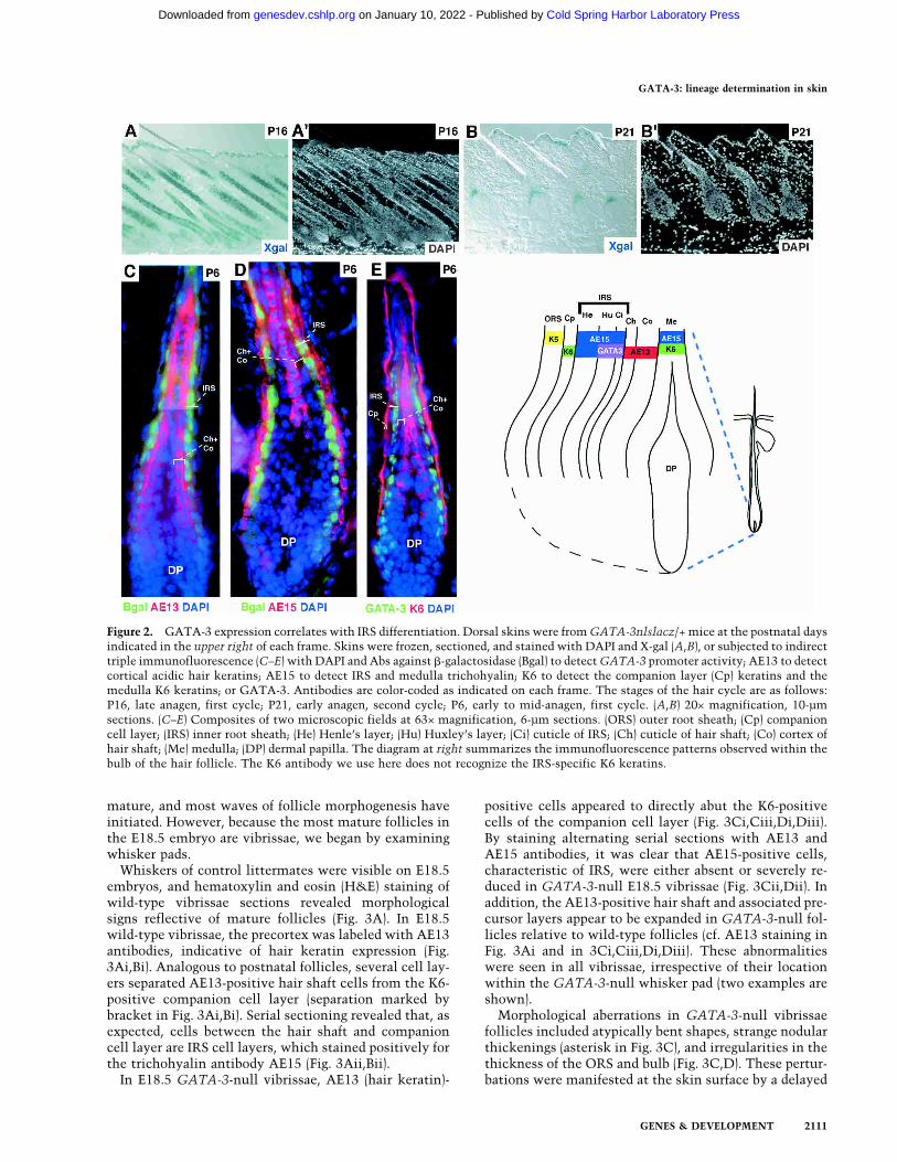
mature and nearing completion of the first growth (ana-gen) phase of the hair cycle (Hardy 1992), GATA-3 wasclearly expressed in follicles as well as epidermis (Fig.2A,A�). Shortly after the synchronous initiation of thefirst postnatal hair cycle, X-gal staining was seen in acone, distinctive of the organization of PreIRS, composedof IRS progenitor cells (Fig. 2B,B�).To determine the precise location of GATA-3 expres-
sion within the hair follicle, we compared GATA-3 pro-moter and protein expression to the expression of well-established biochemical markers for follicles. As judgedby both GATA-3-promoter-driven �-galactosidase ex-pression and GATA-3 protein expression, GATA-3 ex-pression was restricted to two cell layers of the follicle(green in Fig. 2C–E). These layers were located directlyexternal to the hair shaft cuticle and cortex/precortex,marked by an AE13 monoclonal antibody specific forhair-specific keratins (Fig. 2C, red; Lynch et al. 1986). Atthe base of the follicle, the two �-galactosidase-positive
layers were directly internal to a single layer of IRS cellslabeling with the monoclonal antibody AE15 (Fig. 2D,red) specific for trichohyalin, a marker of IRS and post-natal, differentiating medulla (Me) cells (O’Guin et al.1992). Further up, the two �-galactosidase-positive celllayers colabeled with AE15 antibodies, which now de-tected all three IRS layers.Furthermore, a single layer of unlabeled nuclei (Hen-
le’s layer) was present between the K6-positive compan-ion cell layer and the two GATA-3-positive Huxley’s andcuticle layers of IRS (Fig. 2E). Although some K6 genesare also expressed in IRS (Winter et al. 1998; Langbein etal. 2003), the K6 antibody used here did not recognizethese isoforms. In addition, two cell layers, namely, thehair shaft cuticle (Ch) and the cortex (Co), separatedAE15- and K6-positive medulla from GATA-3-positiveIRS cuticle (Fig. 2D,E). The diagram in Figure 2 summa-rizes the localization of markers relative toGATA-3 pro-moter and GATA-3 protein expression.
Defects in GATA-3-null embryonic skin
With a firm grasp of the skin expression patterns ofGATA-3, we began an analysis of GATA-3-null epider-mis in E18.5 embryos, the latest stage to which theseanimals survive. At this stage, back skin epidermis is
Figure 1. GATA-3 is expressed in embryonic skin epi-thelium. (A,B) Whole mount in situ hybridization with aGATA-3 digoxygenin cRNA probe (brown). (A) Whole em-bryo hybridization, depicting GATA-3mRNA in develop-ing vibrissae (Vib) at E14.5. (B) Frozen section of skin fromE17 whole mount in situ hybridization. Note strong hy-bridization in suprabasal epidermis and progenitor cellsfor the IRS (preIRS). (C–E) Frozen sections of GATA-3nlslacZ/+ embryos, at ages indicated in lower left of eachframe. Sections were subjected to either X-gal staining(C,D,E) or immunofluorescence (C�,D�,E�) with DAPI todetect nuclei, and with antibodies (Abs) against �-galac-tosidase (Bgal) to detect GATA-3 promoter activity andagainst K5 to detect the basal epidermal layer and follicleORS. Abs are color-coded in the lower right of each frame.(Epi) Epidermis; (Der) dermis; (HF) hair follicle; (DP) der-mal papilla; (ORS) outer root sheath; (M) matrix.
Kaufman et al.
2110 GENES & DEVELOPMENT
Cold Spring Harbor Laboratory Press on January 10, 2022 - Published by genesdev.cshlp.orgDownloaded from
mature, and most waves of follicle morphogenesis haveinitiated. However, because the most mature follicles inthe E18.5 embryo are vibrissae, we began by examiningwhisker pads.Whiskers of control littermates were visible on E18.5
embryos, and hematoxylin and eosin (H&E) staining ofwild-type vibrissae sections revealed morphologicalsigns reflective of mature follicles (Fig. 3A). In E18.5wild-type vibrissae, the precortex was labeled with AE13antibodies, indicative of hair keratin expression (Fig.3Ai,Bi). Analogous to postnatal follicles, several cell lay-ers separated AE13-positive hair shaft cells from the K6-positive companion cell layer (separation marked bybracket in Fig. 3Ai,Bi). Serial sectioning revealed that, asexpected, cells between the hair shaft and companioncell layer are IRS cell layers, which stained positively forthe trichohyalin antibody AE15 (Fig. 3Aii,Bii).In E18.5 GATA-3-null vibrissae, AE13 (hair keratin)-
positive cells appeared to directly abut the K6-positivecells of the companion cell layer (Fig. 3Ci,Ciii,Di,Diii).By staining alternating serial sections with AE13 andAE15 antibodies, it was clear that AE15-positive cells,characteristic of IRS, were either absent or severely re-duced in GATA-3-null E18.5 vibrissae (Fig. 3Cii,Dii). Inaddition, the AE13-positive hair shaft and associated pre-cursor layers appear to be expanded in GATA-3-null fol-licles relative to wild-type follicles (cf. AE13 staining inFig. 3Ai and in 3Ci,Ciii,Di,Diii). These abnormalitieswere seen in all vibrissae, irrespective of their locationwithin the GATA-3-null whisker pad (two examples areshown).Morphological aberrations in GATA-3-null vibrissae
follicles included atypically bent shapes, strange nodularthickenings (asterisk in Fig. 3C), and irregularities in thethickness of the ORS and bulb (Fig. 3C,D). These pertur-bations were manifested at the skin surface by a delayed
Figure 2. GATA-3 expression correlates with IRS differentiation. Dorsal skins were fromGATA-3nlslacz/+ mice at the postnatal daysindicated in the upper right of each frame. Skins were frozen, sectioned, and stained with DAPI and X-gal (A,B), or subjected to indirecttriple immunofluorescence (C–E) with DAPI and Abs against �-galactosidase (Bgal) to detectGATA-3 promoter activity; AE13 to detectcortical acidic hair keratins; AE15 to detect IRS and medulla trichohyalin; K6 to detect the companion layer (Cp) keratins and themedulla K6 keratins; or GATA-3. Antibodies are color-coded as indicated on each frame. The stages of the hair cycle are as follows:P16, late anagen, first cycle; P21, early anagen, second cycle; P6, early to mid-anagen, first cycle. (A,B) 20× magnification, 10-µmsections. (C–E) Composites of two microscopic fields at 63× magnification, 6-µm sections. (ORS) outer root sheath; (Cp) companioncell layer; (IRS) inner root sheath; (He) Henle’s layer; (Hu) Huxley’s layer; (Ci) cuticle of IRS; (Ch) cuticle of hair shaft; (Co) cortex ofhair shaft; (Me) medulla; (DP) dermal papilla. The diagram at right summarizes the immunofluorescence patterns observed within thebulb of the hair follicle. The K6 antibody we use here does not recognize the IRS-specific K6 keratins.
GATA-3: lineage determination in skin
GENES & DEVELOPMENT 2111
Cold Spring Harbor Laboratory Press on January 10, 2022 - Published by genesdev.cshlp.orgDownloaded from

eruption of GATA-3-null relative to wild-type vibrissaeshafts (Fig. 3E, see insets).Elsewhere within E18.5 skin, few differences were
noted. As a measure of epidermal function, E18.5 knock-out (KO) embryos were able to exclude blue dye, indicat-ing that the epidermal barrier was intact (Fig. 3E). Analy-ses of E17.5 embryos revealed a slight delay in barrierfunction acquisition at this earlier age, as judged by thecomplete penetration of blue dye through GATA-3-nullskin relative to only partial penetration through wild-
type E17.5 skin (Fig. 3E). Overall, however, the mor-phology and biochemistry of GATA-3-null epidermisthroughout E18.5 embryos was largely similar to wild-type epidermis (Fig. 4). Thus, basal (BL), spinous (SL),granular (GL), and cornified (CL) layers were all presentand of comparable size and thickness. Wild-type and KOepidermis displayed normal staining for the basal layerkeratin 5, the suprabasal layer keratin 1, and the late-stage differentiation markers involucrin, loricrin, andfilaggrin.
Figure 3. Whisker follicles develop by E18.5, but they are aberrant in GATA-3-null embryos. Whisker pads of E18.5 wild-type (WT)and GATA-3nlslacz(z/z) (GATA-3-null) embryos were frozen and sectioned (10 µm). Serial sections (denoted by i, ii, iii) of twowild-type follicles (A,B) and two KO follicles (C,D) are shown. Sections were either subjected to H&E staining (A–D) or triple indirectimmunofluorescence microscopy using the antibodies indicated in the lower right of each frame. Color coding reflects the secondaryAbs used for detection. Brackets in Ai and Bi denote a gap between the hair cortex (red, AE13) and companion cell layer (green, K6).This gap represents the IRS (red, AE15; Aii and Bii), which is sandwiched between the companion layer and the cortex of wild-typefollicles. In KO follicles, AE15 staining is largely absent, and K6 and AE13 staining are adjacent to one another. Additional abnor-malities in embryonic KO follicles include nodular thickenings in the vibrissae shafts (marked with an asterisk; C), irregular bulgesin the ORS (e.g., double-headed arrow; D), and a shortened, bent follicle of enlarged diameter at the bulb. Images are shown at 20×magnification. Panels are composites of two or three microscopic fields. (E) Barrier function analyses. The ability of whole embryosto exclude blue dye was used to examine the epidermal barrier, normally acquired beginning at E17.5 and complete by E18.5. Note thecomplete penetration of blue dye in a representative GATA-3-null E17.5 embryo, in contrast to the comparably sized E17.5 wild-typeembryo, which has the characteristic dorsal pattern of dye exclusion. By E18.5, both wild-type and null embryos have acquired barrierfunction based on this assay. Note, however, that whiskers have not protruded from the muzzle of the KO embryo, in contrast to thewild-type littermate.
Kaufman et al.
2112 GENES & DEVELOPMENT
Cold Spring Harbor Laboratory Press on January 10, 2022 - Published by genesdev.cshlp.orgDownloaded from

Morphological and biochemical signs of IRS and hairshaft differentiation are largely absent in back skin priorto birth (e.g., see Fig. 4A,B). The lack of overt abnormali-ties in the back skin of even the oldest living GATA-3KO embryos explains why skin defects had not beennoted previously in these animals (Lim et al. 2000).
Skin grafting reveals gross defects in postnataldevelopment of GATA-3-null hairs
A priori, the selective abnormalities in E18.5 GATA-3-null vibrissae could be attributed either to a special rolefor GATA-3 in vibrissae or to their early development. Inaddition, given the well-established role of GATA-3 inhematopoiesis and CNS/PNS development, it was pos-sible that the hair follicle defects were attributable toindirect rather than autonomous effects. To distinguishbetween these possibilities, we grafted full thicknessE17.5 skin from GATA-3-null and wild-type littermate
embryos onto backs of recipient nu/nu mice. Mice withthe nu/nu mutation are immunocompromised as theylack T cells and cannot reject the graft. They also lacknearly all external hair shaft formation, but are wild typefor GATA-3 (Flanagan 1966; Nehls et al. 1994; Segre etal. 1995).When bandages were removed from recipient mice at 7
d after engraftment, hair shafts were already evident onwild-type grafts but not on GATA-3-null grafts (Fig.5A,B; examples shown are day 9 post engraftment).Grafts were monitored daily, and in all 6 of 6 grafts, KOskin consistently failed to develop the thick hair coatdisplayed by wild-type grafts (Fig. 5C–F). Instead, KOgrafts developed short “stubble” that failed to progressinto a normal hair coat (Fig. 5D,F).In wild-type grafts, all three major hair types, includ-
ing awl, guard, and zig-zag hairs, were readily identifiedupon microscopic examination of plucked hairs (Fig.5G). In contrast, hairs from GATA-3-null grafts were
Figure 4. Classical morphological and biochemicalfeatures of epidermal differentiation are normal inGATA-3-null animals. Panels are from an E18.5 wild-type littermate (left set) and a GATA-3-null embryo(right set). (A,B) Dorsal back skins were fixed in Eponand embedded, and semithin sections (1 µm) werestained with toluidine blue. Note the presence of basal(BL), spinous (SL), granular (GL), and cornified (CL) lay-ers, which are largely similar between wild-type andKO skin. Note also similarities in wild-type and KOpelage hair follicles (HF), which at this stage have notbegun to differentiate into shaft, IRS, and ORS. (C–J)Frozen sections of E18.5 dorsal back skins were sub-jected to triple indirect immunofluorescence usingDAPI to label the nuclei and the following antibodies:K5 to label the epidermal basal layer; K1 and involucrin(Inv; in mouse skin, Inv is more suprabasal than in hu-man) to label the spinous layers; filaggrin (Fil) and loric-rin (Lor) to mark the granular layers (Fil detects pre- andprocessed filaggrin). Similar normal biochemical pat-terns of staining were observed for mature epidermis,obtained from grafted KO and wild-type skins.
GATA-3: lineage determination in skin
GENES & DEVELOPMENT 2113
Cold Spring Harbor Laboratory Press on January 10, 2022 - Published by genesdev.cshlp.orgDownloaded from

shorter and wider and did not fall into any standard clas-sification (Fig. 5H). As judged by scanning electron mi-croscopy, the fine structure of these hairs was highlyirregular. Wild-type follicles displayed a “shingle-like”pattern, characteristic of flat cuticle cells that constitutethe outer layer of normal hair shafts (Fig. 5I). Althoughflat cuticle cells were present in KO shafts, their organi-zation was highly abnormal (Fig. 5J). Additionally, KOhair shafts were short, and their diameter was largeand irregular compared with their wild-type counter-parts. The nodules, bulges, and bends in GATA-3-nullhairs paralleled the follicle defects seen below the skinsurface in embryonic GATA-3-null vibrissae follicles(see Fig. 3).
Expansion of hair shaft and hair shaft precursor cellsin GATA-3-null folliclesThe skin grafts enabled us to examine changes in expres-sion patterns of hair follicle proteins in mature pelagefollicles, replete with hair shafts. Immunofluorescenceanalyses revealed severe abnormalities in the expressionpatterns of hair follicle proteins within GATA-3-nullskin grafts (Fig. 6). As judged by AE13 staining (red) todetect the cortical keratins of hair shafts, GATA-3-nullfollicles exhibited a markedly expanded precortex andcortex as compared with wild-type grafts (Fig. 6A–D).These differences were further documented by a largeincrease in Lef-1-positive precortical cell nuclei in KOversus wild-type follicles (Fig. 6E,F). In addition, there
Figure 5. GATA-3-null skin grafts develop strikingly abnormal hairs. Full thickness skins from six wild-type and six GATA-3-null(KO) E17.5 CD-1 albino embryos were grafted onto nu/numice. At 9 d after engraftment, hairs (white) appeared in all wild-type grafts(A) but were absent in all GATA-3-null grafts (B). By 27–29 d, a thick pelage hair coat had developed on all wild-type grafts (C and Eshow representative examples of grafts from skins of two different wild-type animals), but the KO grafts produced only short, stubbyhairs (D and F show representative examples of grafts from skins of two different KO animals). The three major hair types (awl, guard,and zig-zag) were evident among hairs plucked from wild-type grafts (examples shown in G) at 12 wk postengraftment. In contrast,hairs from equivalent GATA-3-null grafts (representative examples shown in H) did not fit these classifications and were shorter andthicker than wild-type shafts. (I,J) Scanning electron microscopy (SEM) analysis. SEM (600×) of the surface of wild-type and KO graftedskin at 12 wk postengraftment. The outer surface of each hair shaft is the cuticle layer, which is highly ordered in the wild-type butnot KO mice. Note additional irregularities in the shafts, reflecting in detail the differences noted at lower magnification.
Kaufman et al.
2114 GENES & DEVELOPMENT
Cold Spring Harbor Laboratory Press on January 10, 2022 - Published by genesdev.cshlp.orgDownloaded from

was an expansion of differentiated medulla cells express-ing trichohyalin (denoted by ∨ in Fig. 6F; cf. WT in Fig.6E), which was flanked by the AE13-positive cortex (cf. ∨in Fig. 6C and 6F). Ultrastructural analyses later con-firmed the identity of these cells as medulla rather thana misplaced core of IRS cells.
Paucity of differentiated IRS cells but expansionof IRS precursor cells in GATA-3-null skin
The increases in cortical and medulla cells were paral-leled by a paucity of differentiated IRS cells labeled withAE15 antibodies against trichohyalin. Thus, in contrastto the two robust stripes of wild-type IRS stained withAE15 antibodies (denoted by brackets in Fig. 6E), onlywisps of AE15-positive KO IRS cells were detected(bracket in Fig. 6F). Additionally, these remnants of whatappeared biochemically to be IRS were always foundwell above the bulb of the follicle and above the initia-
tion of medulla differentiation (see arrow in Fig. 6F). Inmarked contrast, IRS differentiation in wild-type graftsalways initiated in a horizontal plane near the base of thebulb, comparable to that of the Lef-1-positive corticalprecursors and well below the differentiating medulla(arrows in Fig. 6E).Despite the paucity of trichohyalin-positive IRS cells
of GATA-3-null grafted skin, the K6-positive companionlayer appeared to be largely normal in size and location(Fig. 6G–J). However, whereas the companion layer inwild-type follicles flanked the AE13-negative/AE15-positive IRS cells (marked with brackets in Fig. 6G,I), itflanked the AE13-positive/AE15-negative cortical cellsin KO follicles (Fig. 6H,J). Overall, these results provideadditional evidence that differentiating IRS cells arelargely absent in GATA-3-null follicles.We were intrigued by the presence of a large group of
Lef-1-negative epithelial cells surrounding the Lef-1-positive cortical precursor cells near the base of the hair
Figure 6. Mature GATA-3-null hair follicles exhibit biochemical signs of an expanded cortex and medulla and a severely reducedinner root sheath. (A–J) Matched P35 skin grafts from E17.5 GATA-3-null (KO) and wild-type littermates were frozen and sectioned(10 µm) and subjected to triple indirect immunofluorescence microscopy. Most follicles were in the anagen phase of the hair cycle. Theupper right denotes genotype of skin. The antibodies used are noted on each frame and are color-coded according to the secondary Abused for detection. DAPI was used to mark the nuclei, AE13 for cortical keratins, AE15 for IRS and medulla trichoyalin, anti-K6 forthe companion layer and medulla (this antibody does not detect the IRS-K6 keratins), and Lef-1 to mark the precortical cells. ∨ denotesthe wide stripe of AE13-negative, AE15-positive cells, corresponding to the medulla at the hair shaft center. Note the expandedLef-1-positive precortex and AE13-positive cortex in mutant follicles. Brackets denote the AE15-positive IRS, largely absent in the KOfollicles. Arrows indicate the lowest extent of AE15-positive IRS staining.
GATA-3: lineage determination in skin
GENES & DEVELOPMENT 2115
Cold Spring Harbor Laboratory Press on January 10, 2022 - Published by genesdev.cshlp.orgDownloaded from

bulb (Fig. 6F). Could these Lef-1-negative cells be IRSprecursors? To examine this possibility, we took advan-tage of the functional LacZ coding sequence inserteddownstream from the GATA-3 promoter of the KO ani-mals. Previously, we had shown that GATA-3nlslacZ+/−
follicles displayed a pattern of �-galactosidase activitythat faithfully paralleled the two rows of anti-GATA-3immunoreactivity demarcating the Huxley’s and cuticlelayers of the IRS (see Fig. 2). We now tracked the expres-sion pattern of GATA-3 promoter activity under condi-tions in which GATA-3 protein was missing. Surpris-ingly, GATA-3-null follicles displayed a gross expansion
of GATA-3-promoter-active (�-gal-positive) cells (Fig. 7).Thus, rather than two neat rows as in wild-type follicles(Fig. 7A), extensive GATA-3 promoter activity was de-tected within cells of the follicle bulbs of GATA-3 pro-tein-deficient follicles (Fig. 7B,C). Most of these cellswere negative for AE13 (Fig. 7C) and AE15 staining (seeFig. 6). Thus, neither their position nor their expressionpattern was consistent with a precortical identity.To search for markers that might characterize these
GATA-3-promoter-active bulb cells, we examined theexpression of FOG1 (Friend of GATA-1), which oftenfunctions with GATA factors, including GATA-3, to
Figure 7. Expansion and aberrancies in expression patterns of cortical and IRS precursor populations in GATA-3-null follicles. Skinsfrom matched P35 wild-type and GATA-3-null grafts were frozen, sectioned (10 µm), and subjected to either triple indirect immu-nofluorescence (A–K) or in situ hybridization (L,M). The genotype of each section is denoted at the upper right. Abs used arecolor-coded and indicated across the bottom of each frame. Asterisks in B and I denote autofluorescence, an artifact of the knots ofhighly keratinized material in some KO shafts. Boxed areas are shown at higher magnification in the insets. DAPI (blue) denotes nuclei.Abs were against GATA-3, present in the IRS lineage, and absent in the GATA-3-null sections; �-galactosidase (Bgal), specific forGATA-3 promoter activity, which was indistinguishable from the anti-GATA IRS labeling in wild-type follicles, and still present inthe expanded IRS precursor population of KO follicles; AE13, specific for the keratins of differentiating cortical cells; FOG1, acorepressor for GATA-3 protein, present in IRS precursor cells, where it marked the nuclei of wild-type but the cytoplasm of KO cells(D and E, respectively; see inset to E as well); Lef-1, specific for cortical precursor cells, expanded in the KO; Ki67, specific forproliferating cell nuclei; and K5, specific for the ORS of the follicle. The cRNA probe is against Shh, Sonic hedgehog, which marks asmall subset of matrix cells in wild-type follicles, unchanged in KO follicles. Note that in KO follicles, some AE13-positive corticaland Lef-1-positive precortical cells were also positive for GATA-3 promoter activity (representative cells indicated by arrows in C andG). Note also that Ki67 labeling confirms the anagen state of both wild-type and KO follicles, and some Ki67-positive cells were alsopositive for GATA-3 promoter activity (J,K).
Kaufman et al.
2116 GENES & DEVELOPMENT
Cold Spring Harbor Laboratory Press on January 10, 2022 - Published by genesdev.cshlp.orgDownloaded from
suppress GATA’s action as an activator (Tsang et al.1997; Zhou et al. 2001). Interestingly, in wild-type fol-licles, an anti-FOG1 antibody labeled the same two IRSlayers that labeled with GATA-3 antibodies (Fig. 7D).Antibodies against GATA-3 and FOG1 both localized tothe nuclei of these wild-type IRS precursor cells. InGATA-3-null follicles, anti-FOG1 costained with anti-�-galactosidase antibodies in the expanded zone of GATA-3-promoter-active cells (Fig. 7E). In the absence ofGATA-3, however, anti-FOG labeling shifted fromnucleus to cytoplasm (Fig. 7E, inset), opening the possi-bility that GATA-3 and FOG-1 may function together atsome point in IRS specification. The increased popula-tion of Lef-1-positive precortical cells in GATA-3-nullfollicles was interesting and suggested an interdepen-dency of the development of IRS and cortical progeni-tors. Given the marked aberrations in hair shaft, how-ever, we wondered whether the expanded compartmentsof IRS and cortical precursors were truly normal. Closerinspection of GATA-3-promoter-active cells revealed anumber of cells whose nuclei were positive for both �-ga-lactosidase and cortical keratins (examples marked byarrows in magnified photos in Fig. 7C). Conversely, someof the Lef-1-positive cortical precursor cells were alsopositive for �-galactosidase (Fig. 7G, arrows in enlargedphotos indicate an example of a cell expressing bothLef-1 and �-gal). Such mixing of biochemical markerswas not seen in wild-type follicles, where Lef-1-positiveprecortical cells were distinct in their expression patternfrom the compartment of GATA-3/FOG1/GATA-3-pro-moter-active IRS precursor cells (Figs. 2C, 7F).Given the expansion of precursor compartments for
GATA-3-null IRS and cortex, we examined the status ofmatrix cells, which are thought to give rise to these pre-cursor populations. In both wild-type and KO follicles,antibodies against the proliferating nuclear antigen,Ki67, revealed intense labeling of the matrix cells, whichin anagen, are rapidly proliferating (Fig. 7H,I, green).Overall, the number of Ki67-positive cells was similar inwild-type and KO follicles.Finally, we examined the status of Sonic hedgehog
(Shh), a regulator of follicle morphogenesis that is nor-mally expressed in a small zone of matrix cells (Gam-bardella et al. 2000). In mutant mice deficient in theCCATT box displacement factor, CDP, Shh mRNAs areabsent from this zone and instead activated in the regioncorresponding to the IRS (Ellis et al. 2001). Because bothCDP and GATA-3 mutations lead to IRS abnormalities,we assessed whether such misregulation might occur inthe absence of GATA-3 as well. As shown in Figure 7Land M, the expression of Shh mRNAs appeared to beunaffected in the GATA-3 mutant follicles. The signifi-cance of this and other differences is discussed in moredetail below.
Morphological confirmation of the biochemical defectsin GATA-3-null hair follicles
Ultrastructural analysis of wild-type and GATA-3-nullgraft skin by transmission electron microscopy helped to
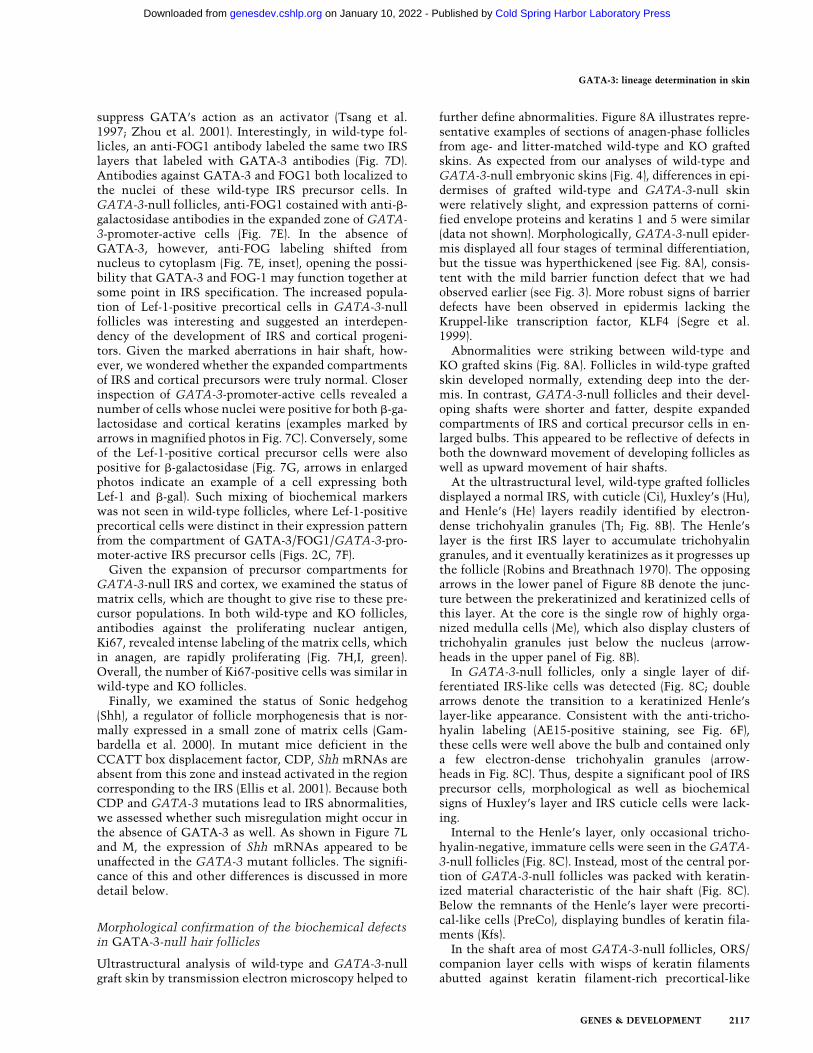
further define abnormalities. Figure 8A illustrates repre-sentative examples of sections of anagen-phase folliclesfrom age- and litter-matched wild-type and KO graftedskins. As expected from our analyses of wild-type andGATA-3-null embryonic skins (Fig. 4), differences in epi-dermises of grafted wild-type and GATA-3-null skinwere relatively slight, and expression patterns of corni-fied envelope proteins and keratins 1 and 5 were similar(data not shown). Morphologically, GATA-3-null epider-mis displayed all four stages of terminal differentiation,but the tissue was hyperthickened (see Fig. 8A), consis-tent with the mild barrier function defect that we hadobserved earlier (see Fig. 3). More robust signs of barrierdefects have been observed in epidermis lacking theKruppel-like transcription factor, KLF4 (Segre et al.1999).Abnormalities were striking between wild-type and
KO grafted skins (Fig. 8A). Follicles in wild-type graftedskin developed normally, extending deep into the der-mis. In contrast, GATA-3-null follicles and their devel-oping shafts were shorter and fatter, despite expandedcompartments of IRS and cortical precursor cells in en-larged bulbs. This appeared to be reflective of defects inboth the downward movement of developing follicles aswell as upward movement of hair shafts.At the ultrastructural level, wild-type grafted follicles
displayed a normal IRS, with cuticle (Ci), Huxley’s (Hu),and Henle’s (He) layers readily identified by electron-dense trichohyalin granules (Th; Fig. 8B). The Henle’slayer is the first IRS layer to accumulate trichohyalingranules, and it eventually keratinizes as it progresses upthe follicle (Robins and Breathnach 1970). The opposingarrows in the lower panel of Figure 8B denote the junc-ture between the prekeratinized and keratinized cells ofthis layer. At the core is the single row of highly orga-nized medulla cells (Me), which also display clusters oftrichohyalin granules just below the nucleus (arrow-heads in the upper panel of Fig. 8B).In GATA-3-null follicles, only a single layer of dif-
ferentiated IRS-like cells was detected (Fig. 8C; doublearrows denote the transition to a keratinized Henle’slayer-like appearance. Consistent with the anti-tricho-hyalin labeling (AE15-positive staining, see Fig. 6F),these cells were well above the bulb and contained onlya few electron-dense trichohyalin granules (arrow-heads in Fig. 8C). Thus, despite a significant pool of IRSprecursor cells, morphological as well as biochemicalsigns of Huxley’s layer and IRS cuticle cells were lack-ing.Internal to the Henle’s layer, only occasional tricho-
hyalin-negative, immature cells were seen in theGATA-3-null follicles (Fig. 8C). Instead, most of the central por-tion of GATA-3-null follicles was packed with keratin-ized material characteristic of the hair shaft (Fig. 8C).Below the remnants of the Henle’s layer were precorti-cal-like cells (PreCo), displaying bundles of keratin fila-ments (Kfs).In the shaft area of most GATA-3-null follicles, ORS/
companion layer cells with wisps of keratin filamentsabutted against keratin filament-rich precortical-like
GATA-3: lineage determination in skin
GENES & DEVELOPMENT 2117
Cold Spring Harbor Laboratory Press on January 10, 2022 - Published by genesdev.cshlp.orgDownloaded from

cells, which, in turn, were adjacent to cortex and me-dulla (Fig. 8C�). Additionally, medulla cells were mark-edly disorganized and often embedded in a mass of kera-tinized cortical cells (Fig. 8C�). Some medulla cells dis-played trichohyalin granules (arrowheads in 8C�).However, the organization of granules as well as the or-ganization of cells was aberrant (cf. medulla cells in Fig.8C� and in 8B, upper panel). Overall, this analysis con-firmed the medulla identity of the thickened core ofAE15-positive cells in GATA-3-null follicles (see Fig. 6F)and revealed a level of disorganization of the hair shaftthat was even more prominent than that envisionedfrom the immunohistochemical studies.
Discussion
With each new hair cycle, the IRS and hair shaft mustdevelop simultaneously to provide a channel for the newhair to emerge. Yet despite significant advances in ourunderstanding of the molecular controls governing hairshaft differentiation, little is known about the regulationand function of the IRS. Our present studies have nowuncovered a member of the well-studied GATA family oftranscription factors as a major and essential regulator ofIRS differentiation.The structural consequences of IRS loss are severe.
Without the Huxley’s and cuticle layers, the Henle’s
Figure 8. Ultrastructural abnormalities inGATA-3-null follicles. Matched wild-typeand GATA-3-null (KO) skin grafts were fro-zen in OCT (A,B) or fixed, embedded inEpon, and sectioned for ultrathin analyses(C,D). (A) Light microscopic analysis ofH&E-stained 10-µm sections of 5-wk grafts.Wild-type (WT) composite of two 20× micro-scopic fields; KO is visible in one field be-cause of its shorter and fatter structure at allstages of the hair cycle. (B–C�) Ultrastruc-tural analyses of 12-wk grafts. All folliclesare oriented with the skin surface at the top.Magnification bars are denoted on eachframe. (B) Sagittal section of wild-type fol-licle, depicting the thin Henle’s cell layer(He) with uniform density caused by kerati-nization. On the outside, this layer isflanked by the companion cell layer (Cp) andORS and finally the dermal sheath (DS). Onthe inside, the Henle’s layer is flanked by thetrichohyalin (Th)-rich Huxley’s (Hu) layer ofthe IRS, and the thin IRS cuticle (Ci). Thehair shaft (HS) is internal to the IRS, and iscomposed of three major layers: an innercore of medulla (Me) cells with trichoyalingranules below each nucleus (arrowheads),the cortex (Co), and the cuticle of the hairshaft (Ch). The upper and lower panels arefrom the same hair follicle, but the lowerpanel depicts the base of the keratinizedHenle’s layer of the IRS (double arrowheads);cells below this juncture still possesstrichoyalin granules, but are less mature andless organized, as is characteristic of IRS pre-cursor cells. (C–C�) Sections from GATA-3-null follicles. All are views from the regionjust above the follicle bulb. (C) The origins ofthe keratinized cells of a Henle’s-like layerin a KO follicle (double arrows, IRS*). Notethe presence of only a few trichoyalin gran-ules (arrowheads) in the IRS precursor cellsjust below these keratinizing cells, in contrast to the wild-type IRS precursors, which are abundant in trichohyalin (Th; lower panelin B). Note also the presence of precortical cells (PreCo), rich in keratin filaments (Kf), located atypically below the Henle’s cells; thisirregularity was predicted from our biochemical studies (see Fig. 6F). Note further the mass of highly keratinized cortex and medullacentral to the tiny IRS. The sections in C� andC� show that in some areas, the IRS was missing altogether, and the ORS and companionlayer were abutted against the precortex, cortex, andmedulla. Again, note signs of gross disorganization, with as many as three medullacells (Me in C�) stacked horizontally against each other, instead of in the organized linear vertical array seen in wild-type medulla (B,upper panel). Note trichohyalin (arrowheads) in the medulla but not elsewhere (C�).
Kaufman et al.
2118 GENES & DEVELOPMENT
Cold Spring Harbor Laboratory Press on January 10, 2022 - Published by genesdev.cshlp.orgDownloaded from

layer does not develop properly, and without the IRS,gross abnormalities arise in composition and organiza-tion of cells within the hair shaft. Thus, not only are thecortex and medulla dramatically thickened, but in addi-tion, cells no longer spatially align. The shaft develops asa hyperthickened mass of irregular medulla, cortical, andcuticle cells, rather than the architectural wonder of anormal shaft. Perhaps as no other knockout has sographically illustrated, these two thin layers of IRS cellsare critical for the proper differentiation and/or organi-zation of the hair shaft.In recent years, a myriad of genes encoding transcrip-
tion factors have been genetically implicated in follicledevelopment (Cachon-Gonzalez et al. 1994; Nehls et al.1994; van Genderen et al. 1994; Segre et al. 1995; Zhou etal. 1995; Dai et al. 1998; Li et al. 2000; Pennisi et al.2000; Ellis et al. 2001; Ma et al. 2003; Mill et al. 2003).When mutated, only a few of these genes result in de-fects involving the IRS. Of these, hairless (encoding azinc finger protein required for postnatal hair cycling)and nude (encoding foxn1, a forkhead protein) are ex-pressed in both hair shaft and IRS precursors, and displaydefects in both (Cachon-Gonzalez et al. 1994; Nehls etal. 1994; Segre et al. 1995; Lee et al. 1999; Panteleyev etal. 2000). In contrast, although GATA-3’s absence re-sulted in major abnormalities in hair structure, involv-ing both IRS and shaft, it was only expressed in two ofthe IRS layers. This enabled us to conclude that the se-vere hair shaft defects that occurred in the GATA-3-de-ficient mouse originated specifically from transcrip-tional aberrations within the IRS.To date, the highly restricted pattern of GATA-3 ex-
pression is unique, but it is most similar to that of thetranscriptional repressor CDP, which represses genesregulated by CCAAT sequence motifs (Ellis et al. 2001).The Cutl1 gene encoding CDP seemed to be expressedearlier and more broadly in the IRS pathway, where itwas found in the outermost layer of the lower ORS, inIRS progenitor cells of the matrix, and in all three IRSlayers. A few mice harboring mutations in Cutl1 sur-vived their lung defects long enough to exhibit a sparseand abnormal hair coat. The morphology of Cutl1-nullhair follicles was unusual, as just above a relatively nor-mally structured bulb, the IRS formed, but then both IRSand shaft eventually degenerated to form a large follicu-lar cyst, soon appearing more similar to that of nude oreven hairless mutant mice (Ellis et al. 2001).Although both Cutl1- and GATA-3-null follicles ex-
hibited a failure to develop a proper IRS, GATA-3’s ab-sence was felt most strongly in two major ways thatinvolve cell fate specification within the hair follicle.First, it resulted in an accumulation of IRS precursorcells that appeared to be dysfunctional, unable to differ-entiate into trichohyalin- and keratin-producing IRScells. Second, it led to an expansion of seemingly func-tional Lef-1-expressing precortical cells, which differen-tiated into hair keratin-producing cortical cells. Some ofthese Lef-1-positive cells exhibited mixing of IRS andcortical gene expression patterns (Fig. 6C,E). Because wedid not detect such cells in wild-type, GATA-3-contain-
ing follicles (Figs. 2C, 6F), these findings support the ar-gument that the loss of GATA-3 results not only in ex-pansion of selective cell populations within the follicle,but also in some misexpression of genes in these com-partments. In contrast, Cut1-null follicles exhibited sub-stantial misexpression of both trichohyalin and the com-panion layer K6 in the cortex, features that were notobserved in GATA-3-null follicles, as well as strong ex-pression of the Cutl1 promoter throughout the cortex.Furthermore, in Cut1-null vibrissae, Shh was aberrantlyexpressed in the IRS (Ellis et al. 2001), whereas inGATA-3 mutant mice, Shh expression appeared unaf-fected (Fig. 7L,M). It may still be possible that CDP andGATA-3 function in the same pathway to regulate IRSspecification, but the target genes they affect must be atleast partially nonoverlapping.It is interesting that in T-cell development, both
GATA-3 and Lef-1/Tcf1 are necessary early to directlymphoid precursors to become T cells, and later in thelineage, GATA-3 acts independently of Lef-1 in Th cellcommitment (van Genderen et al. 1994; Verbeek et al.1995; Hattori et al. 1996; Ting et al. 1996; Okamura et al.1998; Schilham et al. 1998; Hendriks et al. 1999). In na-ive Th cells, FOG1 and GATA-3 act to inhibit interleu-kin gene expression, and upon Th commitment, FOG1 isdown-regulated (Kurata et al. 2002). GATA-3, which au-toactivates its own expression, is required for Th2 celldifferentiation (Ouyang et al. 2000; Zhou et al. 2001),whereas suppression of GATA-3 promotes Th1 cell de-termination (for review, see Patient and McGhee 2002).Despite some differences, the transcriptional parallels
between T-cell development and hair follicle differentia-tion are quite striking. In the postnatal hair follicle, Lef-1/�-catenin and GATA-3 seem to act in parallel but dis-tinct pathways governing cortical and IRS differentia-tion, respectively. The expansion of the cortical lineagein the GATA-3-deficient state could be analogous to anincrease in Th1 rather than Th2 cell differentiation, andthe up-regulation of GATA-3 promoter activity inGATA-3-deficient IRS precursor cells might be a signthat FOG1 and GATA-3 normally repress this promoterin wild-type IRS precursors. When taken together withthe paucity of differentiated IRS cells, the expandedpopulation of GATA-3-promoter-active/FOG1-positivecells in GATA-3-deficient follicles might represent apopulation of stalled IRS precursors. The enhancedGATA-3 immunoreactivity as IRS precursor cells differ-entiate into IRS cells in wild-type skin is consistent witha transactivating role for GATA-3 later in IRS differen-tiation, perhaps analogous to Th2 cell differentiation.The fact that the structural genes for IRS differentiationare not expressed in the GATA-3-deficient state, despitethe presence of progenitor cells, further supports a posi-tive role for GATA-3 in their expression.In summary, our studies illuminate a new dimension
in our understanding of how hair follicle morphogenesisis transcriptionally controlled. In many respects,GATA-3 appears to be a master regulator of IRS cell lin-eage determination as Lef-1/�-catenin is for cortical lin-eage specification. The matrix offers a rich milieu of en-
GATA-3: lineage determination in skin
GENES & DEVELOPMENT 2119
Cold Spring Harbor Laboratory Press on January 10, 2022 - Published by genesdev.cshlp.orgDownloaded from

vironmental cues that ultimately lead to the differentialspecification of these lineages. In the future, our effortswill be placed on understanding how these signals im-pact on GATA-3 expression as well as identifying thedownstream target genes for this novel and importantregulator of IRS differentiation.
Materials and methods
Mice
The generation and characterization of the GATA-3nlslacZmice has been described (Hendriks et al. 1999; van Doorninck etal. 1999). Briefly, the lacZ gene fused to a nuclear localizationsignal (nls) was placed in-frame at the ATG translational startsite in the GATA-3 locus, inactivating the GATA-3 gene andexpressing �-galactosidase from the endogenous GATA-3 pro-moter. To obtain GATA-3-null embryos that survive to E18.5,we used a modification of a drug rescue regime (Lim et al. 2000;K.C. Lim, pers. comm.). Beginning at E7.5, pregnant GATA-3nlslacZ/+ dams that were mated to GATA-3nlslacZ/+ maleswere given fresh water daily containing 100 µg/mL L-phenyl-ephrine (Sigma P6126), 100 µg/mL isoproterenol (Sigma I5627)and 2 mg/mL ascorbic acid (Sigma A0278). The presence of theGATA-3nlslacZ allele was determined by a combination of PCRscreening, X-gal staining, phenotypic identification of GATA-3-null embryos, and/or loss of GATA-3 immunoreactivity (Lim etal. 2000). The PCR primers used were 5�-TCCTGCGAGCCTGGCTGTCGGA-3�, which recognizes GATA-3 intron 1; 5�-CCTGTAGCCAGCTTTCATAAC-3�, which recognizes lacZ; and5�-GTTGCCTTGACCATCGATGTT-3�, which recognizesGATA-3 exon 2. The reaction conditions were 94°C for 5 min,followed by 45 cycles of 94°C for 30 sec, 53°C for 30 sec, and72°C for 1.2 min. The band sizes are: ∼ 750 bp (wild-type allele)and ∼ 1.1 kb (GATA-3nlslacz allele).
Histology and electron microscopy
Tissues for immunofluorescence and H&E staining were em-bedded in OCT and then frozen immediately on dry ice. Tissuesfor scanning electron microscopy (EM) were fixed in 2.5% glu-taraldehyde in PBS at room temperature for >1 h, processedusing standard techniques (Fujiwara et al. 2002), and visualizedusing a JEOL JSM-35 scanning electron microscope. For trans-mission EM, samples were fixed in 2% glutaraldehyde, 4%formaldehyde, and 2 mM CaCl2 in 0.05 M sodium cacodylatebuffer at room temperature for >1 h and processed for Eponembedding as described (Segre et al. 1999). Samples were visu-alized with a Tecnai G2 transmission electron microscope.
In situ analysis
Digoxygenin probe synthesis was performed according to themanufacturer’s instructions (Roche). Whole mount in situ hy-bridization was described by Conlon and Rossant (1992), withmodifications (Byrne et al. 1994). In situ hybridizations on 10-µm (Shh) and 15-µm (GATA-3) frozen sections were performedas described (Schaeren-Wiemers and Gerfin-Moser 1993).
X-gal staining and immunofluorescence
OCT sections were fixed for 30 sec in 0.1% glutaraldehyde,washed three times in PBS, and incubated in X-gal staining so-lution at 37°C for 1–2 h. OCT sections were then fixed for 10min in 4% PFA in PBS and washed four times for 5 min in PBS.
When staining with mouse monoclonal antibodies, we used thereagents and protocol from the MOM Basic kit (Vector Labs). Inother cases, the following block/diluent was used: 2.5% NDS,2.5% NGS, 1% BSA, 2% gelatin, and 1% Triton X-100 in PBS.The primary antibodies at the indicated concentrations wereAE13 (mouse, 1:50–1:100; Lynch et al. 1986), AE15 (mouse, 1:10; O’Guin et al. 1992), K5 (gp, 1:250; Fuchs lab), GATA-3(mouse, 1:100; Santa Cruz, HCG3-31), �-galactosidase (rabbit,1:400; mouse, 1:100; Harlan; Sigma), K1 (rabbit, 1:250; Fuchslab), loricrin (rabbit, 1:300; Fuchs lab), Lef-1 (rabbit, 1:250; Fuchslab), filaggrin (rabbit, 1:1000; Covance, PRB-417P), K6 (rabbit,1:500; Fuchs lab), involucrin (mouse, 1:200; Babco), Ki67 (rabbit,1:1000; NovoCastra Laboratories Ltd.), and FOG1 (goat, 1:50;Santa Cruz, sc-9361). Relevant FITC- or TxR-conjugated donkeyor goat antibodies (1:100; Jackson Laboratories) were used fordetection of primary antibodies.
Barrier function assay
Embryos were submerged at 37°C for >8 h in a solution of 1.3mM MgCl2, 100 mM NaPO4, 3 mM K3Fe(CN)6, 3 mMK4Fe(CN)6, 0.01% sodium deoxycholate, 0.2% NP-40, and 1mg/mL X-gal, which was adjusted to a pH of 4.5 with HCl(Hardman et al. 1998). At this pH in the absence of the epider-mal barrier, the solution penetrates epidermis, and an endog-enous �-galactosidase-like activity catalyzes production of ablue precipitate.
Skin grafting
Pregnant GATA-3nlslacZ/+ dams were killed at E17.5, and em-bryos were removed. Full thickness skins were removed fromtorsos of wild-type and GATA-3-null embryos, spread on a ster-ile plastic dish, and stored briefly at 4°C. During this time, eachskin graft recipient site was prepared by removal of a patch offull thickness skin on an anesthetized female nu/nu CD-1mouse. Embryonic donor skin was then placed on the graft bedand secured by sterile gauze and cloth bandages. A total of 12grafts were placed (six wild-type and sixGATA-3-null), and eachshowed a consistent phenotype dependent on the presence orabsence of GATA-3 in the donor skin.
Acknowledgments
A portion of this research was conducted at The University ofChicago, where C.K. and D.B. are enrolled as MD/Ph.D stu-dents. We thank Frank Grosveld (Erasmus University, Rotter-dam, The Netherlands) for GATA-3nlslacZ+/− mice, Linda De-genstein and Lisa Polak for assistance with mouse husbandry,T.T. Sun (NY University School of Medicine) for AE13 andAE15 antibodies, R. Cannon (University of North Carolina,Chapel Hill, NC) for the GATA-3 probe, C. Tabin (HarvardMedical School, Boston, MA) for the Shh probe, and N. Lampen(EM facility at Memorial Sloan-Kettering Cancer Center) forassistance with scanning electron microscopy. C.K. and D.B. aresupported by the Medical Scientist National Research ServiceAward, NIH/National Institute of General Medical Sciences,Grant No. 5 T32 GM07281. D.B. also receives support from TheUniversity of Chicago, Division of Biological Sciences, NaomiRagins Goldsmith Fund. K.-C.L. is supported by NIH Grant GM28896. E.F. is an HHMI Investigator. This research was fundedby a grant from the National Institutes of Health.The publication costs of this article were defrayed in part by
payment of page charges. This article must therefore be hereby
Kaufman et al.
2120 GENES & DEVELOPMENT
Cold Spring Harbor Laboratory Press on January 10, 2022 - Published by genesdev.cshlp.orgDownloaded from

marked “advertisement” in accordance with 18 USC section1734 solely to indicate this fact.
References
Byrne, C., Tainsky, M., and Fuchs, E. 1994. Programming geneexpression in developing epidermis. Development120: 2369–2383.
Cachon-Gonzalez, M.B., Fenner, S., Coffin, J.M., Moran, C.,Best, S., and Stoye, J.P. 1994. Structure and expression of thehairless gene of mice. Proc. Natl. Acad. Sci. 91: 7717–7721.
Cantor, A.B. and Orkin, S.H. 2002. Transcriptional regulation oferythropoiesis: An affair involving multiple partners. Onco-gene 21: 3368–3376.
Carlsson, P., Waterman, M.L., and Jones, K.A. 1993. The hLEF/TCF-1 � HMG protein contains a context-dependent tran-scriptional activation domain that induces the TCR � en-hancer in T cells. Genes & Dev. 7: 2418–2430.
Chan, E.F., Gat, U., McNiff, J.M., and Fuchs, E. 1999. A com-mon human skin tumour is caused by activating mutationsin �-catenin. Nat. Genet. 21: 410–413.
Conlon, R.A. and Rossant, J. 1992. Exogenous retinoic acid rap-idly induces anterior ectopic expression of murine Hox-2genes in vivo. Development 116: 357–368.
Cotsarelis, G., Sun, T.T., and Lavker, R.M. 1990. Label-retain-ing cells reside in the bulge area of pilosebaceous unit: Im-plications for follicular stem cells, hair cycle, and skin car-cinogenesis. Cell 61: 1329–1337.
Dai, X., Schonbaum, C., Degenstein, L., Bai, W., Mahowald, A.,and Fuchs, E. 1998. The ovo gene required for cuticle forma-tion and oogenesis in flies is involved in hair formation andspermatogenesis in mice. Genes & Dev. 12: 3452–3463.
DasGupta, R. and Fuchs, E. 1999. Multiple roles for activatedLEF/TCF transcription complexes during hair follicle devel-opment and differentiation. Development 126: 4557–4568.
Dry, F.W. 1926. The coat of the mouse (Mus musculum). J.Genet. 16: 287–340.
Ellis, T., Gambardella, L., Horcher, M., Tschanz, S., Capol, J.,Bertram, P., Jochum, W., Barrandon, Y., and Busslinger, M.2001. The transcriptional repressor CDP (Cutl1) is essentialfor epithelial cell differentiation of the lung and the hairfollicle. Genes & Dev. 15: 2307–2319.
Flanagan, S.P. 1966. ‘Nude,’ a new hairless gene with pleiotropiceffects in the mouse. Genet. Res. 8: 295–309.
Fuchs, E., Merrill, B.J., Jamora, C., and DasGupta, R. 2001. Atthe roots of a never-ending cycle. Dev. Cell 1: 13–25.
Fujiwara, T., Dehart, D.B., Sulik, K.K., and Hogan, B.L. 2002.Distinct requirements for extra-embryonic and embryonicbone morphogenetic protein 4 in the formation of the nodeand primitive streak and coordination of left–right asymme-try in the mouse. Development 129: 4685–4696.
Gambardella, L., Schneider-Maunoury, S., Voiculescu, O., Char-nay, P., and Barrandon, Y. 2000. Pattern of expression of thetranscription factor Krox-20 in mouse hair follicle. Mech.Dev. 96: 215–218.
Gat, U., DasGupta, R., Degenstein, L., and Fuchs, E. 1998. Denovo hair follicle morphogenesis and hair tumors in miceexpressing a truncated �-catenin in skin. Cell 95: 605–614.
Giese, K. and Grosschedl, R. 1993. LEF-1 contains an activationdomain that stimulates transcription only in a specific con-text of factor-binding sites. EMBO J. 12: 4667–4676.
Hardman, M.J., Sisi, P., Banbury, D.N., and Byrne, C. 1998. Pat-terned acquisition of skin barrier function during develop-ment. Development 125: 1541–1552.
Hardy, M.H. 1992. The secret life of the hair follicle. Trends
Genet. 8: 55–61.Hattori, N., Kawamoto, H., Fujimoto, S., Kuno, K., and Katsura,
Y. 1996. Involvement of transcription factors TCF-1 andGATA-3 in the initiation of the earliest step of T cell devel-opment in the thymus. J. Exp. Med. 184: 1137–1147.
Hendriks, R.W., Nawijn, M.C., Engel, J.D., van Doorninck, H.,Grosveld, F., and Karis, A. 1999. Expression of the transcrip-tion factor GATA-3 is required for the development of theearliest T cell progenitors and correlates with stages of cel-lular proliferation in the thymus. Eur. J. Immunol. 29: 1912–1918.
Kuo, C.T. and Leiden, J.M. 1999. Transcriptional regulation of Tlymphocyte development and function. Annu. Rev. Immu-nol. 17: 149–187.
Kurata, H., Lee, H.J., McClanahan, T., Coffman, R.L., O’Garra,A., and Arai, N. 2002. Friend of GATA is expressed in naiveTh cells and functions as a repressor of GATA-3-mediatedTh2 cell development. J. Immunol. 168: 4538–4545.
Lakshmanan, G., Lieuw, K.H., Lim, K.C., Gu, Y., Grosveld, F.,Engel, J.D., and Karis, A. 1999. Localization of distant uro-genital system-, central nervous system-, and endocardium-specific transcriptional regulatory elements in the GATA-3locus. Mol. Cell. Biol. 19: 1558–1568.
Langbein, L., Rogers, M.A., Praetzel, S., Winter, H., andSchweizer, J. 2003. K6irs1, K6irs2, K6irs3, and K6irs4 repre-sent the inner-root-sheath-specific type II epithelial keratinsof the human hair follicle. J. Invest. Dermatol. 120: 512–522.
Lee, D., Prowse, D.M., and Brissette, J.L. 1999. Association be-tween mouse nude gene expression and the initiation of epi-thelial terminal differentiation. Dev. Biol. 208: 362–374.
Li, M., Indra, A.K., Warot, X., Brocard, J., Messaddeq, N., Kato,S., Metzger, D., and Chambon, P. 2000. Skin abnormalitiesgenerated by temporally controlled RXR� mutations inmouse epidermis. Nature 407: 633–636.
Lim, K.C., Lakshmanan, G., Crawford, S.E., Gu, Y., Grosveld, F.,and Engel, J.D. 2000. Gata3 loss leads to embryonic lethalitydue to noradrenaline deficiency of the sympathetic nervoussystem. Nat. Genet. 25: 209–212.
Lynch, M.H., O’Guin, W.M., Hardy, C., Mak, L., and Sun, T.T.1986. Acidic and basic hair/nail (“hard”) keratins: Their co-localization in upper cortical and cuticle cells of the humanhair follicle and their relationship to “soft” keratins. J. CellBiol. 103: 2593–2606.
Ma, L., Liu, J., Wu, T., Plikus, M., Jiang, T.X., Bi, Q., Liu, Y.H.,Muller-Rover, S., Peters, H., Sundberg, J.P., et al. 2003. ‘Cy-clic alopecia’ in Msx2 mutants: Defects in hair cycling andhair shaft differentiation. Development 130: 379–389.
Mill, P., Mo, R., Fu, H., Grachtchouk, M., Kim, P.C., Dlugosz,A.A., and Hui, C.C. 2003. Sonic hedgehog-dependent activa-tion of Gli2 is essential for embryonic hair follicle develop-ment. Genes & Dev. 17: 282–294.
Millar, S.E. 2002. Molecular mechanisms regulating hair follicledevelopment. J. Invest. Dermatol. 118: 216–225.
Nehls, M., Pfeifer, D., Schorpp, M., Hedrich, H., and Boehm, T.1994. New member of the winged-helix protein family dis-rupted in mouse and rat nude mutations. Nature 372: 103–107.
Niemann, C. and Watt, F.M. 2002. Designer skin: Lineage com-mitment in postnatal epidermis. Trends Cell Biol. 12: 185–192.
O’Guin, W.M., Sun, T.T., and Manabe, M. 1992. Interaction oftrichohyalin with intermediate filaments: Three immuno-logically defined stages of trichohyalin maturation. J. Invest.Dermatol. 98: 24–32.
Okamura, R.M., Sigvardsson, M., Galceran, J., Verbeek, S.,Clevers, H., and Grosschedl, R. 1998. Redundant regulation
GATA-3: lineage determination in skin
GENES & DEVELOPMENT 2121
Cold Spring Harbor Laboratory Press on January 10, 2022 - Published by genesdev.cshlp.orgDownloaded from

of T cell differentiation and TCR� gene expression by thetranscription factors LEF-1 and TCF-1. Immunity 8: 11–20.
Oliver, R.F. and Jahoda, C.A. 1988. Dermal-epidermal interac-tions. Clin. Dermatol. 6: 74–82.
Oosterwegel, M., Timmerman, J., Leiden, J., and Clevers, H.1992. Expression of GATA-3 during lymphocyte differentia-tion and mouse embryogenesis. Dev. Immunol. 3: 1–11.
Oshima, H., Rochat, A., Kedzia, C., Kobayashi, K., and Barran-don, Y. 2001. Morphogenesis and renewal of hair folliclesfrom adult multipotent stem cells. Cell 104: 233–245.
Ouyang, W., Lohning, M., Gao, Z., Assenmacher, M., Ranga-nath, S., Radbruch, A., and Murphy, K.M. 2000. Stat6-inde-pendent GATA-3 autoactivation directs IL-4-independentTh2 development and commitment. Immunity 12: 27–37.
Pandolfi, P.P., Roth, M.E., Karis, A., Leonard, M.W., Dzierzak,E., Grosveld, F.G., Engel, J.D., and Lindenbaum, M.H. 1995.Targeted disruption of the GATA3 gene causes severe abnor-malities in the nervous system and in fetal liver haemato-poiesis. Nat. Genet. 11: 40–44.
Panteleyev, A.A., Paus, R., and Christiano, A.M. 2000. Patternsof hairless (hr) gene expression in mouse hair follicle mor-phogenesis and cycling. Am. J. Pathol. 157: 1071–1079.
Panteleyev, A.A., Jahoda, C.A., and Christiano, A.M. 2001. Hairfollicle predetermination. J. Cell Sci. 114: 3419–3431.
Patient, R.K. and McGhee, J.D. 2002. The GATA family (verte-brates and invertebrates). Curr. Opin. Genet. Dev. 12: 416–422.
Pennisi, D., Bowles, J., Nagy, A., Muscat, G., and Koopman, P.2000. Mice-null for sox18 are viable and display a mild coatdefect. Mol. Cell. Biol. 20: 9331–9336.
Robins, E.J. and Breathnach, A.S. 1970. Fine structure of bulbarend of human foetal hair follicle at stage of differentiation ofinner root sheath. J. Anat. 107: 131–146.
Schaeren-Wiemers, N. and Gerfin-Moser, A. 1993. A single pro-tocol to detect transcripts of various types and expressionlevels in neural tissue and cultured cells: In situ hybridiza-tion using digoxigenin-labelled cRNA probes. Histochemis-try 100: 431–440.
Schilham, M.W., Wilson, A., Moerer, P., Benaissa-Trouw, B.J.,Cumano, A., and Clevers, H.C. 1998. Critical involvementof Tcf-1 in expansion of thymocytes. J. Immunol. 161: 3984–3991.
Segre, J.A., Nemhauser, J.L., Taylor, B.A., Nadeau, J.H., andLander, E.S. 1995. Positional cloning of the nude locus: Ge-netic, physical, and transcription maps of the region andmutations in the mouse and rat. Genomics 28: 549–559.
Segre, J.A., Bauer, C., and Fuchs, E. 1999. Klf4 is a transcriptionfactor required for establishing the barrier function of theskin. Nat. Genet. 22: 356–360.
Taylor, G., Lehrer, M.S., Jensen, P.J., Sun, T.T., and Lavker,R.M. 2000. Involvement of follicular stem cells in formingnot only the follicle but also the epidermis. Cell 102: 451–461.
Ting, C.N., Olson, M.C., Barton, K.P., and Leiden, J.M. 1996.Transcription factor GATA-3 is required for development ofthe T-cell lineage. Nature 384: 474–478.
Tsang, A.P., Visvader, J.E., Turner, C.A., Fujiwara, Y., Yu, C.,Weiss, M.J., Crossley, M., and Orkin, S.H. 1997. FOG, a mul-titype zinc finger protein, acts as a cofactor for transcriptionfactor GATA-1 in erythroid and megakaryocytic differentia-tion. Cell 90: 109–119.
van Doorninck, J.H., van Der Wees, J., Karis, A., Goedknegt, E.,Engel, J.D., Coesmans, M., Rutteman, M., Grosveld, F., andDe Zeeuw, C.I. 1999. GATA-3 is involved in the develop-ment of serotonergic neurons in the caudal raphe nuclei. J.Neurosci. 19: RC12.
van Genderen, C., Okamura, R.M., Farinas, I., Quo, R.G.,Parslow, T.G., Bruhn, L., and Grosschedl, R. 1994. Develop-ment of several organs that require inductive epithelial-mes-enchymal interactions is impaired in LEF-1-deficient mice.Genes & Dev. 8: 2691–2703.
Verbeek, S., Izon, D., Hofhuis, F., Robanus-Maandag, E., te Ri-ele, H., van de Wetering, M., Oosterwegel, M., Wilson, A.,MacDonald, H.R., and Clevers, H. 1995. An HMG-box-con-taining T-cell factor required for thymocyte differentiation.Nature 374: 70–74.
Winter, H., Langbein, L., Praetzel, S., Jacobs, M., Rogers, M.A.,Leigh, I.M., Tidman, N., and Schweizer, J. 1998. A novelhuman type II cytokeratin, K6hf, specifically expressed inthe companion layer of the hair follicle. J. Invest. Dermatol.111: 955–962.
Zhang, D.H., Cohn, L., Ray, P., Bottomly, K., and Ray, A. 1997.Transcription factor GATA-3 is differentially expressed inmurine Th1 and Th2 cells and controls Th2-specific expres-sion of the interleukin-5 gene. J. Biol. Chem. 272: 21597–21603.
Zheng, W. and Flavell, R.A. 1997. The transcription factorGATA-3 is necessary and sufficient for Th2 cytokine geneexpression in CD4 T cells. Cell 89: 587–596.
Zhou, M., Ouyang, W., Gong, Q., Katz, S.G., White, J.M., Orkin,S.H., and Murphy, K.M. 2001. Friend of GATA-1 repressesGATA-3-dependent activity in CD4+ T cells. J. Exp. Med.194: 1461–1471.
Zhou, P., Byrne, C., Jacobs, J., and Fuchs, E. 1995. Lymphoidenhancer factor 1 directs hair follicle patterning and epithe-lial cell fate. Genes & Dev. 9: 700–713.
Kaufman et al.
2122 GENES & DEVELOPMENT
Cold Spring Harbor Laboratory Press on January 10, 2022 - Published by genesdev.cshlp.orgDownloaded from

10.1101/gad.1115203Access the most recent version at doi: 17:2003, Genes Dev.
Charles K. Kaufman, Ping Zhou, H. Amalia Pasolli, et al. GATA-3: an unexpected regulator of cell lineage determination in skin
References
http://genesdev.cshlp.org/content/17/17/2108.full.html#ref-list-1
This article cites 59 articles, 21 of which can be accessed free at:
License
ServiceEmail Alerting
click here.right corner of the article or
Receive free email alerts when new articles cite this article - sign up in the box at the top
Cold Spring Harbor Laboratory Press
Cold Spring Harbor Laboratory Press on January 10, 2022 - Published by genesdev.cshlp.orgDownloaded from