B Lymphoblastic Leukemia/Lymphoma with Burkitt-like ...
15
B Lymphoblastic Leukemia/Lymphoma with Burkitt-like morphology and IGH/MYC rearrangement: report of three cases in adult patients Yiting Li, MD, PhD 1 , Gunjan Gupta, MD 2 , Ari Molofsky, MD 1 , Yi Xie, MD, PhD 1 , Nader Shihabi, MD 3 , Jane McCormick, MD 4 , and Elaine S Jaffe, MD 5 1 Department of Laboratory Medicine, University of California, San Francisco Medical Center, 513 Parnassus Ave, San Francisco, CA 94143 2 Department of Pathology and Laboratory Medicine, West Connecticut Healthcare Network, Danbury Hospital Campus, 24 Hospital Avenue, Danbury, CT 06810 3 Pathology Department, John Muir Health, 399 Taylor Blvd, Suite 200, Pleasant Hill, CA 94523 4 Contra Costa Regional Medical Center, 2500 Alhambra Ave, Martinez, CA 94553 5 Laboratory of Pathology, Center for Cancer Research, National Cancer Institute, 10 Center Drive, Bethesda, MD 20892 Abstract Isolated MYC rearrangement without other recurrent genetic abnormalities is rare in B lymphoblastic leukemia/lymphoma (B-ALL/LBL), with most cases reported in pediatric patients. We report three adult cases with lymphoblasts showing a precursor B cell immunophenotype, and isolated MYC/IGH translocation. All three cases occurred in male patients with initial presentation of diffuse lymphadenopathy. Cases and 1 and 2 had B-ALL with significantly increased lymphoblasts in peripheral blood and bone marrow. Case 3, a patient with human immunodeficiency virus infection, had the diagnosis of B-LBL made on a retroperitoneal lymph node biopsy and had no peripheral blood or bone marrow involvement. The leukemic and lymphoma cells in all three cases demonstrated Burkitt lymphoma-like morphology with deeply basophilic cytoplasm and numerous cytoplasmic vacuoles. However, all three had immature immunophenotypes including expression of TdT, absence of BCL6, and dim-to-negative CD45. CD20 was largely negative in 2 of 3 cases. All three had confirmed MYC/IGH translocation, but lacked rearrangements of BCL2 or BCL6. EBV was negative by EBER in situ hybridization. Treatment protocols varied, including both high risk ALL-type (protocol 8707) and high grade lymphoma regimens (Hyper-CVAD), but no patient achieved continuous complete remission. These cases appear to represent a distinct biological phenomenon, in which a MYC translocation may be acquired at an immature stage of differentiation, thus manifesting features of both B- ALL/LBL and Burkitt lymphoma. Correspondence to: Elaine S Jaffe. The authors report no disclosures or conflicts of interest. HHS Public Access Author manuscript Am J Surg Pathol. Author manuscript; available in PMC 2019 February 01. Published in final edited form as: Am J Surg Pathol. 2018 February ; 42(2): 269–276. doi:10.1097/PAS.0000000000000982. Author Manuscript Author Manuscript Author Manuscript Author Manuscript
-
Upload
khangminh22 -
Category
Documents
-
view
0 -
download
0
Transcript of B Lymphoblastic Leukemia/Lymphoma with Burkitt-like ...

B Lymphoblastic Leukemia/Lymphoma with Burkitt-like morphology and IGH/MYC rearrangement: report of three cases in adult patients
Yiting Li, MD, PhD1, Gunjan Gupta, MD2, Ari Molofsky, MD1, Yi Xie, MD, PhD1, Nader Shihabi, MD3, Jane McCormick, MD4, and Elaine S Jaffe, MD5
1Department of Laboratory Medicine, University of California, San Francisco Medical Center, 513 Parnassus Ave, San Francisco, CA 94143
2Department of Pathology and Laboratory Medicine, West Connecticut Healthcare Network, Danbury Hospital Campus, 24 Hospital Avenue, Danbury, CT 06810
3Pathology Department, John Muir Health, 399 Taylor Blvd, Suite 200, Pleasant Hill, CA 94523
4Contra Costa Regional Medical Center, 2500 Alhambra Ave, Martinez, CA 94553
5Laboratory of Pathology, Center for Cancer Research, National Cancer Institute, 10 Center Drive, Bethesda, MD 20892
Abstract
Isolated MYC rearrangement without other recurrent genetic abnormalities is rare in B
lymphoblastic leukemia/lymphoma (B-ALL/LBL), with most cases reported in pediatric patients.
We report three adult cases with lymphoblasts showing a precursor B cell immunophenotype, and
isolated MYC/IGH translocation. All three cases occurred in male patients with initial presentation
of diffuse lymphadenopathy. Cases and 1 and 2 had B-ALL with significantly increased
lymphoblasts in peripheral blood and bone marrow. Case 3, a patient with human
immunodeficiency virus infection, had the diagnosis of B-LBL made on a retroperitoneal lymph
node biopsy and had no peripheral blood or bone marrow involvement. The leukemic and
lymphoma cells in all three cases demonstrated Burkitt lymphoma-like morphology with deeply
basophilic cytoplasm and numerous cytoplasmic vacuoles. However, all three had immature
immunophenotypes including expression of TdT, absence of BCL6, and dim-to-negative CD45.
CD20 was largely negative in 2 of 3 cases. All three had confirmed MYC/IGH translocation, but
lacked rearrangements of BCL2 or BCL6. EBV was negative by EBER in situ hybridization.
Treatment protocols varied, including both high risk ALL-type (protocol 8707) and high grade
lymphoma regimens (Hyper-CVAD), but no patient achieved continuous complete remission.
These cases appear to represent a distinct biological phenomenon, in which a MYC translocation
may be acquired at an immature stage of differentiation, thus manifesting features of both B-
ALL/LBL and Burkitt lymphoma.
Correspondence to: Elaine S Jaffe.
The authors report no disclosures or conflicts of interest.
HHS Public AccessAuthor manuscriptAm J Surg Pathol. Author manuscript; available in PMC 2019 February 01.
Published in final edited form as:Am J Surg Pathol. 2018 February ; 42(2): 269–276. doi:10.1097/PAS.0000000000000982.
Author M
anuscriptA
uthor Manuscript
Author M
anuscriptA
uthor Manuscript

Keywords
B lymphoblastic leukemia/lymphoma; MYC rearrangement; Burkitt lymphoma; terminal transferase; gene rearrangement
INTRODUCTION
B lymphoblastic leukemia/lymphoma (B-ALL/LBL) is a hematologic malignancy
characterized by proliferation of neoplastic B-lymphoblasts in the bone marrow, peripheral
blood, and lymphoid organs. It is the predominant form of ALL/LBL and accounts for 80–
85% of all cases 1. In the past few decades, the survival rate of B-ALL/LBL has improved
dramatically and is approximately 90% in the pediatric population 2. However, the 5-year
overall survival (OS) remains at 30%–40% in adults and elderly patients despite our current
knowledge of the molecular and cytogenetic underpinnings of B-ALL/LBL 3. Therefore, a
better understanding of the biology of B-ALL/LBL is needed to determine additional factors
related to these poor outcomes.
MYC (c-MYC) is a transcription factor that plays multifunctional roles in cell cycle
regulation, apoptosis, and cellular transformation to high grade malignancies. In the
pathogenesis of lymphoid neoplasms, the MYC gene on chromosome 8q24 is translocated to
an immunoglobulin heavy chain gene on chromosome 14q32, or less commonly, the kappa
and lambda light chain genes on chromosomes 2p12 and 22q11, leading to overexpression
of c-MYC and induction of cellular proliferation 4. MYC rearrangement was first discovered
in Burkitt lymphoma and is commonly seen in other aggressive mature B cell lymphomas
such as diffuse large B cell lymphoma (DLBCL) and plasmablastic lymphoma (PBL) 5, 6.
MYC rearrangement also has been described in B-ALL/LBL with concurrent BCL2 rearrangement. However, these cases are most often secondary, representing progression
from follicular lymphoma 7–10. MYC rearrangement is rarely observed in B-ALL/LBL in
the absence of the t(14;18) IGH/BCL2 rearrangement.
In this report we present three unusual adult cases of B-ALL/LBL with Burkitt lymphoma-
like morphology. Lymphoblasts in all three cases harbored t(8;14)(q24;q32) IGH/MYC translocation. The initial differential diagnosis included Burkitt leukemia/lymphoma, B
ALL/LBL, and high grade B cell lymphoma, NOS, based on the morphology and MYC translocation 6. However, all cases displayed an immature B-cell immunophenotype, leading
to a final diagnosis of MYC+B-ALL/LBL.
MATERIALS AND METHODS
Clinical Features
Case 1—A 70-year-old man presented with dyspnea on exertion, worsening back pain and
acute renal failure. The patient had a past medical history of hypertension, diabetes,
hyperlipidemia, pulmonary embolism and chronic back pain. Imaging studies revealed
massive splenomegaly, anterior pericardial, upper abdominal, and retroperitoneal
lymphadenopathy. A complete blood count (CBC) showed a white cell count of 14.3 ×
109/L, Hemoglobin 11.0 g/dL, MCV 80 fL, and platelet count of 38 × 109/L. The patient
Li et al. Page 2
Am J Surg Pathol. Author manuscript; available in PMC 2019 February 01.
Author M
anuscriptA
uthor Manuscript
Author M
anuscriptA
uthor Manuscript

was also found to have elevated LDH (2309 U/L, reference range 122–222 U/L), uric acid
(11.3 mg/dl, reference range 3.7–8.0 mg/dL), and Beta hydroxybutyrate (1.3 mmol/L,
reference range <0.4 mmol/L). Peripheral blood and bone marrow demonstrated 20% and
80% blasts, respectively. Flow cytometry and immunohistochemical stains confirmed the
diagnosis of B-ALL. The patient received Hyper-CVAD (cyclophosphamide, vincristine,
Adriamycin, and dexamethasone), and intrathecal methotrexate. Unfortunately, the patient
developed kidney failure and expired while undergoing therapy.
Case 2—A 56-year-old previously healthy man presented with two weeks of increasing
fatigue and intermittent headaches. Imaging studies demonstrated mediastinal and axillary
lymphadenopathy and hepatosplenomegaly. CBC showed a white blood cell count of 129.0
× 109/L, Hemoglobin 10.8 g/dL, MCV 91 fL, and platelet count of 13 × 109/L. The patient
was also found to have an elevated LDH 7033 U/L, creatinine (2.6 mg/dL, reference range
0.71–1.22 mg/dL), and markedly increased aspartate transaminase (233U/L, reference range
17–42 U/L). Both peripheral blood and bone marrow showed over 90% blasts. Flow
cytometry and immunohistochemical stains confirmed the diagnosis of B-ALL. After
diagnosis, the patient received intensified and shortened cyclical chemotherapy (protocol
8707) 11 with initial remission and minimal residual disease, but with progression two
months after treatment. The patient refused further intervention and presented with recurrent
leukocytosis seven months after initial diagnosis. Protocol 8707 resumed and patient
achieved remission with pancytopenia. The patient was transferred to another hospital and
was lost to follow up.
Case 3—A 44-year-old man presented with right flank and back pain for one month
without fever or significant weight loss. He had a history of Human Immunodeficiency Virus
(HIV) infection for six years without prior treatment. Imaging studies showed bulky
mesenteric and retroperitoneal lymphadenopathy abutting the right ureter, and a few small
right axillary and mediastinal lymph nodes. CBC showed white cell count of 6.9 × 10⁹/L,
Hemoglobin 12.9 g/dL, MCV 88 fL, and platelet count of 275 × 109/L. No circulating blasts
were seen. The patient had elevated LDH 509 U/L while liver and kidney function tests were
within the normal range. His CD4 T cell count was 316/μL with an HIV viral load of 24,000
copies/mL. A diagnosis of B-LBL was made on the retroperitoneal lymph node biopsy.
Bone marrow biopsy revealed 15–20% polytypic plasma cells with no evidence of
involvement by leukemia/lymphoma. Cerebrospinal fluid also showed no evidence of
malignancy. After diagnosis, patient received Hyper-CVAD and decreased
lymphadenopathy, but with persistent disease. The patient was deemed to be in partial
remission and is being prepared for a stem cell transplant.
Immunohistochemistry & EBER in situ hybridization
Immunohistochemical stains for BCL-2, BCL-6, CD10, CD19, CD20, CD34, CD79a,
CD99, CD117, C-MYC, Cyclin D1, MPO, MUM-1, PAX-5, TdT, and Ki-67 (DAKO, Santa
Clara, CA; LEICA Microsystems, Buffalo Grove, IL; Abcam, Cambridge, MA) were
performed on formalin-fixed, paraffin-embedded (FFPE) tissue sections following routine
procedures. EBER in situ hybridizattion was performed on FFPE sections as previously
described 12.
Li et al. Page 3
Am J Surg Pathol. Author manuscript; available in PMC 2019 February 01.
Author M
anuscriptA
uthor Manuscript
Author M
anuscriptA
uthor Manuscript

Flow cytometry
Bone marrow aspirate and fresh tissue specimens were processed and incubated with
antibodies for 15–20 minutes, washed, and resuspended with phosphate-buffered saline
(PBS). The sample was then analyzed by flow cytometry immunophenotyping.
Fluorochrome-conjugated monoclonal antibodies against the following antigens were used:
CD2, CD3, CD4, CD5, CD7, CD8, CD10, CD11b, CD11c, CD13, CD15, CD16, CD19,
CD20, CD22, CD23, CD33, CD34, CD38, CD45, CD56, CD64, CD79a, CD117, cMPO,
cCD3, HLA-DR, sKappa, sLambda, TdT, (Becton Dickinson, San Diego, CA, DAKO, Santa
Clara, CA). Cell viability was distinguished by staining with 7-amino-actinomycin D or
Propidium Iodide in case 2. All fresh samples and cell lines were gated according to their
light scatter characteristics. Data were analyzed using BD FACS Canto II flow cytometer
(Becton Dickinson, San Jose, CA).
Fluorescence in situ hybridization (FISH)
FISH analysis was performed on bone marrow aspirate and lymph node specimens
according to standard protocols by Mayo Clinic Laboratories (Rochester, MN) in case 1,
Integrated Oncology (Phoenix, AZ) in case 2, and NeoGenomics Laboratories (Fort Myers,
FL) in case 3. At least 200 interphase nuclei were analyzed for each probe. FISH probes
used in this study included C-MYC dual-color, break-apart rearrangement probe; MYC/IGH
dual-color, dual fusion translocation probe; MYC/IGK dual-color, dual-fusion translocation
probe; MYC/IGL dual-color, dual-fusion translocation probe; and CEN8/MYC/IGH three-
color, dual-fusion translocation probe. For investigation of the BCL2 and BCL6 genes, dual-
color break-apart rearrangement probes were used, targeting 18q21 and 3q27 respectively.
Cytogenetic analysis
Conventional cytogenetic analysis was performed on metaphase cells prepared from bone
marrow aspirate specimens by Mayo Clinic Laboratories (Rochester, MN) in case 1, and
UCSF Medical Center in case 2 using standard techniques. Giemsa-banded metaphases were
analyzed, and the results were reported using the International System for Human
Cytogenetic Nomenclature.
RESULTS
Microscopic findings
In case 1, peripheral blood and bone marrow demonstrated 20% and 80% blasts,
respectively. In case 2, both peripheral blood and bone marrow showed over 90% blasts.
Circulating blasts in both cases were intermediate in size, demonstrated round to ovoid
nuclei, fine chromatin, and deeply basophilic cytoplasm with numerous vacuoles,
resembling Burkitt lymphoma (Fig. 1). Bone marrow aspirate and biopsy revealed a diffuse
infiltrate of blasts with similar morphology. Mitotic figures and tingible body macrophages
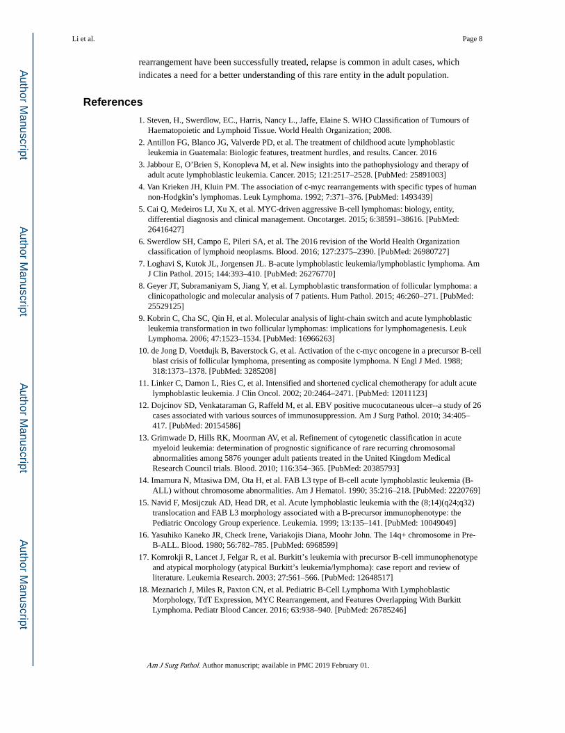
were evident. In case 3, H&E sections of the core biopsy of retroperitoneal lymph node
revealed an infiltrate of atypical lymphoid cells in a fibrotic backgroundl (Fig. 2). The cells
were intermediate-to-large in size with irregular nuclei, fine chromatin, small basophilic
nucleoli and a variably high nuclear-to-cytoplasmic ratio. Mitotic figures were easily
Li et al. Page 4
Am J Surg Pathol. Author manuscript; available in PMC 2019 February 01.
Author M
anuscriptA
uthor Manuscript
Author M
anuscriptA
uthor Manuscript

identified. Scattered small mature lymphocytes were intermixed. The atypical lymphoid
cells on touch preps showed Burkitt lymphoma-like morphology with frequent cytoplasmic
vacuoles.
Immunohistochemical stains were performed on bone marrow and lymph node biopsies. In
case 1, the blasts were positive for CD19, PAX-5, CD79a, CD10, TdT (diffuse and strong),
MUM-1, and C-MYC, and were negative for CD20, CD34, BCL-2, BCL-6, CyclinD1,
CD99, MPO, and CD117. Ki-67 showed a proliferative rate of approximately 90%. In case
2, the blasts were positive for PAX-5, CD79a, CD10, TdT (10%), C-MYC, and were
negative for CD20, CD34, BCL-2, BCL-6, and CyclinD1. Ki-67 showed a very high
proliferative rate of >90%. In case 3, the atypical cells were positive for PAX-5, TdT (diffuse
and strong), CD20, CD10, CD99, and were negative for CD34, BCL-2, BCL-6, MUM-1,
and CD117. Ki-67 showed a proliferative rate of >90%. CD3 highlighted interspersed
background small T lymphocytes. EBER in situ hybridization was negative in all three
cases.
Flow cytometry
In case 1, flow cytometric analysis of bone marrow showed 75% of events were an atypical
population in the CD45 dim gate (Fig. 3A) with expression of CD10 (Fig. 3B), CD19 (Fig.
3C), dim-to absent CD20 (Fig. 3D), TdT (Fig. 3E), and Lambda light chain restriction, and
with no expression of CD34. In case 2, flow cytometry analysis of bone marrow
demonstrated 87% of events were abnormal B-cells with slightly dim CD45 and low side
scatter (Fig. 3F) with expression of CD10, CD19 (Fig. 3G), weak CD20 (15% of gated
events), CD22, weak CD33, bright CD38, and cCD79a, without co-expression of surface
Kappa (Fig. 3H) or Lambda light chains (Fig. 3I). Possible minor subsets expressed dim
CD34 or TdT (<5% of gated events, Fig. 3J). These abnormal B-cells were negative for T-
cell markers, myeloid markers, MPO, and cCD3. In case 3, flow cytometry analysis was
limited by low cellularity, while the CD45 negative cells were positive for CD19, CD20, and
CD10 and were negative for surface light chains.
FISH
FISH studies of all three cases revealed t(8;14)(q24;q32) IGH/MYC translocation. There
was no evidence of t(14;18)(q32;21) IGH/BCL2 or BCL6 rearrangement. In case 1,
Approximately 8% of nuclei had three to four copies of the IGL gene region (at 22q11.2).
This result indicated additional copies of chromosome 22 or a 22q duplication. In case 3,
there were additional copies of the BCL6 gene or chromosome 3, suggesting gains of BCL6 gene or chromosome 3.
Cytogenetic findings
In case 1, 7 of the 20 metaphases were normal, while 8 metaphases had a 5q deletion and 5
metaphases had loss of the Y chromosome. To determine if the metaphases with 5q deletion
were associated with the B-cell lymphoma clone, FISH for MYC and IGH was performed
on several metaphases with the 5q deletion. These results indicated a normal result for MYC and IGH in the 5q deletion metaphases. The combined chromosome and metaphase FISH
results strongly suggested that the metaphases with the 5q deletion represent a neoplastic
Li et al. Page 5
Am J Surg Pathol. Author manuscript; available in PMC 2019 February 01.
Author M
anuscriptA
uthor Manuscript
Author M
anuscriptA
uthor Manuscript

process unrelated to the B-ALL/LBL and were concerning for an evolving/underlying
myeloid neoplasm. It was noted that megakaryocytes were clustered in the marrow with
small hypolobated forms. This was consistent with the cytogenetics finding of 5q- deletion
and a diagnosis of myelodysplastic syndrome 5q- 13. In case 2, chromosomal study was
limited with reduced capability of detecting clonal abnormalities. Only 4 analyzable
metaphase cells were obtained from this specimen and 3 of them had an apparently normal
male karyotype. The remaining cell showed a loss of chromosome 14, which could not be
further evaluated in this study. A full cytogenetic karyotype was not obtained in case 3.
DISCUSSION
In the WHO classification, Burkitt lymphoma is defined as a mature B-cell lymphoma, and
lacks the precursor B cell immunophenotype of B-ALL/LBL. While it occasionally presents
with bone marrow or peripheral blood involvement, as seen in the L3 subtype of ALL in the
FAB classification (1990) 14, these cases share the mature B-cell phenotype of Burkitt
lymphoma. Although implicated in many mature B cell lymphomas, MYC rearrangement
has been reported only rarely in B-ALL/LBL. In a study of 5280 cases of pediatric ALL,
only five with isolated MYC rearrangement were identified, fewer than 0.1% 15. MYC rearrangement has been reported rarely in lymphoblastic malignancies occurring as
progression of underlying follicular lymphoma. These tumors manifest a double-hit, with
rearrangements of both the MYC and BCL2 genes, and can present either in bone marrow or
peripheral lymphoid tissues 8–10. De novo lymphoblastic malignancies with an isolated
MYC translocation have been only rarely identified 7.
Since 1980, only 13 cases of B-ALL/LBL with isolated MYC rearrangement were
reported 15–23. Like other B-ALL/LBLs, the majority of the cases occurred in pediatric
patients and only three cases were reported in adults 17, 24, 25 (Table 1), including two cases
of B-ALL and one case of B-LBL without bone marrow involvement 25. The majority of the
cases demonstrated Burkitt-like morphology. In this study, we report three additional adult
cases with a precursor B cell immunophenotype, Burkitt leukemia/lymphoma-like
morphology, and isolated MYC rearrangement. Similar to previously reported adult cases,
two of our cases were B-ALL and one was B-LBL without bone marrow and peripheral
blood involvement.
In the two cases of adult B-ALL with isolated MYC rearrangement, the morphology of the
blasts resembled that of Burkitt lymphoma, with deeply basophlic cytoplasm and
cytoplasmic vacuoles. In the case of B-LBL, the touch prep showed similar cytological
features as the B-ALL cases, while the nuclei were more pleomorphic on the core biopsy,
probably due to the background sclerosis. Based on these morphological features, Burkitt
leukemia/lymphoma was considered in the differential diagnosis. The distinction between
Burkitt leukemia/lymphoma and B-ALL/LBL is important, as different treatment strategies
would be applied, depending on the final diagnosis.
Among all of the reported adult B-ALL/LBL cases, case 1 in our report is the only one
showing surface light chain immunoglobulin expression. Case 1 was classified as B-
ALL/LBL based on the dim CD45 expression, strong and diffuse TdT expression and dim-
Li et al. Page 6
Am J Surg Pathol. Author manuscript; available in PMC 2019 February 01.
Author M
anuscriptA
uthor Manuscript
Author M
anuscriptA
uthor Manuscript

to-absent CD20 in the lymphoblasts. Although unusual, coexpression of surface light chains
and TdT has been reported in B-ALL previously 23, 26. In fact, the expression of TdT was
variable in the reported pediatric and adult cases B-ALL/LBL with isolated MYC
rearrangement, a finding recapitulated in our series. Case 2 was positive for TdT in only
10% of the cells, but had an immature phenotype by other criteria. CD34 has been reported
negative in all adult cases, and was positive in only one of the pediatric cases 15. Futher
evidence against a diagnosis of Burkitt lymphoma was absence of BCL-6 expression in all
reported cases, including the current report. The variation in CD20, CD34, TdT, and surface
immunoglobulin expression in these cases indicates the arrest of differentiation at varying
stages of precursor B-cell maturation.
Compared to MYC+ B-ALL, B-LBL with isolated MYC rearrangement is extremely rare.
To the best of our knowledge, there was only one case reported previously. Case 3 in this
study may represent the second case, presenting as lymphadenopathy without bone marrow
and peripheral blood involvement. Hepatomegaly or splenomegaly were part of the initial
clinical presentation of the MYC+ B-ALL cases but were not seen in those presenting as
lymphoma. The previously reported MYC+ B-LBL patient was treated with R-CHOP and
our patient was treated with Hyper-CVAD. Both patients showed a good response to
chemotherapy, suggesting that a treatment approach for Burkitt lymphoma rather than B-
ALL/LBL may be warranted in these patients.
MYC rearrangement is most frequently associated with Burkitt lymphoma, yet it also has
been reported in other mature and immature B-cell neoplasms. The rearrangement most
commonly involves MYC at 8q24 and the IGH gene 14q32. A minority of the cases carry
t(2;8)(p11;q24) or t(8;22)(q24;q11) chromosomal rearrangements. These three
rearrangements share the juxtaposition of MYC proto-oncogene on chromosome 8 and
either the heavy or light chain immunoglobulin genes 27. MYC rearrangement was also
reported to involve a non-immunoglobulin chromosomal locus t(4;8)(q31.1;q24.1) in the B-
lymphoblasts of a 4-year-old boy 19. This novel translocation was detected at the time of his
second relapse, and the patient died 3 months later without achieving complete remission.
Due to the rarity of these cases, MYC is not routinely tested in B-ALL/LBL. However, our
study calls attention to this rare event, and additional FISH or cytogenetic studies would be
indicated in cases with variant morphology, as reported here.
Isolated MYC rearrangement without other recurrent genetic abnormalities represents a rare
entity in B-ALL/LBL, especially in adult patients.
These cases appear to represent a distinct biological phenomenon, in which a MYC translocation may be acquired at an immature stage of differentiation, in which the tumor
cells still express TDT and lack a mature B-cell phenotype. The mechanism underlying the
MYC translocation may differ in these novel cases, and requires further investigation.
Differences in the site of the MYC break have been reported in different forms of Burkitt
lymphoma, with differences seen in endemic and sporadic cases 28, 29. Thus, the
maturational stage of the B-cell in which the translocation occurs is thought to influence the
site of the MYC break. Although some pediatric B-ALL/LBL with isolated MYC
Li et al. Page 7
Am J Surg Pathol. Author manuscript; available in PMC 2019 February 01.
Author M
anuscriptA
uthor Manuscript
Author M
anuscriptA
uthor Manuscript
rearrangement have been successfully treated, relapse is common in adult cases, which
indicates a need for a better understanding of this rare entity in the adult population.
References
1. Steven, H., Swerdlow, EC., Harris, Nancy L., Jaffe, Elaine S. WHO Classification of Tumours of Haematopoietic and Lymphoid Tissue. World Health Organization; 2008.
2. Antillon FG, Blanco JG, Valverde PD, et al. The treatment of childhood acute lymphoblastic leukemia in Guatemala: Biologic features, treatment hurdles, and results. Cancer. 2016
3. Jabbour E, O’Brien S, Konopleva M, et al. New insights into the pathophysiology and therapy of adult acute lymphoblastic leukemia. Cancer. 2015; 121:2517–2528. [PubMed: 25891003]
4. Van Krieken JH, Kluin PM. The association of c-myc rearrangements with specific types of human non-Hodgkin’s lymphomas. Leuk Lymphoma. 1992; 7:371–376. [PubMed: 1493439]
5. Cai Q, Medeiros LJ, Xu X, et al. MYC-driven aggressive B-cell lymphomas: biology, entity, differential diagnosis and clinical management. Oncotarget. 2015; 6:38591–38616. [PubMed: 26416427]
6. Swerdlow SH, Campo E, Pileri SA, et al. The 2016 revision of the World Health Organization classification of lymphoid neoplasms. Blood. 2016; 127:2375–2390. [PubMed: 26980727]
7. Loghavi S, Kutok JL, Jorgensen JL. B-acute lymphoblastic leukemia/lymphoblastic lymphoma. Am J Clin Pathol. 2015; 144:393–410. [PubMed: 26276770]
8. Geyer JT, Subramaniyam S, Jiang Y, et al. Lymphoblastic transformation of follicular lymphoma: a clinicopathologic and molecular analysis of 7 patients. Hum Pathol. 2015; 46:260–271. [PubMed: 25529125]
9. Kobrin C, Cha SC, Qin H, et al. Molecular analysis of light-chain switch and acute lymphoblastic leukemia transformation in two follicular lymphomas: implications for lymphomagenesis. Leuk Lymphoma. 2006; 47:1523–1534. [PubMed: 16966263]
10. de Jong D, Voetdujk B, Baverstock G, et al. Activation of the c-myc oncogene in a precursor B-cell blast crisis of follicular lymphoma, presenting as composite lymphoma. N Engl J Med. 1988; 318:1373–1378. [PubMed: 3285208]
11. Linker C, Damon L, Ries C, et al. Intensified and shortened cyclical chemotherapy for adult acute lymphoblastic leukemia. J Clin Oncol. 2002; 20:2464–2471. [PubMed: 12011123]
12. Dojcinov SD, Venkataraman G, Raffeld M, et al. EBV positive mucocutaneous ulcer--a study of 26 cases associated with various sources of immunosuppression. Am J Surg Pathol. 2010; 34:405–417. [PubMed: 20154586]
13. Grimwade D, Hills RK, Moorman AV, et al. Refinement of cytogenetic classification in acute myeloid leukemia: determination of prognostic significance of rare recurring chromosomal abnormalities among 5876 younger adult patients treated in the United Kingdom Medical Research Council trials. Blood. 2010; 116:354–365. [PubMed: 20385793]
14. Imamura N, Mtasiwa DM, Ota H, et al. FAB L3 type of B-cell acute lymphoblastic leukemia (B-ALL) without chromosome abnormalities. Am J Hematol. 1990; 35:216–218. [PubMed: 2220769]
15. Navid F, Mosijczuk AD, Head DR, et al. Acute lymphoblastic leukemia with the (8;14)(q24;q32) translocation and FAB L3 morphology associated with a B-precursor immunophenotype: the Pediatric Oncology Group experience. Leukemia. 1999; 13:135–141. [PubMed: 10049049]
16. Yasuhiko Kaneko JR, Check Irene, Variakojis Diana, Moohr John. The 14q+ chromosome in Pre-B-ALL. Blood. 1980; 56:782–785. [PubMed: 6968599]
17. Komrokji R, Lancet J, Felgar R, et al. Burkitt’s leukemia with precursor B-cell immunophenotype and atypical morphology (atypical Burkitt’s leukemia/lymphoma): case report and review of literature. Leukemia Research. 2003; 27:561–566. [PubMed: 12648517]
18. Meznarich J, Miles R, Paxton CN, et al. Pediatric B-Cell Lymphoma With Lymphoblastic Morphology, TdT Expression, MYC Rearrangement, and Features Overlapping With Burkitt Lymphoma. Pediatr Blood Cancer. 2016; 63:938–940. [PubMed: 26785246]
Li et al. Page 8
Am J Surg Pathol. Author manuscript; available in PMC 2019 February 01.
Author M
anuscriptA
uthor Manuscript
Author M
anuscriptA
uthor Manuscript

19. Seo JY, Lee SH, Kim HJ, et al. MYC rearrangement involving a novel non-immunoglobulin chromosomal locus in precursor B-cell acute lymphoblastic leukemia. Ann Lab Med. 2012; 32:289–293. [PubMed: 22779071]
20. Hassan R, Felisbino F, Stefanoff CG, et al. Burkitt lymphoma/leukaemia transformed from a precursor B cell: clinical and molecular aspects. Eur J Haematol. 2008; 80:265–270. [PubMed: 18005389]
21. Gupta AA, Grant R, Shago M, et al. Occurrence of t(8;22)(q24.1;q11.2) involving the MYC locus in a case of pediatric acute lymphoblastic leukemia with a precursor B cell immunophenotype. J Pediatr Hematol Oncol. 2004; 26:532–534. [PubMed: 15284595]
22. Kaplinsky C, Rechavi G. Acute lymphoblastic leukemia of Burkitt type (L3 ALL) with t(8;14) lacking surface and cytoplasmic immunoglobulins. Med Pediatr Oncol. 1998; 31:36–38. [PubMed: 9607430]
23. Higa B, Alkan S, Barton K, et al. Precursor B-cell acute lymphoblastic leukaemia with FAB L3 (i.e., Burkitt’s leukaemia/lymphoma) morphology and co-expression of monoclonal surface light chains and Tdt: report of a unique case and review of the literature. Pathology. 2009; 41:495–498. [PubMed: 19900092]
24. Slavutsky I, Andreoli G, Gutierrez M, et al. Variant (8;22) translocation in lymphoblastic lymphoma. Leuk Lymphoma. 1996; 21:169–172. [PubMed: 8907285]
25. Shiratori S, Kondo T, Fujisawa S, et al. c-myc rearrangement in B-cell lymphoblastic lymphoma with the involvement of multiple extranodal lesions. Leuk Lymphoma. 2011; 52:716–718. [PubMed: 21314486]
26. Nelson BP, Treaba D, Goolsby C, et al. Surface immunoglobulin positive lymphoblastic leukemia in adults; a genetic spectrum. Leuk Lymphoma. 2006; 47:1352–1359. [PubMed: 16923568]
27. Behm, FCD. Immunophenotyping. In: Pui, C-H., editor. Childhood leukemias. 1999. p. 111
28. Bertrand P, Bastard C, Maingonnat C, et al. Mapping of MYC breakpoints in 8q24 rearrangements involving non-immunoglobulin partners in B-cell lymphomas. Leukemia. 2007; 21:515–523. [PubMed: 17230227]
29. Pelicci P, Knowles D, Magrath I, et al. Chromosomal breakpoints and structural alterations of the c-myc locus differ in endemic and sporadic forms of Burkitt lymphoma. Proc Natl Acad Sci USA. 1986; 83:2984–2988. [PubMed: 3458257]
Li et al. Page 9
Am J Surg Pathol. Author manuscript; available in PMC 2019 February 01.
Author M
anuscriptA
uthor Manuscript
Author M
anuscriptA
uthor Manuscript

Fig. 1. Bone marrow and peripheral blood findings of MYC+ B-ALL/LBLA: Blasts in peripheral blood smear (Wright-Giemsa stain, 1000× (case 2) were
intermediate-in-size, with ovoid-to-irregular nuclei, fine chromatin, occasional prominent
nucleoli, and scant-to-moderate deeply basophilic cytoplasm. B: Bone marrow aspirate
smear, case 1 (Wright-Giemsa stain, 1000×) showed cells with high nuclear:cytoplasmic
ratio, deeply basophilic cytoplasm, and cytoplasmic vacuoles. C: Bone marrow core biopsy
(H&E 1000×, (case 1) showed a diffuse infiltrate of blasts with finely dispersed chromatin
and small nucleoli. Immunohistochemical findings (Case 1) showed positive staining for
CD19 (D), but absence of CD20 (E). TDT showed strong nuclear staining (F). The blasts
Li et al. Page 10
Am J Surg Pathol. Author manuscript; available in PMC 2019 February 01.
Author M
anuscriptA
uthor Manuscript
Author M
anuscriptA
uthor Manuscript

show nuclear staining for MYC (Case 2) (G). BCL-6 was negative (H), but PAX5 was
positive (I).
Li et al. Page 11
Am J Surg Pathol. Author manuscript; available in PMC 2019 February 01.
Author M
anuscriptA
uthor Manuscript
Author M
anuscriptA
uthor Manuscript
Fig 2. Lymph node biopsy in MYC+ B-ALL/LBLA. A core biopsy of retroperitoneal lymph node (H&E, 40×) (case 3) showed diffuse
sclerosis with a dense atypical lymphoid infiltrate. B. The infiltrate (H&E, 400×) was
composed of intermediate-to-large atypical lymphoid cells with irregular nuclei, fine
chromatin, small nucleoli, and variably high nuclear-to-cytoplasmic ratio. Scattered small
lymphocytes were intermixed. The tumor cells were positive for PAX5 (C) and showed
strong nuclear staining for TDT (D).
Li et al. Page 12
Am J Surg Pathol. Author manuscript; available in PMC 2019 February 01.
Author M
anuscriptA
uthor Manuscript
Author M
anuscriptA
uthor Manuscript

Fig. 3. Flow cytometry analysis of bone marrowIn case 1, the atypical lymphoid cells showed dim CD45 and lower side scatter (A, dark
green), with expression of CD10 (B, cyan), CD19 (C, cyan), dim-to-absent expression of
CD20 (D, cyan), and TdT (E, green); In case 2, the atypical lymphoid cells with dim CD45
and low side scatter gate (F) showed expression of CD19 (G), without expression of kappa
(H) and lambda (I) light chains. Only few cells expressed dim CD34 or TdT (<5% of gated
events, J).
Li et al. Page 13
Am J Surg Pathol. Author manuscript; available in PMC 2019 February 01.
Author M
anuscriptA
uthor Manuscript
Author M
anuscriptA
uthor Manuscript

Fig. 4. FISH analysis of bone marrow, Case 1A. Breakapart probe with red fluorochrome labeling 5′MYC and the green fluorochrome
labeling 3′MYC. Result shows split of red-green signals in tumor cells. B. Dual color
double fusion probes with the red fluorochrome directly labeling MYC spanning the known
breakpoints (8q24), the green fluorochrome directly labeling the known breakpoints of IGH
(14q32), and an aqua fluorochrome directed at the alpha satellite centromeric region of
chromosome 8 (D8Z2). Result indicates one separate red signal, one separate green signal
and two orange/green fusion signals (arrows) indicating the MYC/IGH translocation.
Li et al. Page 14
Am J Surg Pathol. Author manuscript; available in PMC 2019 February 01.
Author M
anuscriptA
uthor Manuscript
Author M
anuscriptA
uthor Manuscript

Author M
anuscriptA
uthor Manuscript
Author M
anuscriptA
uthor Manuscript
Li et al. Page 15
Tab
le 1
Clin
ical
and
pat
holo
gica
l fea
ture
s of
adu
lt B
-AL
L/L
BL
with
isol
ated
MY
C tr
ansl
ocat
ion
Pri
or
and
curr
ent
repo
rts
Age
Sex
Init
ial p
rese
ntat
ion
Tre
atm
ent
and
Cou
rse
FIS
H(%
)C
ytog
enet
ics
BC
L2
RB
CL
6 R
CD
10C
D19
CD
20B
CL
-6C
D34
TdT
sIg
EB
ER
Kom
rokj
i R
, et a
l45
MB
sym
ptom
s, s
plen
omeg
aly
Hyp
er-C
VA
D
alte
rnat
ing
with
hig
h do
se M
TX
/AR
A-C
t(8,
14)(
q24,
q32)
, +i(
1)(q
10)
NT
++
+N
T−
−−
NT
Shir
ator
i S,
et a
l64
FC
horo
id a
nd s
kin
invo
lvem
ent b
y B
-LB
LA
chie
ved
CR
aft
er R
-C
HO
P ×
6; r
elap
sed
on
skin
2 y
ears
late
r, ac
hiev
ed C
R a
fter
ra
diat
ion
ther
apy;
a
furt
her
2 ye
ars
late
r, sk
in a
nd c
hori
od r
elap
se
as in
initi
al s
tage
t(8;
14)(
q24;
q32)
; pos
itive
IG
H a
nd T
CR
gen
e re
arra
ngem
ent
−+
++
−N
T+
NT
NT
Slav
utsk
y I,
et a
l20
FB
sym
ptom
s,
hepa
tom
egal
y, la
rge
retr
oper
itone
al
lym
phad
enop
athy
, and
ne
phro
meg
aly
n/a
t(8;
22)(
q24;
q 11
); h
yper
ploi
dN
T+
+N
TN
TN
TN
T−
−
Li Y
, et
al
Cas
e 1
70M
Dif
fuse
lym
phad
nopa
thy,
sp
leno
meg
aly,
PB
and
BM
in
volv
emen
t
Hyp
er-C
VA
D a
nd
intr
athe
cal m
etho
trex
ate.
E
xpir
ed d
urin
g th
e se
cond
cyc
le o
f ch
emot
hera
py
MY
C/I
GH
(55
%);
5q
dele
ion
(7/2
0) m
etap
hase
s−
++
−−
−+
+−
Cas
e 2
56M
Dif
fuse
lym
phad
enop
athy
, he
pato
sple
nom
egal
y, P
B
and
BM
invo
lvm
ent
Hig
h-ri
sk A
LL
pro
toco
l (8
707)
with
initi
al
rem
issi
on, b
ut w
ith
prog
ress
ion
two
mon
ths
afte
r tr
eatm
ent.
MY
C/I
GH
(60
%)
−+
+−
−−
−/+
−−
Cas
e 3
44M
Bul
ky in
trab
dom
inal
ly
mph
aden
opat
hy, n
egat
ive
BM
Hyp
er-C
VA
D w
ith
part
ial r
emis
sion
, pe
ndin
g st
em c
ell
tran
spla
nt
MY
C/I
GH
(63
%)
−+
NT
+−
−+
NT
−
Abb
revi
atio
ns: N
T, n
ot te
sted
; PB
, per
iphe
ral b
lood
; BM
, bon
e m
arro
w
Am J Surg Pathol. Author manuscript; available in PMC 2019 February 01.