RAP-EIV (INVERTER) UNIDADES MODULARES LINHA RVT/RTC E RUV/RUT RAP (FIXO
Crucial role of the Rap G protein signal inNotch activation and leukemogenicity ofT-cell acute lymphoblastic leukemiaKeiko Doi1, Takahiko Imai1, Christopher Kressler1, Hideo Yagita2, Yasutoshi Agata1, Marc Vooijs3,Yoko Hamazaki1, Joe Inoue1 & Nagahiro Minato1
1Department of Immunology and Cell Biology, Graduate School of Medicine, Kyoto University, Sakyo-ku, Kyoto, Japan,2Department of Immunology, Juntendo University School of Medicine, Bunkyo-ku, Tokyo, Japan, 3Maastricht Radiation Oncologyand School for Oncology and Developmental Biology, University of Maastricht, Maastricht, The Netherlands.
The Rap G protein signal regulates Notch activation in early thymic progenitor cells, and deregulated Rapactivation (Raphigh) results in the development of Notch-dependent T-cell acute lymphoblastic leukemia(T-ALL). We demonstrate that the Rap signal is required for the proliferation and leukemogenesis ofestablished Notch-dependent T-ALL cell lines. Attenuation of the Rap signal by the expression of adominant-negative Rap1A17 or Rap1GAP, Sipa1, in a T-ALL cell line resulted in the reduced Notchprocessing at site 2 due to impaired maturation of Adam10. Inhibition of the Rap1 prenylation with ageranylgeranyl transferase inhibitor abrogated its membrane-anchoring to Golgi-network and causedreduced proprotein convertase activity required for Adam10 maturation. Exogenous expression of a matureform of Adam10 overcame the Sipa1-induced inhibition of T-ALL cell proliferation. T-ALL cell linesexpressed Notch ligands in a Notch-signal dependent manner, which contributed to the cell-autonomousNotch activation. Although the initial thymic blast cells barely expressed Notch ligands during the T-ALLdevelopment from Raphigh hematopoietic progenitors in vivo, the ligands were clearly expressed in theT-ALL cells invading extrathymic vital organs. These results reveal a crucial role of the Rap signal in theNotch-dependent T-ALL development and the progression.
The Notch signal is essential for thymic T-cell development1,2. Notch protein is synthesized as a large singlepeptide, which is later cleaved intracellularly at a heterodimerization (HD) domain (S1 cleavage) to generatethe heterodimeric Notch receptor3. Upon engagement with specific ligands, the Notch receptor is activated
through successive proteolytic cleavages at a juxtamembrane site (S2) followed by an intramembranous site (S3)mediated by Adam10 and c-secretase complex, respectively, resulting in the release and nuclear translocation ofNotch intracellular domain (NICD)4. In early T-cell progenitors (ETPs), Notch receptor is activated via Delta-like4 (Dll4), which is expressed on thymic epithelial cells5. The Notch signal also plays a key role in the developmentof T-cell acute lymphoblastic leukemia (T-ALL)6. More than 50% of human T-ALL cell lines show ‘‘activating’’Notch1 mutations7, although more recent studies suggest that these mutations may not alone suffice for T-ALLdevelopment8–10.
We have reported that the Rap G protein signal also plays an important part in thymic T-cell development aswell as T-ALL genesis11–13. The signal switch function of Rap is regulated positively by specific guanine nucleotideexchange factors such as C3G and negatively by GTPase-activating proteins represented by Sipa112. Impaired Rapactivation in ETPs results in arrested thymic T-cell development, whereas deregulated Rap activation (Raphigh)remarkably enhances the Notch-dependent proliferation of ETPs11. Moreover, bone marrow transplantation(BMT) of Raphigh hematopoietic progenitor cells (HPCs) results in the development of T-ALL13. Intriguingly, suchT-ALL cells were dependent on the Notch signal and often showed characteristic Notch1 mutations similar tohuman T-ALL13, suggesting a functional crosstalk between the Rap and Notch signals.
In current study, we demonstrate that the Rap signal controls Notch activation in T-ALL cells by regulatingproprotein convertase activity required for the maturation of Adam10 mediating the Notch processing. Wefurther indicate that the sustained Notch activation in thymic Raphigh blast cells eventually results in the express-ion of Notch ligands, leading to the cell-autonomous Notch activation and systemic T-ALL progression.
OPEN
SUBJECT AREAS:
ACUTE LYMPHOCYTICLEUKAEMIA
LEUKAEMIA
Received23 July 2014
Accepted23 December 2014
Published23 January 2015
Correspondence andrequests for materials
should be addressed toN.M. (minato@imm.
med.kyoto-u.ac.jp)
SCIENTIFIC REPORTS | 5 : 7978 | DOI: 10.1038/srep07978 1

ResultsThe Rap signal is required for Notch activation in T-ALL cell lines.FL0 cell line derived from T-ALL by BMT of Raphigh HPCs expressedintact Notch receptors with no detectable Notch1 mutation andshowed Notch-dependent proliferation (Figure S1). Retroviraltransduction of dominant-negative Rap1 (Rap1A17) in FL0 cellscausing a decrease of the Rap1-GTP resulted in a reduced ex-pression of NICD and its target Hes1 (Figure 1a). Accordingly, theexpression of p27Kip1, a target of Hes1-mediated repression14 wasincreased, and the proliferation was significantly reduced (Figure 1a).The FL0/Rap1A17 cells showed significantly compromised leuke-mogenic activity in scid mice, and 20% of the recipients remainedfree of leukemia, whereas all recipients of control FL0/vect cellsdied within 25 days (Figure 1b, left). Moreover, the leukemiacells developed in the recipients of FL0/Rap1A17 cells revealedsignificantly reduced expression of the retrovirus-driven NGFR(Figure 1b, right). Such an effect was not observed in the recipientsof FL0/vect cells, suggesting a counter selection against FL0/Rap1A17high cells in vivo. We then transduced Rap1-specific GAP,Sipa1, in FL0 cells with a doxycycline (Dox)-inducible system.Induction of Sipa1 expression also resulted in the decreasedNICD expression and cell proliferation in concordance withreduced Rap1-GTP levels in a Dox-dose dependent manner(Figure 1c). Furthermore, treatment of the FL0 cells with ageranylgeranyl transferase inhibitor (GGTI)15, which inhibited the
Rap prenylation required for membrane anchoring, also suppressedthe NICD generation and cell proliferation at a dose-range that didnot affect the proliferation of irrelevant leukemia cells (Figure 1d).Other T-ALL cell lines of mice and humans similarly showedsignificantly higher susceptibility to GGTI than leukemia cells ofnon-T-ALL types (Figure S2). The results suggest that the Rapsignal plays an important role in sustaining Notch activation andproliferation of established T-ALL cell lines.
The Rap signal controls Notch S2 processing by regulating intra-cellular Adam10 maturation. The conditional expression of Sipa1in FL0 cells did not affect the cell surface expression of Notch1(Figure 2a). However, analysis with Notch1-immunoprecipitationfollowed by immunoblotting with S2 (V1711)-specific antibodyrevealed that the Notch S2 product precedent to the NICDgeneration was also decreased by Sipa1 expression (Figure 2a). InT-ALL cells, Notch cleavage at S2 site is mediated by Adam1016,which maturates intracellularly via prodomain-cleavage of theimmature form17. Sipa1 expression in FL0 cells caused a decreaseof the mature form of Adam10 (m-Adam10) with barely affectingthe immature form (i-Adam10) or the transcripts (Figure 2b).Because it is reported that membrane Dll1 is constitutively cleavedextracellularly by Adam1018, we also examined the effect of Sipa1expression on Dll1. Induction of Sipa1 expression in FL0 cellsresulted in the accumulation of unprocessed, full-length (Fl) Dll1
Figure 1 | Attenuation of the Rap signal inhibits Notch activation and the proliferation of T-ALL cell lines. (a) FL0/Rap1A17 and control FL0/vect cells
were immunoblotted with the indicated antibodies. These cells were cultured at 3 3 104 cells/mL in triplicate, and the viable cell numbers were assessed on
day 3. *; p , 0.01. (b) Survival rates of scid mice transplanted with FL0/vect (grey symbols) or FL0/Rap1A17 (solid symbols) cells (10 mice per group).
Expression levels of the retrovirus-derived hNGFR in the BM of FL0/vect- and FL0/Rap1A17-recipients were analyzed at 20 and 25 days after the
transplantation, respectively, in comparison with the original inoculants. The means of mean fluorescence intensities (MFIs) in 3 recipients are indicated.
(c) FL0/rtTA-Sipa1 cells were cultured for 3 days in the absence or presence of varying doses of Dox and immunoblotted with the indicated antibodies.
FL0/rtTA-Sipa1 (open circles) and control FL0/rtTA (solid circles) cells were cultured in triplicate for 3 days in the absence or presence of Dox, and
the viable cell numbers were assessed. *; p , 0.01. (d) FL0 cells were cultured in the absence or presence of GGTI for 2 days and immunoblotted with the
indicated antibodies. FL0 (open circles), EL4 (closed circles), and P3U1 (closed squares) cells were cultured in triplicate with or without GGTI for 3 days,
and the viable cell numbers were assessed. *; p , 0.01. Relevant parts of immunoblot images in (a), (c), and (d) were cropped from full-length blots shown
in Figure S7.
www.nature.com/scientificreports
SCIENTIFIC REPORTS | 5 : 7978 | DOI: 10.1038/srep07978 2
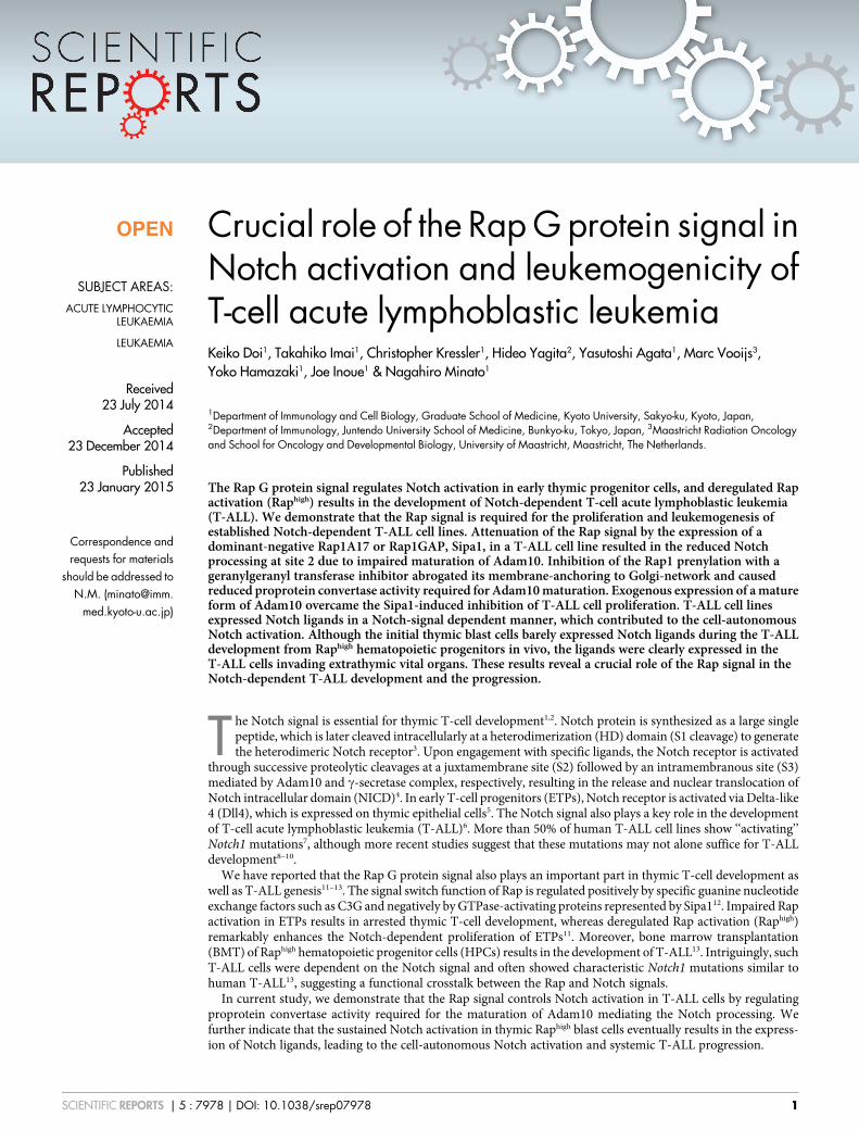
(85 kDa) with a concomitant decrease of the cleaved transmembraneand intracellular (TMIC) form (29 kDa); FACS analysis confirmed aslight yet clear increase in the cell surface Dll1 expression (Figure 2c).We next examined the proprotein convertase activity required for theAdam10 maturation19, with the use of a fluorescence-labeled syntheticsubstrate (Pyr-Arg-Thr-Lys-Arg-AMC)20. Although proprotein con-vertase activity requires Ca21 21,22, early-phase cleavage activity in thecell lysate tended to be resistant to EDTA and was probably attri-buted to other trypsin-like proteases. Sipa1 expression in FL0 cellsrather preferentially inhibited the EDTA-sensitive late-phase activity(Figure 2d). The results suggest that the Rap signal regulates theactivation of proprotein convertase activity required for the matu-ration of Adam10 mediating Notch S2 processing.
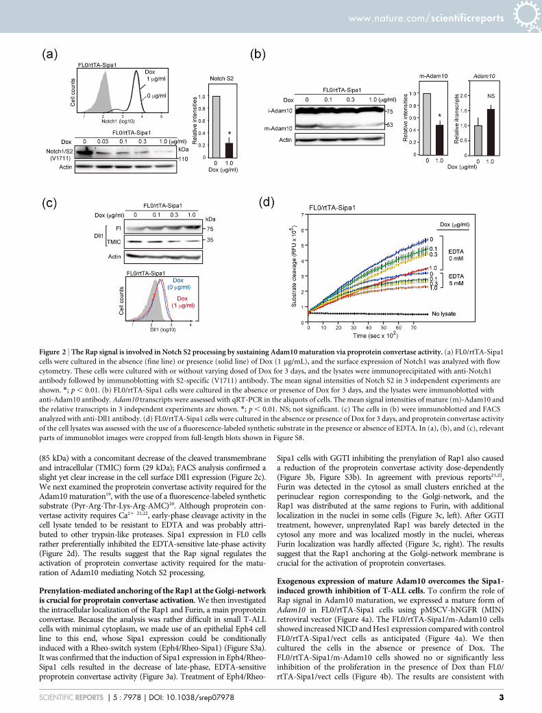
Prenylation-mediated anchoring of the Rap1 at the Golgi-networkis crucial for proprotain convertase activation. We then investigatedthe intracellular localization of the Rap1 and Furin, a main proproteinconvertase. Because the analysis was rather difficult in small T-ALLcells with minimal cytoplasm, we made use of an epithelial Eph4 cellline to this end, whose Sipa1 expression could be conditionallyinduced with a Rheo-switch system (Eph4/Rheo-Sipa1) (Figure S3a).It was confirmed that the induction of Sipa1 expression in Eph4/Rheo-Sipa1 cells resulted in the decrease of late-phase, EDTA-sensitiveproprotein convertase activity (Figure 3a). Treatment of Eph4/Rheo-
Sipa1 cells with GGTI inhibiting the prenylation of Rap1 also causeda reduction of the proprotein convertase activity dose-dependently(Figure 3b, Figure S3b). In agreement with previous reports21,22,Furin was detected in the cytosol as small clusters enriched at theperinuclear region corresponding to the Golgi-network, and theRap1 was distributed at the same regions to Furin, with additionallocalization in the nuclei in some cells (Figure 3c, left). After GGTItreatment, however, unprenylated Rap1 was barely detected in thecytosol any more and was localized mostly in the nuclei, whereasFurin localization was hardly affected (Figure 3c, right). The resultssuggest that the Rap1 anchoring at the Golgi-network membrane iscrucial for the activation of proprotein convertases.
Exogenous expression of mature Adam10 overcomes the Sipa1-induced growth inhibition of T-ALL cells. To confirm the role ofRap signal in Adam10 maturation, we expressed a mature form ofAdam10 in FL0/rtTA-Sipa1 cells using pMSCV-hNGFR (MIN)retroviral vector (Figure 4a). The FL0/rtTA-Sipa1/m-Adam10 cellsshowed increased NICD and Hes1 expression compared with controlFL0/rtTA-Sipa1/vect cells as anticipated (Figure 4a). We thencultured the cells in the absence or presence of Dox. TheFL0/rtTA-Sipa1/m-Adam10 cells showed no or significantly lessinhibition of the proliferation in the presence of Dox than FL0/rtTA-Sipa1/vect cells (Figure 4b). The results are consistent with
Figure 2 | The Rap signal is involved in Notch S2 processing by sustaining Adam10 maturation via proprotein convertase activity. (a) FL0/rtTA-Sipa1
cells were cultured in the absence (fine line) or presence (solid line) of Dox (1 mg/mL), and the surface expression of Notch1 was analyzed with flow
cytometry. These cells were cultured with or without varying dosed of Dox for 3 days, and the lysates were immunoprecipitated with anti-Notch1
antibody followed by immunoblotting with S2-specific (V1711) antibody. The mean signal intensities of Notch S2 in 3 independent experiments are
shown. *; p , 0.01. (b) FL0/rtTA-Sipa1 cells were cultured in the absence or presence of Dox for 3 days, and the lysates were immunoblotted with
anti-Adam10 antibody. Adam10 transcripts were assessed with qRT-PCR in the aliquots of cells. The mean signal intensities of mature (m)-Adam10 and
the relative transcripts in 3 independent experiments are shown. *; p , 0.01. NS; not significant. (c) The cells in (b) were immunoblotted and FACS
analyzed with anti-Dll1 antibody. (d) FL0/rtTA-Sipa1 cells were cultured in the absence or presence of Dox for 3 days, and proprotein convertase activity
of the cell lysates was assessed with the use of a fluorescence-labeled synthetic substrate in the presence or absence of EDTA. In (a), (b), and (c), relevant
parts of immunoblot images were cropped from full-length blots shown in Figure S8.
www.nature.com/scientificreports
SCIENTIFIC REPORTS | 5 : 7978 | DOI: 10.1038/srep07978 3
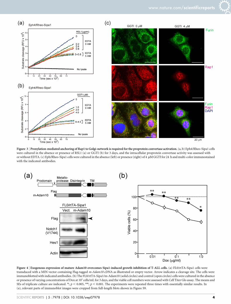
Figure 3 | Prenylation-mediated anchoring of Rap1 to Golgi-network is required for the proprotein convertase activation. (a, b) Eph4/Rheo-Sipa1 cells
were cultured in the absence or presence of RSL1 (a) or GGTI (b) for 3 days, and the intracellular proprotein convertase activity was assessed with
or without EDTA. (c) Eph/Rheo-Sipa1 cells were cultured in the absence (left) or presence (right) of 4 mM GGTI for 24 h and multi-color immunostained
with the indicated antibodies.
Figure 4 | Exogenous expression of mature Adam10 overcomes Sipa1-induced growth inhibition of T-ALL cells. (a) FL0/rtTA-Sipa1 cells were
transduced with a MIN vector containing Flag-tagged m-Adam10 cDNA as illustrated or empry vector. Arrow indicates a cleavage site. The cells were
immunoblotted with indicated antibodies. (b) The FL0/rtTA-Sipa1/m-Adam10 (solid circles) and control (open circles) cells were cultured in the absence
or presence of varying concentrations of Dox at 105 cells/mL for 3 days, and the viable cell numbers were assessed with Cell Titer Glo assay. The means and
SEs of triplicate culture are indicated. *; p , 0.005, **; p , 0.001. The experiments were repeated three times with essentially similar results. In
(a), relevant parts of immunoblot images were cropped from full-length blots shown in Figure S9.
www.nature.com/scientificreports
SCIENTIFIC REPORTS | 5 : 7978 | DOI: 10.1038/srep07978 4
the notion that the Rap signal is required for the Notch-dependentproliferation of T-ALL cells by promoting endogenous Adam10maturation.
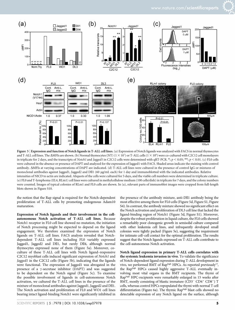
Expression of Notch ligands and their involvement in the cell-autonomous Notch activation of T-ALL cell lines. BecauseNotch1 receptor in FL0 cell line showed no mutation, the initiationof Notch processing might be expected to depend on the ligandengagement. We therefore examined the expression of Notchligands on T-ALL cell lines. FACS analysis revealed that Notch-dependent T-ALL cell lines including FL0 variably expressedJagged1, Jagged2 and Dll1, but rarely Dll4, although normalthymocytes expressed none of them (Figure 5a). Moreover, co-culture of these T-ALL cell lines with Notch ligand–responsiveC2C12 myoblast cells induced significant expression of Notch1 andJagged1 in the C2C12 cells (Figure 5b), indicating that the ligandswere functional. The expression of Jagged1 was abrogated in thepresence of a c-secretase inhibitor (DAPT) and was suggestedto be dependent on the Notch signal (Figure 5c). To examinethe possible involvement of ligands in cell-autonomous Notchactivation, we cultured the T-ALL cell lines in the presence of themixture of monoclonal antibodies against Jagged1, Jagged2 and Dll1.The Notch activation and proliferation of FL0 and WO1 cell linesbearing intact ligand-binding Notch1 were significantly inhibited in
the presence of the antibody mixture, anti-Dll1 antibody being themost effective among them for FL0 cells (Figure 5d, Figure S1, FigureS4). In contrast, the antibody mixture showed no significant effect onthe Notch activation and proliferation of DL3 cell line that lacked theligand-binding region of Notch1 (Figure 5d, Figure S1). Moreover,despite the robust proliferation in liquid culture, the FL0 cells showeda remarkably poor clonogenic growth in semisolid culture comparedwith other leukemia cell lines, and infrequently developed smallcolonies were tightly packed (Figure 5e), suggesting the requirementfor intimate cell–cell contact for the optimal proliferation. The resultssuggest that the Notch ligands expressed on T-ALL cells contribute tothe cell-autonomous Notch activation.
Notch ligand expression in primary T-ALL cells correlates withthe systemic leukemia invasion in vivo. To validate the significanceof Notch-dependent ligand expression during T-ALL development invivo, we performed BMT of Raphigh HPCs. As reported previously13,the Raphigh HPCs caused highly aggressive T-ALL eventually in-volving most vital organs in the BMT recipients. The thymi ofRaphigh HPC-recipients were remarkably enlarged in 15 weeks afterBMT, mostly consisting of blastic immature (CD32 CD41 CD81) Tcells, whereas control HPCs repopulated the thymi with normal T celldifferentiation (Figure 6a). The thymic Raphigh blast cells showed nodetectable expression of any Notch ligand on the surface, although
Figure 5 | Expression and function of Notch ligands in T-ALL cell lines. (a) Expression of Notch ligands was analyzed with FACS in normal thymocytes
and T-ALL cell lines. TheDMFIs are shown. (b) Normal thymocytes (NT) (1 3 107) or T-ALL cells (1 3 106) were co-cultured with C2C12 cell monolayers
in triplicate for 2 days, and the transcripts of Notch1 and Jagged1 in C2C12 cells were determined with qRT-PCR. *; p , 0.05; **; p , 0.01. (c) FL0 cells
were cultured in the absence or presence of DAPT and analyzed for the expression of Jagged1 with FACS. Shaded areas indicate the staining with control
antibody. DMFIs at varying concentrations of DAPT are indicated. (d) T-ALL cell lines were cultured in the presence of control IgG or mixtures of
monoclonal antibodies against Jagged1, Jagged2 and Dll1 (60 mg/mL each) for 1 day and immunoblotted with the indicated antibodies. Relative
intensities of NICD to actin are indicated. Aliquots of the cells were cultured for 5 days, and the viable cell numbers were determined in triplicate culture.
(e) FL0 and T-lymphoma (EL4, RLm1) cell lines were cultured in methylcellulose medium (100 cells/dish) in triplicate for 7 days, and the colony numbers
were counted. Images of typical colonies of RLm1 and FL0 cells are shown. In (a), relevant parts of immunoblot images were cropped from full-length
blots shown in Figure S10.
www.nature.com/scientificreports
SCIENTIFIC REPORTS | 5 : 7978 | DOI: 10.1038/srep07978 5
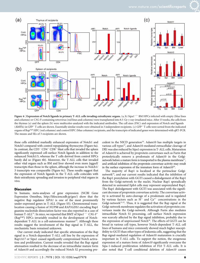
these cells exhibited markedly enhanced expression of Notch1 andNotch3 compared with control repopulating thymocytes (Figure 6a).In contrast, the CD32 CD41 CD81 blast cells that invaded the spleensignificantly expressed cell-surface Notch ligands in addition to theenhanced Notch1/3, whereas the T cells derived from control HPCsbarely did so (Figure 6b). Moreover, the T-ALL cells that invadedother vital organs such as BM and liver showed even more Jagged1transcripts than those in the spleen, although the increase in Notch1/3 transcripts was comparable (Figure 6c). These results suggest thatthe expression of Notch ligands in the T-ALL cells coincides withtheir extrathymic spreading and invasion to peripheral vital organs invivo.
DiscussionIn humans, meta-analyses of gene expression (NCBI GeneExpression Omnibus, http://lifesciencedb.jp/geo/) show that thenegative Rap regulator SIPA1 is one of the most prominentlyunder-expressed genes in T-ALL (Figure S5). Chromosomal trans-location causing a fusion of NUP98 and RAP1GDS1 encoding Rap1guanine nucleotide dissociation factor was also reported in a case ofhuman T-ALL23. In mice, we reported that BMT of Sipa12/2 C3G-F1
(Raphigh) HPCs invariably resulted in the development of Notch-dependent T-ALL in a cell-autonomous manner13. Although theseresults suggest the involvement of the Rap signal in T-ALL, themechanistic basis remained unknown.
Our current study indicated that specific attenuation of the Rapsignal in a Notch-dependent T-ALL cell line by the expression ofRap1A17 or Sipa1 caused significantly compromised Notch activa-tion and proliferation. Current results revealed that the Rap signalattenuation resulted in the decrease of an intracellular mature formof Adam10 and accordingly the reduced Notch S2 processing pre-
cedent to the NICD generation16. Adam10 has multiple targets invarious cell types19, and Adam10-mediated extracellular cleavage ofDll1 was also reduced by Sipa1 expression in T-ALL cells. Maturationof Adam10 is achieved by proprotein convertases such as Furin thatproteolytically remove a prodomain of Adam10 in the Golgi-network before a mature form is transported to the plasma membrane17,and artificial inhibition of the proprotein convertase activity may resultin the surface expression of the immature form of Adam1024.
The majority of Rap1 is localized at the perinuclear Golgi-network25, and our current results indicated that the inhibition ofthe Rap1 prenylation with GGTI caused a dislodgement of the Rap1from the Golgi-network to the nuclei. Nuclear Rap1 sporadicallydetected in untreated Eph4 cells may represent unprenylated Rap1.The Rap1 dislodgement with GGTI was associated with the signifi-cant decrease of proprotein convertase activity. The convertase activ-ity is activated by auto-cleavage of a prodomain and is regulatedby various factors such as H1 and Ca21 concentrations in theGolgi-network21,22. Thus, it is suggested that the Rap signal at theGolgi-network membrane regulates the activation of proprotein con-vertases inside the Golgi-network. Although Furin also mediatesintracellular Notch S1 processing, cell surface Notch expressionwas scarcely affected by the Rap signal inhibition, probably due tothe expression of unprocessed Notch26. The effects of GGTI can bebroad in various cell types, however Notch-dependent T-ALL celllines of humans and mice commonly showed much higher suscept-ibility to GGTI than other types of leukemia cells, suggesting that theRap signal-mediated regulation of Adam10 maturation is cruciallyimportant in T-ALL cells. To support the notion, the exogenousexpression of a mature form of Adam10 significantly overcame theSipa-1-induced proliferation inhibition of FL0 T-ALL cells. It isalso noted that T-cell conditional deletion of Adam10 causes
Figure 6 | Expression of Notch ligands in primary T-ALL cells invading extrathymic organs. (a, b) Sipa12/2 BM HPCs infected with empty (blue lines
and columns) or C3G-F-containing retrovirus (red lines and columns) were transplanted into 8.5 Gy c-ray-irradiated mice. After 15 weeks, the cells from
the thymus (a) and the spleen (b) were multicolor-analyzed with the indicated antibodies. The cell sizes (FSC) and expression of Notch and ligands
(DMFIs) in GFP1 T cells are shown. Essentially similar results were obtained in 3 independent recipients. (c) GFP1 T cells were sorted from the indicated
organs of Raphigh HPC (red columns) and control HPC (blue columns) recipients, and the transcripts of indicated genes were determined with qRT-PCR.
The means and SEs of 3 recipients are shown.
www.nature.com/scientificreports
SCIENTIFIC REPORTS | 5 : 7978 | DOI: 10.1038/srep07978 6
arrested thymic T-cell development, similar to T-cell conditionalSipa1 overexpression13,27.
Although deregulated Rap activation in normal ETPs inducesremarkably enhanced Notch-mediated proliferation, the effect isdependent on the Notch ligands provided by stroma cells, and thusthe enhanced Rap signal alone is incapable of bypassing the ligandrequirement11. How Notch activation occurs cell-autonomously inT-ALL cells without other ligand-donor cells has been an open ques-tion, and several mechanisms have been reported. Activating Notch1mutations, particularly at an HD region, may result in an intrinsicincrease of spontaneous S2 cleavage28. It was also reported thatunique Notch isoforms with spontaneous S2 cleavage are generatedvia cryptic Notch1 promoters in murine T-ALL models29. Our cur-rent results indicated that T-ALL cell lines express functional Notchligands, and that the Notch activation and proliferation of thoseexpressing intact Notch1 are significantly inhibited by the mixtureof antibodies against the ligands. As expected, those of a T-ALL cellline with Notch1 receptor lacking the ligand-binding region wereunaffected by the antibodies despite the ligand expression. Theexpression of Jagged1 in the T-ALL cells was dependent on theNotch signal, apparently forming an auto-amplification circuit asreported in human macrophages30. The results provide anothermechanism for cell-autonomous Notch activation in T-ALL cells,in which Notch receptor engagement is achieved via a paracrinemanner among the T-ALL cells, being reminiscent of ‘‘lateral’’Notch activation31. Requirement of the intimate cell-cell contactsfor the optimal proliferation of T-ALL cells is consistent with thenotion.
Using a T-ALL model by the BMT of Rap1high HPCs13, we alsoinvestigated the Notch ligand expression in the primary T-ALL cellsin vivo. The results indicated that the initial intrathymic blast cellsshowed remarkably enhanced Notch expression but barely expressedNotch ligands. In contrast, the leukemic cells that spread and invadedinto peripheral organs such as spleen, BM and liver significantlyexpressed the ligands on the cell surface with a remarkable increaseof the transcripts. The possible mechanisms leading to the ligandexpression under sustained Notch signaling in T-ALL cells remainto be investigated. A recent report indicates that a Toll-like receptorsignal induces Notch ligand expression in collaboration with theNotch signal in macrophages26. We reported that, unlike Sipa12/2
C3G-F1 HPCs, WT C3G-F1 HPCs caused a remarkable increase inthe oligoclonal thymic blast cells with little systemic leukemia, imply-ing that the Rap signal strength might influence the leukemic spread ofblast cells12. In any case, it may be suggested that the subclones ofthymic balst cells expressing Notch ligands have an apparent advant-age for the survival and proliferation outside the thymic tissues, owingto the liberation from the requirement of other ligand-donor cells. Wethus propose that the Notch ligand expression may represent one ofthe early steps toward systemic T-ALL progression (Figure S6). Itremains to be seen when and where the characteristic Notch mutationstake place in the T-ALL-genic process, however Notch mutationsleading to the Notch activation bypassing the ligand requirement28
as well as other secondary genetic changes bypassing the Notch sig-naling per se32 may further aggravate the disease (Figure S6).
Our current results disclose a mechanistic link between the Rapsignal and Notch activation in T-ALL cells and may provide a novelstrategic clue for therapeutic control of human T-ALL.
MethodsMice. C57BL/6 (B6) and scid mice were purchased from Japan SLC (Shizuoka, Japan)and CLEA Japan (Tokyo, Japan), respectively; Sipa12/2 mice were describedpreviously33. All mice were maintained under specific pathogen–free conditions at theInstitute of Laboratory Animals, Graduate School of Medicine, Kyoto University,Kyoto, Japan, according to the University’s guidelines for the treatment of animals.All protocols were approved by the committee on the ethics of animal experiments ofKyoto University (Permit Number: MedKyo14049). All efforts were made tominimize suffering.
Cell lines. Murine T-ALL cell lines were reported previously13,34, and human T-ALLcell lines were obtained from RIKEN BRC, Saitama, Japan. FL0/rtTA-Sipa1 and FL0/RapA17 cells were established by the transduction of FL0 cells with pRetroX-Tight-Sipa1 plus pRetroX-Tet-On Advanst vector (Clontech, Palo Alto, CA) and pMSCV-hNGFR (MIN)-RapA1711, respectively. Because the addition of tag to RapA17 cDNAabrogated the dominant negative effect, untagged RapA17 cDNA was used. Matureform (m-) of Adam10 cDNA truncated at the 7 residue-upstream Metalloprotease/Distintegrin domain was cloned from FL0 cells with Flag-tagged at the 59-terminus,and was integrated into a MIN retrovirus vector. FL0/rtTA-Sipa1 cells were infectedwith the MIN/m-Adam10. The mammary epithelial Eph4 cell line was transducedwith pNEBRX1-Hygro-fSpa-1Thr and pNEBR-R1 (New England BioLabs Inc.,Beverly, MA) (Eph4/Rheo-Sipa1). The myoblastic cell line (C2C12) was provided byDr. R. Kageyama, Institute for Virus Research, Kyoto University, Kyoto, Japan. Cellviability was assessed with Cell Titer Glo assay (Promega, Madison, WI).
Reagents. The c-secretase inhibitor (GSI) (DAPT, Calbiochem, San Diego, CA), thegeranylgeranyl transferase inhibitor (GGTI) (GGTI298, Sigma-Aldrich, Poole, UK),and the Rheo-switch ligand (RSL1, New England Biolabs Inc., Beverly, MA) wereobtained commercially.
Flow cytometry. Multicolor flow cytometry analysis was performed withFACSCalibur flow cytometers (Becton Dickinson, San Jose, CA). Antibodies includedbiotin-conjugated anti-Notch1, anti-Notch2, anti-Notch3, anti-Notch4, anti-Jagged1, anti-Jagged2, anti-Dll1, anti-Dll435, PE-conjugated anti-mouse Adam10(R&D Systems, Minneapolis, MN), and biotin-conjugated anti-human nerve growthfactor receptor (NGFR) antibodies (BD Bioscience, San Jose, CA). PE/Cy7-conjugated and APC-conjugated streptavidin (BioLegend, San Diego, CA) served assecond reagents.
Immunoblotting, immunoprecipitation, and pull-down assay. Cells were lysedwith lysis buffer (150 mM NaCl, 50 mM Tris [pH 7.6], 0.5% Triton X-100, proteaseand phosphatase inhibitors) and subjected to immunoblotting. Antibodies includedanti-Notch1 (C-20), anti-actin (I-19), anti-unprenylated Rap1 (C-17), anti-Dll1(Santa Cruz Biotechnology, Santa Cruz, CA), anti-Hes1, anti-p27Kip1 (BDTransduction Laboratories, San Diego, CA), anti-Sipa133, anti-Adam10 (ab39180)(Abcam, Cambridge, MA), and anti-Notch1/Val1744 (D3B3) (Cell SignalingTechnology, Danvers, MA) antibodies. For detection of the S2 product of Notch1, thelysate was immunoprecipitated with anti-Notch1 (C-20) and protein A–conjugatedbeads and then immunoblotted with S2-cleavage (V1711) specific antibody asreported previously16. Rap1GTP was assessed by a pull-down assay.
Immunostaining. Cells grown on cover glasses were fixed with chilled 100%methanol, blocked in PBS containing 1% BSA (w/v), and then incubated with primaryantibodies, followed by fluorophore-conjugated secondary antibodies. The primaryantibodies used were mouse anti-mouse Furin (Enzo Life Science, Farmingdale, NY)and rabbit anti-Rap1A (Santa Cruz Biotechnology, Santa Cruz, CA). Secondantibodies were Alexa-Fluor-488- or Cy3-conjugated antibodies. Nuclei were stainedwith 4, 6-diamidino-2-phenylindole (DAPI). Cover glasses were mounted on slidesand examined by Axiovert 200M inverted fluorescent microscope (Carl Zeiss, NewYork, NY).
Notch ligand assay. Notch ligand activity was assessed as reported by Luo et al.36.Briefly, normal thymocytes or T-ALL cells were cultured with C2C12 cell monolayers.Two days later, C2C12 cells were recovered by depleting CD451 cells with rat anti-CD45 magnetic beads (Daynabeads, Life Technologies, Oslo, Norway), and Notch1and Jagged1 transcripts were assessed with qRT-PCR.
Proprotein convertase assay. Cells were lysed with reaction buffer (500 mM HEPES,pH 7.0 2.5% Triton X-100, 5 mM CaCl2, 5 mM b-mercaptoethanol), and the lysateswere incubated in black opaque 96-well plates (Perkin Elmer, Wellesley, MA)containing 0.1 mM Furin fluorogenic substrate (Calbiochem, La Jolla, CA) in theabsence or presence of 5 mM EDTA. Fluorogenic intensity was measured withexcitation at 355 nm and emission at 450 nm, 1 sec in every 1.5 min.
Quantitative real-time PCR. Total RNAs were isolated with TRIzol Reagent andtreated with DNase I (Invitrogen, Carlsbad, CA), and cDNAs were synthesized withSuperScript III (Invitrogen, Carlsbad, CA). Quantitative real-time PCR (qRT-PCR)was performed with LightCycler 480 SYBR Green I Master Kit (Roche, Basel,Switzerland) on a LightCycler480 instrument (Roche, Basel, Switzerland). Therelative transcripts levels were normalized to those of Gapdh. Primers were as follows;Notch1, sense; gcagatgctcagggtgtctt, antisense; agttgtgccatcatgcattc, Notch3, sense;catcaaccgttatgactgtgtct, antisense; ctcattgaatctccacgttgc, Jagged1, sense;agtggctgggtctgttgct-, antisense; cattgttggtggtgttgtcctc, Gapdh, sense;tgttcctacccccaatgtgt, antisense; tgtgagggagatgctcagtg-39, Adam10, sense;gggaagaaatgcaagctgaa, antisense; ctgtacagcagggtccttgac.
Retroviral infection and BMT. Isolation of BM Lin2 HPCs and retroviral expressionof C3G-F were performed as described before13. GFP1 cells were sorted with aFACSAria II (Becton Dickinson, San Jose, CA) and injected into 8.5 Gy c-ray–irradiated B6 mice together with normal rescue BM cells.
www.nature.com/scientificreports
SCIENTIFIC REPORTS | 5 : 7978 | DOI: 10.1038/srep07978 7
Statistical analysis. Statistical analysis was performed using the Student’s t-test.
1. Zuniga-Pflucker, J. C. T-cell development made simple. Nature Rev. Immunol. 4,67–72 (2004).
2. Maillard, I., Fang, T. & Pear, W. S. Regulation of lymphoid development,differentiation, and function by the Notch pathway. Annu. Rev. Immunol. 23,945–974 (2005).
3. Logeat, F. et al. The Notch1 receptor is cleaved constitutively by a furin-likeconvertase. Proc. Natl Acad. Sci. 95, 8108–8112 (1998).
4. Kopan, R. & Ilagan, M. X. The canonical Notch signaling pathway: unfolding theactivation mechanism. Cell 137, 216–233 (2009).
5. Hozumi, K. et al. Delta-like 4 is indispensable in thymic environment specific forT cell development. J. Exp. Med. 205, 2507–2513 (2008).
6. Van Vlierberghe, P. & Ferrando, A. The molecular basis of T cell acutelymphoblastic leukemia. J. Clin. Invest. 122, 3398–3406 (2012).
7. Weng, A. P. et al. Activating mutations of NOTCH1 in human T cell acutelymphoblastic leukemia. Science 306, 269–271 (2004).
8. Chiang, M. Y. et al. Leukemia-associated NOTCH1 alleles are weak tumorinitiators but accelerate K-ras-initiated leukemia. J. Clin. Invest. 118, 3181–3194(2008).
9. Koch, U. & Radtke, F. Notch in T-ALL: new players in a complex disease. TrendsImmunol. 32, 434–442 (2011).
10. Lobry, C., Oh, P. & Aifantis, I. Oncogenic and tumor suppressor functions ofNotch in cancer: it’s NOTCH what you think. J. Exp. Med. 208, 1931–1935 (2011).
11. Kometani, K. et al. Essential role of Rap signal in pre-TCR-mediated beta-selection checkpoint in alphabeta T-cell development. Blood 112, 4565–4573(2008).
12. Minato, N. & Hattori, M. Spa-1 (Sipa1) and Rap signaling in leukemia and cancermetastasis. Cancer Sci. 100, 17–23 (2009).
13. Wang, S. F. et al. Development of Notch-dependent T-cell leukemia byderegulated Rap1 signaling. Blood 111, 2878–2886 (2008).
14. Murata, K. et al. Hes1 directly controls cell proliferation through thetranscriptional repression of p27Kip1. Mol. Cell. Biol. 25, 4262–4271 (2005).
15. Berzat, A. C., Brady, D. C., Fiordalisi, J. J. & Cox, A. D. Using inhibitors ofprenylation to block localization and transforming activity. Methods Enzym. 407,575–597 (2006).
16. van Tetering, G. et al. Metalloprotease ADAM10 is required for Notch1 site 2cleavage. J. Biol. Chem. 284, 31018–31027 (2009).
17. Anders, A., Gilbert, S., Garten, W., Postina, R. & Fahrenholz, F. Regulation of thealpha-secretase ADAM10 by its prodomain and proprotein convertases. FASEB J.15, 1837–1839 (2001).
18. Six, E. et al. The Notch ligand Delta1 is sequentially cleaved by an ADAM proteaseand gamma-secretase. Proc. Natl Acad. Sci. 100, 7638–7643 (2003).
19. Edwards, D. R., Handsley, M. M. & Pennington, C. J. The ADAMmetalloproteinases. Mol. Aspects Med. 29, 258–289 (2008).
20. Bourne, G. L. & Grainger, D. J. Development and characterisation of an assay forfurin activity. J. immunol. Meth. 364, 101–108 (2011).
21. Thomas, G. Furin at the cutting edge: from protein traffic to embryogenesis anddisease. Nat. Rev. Mol. Cell. Biol. 3, 753–766 (2002).
22. Anderson, E. D., VanSlyke, J. K., Thulin, C. D., Jean, F. & Thomas, G. Activation ofthe furin endoprotease is a multiple-step process: requirements for acidificationand internal propeptide cleavage. EMBO J. 16, 1508–1518 (1997).
23. Hussey, D. J., Nicola, M., Moore, S., Peters, G. B. & Dobrovic, A. The (4; 11) (q21;p15) translocation fuses the NUP98 and RAP1GDS1 genes and is recurrent in T-cell acute lymphocytic leukemia. Blood 94, 2072–2079 (1999).
24. Carey, R. M., Blusztajn, J. K. & Slack, B. E. Surface expression and limitedproteolysis of ADAM10 are increased by a dominant negative inhibitor ofdynamin. BMC Cell Biol. 12, 20 (2011).
25. Nomura, K., Kanemura, H., Satoh, T. & Kataoka, T. Identification of a noveldomain of Ras and Rap1 that directs their differential subcellular localizations.J. Biol. Chem. 279, 22664–22673 (2004).
26. Nichols, J. T. et al. DSL ligand endocytosis physically dissociates Notch1heterodimers before activating proteolysis can occur. J. Cell Biol. 176, 445–458(2007).
27. Tian, L. et al. ADAM10 is essential for proteolytic activation of Notch duringthymocyte development. Int. Immunol. 20, 1181–1187 (2008).
28. Malecki, M. J. et al. Leukemia-associated mutations within the NOTCH1heterodimerization domain fall into at least two distinct mechanistic classes. Mol.Cell. Biol. 26, 4642–4651 (2006).
29. Gomez-del Arco, P. et al. Alternative promoter usage at the Notch1 locus supportsligand-independent signaling in T cell development and leukemogenesis.Immunity 33, 685–698 (2010).
30. Foldi, J. et al. Autoamplification of Notch signaling in macrophages by TLR-induced and RBP-J-dependent induction of Jagged1. J. Immunol. 185, 5023–5031(2010).
31. Artavanis-Tsakonas, S., Matsuno, K. & Fortini, M. E. Notch signaling. Science 268,225–232 (1995).
32. Palomero, T. et al. Mutational loss of PTEN induces resistance to NOTCH1inhibition in T-cell leukemia. Nature Med. 13, 1203–1210 (2007).
33. Ishida, D. et al. Myeloproliferative stem cell disorders by deregulated Rap1activation in SPA-1-deficient mice. Cancer cell 4, 55–65 (2003).
34. Lee, J. S., Ishimoto, A., Honjo, T. & Yanagawa, S. Murine leukemia provirus-mediated activation of the Notch1 gene leads to induction of HES-1 in a mouse Tlymphoma cell line, DL-3. FEBS Lett. 455, 276–280 (1999)
35. Koyanagi, A., Sekine, C. & Yagita, H. Expression of Notch receptors and ligandson immature and mature T cells. Biochem. Biophys. Res. Commun. 418, 799–805(2012).
36. Luo, B., Aster, J. C., Hasserjian, R. P., Kuo, F. & Sklar, J. Isolation and functionalanalysis of a cDNA for human Jagged2, a gene encoding a ligand for the Notch1receptor. Mol. Cell. Biol. 17, 6057–6067 (1997).
AcknowledgmentsWe are grateful to Dr. A. Sekine and Dr. E. Nakamura for help with genomic sequencing anddatabase analysis. We also thank Dr. K. Nakayama for helpful discussion on proproteinconvertases and Dr. M. Hikita for helping cDNA cloning. This work was supported bygrants from the Ministry of Education, Culture, Science, Sports and Technology of Japan toN.M. and ERC-St grant 208259 to M.V.
Author contributionsK.D., T.I., C.K. and J.I. performed experiments and collected data; H.Y. and M.V. developedand provided antibodies; Y.A. and Y.H. helped gene construction and immunostaining,respectively; and N.M. designed the research and wrote the paper.
Additional informationSupplementary information accompanies this paper at http://www.nature.com/scientificreports
Competing financial interests: The authors declare no competing financial interests.
How to cite this article: Doi, K. et al. Crucial role of the Rap G protein signal in Notchactivation and leukemogenicity of T-cell acute lymphoblastic leukemia. Sci. Rep. 5, 7978;DOI:10.1038/srep07978 (2015).
This work is licensed under a Creative Commons Attribution-NonCommercial-ShareAlike 4.0 International License. The images or other third party material in thisarticle are included in the article’s Creative Commons license, unless indicatedotherwise in the credit line; if the material is not included under the CreativeCommons license, users will need to obtain permission from the license holderin order to reproduce the material. To view a copy of this license, visit http://creativecommons.org/licenses/by-nc-sa/4.0/
www.nature.com/scientificreports
SCIENTIFIC REPORTS | 5 : 7978 | DOI: 10.1038/srep07978 8
Copyright © 2022 FDOKUMEN