IL-7R-mediated signaling in T-cell acute lymphoblastic leukemia
12
Author's personal copy IL-7R-mediated signaling in T-cell acute lymphoblastic leukemia Daniel Ribeiro 1 , Alice Melão 1 , João T. Barata * Instituto de Medicina Molecular, Faculdade de Medicina, Unversidade de Lisboa, Av. Prof. Egas Moniz, 1649-028 Lisboa, Portugal abstract Interleukin-7 (IL-7), a cytokine produced in the bone marrow, thymus and other organs, is mandatory for normal human T-cell development and peripheral homeostasis. Different studies, including phase I clinical trials, have indicated the potential ther- apeutic value of recombinant IL-7 in the context of anti-cancer immunotherapy and as a booster of immune reconstitution. However, the two main pathways activated by IL-7, JAK/STAT5 and PI3K/Akt/mTOR, have both been implicated in cancer and there is considerable evidence that IL-7 and its receptor (IL-7R), formed by IL-7Ra (encoded by IL7R) and gc, may partake in T-cell acute lymphoblastic leukemia (T-ALL) development. In this context, the most compelling data comes from recent studies demonstrating that around 10% of T-ALL patients display IL7R gain-of-function mutations leading, in most cases, to disulfide bond-dependent homodimerization of two mutant receptors and consequent constitutive activation of downstream signaling, with ensuing cell transformation in vitro and tumorigenic ability in vivo. Here, we review the data on the involvement of IL-7 and IL-7R in T-ALL, further discussing the peculiarities of IL-7R-mediated signaling in human leukemia T-cells that may be of therapeutic value, namely regarding the potential use of PI3K and mTOR pharmacological inhibitors. Ó 2012 Elsevier Ltd. All rights reserved. * Corresponding author. Cancer Biology Unit, Instituto de Medicina Molecular, Faculdade de Medicina, Universidade de Lisboa, Av. Prof. Egas Moniz, 1649-028 Lisboa, Portugal. Tel./fax: þ351217999524. E-mail address: [email protected] (J.T. Barata). 1 These authors contributed equally to this article. Contents lists available at SciVerse ScienceDirect Advances in Biological Regulation journal homepage: www.elsevier.com/locate/jbior 2212-4926/$ – see front matter Ó 2012 Elsevier Ltd. All rights reserved. http://dx.doi.org/10.1016/j.jbior.2012.10.005 Advances in Biological Regulation 53 (2013) 211–222
Transcript of IL-7R-mediated signaling in T-cell acute lymphoblastic leukemia

Author's personal copy
IL-7R-mediated signaling in T-cell acutelymphoblastic leukemia
Daniel Ribeiro 1, Alice Melão 1, João T. Barata*
Instituto de Medicina Molecular, Faculdade de Medicina, Unversidade de Lisboa, Av. Prof. Egas Moniz,1649-028 Lisboa, Portugal
a b s t r a c t
Interleukin-7 (IL-7), a cytokine produced in the bone marrow,thymus and other organs, is mandatory for normal human T-celldevelopment and peripheral homeostasis. Different studies,including phase I clinical trials, have indicated the potential ther-apeutic value of recombinant IL-7 in the context of anti-cancerimmunotherapy and as a booster of immune reconstitution.However, the two main pathways activated by IL-7, JAK/STAT5 andPI3K/Akt/mTOR, have both been implicated in cancer and there isconsiderable evidence that IL-7 and its receptor (IL-7R), formed byIL-7Ra (encoded by IL7R) and gc, may partake in T-cell acutelymphoblastic leukemia (T-ALL) development. In this context, themost compelling data comes from recent studies demonstratingthat around 10% of T-ALL patients display IL7R gain-of-functionmutations leading, in most cases, to disulfide bond-dependenthomodimerization of two mutant receptors and consequentconstitutive activation of downstream signaling, with ensuing celltransformation in vitro and tumorigenic ability in vivo. Here, wereview the data on the involvement of IL-7 and IL-7R in T-ALL,further discussing the peculiarities of IL-7R-mediated signaling inhuman leukemia T-cells that may be of therapeutic value, namelyregarding the potential use of PI3K and mTOR pharmacologicalinhibitors.
� 2012 Elsevier Ltd. All rights reserved.
* Corresponding author. Cancer Biology Unit, Instituto de Medicina Molecular, Faculdade de Medicina, Universidade deLisboa, Av. Prof. Egas Moniz, 1649-028 Lisboa, Portugal. Tel./fax: þ351217999524.
E-mail address: [email protected] (J.T. Barata).1 These authors contributed equally to this article.
Contents lists available at SciVerse ScienceDirect
Advances in Biological Regulationjournal homepage: www.elsevier .com/locate/ jb ior
2212-4926/$ – see front matter � 2012 Elsevier Ltd. All rights reserved.http://dx.doi.org/10.1016/j.jbior.2012.10.005
Advances in Biological Regulation 53 (2013) 211–222

Author's personal copy
Introduction
Although cancer is a genetic disease, there is accumulating evidence for the involvement ofmicroenvironmental cues in tumor progression. T-cell acute lymphoblastic leukemia (T-ALL), anaggressive subtype of the most frequent childhood cancer, is no exception. Interleukin-7 (IL-7) isproduced in the bone marrow, thymus and other organs, and it is absolutely required for normalhuman T-cell development and homeostasis of mature T-cells. Nevertheless, there is also considerableevidence, reviewed in the next sections, that IL-7 and its heterodimeric receptor (IL-7R), formed by IL-7Ra (encoded by IL7R and shared by the receptor for TSLP) and gc (which is shared by the receptors forIL-2, IL-4, IL-9, IL-15 and IL-21), may promote leukemia development. IL-7 stimulates viability and cellcycle progression, and triggers the activation of JAK/STAT5 and PI3K/Akt/mTOR pathways, in bothnormal and malignant T-cells. However, the mechanisms by which IL-7 promotes these effects differbetween normal and malignant T-cells, in ways that may have important therapeutic implications,namely regarding the differential functional consequences of pharmacological modulation of PI3K/Akt/mTOR signaling pathway. We will also discuss this below. Finally, we will briefly present the geneticevidence supporting the notion that aberrant activation of IL-7/IL-7R signaling is a frequent event in T-ALL. This notion stems from the fact that, in addition to the recently found gain-of-function mutationsin IL7R itself, there are alterations identified in T-ALL patients not only in the genes encoding thekinases that directly associate with the receptor, namely JAK1 and JAK3, but also in different elements ofPI3K/Akt/mTOR signaling pathway, particularly PI3K, PTEN and Akt.
The Importance of IL-7 and IL-7R for Normal T-cell Development and Homeostasis
Interleukin-7 (IL-7) was originally cloned from B-cells, more than 30 years ago (Namen et al., 1988).Curiously, IL-7 is essential for normal B- and T-cell development in mice (Peschon et al., 1994), whereasin humans IL-7R receptor inactivating mutation were shown to lead to severe T-cell lymphopeniawithout affecting B-cell numbers (Noguchi et al., 1993; Puel et al., 1998). IL-7 is produced by stromalcells in the thymus and bone marrow, and also by vascular endothelial cells, intestinal epithelium,keratinocytes and follicular dendritic cells in different organs (Jiang et al., 2005). The IL-7 receptor (IL-7R) is a heterodimer comprised of the IL-7Ra (CD127) and the common gamma chain (gc; CD132),which is shared by the receptors for IL-2, -4, -9, -15 and -21. In humans, loss-of-function geneticalterations affecting either the a chain (Puel et al., 1998) or the gc (Noguchi, Yi, 1993) lead to thedevelopment of severe combined immunodeficiency (SCID) resulting from severely impaired thymo-poiesis and T-cell survival (Buckley, 2004; Puel et al., 1998). This syndrome, which is ultimately fatal,can only be corrected by bone marrow transplantation or gene therapy replacement of the defectivegene (Buckley, 2004), illustrating the non-redundant, critical role of IL-7 in T-cell development, whichhas also been demonstrated inmice (Boyman et al., 2008; Peschon et al., 1994; von Freeden-Jeffry et al.,1995).
T-cell development in the thymus is characterized by the sequential expression of CD4 and CD8 co-receptors in T-cell precursors (thymocytes), proceeding from CD8�CD4� double-negative (DN) intoCD8þCD4þ double-positive (DP) and finally, after negative and positive selection, CD8þ or CD4þsingle-positive (SP) thymocytes, that ultimately leave the thymus and move into the periphery asmature naïve T-cells. Inmice, IL-7was shown to be critical for the early thymocyte development, whereits absence results in a major developmental block at the DN stage. This is because IL-7 non-redun-dantly supports cell survival and proliferation of early DN thymocytes (CD44þCD25�, DN1 andCD44þCD25þ, DN2). At this stage IL-7 is also essential for T-cell receptor (TCR) g-chain gene rear-rangements (Ye et al., 2001). The rearrangement of other TCR genes can be promoted by, although notabsolutely dependent on, IL-7R-mediated signals (Chowdhury and Sen, 2003). At the DN3(CD44�CD25þ) stage, signals generated by a productively rearranged b-chain and pre-TCR (pTa) serveto maintain IL-7Ra expression, perhaps allowing responsiveness to limiting concentrations of IL-7 andsuccessful transition to the DN4 (CD44�CD25�) stage (Trigueros et al., 2003). Interestingly, thedownregulation of IL-7Ra appears to be required at the DN4 stage for further development to occurproperly, since forced IL-7Ra expression inhibits the transcription factors necessary for progression tothe DP stage, resulting in a blockade in T cell development (Balciunaite et al., 2005; Tussiwand et al.,
D. Ribeiro et al. / Advances in Biological Regulation 53 (2013) 211–222212

Author's personal copy
2011). The expression of IL-7Ra is maintained at low to absent levels during most of the DP stage.However, after positive selection, thymocytes rapidly upregulate IL-7Ra. Re-expression of IL-7Ra in thethymus and the subsequent abundance of IL-7Ra on peripheral T-cells is not a passive process. It isapparently fine-tuned by positive selection signals in the thymus that have profound implications forT-cell fitness (Sinclair et al., 2011). This process will regulate IL-7 signaling during positive selection,which is required for CD8 T-cell lineage commitment (Park et al., 2010).
The essential role of IL-7 in the context of T-cell biology is not restricted to its functions duringthymocyte development. IL-7 has also a major impact on the homeostasis, differentiation and activityof mature T-cells both in mice (Azevedo et al., 2009; Lenz et al., 2004; Pellegrini et al., 2011; Prlic et al.,2002; Schluns et al., 2000; Seddon et al., 2003) and humans (Azevedo et al., 2009; Soares et al., 1998;Swainson et al., 2007). Notably, availability of IL-7 appears to be a limiting factor in T-cell expansion andit is critically conditioned by its utilization more than by regulation of its production (Guimond et al.,2009; Mazzucchelli and Durum, 2007). The IL-7Ra is expressed bymost resting T-cells as a result of theactivity of the transcription factors FOXO1 (Ouyang et al., 2009) and ETS1 (Grenningloh et al., 2011).However, IL-7Ra is transcriptionally downmodulated following IL-7-mediated signaling and/or T-cellactivation (Fry et al., 2003; Park et al., 2004). The implication is evident. Cells that have been exposedto, and benefited from, IL-7 lose their capacity to keep consuming the cytokine. Why does this happen?It is likely a smart strategy for maximizing the number of T-cells that gain access to this vital resource,inwhat has been proposed as the “altruistic model” (Mazzucchelli and Durum, 2007; Park et al., 2004).In fact, the mechanisms of IL-7-dependent downregulation of IL-7Ra are not restricted to, and canactually precede, the inhibition of IL7R transcription, since IL-7 induces rapid internalization of its ownreceptor in T-cells (Henriques et al., 2010). The prevalence of these mechanisms is suggestive of thebiological importance of diminishing the surface expression of IL-7Ra in activated T-cells. In otherwords, IL-7R downregulationmay have been naturally selected because of its ability to provide optimalimmunity in the context of a limited essential resource. Of course, this also illustrates the relevance offine-tuning the activity of IL-7-mediated signaling in peripheral T-cells. Accordingly, deregulation ofthe IL-7/IL-7R axis has been implicated in autoimmune diseases such as diabetes andmultiple sclerosis(Lee et al., 2012; Mazzucchelli et al., 2012), or chronic inflammatory diseases such as rheumatoidarthritis (Churchman and Ponchel, 2008).
The Bright Side of the Moon: IL-7 as an Anti-cancer Agent
The multiple roles of IL-7 extend beyond its impact on T-cell development and peripheralhomeostasis. IL-7 has been shown to potentiate anti-viral immune responses and anti-tumoralimmunity by different mechanisms (reviewed in (Fry and Mackall, 2002) and (Mackall et al., 2011)).In addition, IL-7 can have an extremely important role in boosting the immune system after chemo-therapy or bonemarrow transplantation (Mackall et al., 2011). This is particularly important since anti-cancer chemotherapeutic regimens frequently induce lymphopenia, and T-cell counts and repertoirediversity may remain affected for several years (Mackall et al., 1997; Mackall et al., 1994). Studies inmice have shown that treatment with IL-7 after administration of chemotherapeutic drugs or bonemarrow transplantation lead to expansion and recovery of the B- and T-cell compartments withincreased frequencies of naïve cells with normal function and BCR/TCR diversity (Komschlies et al.,1994; Morrissey et al., 1991). Furthermore, the reconstituted immune system also showed anti-tumor activity (Komschlies et al., 1994).
Overall, the evidence was strong suggesting that IL-7 administration could benefit cancer patients,not only by potentiating the immune response against the tumor, but also by preventing complicationsthat arise from therapy-induced immunodeficiency. This prompted the clinical testing of recombinanthuman IL-7 (rhIL-7) in cancer patients with refractory disease. At the time this manuscript is beingwritten there are, as far as we know, four registered cancer-related phase I and II clinical trials in theClinicalTrials.gov website that use rhIL-7 administration in their regimens (Table 1).
In the first study (NCT ID: NCT00062049) (Sportes et al., 2010; Sportes et al., 2008), rhIL-7 wasadministered to 16 lymphopenic subjects with refractory non-hematological malignancies. Treatmentwith rhIL-7 induced sustained peripheral CD4þ and CD8þ T-cell expansion, increased T-cell survivaland diversity of the TCR repertoire, independently of the age of the subject. Both CD4þ and CD8þ T-cell
D. Ribeiro et al. / Advances in Biological Regulation 53 (2013) 211–222 213
Author's personal copy
subsets were functional, and displayed a profile with increased proportion of naïve recent thymicemigrants and decreased proportion of regulatory T-cells. Importantly, no significant toxicities wereobserved. This suggests that administration of IL-7 could successfully promote immune reconstitutionafter chemotherapy-derived lymphopenia. Additionally, although this was a phase I trial, the study alsoevaluated potential anti-tumor effects of rhIL-7 administration (Sportes et al., 2010), demonstratingthat one of the patients had transient reduction in tumor mass size, whereas the remaining patientshad progressive disease.
In the second phase I trial, NCT ID: NCT00091338, twelve patients with metastatic melanoma weretreatedwithmelanoma antigen peptides together with different doses of rhIL-7. In accordancewith theprevious study, administration of exogenous IL-7 lead to increased circulating levels of functionalCD4þ and CD8þ T-cells, with a decrease in the proportion of regulatory T-cells. However, theadministration of rhIL-7 was not able to selectively promote the generation of immune precursorsagainst the peptides, when compared to the administration of the peptides alone (Rosenberg et al.,2006).
The two remaining clinical trials have no published data yet. The NCT ID: NCT00492440 study, wasa phase I trial to determine the optimal dose and biological activity of IL-7 in reconstituting the immunesystem and potentially promoting anti-tumor activity in patients with advanced-stage melanoma orkidney cancer. The trial has apparently been terminated. Themost recent study (NCT ID: NCT01368107)is a phase II clinical trialwhich is currently recruitingpatients. Theobjective is todetermine the impactofIL-7 on CD4þ T-cell lymphopenia and tumor progression in metastatic breast cancer.
Another phase I study (not registered in the ClinicalTrials.gov site) was performed using vaccinationwith autologous melanoma tumor cells engineered to express IL-7 (Moller et al., 1998). Three out of sixpatients developed anti-tumor reactive T-cells. However, although the vaccination was well tolerated,no major clinical responses were achieved.
The clinical trials already conducted provided good evidence for the potential of IL-7 in the contextof anti-cancer therapies, in the least as a booster of T-cell numbers and consequent improvement ofimmune reconstitution. However, it is important to take into account, in the design and interpretationof human studies, that IL-7 and its receptor may also have a “dark side” that relates to their capacity topromote the growth of tumor cells. For instance, in vitro evidence suggests that IL-7/IL-7R-mediatedsignaling may play a pro-tumorigenic role in some solid cancers, including breast, lung, melanoma,and colon cancer (Al-Rawi et al., 2003, 2004; Cosenza et al., 2002). Most strikingly, numerous studieshave gathered data suggesting the involvement of IL-7 and IL-7R in different lymphoid malignancies,which range from Hodkin’s lymphoma to chronic lymphocytic leukemia (Brown et al., 2003;Cattaruzza et al., 2009; Digel et al., 1991; Foss et al., 1994; Frishman et al., 1993; Long et al., 1995; Sassonet al., 2010). In the next sections, we will focus on T-cell acute lymphoblastic leukemia (T-ALL), forwhich the most convincing evidence has been gathered pinpointing the tumorigenic potential of theIL-7/IL-7R axis.
Table 1Cancer clinical trials using recombinant human IL-7.
Study Clinicaltrial.gov identifier(phase)
Status Startyear
Reference
Interleukin-7 in Treating PatientsWith Refractory Solid Tumors
NCT ID: NCT00062049(Phase I)
Completed 2003 (Sportes et al., 2010, 2008)
Interleukin-7 and Vaccine Therapyin Treating Patients With MetastaticMelanoma
NCT ID: NCT00091338(Phase I)
Completed 2004 (Rosenberg et al., 2006)
Interleukin-7 in Treating PatientsWith Metastatic Melanoma orLocally Advanced or MetastaticKidney Cancer
NCT ID: NCT00492440(Phase I)
Terminated 2007 –
Study Evaluating Impact of IL-7 onCD4 Lymphopenia, Risks of SevereHematological Toxicity and TumorProgression in Metastatic BreastCancer Patients
NCT ID: NCT01368107(Phase II)
Recruiting 2011 –
D. Ribeiro et al. / Advances in Biological Regulation 53 (2013) 211–222214

Author's personal copy
The Dark Side of the Moon: IL-7 and IL-7R Involvement in T-ALL
T lymphocytes do not produce IL-7 and thus they depend exclusively of exogenous, microenvi-ronmental IL-7, which is produced by different cell types in several organs, most notably in the bonemarrow and the thymus (Alves et al., 2009; Goodwin et al., 1990; Hara et al., 2012; Mazzucchelli et al.,2009; Repass et al., 2009; Sakata et al., 1990; Tokoyoda et al., 2004; Wiles et al., 1992). T-ALL cells arethought to derive from thymic T-cell progenitors that colonize the bone marrow. It is therefore verylikely that T-cells undergoing malignant transformation encounter IL-7 in their microenvironment.This, in turn, implicates that IL-7 has the potential to modulate leukemogenesis. Several comple-mentary lines of evidence suggest that this may be the case.
It is known for long that IL-7 transgenic mice display accelerated mortality due to T- and B-lymphoma development (Abraham et al., 2005; Osborne et al., 2010; Rich et al., 1993). Moreover, AKR/J mice, which naturally overexpress IL-7Ra, tend to spontaneously develop thymic T-cell lymphomas.Overexpression of IL-7Ra in thymocytes from AKR/J mice associates with increased survival andselective advantage in competitive transplantation assays (Laouar et al., 2004). Notably, IL-7Ra istranscriptionally upregulated by NOTCH1, one of the most commonly mutated genes in T-ALL (Wenget al., 2004), and appears to be involved in Notch-mediated leukemia cell maintenance (Gonzalez-Garcia et al., 2009; Li and von Boehmer, 2011). These studies suggest that deregulation of the IL-7/IL-7R signaling axis can be oncogenic or, at least, partake in leukemia progression. In contrast,there is evidence from mouse studies with Il7r -/- mice that restraining IL-7R-mediated signaling canaccelerate lymphomagenesis in the context of p53 deficiency (Kibe et al., 2012), which suggest thatthe leukemogenic potential of the IL-7/IL-7R axis may depend on particular cellular scenarios.Nonetheless, in agreement with the notion that IL-7R-mediated signal transduction can promoteleukemia development, malignant cells collected from human T-ALL patients at diagnosis frequentlyexpress the IL-7 receptor (Digel et al., 1991; Eder et al., 1990; Scupoli et al., 2007; Silva et al., 2011a;Touw et al., 1990) and the anti-apoptotic effect of IL-7 in vitro on T-ALL cells (Barata et al., 2001; Digelet al., 1991; Karawajew et al., 2000) appears to correlate with their IL-7Ra surface levels (Karawajewet al., 2000). Moreover, IL-7 clearly promotes T-ALL cell proliferation in vitro, as demonstrated bynumerous early independent studies (Barata et al., 2001; Barata et al., 2004b; Digel et al., 1991; Ederet al., 1990; Makrynikola et al., 1991; Masuda et al., 1990). Notably, we have shown that the prolif-erative effect of IL-7 occurs in a majority of T-ALL cases (more than 70%), independently of their stageof maturation block (Barata et al., 2004b). These observations extend to in vitro systems that mimicthe thymic and bone marrow microenvironments. Scupoli et al showed that thymic epithelial cellsand bone marrow stromal cells cultured in vitro promote T-ALL cell viability via IL-7 production(Scupoli et al., 2007, 2003), and our studies using chimeric fetal thymus organ cultures providedevidence for the importance of IL-7 produced in the thymus for T-ALL cell proliferation (Barata et al.,2006). Most importantly, IL-7 can accelerate T-cell leukemia development in vivo. We have recentlyshown that human T-ALL cells initiate leukemia more slowly when engrafted to immunocompro-mised Rag2-/- Il2rg-/- mice lacking IL-7 as compared to control Rag2-/- Il2rg-/- mice. IL-7 deficiencydecreases the expansion of leukemia cells in the bone marrow, diminishes the efficiency of infiltrationinto other organs and delays leukemia-associated death of transplanted mice (Silva et al., 2011b).Importantly, clinical measurements of IL-7 plasma levels and IL-7 receptor (IL-7R) expression in T-ALLpatients versus healthy controls strongly suggest that IL-7 effectively stimulates human leukemia cells(Silva et al., 2011b).
While the studies summarized above were generated from in vitro analyses, mouse models orcorrelative data collected from patient samples, their biological relevance for human T-cell leukemiahas very recently been validated by the work of different groups providing direct evidence for theinvolvement of IL-7R signaling in T-ALL. We and others have found that around 10% of T-ALL patientsdisplay gain-of-function mutations in the IL7R gene (encoding IL-7Ra), which lead to constitutiveactivation of the receptor promoting cell transformation in vitro (Shochat et al., 2011; Zenatti et al.,2011) and tumor formation in vivo (Zenatti et al., 2011). The mutations are heterozygous, somatic andlocated in exon 6. Most of them create an unpaired cysteine in the extracellular juxtamembrane–transmembrane portion of IL-7Ra, leading to disulfide bond-dependent receptor homodimerization(Zenatti et al., 2011) and constitutive signaling in a manner that does not require the ligand, the other
D. Ribeiro et al. / Advances in Biological Regulation 53 (2013) 211–222 215

Author's personal copy
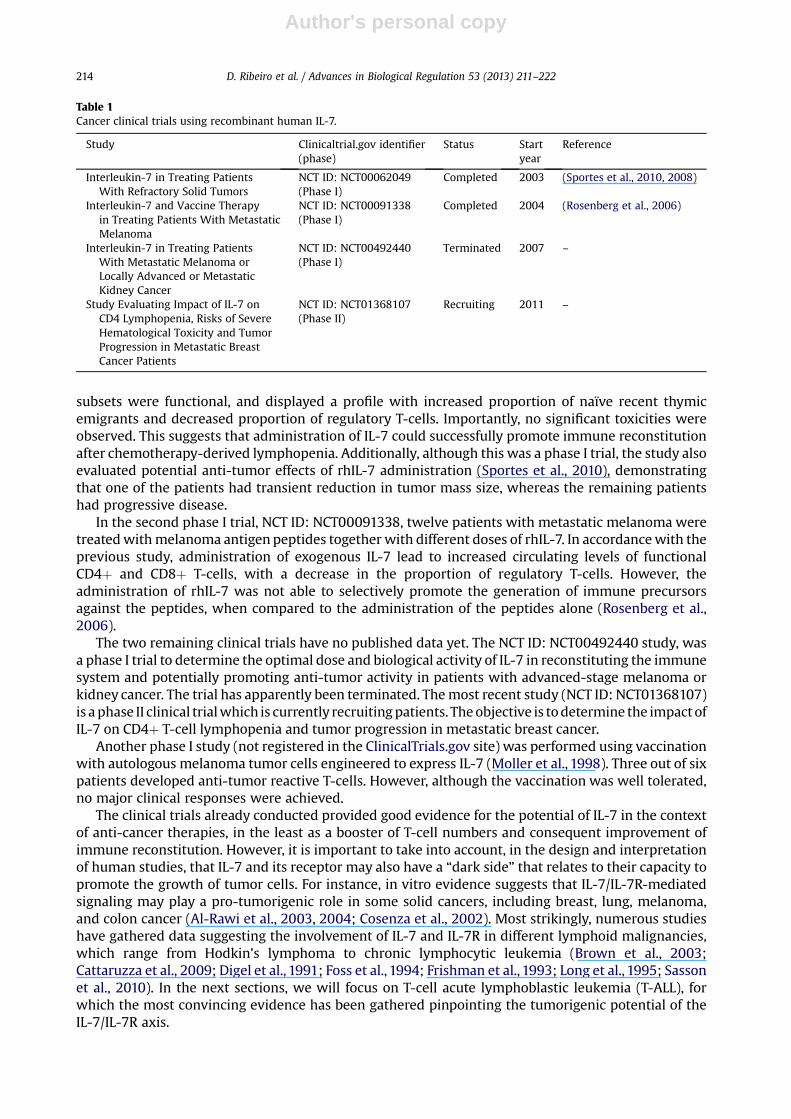
IL-7R subunit (gc), or JAK3 (which is normally associated with gc), and is dependent on JAK1 (whichis normally associated with IL-7Ra) (Fig. 1) (Zenatti et al., 2011). The remaining IL7R mutationsintroduce either a tryptophan residue or an SxxxG related motif within the transmembrane region,both of which have been implicated in facilitating homo- or heterodimer formation (Ridder et al.,2005; Russ and Engelman, 2000). However, the exact mechanism by which these mutations oper-ate and the evaluation of their in vivo leukemogenic potential remain to be determined. Nonetheless,these results revealing the presence of IL7R gain-of-function mutations in T-ALL, which have beenconfirmed by subsequent studies (Bains et al., 2012; Porcu et al., 2012; Zhang et al., 2012), indicatethat targeting IL-7R-mediated downstream signaling might be a potentially useful therapeuticapproach for the treatment of IL7R-mutated patients. We have generated proof-of-concept data inthis direction by showing that different clinical-stage JAK small molecule inhibitors are able to killIL7R-mutant cells (Zenatti et al., 2011). Similar to IL-7 stimulation (Barata et al., 2004a; Barata et al.,2004c; Batista et al., 2011; Silva et al., 2011a), IL7R mutations trigger not only JAK/STAT5 but alsoPI3K/Akt/mTOR signaling pathway (Shochat et al., 2011; Zenatti et al., 2011) in T-ALL cells. Thus,targeting the latter pathway might be therapeutically beneficial not only to prevent the effects ofmicroenvironmental IL-7 stimulation of T-ALL cells but also in the context of cell-autonomousaberrations involving the IL-7R. In this context, it is important to emphasize the singularities of IL-7/IL-7R-mediated signaling in T-ALL cells versus normal T-cells, and how subtle differences maypossibly open important therapeutic windows.
Fig. 1. Physiological (IL-7-dependent) versus mutant IL-7R-mediated (constitutive) signaling in leukemia. Physiological, IL-7-dependent, signaling relies on heterodimerization of the two IL-7R subunits (IL-7Ra/CD127 and gc/CD132), which is required foractivation of both JAK1 and JAK3 and consequent downstream signaling. It fluctuates between ON and OFF states according to ligandavailability. In contrast, IL7R cysteine-introducing mutations promote disulfide bond formation between two IL-7Ra/CD127 mutantchains and receptor homodimerization. This leads to the close proximity and consequent activation of two JAK1 molecules asso-ciated with each IL-7Ra cytoplasmic tail. The corollary is that IL-7 is no longer required for triggering signaling, which is perma-nently turned ON.
D. Ribeiro et al. / Advances in Biological Regulation 53 (2013) 211–222216

Author's personal copy
IL-7/IL-7R-Mediated Signaling in T-ALL versus Normal T-cells: Subtle Differences – MajorConsequences?
IL-7 binding to its receptor initiates signaling by promoting IL-7Ra and gc heterodimerization andassociated conformational changes (McElroy et al., 2012) that allow for JAK1 and JAK3 trans-phosphorylation and consequent phosphorylation of tyrosine residues in the cytoplasmic tail of IL-7Ra (of which the conserved Y449 residue appears to be critical for downstream signaling), therebycreating docking sites for effector molecules, most notably PI3K and STAT5 (reviewed in (Kittipatarinand Khaled, 2007; Mazzucchelli and Durum, 2007)).
STAT5 is phosphorylated by JAKs following recruitment to the IL-7Ra, consequently dimerizing andtranslocating to the nucleus where it induces transcription of several genes involved in cell survival andproliferation. The JAK/STAT pathway is critical for IL-7-dependent homeostasis of naïve CD4 and CD8 T-cells, as demonstrated by the fact that overexpression of SOCS1 (a negative regulator of JAK-STATsignaling) results in reduction of the number of naïve cells in the peripheral T-cell pool(Ramanathan et al., 2006). During thymic development, there is evidence that STAT5 is involved in IL-7-mediated immature thymocyte differentiation rather than mediating the survival/proliferativeeffects of the cytokine, which are PI3K-dependent (Pallard et al., 1999). The role of JAK/STAT5 pathwayin the development of human T-ALL is not clear, although there is evidence from mouse studies thatSTAT5 is required for IL-7-dependent T-cell lymphomagenesis (Abraham et al., 2005). However, thedetails and possible specificities of JAK/STAT pathway modulation by IL-7 stimulation in T-ALL cells ascompared to their normal counterparts remain to be dissected.
The PI3K/Akt/mTOR pathway is anothermajor signalingmodule involved in thymocyte developmentand IL-7-mediated effects. Phosphatidylinositol 3-kinase (PI3K) catalyzes the production of phosphati-dylinositol 3,4,5 trisphosphate (PIP3), therebyactivatinganumberofdownstreamtargets, includingmostprominently Akt/PKB and its downstream targetmTOR. In vitro studieswith human thymocytes indicatethat activationofPI3K/Aktpathway is required for IL-7-inducedproliferationand/or survival of immatureCD34þ thymocytes (Pallard et al.,1999). Interestingly, IL-7was shown to induce rapid phosphorylation ofAkt/PKB in CD34þ thymocytes, which was not detected in more differentiated CD34- thymocytes(Johnson et al., 2008). In humanmature peripheral T-cells IL-7 induces PI3K/Akt pathway activation onlyafter at least one hour of stimulation. This late, but sustained, induction of PI3K/Akt signaling activity isnecessary for T-cell proliferation (Lali et al., 2004) and, as judged by studies in mouse peripheral T-cells,STAT5 transcriptional activitymight be required for Akt activation (Wofford et al., 2008). This contrasts towhat happens in T-ALL cells, which generally respond to IL-7 in a manner that resembles that of CD34þthymocytes in the sense that PI3K/Akt/mTOR pathway is rapidly activated even in CD34- T-ALL cases((Barata et al., 2004a; Barataet al., 2004c; Batista et al., 2011; Silva et al., 2011a) andourunpublisheddata).This ability to promote early, STAT5-independent activation of PI3K/Akt/mTORpathwaymight reflect theexistence of an immature precursor program that remains active in T-ALL cells irrespectively of theirdevelopmental stage. Whether this is indeed the case requires investigation.
IL-7 prevents T-cell apoptosis and induces cell cycle entry, thereby promoting in vitro proliferation ofboth normal and malignant T-cells (e.g. (Barata et al., 2001; Barata et al., 2004c; Fox et al., 2005; Khaledet al., 2002; Lali et al., 2004; Pellegrini et al., 2004; Rathmell et al., 2001; Sereti et al., 2009; Swainsonet al.,2007; vonFreeden-Jeffryet al.,1997)–however, themechanismsbywhich IL-7does sodiffer in each case.In healthy T-cells PI3K/Akt/mTOR activation is indispensable for T-cell cell cycle progression (Lali et al.,2004; Swainson et al., 2007), but does not regulate Bcl-2 expression or cell survival (Fox et al., 2005;Rathmell et al., 2001). In sharp contrast, PI3K/Akt/mTOR activation in IL-7-stimulated T-ALL cells isfundamental not only for p27kip1 downregulation and cell cycle progression but also for Bcl-2 upregu-lation and increased cell viability (Barata et al., 2001; Barata et al., 2004c). Although the exact reasons forthese functional differences warrant investigation, they have clear therapeutic implications. PI3K ormTORsmallmolecule inhibitorsareessentiallycytostatic for IL-7-exposedhealthyT-cells (Swainsonetal.,2007) and do not significantly affect the viability ofmature T-cells (Fox et al., 2005; Rathmell et al., 2001)or thymocytes (our unpublished data). In contrast, the same class of drugs is clearly cytotoxic to IL-7-stimulated T-ALL cells, blocking cell cycle progression and promoting apoptosis (Barata et al., 2001;Barata et al., 2004c). Most patients at diagnosis present with leukemia cells that are IL-7-responsivein vitro (Barata et al., 2004b) and that are likely benefiting from IL-7 exposure in vivo (Silva et al.,
D. Ribeiro et al. / Advances in Biological Regulation 53 (2013) 211–222 217

Author's personal copy
2011b). Therefore, if integrated into combinatorial therapeutic protocols these drugs have thepotential tocontribute to the selective elimination of the malignant cells without having a major impact on theirnormal counterparts. Moreover, the therapeutic potential of PI3K/Akt/mTOR signaling inhibitors mayextend beyond the context of IL-7R-mediated signaling, since it is known that the vast majority of T-ALLdisplayconstitutive activationof the pathway (Silva et al., 2008). In this regard it isworthmentioning thatpre-clinical in vitro studies with several clinical-stage PI3K/Akt/mTOR pharmacological inhibitors haveshownpromising results (Chiarini et al., 2010; Evangelisti et al., 2011; Grimaldi et al., 2012; Simioni et al.,2012).
Information on other pathways that have been reported to be activated by IL-7 in lymphocytes canbe found elsewhere (Barata et al., 2005; Jiang et al., 2005; Khaled and Durum, 2002, 2003).
Genetic Evidence for the Relevance of IL-7R-Mediated Signaling in T-ALL
In the previous section, we have summarized the main signaling pathways elicited by IL-7 stimu-lation of normal and malignant T-cells. This allows us to better contextualize the genetic evidence onthe relevance of cell-autonomous deregulation of IL-7R-mediated signaling networks in T-ALL biology,which extends beyond the IL7R mutations we already described.
Somatic JAK1 gain-of-function mutations are relatively prevalent in adult T-ALL (18% of the cases)and can also be found in childhood T-ALL (2% of the cases) (Flex et al., 2008; Zhang et al., 2012). Themutations affect conserved residues within the FERM, SH2, pseudokinase, and kinase domains of JAK1.Importantly, the mutants induce constitutive activation of STATs, Akt and ERK proteins, and confergrowth factor independence to Ba/F3 cells, indicative of their transforming ability. Clinically, JAK1mutations are associated with relapse and decreased overall survival (Flex et al., 2008). Themechanismof action of these mutations is not completely understood, particularly given their heterogeneousdomain location, although some data indicate that they might play a role in inducing conformationalchanges which promote the catalytic activation of the protein (Radtke et al., 2005; Zhou et al., 2001).
JAK3 gain-of-functionmutations have been reported in differentmalignancies, such as breast cancer(Jeong et al., 2008), adult T-cell leukemia/lymphoma (Elliott et al., 2011) and megakaryoblasticleukemia (Walters et al., 2006). They have been recently described also in T-ALL (Bains et al., 2012;Kalender Atak et al., 2012; Zhang et al., 2012). Similar to JAK1, the JAK3 mutations occur within theFERM, pseudokinase and kinase domains and are transforming in vitro. However, their mechanism ofaction still needs to be elucidated.
In addition to these alterations in IL-7R-associated JAKs, there is evidence that cell-autonomouslesions in downstream mediators of IL-7R signaling are frequent in T-ALL cells, most notably withinPI3K/Akt pathway. Hyperactivation of this pathway is very frequent in T-ALL and contributes toleukemia cell maintenance (Silva et al., 2008). The major negative regulator of PI3K/Akt pathway is thelipid phosphatase PTEN (phosphatase and tensin deleted on chromosome 10), which removes the 3-phosphate from PIP3, dephosphorylating PIP3 into PIP2 and thereby down-modulating Akt andmTOR signaling (Fruman and Bismuth, 2009). PTENmutations aremostly clustered in exon 7 (Gutierrezet al., 2009; Jotta et al., 2010; Maser et al., 2007; Silva et al., 2008), which encodes the C2 domain of theprotein, and are usually nonsense or frame-shift, resulting in a truncated, non-functional protein(Gutierrez et al., 2009). They occur in 5–30% of the T-ALL samples and have been associated withdecreased overall survival and risk of relapse (Jotta et al., 2010). PTEN gene deletion is less frequent,occurs in up to 9% of primary T-ALL and has also been associated with poor prognosis (Gutierrez et al.,2009; Remke et al., 2009). PIK3CA (p110a) gain-of-function mutations in T-ALL are uncommon (5% ofthe cases) and affect mostly exons 9 and 20. PI3KR1 (p85a) inactivating mutations have also beenreported in 5% of T-ALL patients. AKTmutations are even less frequent (around 2% of T-ALLs) (Gutierrezet al., 2009). The frequencies of mutations for each gene are relatively low. However, if analyzedtogether as part of a common pathway, genetic alterations in the PTEN-PI3K-AKT pathway can accountup to 48% of the T-ALL cases (Gutierrez et al., 2009). Evidently, this pathway is involved in manyprocesses in the cell that do not relate to IL-7R-mediated signaling. Nonetheless, one should take intoaccount that PI3K/Akt/mTOR is one of the major IL-7R downstream signaling effectors, with a clearimpact on T-ALL cell cycle progression and cell viability. Therefore, it seems logical to include thegenetic alterations summarized above that affect PI3K/Akt/mTOR signaling as evidence, together with
D. Ribeiro et al. / Advances in Biological Regulation 53 (2013) 211–222218

Author's personal copy
the mutations affecting JAK1, JAK3 and IL7R itself, for the high prevalence of mutations affecting the IL-7/IL-7R signaling axis in T-ALL. Overall, the studies summarized in this review hint on the majorimportance of IL-7/IL-7R-mediated signaling deregulation for human T-cell leukemogenesis.
Conflict of Interest Statement
The authors have no conflict of interest to declare.The research work in JTB’s lab related to the present reviewwas supported by grants from Fundação
para a Ciência e a Tecnologia, FCT, Portugal. The funding agency did not have any influence on theplanning, execution or writing of the manuscript.
Acknowledgments
The research work in JTB’s lab related to the present reviewwas supported by grants from Fundaçãopara a Ciência e a Tecnologia, FCT, Portugal (PTDC/SAU-OBD/104816/2008, PTDC/SAU-ONC/122428/2010 and PIC/IC/83023/2007). DR and AM have FCT PhD fellowships. We apologize to the authorswhose work, although of relevance to the field, may have not been included in this review.
References
Abraham N, Ma MC, Snow JW, Miners MJ, Herndier BG, Goldsmith MA. Haploinsufficiency identifies STAT5 as a modifier of IL-7-induced lymphomas. Oncogene 2005;24:5252–7.
Al-Rawi MA, Mansel RE, Jiang WG. Interleukin-7 (IL-7) and IL-7 receptor (IL-7R) signalling complex in human solid tumours.Histol Histopathol 2003;18:911–23.
Al-Rawi MA, Rmali K, Watkins G, Mansel RE, Jiang WG. Aberrant expression of interleukin-7 (IL-7) and its signalling complex inhuman breast cancer. Eur J Cancer 2004;40:494–502.
Alves NL, Richard-Le Goff O, Huntington ND, Sousa AP, Ribeiro VS, Bordack A, et al. Characterization of the thymic IL-7 nichein vivo. Proc Natl Acad Sci U S A 2009;106:1512–7.
Azevedo RI, Soares MV, Barata JT, Tendeiro R, Serra-Caetano A, Victorino RM, et al. IL-7 sustains CD31 expression in human naiveCD4þ T cells and preferentially expands the CD31þ subset in a PI3K-dependent manner. Blood 2009;113:2999–3007.
Bains T, Heinrich MC, Loriaux MM, Beadling C, Nelson D, Warrick A, et al. Newly described activating JAK3 mutations in T-cellacute lymphoblastic leukemia. Leukemia 2012;26:2144–6.
Balciunaite G, Ceredig R, Fehling HJ, Zuniga-Pflucker JC, Rolink AG. The role of Notch and IL-7 signaling in early thymocyteproliferation and differentiation. Eur J Immunol 2005;35:1292–300.
Barata JT, Boussiotis VA, Yunes JA, Ferrando AA, Moreau LA, Veiga JP, et al. IL-7-dependent human leukemia T-cell line asa valuable tool for drug discovery in T-ALL. Blood 2004a;103:1891–900.
Barata JT, Cardoso AA, Boussiotis VA. Interleukin-7 in T-cell acute lymphoblastic leukemia: an extrinsic factor supportingleukemogenesis? Leuk Lymphoma 2005;46:483–95.
Barata JT, Cardoso AA, Nadler LM, Boussiotis VA. Interleukin-7 promotes survival and cell cycle progression of T-cell acutelymphoblastic leukemia cells by down-regulating the cyclin-dependent kinase inhibitor p27(kip1). Blood 2001;98:1524–31.
Barata JT, Keenan TD, Silva A, Nadler LM, Boussiotis VA, Cardoso AA. Common gamma chain-signaling cytokines promoteproliferation of T-cell acute lymphoblastic leukemia. Haematologica 2004b;89:1459–67.
Barata JT, Silva A, Abecasis M, Carlesso N, Cumano A, Cardoso AA. Molecular and functional evidence for activity of murine IL-7on human lymphocytes. Exp Hematol 2006;34:1133–42.
Barata JT, Silva A, Brandao JG, Nadler LM, Cardoso AA, Boussiotis VA. Activation of PI3K is indispensable for interleukin 7-mediated viability, proliferation, glucose use, and growth of T cell acute lymphoblastic leukemia cells. J Exp Med 2004c;200:659–69.
Batista A, Barata JT, Raderschall E, Sallan SE, Carlesso N, Nadler LM, et al. Targeting of active mTOR inhibits primary leukemia Tcells and synergizes with cytotoxic drugs and signaling inhibitors. Exp Hematol 2011;39:457–72. e3.
Boyman O, Ramsey C, Kim DM, Sprent J, Surh CD. IL-7/anti-IL-7 mAb complexes restore T cell development and inducehomeostatic T cell expansion without lymphopenia. J Immunol 2008;180:7265–75.
Brown VI, Fang J, Alcorn K, Barr R, Kim JM, Wasserman R, et al. Rapamycin is active against B-precursor leukemia in vitro andin vivo, an effect that is modulated by IL-7-mediated signaling. Proc Natl Acad Sci U S A 2003;100:15113–8.
Buckley RH. Molecular defects in human severe combined immunodeficiency and approaches to immune reconstitution. AnnRev Immunol 2004;22:625–55.
Cattaruzza L, Gloghini A, Olivo K, Di Francia R, Lorenzon D, De Filippi R, et al. Functional coexpression of Interleukin (IL)-7 andits receptor (IL-7R) on Hodgkin and Reed-Sternberg cells: involvement of IL-7 in tumor cell growth and microenviron-mental interactions of Hodgkin’s lymphoma. Int J Cancer 2009;125:1092–101.
Chiarini F, Grimaldi C, Ricci F, Tazzari PL, Evangelisti C, Ognibene A, et al. Activity of the novel dual phosphatidylinositol3-kinase/mammalian target of rapamycin inhibitor NVP-BEZ235 against T-cell acute lymphoblastic leukemia. Cancer Res2010;70:8097–107.
Chowdhury D, Sen R. Transient IL-7/IL-7R signaling provides a mechanism for feedback inhibition of immunoglobulin heavychain gene rearrangements. Immunity 2003;18:229–41.
Churchman SM, Ponchel F. Interleukin-7 in rheumatoid arthritis. Rheumatology (Oxford) 2008;47:753–9.
D. Ribeiro et al. / Advances in Biological Regulation 53 (2013) 211–222 219

Author's personal copy
Cosenza L, Gorgun G, Urbano A, Foss F. Interleukin-7 receptor expression and activation in nonhaematopoietic neoplastic celllines. Cell Signal 2002;14:317–25.
Digel W, Schmid M, Heil G, Conrad P, Gillis S, Porzsolt F. Human interleukin-7 induces proliferation of neoplastic cells fromchronic lymphocytic leukemia and acute leukemias. Blood 1991;78:753–9.
Eder M, Ottmann OG, Hansen-Hagge TE, Bartram CR, Gillis S, Hoelzer D, et al. Effects of recombinant human IL-7 on blast cellproliferation in acute lymphoblastic leukemia. Leukemia 1990;4:533–40. Official Journal of the Leukemia Society ofAmerica, Leukemia Research Fund, UK.
Elliott NE, Cleveland SM, Grann V, Janik J, Waldmann TA, Dave UP. FERM domain mutations induce gain of function in JAK3 inadult T-cell leukemia/lymphoma. Blood 2011;118:3911–21.
Evangelisti C, Ricci F, Tazzari P, Tabellini G, Battistelli M, Falcieri E, et al. Targeted inhibition of mTORC1 and mTORC2 by active-site mTOR inhibitors has cytotoxic effects in T-cell acute lymphoblastic leukemia. Leukemia 2011;25:781–91. Official Journalof the Leukemia Society of America, Leukemia Research Fund, UK.
Flex E, Petrangeli V, Stella L, Chiaretti S, Hornakova T, Knoops L, et al. Somatically acquired JAK1 mutations in adult acutelymphoblastic leukemia. J Exp Med 2008;205:751–8.
Foss FM, Koc Y, Stetler-Stevenson MA, Nguyen DT, O’Brien MC, Turner R, et al. Costimulation of cutaneous T-cell lymphoma cellsby interleukin-7 and interleukin-2: potential autocrine or paracrine effectors in the Sezary syndrome. J Clin Oncol 1994;12:326–35.
Fox CJ, Hammerman PS, Thompson CB. The Pim kinases control rapamycin-resistant T cell survival and activation. J Exp Med2005;201:259–66.
Frishman J, Long B, Knospe W, Gregory S, Plate J. Genes for interleukin 7 are transcribed in leukemic cell subsets of individualswith chronic lymphocytic leukemia. J Exp Med 1993;177:955–64.
Fruman DA, Bismuth G. Fine tuning the immune response with PI3K. Immunol Rev 2009;228:253–72.Fry TJ, Mackall CL. Interleukin-7: from bench to clinic. Blood 2002;99:3892–904.Fry TJ, Moniuszko M, Creekmore S, Donohue SJ, Douek DC, Giardina S, et al. IL-7 therapy dramatically alters peripheral T-cell
homeostasis in normal and SIV-infected nonhuman primates. Blood 2003;101:2294–9.Gonzalez-Garcia S, Garcia-Peydro M, Martin-Gayo E, Ballestar E, Esteller M, Bornstein R, et al. CSL-MAML-dependent Notch1
signaling controls T lineage-specific IL-7R{alpha} gene expression in early human thymopoiesis and leukemia. J Exp Med2009;206:779–91.
Goodwin RG, Friend D, Ziegler SF, Jerzy R, Falk BA, Gimpel S, et al. Cloning of the human and murine interleukin-7 receptors:demonstration of a soluble form and homology to a new receptor superfamily. Cell 1990;60:941–51.
Grenningloh R, Tai TS, Frahm N, Hongo TC, Chicoine AT, Brander C, et al. Ets-1 maintains IL-7 receptor expression in peripheral Tcells. J Immunol 2011;186:969–76.
Grimaldi C, Chiarini F, Tabellini G, Ricci F, Tazzari PL, Battistelli M, et al. AMP-dependent kinase/mammalian target of rapamycincomplex 1 signaling in T-cell acute lymphoblastic leukemia: therapeutic implications. Leukemia 2012;26:91–100. OfficialJournal of the Leukemia Society of America, Leukemia Research Fund, UK.
Guimond M, Veenstra RG, Grindler DJ, Zhang H, Cui Y, Murphy RD, et al. Interleukin 7 signaling in dendritic cells regulates thehomeostatic proliferation and niche size of CD4þ T cells. Nat Immunol 2009;10:149–57.
Gutierrez A, Sanda T, Grebliunaite R, Carracedo A, Salmena L, Ahn Y, et al. High frequency of PTEN, PI3K, and AKT abnormalitiesin T-cell acute lymphoblastic leukemia. Blood 2009;114:647–50.
Hara T, Shitara S, Imai K, Miyachi H, Kitano S, Yao H, et al. Identification of IL-7-Producing Cells in Primary and SecondaryLymphoid Organs Using IL-7-GFP Knock-In Mice. J Immunol 2012;189:1577–84.
Henriques CM, Rino J, Nibbs RJ, Graham GJ, Barata JT. IL-7 induces rapid clathrin-mediated internalization and JAK3-dependentdegradation of IL-7Ralpha in T cells. Blood 2010;115:3269–77.
Jeong EG, Kim MS, Nam HK, Min CK, Lee S, Chung YJ, et al. Somatic mutations of JAK1 and JAK3 in acute leukemias and solidcancers. Clin Cancer Res 2008;14:3716–21.
Jiang Q, Li WQ, Aiello FB, Mazzucchelli R, Asefa B, Khaled AR, et al. Cell biology of IL-7, a key lymphotrophin. Cytokine GrowthFactor Rev 2005;16:513–33.
Johnson SE, Shah N, Bajer AA, LeBien TW. IL-7 activates the phosphatidylinositol 3-kinase/AKT pathway in normal humanthymocytes but not normal human B cell precursors. J Immunol 2008;180:8109–17.
Jotta PY, Ganazza MA, Silva A, Viana MB, da Silva MJ, Zambaldi LJ, et al. Negative prognostic impact of PTEN mutation inpediatric T-cell acute lymphoblastic leukemia. Leukemia 2010;24:239–42.
Kalender Atak Z, De Keersmaecker K, Gianfelici V, Geerdens E, Vandepoel R, Pauwels D, et al. High accuracy mutation detectionin leukemia on a selected panel of cancer genes. PLoS One 2012;7:e38463.
Karawajew L, Ruppert V, Wuchter C, Kosser A, Schrappe M, Dorken B, et al. Inhibition of in vitro spontaneous apoptosis by IL-7correlates with bcl-2 up-regulation, cortical/mature immunophenotype, and better early cytoreduction of childhood T-cellacute lymphoblastic leukemia. Blood 2000;96:297–306.
Khaled AR, Durum SK. Lymphocide: cytokines and the control of lymphoid homeostasis. Nat Rev Immunol 2002;2:817–30.Khaled AR, Durum SK. Death and Baxes: mechanisms of lymphotrophic cytokines. Immunol Rev 2003;193:48–57.Khaled AR, Li WQ, Huang J, Fry TJ, Khaled AS, Mackall CL, et al. Bax deficiency partially corrects interleukin-7 receptor alpha
deficiency. Immunity 2002;17:561–73.Kibe R, Zhang S, Guo D, Marrero L, Tsien F, Rodriguez P, et al. IL-7Ralpha deficiency in p53null mice exacerbates thymocyte
telomere erosion and lymphomagenesis. Cell Death Differ 2012;19:1139–51.Kittipatarin C, Khaled AR. Interlinking interleukin-7. Cytokine 2007;39:75–83.Komschlies KL, Gregorio TA, Gruys ME, Back TC, Faltynek CR, Wiltrout RH. Administration of recombinant human IL-7 to mice
alters the composition of B-lineage cells and T cell subsets, enhances T cell function, and induces regression of establishedmetastases. J Immunol 1994;152:5776–84.
Lali FV, Crawley J, McCulloch DA, Foxwell BM. A late, prolonged activation of the phosphatidylinositol 3-kinase pathway isrequired for T cell proliferation. J Immunol 2004;172:3527–34.
Laouar Y, Crispe IN, Flavell RA. Overexpression of IL-7R alpha provides a competitive advantage during early T-cell development.Blood 2004;103:1985–94.
D. Ribeiro et al. / Advances in Biological Regulation 53 (2013) 211–222220

Author's personal copy
Lee LF, Logronio K, Tu GH, Zhai W, Ni I, Mei L, et al. Anti-IL-7 receptor-alpha reverses established type 1 diabetes in nonobesediabetic mice by modulating effector T-cell function. Proc Natl Acad Sci U S A 2012;109:12674–9.
Lenz DC, Kurz SK, Lemmens E, Schoenberger SP, Sprent J, Oldstone MB, et al. IL-7 regulates basal homeostatic proliferation ofantiviral CD4þT cell memory. Proc Natl Acad Sci U S A 2004;101:9357–62.
Li X, von Boehmer H. Notch Signaling in T-Cell Development and T-ALL. ISRN Hematol 2011:921706.Long BW, Witte PL, Abraham GN, Gregory SA, Plate JM. Apoptosis and interleukin 7 gene expression in chronic B-lymphocytic
leukemia cells. Proc Natl Acad Sci U S A 1995;92:1416–20.Mackall CL, Fleisher TA, Brown MR, Andrich MP, Chen CC, Feuerstein IM, et al. Distinctions between CD8þ and CD4þ T-cell
regenerative pathways result in prolonged T-cell subset imbalance after intensive chemotherapy. Blood 1997;89:3700–7.Mackall CL, Fleisher TA, Brown MR, Magrath IT, Shad AT, Horowitz ME, et al. Lymphocyte depletion during treatment with
intensive chemotherapy for cancer. Blood 1994;84:2221–8.Mackall CL, Fry TJ, Gress RE. Harnessing the biology of IL-7 for therapeutic application. Nat Rev Immunol 2011;11:330–42.Makrynikola V, Kabral A, Bradstock K. Effects of interleukin 7 on the growth of clonogenic cells in T-cell acute lymphoblastic
leukaemia. Leukemia Res 1991;15:879–82.Maser RS, Choudhury B, Campbell PJ, Feng B, Wong KK, Protopopov A, et al. Chromosomally unstable mouse tumours have
genomic alterations similar to diverse human cancers. Nature 2007;447:966–71.Masuda M, Motoji T, Oshimi K, Mizoguchi H. Effects of interleukin-7 on proliferation of hematopoietic malignant cells. Exp
Hematol 1990;18:965–7.Mazzucchelli R, Durum SK. Interleukin-7 receptor expression: intelligent design. Nat Rev Immunol 2007;7:144–54.Mazzucchelli RI, Riva A, Durum SK. The human IL-7 receptor gene: deletions, polymorphisms and mutations. Semin Immunol
2012;24:225–30.Mazzucchelli RI, Warming S, Lawrence SM, Ishii M, Abshari M, Washington AV, et al. Visualization and identification of IL-7
producing cells in reporter mice. PloS ONE 2009;4:e7637.McElroy CA, Holland PJ, Zhao P, Lim JM, Wells L, Eisenstein E, et al. Structural reorganization of the interleukin-7 signaling
complex. Proc Natl Acad Sci U S A 2012;109:2503–8.Moller P, Sun Y, Dorbic T, Alijagic S, Makki A, Jurgovsky K, et al. Vaccination with IL-7 gene-modified autologous melanoma cells
can enhance the anti-melanoma lytic activity in peripheral blood of patients with a good clinical performance status:a clinical phase I study. Br J Cancer 1998;77:1907–16.
Morrissey PJ, Conlon P, Braddy S, Williams DE, Namen AE, Mochizuki DY. Administration of IL-7 to mice withcyclophosphamide-induced lymphopenia accelerates lymphocyte repopulation. J Immunol 1991;146:1547–52.
Namen AE, Schmierer AE, March CJ, Overell RW, Park LS, Urdal DL, et al. B cell precursor growth-promoting activity. Purificationand characterization of a growth factor active on lymphocyte precursors. J Exp Med 1988;167:988–1002.
Noguchi M, Yi H, Rosenblatt HM, Filipovich AH, Adelstein S, Modi WS, et al. Interleukin-2 receptor gamma chain mutationresults in X-linked severe combined immunodeficiency in humans. Cell 1993;73:147–57.
Osborne LC, Duthie KA, Seo JH, Gascoyne RD, Abraham N. Selective ablation of the YxxM motif of IL-7Ralpha suppresseslymphomagenesis but maintains lymphocyte development. Oncogene 2010;29:3854–64.
Ouyang W, Beckett O, Flavell RA, Li MO. An essential role of the Forkhead-box transcription factor Foxo1 in control of T cellhomeostasis and tolerance. Immunity 2009;30:358–71.
Pallard C, Stegmann AP, van Kleffens T, Smart F, Venkitaraman A, Spits H. Distinct roles of the phosphatidylinositol 3-kinase andSTAT5 pathways in IL-7-mediated development of human thymocyte precursors. Immunity 1999;10:525–35.
Park JH, Adoro S, Guinter T, Erman B, Alag AS, Catalfamo M, et al. Signaling by intrathymic cytokines, not T cell antigen receptors,specifies CD8 lineage choice and promotes the differentiation of cytotoxic-lineage T cells. Nat Immunol 2010;11:257–64.
Park JH, Yu Q, Erman B, Appelbaum JS, Montoya-Durango D, Grimes HL, et al. Suppression of IL7Ralpha transcription by IL-7 andother prosurvival cytokines: a novel mechanism for maximizing IL-7-dependent T cell survival. Immunity 2004;21:289–302.
Pellegrini M, Bouillet P, Robati M, Belz GT, Davey GM, Strasser A. Loss of Bim increases T cell production and function ininterleukin 7 receptor-deficient mice. J Exp Med 2004;200:1189–95.
Pellegrini M, Calzascia T, Toe JG, Preston SP, Lin AE, Elford AR, et al. IL-7 engages multiple mechanisms to overcome chronic viralinfection and limit organ pathology. Cell 2011;144:601–13.
Peschon JJ, Morrissey PJ, Grabstein KH, Ramsdell FJ, Maraskovsky E, Gliniak BC, et al. Early lymphocyte expansion is severelyimpaired in interleukin 7 receptor-deficient mice. J Exp Med 1994;180:1955–60.
Porcu M, Kleppe M, Gianfelici V, Geerdens E, De Keersmaecker K, Tartaglia M, et al. Mutation of the receptor tyrosine phos-phatase PTPRC (CD45) in T-cell acute lymphoblastic leukemia. Blood 2012;119:4476–9.
Prlic M, Lefrancois L, Jameson SC. Multiple choices: regulation of memory CD8 T cell generation and homeostasis by interleukin(IL)-7 and IL-15. J Exp Med 2002;195:F49–52.
Puel A, Ziegler SF, Buckley RH, LeonardWJ. Defective IL7R expression in T(-)B(þ)NK(þ) severe combined immunodeficiency. NatGenet 1998;20:394–7.
Radtke S, Haan S, Jorissen A, Hermanns HM, Diefenbach S, Smyczek T, et al. The Jak1 SH2 domain does not fulfill a classical SH2function in Jak/STAT signaling but plays a structural role for receptor interaction and up-regulation of receptor surfaceexpression. J Biol Chem 2005;280:25760–8.
Ramanathan S, Gagnon J, Leblanc C, Rottapel R, Ilangumaran S. Suppressor of cytokine signaling 1 stringently regulates distinctfunctions of IL-7 and IL-15 in vivo during T lymphocyte development and homeostasis. J Immunol 2006;176:4029–41.
Rathmell JC, Farkash EA, Gao W, Thompson CB. IL-7 enhances the survival and maintains the size of naive T cells. J Immunol2001;167:6869–76.
Remke M, Pfister S, Kox C, Toedt G, Becker N, Benner A, et al. High-resolution genomic profiling of childhood T-ALL revealsfrequent copy-number alterations affecting the TGF-beta and PI3K-AKT pathways and deletions at 6q15-16.1 as a genomicmarker for unfavorable early treatment response. Blood 2009;114:1053–62.
Repass JF, Laurent MN, Carter C, Reizis B, Bedford MT, Cardenas K, et al. IL7-hCD25 and IL7-Cre BAC transgenic mouse lines: newtools for analysis of IL-7 expressing cells. Genesis 2009;47:281–7.
Rich BE, Campos-Torres J, Tepper RI, Moreadith RW, Leder P. Cutaneous lymphoproliferation and lymphomas in interleukin 7transgenic mice. J Exp Med 1993;177:305–16.
D. Ribeiro et al. / Advances in Biological Regulation 53 (2013) 211–222 221

Author's personal copy
Ridder A, Skupjen P, Unterreitmeier S, Langosch D. Tryptophan supports interaction of transmembrane helices. J Mol Biol 2005;354:894–902.
Rosenberg SA, Sportes C, Ahmadzadeh M, Fry TJ, Ngo LT, Schwarz SL, et al. IL-7 administration to humans leads to expansion ofCD8þ and CD4þ cells but a relative decrease of CD4þ T-regulatory cells. J Immunother 2006;29:313–9.
Russ WP, Engelman DM. The GxxxG motif: a framework for transmembrane helix-helix association. J Mol Biol 2000;296:911–9.Sakata T, Iwagami S, Tsuruta Y, Teraoka H, Tatsumi Y, Kita Y, et al. Constitutive expression of interleukin-7 mRNA and production
of IL-7 by a cloned murine thymic stromal cell line. J Leukoc Biol 1990;48:205–12.Sasson SC, Smith S, Seddiki N, Zaunders JJ, Bryant A, Koelsch KK, et al. IL-7 receptor is expressed on adult pre-B-cell acute
lymphoblastic leukemia and other B-cell derived neoplasms and correlates with expression of proliferation and survivalmarkers. Cytokine 2010;50:58–68.
Schluns KS, Kieper WC, Jameson SC, Lefrancois L. Interleukin-7 mediates the homeostasis of naive and memory CD8 T cellsin vivo. Nat Immunol 2000;1:426–32.
Scupoli MT, Perbellini O, Krampera M, Vinante F, Cioffi F, Pizzolo G. Interleukin 7 requirement for survival of T-cell acutelymphoblastic leukemia and human thymocytes on bone marrow stroma. Haematologica 2007;92:264–6.
Scupoli MT, Vinante F, Krampera M, Vincenzi C, Nadali G, Zampieri F, et al. Thymic epithelial cells promote survival of humanT-cell acute lymphoblastic leukemia blasts: the role of interleukin-7. Haematologica 2003;88:1229–37.
Seddon B, Tomlinson P, Zamoyska R. Interleukin 7 and T cell receptor signals regulate homeostasis of CD4 memory cells. NatImmunol 2003;4:680–6.
Sereti I, Dunham RM, Spritzler J, Aga E, Proschan MA, Medvik K, et al. IL-7 administration drives T cell-cycle entry and expansionin HIV-1 infection. Blood 2009;113:6304–14.
Shochat C, Tal N, Bandapalli OR, Palmi C, Ganmore I, te Kronnie G, et al. Gain-of-function mutations in interleukin-7 receptor-alpha (IL7R) in childhood acute lymphoblastic leukemias. J Exp Med 2011;208:901–8.
Silva A, Girio A, Cebola I, Santos CI, Antunes F, Barata JT. Intracellular reactive oxygen species are essential for PI3K/Akt/mTOR-dependent IL-7-mediated viability of T-cell acute lymphoblastic leukemia cells. Leukemia 2011a;25:960–7. Official Journalof the Leukemia Society of America, Leukemia Research Fund, UK.
Silva A, Laranjeira AB, Martins LR, Cardoso BA, Demengeot J, Yunes JA, et al. IL-7 contributes to the progression of human T-cellacute lymphoblastic leukemias. Cancer Res 2011b;71:4780–9.
Silva A, Yunes JA, Cardoso BA, Martins LR, Jotta PY, Abecasis M, et al. PTEN posttranslational inactivation and hyperactivation ofthe PI3K/Akt pathway sustain primary T cell leukemia viability. J Clin Invest 2008;118:3762–74.
Simioni C, Neri LM, Tabellini G, Ricci F, Bressanin D, Chiarini F, et al. Cytotoxic activity of the novel Akt inhibitor, MK-2206, in T-cell acute lymphoblastic leukemia. Leukemia 2012. Official Journal of the Leukemia Society of America, Leukemia ResearchFund, UK.
Sinclair C, Saini M, van der Loeff IS, Sakaguchi S, Seddon B. The long-term survival potential of mature T lymphocytes isprogrammed during development in the thymus. Sci Sigal 2011;4:ra77.
Soares MV, Borthwick NJ, Maini MK, Janossy G, Salmon M, Akbar AN. IL-7-dependent extrathymic expansion of CD45RAþ T cellsenables preservation of a naive repertoire. J Immunol 1998;161:5909–17.
Sportes C, Babb RR, Krumlauf MC, Hakim FT, Steinberg SM, Chow CK, et al. Phase I study of recombinant human interleukin-7administration in subjects with refractory malignancy. Clin Cancer Res 2010;16:727–35.
Sportes C, Hakim FT, Memon SA, Zhang H, Chua KS, Brown MR, et al. Administration of rhIL-7 in humans increases in vivo TCRrepertoire diversity by preferential expansion of naive T cell subsets. J Exp Med 2008;205:1701–14.
Swainson L, Kinet S, Mongellaz C, Sourisseau M, Henriques T, Taylor N. IL-7-induced proliferation of recent thymic emigrantsrequires activation of the PI3K pathway. Blood 2007;109:1034–42.
Tokoyoda K, Egawa T, Sugiyama T, Choi BI, Nagasawa T. Cellular niches controlling B lymphocyte behavior within bone marrowduring development. Immunity 2004;20:707–18.
Touw I, Pouwels K, van Agthoven T, van Gurp R, Budel L, Hoogerbrugge H, et al. Interleukin-7 is a growth factor of precursor Band T acute lymphoblastic leukemia. Blood 1990;75:2097–101.
Trigueros C, Hozumi K, Silva-Santos B, Bruno L, Hayday AC, Owen MJ, et al. Pre-TCR signaling regulates IL-7 receptor alphaexpression promoting thymocyte survival at the transition from the double-negative to double-positive stage. Eur JImmunol 2003;33:1968–77.
Tussiwand R, Engdahl C, Gehre N, Bosco N, Ceredig R, Rolink AG. The preTCR-dependent DN3 to DP transition requires Notchsignaling, is improved by CXCL12 signaling and is inhibited by IL-7 signaling. Eur J Immunol 2011;41:3371–80.
von Freeden-Jeffry U, Solvason N, Howard M, Murray R. The earliest T lineage-committed cells depend on IL-7 for Bcl-2expression and normal cell cycle progression. Immunity 1997;7:147–54.
von Freeden-Jeffry U, Vieira P, Lucian LA, McNeil T, Burdach SE, Murray R. Lymphopenia in interleukin (IL)-7 gene-deleted miceidentifies IL-7 as a nonredundant cytokine. J Exp Med 1995;181:1519–26.
Walters DK, Mercher T, Gu TL, O’Hare T, Tyner JW, Loriaux M, et al. Activating alleles of JAK3 in acute megakaryoblasticleukemia. Cancer Cell 2006;10:65–75.
Weng AP, Ferrando AA, Lee W, Morris JPt, Silverman LB, Sanchez-Irizarry C, et al. Activating mutations of NOTCH1 in human Tcell acute lymphoblastic leukemia. Science 2004;306:269–71.
Wiles MV, Ruiz P, Imhof BA. Interleukin-7 expression during mouse thymus development. Eur J Immunol 1992;22:1037–42.Wofford JA, Wieman HL, Jacobs SR, Zhao Y, Rathmell JC. IL-7 promotes Glut1 trafficking and glucose uptake via STAT5-mediated
activation of Akt to support T-cell survival. Blood 2008;111:2101–11.Ye SK, Agata Y, Lee HC, Kurooka H, Kitamura T, Shimizu A, et al. The IL-7 receptor controls the accessibility of the TCRgamma
locus by Stat5 and histone acetylation. Immunity 2001;15:813–23.Zenatti PP, Ribeiro D, Li W, Zuurbier L, Silva MC, Paganin M, et al. Oncogenic IL7R gain-of-function mutations in childhood T-cell
acute lymphoblastic leukemia. Nat Genet 2011;43:932–9.Zhang J, Ding L, Holmfeldt L, Wu G, Heatley SL, Payne-Turner D, et al. The genetic basis of early T-cell precursor acute
lymphoblastic leukaemia. Nature 2012;481:157–63.Zhou YJ, Chen M, Cusack NA, Kimmel LH, Magnuson KS, Boyd JG, et al. Unexpected effects of FERM domain mutations on
catalytic activity of Jak3: structural implication for Janus kinases. Mol Cell 2001;8:959–69.
D. Ribeiro et al. / Advances in Biological Regulation 53 (2013) 211–222222