Idiopathic" mental retardation and new chromosomal abnormalities
Upload
independentCategory
view
0download
0
Diagnostic and Prognostic Significance of Chromosome Abnormalities
in Childhood Acute Lymphoblastic Leukemia"
8 . 0 L h , 4 c E. BALOGH,' P. WAR:' L. PAJOR,d Z. JAKAB,' AND C. KISS'
'Department of Pediatrics University Medical School of Debrecen
H-4012 Debrecen, P.O. Box 32 Hungary
dDepartment of Pediatrics and Institute of Pathology
Medical School of Pdcs Pdcs, Hungary
INTRODUCTION
Current intensive chemotherapy cures about 70% of children with acute lympho- cytic leukemia (ALL). Although this percentage is relatively high, it means that a major proportion of the children with ALL is st i l l not cured despite intensive treatment. At the same time some highly curable patients are treated too intensively and suffer from unnecessary side effects of the chemo- and radiotherapy applied, for example, anthracyline-related cardiomyopathy, infertility, and second malignancies associated with alkylating drugs and epipodophyllotoxins. In order to further im- prove the therapeutic results and reduce the therapy-related toxicity in childhood ALL, we have to distinguish between the cases with a better and those with a worse prognosis at the time of determining the diagnosis. The initial cytogenetic changes (both structural and numerical abnormalities) proved to be one of the most reliable prognostic parameters leading to the suggestion of developing genotype-specific therapy.'-"
In the spontaneously dividing blast cells of 70-75% of ALL children, acquired clonal chromosome abnormalities can be detected at the time of the diagnosi~.'"'~ About half of these abnormalities seem to be specific to the tumor type involved. The cell specificity in this disease means that there is a close correlation be- tween the aberrations and the immunophenotype of the blasts. Among the karyo- type changes structural aberrations and numerical disturbances can be distin- guished.
Structural abnormalities, mostly translocations and deletions involving specific breakpoints, are present in two-thirds of patients with abnormal karyotypes de-
Supported by grants from Hungarian Ministry of Welfare (BlT1220). Address for correspondence: &a OlBh, M.D., D.Sc., Department of Pediatrics, University
Medical School of Debrecen, Debrecen H-4012, P.O. Box 32, Hungary. Telephone/Fax: (36) 52 414 992, e-mail: [email protected]
8

OLAH et UL: CHROMOSOME ABNORMALITIES IN ALL 9
scribed to date. At present more than 15 different, consistently occurring structural rearrangements are known in ALL.'1~'s-17 Most of them are specifically associated with distinctive immunophenotypes and are of particular clinical (diagnostic and prognostic) importance (TABLE l).5~R.11J2J8J9 A molecular analysis of the breakpoints involved in these aberrations provides an insight into the molecular mechanisms of the pathogenesis of the disease.12*20-"
Patients without ALL-specific structural abnormalities can be classified in terms of the modal chromosome number into five subgroups: diploid (46 chromosomes), pseudodiploid (46 chromosomes with structural or numerical abnormalities), hypo- diploid (less than 46 chromosomes), hyperdiploid A (47 to 50 chromosomes) and hyperdiploid B (more than 50 chromosome^).^^^^^^^'^ The latter group can be further subdivided into the hyperdiploid (50-57 chromosomes), the near-triploid (58-80 chromosomes), and the near-tetraploid subgroup (81 -103 chromosomes).u
Hyperdiploidy with a chromosome number between 50 and 57 with a mean peak at 55 chromosomes can be considered as a distinct karyotype pattern occurring in about 25-30% of childhood ALL.'o-12J423 In Sweden Heim and his co-workers found more than 50 chromosomes in 49% of ALL patients with abnormal karyo- t~pe .2~ The main feature of this group is the presence of nonrandom numerical abnormalities due to a gain of chromosomes 4, 6, 10, 14, 17, 18, 21, and X.s.'0J4 In contrast, chromosomes 1, 2, 3, 12, and 16 are rarely inv~lved.'~.'~
Near-triploid and near-tetraploid cases are rare and account only for 1-1.5% of all childhood ALL cases.= Although near-triploid cases do not appear to differ from the general ALL population in clinical features, the near-tetraploid patients more often show a T-cell phenotype and have a poorer prognosis.u
Because these structural and numerical abnormalities together with genetic changes appearing at the molecular level determine the biological features of the blasts and the morphological, cytochemical, and immunological features as well as
TABLE I. Most Common Structural Abnormalities Related to TvDical ImmunoDhenotvDe
Typical Immunophenotype Rearrangement B-cell ALL t(8;14)(q24;q32)
t(2;8)(~12;q24) t( 8;22)(q24;qll)
Pre-B-cell ALL t( l;ll)(p32;q23) t( 1;19)(q23;p13) t(9;22)(q34;qll)
Early B precursor t(4;11)(q21;q23) t (9;22)( q34;qll)
Common ALL t(9;22)( q34;qll)
T-cell ALL t(8;14)(q24;qll) t( 10;14)(q24;qll) t(ll;l4)(pl3;qll)
del/t(9)(p21) dic(9;20)(p1?3;qll)
del/t( 12p)
del/t(9)(p21) other t/del(l4)(qll)
Mixed phenotype t(4;11)(q21;q23) t(9;22)(q34;qll)
NOTE: Due to cell specificity, the aberrations are of diagnostic siflcance.

10 ANNALS NEW YORK ACADEMY OF SCIENCES
TABLE 2. Qtogenetic Subgroups with Different Prognosis in ALL at Diagnosis Karyotype
> 50 chromosomes c46 t(4;ll)
t(1;19) t(12;21)
del(6q) 14q+ normal t(9J2) t(8;14), including variants
46 abnormal 47-50
NOTE: Complete remission rates and complete remission durations are different in these cytogenetic subgroups.’*
the tumor’s invasivity, metastatic capacity, and response to therapy, it is evident that these genetic changes are of clinical significance. Their detection gives invaluable assistance to clinicians in confirming the diagnosis, subclassification of the disease, predicting the prognosis, and making the therapeutic decision.
From a clinical and therapeutic point of view, the prognostic implication of the initial karyotype is of utmost importance. Several cytogenetic subgroups with different prognoses can be distinguished (TABLE 2). Of these, three main subgroups should be pointed out (TABLE 3). The t(9;22), and t(4;ll) subgroups are associated with an unfavorable prognosis and serve as an indication for bone marrow trans- plantation. In contrast, the hyperdiploidy (with >SO chromosomes and/or >1.16 DNA index) proved to be the strongest predictor of a favorable course of the disease and of a good response to therapy.1.6’225 These patients can be characterized by favorable clinical and laboratory parameters, by a lower mean age, a usually moderate tumor load, relatively low initial white blood cell count, and an early B- cell precursor immunphenotype.26 They can achieve remission on a metabolite- based therapy; therefore the toxic effect of a more intensive treatment with anthracy- cline or genotoxic agents may be avoided. There are data available currently sug- gesting that the prognostic value of hyperdiploidy may further be related to the particular karyotype composition, namely the chromosomes involved in the aberra- tions and/or the structural abnormalities associated? For example, trisomy 6, or the combined trisomy 4 and 10, has proved to indicate an extremely favorable progno~is.”~ In contrast, recent data suggested that there is a small subset of patients with hyperdiploidy associated with chromosome 7 aberrations that has a relatively high relapse Structural aberrations added to numerical ones makes
TABLE 3. Three Main Prognostically Different Qtogenetic Subgroups of ALL Patients
Unfavorable Favorable Others
t(9;22) Hyperdiploid y Diploidy t(4;ll) t(1221) Translocations
Deletions .1 .1
Less aggressive Genotype-specific .1
Bone marrow transplantation chemotherapy chemotherapy
NOTE: Presence of t(9;22) and t(4;ll) serves as indication for bone marrow transplantation. Hyperdiploid karyotype with >50 chromosomes seems to be associated with a favorable prognosis. All the other patients, with various cytogenetic changes of different prognoses, belong to the third group. These patients need genotype-specific therapy.

O L h et 01.: CHROMOSOME ABNORMALITIES IN ALL ll
the prognosis worse.s All other patients left with various cytogenetic abnormalities belong to the third group. These patients require a genotype-specific, individually chosen therapy.B31 Based on these observations, the initial cytogenetic data may serve as a guide for the stratification of therapy, for the choice of the genotype- specific, effective but not too agressive chemotherapy for each individual. Foremost, a reliable and accurate determination of patients with hyperdiploid karyotype and of their particular karyotype composition is becoming an increasingly important task.
Several laboratory methods have been proved to be useful for determination of leukemia-associated initial genetic alterations, first of all of hyperdiploidy in childhood ALL. To identify the patients with more than 50 chromosomes, three standard methods can be used: the classical cytogenetic analysis, flow cytometry, and interphase fluorescence in situ hybridization (FISH). A recently developed FISH technique, the so-called comparative genomic hybridization (CGH) offers a new possibility to determine all chromosomal gains and losses in tissues that are otherwise not suitable for traditional cytogenetic analysis?* Unfortunately, none of these methods give complete information about all genetic changes. Each of them has both advantages and disadvantages, which are explained as follows.
Obviously, the chromosome analysis provides the most complete information about the karyotype changes, about the numerical and structural aberrations, the chromosomes involved in the aberrations, the different cell lines, and so forth. On the other hand, it requires a great number of dividing cells and the commonly poor quality of abnormal metaphases often hampers accurate evaluation. In addition, it is occasionally difficult to decide whether the abnormalities detected in the few analyzable metaphases are representative of the whole leukemic clone. A further disadvantage of this method is that it requires a lot of expertise and is a rather time-consuming procedure.
To identify the relative DNA content of the blasts and ploidy level, a simple and rapid technique, flow cytometry, can be used. However, it gives information only about the gross quantitative deviations of DNA content.D333 No information about structural chromosome aberrations, the individual chromosomes, and the different cell lines is provided. Using this technique alone, diploidy and pseudodip- loidy cannot be distinguished. The main disadvantage of the method is that in cases of trisomies of the smallest chromosomes the hyperdiploid chromosome number (>SO chromosomes) may be associated with a DNA index less than 1.16. As a result of this, about 30% of hyperdiploid patients are lost when we use flow cytometry as the only method.53s
The two methods mentioned can be complemented by the fluorescence in situ hybridization (FISH) techniques offering a strong possibility of identifying some cytogenetic abnormalities even in interphase nuclei.M41 The method is suitable for screening a large number of cells. Applying centromere-specific probes it makes possible for us to identlfy the extra chromosomes in hyperdiploid ALL patients. However, the number of simultaneously applicable probes is limited, and some of the structural abnormalities cannot be detected in this way.
In order to identify the initial genetic abnormalities in childhood ALL as accu- rately as possible, the three methods-conventional chromosome analysis, flow cytometry, and occasionally the complementary FISH technique-should be used simultaneously.
In 1993 it was decided to develop a comprehensive, nationwide project in order to perform an initial genetic analysis on all ALL children diagnosed in the hematological/oncological centers of Hungary. Before starting with this project, it seemed to be important to evaluate the results of all genetic investigations performed

12 ANNALS NEW YORK ACADEMY OF SCIENCES
on ALL children in Hungary until that time and, on the basis of the results obtained, prepare a proper strategy for our future work. Chromosome findings of 165 pre- viously investigated patients were collected and evaluated. Comparing the results to those in the literature we come to the following conclusions:
Cytogenetic analyses were carried out only in the minority of ALL children (30%). Clonal chromosome aberrations were found in 40% of patients studied. The proportion of hyperdiploid patients with more than 50 chromosomes proved to be 12% instead of 30% of the expected proportion based on a large series.'"'*"" The frequent involvement of chromosomes 8,11, and 13 in the trisomies differed from data of other a~thors.S9'~*'~~~ The survival data of patients with hyperdiploidy did not demonstrate a favorable prognosis.
All these clinical and cytogenetic differences were suggested to be due to techni- cal problems, as well as the small number of patients investigated, but the role of real geographic differences of prognostic importance was also considered. That is why it was important to study the genetic characteristics of ALL patients in our country and compare them to observations made by other authors. The aim of our study was to answer the following questions:
What is the frequency of clonal chromosome aberrations in ALL children at diagnosis in Hungary? What is the distribution of the leukemic children in terms of the modal chromosome number? What is the frequency of hyperdiploid (>50 chromosomes) cases at diagno- sis?
0 Which chromosomes are most frequently involved in the numerical and struc- tural aberrations?
0 Is there any difference in the frequency of hyperdiploid cases and in the spectrum of chromosomes involved as compared with the data in the liter- ature?
0 What is the frequency of additional structural aberrations in hyperdiploid pa- tients? How can the patients with more than 50 chromosomes be characterized from a clinical and immunological point of view?
0 Is there any correlation between the cytogenetic findings and the progno- sis? From a methodological point of view, we planned to study the reliability of the three methods in order to determine a more effective strategy for future investigations.
In spite of the fact that the present situation in health service in Hungary is not favorable either in terms of financial support or instrumental and personnel facilities of genetic laboratories for the introduction of any new investigations, great efforts have been made in order to perform as many genetic analyses on ALL patients as possible in Hungary. Here we summarize our results, which were obtained during the period from 1993 to 1995, and we will also try to outline some guidelines for our future work.
PATIENTS AND METHODS
Diagnosis and treatment of children with malignant diseases have been per- formed for more than two decades at 10 hematological/oncological centers of the

O L ~ er aL: CHROMOSOME ABNORMALITIES M ALL w
Hungarian Pediatric Oncology Working Group established in 1971.’ The diagnostic and therapeutic work of these centers is coordinated by the main center in the Department of Pediatrics No. 2. of Semmelweis Medical School, Budapest; and the work is based on standardized diagnostic and therapeutic principles.
Patients
Each ALL patient diagnosed in one of the 10 centers is registered, and the diagnosis of each case is confirmed by the coordinating center in Budapest. Cyto- chemical, immunologic, and genetic analyses are performed at the local laboratories of hematological centers. The treatment of patients included in this study was carried out in accordance with the “BFM 90” regimen recommended by the German Berlin-Frankfurt-Mtster (BFM) Study Group.
Regarding the genetic analysis, the following strategy was recommended: To identtfy the hyperdiploid patients, cytogenetic analysis and flow cytometric evalua- tion should be performed simultaneously. In cases of successful chromosome analy- ses, no further investigation is needed. However, if the cytogenetic investigation fails to provide accurate information about the karyotypic changes and the DNA index is above 1.0, FISH technique is recommended for the identification of supernu- merary chromosomes. Considering the chromosomes mostly involved in numerical aberrations, the application of probes specific for the centromeric region of chromo- somes 4, 6, 10, 17, 18, 21, and X seems to be necessary. Besides, based on our earlier experience, it seemed worthwhile to complete this spectrum of chromosomes with probes for chromosome 8, 11, and 13.
Clinical, hematological, and genetic data were collected and evaluated by the hematological/oncological center and the genetic working team of the Department of Pediatrics of the University Medical School, Debrecen.
Methodr
Cytogenetic Analysis
Unstimulated, isolated bone marrow andlor peripheral blood cells were cultured for 24 hours. Chromosomes were prepared according to standard procedures. Slides were G-banded with trypsin-Giemsa. Karyotyping was performed on a KaryoASK version 4.11 chromosome analysis system. In evaluating the karyotype changes, the International System for Human Cytogenetic Nomenclature (ISCN) was followed.42
The hematologicalloncological centers are the following: Department of Pediatrics No. 2, Semmelweis University Medical School, Budapest; Department of Pediatrics No. 1, Semmel- weis University Medical School, Budapest; Heim Pal Children’s Hospital, Budapest; Bethesda Children’s Hospital, Budapest, Madarasz Children’s Hospital, Budapest; Department of Pedi- atrics, University Medical School of Debrecen, Debrecen; County Children’s Hospital, Mis- kolc; Department of Pediatrics, University Medical School of Pea, Pea; Department of Pediatrics, Albert Szent-Gyorgyi University Medical School, Szeged; County Children’s Hospi- tal, Szombathely.

14 ANNALS NEW YORK ACADEMY OF SCIENCES
Flow Cytometry
For the analysis of DNA content, freshly isolated, ethanol-fixed cell pellets were prepared. Fixed cells were resuspended in a solution containing 3% citric acid and 0.5% Tween 20 and incubated for 20 min at room temperature with slow agitation on a shaker board. Nuclei were passed through a 30-pm nylon mesh to remove cell clumps and centrifuged at 1200 rpm for 8 min. The cell pellet was resuspended in 70% ethanol. After two hours at 2OT, the material was centrifuged again, and the pellet suspended in 0.3 ml 0.5% Tween 20 for 10 min at room temperature. Cells were stained with propidium iodide for one hour at room temperature and simultaneously digested with RNAse. The samples were analyzed with a Beckton Dickinson Fascan flow cytometer. As a control, fresh samples from Ficoll-separated peripheral blood lymphocytes from healthy donors were used (CV < 2.5%). At least 20,000 nuclei were analyzed and plotted. A DNA index < 0.95 is considered as hypodiploidy, and a DNA index > 1.05 means hyperdiploidy. The hyperdiploidy with more than 50 chromosomes equals a DNA index 1.16.’3
Fluorescence in situ Hybridization (FISH)
Numerical abnormalities were analyzed in interphase cells with probes specific for the centromeric regions of chromosomes 1, 8, 17, 18, 13/21, and X according to standard methods described by Berger ef aL3 In some patients a total panel of centromere-specific probes could be applied.
RESULTS
From 1993 until 1995, a total of 187 ALL children were diagnosed in 9 of the 10 hematological/oncological centers in Hungary. In 74% of newly diagnosed cases (140/187), chromosome analysis was performed. The proportion of successful cyto- genetic investigations proved to be 55.7% (78/140). In 1994 the ratio of patients studied cytogenetidy was rather high (92%), but this value was associated with the lowest ratio of successful investigations (TABLE 4). Flow cytometric evaluation was performed in 31 of 187 patients (16.5%), mostly together with a simultaneous chromosome analysis. FISH technique was applied in 12 cases (6.4%) when chromo-
TABLE 4. Ratio of Cytogenetically Successfully Investigated ALL Cases
Total No. of ALL Patients 57 52 78 187 No. of cytogenetically investigated cases 39 48 53 140
1993 1994 1995 Total
(68.4%) (92.4%) (67.9%) (74.8%)
(56.4%) (43.7%) (65%) (55.7%)
NOTE: Number of patients diagnosed and cytogenetically investigated in hematologid centers of the Hungarian Pediatric Oncology Study Group in 1993-95. The ratio of successful investigations indicates an improving tendency from 1994 to 1995.
No. of successful analyses 22 21 35 78

O L h ct aL: CHROMOSOME ABNORMALUTES IN ALL
Flow cytometry alone: 3 cytogenetics alone: 48 FISH alone: -
flow cytometry + cytogenetics: 18 cytogenetics + FISH : 2
flow cytometry + cytogeietics + FISH: 10
FIGURE 1. Distribution of patients -died genet idy according to the methods rpplkd Total number of chromosome analyses: 78; total number of flow cytometry evaluations: 31; total number of FISH methods: 12. Although the number of chromosome analyses, flow cytometric evaluations, and FISH methods are separately rather low; in 80 of 187 (42.7%) newly diagnosed patients, evaluable information about the genotype could be obtained.
some study failed to offer full information about the genetic changes (FIG. 1). Evaluable information about the genetic abnormality is available in 80 of 187 patients (42.7%).
Chromosome Analysis
Out of 78 cytogenetically investigated patients, 42 (53.8%) proved to have a diploid chromosome set; 11 (14.1%) showed pseudodiploid; four (5.1%) hypodiploid; 8 (10.2%) hyperdiploid A (47-50 chromosomes); and 13 (17.1%) hyperdiploid B (>50 chromosomes) karyotype. The ratio of children with abnormal karyotype is 36/78 (46.1%). (FIG. 2).
Flow Cytometric Evaluation
In 21 of 31 patients (67.7%), the DNA index showed a diploid DNA content. Only one patient had a DNA index less than 1.0, and two between 1.05 and 1.16.
, pseudodiploid: 1 I / Total No. of
patient3 suo I C-Y -, hypodiploid : 4
hyperdiploid A (47-50): 8
\ hyperdiploid B (SO): 13
studied cytogenctically: 78
FIGURE 2. Distribution of patients according to m d ebromosome number (ploidy). The ratio of patients with abnormal karyotype is rather low: 36 out of 78 (46.1%). Seven of 78 patients studied cytogenetically (16.6%) proved to have >50 chromosomes.

16 ANNALS NEW YORK ACADEMY OF SCIENCES
On the basis of flow cytometry, seven patients (7/31,22.5%) could be classified as hyperdiploid (>50 chromosomes) (FIG. 3).
Taken together, 16 of 80 patients (20.0%) proved to have hyperdiploid (>50 chromosomes and/or DNA index above 1.16) karyotype: eight patients were as- sessed by chromosome analysis alone, three by flow cytometry alone, and five by using the two methods simultaneously (TABLE 5).
The hyperdiploid A subgroup (47-50 chromosomes) includes eight patients with various karyotype abnormalities (TABLE 6). The supernumerary chromosomes were as follows: +1,+6, +12, +D, +18, + 20. In three of the eight patients structural aberrations could also be detected (TABLE 6, No. 3, 6,7). In a patient with B-cell ALL a t(8;22) was identified associated with kappa and lambda chain rearrangement. The patient died within three weeks. Median survival of the subgroup is 14.5 months. Three of eight children were lost.
Pseudodiploidy was identified in 11 patients. In 9 of the 11 children, ALL- specific translocations or structural aberrations involving specific breakpoints could be detected. Translocations were associated with the expected immunophenotype: t(1;19) and t(2;12) with pre-B cell ALL, t(9;22) and t(4;ll) with biphenotypic (lymphoid/myeloid) diseases, and t( 14;ll) with a T-cell disease involving one patient each (TABLE 7). Although the exact breakpoints of aberrations were not given, the involvement of the well-known specific regions can be supposed 7q+: 7q35, del(2p) : 2pl1, del(l1q): llq23, del(6q): 6q21,6q23.
In some patients (TABLE 7, No. 1,8) combined structural and numerical aberra- tions were found.
The prognosis of pseudodiploid patients proved to be unfavorable. Median survival was 15 months. Two of them died.
Interphase Cytogenetics
FISH was performed in 12 patients (TABLE 8). In 10 cases centromere-specific probes for all chromosomes were used, whereas in two additional patients (No. 21, 24) probes only for chromosomes 8,17,18,13/21, and X could be applied. In three of six patients with normal karyotype and diploid DNA index (1.0) (No. 11, 12, 13) and in a Down’s syndrome case besides the 21-trisomy (No. 19), FISH revealed trisomies and monosomies of a few chromosomes in a small proportion of the cells. In two patients with >50 chromosomes and with a DNA index >1.16 (TABLE 8, No. 23,25) accurate information about the chromosomes involved could be obtained only by FISH. In two additional cases, when detailed cytogenetic data based on
diploid (0.97 - 1.05): 21
hypodiploid (c 0.97): 1 Number of patients
studied by flow - hyperdiploid A (1.05 - 1.16): 2
cytornetry :3 1 hyperdiploid B (> 1.16): 7
FIGURE 3. Subgroups of patients with different ploidy aa indicated by DNA Bow cytometry. The ratio of patients with hyperdiploidy (DNA index is above 1.16) detected by flow cytometry proved to be higher than that by chromosome analysis (7/31: 22.5%). The difference indicates that flow cytometry is a useful method for screening patients for ploidy.
O L h cf aL: CHROMOSOME ABNORMALITIES IN ALL 17
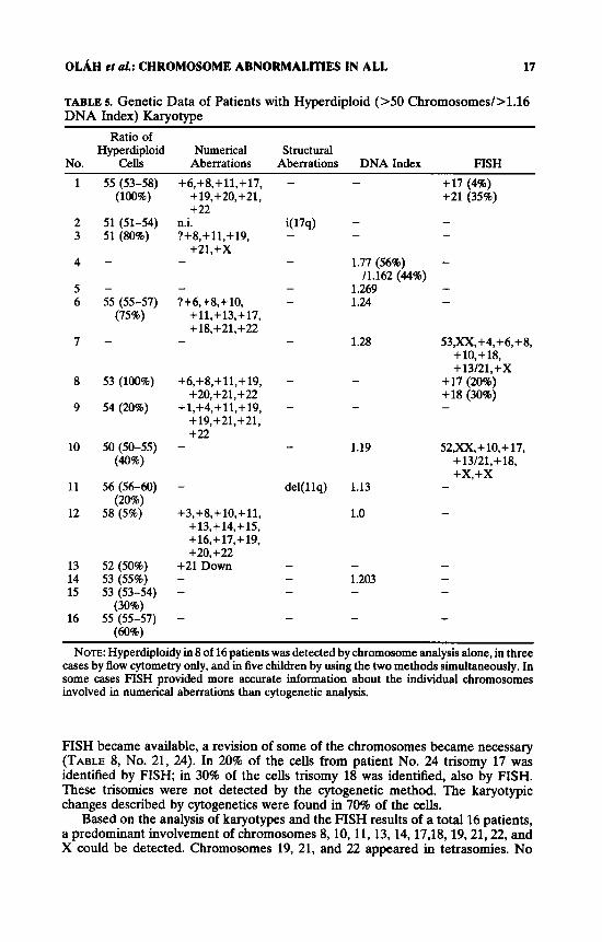
TABLE 5. Genetic Data of Patients with Hyperdiploid (>50 Chromosomes/>l.l6 DNA Index) Karyotype
~ ~~~~
Ratio of Hyperdiploid Numerical StNCtUId
No. Cells Aberrations Aberrations DNA Index FISH 1
2 3
4
5 6
7
8
9
10
11
12
13 14 15
16
55 (53-58) +6,+8,+11,+17, +19,+20,+21, + 22
(100%)
51 (51-54) n.i. 51 (80%) ?+8,+11,+19,
+21,+x
- - 55 (55-57) ?+6,+8,+ 10,
(75%) +11,+13,+17, +18,+21,+22
- -
53 (100%) +6,+8,+11,+19, +20,+21,+22
54 (20%) +1,+4,+11,+19, + 19, +21, +21, + 22
50 (50-55) - (40%)
56 (56-60) (20%)
58 (5%)
52 (50%) 53 (55%) 53 (53-54)
(30%) 55 (55-57) (60%)
+3,+8,+ lo,+ 11, +13,+14,+15, + 16,+ 17,+ 19, +20,+22
+21 Down -
- +17 (4%) +21 (35%)
1.77 (56%) - 1.269 - 1.24
11.162 (44%)
-
1.28 53,X?C, +4, +6, +8, +10,+18, +13/21,+X
- +17 (20%) +18 (30%) - -
52,XX,+ lo,+ 17, + 13/21, + 18, + x , + x
1.19
1.13 -
- 1.0
. , NOTE: Hyperdiploidy in 8 of 16 patients was detected by chromosome analysis alone, in three
cases by Bow cytometry only, and in five children by using the two methods simultaneously. In some cases FISH provided more accurate information about the individual chromosomes involved in numerical aberrations than cytogenetic analysis.
FISH became available, a revision of some of the chromosomes became necessary (TABLE 8, No. 21, 24). In 20% of the cells from patient No. 24 trisomy 17 was identified by FISH; in 30% of the cells trisomy 18 was identified, also by FISH. These trisomies were not detected by the cytogenetic method. The karyotypic changes described by cytogenetics were found in 70% of the cells.
Based on the analysis of karyotypes and the FISH results of a total 16 patients, a predominant involvement of chromosomes 8,10,11,13,14,17,18,19,21,22, and X could be detected. Chromosomes 19, 21, and 22 appeared in tetrasomies. No

18 ANNALS NEW YORK ACADEMY OF SCIENCES
pentasomy of any of the chromosomes was found. Structural aberrations added to the numerical ones were observed in only two of 16 patients (see TABLE 5, TABLE 8, FIG. 4).
Comparison of the Results Obtained by Using the Three Methods Simultaneously
We had an opportunity to compare the results of cytogenetic analysis, DNA content, and FISH performed (two or all three) simultaneously in 29 patients. Chromosome analysis and flow cytometry were compared in 17 cases, chromosome and FISH in two, flow cytometry and FISH in one, and the three methods were applied simultaneously in nine patients (see TABLE 8).
Cytogenetics andflow cytometry: In 18 cases a diploid karyotype (46,XX /46,XY) (TABLE 8, No. 1-18) and in another patient with Down’s syndrome (No. 19), the 21-trisomy was associated with a diploid DNA index of 1.0. In an additional patient (No. 20) a normal karyotype was found together with a DNA index of 1.11. In three patients with >50 chromosomes (No. 22,25,28), the DNA index proved to be above 1.16. In two more cases (No. 26,27) where the modal chromosome number
TABLE 6. Cytogenetic Data and Survival of Patients with Hyperdiploid A (47-50 ChromosomesY
Modal Chromosome Numerical Structural DNA Survival
No. Number Karyotype Aberrations Aberrations Index (months) 47 46,xX/47Jo(, +20
47 46,XX/47,XX, +D
47 46,XX,t(16;17)
(65%)
(50%)
(q24;q11)(30%)/ 47,XX,+18
47 46,XY(30%)/ (70%)
(70%)
(10%)
47,XY,+mar
46 46,XY/48-51,XY
49 46,XX/49-50,XX,
48 (47-50) 47,XY,+(7q), +mar,Ph+ (90%)
t(822)(q22;qll)/ 48,XY9+i(7q),
+i(7q),+6, +6/49JCY,
+ 12/50,XY,+ 1, +6,+ 12,+i(7q)
41
+4
24
- - + 20
+D - -
+18 t(16;17) -
- +13 - ?
1 .o 16
19
+0.7
- -
- Ph - +1,+6,+12 i(7q) -
t(8;22)
10 47 46,XX/47Jcx;+20 +20 - - Patients who died are signed by +. In patient No. 7, a complex karyotype change with
approximately equal proportions of metaphases with 47,48,49, and 50 chromosomes could be identified. A B-cell-specific t(8;22) was detected in cells with 47 chromosomes, and an i(17q) occurred in each cell line. Additional supernumerary chromosomes representing a clonal evolution were also identified. The patient died in three weeks.

O L h d aL: CHROMOSOME ABNORMALITIES IN ALL 19
TABLE 7. Karyotype Changes, Immunophenotype, and Survival of Patients with Pseudodiploid Karyotype
~~~ ~~
survival No. Karyotype Immunophenotype (months)
1
2 3 4 5” 6 7 8
9 10 11
46,XX(4)/46,XX,7q+ (5)/48,XX,del(lq), +marl, +mar2( 1)
46,XX7de1(2p) 46,XY/46,XY,t(l;l9)(q2l;p13) 46,XY/46,XY,t( 14;ll) 47,XY,+21,del(5)(p12) 46,XX,t(9;11) 46W,,t(9;22) 46,XX(2)/46,XX,t(4;11)(6)/46;rM,t(4;11),
46,XY/46,,XY,lp+ 46,XY/46JCY,del( 1 lq) 46,XY,t(2;12)
de1(6q)(5)/45,XX,t(4;11),- 17(3)
N.D.
CALL Pre-B cell T cell N.D. Pre-B cell Bipheno Bipheno
CALL Bipheno Pre-B cell
34
+1 N.D.
15 15 15 15 +9
16 1 8
NOTE: In 9 of 11 patients, ALL-specific translocations or aberrations with specific breakpoints could be detected. The pattern of aberrations observed together with the typical immunophenotype may explain the unfavorable prognosis of patients belonging to this group. N.D., no data.
* Down’s syndrome.
was more than 50, and in another one with 47-48 chromosomes (No. 29), the DNA index remained below 1.16. In these latter cases the proportion of hyperdiploid cells was rather low (5-20%).
Conventional cytogenetics and FISH: Chromosome analysis and FISH technique without flow cytometry were carried out in only two cases (No. 21, 24). On the basis of FISH, some revision of the karyotype was needed. In patient No. 21 FISH analysis detected trisomy 8 in one-third and trisomy X in one-fifth of the cells. We think that trisomy 6 of the karyotype should be revised as trisomy X and trisomy 12 as trisomy 8. In the other case (No. 24) trisomy 17 and trisomy 18 were revealed by FISH. In our opinion, not instead of but in addition to trisomies of the other chromosomes identified by chromosome analysis.
Flow cytometry and FISH: These two methods, without cytogenetic analysis, were performed simultaneously in one patient only (No. 23). In accordance with a hyperdiploid DNA index (1.28), FISH showed trisomy 4, 6, 8, 10, 18, 13/21, and X in 80% of the cells.
Conventional cytogenetics, flow cytometry, and FISH: The three methods were simultaneously applied in a total of ten cases. In seven of 18 patients with diploid karyotype and diploid DNA content, FISH analysis was also performed. Random trisomies and monosomies were found in three of seven cases (No. 11, 12, 13). In a patient with Down’s syndrome (No. 19), besides trisomy 21, trisomy X could also be detected in a few cells by FISH. The three methods were applied simultaneously in one hyperdiploid patient (No. 25). Using chromosome analysis, only the hyperdip- loid modal chromosome number (55) could be determined in accordance with a DNA index 1.19. Applying the FISH method, the extra chromosomes could be clarified.
Using one of the three methods or their combinations, 16 of the 80 patients (20.0%) proved to have hyperdiploid karyotype with more than 50 chromosomes and/or a >1.16 DNA index. These cases were associated with favorable clinical,

TABLE
a C
ompa
riso
n of
Res
ults
Obt
aine
d by
Cyt
ogen
etic
, Flow
Cyt
omet
ric,
and
FIS
H A
naly
sis
Perc
ent w
ith
Mod
al
Mod
al
Chr
omos
ome
Chr
omos
ome
No.
N
umbe
r R
ange
N
umbe
r K
aryo
type
D
NA
FI
SH
Rev
isio
n 1-
7 8-
10
11
12
13
14
15
16
17
18
19
20
21
22
23
24
25
26
27
28
29
46
46
46
46
46
46
46
46
46
46
47
46
55
55
-
53
46
46
46
46
46
46
46
46
46
46
46
46
46
46
45
46
47
46
53-5
8
55-5
7
-
51-5
4
46-5
5
46-60
46-5
9
46-5
6 46
-51
100
100
100
100
100
100
100
100
100
75
100
100 50
75
-
70
40
80
95
40
95
4x46
m3x
46Jc
y 46Jcy12x 4
6,X
X
46,X
X
GJcy
46Jc
y 46xy
46m
46xy
46Jc
y 46
Jcy1
453,
- C
, - D
47
Xy,
+ 21
46JC
YQh+
53
,XX
,+6,
+12,
+ 17
,+19
, +2
0,+2
1,+2
2 55
,XX
, +6,
+ 8,
+ 10,
+ 11,
+1
3,+1
7,+1
8,+2
1,
+22
53xy
,+6,
+ 1
1,+ 1
2, + 1
9,
+20,
+21,
+22
46,X
X,d
el(W
58
,XX
,+3,
+10,
+11,
+13,
+1
4,+1
5,+1
6,+
17,
+19,
+19,
+20,
+22
1.0
1.0
1.0
1.0
1.0
1.02
3 1.
03
1.02
3 1.1
1 1.
0 1 .o
1.0 -
1.24
1.28
-
1.19
1.13
1.
0
1.20
3 1.
0
-
46Jc
yI2x
46xx
44xy
,-7,
-20
(5%
)
46,X
X,+
1,+3
,-6,-X
(5
%)
46,X
Y,-4
,+15
(10
%)
-
-
46xy
,+21
,+x
(5%
) 46
JcyP
h+
+8(
35%
), +X (2
096)
, +2
1(80
%) -
53,X
X,+
4,+6
,+8,
+10,
+1
8,+1
3/21
,+X
+1
7 (2
0%)
+18
(30%
)
52,X
X,+
10,+
17,+
18,
+13/
21,+
X,+
X
-
-
+ 1,+
3,-6
,-X
-7,-2
0 -4
,+15
+X
6 =
X, 1
2 =
8
? 19
= 1
7 ?
20 =
18
or: +
17,+
18
NO
TE: C
ompa
riso
n of
the
res
ults
obt
aine
d by
chr
omos
ome
anal
ysis
and
flow
cyto
met
ry, t
he l
atte
r on
e an
d FI
SH, o
r by
the
thr
ee m
etho
ds
sim
ulta
neou
sly d
emon
stra
tes
that
the
se m
etho
ds a
re c
ompl
emen
tary
to e
ach
othe
r an
d th
eir
sim
ulta
neou
s ap
plic
atio
n is
esse
ntia
l. In
som
e ca
ses
the
FISH
met
hod
prov
ided
new
pie
ces
of information a
bout
the
chro
mos
omes
invo
lved
. In
patie
nts
No.
22
and
25, a
revi
sion
of t
he k
aryo
type
was
need
ed. (
In p
atie
nt No. 22
, chr
omos
ome 6
= X
and
12
= 8
; in
patie
nt No. 25
,19
= 1
7,20
= 1
8?).
0 L . h et al.: CHROMOSOME ABNORMALITIES IN ALL 21
ae 10
c 0 E g 4
5 - 6 0 c
- P - e 2 0
n 1 " 1 2 3 4 5 3 7 8 D 1 0 1 1 1 2 1 3 1 4 1 5 1 3 1 7 1 8 1 0 ~ 2 1 2 2 X Y
Chromosome
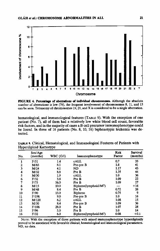
FIGURE 4. Percentage of aberrations of iodividd chromosomes. Although the absolute number of aberrations is low (76), the frequent involvement of chromosomes 8, 11, and 13 can be seen. Tetrasomy of chromosomes 19,21, and X is considered to be a single aberration.
hematological, and immunological features (TABLE 9). With the exception of one patient (No. 7), all of them had a relatively low white blood cell count, favorable risk factors, and in the majority of cases a B-cell precursor immunophenotype could be found. In three of 16 patients (No. 8, 10, 16) biphenotypic leukemia was de- tected.
TABLE 9. Clinical, Hematological, and Immunological Features of Patients with Hyperdiploid Karyotype
No. (months) WBC (GI1) Immunophenotype Factor (months) SexlAge Risk survival
1 F151 1.4 CALL 0.7 35 2 MI83 8.1 Pre-pre B 1.8 41 3 MI24 4.1 ND 1.17 43 4 MI32 8.8 Pre B 1.35 44 5 MI30 1.9 CALL 1 .o 36 6 Fl32 3.8 Pre B 1.09 22 7 Fl75 56.0 Pre B 1.09 27 8 MI15 10.0 Bipheno(lymphoidlM7) - + 16 9 MI48 8.4 Pre B 0.72 26
10 F190 13.0 Bipheno 0.75 9 11 F1198 9.0 Pre-pre B 1 .o 16 12 MI120 6.2 CALL 1.08 15 13 MI30 8.4 Pre-pre B 1.09 36 14 F1188 28.0 Pre B 1.07 28 15 F1% 5.0 Pre B 1.0 14 16 F132 6.0 Bipheno(lymphoidlM7) 0.08 +0.1 NOTE: With the exception of three patients with mixed immunophenotype hyperdiploidy
proved to be associated with favorable clinical, hematological and immunological parameters. ND. no data.

22
1.0
0.9
3 0.8
-2 >
3 0.7
0.6
n *
ANNALS NEW YORK ACADEMY OF SCIENCES
;------------------------------------------ I * 5; \
.) .................... ! :
j \\ ..+ - :I ................................................ :!
+ _.__ *-., \ I - \ I \ i : - -.-.-. L
3 ), i:
1 !
- I.... ................................................................. I
-
I I I I
Diploid
.......
........ Hypodiploid
"." ___
0 10 20 30 40
Months FIGURE 5. Survival curve8 of 80 patients with different modal chromosome numbers. Calcu- lation of the curves was made based on the Kaplan-Meyer life table analysis. The number of cases analyzed diploid, 42; pseudodiploid, 11; hypodiploid, 4; hyperdiploid A, 8; and hyperdip- loid B, 16. A significant difference was found between diploid and pseudodiploid, and diploid and hyperdiploid A subgroups (p < 0.01). Survival curve of patients with hyperdiploid B karyotype remained under that of diploid patients.
Comparing the overall survival curves of subgroups with various modal chromo- some number, an unfavorable prognosis of patients with a pseudodiploid and with a hyperdiploid A karyotype could be observed as compared to that of patients with a diploid chromosome set ( p < 0.01). No significant difference between the other subgroups could be observed. The favorable prognosis of patients with more than 50 chromosomes could not be proved (FIG. 5). The number of patients in each group, however, is too small to avoid a possible statistical error. Thus, it is not possible to make any conclusions related to prognostic significance of hyperdiploidy in this study.
DISCUSSION
During the last three years ALL was diagnosed in 187 patients. In 140 of 187 patients, chromosome analysis was attempted, in 78 cases successfully. Although the ratio of successful investigations is rather low, it is important to point out that efforts have been made to perform these initial genetic analyses in various genetic laboratories working in collaboration with the hematological centers of the country. It represents a step forward that, in 80 of a total of 187 ALL patients (42.7%), the genetic diagnosis was available for the clinicians. This number will hopefully increase

0 L . h ct aL: CHROMOSOME ABNORMALITIES IN ALL 23
in the future if the staffing and instrumental facilities as well as the costs of the analyses will be provided.
Although chromosome analysis was performed in 78 patients, the DNA analysis and FISH were carried out in many fewer cases. Although chromosome analysis is the most informative method indicating the accurate numerical and structural aberrations, its value is decreased by the great number of unsuccessful cases. This is confirmed by our results presented here. Clonal chromosome aberrations were detected only in 35 of 78 successfully investigated patients (44.8%), and the ratio of cases with >50 chromosomes proved to be only 13 : 78 (16.6%). The low propor- tion of cases with abnormal karyotype may be the consequence of technical difficul- ties. When few analyzable metaphases are available, only a small number of cells are evaluated, and the normal metaphases of better quality are considered first of all. Furthermore, because of the poor quality of chromosome morphology, a great majority of the fine structural abnormalities remains unidentified. In addition, there are "hidden" structural abnormalities that cannot be detected by the traditional cytogenetic methods. For this reason it is essential to improve the standard of cytogenetic analyses and to complete them with molecular techniques.
The ratio of patients with more than 50 chromosomes also remains far behind the data in the literature. A frequency of 30% is usually menti~ned, '" '~. '~~ but a much higher ratio (49%) was also found.24 The low proportion of hyperdiploid cases in our material may also result from technical difficulties mentioned above, for example, selection of normal metaphases of better quality, or from the small number of patients studied, but the fact that our experience is characteristic of this geographic area cannot be excluded. On the other hand, the fact that the proportion of hyperdip- loid cases proved to be higher when we used flow cytometry (7/31: 22.5%) may contradict the above hypothesis. These results call attention to the significance of simultaneous application of the two methods mentioned.
In evaluating the involvement of individual chromosomes in numerical aberra- tions of hyperdiploid patients, we have to point out the question of geographic factors anew. In accordance with our previous results, a difference in the spectrum of chromosomes involved compared to the data in the literature could be observed. In addition to the trisomies of chromosomes 4, 6, 10, 17, 18, 21, 22, and X, a supernumerary occurrence of chromosomes 3,8, 11, and 13 was also found with a high frequency. This is not a unique observation. A geographic heterogeneity in the occurrence and frequency of chromosome aberrations was also observed in other diseases like that of t(8;21) in M2-type acute nonlymphocytic leukemia, and of t(15;17) in acute promyelocytic leukemia, respectively." A difference in the frequency of the secondary aberrations of blastic-phase Ph-positive chronic myeloid leukemia was also proved.M The geographic differences in the Occurrence and frequency of tumor-specific aberrations may reflect the role of different environmen- tal mutagen/carcinogenic effects?' In order to determine whether these are or not real differences characteristic of our geographic area, further investigations are needed.
From a clinical point of view, the prognostic significance of the initial karyotype is of particular significance. Because the genetic changes determine the biological behavior of tumor cells, cell-to-cell interaction, invasivity, metastatic capacity, and response to therapy, the exact determination of the initial genetic alteration makes it possible to develop a genotype-specific therapy.M3' While certain translocations, t(9;22), t(4;ll) suggest the necessity of a bone marrow transplantation, patients with hyperdiploidy of a favorable prognosis need only a standard therapy. In our study, however, the small number of patients in each group is too small to avoid a possible statistical error; therefore, it is not possible to make any conclusion related

24 ANNALS NEW YORK ACADEMY OF SCIENCES
to prognostic significance of karyotype changes. Despite this, our observations might be indicative of probable geographic characteristics and might call our attention to a few avenues of study. In our study the comparison of survival curves of patients with different modal chromosome numbers proved a significantly worse prognosis for patients with pseudodiploid and hyperdiploid A karyotypes compared to that of the diploid group. The favorable prognosis of hyperdiploid patients (>50 chromo- somes) could not be proved. The lack of the expected good prognosis of hyperdiploid patients may result from the small number of patients, perhaps the unfavorable prognosis of three biphenotypic cases (two of them had lymphoid/M7 phenotype), but may also be related to the different spectrum of chromosomes involved, such as the high frequency of trisomy 8 and 11. The unfavorable effect of chromosome 8 on the course of the disease is well Similarly, the aberrations of chromo- some 11 in acute leukemias are associated with a rather severe di~ease. '~. '~.~~ Besides, there are data regarding the special prognostic value of the particular extra chromo- somes in these patients, for example, cases with trisomy 6 and with combined trisomy of 4 and 10 seem to have a particularly good prognosis.2728 Our findings and the latter observations call attention to the necessity for a more detailed analysis of involvement of chromosomes in numerical aberrations. An additional possible explanation for the lack of prognostic difference between the diploid and hyperdip- loid groups may be that the diploid group also includes the cases with t(12;21), an aberration that is associated with a favorable prognosis." This translocation cannot be detected by standard cytogenetic methods, only by molecular analysis. In this way this hidden aberration may improve the prognosis of the diploid group and decrease the prognostic difference compared to the hyperdiploid one. This aspect points to the importance of the molecular analysis of cases with a virtual normal karyotype.
Based on the data of patients studied in the last three years, we can say that genetic findings of ALL patients in Hungary seem to be different from those in the literature. The proportion of hyperdiploid cases is lower, the spectrum of chromosomes involved in numerical aberrations is different, and the favorable prognosis of the hyperdiploid group could not be proved. To clarify the significance of these observations, it seems to be essential to determine whether or not these clinical and genetic differences really exist, and if they do we have to reveal the causes leading to these differences. For this purpose we have to extend the genetic investigations to all ALL cases in Hungary to improve the proportion of successful investigations and complete them with molecular techniques, probably with compar- ative genomic hybridization. Besides, a detailed analysis of the individual chromo- somes and a nationwide prospective study of the clinical-genetic correlation seem to be essential.
SUMMARY
Current intensive chemotherapies cure about 70% of the children with ALL. On the other hand a significant number of the children are not cured despite intensive treatment. At the same time some highly curable patients are treated too intensively and suffer from unnecessary side effects of the chemo- and radiotherapy applied. In order to further improve the therapeutic results in this disease, we have to distinguish between the cases with a better and a worse prognosis. The initial karyotype (both numerical and structural chromosome abnormalities) proved to be one of the most reliable prognostic parameters, leading to the suggestion of

O L h cf al.: CHROMOSOME ABNORMALmS IN ALL 25
developing genotype-specific therapies. Although the prognosis in patients with pseudodiploid karyotype is usually unfavorable, a significantly better prognosis can be observed in those with more than 50 chromosomes. Because the latter patients can achieve remission on a metabolite-based therapy, the toxic effects of more aggressive chemotherapy with anthracyclines and genotoxic agents can be avoided; thus, the reliable and accurate identification of patients with >SO chromosomes is of particular importance. For this purpose three methods: chromosome analysis, DNA flow cytometry, and fluorescence in situ hybridization can be used.
In 1993 it was decided to develop a comprehensive nationwide project in order to perform the initial genetic analysis of all ALL children diagnosed in the hematologicalloncological centers of Hungary. Here the data obtained on 187 ALL patients diagnosed in the period from 1993 to 1995 are presented. In about 75% of patients (in 140 of 187) chromosome analysis was performed, in 78 cases (55.7%) successfully. The proportion of patients with abnormal karyotype was 36 of 78 (46.1%), and hyperdiploidy with more than 50 chromosomes was detected in 13 of 78 (16.6%) children. The lower ratio of hyperdiploid cases in our patients as com- pared to the data in the literature may be due to technical difficulties and the small number of patients studied, but it may reflect real geographic characteristics. Using flow cytometry, seven of 31 patients investigated (22.5%) proved to be hyperdiploid with a DNA index above 1.16. A higher ratio of hyperdiploid patients in this study calls attention to the significance of simultaneous application of the two methods. Taken together, 16 of 80 (20.0%) succesfully studied patients proved to be hyperdip- loid (>SO chromosomes and/or DNA index above 1.16).
The pattern of chromosome involvement in our study determined by chromo- some analysis and/or FISH technique proved also to be different from the data of large international series. In addition to trisomies of chromosomes 4, 6, 10, 14, 17, 18,21, and X, which are known to be the most frequently involved chromosomes, trisomies of chromosomes 3,8,11, and 13 were also observed with a high frequency. Comparison of survival curves of various cytogenetic subgroups showed a significant difference between diploid-pseudodiploid and diploid-hyperdiploid A (with 47-50 chromosomes) subgroups. No favorable prognosis of hyperdiploid patients (>SO chromosomes) could be proved. Because of the small number of patients studied, prognostic differences of cytogenetic subgroups need further confirmation. The clinical and genetic differences observed, however, call attention to the necessity for further genetic studies of ALL patients in Hungary, because these differences may reflect real geographic characteristics and may be related to different environ- mental mutagen/carcinogen effects of the given geographic area. It is essential to determine whether or not these differences really exist and if they do to reveal the causes leading to these differences. In our view this is one of the routes by which the therapeutic results in childhood ALL can be further improved simultaneously with the avoidance of early and late toxicity of chemotherapy.
ACKNOWLEDGMENT
We thank all clinicians of the Hematological/Oncological Centers of the Hungar- ian Pediatric Oncology Study Group for providing the patient data and the geneti- cists and technicians working at different laboratories for their valuable contribution.
REFERENCES
1. BLOOMFIELD, C. D., era/. 1990. Six-year follow up of the clinical significance of karyotype in acute lymphoblastic leukemia. Cancer Genet. Cytogenet. 40: 171.

26 ANNALS NEW YORK ACADEMY OF SCIENCES
2. CASSANO, W. F., er al. 1993. Therapy for childhood acute lymphoblastic leukemia. Curr.
3. CHESSELS, J. M. 1986. Acute leukaemia in children. Clin. Haematol. 15.727. 4. CRIST, W., et al. 1986. Clinical and biological features predict a poor prognosis in acute
lymphoid leukemias in infants. A. Pediatric Oncology Group Study. Blood 67: 135. 5. W o n , J., et al. 1993. Clinical significance of cytogenetic studies in childhood acute
lymphoblastic leukemia. Experience of the BFM trials. Rec. Res. Cancer Res. l31: 123.
6. MILLER, D. R. 1988. Childhood acute lymphoblastic leukemia. 1. Biological features and their use in predicting outcome of treatment. Am. J. Ped. Hematol. Oncol. 10: 163.
7. POPLACK, D. G. &t G. REAMAN. 1988. Acute lymphoblasticleukemia inchildhood. Pediatr. Clin. N. Am. 35: 903.
8. Put, C. H., et al. 1988. Correlation of karyotype and immunophenotype in childhood acute lymphoblastic leukemia. J. Clin. Oncol. 6: 56.
9. PUI, C. H., et al. 1989. Prognostic importance of structural chromosomal abnormalities in children with hyperdiploid (>50 chromosomes) acute lymphoblastic leukemia. Blood 73: 1963.
10. Pui, C. H. & W. M. CRIST. 1992. Cytogenetic abnormalities in childhood acute lymphoblas- tic leukemia correlates with clinical features and treatment outcome. Leuk. Lymphoma 7: 259.
11. SECKER-WALKER, L. M. 1990. Prognostic and biological importance of chromosome findings in acute lymphoblastic leukemia. Cancer Genet. Cytogenet. 49: 1.
12. HEIM, S. & F. MITELMAN. 1987. Acute lymphoblastic leukemia. In Cancer Cytogenetics. Chapt. 8.: 141-173. Alan R. Liss, Inc. New York.
13. h i , C. H., et al. 1990. Biology and clinical significance of cytogenetic abnormalities in childhood acute lymphoblastic leukemia. Blood 76 1449.
14. RAIMONDI, S. C. 1993. Current status of cytogenetic research in childhood acute lympho- blastic leukemia. Blood 81: 2237.
15. MURPHY, S. B., et al. 1989. Nonrandom abnormalities of chromosome 9p in childhood acute lymphoblastic leukemia: Association with high risk clinical features. Blood 74: 409.
16. RAIMONDI, S. C., et al. 1989. Childhood acute lymphoblastic leukemia with chromosomal breakpoints at llq23. Blood 73: 1627.
17. SLATER, R., et al. 1995. A nonrandom chromosome abnormality found in precursor-B lineage acute lymphoblastic leukemia: dic(9;2O)(p173;qll). Leukemia 9: 1613.
18. CRIST, W., et al. 1989. Prognostic importance of the pre-Ball immunophenotype and other presenting features in B lineage childhood acute lymphoblastic leukemia. A. Pediatric Oncology Group Study. Blood 74: 1252.
19. Put, C. H., et al. 1993. Clinical and biological relevance of immunologic marker studies in childhood acute lymphoblastic leukemia. Blood 82: 343.
20. BOROWITZ, M. J.. et al. 1993. Predictability of the t(1;19)(q23;p13) from surface antigen phenotype: Implications for screening cases of childhood acute lymphoblastic leukemia for molecular analysis: A Pediatric Oncology Group Study. Blood 82: 1086.
21. DEVARAJ, P. E., et al. 1995. Expression of the E2A-PBxl fusion transcripts in t(1;19)(q23;p13) and leukaemia with different B lineage immunophenotypes. Leukae- mia 9: 821.
22. ROMANA, S. P. 1995. The t(12;21) of acute lymphoblastic leukemia results in a tellAML1 gene fusion. Blood 85: 3662.
23. Pur, C. H., et al. 1990. Near-triploid and near-tetraploid acute lymphoblastic leukemia of childhood. Blood 76r 590.
24. HEIM, S., et al. 1990. Bone marrow karyotypes in 94 children with acute leukemia. Eur. J. Haematol. 44(4): 227.
25. TRUEWORTHY, R., et al. 1992. Ploidy of lymphoblasts is the strongest predictor of treat- ment outcome in B-progenitor-cell acute lymphoblastic leukemia of childhood: A Pediatric Oncology Group Study. J. Clin. Oncol. 10: 606.
26. HAMMOND, D., et al. 1986. Analysis of prognostic factors in acute lymphoblastic leukemia. Med. Ped. Oncol. 14: 124.
Opin. Oncol. 5: 42.

O L h er aL: CHROMOSOME ABNORMALITIES IN ALL 27
27.
28.
29.
30. 31.
32.
33.
34.
35.
36.
HARRIS, M. B., et af. 1992. Trisomy of leukemic cell chromosomes 4 and 10 identi6es children with B progenitor cell acute lymphoblastic leukemia with a very low risk of treatment failure: A Pediatric Oncology Group Study. Blood 79: 3316.
JACKSON, J. F., et al. 1990. Favorable prognosis associated with hyperdiploidy in children with acute lymphocytic leukemia correlates with extra chromosome 6. A Pediatric Oncology Group Study. Cancer 66: 1183.
HARBOR, J., et al. 1994. Cytogenetics and clonal evolution in childhood acute lyrnphoblas- tic leukemia. Ann. Haematol. 68(Suppl. I): A13.
PINKEL, D. 1987. Curing children of leukemia. Cancer 10: 1683. PINKEL, D. 1989. Species-specific therapy of acute lymphoid leukemia. Haematol. Blood
Transf. 32: 27. Du MANOIR, S., et af. 1993. Detection of complete and partial chromosome gains and
losses by comparative genomic in situ hybridization. Hum. Genet. 90: 590. ANDREEFF, M. 1990. Flow cytometry of leukaemia. In Flow Cytometry and Sorting. 2nd
edit. M. R. Melamed, T. Lindmo, M. L. Mendelsohn, (Eds.: 697-724. Willy-Liss, Inc. New York.
HIDDEMANN, W., et of. 1987. Nachweis von aberrationen des Karyotyps bei Kindern mit akuten Leukiimien: Eine vergleichende Analyse von Zytogenetik und Durchflubzyto- photometrie. Klinische Piidiatrie 199/3: 161.
LOOK, T. A., et al. 1985. Prognostic importance of blast cell DNA content in childhood acute lymphocytic leukaemia. Blood 65: 1079.
ANASTASI, J. 1993. Fluorescence in situ hybridization in leukemia. Applications in diagno- sis, subclassification and monitoring the response to therapy. AM. N. Y. Acad. Sci. 677: 214. - . . . - - . .
37. ANASTASI, J., er al. 1990. Detection of numerical chromosomal abnormalities in neoplastic hematopoietic cells by in situ hybridization with a chromosome-specific probe. Am. J. Pathol. w6: 131.
38. BERGER, A., et af. 1994. Interphase cytogenetic study of childhood acute lymphoblastic leukemia. Med. Ped. Oncol. U: 413.
39. PINKEL, D., et al. 1986. Cytogenetic analysis using quantitaive high sensitivity fluorescence hybridization. Proc. Natl. Acad. Sci. USA 83: 2934.
40. PODDIGHE, P. J., etal. 1991. Interphase cytogenetics of hematological cancer: Comparison of classical karyotyping and in situ hybridization using a panel of eleven chromosome specific DNA probes. Cancer Res. 51: 1959.
41. Tossi, S., etal. 1994. Double target in situ hybridization applied to the study of numerical aberrations in childhood acute lymphoblastic leukemia. Cancer Genet. Cytogenet. 73: 10.
42. ISCN. 1995. An International System for Human Cytogenetic Nomenclature. S. Karger. Basel, Switzerland.
43. HEIM, S. & F. MITELMAN. 1987. Acute nonlymphocytic leukemia. Cancer Cytogenetics. Chapt. 5.: 65-110. Alan R. Liss, Inc. New York.
44. HEIM, S. & F. MITELMAN. 1987. Chronic myeloid leukemia. In Cancer Cytogenetics. Chapt. 4.: 41-46. Alan R. Liss, Inc. New York.
Copyright © 2022 FDOKUMEN