Silencer of death domains controls cell death through tumour necrosis factor-receptor 1 and...
10
Silencer of Death Domains Controls Cell Death through Tumour Necrosis Factor-Receptor 1 and Caspase-10 in Acute Lymphoblastic Leukemia Adam Cisterne 1. , Rana Baraz 1. , Naveed I. Khan 1 , Robert Welschinger 1 , Jordan Basnett 1 , Carina Fung 1 , Helen Rizos 1 , Kenneth F. Bradstock 2 , Linda J. Bendall 1 * 1 Centre for Cancer Research, Westmead Millennium Institute, University of Sydney, Sydney, New South Wales, Australia, 2 Blood and Marrow Transplant Service, Department of Haematology, Westmead Hospital, Sydney, New South Wales, Australia Abstract Resistance to apoptosis remains a significant problem in drug resistance and treatment failure in malignant disease. NO- aspirin is a novel drug that has efficacy against a number of solid tumours, and can inhibit Wnt signaling, and although we have shown Wnt signaling to be important for acute lymphoblastic leukemia (ALL) cell proliferation and survival inhibition of Wnt signaling does not appear to be involved in the induction of ALL cell death. Treatment of B lineage ALL cell lines and patient ALL cells with NO-aspirin induced rapid apoptotic cell death mediated via the extrinsic death pathway. Apoptosis was dependent on caspase-10 in association with the formation of the death-inducing signaling complex (DISC) incorporating pro-caspase-10 and tumor necrosis factor receptor 1 (TNF-R1). There was no measurable increase in TNF-R1 or TNF-a in response to NO-aspirin, suggesting that the process was ligand-independent. Consistent with this, expression of silencer of death domain (SODD) was reduced following NO-aspirin exposure and lentiviral mediated shRNA knockdown of SODD suppressed expansion of transduced cells confirming the importance of SODD for ALL cell survival. Considering that SODD and caspase-10 are frequently over-expressed in ALL, interfering with these proteins may provide a new strategy for the treatment of this and potentially other cancers. Citation: Cisterne A, Baraz R, Khan NI, Welschinger R, Basnett J, et al. (2014) Silencer of Death Domains Controls Cell Death through Tumour Necrosis Factor- Receptor 1 and Caspase-10 in Acute Lymphoblastic Leukemia. PLoS ONE 9(7): e103383. doi:10.1371/journal.pone.0103383 Editor: Halvard Boenig, German Red Cross Blood Service Frankfurt, Germany Received December 2, 2013; Accepted July 1, 2014; Published July 25, 2014 Copyright: ß 2014 Cisterne et al. This is an open-access article distributed under the terms of the Creative Commons Attribution License, which permits unrestricted use, distribution, and reproduction in any medium, provided the original author and source are credited. Funding: This work was funded by a grant from NHMRC (Grant No. 352326) to LJB. LJB and HR are NHMRC Senior Research Fellows and recipients of Cancer Institute New South Wales Fellowships. The funders had no role in study design, data collection and analysis, decision to publish, or preparation of the manuscript. Competing Interests: The corresponding author is an academic editor for PLOS ONE but this does not alter the authors’ adherence to PLOS ONE editorial policies and criteria. * Email: [email protected] . These authors contributed equally to this work. Introduction Acute lymphoblastic leukemia (ALL) is the most common malignancy in children and although remission is almost always attained, up to 20% of children will relapse, with subsequent poor prognosis [1]. Adult patients have a worse outlook, with more than half relapsing [2]. Current management of ALL in both children and adults is dependent on treatment with multiple chemotherapy drugs, such as corticosteroids and vincristine, which induce apoptosis in the leukemia cells. However, resistance to drug- induced apoptosis is a common problem, and there is an urgent requirement for new drugs with efficacy against leukemic cells in ALL. Apoptosis can be mediated via the extrinsic death receptor- mediated pathway, or the intrinsic mitochondrial pathway. Cell death is ultimately mediated by activation of effector caspases including caspase-3 and -7. However, the initiator caspases differ between the two pathways, with caspases-8 and -10 being involved in the extrinsic, and caspase-9 in the intrinsic pathway [3]. Commitment to the intrinsic pathway occurs when cytochrome c is released from mitochondria as a result of mitochondrial outer membrane permeabilization (MOMP) [4]. This results in the formation of the apoptosome by recruitment of APAF-1 and pro- caspase-9 [5]. Here caspase-9 is activated by cleavage and in turn activates the effector caspases. The extrinsic death pathway is initiated by the binding of death ligands, such as tumour necrosis factor alpha (TNF-a), TNF-related apoptosis inducing ligand (TRAIL) or FasL to their respective cell surface death receptors, tumour necrosis factor receptor 1 (TNF-R1), death receptors (DR) 4 or 5, and Fas. Oligomerization of the death domains in the cytoplasmic regions of these receptors is the initial event in signalling through these receptors. This can be inhibited by silencer of death domain (SODD), alternatively known as BCL2- associated athanogene 4 (BAG4) in the case of TNF-R1, Fas and DR3 [6,7]. Once oligomerization has occurred, binding of the adaptor molecules, TNF-R1-associated death domain protein (TRADD) or Fas-associated protein with death domain (FADD), depending on the receptor involved, and pro-caspases-8 or -10 produces the death-inducing signaling complex (DISC) [8]. In some cells activation of caspases-8 or -10 within the DISC is sufficient to facilitate direct activation of effector caspases and cell death, while in others linkage to the intrinsic pathway is needed. PLOS ONE | www.plosone.org 1 July 2014 | Volume 9 | Issue 7 | e103383
Transcript of Silencer of death domains controls cell death through tumour necrosis factor-receptor 1 and...

Silencer of Death Domains Controls Cell Death throughTumour Necrosis Factor-Receptor 1 and Caspase-10 inAcute Lymphoblastic LeukemiaAdam Cisterne1., Rana Baraz1., Naveed I. Khan1, Robert Welschinger1, Jordan Basnett1, Carina Fung1,
Helen Rizos1, Kenneth F. Bradstock2, Linda J. Bendall1*
1 Centre for Cancer Research, Westmead Millennium Institute, University of Sydney, Sydney, New South Wales, Australia, 2 Blood and Marrow Transplant Service,
Department of Haematology, Westmead Hospital, Sydney, New South Wales, Australia
Abstract
Resistance to apoptosis remains a significant problem in drug resistance and treatment failure in malignant disease. NO-aspirin is a novel drug that has efficacy against a number of solid tumours, and can inhibit Wnt signaling, and although wehave shown Wnt signaling to be important for acute lymphoblastic leukemia (ALL) cell proliferation and survival inhibitionof Wnt signaling does not appear to be involved in the induction of ALL cell death. Treatment of B lineage ALL cell lines andpatient ALL cells with NO-aspirin induced rapid apoptotic cell death mediated via the extrinsic death pathway. Apoptosiswas dependent on caspase-10 in association with the formation of the death-inducing signaling complex (DISC)incorporating pro-caspase-10 and tumor necrosis factor receptor 1 (TNF-R1). There was no measurable increase in TNF-R1 orTNF-a in response to NO-aspirin, suggesting that the process was ligand-independent. Consistent with this, expression ofsilencer of death domain (SODD) was reduced following NO-aspirin exposure and lentiviral mediated shRNA knockdown ofSODD suppressed expansion of transduced cells confirming the importance of SODD for ALL cell survival. Considering thatSODD and caspase-10 are frequently over-expressed in ALL, interfering with these proteins may provide a new strategy forthe treatment of this and potentially other cancers.
Citation: Cisterne A, Baraz R, Khan NI, Welschinger R, Basnett J, et al. (2014) Silencer of Death Domains Controls Cell Death through Tumour Necrosis Factor-Receptor 1 and Caspase-10 in Acute Lymphoblastic Leukemia. PLoS ONE 9(7): e103383. doi:10.1371/journal.pone.0103383
Editor: Halvard Boenig, German Red Cross Blood Service Frankfurt, Germany
Received December 2, 2013; Accepted July 1, 2014; Published July 25, 2014
Copyright: � 2014 Cisterne et al. This is an open-access article distributed under the terms of the Creative Commons Attribution License, which permitsunrestricted use, distribution, and reproduction in any medium, provided the original author and source are credited.
Funding: This work was funded by a grant from NHMRC (Grant No. 352326) to LJB. LJB and HR are NHMRC Senior Research Fellows and recipients of CancerInstitute New South Wales Fellowships. The funders had no role in study design, data collection and analysis, decision to publish, or preparation of themanuscript.
Competing Interests: The corresponding author is an academic editor for PLOS ONE but this does not alter the authors’ adherence to PLOS ONE editorialpolicies and criteria.
* Email: [email protected]
. These authors contributed equally to this work.
Introduction
Acute lymphoblastic leukemia (ALL) is the most common
malignancy in children and although remission is almost always
attained, up to 20% of children will relapse, with subsequent poor
prognosis [1]. Adult patients have a worse outlook, with more than
half relapsing [2]. Current management of ALL in both children
and adults is dependent on treatment with multiple chemotherapy
drugs, such as corticosteroids and vincristine, which induce
apoptosis in the leukemia cells. However, resistance to drug-
induced apoptosis is a common problem, and there is an urgent
requirement for new drugs with efficacy against leukemic cells in
ALL.
Apoptosis can be mediated via the extrinsic death receptor-
mediated pathway, or the intrinsic mitochondrial pathway. Cell
death is ultimately mediated by activation of effector caspases
including caspase-3 and -7. However, the initiator caspases differ
between the two pathways, with caspases-8 and -10 being involved
in the extrinsic, and caspase-9 in the intrinsic pathway [3].
Commitment to the intrinsic pathway occurs when cytochrome c
is released from mitochondria as a result of mitochondrial outer
membrane permeabilization (MOMP) [4]. This results in the
formation of the apoptosome by recruitment of APAF-1 and pro-
caspase-9 [5]. Here caspase-9 is activated by cleavage and in turn
activates the effector caspases. The extrinsic death pathway is
initiated by the binding of death ligands, such as tumour necrosis
factor alpha (TNF-a), TNF-related apoptosis inducing ligand
(TRAIL) or FasL to their respective cell surface death receptors,
tumour necrosis factor receptor 1 (TNF-R1), death receptors (DR)
4 or 5, and Fas. Oligomerization of the death domains in the
cytoplasmic regions of these receptors is the initial event in
signalling through these receptors. This can be inhibited by
silencer of death domain (SODD), alternatively known as BCL2-
associated athanogene 4 (BAG4) in the case of TNF-R1, Fas and
DR3 [6,7]. Once oligomerization has occurred, binding of the
adaptor molecules, TNF-R1-associated death domain protein
(TRADD) or Fas-associated protein with death domain (FADD),
depending on the receptor involved, and pro-caspases-8 or -10
produces the death-inducing signaling complex (DISC) [8]. In
some cells activation of caspases-8 or -10 within the DISC is
sufficient to facilitate direct activation of effector caspases and cell
death, while in others linkage to the intrinsic pathway is needed.
PLOS ONE | www.plosone.org 1 July 2014 | Volume 9 | Issue 7 | e103383

This occurs by caspase-8 or -10-mediated cleavage of Bid and
induction of MOMP [9].
Despite expression of surface death receptors, including TNF-
R1, Fas and TRAIL-R1 and R2, cells from a significant
proportion of ALL patients are resistant to ligand-induced
apoptosis when exposed to TNF-a, FasL or TRAIL [10–12].
The reasons for this are unclear but are thought to involve
alterations to death receptor signalling pathways. The specific role
of caspase-10 in the induction of cell death is not clear and in most
settings it takes a subordinate role to caspase-8. Mice naturally lack
caspase-10 [13] and in humans it can substitute for caspase-8 in
certain cell types [14]. However, mutations in CASP10 are
associated with type II autoimmune lymphoproliferative syndrome
suggesting it has a significant role in lymphoid cells [15]. Caspase-
10 is highly expressed in lymphoid cells and can be mutated in
lymphoid malignancies [16], including in ALL, although this
appears to be rare [17]. Activity of caspase-10 has been implicated
in the response to a number of chemotherapeutic agents including
etoposide, doxorubicin, arsenic trioxide and paclitaxel [18–20].
This can be mediated by p53-dependent, or histone-H3 acetyla-
tion-dependent modulation of the CASP10 locus [18].
We have previously reported that the nitric oxide donating non-
steroidal anti-inflammatory drug (NO-NSAID) para-NO-aspirin
(para-NO-ASA) has anti-leukemic activity against human ALL by
induction of apoptosis in association with suppression of NF-kB
signalling [21]. NO-NSAID drugs consist of traditional NSAID to
which an NO-donating moiety is covalently attached via an
aromatic or aliphatic spacer [22]. These compounds demonstrate
substantial anti-cancer activity and are up to 20 fold more potent
than the parental NSAID [23]. para-NO-ASA is the best
characterized NO-NSAID and para-NO-ASA demonstrated a
several hundred-fold increase in potency compared with aspirin
[24].
In this study we examined the mechanisms involved in para-
NO-ASA induced apoptosis. We found that para-NO-ASA
resulted in caspase-10 dependent cell death. Although associated
with caspase-10 binding to TNF-R1, there was no evidence to
suggest that cell death was ligand dependent. Cell death was
associated with suppression of SODD, suggesting that SODD
provides protection from death receptor induced death. Agents
that specifically suppress SODD may be of therapeutic benefit for
the treatment of this disease. Furthermore SODD is overexpressed
in a number of malignancies including ovarian and pancreatic
cancers [25,26] and suppression of SODD has recently been
associated with the induction of cell death in melanoma [27],
suggesting that modulation of SODD may have broader
application in cancer treatment.
Experimental Procedures
Chemicals and reagentspara-NO-ASA [2-(acetyloxy)benzoic acid 4-(nitrooxymethyl)-
phenyl ester] was purchased from NicOx (Sophia Antipolis,
France). The inhibitors of caspase-8 (Z-IETD-FMK) and caspase-
9 (Z-LEHD-FMK), Fix/Perm and Perm/Wash solutions, mouse
anti-human TNF-a PE, annexin V FITC and AlexaFluor 647, and
7AAD were obtained from Becton Dickinson (North Ryde, NSW,
Australia). Tetramethylrhodamine methyl ester (TMRM) was
purchased from Molecular Probes (Invitrogen, Grand Island, NY,
USA), N-acetyl cysteine (NAC) from Sigma-Aldrich (St Louis,
MO, USA), the InnoCyte Flow Cytometric Cytochrome C release
Kit from Calbiochem (San Diego, CA, USA) and the Caspase-8
(BF4100) and caspase-9 (BF10100) activity assays were purchased
from Bioscientific (Sydney, Australia). The following antibodies to
human proteins were purchased: mouse anti-b-actin (Sigma-
Aldrich); rabbit anti-SODD (Chemicon, Boronia, VIC, Australia);
rabbit anti-PUMA, rabbit anti-Bcl-XL Alexa Fluor-488, rabbit
anti-BIM, rabbit anti-XIAP, rabbit anti-TNF-R1, rabbit anti-Bid,
rabbit anti-cleaved caspase-9, rabbit anti-cleaved caspase-8, rabbit
anti-full length caspase-10 (Cell Signaling Technologies, Boston,
MA, USA); mouse anti-Noxa (Abcam, Cambridge, MA, USA).
Goat anti-mouse Alexa Fluor-488 was purchased from Invitrogen.
Human recombinant TNF-a was purchased from Millipore (Nth
Ryde, NSW, Australia). Caspase-10 inhibitor AEVD-FMK was
purchased from Biovision (Mountain View, CA, USA) and FAM
Caspase-10 kit from AbD Serotec (Raleigh, NC, USA). Primers for
PCR amplification were synthesized by Proligo (Lismore, NSW,
Australia) or Sigma Aldrich.
CellsPre-B ALL cell lines NALM6 (DSMZ Braunschweig, Ger-
many), Reh (ATCC, Manassas, VA, USA) and LK63 (gift of Prof
A. Boyd, QMIR, Australia) [28], the Burkitt’s lymphoma lines
Raji and Daudi and T-ALL lines Jurkat and CEMT4 (ATCC)
were cultured in RPMI medium with 10% fetal calf serum. The
stromal-dependent cells were grown in RPMI with 10% fetal calf
serum, on confluent layers of either a human bone marrow
stromal cell line transformed with hTERT, here termed
hTERT.BMS, (kind gift of Dr D. Campana, Memphis TN,
USA) [29] or bone marrow stromal cells prepared as previously
described [30].
Leukemic blasts were isolated from the bone marrow samples
from five patients with B cell progenitor ALL and cryopreserved as
previously described [31]. Samples were collected following
informed written consent from the donor or guardian as
appropriate and with ethics approval from the Sydney West Area
Heath Service Human Research Ethics Committee. Details for
0407 cells have been previously published [32]. The details of the
stromal-dependent lines, including the patient characteristics have
been previously published [33,34]. A summary of the clinical
information for all patient-derived samples and cell lines used in
this study has been provided in Table S1 for convenience.
Proliferation and cell cycle analysisCell proliferation was determined by [3H]-thymidine (Amer-
sham Biosciences, NSW, Australia) incorporation as previously
described [35]. The proliferation index = CPM of treated cells/
CPM of control cells. Cell cycle analysis was performed by
assessment of DNA content after staining with propidium iodide as
previously described [33].
Apoptosis and Caspase activationApoptosis was assessed by Annexin V/PI or 7AAD staining as
previously described [35,36]. Cells negative for annexin V and PI
(or 7AAD) were considered viable. Pan-caspase activation was
measured by incubation of cells with 10 mM CaspACETM FITC-
VAD-FMK (Promega) for 20 min in the dark at 37uC. Washed
cells were analysis on FACSCalibur flow cytometer (Becton
Dickinson) using Cell Quest software. Activation of specific
caspases was assessed by flow cytometry using specific antibodies.
Assessment of TNF-a ProductionFor detection of TNF-a protein, cells were treated with 2 mM
monensin and 1 mg/ml Brefeldin A for 4 h at 37uC prior to
fixation with Fix/Perm solution for 20 min at 4uC. Cells were
washed in Perm/Wash buffer and directly labelled for TNF-ausing PE conjugated anti-TNF-a antibody.
NO-Aspirin Induced Extrinsic Cell Death in ALL
PLOS ONE | www.plosone.org 2 July 2014 | Volume 9 | Issue 7 | e103383

Mitochondrial Depolarization and Cytochrome c ReleaseMitochondrial depolarization was measured using 143 nM
TMRM in PBS at 37uC for 20 mins, followed by labelling with
annexin V Alexafluor 647 and 7AAD. A 5 min exposure to
12.5 mM CCCP (carbonyl cyanide 3-chlorophenylhydrazone) was
used as a positive control. Cytochrome c release was performed
using the InnoCyte Flow Cytometric Cytochrome C release Kit
according to manufacturer’s instructions except that goat anti-
mouse Alexa Fluor 647 was used as the second layer.
RT-PCR and qRT-PCRTotal RNA was extracted using Trizol reagent and reverse
transcribed. The relative expression ratio of the target genes was
compared with the housekeeping gene glyceraldehyde-3-phos-
phate dehydrogenase (GAPDH) using the same cDNA. Values
were then normalised to the untreated sample. All samples were
run in quadruplicates. The primers for SODD were: Forward:
acccaagtacatatcctgtaagacc; Reverse: aggagaagcccacgcatcttc. Those
for GAPDH have been previously published (Khan, et al 2008).
For the qRT-PCR cDNA was prepared as above. SODD
mRNA expression levels were quantitated from standard curves
generated using cDNA prepared from NALM6 cells by real-time
RT-PCR using SYBR Green JumpStart Taq Ready Mix (Sigma-
Aldrich, Sydney, Australia) and a Corbett 6000 thermocycler
(Corbett Life Science, NSW, Australia) and specific primers:
SODD forward – acatgtgctggagaaggtccagt; SODD reverse –
acagagtcctggcccccagt. Values were normalised to the expression
level of GAPDH using the same cDNA to generate the standard
curve. The primers for GAPDH have been previously published
(Bendall, et al 2004).
Immunoprecipitation and Western blottingCell lysates were prepared as previously described [35].
Immunoprecipitations were performed using Protein G Dyna-
beads (Invitrogen) according to the manufacturer’s instructions.
Beads were crosslinked to antibodies to TNF-R1 (Genesearch,
Arundel, QLD, Australia) or Mouse IgG2b control (DakoCytoma-
tion), using bis(sulfosuccinimidyl)suberate reagent as described by
the manufacturer. Immunoprecipitated material was eluted
directly into SDS-PAGE reducing buffer. Western blotting was
performed as previously described [35].
Lentiviral Knockdown of SODDLentiviral constructs were prepared as previously described [37]
using the following shRNA sequences to SODD: SODD1 -
acatatacttcatgtgtaatt or SODD2 - ccaacaatcaagatcaaagta. Lenti-
viral transduction was performed with Institutional Biosafety
Committee approval. NALM6, LK63 or REH cells (16106/mL)
were transduced in RPMI containing 10% FCS, 8 mg/ml
polybrene and 406106 viral particles/ml containing shRNA for
either SODD1 or SODD2 or empty vector (control) for 48 h.
Viral particles were removed and the cells cultured in complete
media for the indicated times. Successfully transduced cells were
identified by GFP expression.
Statistical AnalysisComparisons between 2 groups were performed using Student’s
t-tests and between multiple groups using ANOVA analysis.
Pairwise comparisons between groups were adjusted for multiple
comparisons using Bonferroni’s method. Significance was defined
as having a p value of less than 0.05.
Results
para-NO-ASA induces apoptotic cell death in pre-B ALLcell lines
We have recently shown that para-NO-ASA suppresses NF-kB
signalling in ALL cells and that this is associated with apoptotic
cell death [21]. Here we investigated the mechanism of this in
more detail and extend the findings to primary patient cells. para-
NO-ASA inhibited the proliferation of all three B cell progenitor
ALL cell lines (NALM6, REH and LK63) tested in a dose
dependent manner, with IC50 values in the low micromolar range
(Fig 1A). Despite suppression of NF-kB signalling, changes in cell
cycle distribution in response to para-NO-ASA were not
consistently observed (Fig 1B and data not shown). However, a
significant sub-G0/1 peak was detected in all three cell lines,
consistent with apoptotic cell death as previously reported in ALL
cell lines [21]. para-NO-ASA also induced cell death in 5 freshly
thawed patient samples (p = 0.016 for 5 mM and 0.006 for 10 mM
para-NO-ASA) as well as 4 patient samples that had been
expanded in vitro by co-culture with human stromal layers
(p = 0.029 for 5 mM and p = 0.0006 for 10 mM para-NO-ASA)
(Fig 1C), demonstrating that the effect is not confined to cell lines.
Closer investigation of the cell death mechanism revealed
activation of the executioner caspases -3 and -7 after 12 h (Fig 2A
and data not shown) with cleavage of caspase-3 in response to
5 mM and 10 mM para-NO-ASA in NALM6 cells (Fig 2B)
showing similar kinetics to those previously reported for pan-
caspase detection by Z-VAD FITC [21]. Activation of caspase-3
correlated with a dose-dependent cleavage of PARP detected by
Western blotting, 12 h after drug treatment in NALM6 cells
(Fig 2C). These data are consistent with our previous demonstra-
tion that para-NO-ASA induced cell death is caspase dependent
[21].
para-NO-ASA induces mitochondrial depolarisation(DYm) and mitochondrial outer membranepermeabilization (MOMP)
Loss of the inner mitochondrial transmembrane potential
(DYm) and outer membrane permeablization (MOMP) are
features of a number of cell death mechanisms including apoptotic
cell death [38]. Treatment of cells with 5 mM para-NO-ASA
resulted in a loss of DYm and release of cytochrome c. Loss of
DYm occurred prior to the loss of plasma membrane integrity as
determined by 7AAD staining (Fig 2D) and cytochrome c release
(Fig 2E and F). Both occurred in close association with the
induction of apoptosis as determined by annexin V staining.
During cell death mediated by the intrinsic pathway, caspase
activation occurs following loss of DYm, MOMP and cytochrome
c release and formation of the apoptosome [4]. In contrast, when
death is mediated via the extrinsic pathway, loss of DYm and
cytochrome c release is the result of caspase-8 and/or caspase-10
activation, and truncation of Bid [39]. Pan-caspase inhibition
completely prevented mitochondrial depolarization implicating
the extrinsic death mechanism (Fig 2G). We have previously
reported that para-NO-ASA induced apoptosis is dependent on
the generation of reactive oxygen species (ROS) [21] and here we
found that inhibition of ROS by the anti-oxidant NAC also
attenuated mitochondrial depolarization (Fig 2G).
para-NO-ASA activates the initiator caspases-8, -9 and -10but only caspase-10 activation is required for cell death
para-NO-ASA treatment activated the initiator caspases of the
extrinsic cell death mechanism, caspase-8, and -10, as well as the
NO-Aspirin Induced Extrinsic Cell Death in ALL
PLOS ONE | www.plosone.org 3 July 2014 | Volume 9 | Issue 7 | e103383

initiator caspase for the intrinsic pathway, caspase-9. Cleavage of
caspases-8 and -9, and loss of full-length caspase-10 following
treatment with para-NO-ASA was demonstrated by Western
blotting in NALM6 cells (Fig 3A). The activity of caspase-8 and -9
were confirmed using a colormetric activity assay (Fig 3B). The
cleavage of caspase-10 could not be determined by western
blotting as the antibody only recognised the full-length protein.
However, activation of caspase-10 was demonstrated by flow
cytometry (Fig 3C). Cleavage of all 3 caspases occurred between 1
and 3 h with no activation detectable at 1 h and some activation of
all cases present at 3 h. Caspase cleavage preceded the disruption
of mitochondrial function with greater than 60% of cells having
activated caspase-10 by 6 h, but less than 30% of cells having
disrupted mitochondria (Fig 3C&D). The percentage of cells with
Figure 1. para-NO-ASA inhibit the proliferation and induces celldeath in ALL cell lines and patient samples. (A) The proliferationof the indicated ALL cell lines as determined by 3H-thymidineincorporation following 24 hours incubation with para-NO-ASA. Verticalbars represent standard deviation of quadruplicate determinations fromone of 2 independent experiments. Curves were generated using theline of best fit in Excel software. IC50 values are indicated. (B) Cell cycleanalysis of ALL cell lines treated with indicated concentrations of para-NO-ASA for 24 hours. Representative plots including the mean fromduplicate assessments are shown. (C) The viability of pre-B ALL patientsamples or patient derived stromal dependent cell lines was assessedafter 24 hours of culture in the presence of DMSO (Cont), 5 or 10 mMpara-NO-ASA by annexin V and 7AAD staining with dual negative cellsbeing considered viable. Vertical bars represent standard deviation ofquadruplicate determinations.doi:10.1371/journal.pone.0103383.g001
Figure 2. para-NO-ASA results in activation of executionercaspases and mitochondrial depolarization. (A) Cleavage ofcaspase-3 in NALM6 cells by intracellular flow cytometry following6 hour exposure to 10 mM para-NO-ASA. (B) Caspase-3 activation asmeasured by flow cytometry, in NALM6 cells untreated or treated with5 mM or 10 mM para-NO-ASA for the given time. Bars indicate the meanand s.d. of 3 independent experiments. (C) Western blot analysis ofPARP activation in NALM6 cells stimulated with the indicatedconcentrations of para-NO-ASA for 12 hours. The cleaved (89 kD) anduncleaved (116 kD) forms of PARP are indicated. Data is representativeof 2 independent experiments. (D) NALM6 cells were treated with 5 mMpara-NO-ASA or vehicle for 6 hours and assessed for DYm using TMRMto label cells with polarized mitochondria, and apoptosis using annexinV and 7AAD. Representative plots are shown and the mean percentageof cells from 2 experiments in each quadrant indicated. (E) Represen-tative histograms showing cytochrome c release following exposure ofNALM6 cells to 5 mM para-NO-ASA for 6 hours. The thin line representsisotype control (Iso) staining and the heavy line cytochrome c (Cyto c)staining. (F) Quantitation of cytochrome c release at the indicated timepoints following addition of 5 mM para-NO-ASA. The mean and s.d. ofreplicates is shown. *p = 0.02. (G) NALM6 cells were pre-treated with100 mM Z-VAD or 2.5 mM NAC for 1 hour prior to exposure to 5 mMpara-NO-ASA for 6 h the mean 6 s.e. is shown (n$5). *p,0.05compared to NO-ASA alone.doi:10.1371/journal.pone.0103383.g002
NO-Aspirin Induced Extrinsic Cell Death in ALL
PLOS ONE | www.plosone.org 4 July 2014 | Volume 9 | Issue 7 | e103383

cleaved caspase-8 and -9 could not be determined, as Western
blotting does not permit the identification of individual cells with
activated caspases. Consistent with an extrinsic death mechanism
Bid was processed to form tBid (Fig 3A). The presence of tBid, as
well as the increase in Puma and Noxa (Fig 3A) provides a
mechanism for inhibiting anti-apoptosis proteins including Bcl-2
and Bcl-XL despite the lack of change in the level of these proteins
(Fig S1). Furthermore, Mcl1 was down regulated in response to
para-NO-ASA (Fig 3D). These results indicate that ultimately
both intrinsic and extrinsic pathways are activated.
Surprisingly inhibition of caspase-8 and/or caspase-9 failed to
completely inhibit apoptosis induced by para-NO-ASA, although
caspase-9 inhibition did have a partial but statistically insignificant
effect (Fig 4A left panel and data not shown). In contrast,
inhibition of caspase-10 resulted in a complete block of para-
NO-ASA-induced apoptosis (Fig 4A). Inhibition of caspase-10 also
completely prevented DYm (Fig 4A right panel), suggesting that
caspase-10 activation is an early and essential component of para-
NO-ASA-mediated cell death. Indeed inhibition of caspase-10 also
prevented activation of caspases-8 and -9 (Fig 4B). We have shown
in Figure 1 that para-NO-ASA induces cell death in ALL cells
from patients and as expected it also activated caspase-10 in these
samples (Fig 4C).
Caspase-10 associates with TNF-R1 following treatmentwith para-NO-ASA
Caspase-10 forms part of the extrinsic death pathway, normally
activated by factors such as TNF-a [40]. We were unable to detect
any increase in TNF-R1 expression by quantitative RT-PCR
(Fig 3D) or flow cytometry in response to para-NO-ASA (Fig 5A).
Similarly there was no apparent induction of TNF-a expression in
NALM6 cells following exposure to para-NO-ASA (Fig 5B) and
there was no evidence of intracellular stores of the cytokine that
could have been released following treatment. However, exami-
nation of complexes that co-precipitated with TNF-R1 demon-
strated that addition of para-NO-ASA increases caspase-10
association with TNF-R1 (Fig 5C). We were unable to detect
caspase-8 in the precipitated complexes (data not shown), however
caspase-8 was also not detected in TNF-a induced complexes so it
is possible this was a sensitivity issue and firm conclusions
regarding the absence of caspase-8 should not be drawn from
this data. Regardless, this suggests that TNF-R1 is involved in
para-NO-ASA mediated apoptosis.
SODD is Over-expressed in ALL Cells and is Required for
the Maintenance of Cell Viability. SODD is thought to
prevent spontaneous, and suppress TNF-a-induced, trimerisation
of TNF-R1 and signalling [6,7], and is also reportedly over-
expressed in ALL cells [41], a finding which we have confirmed
and extended here (Fig 6C). Interestingly the smaller isoform was
predominant in leukemic cells, while the larger isoform was more
abundant in normal peripheral blood mononuclear cells (PBMCs),
although both forms were present in normal PBMC on longer
exposure. para-NO-ASA resulted in down regulation of SODD
mRNA and protein levels (Fig 6A&B). Consistent with SODD
preventing activation of TNF-R1, ALL cells were found to be
refractory to cell death induced by exogenous TNF-a (Fig S2). In
contrast, a small but significant proliferative response to TNF-awas detected, probably via activation of TNF-R2 (Fig S2).
Lentiviral based constructs to knockdown of SODD were
prepared, and although both significantly reduced SODD
expression, the first construct, SODD1, was superior, producing
a log greater reduction in SODD mRNA expression in NALM6
cells on day 6 of the culture than SODD2 (Fig 6E): similar results
were obtained for REH cells. In LK63 cells sufficient transduced
Figure 3. Multiple caspases are activated by para-NO-ASA. (A)Western blot of cell lysates from cells treated with para-NO-ASA for theindicated times. Western Blot of caspases-8 (cleaved forms only), -9 (fulllength, p47 and cleaved forms), caspase-10 (full length only), Bid (fulllength and cleaved forms) and full length Noxa and Puma are shown.Note that the caspase-8 antibody (#9496) only recognised the cleavedforms and not the intact protein, while the caspase-10 antibody onlyrecognised the full-length form of the protein. (B) NALM6 cells weretreated with 5 mM NO-ASA for 6 h and cell lysates analysed for caspase-8or caspase-9 activity. The mean6SD of 3 experiments is shown. *p,0.05compared to control. (C) Representative histograms showing para-NO-ASA mediated activation of caspase 10 as determined by flow cytometry.Time of exposure to para-NO-ASA is indicated on the histograms. Themean and s.d. from one of two independent experiments is shown. (D)Gene expression was assessed using quantitative RT-PCR in NALM6 cellstreated with 10 mM para-NO-ASA for 12 hours. *p,0.05.doi:10.1371/journal.pone.0103383.g003
NO-Aspirin Induced Extrinsic Cell Death in ALL
PLOS ONE | www.plosone.org 5 July 2014 | Volume 9 | Issue 7 | e103383

cells were obtained to examine the knockdown of the SODD
protein. Here the SODD1 construct was also superior to SODD2
(Fig 6D), although neither reduced SODD to the level seen in
normal peripheral blood cells (Fig 6D). Culture of NALM6, REH
and LK63 cells following infection with the control and SODD2
lentiviral constructs resulted in the expected cell expansion, but
those transduced with SODD1 showed significant growth
retardation (Fig 6F, left panels). The transduction efficiency in
cells transduced with control and SODD2 constructs was high and
sustained over time, while that in cells transduced with the
SODD1 construct was significantly lower, resulting in a major
reduction in the number of GFP expressing cells in these cultures
(Fig 6F, centre and left panels). In LK63 cells, where the culture
time was further extended, the cells expressing the SODD2
construct also started to decline in the culture, albeit at a slower
rate. This suggests that a threshold level of SODD is required for
the survival of ALL cells and by inference that para-NO-ASA
induced cell death is at least partially the result of the reduction of
SODD expression.
Discussion
We report here that para-NO-ASA results in apoptotic cell
death mediated through activation of caspase-10. The biochemical
process appears to relate closely to death receptor mediated
apoptosis, with activation of caspase-10 being the initiating event
followed by cleavage of Bid, and loss of DYm and MOMP.
Ultimately the intrinsic death mechanism is triggered with
activation of caspase-9, and the executioner caspases-3 and -7.
Cleavage of caspase-10 was subsequent to its association with
TNF-R1. This demonstrates that para-NO-ASA is mediating cell
death via activation of the extrinsic cell death mechanism.
Caspase-8 plays the predominant role in most scenarios
involving extrinsic cell death, including those triggered by TNF-
a. In contrast, para-NO-ASA resulted in caspase-10 dependent
death in ALL cells with no measurable role for caspase-8. Caspase-
10 is highly expressed in lymphoid cells and is frequently down
regulated in lymphoid cell lines [40], and other cancers [42,43].
Mutations in caspase-10 are associated with autoimmune
lymphoproliferative syndrome type II, suggesting that caspase-10
is important for the regulation of normal lymphoid cells [15].
Although mutations in caspase-10 have been demonstrated in
Non-Hodgkin’s lymphoma and are thought to contribute to
disease pathogenesis [16], such mutations have not been detected
in B lineage ALL [17], and are not associated with breast or
ovarian cancer risk [44]. It is therefore likely that the majority of
ALL samples will have functional caspase-10. Cell death that is
exclusively or predominantly caspase-10 dependent has only rarely
been reported, even within lymphoid populations. In most of these
settings, caspase-10 activation occurred downstream of the
mitochondria and independently of death receptors. Examples
include cell death induced by Taxol in the human lymphoblastic
Figure 4. Caspase-10 activation is required for para-NO-ASAinduced cell death. (A) The viability (top panel), as determined by7AAD and annexin V staining, and mitochondrial depolarization (lowerpanel) of NALM6 cells treated with vehicle or 5 mM para-NO-ASA for the30 min following pre-incubation with inhibitors of caspase-8, -9 orcaspase-10. Mean and s.d. of 3 independent experiments is shown. *p,0.05. (B) Western blot showing the cleavage of caspase-8 and -9following treatment with 10 mM para-NO-ASA with or without a 30 minpre-incubation with the caspase-10 inhibitor. (C) Activation of caspase-10 in patient samples exposed to 5 mM para-NO-ASA for 6 h as assessedby flow cytometry. The mean and s.d. of duplicate samples is shown. (D)Activation of caspase-10 in control treated NALM6 cells (left) or NALM6cells treated with 10 mM para-NO-ASA (right) for 6 h. The mean and s.d.of duplicate assessments of caspase-10 activation is shown in the lowerright panel.doi:10.1371/journal.pone.0103383.g004
Figure 5. para-NO-ASA induces formation of the deathinitiating signalling complex containing caspase-10 and sup-pression of SODD. (A) NALM6 and LK63 cell lines were treated with10 mM para-NO-ASA for 6 h and labelled for surface TNF-R1 (left panels).(B) NALM6 and REH cells were treated with para-NO-ASA for 6 h andassessed for TNF-a production by flow cytometry. PBMC treated with50 ng/ml PMA and 1 mg/ml ionomycin are included as a positivecontrol. The percentage of cells expressing TNF-a is shown on eachplot. (C) NALM6 cells were treated with TNF-a (100 ng/ml), or para-NO-ASA (10 mM) as indicated and cell lysates prepared. TNF-R1 wasimmunoprecipitated and recovered complexes probed for caspase-10or TNF-R1.doi:10.1371/journal.pone.0103383.g005
NO-Aspirin Induced Extrinsic Cell Death in ALL
PLOS ONE | www.plosone.org 6 July 2014 | Volume 9 | Issue 7 | e103383
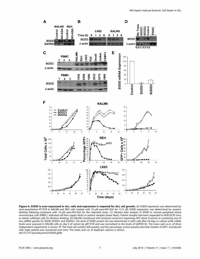
Figure 6. SODD is over-expressed in ALL cells and expression is required for ALL cell growth. (A) SODD expression was determined bysemi-quantitative RT-PCR in NALM6 and REH cells treated with 10 mM para-NO-ASA for 12 h. (B) SODD expression was determined by westernblotting following treatment with 10 mM para-NO-ASA for the indicated times. (C) Western blot analysis of SODD in normal peripheral bloodmononuclear cells (PBMC), indicated cell lines (upper blots) or patient samples (lower blots). Patient samples had been expanded in NOD/SCID miceto obtain sufficient cells for Western blotting. (D) NALM6 transduced with lentiviral constructs expressing GFP alone (Control) or containing one oftwo shRNA specific for SODD (SODD1 and SODD2). The level of SODD protein (D) was determined in LK63 cells after 44 days in culture while mRNAlevels were assessed in NALM6 cells on day 6 of culture by qRT-PCR and was normalised to the levels of GAPDH (E). The mean and s.e.m. of threeindependent experiments is shown. (F) The total cell number (left panels), and the percentage (centre panels) and total number of GFP+ transducedcells (right panels) was monitored over time. The mean and s.d. of duplicate cultures is shown.doi:10.1371/journal.pone.0103383.g006
NO-Aspirin Induced Extrinsic Cell Death in ALL
PLOS ONE | www.plosone.org 7 July 2014 | Volume 9 | Issue 7 | e103383

leukemia cell line, CCRF-HSB-2 [20], arsenic trioxide in acute
promyelocytic leukemia cells [19], etoposide in U937 cells [45]
and a number of other cell lines including Jurkat T cells [46], as
well as in a colon cancer model [43]. In our study there was clear
association between caspase-10 and TNF-R1, which preceded
caspase activation, suggesting that TNF-R1 plays an active role in
the process. However our inability to perform gene knockdown
studies for caspase-10 and potential off target effects of the
caspase-10 inhibitor used leaves room for the role of other caspases
in para-NO-ASA induced cell death.
How the extrinsic death mechanism is triggered by para-NO-
ASA is not entirely clear. B-lineage ALL cells only rarely express
TNF-a [10,47], and we were unable to demonstrate any
background, or para-NO-ASA-induced TNF-a production by
NALM6 cells. This suggests that activation of TNF-R1 may be
occurring in a ligand independent manner. Death receptors
including TNF-R1 are known to self-aggregate, particularly when
over-expressed and SODD is believed to play a role in keeping this
in check [6]. Ligand-independent association has been previously
reported in lung and breast carcinoma and although associated
with DNA damage, DNA damage alone was not sufficient to
activate TNF-R1 [48]. However the possibility remains that TNF-
a, below the limit of detection, may be present and activates TNF-
R1. We were unable to detect evidence of para-NO-ASA induced
DNA damage using c-H2AX staining prior to apoptosis driven
DNA fragmentation (data not shown). This suggests that the DNA
damage response is not involved. More recently ligand-indepen-
dent activation of TNF-R1 has been associated with oxidative
conditions and modification of cysteine residues within the
extracellular cysteine rich domains of TNF-R1 in the model cell
systems of renal and cervical cancer, most likely in the pre-ligand
assembly domain (PLAD) [49]. This may be important in
para-NO-ASA-induced cell death, as we have previously shown
that reactive oxygen species are produced and are required for cell
death in this setting [21].
Another possibility is that suppression of SODD by para-NO-
ASA permits constitutive activation through TNF-R1 self-associ-
ation. SODD is an anti-apoptotic protein that associates with the
death domain of TNF-R1, Fas and death receptor-3 (DR3), and
thereby negatively regulates cell death signalling [6,7]. The
importance of SODD in maintaining TNF-R1 in an inactive
conformation under physiological conditions has not been fully
resolved, with one study finding no phenotype in SODD deficient
mice, while another reported enhanced cytokine production
[50,51]. Here we observed loss of SODD concomitantly with
caspase-10 association with TNF-R1, which is consistent with
SODD playing an active role in keeping TNF-R1 signalling in
check. SODD is also reportedly over expressed in ALL [41], a
finding that we confirmed in this study.
The death inducing ligands, TNF-a, Fas and TRAIL have not
demonstrated the hoped for potency in killing malignant cells and
methods to enhance the efficacy of these agents have been
explored with varying degrees of success [52,53]. The role of TNF-
a in ALL cell biology is unclear with the initial report showing
suppression of ALL cell proliferation [47], while subsequent
reports suggest that TNF-a enhances ALL cell growth [54]. The
co-expression of TNF-R2, which lacks death domains in its
cytoplasmic tail and hence only activates pro-survival NF-kB
signalling [55], would also be expected to promote proliferation
and survival in ALL cells. The pro-survival signals activated by
NF-kB are known to limit the cytotoxic effects of TNF-a and other
death ligands [56]. Over expression of SODD would be expected
to further enhance ALL cell survival by preventing signalling
through TNF-R1 and other death receptors such as Fas and DR3,
but not TNF-R2 [6,41]. It is tempting to speculate that the
potency of para-NO-ASA results from activation of signalling
through TNF-R1, due to suppression of SODD, in the absence of
any increase in NF-kB signalling through TNF-R2 (Fig 7).
Furthermore, we have previously shown that para-NO-ASA
suppresses NF-kB signalling [21]. Therefore, unlike TNF-a,
para-NO-ASA activates death receptor signalling by inhibition
of SODD while simultaneously suppressing pro-survival NF-kB
signalling. We were surprised to discover that suppression of
SODD expression alone was sufficient to significantly impair the
growth of ALL cell lines. This finding is reminiscent of cells
displaying oncogene addiction, which are killed following onco-
gene withdrawal, while normal cells are relatively unaffected. This
suggests that targeting SODD may be a new therapeutic strategy
for the treatment of ALL and other malignancies where SODD is
overexpressed.
Supporting Information
Figure S1 Flow cytometric analysis of Bcl-XL and Bcl-2.
(DOCX)
Figure S2 Expression of TNF-R2 and effect of TNF-a on ALL
cell lines.
(DOCX)
Table S1 Patient Information.
(DOCX)
Acknowledgments
The authors would like to thank Dr Lisa Sedger for helpful discussions that
assisted in the finalization of the study and the writing of the manuscript.
Figure 7. Schematic diagram illustrating the proposed mech-anism of action of para-NO-ASA. para-NO-ASA down regulatesSODD allowing self-aggregation, or enhancing TNF-a-induced, activa-tion of signalling through TNF-R1. TNF-R1 signalling triggers theextrinsic apoptosis cascade including cleavage of pro-caspase-10 andBid, mitochondrial depolarization, outer membrane permeabilizationand release of cytochrome c. This is followed by cleavage of pro-caspase-9 and the executioner caspases-3 and -7. While signallingthrough TNF-R1 can activate NF-kB this may be suppressed by caspase-mediated cleavage of TRAF. In contrast, TNF-R2 signal induces cellsurvival and proliferation pathways, predominantly through NF-kB butalso MAPK signalling. In ALL cells where SODD is over expressed,exogenous TNF-a is likely to predominantly signal through TNF-R2resulting in increased survival and proliferation. Activating TNF-R1 bydown regulation of SODD provides a mechanism for inducing cell deathwithout increasing proliferation and survival signals. FADD, Fas-associated death domain; IKK, inhibitor of kB kinase; I-kB, inhibitor ofkB; TRADD, TNF-R1-associated death domain; TRAF, TNF receptor-associated factor.doi:10.1371/journal.pone.0103383.g007
NO-Aspirin Induced Extrinsic Cell Death in ALL
PLOS ONE | www.plosone.org 8 July 2014 | Volume 9 | Issue 7 | e103383

Author Contributions
Conceived and designed the experiments: NIK LJB KFB HR. Performed
the experiments: AC RB NIK CF RW JB. Analyzed the data: AC RB LJB
NIK. Contributed reagents/materials/analysis tools: HR CF. Wrote the
paper: LJB KFB.
References
1. Pui CH (2010) Recent research advances in childhood acute lymphoblastic
leukemia. J Formos Med Assoc 109: 777–787.
2. Gokbuget N, Hoelzer D (2009) Treatment of adult acute lymphoblastic
leukemia. Semin Hematol 46: 64–75.
3. Movassagh M, Foo R (2008) Simplified apoptotic cascades. Heart Fail Rev 13:
111–119.
4. Goldstein J, Waterhouse N, Juin P, Evan G, Green D (2000) The coordinate
release of cytochrome c during apoptosis is rapid, complete and kinetically
invariant. Nat Cell Biol 2(3):156–62: 2(3):156–162.
5. Li P, Nijhawan D, Budihardjo I, Srinivasula SM, Ahmad M, et al. (1997)
Cytochrome c and dATP-dependent formation of Apaf-1/caspase-9 complex
initiates an apoptotic protease cascade. Cell 91: 479–489.
6. Jiang Y, Woronicz JD, Liu W, Goeddel DV (1999) Prevention of constitutive
TNF receptor 1 signaling by silencer of death domains. Science 283: 543–546.
7. Eichholtz-Wirth H, Fritz E, Wolz L (2003) Overexpression of the ’silencer of
death domain’, SODD/BAG-4, modulates both TNFR1- and CD95-dependent
cell death pathways. Cancer Lett 194: 81–89.
8. Kischkel FC, Hellbardt S, Behrmann I, Germer M, Pawlita M, et al. (1995)
Cytotoxicity-dependent APO-1 (Fas/CD95)-associated proteins form a death-
inducing signaling complex (DISC) with the receptor. EMBO J 14: 5579–5588.
9. Luo X, Budihardjo I, Zou H, Slaughter C, Wang X (1998) Bid, a Bcl2
interacting protein, mediates cytochrome c release from mitochondria in
response to activation of cell surface death receptors. Cell 94: 481–490.
10. Pituch-Noworolska A, Gawlicka M, Wotoszyn M, Balwierz W, Strojny W, et al.
(1998) Tumour necrosis factor alpha (TNF alpha) and leukaemic cells: secretion
and response. Clin Lab Haematol 20: 231–238.
11. Karawajew L, Wuchter C, Ruppert V, Drexler H, Gruss H, et al. (1997)
Differential CD95 expression and function in T and B lineage acute
lymphoblastic leukemia cells. Leukemia 11: 1245–1252.
12. Ehrhardt H, Fulda S, Schmid I, Hiscott J, Debatin KM, et al. (2003) TRAIL
induced survival and proliferation in cancer cells resistant towards TRAIL-
induced apoptosis mediated by NF-kappaB. Oncogene 22: 3842–3852.
13. Reed JC, Doctor K, Rojas A, Zapata JM, Stehlik C, et al. (2003) Comparative
analysis of apoptosis and inflammation genes of mice and humans. Genome Res
13: 1376–1388.
14. Kischkel FC, Lawrence DA, Tinel A, LeBlanc H, Virmani A, et al. (2001) Death
receptor recruitment of endogenous caspase-10 and apoptosis initiation in the
absence of caspase-8. J Biol Chem 276: 46639–46646.
15. Wang J, Zheng L, Lobito A, Chan FK, Dale J, et al. (1999) Inherited human
Caspase 10 mutations underlie defective lymphocyte and dendritic cell apoptosis
in autoimmune lymphoproliferative syndrome type II. Cell 98: 47–58.
16. Shin MS, Kim HS, Kang CS, Park WS, Kim SY, et al. (2002) Inactivating
mutations of CASP10 gene in non-Hodgkin lymphomas. Blood 99: 4094–4099.
17. Rozenfeld-Granot G, Toren A, Amariglio N, Brok-Simoni F, Rechavi G (2001)
Mutation analysis of the FAS and TNFR apoptotic cascade genes in
hematological malignancies. Exp Hematol 29: 228–233.
18. Rikhof B, Corn PG, El-Deiry WS (2003) Caspase 10 levels are increased
following DNA damage in a p53-dependent manner. Cancer Biol Ther 2: 707–
712.
19. Li J, Chen P, Sinogeeva N, Gorospe M, Wersto RP, et al. (2002) Arsenic trioxide
promotes histone H3 phosphoacetylation at the chromatin of CASPASE-10 in
acute promyelocytic leukemia cells. J Biol Chem 277: 49504–49510.
20. Park SJ, Wu CH, Gordon JD, Zhong X, Emami A, et al. (2004) Taxol induces
caspase-10-dependent apoptosis. J Biol Chem 279: 51057–51067.
21. Khan NI, Cisterne A, Baraz R, Bradstock KF, Bendall LJ (2012) Para-NO-
Aspirin inhibits NF-kB and induces apoptosis in B-cell progenitor acute
lymphoblastic leukemia. Exp Hematol 40: 207–215.
22. del Soldato P, Sorrentino R, Pinto A (1999) NO-aspirins: a class of new anti-
inflammatory and antithrombotic agents. Trends Pharmacol Sci 20: 319–323.
23. Kashfi K, Ryan Y, Qiao L, Williams J, Chen J, et al. (2002) Nitric oxide-
donating nonsteroidal anti-inflammatory drugs inhibit the growth of various
cultured human cancer cells: evidence of a tissue type-independent effect.
J Pharmacol Exp Ther 303: 1273–1282.
24. Williams J, Borgo S, Hasan I, Castillo E, Traganos F, et al. (2001) Nitric oxide-
releasing nonsteroidal anti-inflammatory drugs (NSAIDs) alter the kinetics of
human colon cancer cell lines more effectively than traditional NSAIDs:
implications for colon cancer chemoprevention. Cancer Res 61: 3285–3289.
25. Annunziata CM, Kleinberg L, Davidson B, Berner A, Gius D, et al. (2007)
BAG-4/SODD and associated antiapoptotic proteins are linked to aggressive-
ness of epithelial ovarian cancer. Clin Cancer Res 13: 6585–6592.
26. Ozawa F, Friess H, Zimmermann A, Kleeff J, Buchler MW (2000) Enhanced
expression of Silencer of death domains (SODD/BAG-4) in pancreatic cancer.
Biochem Biophys Res Commun 271: 409–413.
27. Reuland SN, Smith SM, Bemis LT, Goldstein NB, Almeida AR, et al. (2013)
MicroRNA-26a is strongly downregulated in melanoma and induces cell death
through repression of silencer of death domains (SODD). J Invest Dermatol 133:
1286–1293.
28. Boyd AW, Ward LD, Wicks IP, Simpson RJ, Salvaris E, et al. (1992) Isolation
and characterization of a novel receptor-type protein tyrosine kinase (hek) from a
human pre-B cell line. J Biol Chem 267: 3262–3267.
29. Mihara K, Imai C, Coustan-Smith E, Dome J, Dominici M, et al. (2003)
Development and functional characterization of human bone marrow
mesenchymal cells immortalized by enforced expression of telomerase.
Br J Haematol 120: 846–849.
30. Kortlepel K, Bendall LJ, Gottlieb DJ (1993) Adhesion molecule expression on
human myeloid leukemic cells:Regulation by growth factors. Leukemia 7: 1174–
1179.
31. Bendall LJ, Kortlepel K, Gottlieb DJ (1993) Human acute myeloid leukemia
cells bind to bone marrow stroma via a combination of beta 1 and beta 2
integrin mechanisms. Blood 82: 3125–3132.
32. Khan NI, Bradstock KF, Bendall LJ (2007) Activation of Wnt/beta-catenin
pathway mediates growth and survival in B-cell progenitor acute lymphoblastic
leukaemia. Br J Haematol 138: 338–348.
33. Juarez J, Baraz R, Gaundar S, Bradstock K, Bendall L (2007) Interaction of
interleukin-7 and interleukin-3 with the CXCL12-induced proliferation of B-cell
progenitor acute lymphoblastic leukemia. Haematologica 92: 450–459.
34. Crazzolara R, Cisterne A, Thien M, Hewson J, Baraz R, et al. (2009)
Potentiating effects of RAD001 (Everolimus) on vincristine therapy in childhood
acute lymphoblastic leukemia. Blood 113: 3297–3306.
35. Juarez J, Bradstock K, Gottlieb D, Bendall L (2003) Effects of inhibitors of the
chemokine receptor CXCR4 on acute lymphoblastic leukemia cells in vitro.
Leukemia 17: 1294–1300.
36. Saunders P, Cisterne A, Weiss J, Bradstock KF, Bendall LJ (2011) The
mammalian target of rapamycin inhibitor RAD001 (everolimus) synergizes with
chemotherapeutic agents, ionizing radiation and proteasome inhibitors in pre-B
acute lymphocytic leukemia. Haematologica 96: 69–77.
37. Rizos H, Scurr L, Irvine M, Alling N, Kefford R (2007) p14ARF regulates E2F-1
ubiquitination and degradation via a p53-dependent mechanism. Cell Cycle 6:
1741–1747.
38. Green DR, Kroemer G (2004) The pathophysiology of mitochondrial cell death.
Science 305: 626–629.
39. Li H, Zhu H, Xu C, Yuan J (1998) Cleavage of BID by caspase 8 mediates the
mitochondrial damage in the Fas pathway of apoptosis. Cell 94: 491–501.
40. Wang J, Chun HJ, Wong W, Spencer DM, Lenardo MJ (2001) Caspase-10 is an
initiator caspase in death receptor signaling. Proc Natl Acad Sci U S A 98:13884–13888.
41. Tao H, Hu Q, Fang J, Liu A, Liu S, et al. (2007) Expression of SODD and P65
in ALL of children and its relationship with chemotherapeutic drugs.
J Huazhong Univ Sci Technolog Med Sci 27: 326–329.
42. Shen XG, Wang C, Li Y, Zhou B, Xu B, et al. (2011) Downregulation of
caspase-10 predicting poor survival after resection of stage II colorectal cancer.
Int J Colorectal Dis 26: 1519–1524.
43. Meng L, Sefah K, O’Donoghue MB, Zhu G, Shangguan D, et al. (2010)
Silencing of PTK7 in colon cancer cells: caspase-10-dependent apoptosis via
mitochondrial pathway. PLoS One 5: e14018.
44. Engel C, Versmold B, Wappenschmidt B, Simard J, Easton DF, et al. (2010)
Association of the variants CASP8 D302H and CASP10 V410I with breast and
ovarian cancer risk in BRCA1 and BRCA2 mutation carriers. Cancer Epidemiol
Biomarkers Prev 19: 2859–2868.
45. Filomenko R, Prevotat L, Rebe C, Cortier M, Jeannin JF, et al. (2006) Caspase-
10 involvement in cytotoxic drug-induced apoptosis of tumor cells. Oncogene
25: 7635–7645.
46. Lee HJ, Pyo JO, Oh Y, Kim HJ, Hong SH, et al. (2007) AK2 activates a novel
apoptotic pathway through formation of a complex with FADD and caspase-10.
Nat Cell Biol 9: 1303–1310.
47. Zhou M, Findley H, Ma L, Zaki S, Hill T, et al. (1991) Effect of tumor necrosis
factor-alpha on the proliferation of leukemic cells from children with B-cell
precursor-acute lymphoblastic leukemia (BCP-ALL): studies of primary leukemic
cells and BCP-ALL cell lines. Blood 77: 2002–2007.
48. Sheikh MS, Antinore MJ, Huang Y, Fornace AJ Jr (1998) Ultraviolet-
irradiation-induced apoptosis is mediated via ligand independent activation of
tumor necrosis factor receptor 1. Oncogene 17: 2555–2563.
49. Ozsoy HZ, Sivasubramanian N, Wieder ED, Pedersen S, Mann DL (2008)
Oxidative stress promotes ligand-independent and enhanced ligand-dependent
tumor necrosis factor receptor signaling. J Biol Chem 283: 23419–23428.
50. Takada H, Chen NJ, Mirtsos C, Suzuki S, Suzuki N, et al. (2003) Role of SODD
in regulation of tumor necrosis factor responses. Mol Cell Biol 23: 4026–4033.
51. Endres R, Hacker G, Brosch I, Pfeffer K (2003) Apparently normal tumor
necrosis factor receptor 1 signaling in the absence of the silencer of death
domains. Mol Cell Biol 23: 6609–6617.
NO-Aspirin Induced Extrinsic Cell Death in ALL
PLOS ONE | www.plosone.org 9 July 2014 | Volume 9 | Issue 7 | e103383

52. Bernhard D, Skvortsov S, Tinhofer I, Hubl H, Greil R, et al. (2001) Inhibition of
histone deacetylase activity enhances Fas receptor-mediated apoptosis in
leukemic lymphoblasts. Cell Death Differ 8: 1014–1021.
53. Balsas P, Lopez-Royuela N, Galan-Malo P, Anel A, Marzo I, et al. (2009)
Cooperation between Apo2L/TRAIL and bortezomib in multiple myeloma
apoptosis. Biochem Pharmacol 77: 804–812.
54. Makrynikola V, Kabral A, Bradstock KF (1991) Effects of recombinant human
cytokines on precursor-B acute lymphoblastic leukemia cells. Exp Hematol 19:674–679.
55. Faustman D, Davis M (2010) TNF receptor 2 pathway: drug target for
autoimmune diseases. Nat Rev Drug Discov 9: 482–493.56. Dutta J, Fan Y, Gupta N, Fan G, Gelinas C (2006) Current insights into the
regulation of programmed cell death by NF-kappaB. Oncogene 25: 6800–6816.
NO-Aspirin Induced Extrinsic Cell Death in ALL
PLOS ONE | www.plosone.org 10 July 2014 | Volume 9 | Issue 7 | e103383