Angiogenesis selectively requires the p110α isoform of PI3K to control endothelial cell migration
Upload
independentCategory
view
4download
0
The
Journ
al o
f Exp
erim
enta
l M
edic
ine
J. Exp. Med.
©
The Rockefeller University Press • 0022-1007/2004/09/659/11 $8.00Volume 200, Number 5, September 6, 2004 659–669http://www.jem.org/cgi/doi/10.1084/jem.20040789
659
Activation of PI3K Is Indispensable for Interleukin 7–mediated Viability, Proliferation, Glucose Use, and Growth of T Cell Acute Lymphoblastic Leukemia Cells
Joao T. Barata,
1
Ana Silva,
1
Joana G. Brandao,
1
Lee M. Nadler,
2
Angelo A. Cardoso,
2
and Vassiliki A. Boussiotis
2
1
Tumor Biology Unit, Institute of Molecular Medicine, University of Lisbon Medical School, 1649-028, Lisbon, Portugal
2
Department of Medical Oncology, Dana-Farber Cancer Institute, Harvard Medical School, Boston, MA 02115
Abstract
Interleukin (IL)-7 is essential for normal T cell development. Previously, we have shown thatIL-7 increases viability and proliferation of T cell acute lymphoblastic leukemia (T-ALL) cellsby up-regulating Bcl-2 and down-regulating the cyclin-dependent kinase inhibitor p27
kip1
.Here, we examined the signaling pathways via which IL-7 mediates these effects. We investi-gated mitogen-activated protein kinase (MEK)–extracellular signal-regulated kinase (Erk) andphosphatidylinositol-3-kinase (PI3K)–Akt (protein kinase B) pathways, which have active rolesin T cell expansion and have been implicated in tumorigenesis. IL-7 induced activation of theMEK–Erk pathway in T-ALL cells; however, inhibition of the MEK–Erk pathway by the useof the cell-permeable inhibitor PD98059, did not affect IL-7–mediated viability or cell cycleprogression of leukemic cells. IL-7 induced PI3K-dependent phosphorylation of Akt and itsdownstream targets GSK-3, FOXO1, and FOXO3a. PI3K activation was mandatory for IL-7–mediated Bcl-2 up-regulation, p27
kip1
down-regulation, Rb hyperphosphorylation, and conse-quent viability and cell cycle progression of T-ALL cells. PI3K signaling was also required forcell size increase, up-regulation of CD71, expression of the glucose transporter Glut1, uptakeof glucose, and maintenance of mitochondrial integrity. Our results implicate PI3K as a majoreffector of IL-7–induced viability, metabolic activation, growth and proliferation of T-ALLcells, and suggest that PI3K and its downstream effectors may represent molecular targets fortherapeutic intervention in T-ALL.
Key words: T cell acute lymphoblastic leukemia • IL-7 • PI3K–Akt • MEK–Erk • Glut1
Introduction
IL-7–mediated signals are linked to survival and cell cycleprogression (1). In normal T cell development, IL-7 plays anonredundant role as an antiapoptotic factor by up-regulatingBcl-2 expression (1). Similar to normal immature thy-mocytes, leukemic blasts from T cell acute lymphoblasticleukemia (T-ALL), patients can express functional IL-7Rs(2). Moreover, different studies demonstrated that IL-7 caninduce proliferation and prevent spontaneous apoptosis ofT-ALL cells in vitro (2, 3). Importantly, IL-7 is present in
the microenvironments where the leukemia arises becauseit is produced by thymic epithelial and bone marrow stromalcells (4, 5), and significantly contributes to the increasedsurvival of T-ALL cells cocultured with thymic epithelialcells (6). These data suggest that IL-7 may play an importantrole in the biology of T-ALL. We have shown previouslythat IL-7 down-regulates the expression of the cyclin-dependent kinase (cdk) inhibitor p27
kip1
, leading not onlyto cell cycle progression but also to up-regulation of Bcl-2protein expression and viability of T-ALL cells (7). Further-more, IL-7 can support the long-term expansion of pri-mary T-ALL cells, as shown by the establishment an IL-7–
A.A. Cardoso and V.A. Boussiotis contributed equally to the supervisionof this work.
The online version of this article contains supplemental material.Address correspondence to Vassiliki A. Boussiotis, Dana-Farber Can-
cer Institute, 44 Binney St., Room Mayer 547, Boston, MA 02115.Phone: (617) 632-4586; Fax: (617) 632-5167;email: [email protected]
Abbreviations used in this paper:
cdk, cyclin-dependent kinase; Erk, extra-cellular signal-regulated kinase; MEK, mitogen-activated protein kinase;MIF, mean intensity of fluorescence; PI3K, phosphatidylinositol-3-kinase;T-ALL, T cell acute lymphoblastic leukemia.
on May 14, 2014
jem.rupress.org
Dow
nloaded from
Published September 7, 2004
http://jem.rupress.org/content/suppl/2004/09/07/jem.20040789.DC1.html Supplemental Material can be found at:
Regulation of T-ALL Growth via PI3K–Akt
660
dependent cell line (TAIL7) that maintains the essentialfeatures of IL-7–responsive primary T-ALL blasts (8).
Mitogen-activated protein kinase (MEK)–extracellularsignal-regulated kinase (Erk) (MEK–Erk) signals have beenshown to promote viability (9) and induce cell cycle pro-gression by regulating the expression of c-Myc, cyclinD1, p27
kip1
, and p21
cip1
(10). Phosphatidylinositol-3-kinase(PI3K) and Akt/protein kinase B (hereafter referred to asAkt) have also been associated with prevention of apoptosisand cell cycle progression (11–13). These effects are impli-cated in PI3K–Akt-mediated tumorigenesis (12, 14–16).Strikingly, Jurkat and other T cell leukemia cell lines lackPTEN and/or SHIP, and consequently have high PI3K andAkt basal activities (17). Both MEK–Erk and PI3K–Aktpathways are involved in T cell survival, expansion, and dif-ferentiation (18–22). However, IL-7 appears to activatePI3K–Akt but not MEK–Erk in normal T cells (18, 23).
Our present studies demonstrate that IL-7 activated theMEK–Erk pathway in T-ALL cells, contrary to what oc-curs in normal T cells (18, 23). However, MEK–Erk didnot appear to be essential for IL-7–mediated viability andproliferation of T-ALL cells. IL-7 also triggered PI3K-dependent phosphorylation of Akt, GSK-3, FOXO1, andFOXO3a. Inhibition of PI3K prevented up-regulation ofBcl-2, down-regulation of p27
kip1
, and hyperphosphoryla-tion of Rb, and abrogated IL-7–mediated survival and pro-liferation of T-ALL cells. IL-7 induced expression of theglucose transporter Glut1 in a PI3K-dependent fashion,and this event correlated with glucose use, mitochondrialintegrity, increase of cell size, and up-regulation of CD69and CD71. These results demonstrate that PI3K down-stream signals are fundamental for IL-7–mediated survival,activation, proliferation, and growth of T-ALL cells, andmay regulate clonal expansion of T cell acute leukemia.
Materials and Methods
Primary T-ALL Samples and the TAIL7 Cell Line.
T-ALLcells were obtained from the peripheral blood and/or the bonemarrow of patients with high leukemia involvement (85–100%).Informed consent and Institutional Review Board approval wasobtained for all sample collections. Samples were enriched bydensity centrifugation over Ficoll-Hypaque, washed twice inRPMI 1640 supplemented with 10% (vol/vol) FBS and 2 mM
l
-glutamine (hereafter referred to as RPMI 10 medium), subjectedto immunophenotypic analysis by flow cytometry as describedpreviously (7), and classified according to their maturation stageusing the criteria defined by the European Group for Immuno-logical Characterization of Leukemias (see Table I; reference 24).The TAIL7 cell line was established from the peripheral blood ofa pediatric T-ALL patient. It is IL-7 dependent and has been de-scribed previously (8).
In Vitro Culture.
Primary T-ALL or TAIL7 cells isolated bydensity centrifugation over Ficoll-Hypaque were cultured in 24-well plates at 2
�
10
6
cells/ml at 37
�
C with 5% CO
2
in the fol-lowing: RPMI 10 (control medium), 10 ng/ml IL-7 (Endogen),IL-7 plus 10
�
M MEK-specific inhibitor PD98059 (Calbio-chem), or IL-7 plus 10
�
M PI3K-specific inhibitor LY294002(Calbiochem). At the indicated time points, cells were harvested
and processed as indicated below for assessment of viability, acti-vation, cell cycle progression, and preparation of lysates for West-ern blotting. TAIL7 cells were previously starved in RPMI 10without IL-7 for 5–7 d or in RPMI without FBS for 1–4 d, withsimilar results.
Proliferation Assays.
Cells were cultured in triplicates in flat-bottom 96-well plates at 2
�
10
6
cells/ml at 37
�
C with 5% CO
2
in RPMI 10 without any cytokine or in the experimental condi-tions mentioned above. Cells were incubated with [
3
H]thymidine(1
�
Ci/well) for 16 h before harvest. DNA synthesis, as measuredby [
3
H]thymidine incorporation, was assessed using a liquid scin-tillation counter. Average and standard deviation of triplicateswere calculated.
Assessment of Cell Viability, Size, and Activation.
Quantitativedetermination of viability of the malignant cells was performedusing an annexin V–based apoptosis detection kit (R&D Sys-tems), as described previously (7). Cell size was assessed by analy-sis of SSC versus FSC flow cytometry plots gated on the live cellpopulation. Surface expression of activation markers CD71 andCD69 was measured by flow cytometry using FITC-conjugatedanti-CD71 (DakoCytomation) and PE-conjugated anti-CD69(Beckman Coulter) antibodies and appropriately matched isotypecontrols. Samples were analyzed using a FACSCalibur flow cy-tometer and CELLQuest software (Becton Dickinson). Resultswere expressed as the percentage of positive cells as comparedwith the negative control, and as the specific mean intensity offluorescence (MIF), defined as the ratio of MIF of the specific an-tibody stain over the MIF of negative control antibody.
Cell Cycle Analysis.
Determination of the percentage of cellsat each stage of the cell cycle was performed by assessment ofDNA content after staining with propidium iodide. In brief, 5
�
10
5
cells per sample were resuspended in 0.5 ml PBS and thenfixed with ice-cold 80% ethanol. Propidium iodide was added ata final concentration of 2.5
�
g/ml, ribonuclease A was added at50
�
g/ml, and samples were incubated for 30 min at 37
�
C in thedark. Analysis of flow cytometry cell cycle histograms was per-formed using ModFit LT software (Verity).
Short-Term Stimulation with IL-7.
For the initial experiments,IL-7–deprived TAIL7 cells were washed twice with PBS and incu-bated for the indicated periods at 37
�
C with prewarmed PBS aloneor with the indicated concentrations of IL-7. IL-7–deprivedTAIL7 cells were then incubated for 15 min at 37
�
C with PBSalone or with 50 ng/ml IL-7. In defined experiments, the cellswere pretreated in PBS with 10
�
M LY294002, 10
�
M PD98059,or the corresponding volume of vehicle (DMSO) for 2 h beforestimulation. Reactions were stopped by placing samples on ice andadding ice-cold PBS. Cells were washed twice with cold PBS andlysates were prepared for Western blot analysis (immunoblotting).
Immunoblotting, Immunoprecipitation, and In Vitro Kinase Reac-tions.
After the indicated conditions and time intervals ofculture, cell lysates were prepared and equal amounts of pro-tein were analyzed by 10% SDS-PAGE, transferred onto nitro-cellulose membranes, and immunoblotted with the followingmAbs or antiserum: p27
kip1
(BD Transduction Laboratories), actin,STAT5, and Glut1 (Santa Cruz Biotechnology, Inc.), ZAP-70and phospho-STAT5A/B (Y694/Y699; Upstate Biotechnology),and phospho-Akt (S473), phospho-GSK-3
�
(S9), phospho-FKHR(FOXO1) (T24)/phospho-FKHRL1(FOXO3a) (T32),phospho-MEK1/2 (S217/S221), phospho-Erk1/2 (T202/Y204),Akt, and Erk1/2 (Cell Signaling Technology). To examine thephosphorylation status of Rb, proteins were analyzed by 6%SDS-PAGE, transferred onto nitrocellulose membrane, and blot-ted with Rb-specific mAbs (BD Biosciences). After immunoblot-
on May 14, 2014
jem.rupress.org
Dow
nloaded from
Published September 7, 2004

Barata et al.
661
ting with mAbs or antiserum, immunodetection was performedby incubation with horseradish peroxidase–conjugated anti–mouse IgG (1:5,000), anti–rabbit IgG (1:10,000), or anti–goatIgG (1:5,000; Promega), as indicated by the host origin of theprimary antibody and developed by chemiluminescence (Amer-sham Biosciences).
Akt in vitro kinase reactions were performed using a nonra-dioactive Akt kinase assay kit purchased from Cell SignalingTechnology according to the manufacturer’s instructions. Inbrief, cell lysates with equal amounts of protein were immuno-precipitated using agarose hydrazide–conjugated Akt antibody,washed twice, and resuspended in kinase buffer supplementedwith 200
�
M cold ATP. Kinase reactions were performed usingparamyosin-crosstide GSK-3
�
/
�
fusion protein as exogenoussubstrate. Reactions were analyzed by 12% SDS-PAGE, trans-ferred to nitrocellulose membrane, and GSK-3 phosphoryla-tion was detected by immunoblotting with phospho-GSK-3
�
/
�
(Ser21/Ser9) antibody. Even loading was confirmed by strippingand reprobing the membranes with an Akt antibody (Cell Signal-ing Technology). Relative quantification of Western blot bandswas performed by densitometry analysis using ImageQuant ImageAnalysis software (Amersham Biosciences).
Intracellular Staining.
Bcl-2 protein expression was assessed byintracellular staining. Cells were fixed in 0.1% formaldehyde for30 min at 4
�
C, washed in PBS, resuspended in 1
�
Perm/WashSolution (BD Biosciences), and incubated with mouse monoclo-nal FITC-conjugated anti–Bcl-2 antibody (DakoCytomation).Irrelevant isotype-matched antibody was used as negative control.Samples were analyzed by flow cytometry. Results were ex-pressed as the percentage of positive cells in comparison to thenegative control, and as specific MIF.
Assessment of Mitochondrial Membrane Potential (
�
�
m
).
Cellswere harvested, stained in culture medium with TMRE (Sigma-Aldrich) to a final concentration of 100 nM, and incubated for 30min at 37
�
C with 5% CO
2
. CCCP (Sigma-Aldrich) was added toduplicate tubes to a final concentration of 50
�
M to collapse
�
�
m
and therefore validate the assay and serve as a control for back-ground levels of fluorescence. Cells were analyzed for TMRE in-tensity by flow cytometry.
Glucose Uptake Assay.
After the indicated culture conditions,10
6
TAIL7 cells were starved in PBS at room temperature for30 min and incubated at 37
�
C for 10 min in PBS containing 5
�
M 2-{
14
C(U)}-deoxy-
d
-glucose (PerkinElmer). Cells were har-vested on filtermats and counted for
14
C-glucose content. Aver-age and standard deviation of triplicates were calculated.
Online Supplemental Material.
In Fig. S1, primary T-ALL cellswere cultured with 10 ng/ml IL-7, either alone or in the pres-ence of 10
�
M PD098059 or 10
�
M LY294002. Viability andproliferation were assessed as described above. Fig. S1 is availableat http://www.jem.org/cgi/content/full/jem.20040789/DC1.
Results
IL-7 Activates MEK–Erk and PI3K–Akt Pathways inT-ALL Cells.
PI3K–Akt and MEK–Erk pathways arethought to play an active role in normal thymocyte andmature T cell expansion (18–20). For this reason we inves-tigated whether these pathways are involved in IL-7–medi-ated proliferation and viability of T-ALL cells that we havepreviously reported. We used both primary T-ALL samplesand TAIL7, a cell line that displays IL-7–mediated re-sponses identical to primary leukemia cells (8) and provides
a useful tool for experiments that require high cell numbersthat cannot be obtained using primary leukemia cells. Cy-tokine-deprived TAIL7 cells were stimulated by 50 ng/ml
Figure 1. IL-7 activates MEK–Erk and PI3K–Akt pathways in T-ALL.IL-7–deprived TAIL7 cells were stimulated with IL-7 for the indicatedperiods (A and B). Cell lysates were resolved with 10% SDS-PAGE and im-munoblotted with the indicated antibodies. Results representative of three in-dependent experiments are shown. Levels of phosphorylated MEK1/2 andErk1/2 were detected with antisera that selectively recognize the activatedforms of the following kinases: Ser217/Ser221-phosphorylated MEK1/2(P-MEK1/2) and Thr202/Tyr204-dual-phosphorylated Erk1/2 (P-Erk1/2).Levels of phosphorylated Akt and GSK-3 were analyzed with antibodies thatspecifically recognize Ser473-phosphorylated Akt (P-Akt) and Ser9-phos-phorylated GSK-3� (P-GSK-3�), respectively. Levels of phosphorylatedFOXO1/FOXO3a were detected with antiserum that reacts with Thr24-phosphorylated FOXO1 (P-AFOXO1) and Thr32-phosphorylated FOXO3a(P- FOXO3a). Akt and Erk1/2 protein levels were assessed with specific an-tibodies and remained unchanged (not depicted). Blots were reprobed withan anti–ZAP-70 antibody to confirm even protein loading. (C) IL-7 activatesAkt and induces in vitro phosphorylation of GSK-3 by Akt. IL-7–deprivedTAIL7 cells were stimulated with IL-7 for 15 min. To compare Akt enzy-matic activity in unstimulated (Unst.) versus IL-7–stimulated cells (IL-7), celllysates were immunoprecipitated with agarose-conjugated anti-Akt antibodyand in vitro kinase reactions were performed using crosstide-GSK-3�/� asexogenous substrate. Reactions were analyzed by 12% SDS-PAGE, trans-ferred to nitrocellulose membrane, and GSK-3 phosphorylation was detectedby immunoblotting with anti–phospho-GSK-3�/� (Ser21/Ser9) antibody.Even loading was confirmed with an anti-Akt antibody. Relative quantifica-tion of phosphorylated GSK-3� and GSK-3� bands was performed by densi-tometry analysis. Results were normalized in relation to the loading control(Akt) and expressed as relative units. IL-7 induced a 1.63-fold increase inGSK-3� and a 3.07-fold increase in GSK-3� Akt-mediated phosphorylation.Results are representative of two independent experiments.
on May 14, 2014
jem.rupress.org
Dow
nloaded from
Published September 7, 2004
Regulation of T-ALL Growth via PI3K–Akt
662
IL-7 for increasing periods of time for up to 120 min, andprotein phosphorylation was assessed by Western blot.Erk1/2 was phosphorylated and activated upon IL-7 stimu-lation of TAIL7 cells, as determined by immunoblottingwith an antibody that exclusively detects Erk1/2 when cat-alytically activated by phosphorylation at both Thr202 andTyr204. Phosphorylation of Erk1/2 was detectable after 5min of stimulation, increased at 15 min, peaked by 30 min(Fig. 1 A), and was still detected by 2 h of stimulation (notdepicted). A similar pattern of phosphorylation was ob-served for MEK1/2 (Fig. 1 A). When TAIL7 cells werestimulated with increasing doses of IL-7, phosphorylationof MEK and Erk was detected with 1 ng/ml and reached aplateau at 10 ng/ml (not depicted).
Next, we analyzed whether IL-7 activated the PI3K–Aktpathway in TAIL7 cells. Phosphorylation of Akt at Ser473,which is necessary for full activation of Akt kinase, oc-curred within 1 min, peaked at 30 min, and was undetect-able after 1 h of IL-7 stimulation (Fig. 1 B). Members ofthe FOXO family of transcription factors are direct targetsof Akt (25). In TAIL7 cells, IL-7 induced phosphorylationof FOXO1 (at Thr24) and FOXO3a (at Thr32) in a time-dependent manner (Fig. 1 B). FOXO1 phosphorylation atSer256 did not appear to be regulated by IL-7 in these cells(not depicted). GSK-3, another known downstream targetof Akt, was also phosphorylated by stimulation with IL-7(Fig. 1 B). IL-7 mediated a dose-dependent phosphoryla-tion of Akt and its downstream targets, which was detectedat
�
1 ng/ml and reached a plateau at 10 ng/ml (not de-picted). These results suggest that Akt becomes enzymati-cally active after stimulation with IL-7. To confirm that IL-7can induce the enzymatic activity of Akt in TAIL7 cells, ly-sates from unstimulated or 15 min IL-7–stimulated cellswere immunoprecipitated with an anti-Akt antibody andan in vitro kinase reaction was performed using GSK-3
�
/
�
as exogenous substrate. Phosphorylation of GSK-3
�
, andmore prominently of GSK-3
�
, was up-regulated (1.6- andthreefold, respectively) by stimulation with IL-7 (Fig. 1 C).These results confirm that IL-7 induced Akt phosphoryla-tion, leading to its enzymatic activation and consequentphosphorylation of GSK-3.
Because we observed that phosphorylation of Akt, GSK-3,and FOXO family members was mediated by IL-7, wesought to confirm that these events were dependent uponPI3K activation. IL-7–deprived TAIL7 cells were pretreatedwith 10
�
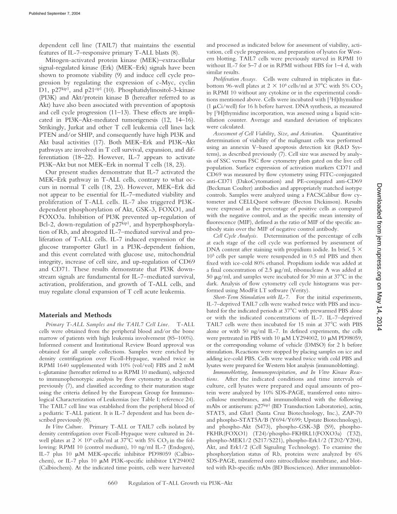
M of the cell-permeable PI3K-specific inhibitorLY294002 or MEK-specific inhibitor PD98059 before IL-7stimulation. LY294002 specifically abrogated phosphoryla-tion of Akt, without affecting phosphorylation of Erk1/2or STAT5, whereas PD98059 specifically inhibited phos-phorylation of Erk1/2 without affecting phosphorylation ofAkt or STAT5 (Fig. 2 A). Furthermore, LY294002 but notPD98059 inhibited GSK-3, FOXO1, and FOXO3a phos-phorylation (Fig. 2 B). These findings indicate that IL-7–induced phosphorylation of Akt and downstream targets inT-ALL cells are dependent on PI3K activity and can bespecifically disrupted by LY294002.
PI3K But Not MEK Is Required for IL-7–mediated Increasein Viability and Cell Cycle Progression of T-ALL Cells.
Toanalyze the functional role of PI3K–Akt and MEK–Erk ac-tivation by IL-7 in T-ALL cells, we investigated the ef-fect of specific inhibition of these pathways on IL-7–medi-ated viability and proliferation of TAIL7 cells by usingLY294002 and PD98059. Initial experiments determinedthat IL-7 stimulated a dose-dependent increase in viabilityand proliferation of TAIL7 cells, which was most evidentbetween 72 and 96 h of culture with 10 ng/ml IL-7 (notdepicted). Therefore, we used this concentration and timepoints in all subsequent experiments. Staining of leukemiccells with annexin V–FITC and propidium iodide followedby flow cytometry analysis revealed that LY294002 inhib-ited IL-7–mediated increase in the percentage of viablecells, whereas PD98059 showed no effect (Fig. 3 A). Thesame pattern was obtained when we examined cell prolifer-ation by assessing the incorporation of [
3
H]thymidine. Asshown in Fig. 3 B, PD98059 did not affect proliferation ofIL-7–cultured TAIL7 cells. In contrast, LY294002 com-pletely abrogated IL-7–induced proliferation (Fig. 3 B).Next, we examined whether inhibition of PI3K prevented
Figure 2. IL-7 induces PI3K-dependent phosphorylation of Akt,GSK-3, FOXO1, and FOXO3a, and MEK-dependent phosphorylationof Erk1/2 in T-ALL cells. IL-7–deprived TAIL7 cells were pretreatedwith 10 �M LY294002 (LY) or 10 �M PD098059 (PD) for 2 h, andthen stimulated with IL-7 for 15 min. (A) Western blot analysis was per-formed with P-Erk1/2, P-Akt antibodies (see legend to Fig. 1), and an anti-body specific for Tyr694/Tyr699-phosphorylated-STAT5A/B (P-STAT5)to confirm that LY294002 and PD98059 were specific inhibitors of thePI3K–Akt and MEK–Erk pathway, respectively. (B) GSK-3�, FOXO1,and FOXO3a phosphorylation is dependent on PI3K activity. Westernblot analysis was performed with P-Akt, P-GSK3�, and P-FOXO1/FOXO3a antibodies. Anti-STAT5 (A) and actin (B) antibodies were usedto confirm equal loading. Representative results from three independentexperiments are shown.
on May 14, 2014
jem.rupress.org
Dow
nloaded from
Published September 7, 2004

Barata et al.
663
[
3
H]thymidine incorporation only because it impaired cellviability, or whether it also blocked cell cycle progression.TAIL7 cells were fixed and stained with propidium iodideand analyzed for cell cycle progression by flow cytometry.As shown in Fig. 2 C, the PI3K inhibitor LY294002 abro-gated cell cycle progression of IL-7–cultured cells, whereasthe MEK inhibitor PD98059 had no effect. Thus, PI3Kseems to have a dominant role in IL-7–mediated viabilityand cell cycle progression in TAIL7 leukemia cells.
To confirm the biological significance of these resultsobtained in TAIL7 cells, we examined whether PI3Kplayed a similar critical role on IL-7–mediated effects inprimary T-ALL cells. Leukemic cells were collected fromthe peripheral blood or bone marrow of pediatric T-ALLpatients with high leukemia involvement and tested for invitro responsiveness to IL-7 by assessing cellular prolifera-tion. IL-7–responsive samples were immunophenotyped,classified according to their maturation stage (Table I), andused in subsequent experiments. The increase in viability(Fig. 3 D and Fig. S1, available at http://www.jem.org/cgi/content/full/jem.20040789/DC1) and proliferation(Fig. 3 E and Fig. S1) induced with IL-7 was completelyabrogated by LY294002, whereas PD98059 had no signifi-cant impact on IL-7–mediated effects in primary leukemiacells. Our data demonstrate that PI3K is indispensable forboth viability and proliferation mediated by IL-7 not onlyin the TAIL7 line, but also in primary T-ALL cells. In con-trast, MEK–Erk pathway albeit activated by IL-7 does notappear to play a functionally significant role in mediatingthese effects of IL-7 in T-ALL.
IL-7–mediated Down-regulation of p27
kip1
, Hyperphosphory-lation of Rb, and Up-regulation of Bcl-2 in T-ALL Cells Is De-pendent upon PI3K Activity.
Cell cycle progression fromG0/G1 to S phase is modulated by exogenous factors andis positively regulated by cyclin–cdk holoenzymes, whichphosphorylate several intracellular substrates including Rb.Phosphorylation and inactivation of Rb result in release ofE2F and transcription of genes that are required for progres-sion through S phase (26). The enzymatic activity of cyclin–cdk complexes is negatively regulated by cdk inhibitors,including the cip/kip family member p27
kip1
(27). We pre-viously demonstrated that culture with IL-7 down-regulatesp27
kip1
protein expression, resulting in cdk activation, ensu-ing hyperphosphorylation of Rb, and cell cycle progression(7). Because LY294002 inhibited IL-7–regulated cell cycleprogression, we next evaluated whether PI3K-dependentevents could link IL-7 to the cell cycle machinery. Cultureof TAIL7 cells with IL-7 for 96 h resulted in down-regula-tion of p27
kip1
(Fig. 4 A) and hyperphosphorylation of Rb(Fig. 4 B). LY294002 completely reversed these IL-7–medi-ated effects, up-regulating p27
kip1
to the same levels as those
Figure 3. PI3K but not MEK is critical for IL-7–mediated viability andcell cycle progression of T-ALL cells. (A–C) TAIL7 cells were culturedfor 96 h with 10 ng/ml IL-7, either alone or in the presence of 10 �MPD098059 (IL-7PD) or 10 �M LY294002 (IL-7LY). (A) TAIL7 cellswere stained with annexin V–FITC plus propidium iodide and viabilitywas determined by flow cytometry analysis. (B) Proliferation was deter-mined by assessment of [3H]thymidine incorporation. (C) Percentage ofcells at SG2/M phases of the cell cycle was determined by propidiumiodide staining followed by flow cytometry analysis. (D and E) PrimaryT-ALL cells were obtained as described in Materials and Methods, andcultured under the indicated conditions. Viability at 96 h (D) and prolif-eration at 72 h (E) of culture were assessed as described for TAIL7 cells.Results are representative of three to six independent experiments withTAIL7 cell line and two independent experiments with all five primaryT-ALL samples. Results from remaining primary T-ALL samples areshown in Fig. S1.
Table I.
Immunophenotype and Classification of T-ALL Patients
Immunophenotype
T-ALL no. CD1 CD2 CD3 CD4 CD5 CD7 CD8 CD10 CD14 CD19 CD33 CD34 CD56 Maturation stage
1
III2
III3
II4
ND ND
ND III5 II
T cell maturation stages of primary samples were defined as described previously (reference 24). Stage II, pre–T-ALL; stage III, cortical T-ALL.
on May 14, 2014
jem.rupress.org
Dow
nloaded from
Published September 7, 2004

Regulation of T-ALL Growth via PI3K–Akt664
presented by leukemic cells cultured in medium alone, andpreventing Rb hyperphosphorylation. MEK inhibition us-ing PD98059 did not affect p27kip1 levels or Rb phosphory-lation (not depicted). Similar results were observed for pri-mary T-ALL cells (Fig. 4, C and D). These results indicatethat PI3K activation is essential for IL-7 to induce p27kip1
down-regulation and cell cycle progression in T-ALL cells.Prevention of spontaneous apoptosis of in vitro–cultured
primary T-ALL cells by IL-7 was shown to correlate withincreased Bcl-2 protein expression (3). We subsequentlydemonstrated that Bcl-2 up-regulation was mandatory forIL-7–mediated promotion of viability. Moreover, we ob-served a causative link between p27kip1 down-regulation andincrease in Bcl-2 expression (7). Because the addition ofLY294002 completely blocked IL-7–mediated viability ofT-ALL cells, we examined whether IL-7 regulated Bcl-2
via activation of PI3K. TAIL7 cells were cultured with ei-ther medium alone, IL-7, or IL-7 plus LY294002. After 96 h,cells were analyzed for intracellular expression of Bcl-2by flow cytometry. IL-7–mediated up-regulation of Bcl-2was significantly impaired by LY294002 (Fig. 4 E). PrimaryT-ALL samples showed similar results (Fig. 4 F). Thus, up-regulation of Bcl-2 mediated by IL-7 in T-ALL cells re-quires activation of PI3K.
IL-7 Induces Increased Cell Size and Activation of T-ALLCells in a PI3K-dependent Manner. T cell activation can bemeasured by increased cell size (cell growth) and by thesurface expression of CD69 and CD71. After 72 h of cul-ture, IL-7 strikingly up-regulated cell size of TAIL7 cells
Figure 4. IL-7 mediates p27kip1 down-regulation, Rb hyperphosphory-lation, and Bcl-2 up-regulation via activation of PI3K in T-ALL cells.TAIL7 cells (A and B) or primary T-ALL cells (C and D) were culturedfor 96 or 72 h, respectively, under the indicated conditions. (A and C)Cell lysates were resolved by 10% SDS-PAGE and immunoblotted withanti-p27kip1 antibody. Membranes were stripped and reprobed with ZAP-70 to confirm equal loading. (B and D) Lysates from the same sampleswere analyzed by 6% SDS-PAGE and immunoblotted with an Rb-specificantibody. The hyperphosphorylated form of Rb corresponds to the bandwith the higher apparent molecular weight. Blasts from T-ALL number 3were used in this experiment. (E) Bcl-2 protein levels at 96 h of culturewere assessed by flow cytometry after intracellular staining of TAIL7 cellswith FITC-conjugated anti–Bcl-2 antibody. (F) Expression of Bcl-2 inprimary T-ALL cells was assessed at 72 h of culture. Results are represen-tative of four different patient samples analyzed. Specific MIF, as described inMaterials and Methods, is indicated in each histogram. Results were similar insix independent experiments.
Figure 5. PI3K is critical for IL-7–mediated cell growth and activationof T-ALL cells. TAIL7 (A) and primary T-ALL (B) cells were culturedfor 72 h in medium alone or with 10 ng/ml IL-7, and then analyzed byflow cytometry for changes in cell size (determined by FSC) in the livecell population. Percentage of “activated” cells was calculated by defininga threshold gate that excluded the bulk, small-sized population of medium-cultured cells. Results from one representative patient of five tested areshown in B. Results shown in A are similar to those found in numerousindependent experiments. TAIL7 cells (C) and primary T-ALL cells (D)were cultured for 96 h under the indicated conditions, and the percentageof activated cells was calculated. TAIL7 cells cultured for 96 h (E) and pri-mary T-ALL cells cultured for 72 h (F) under the indicated conditionswere stained with anti-CD71 antibody. Results were expressed as thepercentage of positive cells and as the specific MIF (in brackets). Resultsfrom primary T-ALL cells and the TAIL7 cell line were similar in threeindependent experiments.
on May 14, 2014
jem.rupress.org
Dow
nloaded from
Published September 7, 2004

Barata et al.665
(Fig. 5 A), consistently with previous results (8). Likewise,IL-7 increased cell size in all primary T-ALL samples (fivecases analyzed; Fig. 5 B). To determine which intracellularpathways were involved in mediating IL-7–inducedgrowth of T-ALL cells, we blocked PI3K–Akt and MEK–Erk pathways with the specific inhibitors. LY294002 com-pletely inhibited or greatly impaired IL-7–mediated in-crease of cell size in TAIL7 (Fig. 5 C) and in primaryT-ALL cells (Fig. 5 D). In contrast, PD98059 induced onlya minor decrease in the percentage of activated cells.
Next, we used TAIL7 (Fig. 5 E) and primary T-ALL cells(Fig. 5 F) to compare the surface expression of activationmarkers CD71 and CD69 between medium- and IL-7–cul-tured cells. Flow cytometry analysis revealed that both CD71(Fig. 5, E and F) and CD69 (not depicted) were strongly up-regulated by IL-7. Consistently with the effects on IL-7–mediated cell size increase, LY294002, but not PD985002,completely inhibited IL-7–mediated surface expression ofCD71 in TAIL7 cells (Fig. 5 E) and primary T-ALL cells(Fig. 5 F), as assessed by both specific MIF and percentage ofCD71 cells. Similar results were obtained with CD69 (notdepicted). These results indicate that the MEK–Erk pathwaydoes not significantly affect IL-7–mediated cell growth andactivation of T-ALL cells, whereas PI3K-dependent signal-ing is critical for the regulation of these effects.
IL-7 Induces Glut1 Expression and Promotes Glucose Uptakeby T-ALL Cells. Activation and growth of normal T cellsis associated with increased glycolysis (28). Cytokines caninduce expression of glucose transporters (29, 30) and aug-ment glucose uptake (31) and glycolytic rates (32). ThePI3K–Akt pathway is specifically involved in these pro-cesses in normal T lymphocytes (29). Because PI3K had acritical role in IL-7–mediated promotion of viability andinduced cell growth and activation of T-ALL cells, we ex-amined whether this correlated with the expression of theglucose transporter Glut1. As shown in Fig. 6 A, IL-7 up-regulated Glut1 in TAIL7 cells and this effect was depen-dent on PI3K activity because it was abrogated by the useof LY (Fig. 6 A). Subsequent to PI3K-dependent inductionof Glut1 expression, IL-7 also increased glucose uptake byTAIL7 cells (Fig. 6 B). Thus, IL-7 provides the machineryfor nutrient use by T-ALL cells and this effect is mediatedin a PI3K-dependent manner.
IL-7 Regulates Mitochondrial Homeostasis in a PI3K-depen-dent Manner in T-ALL Cells. Glucose metabolism, simi-larly to antiapoptotic Bcl-2 family members, can maintainmitochondrial integrity and homeostasis (30), and conse-quently negatively regulate apoptosis (31, 32). In our sys-tem, IL-7 up-regulated Bcl-2, which is directly involved inregulation of mitochondrial integrity. In addition, IL-7 in-duced expression of Glut1, a regulator of glucose transport,which indirectly controls mitochondrial integrity (32). Forthese reasons, we analyzed the effect of IL-7 on mitochon-drial homeostasis of TAIL7 cells. Mitochondrial homeosta-sis can be evaluated by measuring mitochondrial membranepotential (��m) using the potentiometric dye TMRE andflow cytometry analysis (30). Initial analyses of the wholecell population demonstrated that IL-7 up-regulated ��m in
TAIL7 cells, and LY294002 completely abrogated this ef-fect (Fig. 6 C). The percentage of TMRE high cells (Fig. 6C, right peak of the histograms) was similar to the percentof viable cells identified using annexin V/propidium iodidestaining (not depicted). Because differences in ��m can bemeasured even before early manifestations of apoptosis(33), we next focused our analysis on the live population(Fig. 6 D). IL-7–mediated up-regulation of ��m on thispopulation was abrogated by LY294002 (Fig. 6 D), sug-gesting that maintenance of mitochondrial integrity andhomeostasis is an early event in the regulation of PI3K-dependent cell viability mediated by IL-7.
DiscussionIL-7 is expressed in the bone marrow and thymus and
has been shown to stimulate the expansion of immature
Figure 6. IL-7 regulates Glut1 expression and mitochondrial homeosta-sis via PI3K activation. TAIL7 cells were cultured for 96 h under the indi-cated conditions. (A) Cell lysates were resolved with 10% SDS-PAGE andimmunoblotted with anti-Glut1 antibody. Anti–ZAP-70 antibody wasused in the same membrane to ascertain even protein loading. (B) IL-7promotes glucose uptake in a PI3K-dependent manner. TAIL7 cells werecultured as indicated for 96 h. Cells were then assayed for glucose uptakeas described in Materials and Methods. (C) Mitochondrial membrane poten-tial (��m) was assessed at 96 h of culture by staining TAIL7 cells with thepotentiometric dye TMRE and analyzing the whole population by flowcytometry. TMRE intensity reflects the ��m. Results are expressed asMIF. (D) Viable cells were identified and selected by flow cytometry bygating on the live cell population, as determined by FSC � SSC and/orannexin V–FITC staining. ��m of this population was determined usingTMRE. Results are representative of three independent experiments.
on May 14, 2014
jem.rupress.org
Dow
nloaded from
Published September 7, 2004
Regulation of T-ALL Growth via PI3K–Akt666
double negative and mature single positive thymocytes, inpart by up-regulating Bcl-2 expression and viability, andalso by inducing cell cycle progression (1, 33). In humansand mice, defective IL-7R expression results in severe Tcell deficiency (34, 35), indicating that IL-7 plays an essen-tial role during T cell ontogeny. Primary leukemic T cellsshow increased proliferation (2) and viability (3) when cul-tured with IL-7, suggesting that IL-7 might also be in-volved in the pathobiology of T-ALL. Up-regulation ofcdk activity with consequent Rb hyperphosphorylationand progression toward S phase are absolutely dependentupon IL-7–induced down-regulation of p27kip1. In addi-tion, p27kip1 down-regulation is associated with up-regula-tion of Bcl-2, which in turn is essential for IL-7–mediatedsurvival of T-ALL cells (7).
Cytokines and growth factors can not only influencesurvival, but also cell growth through effects on glucosetransporter expression, glucose uptake, and glycolysis (29,30, 32). Lymphocytes require extrinsic stimulation to in-duce expression of surface receptors such as Glut1 that pro-mote nutrient uptake and increase metabolic activity (36).TCR and IL-7R signals are among the signals that induceGlut1 in mature T cells. The importance of these extrinsicsignals to promote nutrient intake in mature T cells wasoriginally revealed by the observation that Bcl-2 transgeneexpression maintains cell survival, but does not prevent cellatrophy resulting from limited energy supplies due to inad-equate nutrient uptake (36, 37). In this study we demon-strated that Glut1 is induced in high amounts in T-ALLcells by IL-7 and that its expression is dependent on IL-7–mediated signaling. In parallel with increased Glut1 expres-sion, IL-7 also promoted nutrient uptake, increase of cellsize, activation, and subsequent cell cycle progression andproliferation of T-ALL. Our results strongly suggest thatIL-7 signals are needed not only to promote survival ofleukemia cells by up-regulating Bcl-2, but also to providethe means for the generation of metabolic energy for initi-ating the cell cycle progression program that will eventuallylead to cellular proliferation and expansion.
We examined the signaling pathways that might link IL-7to the downstream regulators of viability and cell cycle, par-ticularly to Bcl-2, Glut1, and p27kip1. Knowledge regardingIL-7–mediated pathways in T cells is rather incomplete andvery little is known about the integrity and biological role ofthose pathways in T-ALL cells. PI3K–Akt and MEK–Erkpathways have been associated with TCR- or cytokine-mediated expansion of T cell precursors and mature T cells(19, 20, 38). The PI3K–Akt pathway is activated by IL-7 innormal T cells (18, 23). In contrast, most studies with pri-mary human mature T cells and murine T cell lines haveshown that IL-7 does not mediate MEK–Erk activation(23), nor does it phosphorylate the MEK–Erk upstreammolecules Shc (23, 39) and Ras (40). Our studies showedthat in T-ALL cells, IL-7 activates Erk1/2 in a time- anddose-dependent manner that relies on MEK activity. How-ever, inhibition of the MEK–Erk pathway does not affectIL-7–mediated viability or cell cycle progression of TAIL7
cells, indicating that these events occur in a MEK–Erk-independent manner. Studies in different cell types supportan active role for Ras and MEK–Erk in p27kip1 phosphoryla-tion and consequent degradation by the ubiquitin–proteasomesystem (41). In T-ALL cells we found that down-regulationof p27kip1 protein expression and Rb hyperphosphorylationthat result from culture with IL-7 were not reverted byMEK inhibition. Hence, although the MEK–Erk pathwayis activated by IL-7 in T-ALL cells, its exact biological roleremains to be determined.
Our studies showed that IL-7 induced phosphorylationof Akt and its downstream targets GSK-3, FOXO1, andFOXO3a in a PI3K-dependent manner, indicating the ex-istence of a functional IL-7–mediated PI3K–Akt pathwayin T-ALL cells. Our subsequent studies with the PI3K in-hibitor LY294002 demonstrated that activation of PI3K ismandatory for Bcl-2 up-regulation, Glut1 induction, glu-cose uptake, p27kip1 down-regulation, and Rb hyperphos-phorylation in IL-7–cultured T-ALL cells. Accordingly,IL-7 mediates cell cycle progression and viability of T-ALLcells via PI3K-dependent signals. Several studies haveshown that engagement of the IL-7R induces activation ofPI3K and PI(3,4,5)P3 production in human thymocytes, Tlineage ALL blasts, and T-ALL cell lines (2, 40, 42), leadingto their survival and proliferation (2, 18, 40). However, theexact PI3K-dependent mechanisms through which IL-7exerts its effects in T cells are still under investigation. Al-though we cannot rule out the possibility that other PI3Kdownstream targets such as PKC or ILK (43, 44) mightcontribute to IL-7–induced functional outcomes, we favorthe possibility that Akt is the main effector of IL-7–stimu-lated PI3K in T-ALL. First, IL-7 induced phosphorylationof FOXO1 and FOXO3a at threonine residues Thr24 andThr32, which are targets for Akt kinase activity. Second,IL-7 induced phosphorylation of the Akt target GSK-3.
Phosphorylation of members of the Forkhead Box O(FOXO) family of transcription factors FOXO1, FOXO3a,and FOXO4 by Akt induces their inactivation and nuclearexport (25). FasL and p27kip1, which can be involved inapoptosis, are transcriptionally up-regulated by FOXOfamily members (25, 45). Thus, FOXO inactivation by thePI3K–Akt pathway may contribute to down-regulation ofp27kip1 by IL-7. Phosphorylation of GSK-3 results in its in-activation. Because active GSK-3 can mediate cell death,inactivation of GSK-3 by phosphorylation may promotecell viability (46). GSK-3� may also phosphorylate cyclinD1 (47) and c-Myc (48), promoting their protein degrada-tion and contributing to cell cycle arrest. In addition, GSK-3can also phosphorylate and inhibit NF-ATc, a transcriptionfactor involved in proliferation (49, 50) and Bcl-2 genetranscription (51). Thus, GSK-3 phosphorylation and sub-sequent inactivation could result in up-regulation of Bcl-2via activation of NF-ATc transcriptional activity (51) anddown-regulation of p27kip1 via c-Myc protein stabilization(48, 52).
A drop in ��m occurs very early during apoptosis (53).We showed that IL-7 up-regulates ��m in T-ALL cells in a
on May 14, 2014
jem.rupress.org
Dow
nloaded from
Published September 7, 2004

Barata et al.667
PI3K-dependent manner. This could be achieved via regu-lation of Bcl-2 expression (54, 55). Another possible mech-anism may involve IL-7–mediated induction of glucoseuptake and metabolism, which subsequently regulates mi-tochondrial homeostasis and ��m. Consistently with thesecond mechanism, our results showed that IL-7 inducedGlut1 glucose transporter expression and glucose uptake.Cytokine- or oncogene-induced glucose uptake appears toregulate mitochondrial homeostasis, thereby maintainingmitochondrial integrity and preventing apoptosis (31, 32).Conversely, glucose depletion or inhibition of glucose up-take is linked with cell death (31, 36). Here we showedthat IL-7 up-regulates the expression of the glucose trans-porter Glut1 via PI3K activation. Thus, PI3K might con-trol mitochondrial integrity and prevent apoptosis by regu-lating both Bcl-2 expression and glucose metabolism inT-ALL cells. Further studies are required to dissect the in-dividual contribution of Bcl-2 and glucose metabolism inIL-7–regulated mitochondrial homeostasis.
Our studies have shown that IL-7–mediated up-regula-tion of Glut1 is associated with an increase in cell size. Thisfinding may have significant implications on T-ALL patho-biology. Recent evidence suggests that there might be acorrelation between increased cell size and oncogenesis(16). Moreover, tumor progression may not only dependupon uncontrolled cell cycle progression, but also uponunbalance of cell size regulatory mechanisms (56). Activa-tion of lymphocytes is associated with increased size andmetabolic activity (28, 36). The transferrin receptor CD71is up-regulated by lymphocytes upon activation as a mech-anism to meet the increased iron demands associated withincreased metabolism (57). Increased Glut1 expression, glu-cose uptake, and glycolytic rates mediated by external sig-nals allow T cells to anticipate energetic and biosyntheticneeds associated with activation and cell growth (28). Ourstudy showed that IL-7 contributes to T-ALL cell growthand activation, as shown by a dramatic increase in cell sizeand surface expression of CD71 and CD69, which corre-late with induction of Glut1 expression. All of these eventsare dependent on PI3K activation. In mature T cells, thePI3K–Akt pathway regulates glucose metabolism mediatedby CD28 costimulation (28), and Akt-controlled glucoseuptake can promote survival and cell growth in other celltypes (30, 58). Interestingly, in developing CD8 singlepositive thymocytes, IL-7 up-regulates Glut1 expressionand glucose uptake (59).
There is mounting evidence that exogenous stimuli, par-ticularly IL-7, may confer a selective advantage to leukemicT cells and play a fundamental role in leukemia pathophys-iology (3, 6, 7, 60–62). Our studies presented here showedthat IL-7–mediated activation of PI3K–Akt is not only es-sential for increased viability, but also critically involved inthe regulation of metabolic activity, cell size, and prolifera-tion of T-ALL cells, suggesting that this pathway may havean indispensable role in T-ALL biology. Importantly, over-activation of the PI3K–Akt pathway is associated with tu-morigenesis (12, 16). Consistently, Jurkat and other T-ALL
cell lines lack expression of PTEN, a phosphatase that tar-gets PI(3,4,5,)P3, and consequently have high constitutiveAkt activity (17). Taken together, our results support theconclusion that PI3K is a pivotal mediator of IL-7 signalingin T-ALL cells with a striking impact on several biologicalmechanisms necessary for tumorigenesis. These observa-tions indicate that PI3K and its downstream targets mightbe essential for expansion of malignant T cells in vivo andmay represent molecular targets for pharmacological inter-vention in T-ALL.
We thank Alla Berezovskaya for technical support.This work was supported by grants from Fundação para a Cien-
cia e a Tecnologia FCT-Portugal (POCTI-34914 and SAU-13240)and by National Institutes of Health grants P01-CA68484 and AI46548. J.T. Barata was supported by Praxis XXI and SFRH fellow-ships from FCT-Portugal. J.G. Brandao and A. Silva were sup-ported by FCT-Portugal.
The authors have no conflicting financial interests.
Submitted: 21 April 2004Accepted: 30 June 2004
References1. von Freeden-Jeffry, U., N. Solvason, M. Howard, and R.
Murray. 1997. The earliest T lineage-committed cells dependon IL-7 for Bcl-2 expression and normal cell cycle progres-sion. Immunity. 7:147–154.
2. Dibirdik, I., M.C. Langlie, J.A. Ledbetter, L. Tuel-Ahlgren,V. Obuz, K.G. Waddick, K. Gajl-Peczalska, G.L. Schieven,and F.M. Uckun. 1991. Engagement of interleukin-7 recep-tor stimulates tyrosine phosphorylation, phosphoinositide turn-over, and clonal proliferation of human T-lineage acute lym-phoblastic leukemia cells. Blood. 78:564–570.
3. Karawajew, L., V. Ruppert, C. Wuchter, A. Kosser, M.Schrappe, B. Dorken, and W.D. Ludwig. 2000. Inhibition ofin vitro spontaneous apoptosis by IL-7 correlates with bcl-2up-regulation, cortical/mature immunophenotype, and bet-ter early cytoreduction of childhood T-cell acute lympho-blastic leukemia. Blood. 96:297–306.
4. Funk, P.E., R.P. Stephan, and P.L. Witte. 1995. Vascular celladhesion molecule 1-positive reticular cells express interleu-kin-7 and stem cell factor in the bone marrow. Blood. 86:2661–2671.
5. Oosterwegel, M.A., M.C. Haks, U. Jeffry, R. Murray, andA.M. Kruisbeek. 1997. Induction of TCR gene rearrange-ments in uncommitted stem cells by a subset of IL-7 produc-ing, MHC class-II-expressing thymic stromal cells. Immunity.6:351–360.
6. Scupoli, M.T., F. Vinante, M. Krampera, C. Vincenzi, G.Nadali, F. Zampieri, M.A. Ritter, E. Eeren, F. Santini, andG. Pizzolo. 2003. Thymic epithelial cells promote survival ofhuman T-cell acute lymphoblastic leukemia blasts: the role ofinterleukin-7. Haematologica. 88:1229–1237.
7. Barata, J.T., A.A. Cardoso, L.M. Nadler, and V.A. Boussi-otis. 2001. Interleukin-7 promotes survival and cell cycleprogression of T-cell acute lymphoblastic leukemia cells bydown-regulating the cyclin-dependent kinase inhibitor p27(kip1).Blood. 98:1524–1531.
8. Barata, J.T., V.A. Boussiotis, J.A. Yunes, A.A. Ferrando, L.A.Moreau, J.P. Veiga, S.E. Sallan, A.T. Look, L.M. Nadler,
on May 14, 2014
jem.rupress.org
Dow
nloaded from
Published September 7, 2004

Regulation of T-ALL Growth via PI3K–Akt668
and A.A. Cardoso. 2004. IL-7-dependent human leukemiaT-cell line as a valuable tool for drug discovery in T-ALL.Blood. 103:1891–1900.
9. Scheid, M.P., K.M. Schubert, and V. Duronio. 1999. Regu-lation of bad phosphorylation and association with Bcl-x(L)by the MAPK/Erk kinase. J. Biol. Chem. 274:31108–31113.
10. Kerkhoff, E., and U.R. Rapp. 1998. Cell cycle targets ofRas/Raf signalling. Oncogene. 17:1457–1462.
11. Kennedy, S.G., A.J. Wagner, S.D. Conzen, J. Jordan, A. Bel-lacosa, P.N. Tsichlis, and N. Hay. 1997. The PI 3-kinase/Akt signaling pathway delivers an anti-apoptotic signal. GenesDev. 11:701–713.
12. Klippel, A., M.A. Escobedo, M.S. Wachowicz, G. Apell,T.W. Brown, M.A. Giedlin, W.M. Kavanaugh, and L.T.Williams. 1998. Activation of phosphatidylinositol 3-kinase issufficient for cell cycle entry and promotes cellular changescharacteristic of oncogenic transformation. Mol. Cell. Biol.18:5699–5711.
13. del Peso, L., M. Gonzalez-Garcia, C. Page, R. Herrera, andG. Nunez. 1997. Interleukin-3-induced phosphorylation ofBAD through the protein kinase Akt. Science. 278:687–689.
14. Cantley, L.C., and B.G. Neel. 1999. New insights into tu-mor suppression: PTEN suppresses tumor formation by re-straining the phosphoinositide 3-kinase/AKT pathway. Proc.Natl. Acad. Sci. USA. 96:4240–4245.
15. Chang, H.W., M. Aoki, D. Fruman, K.R. Auger, A. Bella-cosa, P.N. Tsichlis, L.C. Cantley, T.M. Roberts, and P.K.Vogt. 1997. Transformation of chicken cells by the gene en-coding the catalytic subunit of PI 3-kinase. Science. 276:1848–1850.
16. Malstrom, S., E. Tili, D. Kappes, J.D. Ceci, and P.N. Tsich-lis. 2001. Tumor induction by an Lck-MyrAkt transgene isdelayed by mechanisms controlling the size of the thymus.Proc. Natl. Acad. Sci. USA. 98:14967–14972.
17. Astoul, E., C. Edmunds, D.A. Cantrell, and S.G. Ward. 2001.PI 3-K and T-cell activation: limitations of T-leukemic celllines as signaling models. Trends Immunol. 22:490–496.
18. Pallard, C., A.P. Stegmann, T. van Kleffens, F. Smart, A.Venkitaraman, and H. Spits. 1999. Distinct roles of the phos-phatidylinositol 3-kinase and STAT5 pathways in IL-7-mediated development of human thymocyte precursors. Im-munity. 10:525–535.
19. Cantrell, D. 2002. Protein kinase B (Akt) regulation andfunction in T lymphocytes. Semin. Immunol. 14:19–26.
20. Dong, C., R.J. Davis, and R.A. Flavell. 2002. MAP kinasesin the immune response. Annu. Rev. Immunol. 20:55–72.
21. Alberola-Ila, J., K.A. Forbush, R. Seger, E.G. Krebs, andR.M. Perlmutter. 1995. Selective requirement for MAP ki-nase activation in thymocyte differentiation. Nature. 373:620–623.
22. Appleman, L.J., A. Berezovskaya, I. Grass, and V.A. Boussi-otis. 2000. CD28 costimulation mediates T cell expansion viaIL-2-independent and IL-2-dependent regulation of cell cy-cle progression. J. Immunol. 164:144–151.
23. Crawley, J.B., J. Willcocks, and B.M. Foxwell. 1996. Inter-leukin-7 induces T cell proliferation in the absence of Erk/MAP kinase activity. Eur. J. Immunol. 26:2717–2723.
24. Bene, M.C., G. Castoldi, W. Knapp, W.D. Ludwig, E. Ma-tutes, A. Orfao, and M.B. van’t Veer. 1995. Proposals for theimmunological classification of acute leukemias. EuropeanGroup for the Immunological Characterization of Leukemias(EGIL). Leukemia. 9:1783–1786.
25. Brunet, A., A. Bonni, M.J. Zigmond, M.Z. Lin, P. Juo, L.S.
Hu, M.J. Anderson, K.C. Arden, J. Blenis, and M.E. Green-berg. 1999. Akt promotes cell survival by phosphorylating andinhibiting a Forkhead transcription factor. Cell. 96:857–868.
26. Weinberg, R.A. 1995. The retinoblastoma protein and cellcycle control. Cell. 81:323–330.
27. Polyak, K., M.H. Lee, H. Erdjument-Bromage, A. Koff, J.M.Roberts, P. Tempst, and J. Massague. 1994. Cloning ofp27Kip1, a cyclin-dependent kinase inhibitor and a potentialmediator of extracellular antimitogenic signals. Cell. 78:59–66.
28. Frauwirth, K.A., J.L. Riley, M.H. Harris, R.V. Parry, J.C.Rathmell, D.R. Plas, R.L. Elstrom, C.H. June, and C.B.Thompson. 2002. The CD28 signaling pathway regulatesglucose metabolism. Immunity. 16:769–777.
29. Chakrabarti, R., C.Y. Jung, T.P. Lee, H. Liu, and B.K.Mookerjee. 1994. Changes in glucose transport and trans-porter isoforms during the activation of human peripheralblood lymphocytes by phytohemagglutinin. J. Immunol. 152:2660–2668.
30. Edinger, A.L., and C.B. Thompson. 2002. Akt maintains cellsize and survival by increasing mTOR-dependent nutrientuptake. Mol. Biol. Cell. 13:2276–2288.
31. Kan, O., S.A. Baldwin, and A.D. Whetton. 1994. Apoptosisis regulated by the rate of glucose transport in an interleukin3–dependent cell line. J. Exp. Med. 180:917–923.
32. Vander Heiden, M.G., D.R. Plas, J.C. Rathmell, C.J. Fox,M.H. Harris, and C.B. Thompson. 2001. Growth factors caninfluence cell growth and survival through effects on glucosemetabolism. Mol. Cell. Biol. 21:5899–5912.
33. Hare, K.J., E.J. Jenkinson, and G. Anderson. 2000. An essen-tial role for the IL-7 receptor during intrathymic expansionof the positively selected neonatal T cell repertoire. J. Immu-nol. 165:2410–2414.
34. Puel, A., S.F. Ziegler, R.H. Buckley, and W.J. Leonard.1998. Defective IL7R expression in T()B()NK() severecombined immunodeficiency. Nat. Genet. 20:394–397.
35. Peschon, J.J., P.J. Morrissey, K.H. Grabstein, F.J. Ramsdell,E. Maraskovsky, B.C. Gliniak, L.S. Park, S.F. Ziegler, D.E.Williams, C.B. Ware, et al. 1994. Early lymphocyte expan-sion is severely impaired in interleukin 7 receptor–deficientmice. J. Exp. Med. 180:1955–1960.
36. Rathmell, J.C., M.G. Vander Heiden, M.H. Harris, K.A.Frauwirth, and C.B. Thompson. 2000. In the absence of ex-trinsic signals, nutrient utilization by lymphocytes is insuffi-cient to maintain either cell size or viability. Mol. Cell. 6:683–692.
37. Rathmell, J.C., E.A. Farkash, W. Gao, and C.B. Thompson.2001. IL-7 enhances the survival and maintains the size of na-ive T cells. J. Immunol. 167:6869–6876.
38. Appleman, L.J., A.A. van Puijenbroek, K.M. Shu, L.M.Nadler, and V.A. Boussiotis. 2002. CD28 costimulation me-diates down-regulation of p27kip1 and cell cycle progressionby activation of the PI3K/PKB signaling pathway in primaryhuman T cells. J. Immunol. 168:2729–2736.
39. Dorsch, M., H. Hock, and T. Diamantstein. 1994. Genetransfer of the interleukin (IL)-2 receptor beta chain into anIL-7-dependent pre-B cell line permits IL-2-driven prolifera-tion: tyrosine phosphorylation of Shc is induced by IL-2 butnot IL-7. Eur. J. Immunol. 24:2049–2054.
40. Sharfe, N., H.K. Dadi, and C.M. Roifman. 1995. JAK3 pro-tein tyrosine kinase mediates interleukin-7-induced activa-tion of phosphatidylinositol-3� kinase. Blood. 86:2077–2085.
41. Takuwa, N., and Y. Takuwa. 1997. Ras activity late in G1phase required for p27kip1 downregulation, passage through
on May 14, 2014
jem.rupress.org
Dow
nloaded from
Published September 7, 2004

Barata et al.669
the restriction point, and entry into S phase in growth factor-stimulated NIH 3T3 fibroblasts. Mol. Cell. Biol. 17:5348–5358.
42. Uckun, F.M., L. Tuel-Ahlgren, V. Obuz, R. Smith, I. Di-birdik, M. Hanson, M.C. Langlie, and J.A. Ledbetter. 1991.Interleukin 7 receptor engagement stimulates tyrosine phos-phorylation, inositol phospholipid turnover, proliferation,and selective differentiation to the CD4 lineage by human fe-tal thymocytes. Proc. Natl. Acad. Sci. USA. 88:6323–6327.
43. Delcommenne, M., C. Tan, V. Gray, L. Rue, J. Woodgett,and S. Dedhar. 1998. Phosphoinositide-3-OH kinase-depen-dent regulation of glycogen synthase kinase 3 and protein ki-nase B/AKT by the integrin-linked kinase. Proc. Natl. Acad.Sci. USA. 95:11211–11216.
44. Le Good, J.A., W.H. Ziegler, D.B. Parekh, D.R. Alessi, P.Cohen, and P.J. Parker. 1998. Protein kinase C isotypes con-trolled by phosphoinositide 3-kinase through the protein ki-nase PDK1. Science. 281:2042–2045.
45. Dijkers, P.F., R.H. Medema, C. Pals, L. Banerji, N.S.Thomas, E.W. Lam, B.M. Burgering, J.A. Raaijmakers, J.W.Lammers, L. Koenderman, et al. 2000. Forkhead transcriptionfactor FKHR-L1 modulates cytokine-dependent transcriptionalregulation of p27(KIP1). Mol. Cell. Biol. 20:9138–9148.
46. Pap, M., and G.M. Cooper. 1998. Role of glycogen synthasekinase-3 in the phosphatidylinositol 3-Kinase/Akt cell sur-vival pathway. J. Biol. Chem. 273:19929–19932.
47. Diehl, J.A., M. Cheng, M.F. Roussel, and C.J. Sherr. 1998.Glycogen synthase kinase-3beta regulates cyclin D1 proteoly-sis and subcellular localization. Genes Dev. 12:3499–3511.
48. Smith, E., G.A. Coetzee, and B. Frenkel. 2002. Glucocorti-coids inhibit cell cycle progression in differentiating osteo-blasts via glycogen synthase kinase-3beta. J. Biol. Chem. 277:18191–18197.
49. Beals, C.R., C.M. Sheridan, C.W. Turck, P. Gardner, andG.R. Crabtree. 1997. Nuclear export of NF-ATc enhancedby glycogen synthase kinase-3. Science. 275:1930–1934.
50. Neal, J.W., and N.A. Clipstone. 2001. Glycogen synthase ki-nase-3 inhibits the DNA binding activity of NFATc. J. Biol.Chem. 276:3666–3673.
51. Gomez, J., A.C. Martinez, A. Gonzalez, A. Garcia, and A.Rebollo. 1998. The Bcl-2 gene is differentially regulated byIL-2 and IL-4: role of the transcription factor NF-AT. Onco-
gene. 17:1235–1243.52. O’Hagan, R.C., M. Ohh, G. David, I.M. de Alboran, F.W.
Alt, W.G. Kaelin Jr., and R.A. DePinho. 2000. Myc-enhancedexpression of Cul1 promotes ubiquitin-dependent proteolysisand cell cycle progression. Genes Dev. 14:2185–2191.
53. Petit, P.X., H. Lecoeur, E. Zorn, C. Dauguet, B. Mignotte,and M.L. Gougeon. 1995. Alterations in mitochondrial struc-ture and function are early events of dexamethasone-inducedthymocyte apoptosis. J. Cell Biol. 130:157–167.
54. Yang, J., X. Liu, K. Bhalla, C.N. Kim, A.M. Ibrado, J. Cai,T.I. Peng, D.P. Jones, and X. Wang. 1997. Prevention ofapoptosis by Bcl-2: release of cytochrome c from mitochon-dria blocked. Science. 275:1129–1132.
55. Hengartner, M.O. 2000. The biochemistry of apoptosis. Na-ture. 407:770–776.
56. Saucedo, L.J., and B.A. Edgar. 2002. Why size matters: alter-ing cell size. Curr. Opin. Genet. Dev. 12:565–571.
57. Aisen, P., C. Enns, and M. Wessling-Resnick. 2001. Chem-istry and biology of eukaryotic iron metabolism. Int. J. Bio-chem. Cell Biol. 33:940–959.
58. Rathmell, J.C., C.J. Fox, D.R. Plas, P.S. Hammerman,R.M. Cinalli, and C.B. Thompson. 2003. Akt-directed glu-cose metabolism can prevent Bax conformation change andpromote growth factor-independent survival. Mol. Cell. Biol.23:7315–7328.
59. Yu, Q., B. Erman, A. Bhandoola, S.O. Sharrow, and A.Singer. 2003. In vitro evidence that cytokine receptor signalsare required for differentiation of double positive thymocytesinto functionally mature CD8 T cells. J. Exp. Med. 197:475–487.
60. Laouar, Y., I.N. Crispe, and R.A. Flavell. 2004. Overexpres-sion of IL-7R alpha provides a competitive advantage duringearly T-cell development. Blood. 103:1985–1994.
61. Amarante-Mendes, J.G., R. Chammas, P. Abrahamsohn,P.C. Patel, E.F. Potworowski, and M.S. Macedo. 1995.Cloning of a thymic stromal cell capable of protecting thy-mocytes from apoptosis. Cell. Immunol. 161:173–180.
62. Rich, B.E., J. Campos-Torres, R.I. Tepper, R.W. Mo-readith, and P. Leder. 1993. Cutaneous lymphoproliferationand lymphomas in interleukin 7 transgenic mice. J. Exp.Med. 177:305–316.
on May 14, 2014
jem.rupress.org
Dow
nloaded from
Published September 7, 2004
Copyright © 2022 FDOKUMEN