Guillain-Barré Syndrome with Acute Lymphoblastic Leukemia
14
INDIAN PEDIATRICS 791 VOLUME 50 __ AUGUST 15, 2013 CASE R R R R REPORTS Guillain-Barré Syndrome with Acute Lymphoblastic Leukemia BINITHA RAJESWARI, *SYAM KRISHNAN, *C SARADA, AND PARUKUTTYAMMA KUSUMAKUMARY From Division of Pediatric Oncology, Regional Cancer Centre; and *Department of Neurology, Sree Chitra Tirunal Institute for Medical Sciences and Technology, Thiruvananthapuram, India. Guillain-Barré syndrome (GBS) is rarely reported in children with acute lymphoblastic leukemia and may be difficult to differentiate from vincristine induced neuropathy. We report two children with acute lymphoblastic leukemia on induction chemotherapy who developed GBS. The diagnostic issues and potential pathogenic mechanisms underlying GBS in pediatric patients with ALL are discussed. Keywords: Acute Lymphoblastic Leukemia, Chemotherapy, Guillain-Barré Syndrome, Vincristine. Correspondence to: Dr Binitha Rajeswari, Lecturer, Division of Pediatric Oncology, Regional Cancer Centre, Thiruvananthapuram, Kerala 695 011, India. [email protected] Received: March 08, 2013; Initial review: March 26, 2013; Accepted: April 09, 2003. T here is substantial evidence for autoimmune cause for Guillain-Barré syndrome (GBS), the most frequent cause of acute flaccid paralysis [1]. GBS has been reported in association with hematologic malignancies like non-Hodgkin lymphoma, chronic lymphocytic leukemia and acute lymphoblastic leukemia (ALL) in adults. There are only few reports of GBS in children with ALL [2,3]. Differentiation from other neuropathies is important from the therapeutic point of view [3,4]. We report two cases of GBS in children on induction chemotherapy for ALL and discuss the clinical and electrophysiological features and potential mechanisms of pathogenesis. CASE REPORT Case 1: A 6-year-old boy was evaluated for fever, pallor, cervical lymphadenopathy and hepatosplenomegaly. Bone marrow aspiration and flow cytometry was suggestive of T-cell acute lymphoblastic leukemia. His CSF did not show blasts. He was started on induction chemotherapy with prednisolone, vincristine, daunorubicin and L-asparaginase. In the fifth week, he developed symmetrical and gradually progressive proximal and distal weakness of upper and lower limbs, progsessing to dense quadriplegia over a period of 3 days. Weakness of facial muscles was noticed on third day. He did not have any sensory symptoms. The tendon reflexes were depressed initially and totally disappeared by the third day. Electrophysiological evaluation done on fourth day of illness was suggestive of a motor axonopathic polyradiculoneuropathy. The common peroneal nerves were bilaterally unexcitable. Stimulation of the tibial, median and ulnar nerves resulted in compound muscle action potentials (CMAPs) with marked reduction of amplitude bilaterally; conduction velocities were normal. F waves were not elicitable from peroneal nerves; F waves from other nerves showed prolonged latency. The sensory nerve action potentials (SNAPs) were normal from all the tested nerves. Correlating the clinical and electrophysiological findings, acute motor axonal neuropathy (AMAN) – a subtype of GBS was diagnosed. He was given a course of intravenous immunoglobulin (IVIg) (0.4 g/kg/day) for 5 days. CSF was re-examined on the eighth day which showed: glucose-55 mg/dL; protein-163 mg/dL; cell count-2 lymphoayties/mm 3 . No blasts were detected in the CSF. The weakness began to improve on the third day of treatment with immunoglobulin. Eight weeks later, he had normal power of all the limbs and was ambulant normally. He is presently on chemotherapy. Case 2: A 2-year-old boy was evaluated for fever, pallor and hepatosplenomegaly. Bone marrow flow cytometry was diagnostic of precursor B acute lymphoblastic leukemia with co-expression of CD 13 and CD33. His CSF did not show blasts. He was started on induction chemotherapy with prednisolone, vincristine, daunorubicin and L-asparaginase. In the third week of chemotherapy, he developed fever, cough and loose stools and was started on broad-spectrum antibiotics and antifungals. One week later, he developed rapidly progressive ascending areflexic weakness of the limbs; within 24 hours, he had paralysis of respiratory muscles necessitating emergency endotracheal intubation and mechanical ventilation. His nerve conduction study showed marked reduction of CMAPs from all upper and lower limb nerves tested. None of the F waves was elicitable. Conduction velocities and distal motor latencies were relatively preserved. All the SNAPs
-
Upload
khangminh22 -
Category
Documents
-
view
7 -
download
0
Transcript of Guillain-Barré Syndrome with Acute Lymphoblastic Leukemia

INDIAN PEDIATRICS 791 VOLUME 50__AUGUST 15, 2013
CCCCC AAAAA SSSSS EEEEE R R R R R EEEEE PPPPP OOOOO RRRRR TTTTT SSSSS
Guillain-Barré Syndrome with Acute Lymphoblastic LeukemiaBINITHA RAJESWARI, *SYAM KRISHNAN, *C SARADA, AND PARUKUTTYAMMA KUSUMAKUMARYFrom Division of Pediatric Oncology, Regional Cancer Centre; and *Department of Neurology, Sree Chitra Tirunal Institute forMedical Sciences and Technology, Thiruvananthapuram, India.
Guillain-Barré syndrome (GBS) is rarely reported in children with acute lymphoblasticleukemia and may be difficult to differentiate from vincristine induced neuropathy. We reporttwo children with acute lymphoblastic leukemia on induction chemotherapy who developedGBS. The diagnostic issues and potential pathogenic mechanisms underlying GBS inpediatric patients with ALL are discussed.
Keywords: Acute Lymphoblastic Leukemia, Chemotherapy, Guillain-Barré Syndrome,Vincristine.
Correspondence to:Dr Binitha Rajeswari, Lecturer, Division ofPediatric Oncology, Regional CancerCentre, Thiruvananthapuram, Kerala 695011, India. [email protected]: March 08, 2013;Initial review: March 26, 2013;Accepted: April 09, 2003.
There is substantial evidence for autoimmunecause for Guillain-Barré syndrome (GBS), themost frequent cause of acute flaccid paralysis[1]. GBS has been reported in association with
hematologic malignancies like non-Hodgkin lymphoma,chronic lymphocytic leukemia and acute lymphoblasticleukemia (ALL) in adults. There are only few reports ofGBS in children with ALL [2,3]. Differentiation fromother neuropathies is important from the therapeuticpoint of view [3,4]. We report two cases of GBS inchildren on induction chemotherapy for ALL and discussthe clinical and electrophysiological features andpotential mechanisms of pathogenesis.
CASE REPORT
Case 1: A 6-year-old boy was evaluated for fever, pallor,cervical lymphadenopathy and hepatosplenomegaly.Bone marrow aspiration and flow cytometry wassuggestive of T-cell acute lymphoblastic leukemia. HisCSF did not show blasts. He was started on inductionchemotherapy with prednisolone, vincristine,daunorubicin and L-asparaginase. In the fifth week, hedeveloped symmetrical and gradually progressiveproximal and distal weakness of upper and lower limbs,progsessing to dense quadriplegia over a period of 3 days.Weakness of facial muscles was noticed on third day. Hedid not have any sensory symptoms. The tendon reflexeswere depressed initially and totally disappeared by thethird day.
Electrophysiological evaluation done on fourth dayof illness was suggestive of a motor axonopathicpolyradiculoneuropathy. The common peroneal nerveswere bilaterally unexcitable. Stimulation of the tibial,median and ulnar nerves resulted in compound muscle
action potentials (CMAPs) with marked reduction ofamplitude bilaterally; conduction velocities were normal.F waves were not elicitable from peroneal nerves; Fwaves from other nerves showed prolonged latency. Thesensory nerve action potentials (SNAPs) were normalfrom all the tested nerves. Correlating the clinical andelectrophysiological findings, acute motor axonalneuropathy (AMAN) – a subtype of GBS was diagnosed.He was given a course of intravenous immunoglobulin(IVIg) (0.4 g/kg/day) for 5 days. CSF was re-examined onthe eighth day which showed: glucose-55 mg/dL;protein-163 mg/dL; cell count-2 lymphoayties/mm3. Noblasts were detected in the CSF. The weakness began toimprove on the third day of treatment withimmunoglobulin. Eight weeks later, he had normal powerof all the limbs and was ambulant normally. He ispresently on chemotherapy.
Case 2: A 2-year-old boy was evaluated for fever, pallorand hepatosplenomegaly. Bone marrow flow cytometrywas diagnostic of precursor B acute lymphoblasticleukemia with co-expression of CD 13 and CD33. His CSFdid not show blasts. He was started on inductionchemotherapy with prednisolone, vincristine, daunorubicinand L-asparaginase. In the third week of chemotherapy, hedeveloped fever, cough and loose stools and was started onbroad-spectrum antibiotics and antifungals. One weeklater, he developed rapidly progressive ascending areflexicweakness of the limbs; within 24 hours, he had paralysis ofrespiratory muscles necessitating emergency endotrachealintubation and mechanical ventilation. His nerveconduction study showed marked reduction of CMAPsfrom all upper and lower limb nerves tested. None of the Fwaves was elicitable. Conduction velocities and distalmotor latencies were relatively preserved. All the SNAPs
INDIAN PEDIATRICS 792 VOLUME 50__AUGUST 15, 2013
CASE REPORTS
were elicited normally. Repetitive nerve stimulation fromulnar and facial nerves did not result in any decrementalresponse. He was started on IVIg and he showed signs ofimprovement in the form of voluntary movements of thelimbs, on the fourth day. However, he succumbed to sepsis.
Neither of the children had any known family historyof hereditary neuropathies or foot deformity. The parentswere evaluated retrospectively for evidence of hereditaryneuropathies and they were normal.
DISCUSSION
We report two patients who developed GBS duringtreatment for ALL. The pattern and evolution of theneurological syndrome and electrodiagnostic features inboth children were consistent with AMAN variant ofGBS [5]. The CSF study was supportive of the diagnosisin the first child but could not be repeated after the onsetof polyneuropathy in the second case because of severethrombocytopenia and coagulopathy.
Guillain-Barre syndrome is an acute immunepolyneuropathy and demyelinating and axonal subtypesare described [1]. There are only very few reports of GBSin children with ALL [2,3,6]. Out of the five cases so farreported, three were from a single centre [3].Autoimmune disorders are known to occur in ALL [7,8].Depletion of the regulatory T cells which suppress auto-reactive T cells, either resulting from ALL or intensivechemotherapy has been postulated as the mechanismunderlying immune thrombocytopenia in ALL[7]; similarmechanisms may underlie the genesis of acute immuneneuropathies in ALL. The immunological vulnerability ofthe peripheral nervous system could be increased inlymphoproliferative disorders; known infective triggerscould precipitate an immune neuropathy in this setting.The association between GBS and ALL could becoincidental or causal. However, the occurrence ofimmune neuropathy in immunocompromised children isinteresting. Improvement of GBS with immunotherapy(before remission of ALL) is not unexpected as GBS is anautoimmune disorder, not directly related to thehematologic malignancy.
An important consideration in children with ALLdeveloping neuropathy while on chemotherapy isvincristine-induced neuropathy. But the clinical andelectrodiagnostic findings will be distinct for vincristineinduced neuropathy, [9] which is a toxic, “dying-back”neuropathy with prominent sensory involvement [10].Vincristine may also cause a fulminant neuropathy withsevere weakness in patients with Charcot-Marie-Toothdisease; the clinical and electrophysiological features inour cases were not supportive of this. Critical illness
polyneuropathy could be considered in critically ill ALLpatients; however, our patients did not satisfy the definitionfor “critical illness” (no multi-organ failures / mechanicalventilation) prior to onset of neuro-muscular illness.
It has been recently suggested that GBS in ALL isprobably more common than expected[3]; a high index ofsuspicion is needed for differentiation from otherneuropathies. Electrophysiological studies guide to thecorrect diagnosis. The differentiation is important toinitiate timely immunomodulatory therapies for GBS andavoid unnecessary withdrawal of vincristine, which couldworsen the outcome of ALL.
Contributors: BR and SK were involved in review of literatureand preparation of manuscript. SC and KP prepared the finalmanuscript. All were involved in management of the patients.Funding: None; Competing interests: None stated.
REFERENCES
1. Yuki N, Hartung H. Guillain–Barré syndrome. N Engl JMed. 2012;366:2294-304.
2. Aral YZ, Gursel T, Ozturk G, Serdaroglu A. Guillain-Barre´ syndrome in a child with acute lymphoblasticleukemia. Pediatr Hematol Oncol. 2001;18:343-6.
3. Brigo F, Balter R, Marradi P, Ferlisi M, Zaccaron A,Fiaschi A, et al. Vincristine-related neuropathy versusacute inflammatory demyelinating polyradiculoneuropathyin children with acute lymphoblastic leukemia. J ChildNeurol. 2012;27:867-74.
4. Bahl A, Chakrabarty B,Gulati S, Vykunta Raju KN, RajaA, Bakhshi S. Acute onset flaccid quadriparesis in pediatricnon-Hodgkin lymphoma: vincristine induced or Guillain-Barr´e syndrome? Pediatr Blood Cancer. 2010;55:1234-5.
5. McKhann GM, Cornblath DR, Griffin JW, Ho TW, Li CY,Jiang Z, et al. Acute motor axonal neuropathy: a frequentcause of acute flaccid paralysis in China. Ann Neurol.1993;33:333-42.
6. Norman M, Elinder G, Finkel Y. Vincristine neuropathyand a Guillain Barre syndrome: a case with acute lymphaticleukemia and quadriparesis. Eur J Haematol. 1987;39:75-6.
7. Horino S, Rikiishi T, Niizuma H, Abe H, Watanabe Y,Onuma M, et al. Refractory chronic immunethrombocytopenic purpura in a child with acutelymphoblastic leukemia. Int J Hematol. 2009; 90: 483-5.
8. Teachey DT, Felix CA. Development of cold agglutininautoimmune hemolytic anemia during treatment forpediatric acute lymphoblastic leukemia. J Pediatr HematolOncol. 2005; 27: 397-9.
9. Pal PK. Clinical and electrophysiological studies invincristine induced neuropathy. Electromyogr ClinNeurophysiol. 1999;39:323-30.
10. Guiheneuc P, Ginet J, Groleau JY, Rojouan J. Early phaseof vincristine neuropathy in man. Electrophysiologicalevidence for a dying-back phenomenon, with transitoryenhancement of spinal transmission of the monosynapticreflex. J Neurol Sci. 1980;45:355-66.
INDIAN PEDIATRICS 793 VOLUME 50__AUGUST 15, 2013
CASE REPORTS
Atypical Hemolytic Uremic Syndrome with MembranoproliferativeGlomerulonephritis*KUMUD MEHTA, VAISHALI MORE, ARUN CHITALE AND SHAILA KHUBCHANDANIFrom the *Department of Pediatric Nephrology, Jaslok Hospital and Research Center and Department of Pediatrics, NephrologyDivision, Dr. D.Y.Patil Medical College, Hospital and Research Center, Nerul, Navi-Mumbai, Maharashtra, India.
Atypical hemolytic uremic syndrome (aHUS) associated with membranoproliferativeglomerulonephritis (MPGN) is an uncommon clinical presentation, especially in children. Wereport a 8-year-old-boy who presented like aHUS but the kidney biopsy showed MPGNtype 1.
Keywords: Atypical hemolytic uremic syndrome, Hypocomplementemia,Membranoproliferative glomerulonephritis.
Correspondence to:Dr Vaishali More, 201, M-5, C-wing, PalmAcres CHS, Pratiksha Nagar, Sion, Mumbai400 022, India, [email protected]: May 05, 2012;Initial review: May 31, 2012;Accepted: April 27, 2013
Atypical hemolytic uremic syndrome (aHUS)and membranoproloiferative glomerulo-nephritis (MPGN) are uncommon diseases.Both these conditions are widely studied and
reported to be associated [1,2]. Previously they wereconsidered to be chance association but now there arereports of the role of factor H in the etiopathogenesis ofMPGN and aHUS.
CASE REPORT
An 8-year-old boy was brought with the complaints ofdecreased urine output, generalized edema and feversince one week. He had no history of similar episodes inthe past or history suggestive of chronic kidney disease inthe family. There was no history of diarrhea, sore throat,rash or hematuria preceding this illness. Examinationrevealed anasarca, periorbital puffiness and pallor.Patient did not have rash, joint involvement or purpura.Blood pressure was normal. He had abdominal distensiondue to ascites. Liver was tender and palpable 4 cm belowthe subcostal margin with a span of 10 cm. Spleen was notpalpable.
Investigations on admission, revealed hemoglobinlevel of 4.6 g/dL, reticulocyte count of 2%, leukocytecount of 12,200/cu mm with 67% neutrophils, 25%lymphocytes and platelets 1,72,000/cu mm. Peripheralsmear showed microcytosis, anisocytosis, schistocytes,few tear drop and target cells. Blood level of albumin was3.1 g/dL, total proteins 5.6 g/dL, pH 7.4, pCO2 26 mm Hgand bicarbonate 16.1 mEq/L, creatinine 1.2 mg /dL, ureanitrogen 65 mg/dL, sodium 136 mEq/L and potassium 5.7mEq/L. Antistreptolysin O (ASO) was negative, C3 was28 mg/dL and hepatitis B and C serology were negative.Urinalysis showed 3+ proteinuria, 10-15 red cells /hpfand 5-10 leukocytes/hpf. Prothrombin time was 17seconds (control 19 seconds) and aPTT was 27 seconds.Lactate dehydrogenase and D-dimer were 772 IU/L and
2000 ng/dL, respectively. Serum C4 was normal andantinuclear antibodies anti-ds, DNA were negative. Adiagnosis of acute renal failure with microangiopathichemolytic anemia due to HUS was made. There was asteady increase in blood urea, creatinine and potassiumwith a fall in urine output over the next two days.Peritoneal dialysis was done for 72 hours followingwhich these levels normalized and the urine outputimproved.
Renal histology showed ten enlarged glomeruli withdiffusely thick basement membrane, proliferation ofmesangial cells, fewer endothelial cells and neutrophilinfiltration. The loops were obliterated. The tubulesshowed focal necrosis, hydrophobic changes and atrophy.The vessels showed mild luminal narrowing due tomyointimal thickening. The interstitium revealed fewlymphocytes. Immunofluorescence revealed depositionof IgM, C3 and C1q in the mesangium and capillaryloops. IgG and IgA were negative. Electron microscopyrevealed proliferation of endothelial and mesangial cells.The lamina densa showed excess of basement membranematerial with zones of interpositioning andsubendothelial electron dense deposits. The footprocesses were flat. The findings were suggestive ofmembranoproloiferative glomerulonephritis (MPGN)type 1.
The patient was treated with oral prednisolone at adose of 2 mg/kg/day and fresh frozen plasma for the firstfew days followed by alternate day transfusions to whichpatient responded well. These were then tapered andstopped when the activity of HUS decreased. During thecourse of prednisolone therapy, blood pressure increasedrequiring multiple agents. Urine albumin reduced, but 5months later showed significant albuminuria. Therapywith mycophenolate mofetil resulted in decrease inproteinuria.

INDIAN PEDIATRICS 794 VOLUME 50__AUGUST 15, 2013
CASE REPORTS
Two months later, he was admitted with fever,vomiting, diarrhea and abdominal pain and 4+proteinuria. He rapidly developed septicemia withhypotension and multiorgan failure, and died after fourdays of intensive care and ventilator support.
DISCUSSION
HUS with MPGN has been widely studied and reportedin the past [1,3]. The earliest reports just mention it as achance association or as HUS secondary to MPGNwhereas, recent reports implicate the role of factor Hwhich is an important component of the alternatecomplement pathway in both HUS as well as MPGN[1,2]. Ten percent of HUS patients are due to atypicalform which is distinct and different from the typical HUS.The atypical form of HUS has a poor prognosis andterminal renal insufficiency occurs in over 50% withdeath rates close to 25% [3].
Our patient had a clinical presentation of aHUS onadmission and his renal biopsy was suggestive of MPGNtype I. There was no evidence of HUS on the biopsyfindings, yet the patient responded well to plasmainfusions. The co-existence of HUS with MPGN can beexplained on the basis of factor H deficiency.
Mutations in the factor H gene are associated withsevere and diverse diseases including the rare renaldisorders of HUS and MPGN, and and the more frequentage related macular degeneration [4-6]. In a study on 19patients of glomerulonephritis with C3 deposits, assayswere performed for factor H, factor I and membranecofactor protein to determine whether they share acommon genetic susceptibility. The study suggested thatdysregulation of the complement alternative pathway isprobably associated with a wide spectrum of diseasesranging from HUS to MPGN with C3 deposits [6]. Theclinical clue to the diagnosis of MPGN in this case waspersistent low C3 complement, which is found only infactor H deficient or complement associated HUS. In astudy of 16 factor H-deficient patients, six had a
homozygous deficiency of which four presented withMPGN and two had aHUS. Patients with heterozygousmutations in factor H gene are also reported to developaHUS [7].
Treatment options include control of hypertension,plasma infusions or plasmapheresis and use of steroids,which will help in control of MPGN as well as a HUS.Eculizimab has also been used in the treatment ofrefractory MPGN [8] and has been found useful inpatients where mycophenolate mofetil does not result in asatisfactory response.
Competing interests: None stated; Funding: None.
REFERENCES
1. Jha V, Murthy MS, Kohli S, Sud K, Gupta KL, Joshi K, etal. Secondary membranoproliferative glomerulonephritisdue to hemolytic uremic syndrome: an unsual presentationRen Fail. 1998;20:6:845-50.
2. Siegler RL, Brewer ED, Pysher TJ. Hemolytic uremicsyndrome associated with glomerular disease Am J KidneyDis. 1989;13:2:144-7.
3. Noris M, RemuzziG. Hemolytic Uremic Syndrome. J AmSoc Nephrol. 2005;16:1035-50.
4. Zipfel PF, Wolf G, John U, Kentouche K, Skerka C. Noveldevelopments in thrombotic microangiopathies: is there acommon link between hemolytic uremic syndrome andthrombotic thrombocytic purpura Pediatr Nephrol.2011;26:1947-56.
5. Zipfel PF, Skerka C. Complement regulators andinhibitors. Nat Rev Immunol. 2009;9:729-40.
6. Skerka C, Zipfel PF. Complement factor H related proteinsin immune diseases Vaccine. 2008;30:19-24.
7. Servais A, Fremeaux-Bacchi V, Lequintrec M, Salomon R,Blouin J, Knebelmann B, et al. Primaryglomerulonephritis with isolated C3 deposits: a new entitywhich shares common genetic risk factors with haemolyticuraemic syndrome. J Med Genet. 2007;44: 193-9.
8. Radhakrishnan S, Lunn A, Kirschfink M, Thorner P,Hebert D, Langlois V, et al. Eculizumab and refractorymembranoproliferative glomerulonephritis.N Engl J Med.2012;366:1165-6.
INDIAN PEDIATRICS 795 VOLUME 50__AUGUST 15, 2013
CASE REPORTS
The 3p deletion syndrome has been proposed asa contiguous gene syndrome with the spectrumof defects depending upon the overall size ofthe deleted segment [1]. The phenotype of
individuals with 3p deletions varies from normal tosevere. The 3p25 chromosome deletion syndrome wasfirst reported in 1978 [2]. Since the first case, less than 50cases with distal 3p deletions have been reported.Characteristic features of the syndrome include low birthweight, microcephaly, trigonocephaly, hypotonia, mentaland growth retardation, ptosis and micrognathia. Otherfeatures that may be seen include polydactyly, renalanomalies, congenital heart defects, ear anomalies, andgastrointestinal tract anomalies. It has been suggestedthat a 1.5 Mb minimal terminal deletion including the twogenes CRBN and CNTN4 in chromosome 3 are sufficientto cause the syndrome. In addition the CHL1 gene,mapping at 3p26.3 distally to CRBN and CNTN4, wasproposed as candidate gene for a non specific mentalretardation because of its high level of expression in thebrain [3].
CASE REPORT
A 3-months-old male child with congenitalmalformations was referred to us for cytogeneticinvestigations. Detailed pedigree analysis and in-depthevaluation of the clinical reports was undertaken.Chromosome preparations were made from peripherallymphocytes using RPMI 1640 medium andphytohemagglutinin using standard method withmodifications [4] and G-banding was done. Fiftymetaphases were examined for numerical as well asstructural abnormalities and five metaphases werekaryotyped with Applied Imaging Software (Cytovision).A written consent was obtained from the parents beforeall the investigations.
A full term male child, weighing 2.10 kg born bycaesarian section was presented with multiple congenitalanomalies. The proband was the first child of healthy,
non-consanguineous parents. The mother had anuneventful pregnancy. He had a delayed and weak cry. Atthe time of examination, the infant was 3 months old withtriangular face, hypertrichosis, bilateral exophthalmouseyeballs, depressed nasal bridge with wide nostrils,retrognathia and high arched palate. He presentedbilateral flexion deformity of wrist and elbow, andcalceneovalgus deformity of right foot. He was lethargic,his neurological and motor milestones were delayed. Hehad history of neonatal asphyxia, cyanotic spells,recurrent vomiting and sleep apnea. X-ray skull showedpartial closure of sutures. The CT scan revealedanteriorly pointed frontal closure of the metopic suturesuggesting craniosynostosis with trignocephaly. Thevenous sinuses were prominent and hyperdense. Routineperipheral blood film showed normochromic picture.Karyotyping of the case showed terminal deletion of thechromosome 3. The child expired a week afterpresentation.DISCUSSION
3p deletion syndrome is a rare disorder involving theshort arm of chromosome 3. The clinical findings of ourcase were very severe and similar to the description inliterature. The flexion deformity has previously not beenreported in the literature. Karyotyping of the present caseshowed 46, XY, del(3)(p25-p26.34). Parents were notavailable for further investigations. This syndromepresents a strong connection between the severity of thedisease and the portion of the deletion. Despiteinvestigations of several genes in the 3p region involvedin CNS development, a causative relationship betweenany particular transcript and the range of observedclinical manifestations has remained elusive. Theminimal candidate region for 3p deletion, implicateshaploinsufficiency of various genes and demonstrates theutility of high-resolution investigations of rarechromosomal rearrangements [6]. Some of the knowngenes in the 3p” phenotype have been previouslydescribed [1,5-10].
3p Deletion SyndromeANUPAM KAUR AND *S KHETARPALFrom the Department of Human Genetics, Guru Nanak Dev University; and , *39-C, Circular Road; Amritsar, Punjab, India.
3p deletion is a rare cytogenetic finding. Here we describe a 3 months old male withcongenital malformations. His karyotype revealed 3p deletion 46,XY,del(3)(p25-pter). Thechild had flexion deformity of wrist and elbow which has never been reported before.
Keywords: 3p deletion, trignocephaly, micrognathia.
Correspondence to:Dr Anupam Kaur, Reader, Human Genetics,Guru Nanak Dev UniversityAmritsar, Punjab 143 005, [email protected]: January 10, 2013;Initial review: January 11, 2013;

INDIAN PEDIATRICS 796 VOLUME 50__AUGUST 15, 2013
CASE REPORTS
Karyotyping remains the gold standard for detectingchromosomal aberrations in cases with congenitalanomalies. A meaningful correlation between the deletionand the clinical phenotype is not possible until further useof high-resolution investigations like CGH array to fullycharacterize the case, which was not possible due tofinancial constraints.
Acknowledgement: Dr Jai Rup Singh, Central UniversityBathinda and Dr. Surbhi Mahajan, Gangaram Hospital, NewDelhi, India.Contributors: Both authors contributed to cytogenetic analysis,clinical examination, diagnosis and writing the manuscript.Funding: None; Competing interests: None stated.
REFERENCES
1. Cargile CB, Goh DL, Goodman BK, Chen XN, KorenbergJR, Semenza GL, et al. Molecular cytogeneticcharacterization of a subtle interstitial del(3)(p25.3p26.2)in a patient with deletion 3p syndrome. Am J Med Genet.2002;109:133-8.
2. Verjaal M, De Nef J. A patient with a partial deletion ofthe short arm of chromosome 3: karyotype: 46, XY, del(3)(p25). Am J Dis Child. 1978;132:43-5.
3. Cuoco C, Ronchetto P, Gimelli S, Béna F, Divizia MT,Lerone M, et al. Microarray based analysis of an inheritedterminal 3p26.3 deletion, containing only the CHL1 gene,
Thoracoscopic Ligation of Thoracic Duct for Spontaneous ChylothoraxARVIND KUMAR, BELAL BIN ASAF, *KRISHAN CHUGH AND *NEETU TALWARFrom Centre for Chest Surgery and Lung Transplantation, Institute of Robotic Surgery, and *Institute of Child Health;Sir Ganga Ram Hospital, New Delhi.
Spontaneous chylothorax, without a predisposing factor is an uncommon cause of pleuraleffusion beyond the neonatal period. We present a case of left sided spontaneouschylothorax in a 20-month-old boy. We report successful management of this difficultproblem with thoracoscopic ligation of thoracic duct after a failed trial with conservativemanagement.
Keywords: Chylothorax, Thoracoscopy, Thoracic duct, VATS.
Correspondence to:Prof. Arvind Kumar, Room No. 2328,Institute of Robotic Surgery, Sir Ganga RamHospital, New Delhi, [email protected]: January 02, 2013;Initial review: January 24, 2013;Accepted: May 03, 2013.
Chylothorax has various causes, includingmalignancy, trauma (including surgery), andmiscellaneous disorders (such as deep veinthrombosis, sarcoidosis, and congestive heart
failure), and can also be idiopathic [1,2]. Undetectedmalformations of the lymphatic trunks are implicated as acause of spontaneous chyle accumulation. Managementof spontaneous chyle accumulation in a child is achallenging task. We present a child with left sidedspontaneous chylothorax who was managed withthoracoscopic ligation of thoracic duct on right side.
CASE REPORT
A 20-month-old boy presented with fever and breathingdifficulty for one week. There was no history of trauma oroperative intervention in the child. The mother gave nohistory of excessive cough or vomiting. The child wasotherwise healthy, with no significant past medicalhistory. There was no history of recent trauma or historysuggestive of cardiopulmonary disease. The child’simmunization was up to date.
On examination, the child weighed 12 kg, was febrile,
from a normal father to his two affected children.Orphanet J Rare Diseases. 2011;6:12.
4. Kaur A, Mahajan S, Singh JR. Cytogenetic profile ofindividuals with mental retardation. Int J Hum Genet.2003;3:13-6.
5. Shuib S, McMullan D, Rattenberry E, Barber RM,Rahman F, Zatyka M, et al. Microarray based analysis of3p25-p26 deletions (3p” syndrome), Am J Med Genet.Part A. 2009;149:2099-105.
6. Fernandez T, Morgan T, Davis N, Klin A, Morris A, FarhiA, et al. Disruption of Contactin 4 (CNTN4) results indevelopmental delay and other features of 3p deletionsyndrome state Am J Hum Genet. 2004;74:1286-93.
7. Malmgren H, Sahlen S, Wide K, Lundvall M, BlennowE. Distal 3p deletion syndrome: detailed molecularcytogenetic and clinical characterization of three smalldistal deletions and review. Am J Med Genet. 2007;143A:2143-9.
8. Gunnarsson C, Foyn BC. Molecular characterization andclinical features of a patient with an interstitial deletion of3p25.3-p26.1. Am J Med Genet. 2010;152 A:3110-4.
9. McCullough BJ, Adams JC, Shilling DJ, Feeney MP, SieKCY, Tempel BL. 3p” syndrome defines a hearing losslocus in 3p25.3. Hearing Research. 2007;224:1-2:51-60.
10. Carayol J, Sacco R, Tores F, Rousseau F, Lewin P, HagerJ, et al. Converging evidence for an association ofATP2B2 allelic variants with autism in male subjects.Biological Psychiatry. 2011; 170:880-7.
INDIAN PEDIATRICS 797 VOLUME 50__AUGUST 15, 2013
CASE REPORTS
pulse rate was 130/ minute, blood pressure was 90/64 mmHg and respiratory rate was 56/minute. His bloodbiochemistry and hematological parameters (totalleucocyte count was 9600 with 80 % neutrophils, 12%lymphocytes, 7% monocytes and 1% eosinophils) wereall within normal limits. The CRP level was 1.8 mg/L andESR was 4 mm. Chest examination revealed absentbreath sound on left side. Chest X-ray revealed an opaqueleft hemithorax. Computed tomography of chest showedmassive left sided pleural effusion with mediastinumshifted to right side.There was no mass or any otherabnormality. Left side intercostal drain (24 Fr) wasinserted and 640 mL milky fluid was drained from thechest. Fluid analysis revealed total cell count of 10600per cubic mm with lymphocytic predominance, and hightriglyceride (150 mg/dL). He was started on intravenousantibiotics and octreotide infusion at 240 μg/day in 12 ccNS at the rate of 0.5 mL/h for 7 days. He became afebrilewith the treatment but chest tube continued to drain 600-700 mL of milky fluid every day. Lymphoscintigraphyperformed after 5 days of conservative approach showeda large leak of dye in left pleural space; however, the siteof leak could not be identified. The chest tube output andcharacter remained unchanged despite starting the childon fat restricted diet. Subsequently, oral feeds werestopped and total parenteral nutrition (TPN) was started;octreotide was continued for further two weeks. The chesttube output reduced to 400 mL/day of rice water colorfluid in the first few days and remained unchangedthereafter. A surgical consultation was sought whenconservative treatment failed even after 3 weeks. At thisstage, thoracoscopic ligation of thoracic duct on rightside was offered.
The procedure was performed under generalanesthesia. Selective deflation of right lower lobe wasachieved using Fogarty balloon tipped catheter insertedthrough the ETT and advanced to the right lower lobebronchus under fiber optic bronchoscope guidance. Thechild was placed supine with 30 degrees right up position.Three 5-mm ports were used: the port positions includedthe mid-axillary line 5th intercostal space, mid clavicularline 7th intercostal space, and posterior axillary line 7thintercostal space. The procedure was begun with incisionof mediastinal pleura between azygous vein and thoracicaorta in the lower chest (just above the diaphragm). Thethoracic duct was identified as a thin walled tubularstructure lying between the azygous vein and the aorta.Butter milk was given to the child through nasogastric(Ryle’s) tube to make the duct prominent forvisualization. Metallic clips were applied to occlude theduct. The milky output through the left chest tube stoppedimmediately after clipping of the duct and the closure
done after insertion of 20 Fr chest tube into right chest.The child tolerated the procedure well, was extubated onthe table and allowed orally from the next day. The rightchest tube drained 100 mL of serosanguineous fluid onthe first post-operative day. It was removed on the secondpost-operative day when the drainage was 40 mL.Despite normal food intake, the left chest tube outputstarted reducing with each passing day, stoppingcompletely on day 14 and the tube was removed on day16. The child was discharged the next day. He has been onnormal diet and is doing well at 6-months follow up.
DISCUSSION
Chylothorax needs prompt treatment. Drainage of thepleural cavity by chest-tube relieves the patient ofbreathing difficulty. Thereafter, conservative treatmentshould be started, consisting of restriction of dietary fat tomedium-chain triglyceride and fluid and electrolytereplacement. If there is no improvement, all oral intakesshould be stopped and total parenteral nutrition should beimplemented [2-4]. Although, there is no uniformagreement in medical literature, several authors havereported successful treatment of cases of congenital andpostoperative chylothorax in children with octreotideinfusion [4,5]. TPN has its own attendant complicationand it is a challenging task to keep a 20-month-old childnil orally for a long time.
If loss of chyle persists, surgical treatment should beconsidered. Pleurodesis with talc or povidone-iodine,fluoroscopically guided embolization of the thoracicduct, pleuro-peritoneal shunt and pleurectomy have beentried with variable success. Surgical ligation of thethoracic duct represents the most definitive treatment ofchylothorax. There are a very few reports of use ofthoracoscopic thoracic duct ligation for spontaneouschylothorax in a child in the English literature. We couldfind only two reports describing the successfulthoracoscopic ligation in such circumstances [6,7].Martinez, et al. [7] subsequently published an erratumstating that there case report was not idiopathic butprobably secondary to trauma [7]. In its course throughthe left chest, the thoracic duct is quite inaccessible.Hence it is accessed and ligated in the right lower chest,which takes care of left side leaks also. Some of thesepatients, however, do drain the chylous fluid, for fewdays, from the thoracic duct segment distal to the site ofligation, which closes in a few days. Traditionally,thoracic duct ligation in the right chest has been done bythoracotomy. Now, with the advancement of minimalaccess techniques, the same is possible throughthoracoscopic approach, providing the benefits ofminimal access surgery. Ample evidence in the literature
INDIAN PEDIATRICS 798 VOLUME 50__AUGUST 15, 2013
CASE REPORTS
has proved the effectiveness of thoracoscopic ligation ofthoracic duct in the management of chylothorax [8,9].After thoracoscopic thoracic duct ligation, the output inour patient dropped to half of the usual output in theimmediate postoperative period and decreased slowlyover next few days to stop completely in 14 days. Thiswas either due to leakage from the segment distal to thesite of ligation (which healed slowly) or due to anaccessory duct leaking into left pleural cavity.
Contributors: All the authors have designed, contributed andapproved the study.Funding: None; Competing interests: None stated.
REFERENCES
1. Ferguson MK, Little AG, Skinner DB. Current concepts inthe management of postoperative chylothorax. Ann ThoracSurg. 1985;40:542-5.
2. Bond SJ, Guzzetta PC, Snyder ML, Randolph JG.
Management of pediatric postoperative chylothorax. AnnThorac Surg. 1993;56:469-73.
3. Valentine VG, Raffin TA. The management ofchylothorax. Chest. 1992;102:586-91.
4. Au M, Weber T, Fleming R. Successful use of somatostatinin a case of neonatal chylothorax. J Pediatr Surg.2003;38:1106–7.
5. Demos N, Kozel J, Screbo J. Somatostatin in the treatmentof chylothorax. Chest. 2001; 119:964–6.
6. Achildi O, Smith BP, Grewal H. Thoracoscopic ligation ofthe thoracic duct in a child with spontaneous chylothorax..J Laparoendosc Adv Surg Tech A. 2006;16:546-9.
7. Soto-Martinez ME, Clifford V, Clarnette T, RanganathanS, Massie RJ. Spontaneous chylothorax in a 2-year-oldchild. Med J Aust. 2009;190:262-4.
8. Janssen JP, Joosten HJ, Postmus PE. Thoracoscopictreatment of postoperative chylothorax after coronarybypass surgery. Thorax. 1994;49:1273.
9. Zoetmulder F, Rutgers E, Baas P. Thoracoscopic ligationof a thoracic duct leakage. Chest. 1994;106:1233-4.
INDIAN PEDIATRICS 799 VOLUME 50__AUGUST 15, 2013
CCCCC OOOOO RRRRR RRRRR EEEEE SSSSS PPPPP OOOOO NNNNN DDDDD EEEEE NNNNN CCCCC EEEEE
Is INH Waging a Lonely LosingBattle
The Updated National Guidelines for PediatricTuberculosis in India, 2012, [1] has rightly emphasized,at length, the need for becoming more aggressive in ourtreatment of pediatric tuberculosis (dropping out 3 drugsfrom all regimens i.e. HRZ). The area of chemopro-phylaxis, however, has been left untouched [except forraising the dose of INH to 10 mg/kg/day). We all knowand accept that resistance to first line AKT is rapidlyemerging and the article itself acknowledges that “ thedrug Category III has been withdrawn in view of highINH resistance [>5%] in our community”. I am sure thatthere is a lurking fear that this figure may be much higher.In such a setting, are we justified in offering a single drugas prophylaxis? Many years ago, I had suggested that
INH may not be enough for prophylaxis [2]. Today Istrongly feel that this is an idea whose time has come.Serious thought needs to be given to the case of adding asecond drug so that no contact is exposed to a bacilluswhich is resistant to the drug he is using, thus negatingany benefit to him. This scenario also exposes the contactto the risk of developing tuberculous disease and it’scomplications.
MUKESH SANKLECHA AND S SUNDARESANBombay Hospital Institute of Medical Sciences,
Mumbai, [email protected]
REFERENCES
1. Kumar A, Gupta D, Sharath BN, Singh V, Sethi GR, PrasadJ. Updated National Guidelines for Pediatric Tuberculosisin India, 2012. Indian Pediatr. 2013;50:301-6.
2. Sanklecha M, Raghavan K, Mehta M. Is INH alone enoughfor prophylaxis? Indian Pediatr. 1995;32:105.
Substance Abuse in Urban SchoolGoing Adolescents in India: AGrowing Challenge
The epidemic of substance abuse in the young hasassumed alarming dimensions in India. Changing culturalvalues, increasing economic stress and dwindlingsupportive bonds are contributing factors.The GlobalYouth Tobacco Survey (GYTS) showed 3.8% children tobe smokers and 11.9% using smokeless tobacco [1]. Moststudies in India were done on the lower socioeconomicsection such as the survey by Bansal, et al. [2], whichshowed 45% street children using varied substances.Most previous studies demonstrate alcohol as thecommonest substance used (60-98%) followed bycannabis (4-20%) [3].
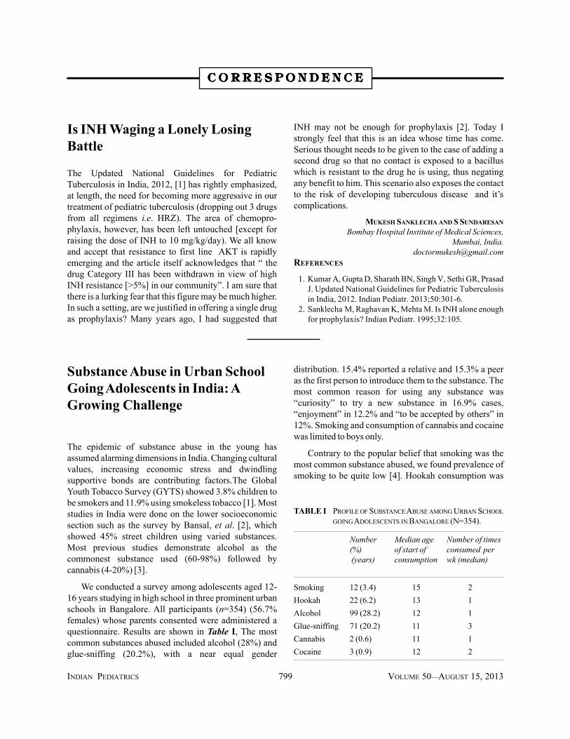
We conducted a survey among adolescents aged 12-16 years studying in high school in three prominent urbanschools in Bangalore. All participants (n=354) (56.7%females) whose parents consented were administered aquestionnaire. Results are shown in Table I, The mostcommon substances abused included alcohol (28%) andglue-sniffing (20.2%), with a near equal gender
distribution. 15.4% reported a relative and 15.3% a peeras the first person to introduce them to the substance. Themost common reason for using any substance was“curiosity” to try a new substance in 16.9% cases,“enjoyment” in 12.2% and “to be accepted by others” in12%. Smoking and consumption of cannabis and cocainewas limited to boys only.
Contrary to the popular belief that smoking was themost common substance abused, we found prevalence ofsmoking to be quite low [4]. Hookah consumption was
TABLE I PROFILE OF SUBSTANCE ABUSE AMONG URBAN SCHOOLGOING ADOLESCENTS IN BANGALORE (N=354).
Number Median age Number of times(%) of start of consumed per (years) consumption wk (median)
Smoking 12 (3.4) 15 2Hookah 22 (6.2) 13 1Alcohol 99 (28.2) 12 1Glue-sniffing 71 (20.2) 11 3Cannabis 2 (0.6) 11 1Cocaine 3 (0.9) 12 2

INDIAN PEDIATRICS 800 VOLUME 50__AUGUST 15, 2013
CORRESPONDENCE
tried and used by a significant number of adolescents.Inspite of a ban issued in Bangalore against Hookahcafes, they continue to thrive in the city and contribute toa huge number of children being addicted to the same.This; however, may not reflect the situation in other partsof India, as hookah consumption is closely linked to theavailability and presence of joints in the vicinity. We alsofound ‘sniffing’ being high prevalent among urbanadolescents. A previous review of all substance abuse inIndia has not reported this finding [5].
KR BHARATH KUMAR REDDY AND ASTHIK BISWASDepartment of Paediatrics,
Raju Multispeciality Hospital,CMR Layout, Lingarajapuram,
Bangalore 560 084, Karnataka, [email protected]
REFERENCES
1. Sinha DN, Reddy KS, Rahman K, Warren CW, Jones NR,Asma S. Linking Global Youth Tobacco Survey (GYTS)data to the WHO framework convention on tobaccocontrol: The case for India. Indian J PublicHealth. 2006;50:76-89.
2. Bansal RK, Banerjee S. Substance use in child labourers.Indian J Psychiatry. 1993;35:159-61.
3. Jena R, Shukla TR, Hemraj P. Drug abuse in a ruralcommunity in Bihar: Some psychosocialcorrelates. Indian J Psychiatry. 1996;38:43–6.
4. Narain R, Satyanarayana L. Tobacco use among schoolstudents in India: the need for behavioral change. IndianPediatr. 2005;42:732-3.
5. Murthy P, Manjunatha N, Subodh BN, Chand PK,Benegal V. Substance use and addiction research in India.Indian J Psychiatry. 2010;52:189-99.
Updated National Guidelines forPediatric Tuberculosis: ConcernsRegarding Neurotuberculosis
I read with interest the recent Updated Nationalguidelines for pediatric tuberculosis in India [1]. Thereare few important concerns in guidelines regardingneurotuberculosis which I wish to highlight.
First, there are discrepancies in dose ranges ofisoniazid, rifampicin and pyrazinamide from the latestWHO guidelines and should be corrected. WHOcurrently recommends the following daily doses ofantituberculosis medicines for the treatment oftuberculosis in children: isoniazid–10 mg/kg (range 10-15 mg/kg); rifampicin–15 mg/kg (range 10–20 mg/kg),pyrazinamide-35 mg/kg (30-40 mg/kg); ethambutol–20mg/kg (15-25 mg/kg) [2]. It is important as upper end ofthe recommended dose range should be considered inneurotuberculosis in view of uncertain penetration ofantituberculosis medicines into the central nervoussystem. The dose range suggested in published nationalguidelines probably follows WHO 2006 guidelines andshould be corrected according to WHO 2009 guidelines.Second concern is regarding the duration ofantitubercular therapy in neurotuberculosis. WHOrecommends that duration of antitubercular therapyshould be at least 12 months [3]. Similarly, a systemicreview also identifies that there is no evidence base forshorter duration regime [4]. So, the recommendation of
shorter 9 months duration is inappropriate and notevidence-based. Third, selection of third drug asethambutol in continuation phase of previously treatedcases has poor evidence-base with regard to neuro-tuberculosis. As pyrazinamide has better central nervoussystem penetration and bactericidal effect, it is probably abetter choice as the third drug in continuation phase ofpreviously treated cases.
Overall, I must congratulate authors for verycomprehensive guideline and I hope the revised versionwould focus on the concerns regarding neuro-tuberculosis.
JITENDER KUMAR SAHUAssistant Professor, Pediatric Neurology Division,
Department of Pediatrics,Post Graduate Institute of Medical Education & Research,
Chandigarh, India. [email protected]
1. Kumar A, Gupta D, Sharath BN, Singh V, Sethi GR,Prasad J. Updated National Guidelines for PediatricTuberculosis in India, 2012. Indian Pediatr. 2013;50:301-6.
2. World Health Organization. Dosing instructions for the useof currently available fixed-dose combination TBmedicines for children, 2009. Available from: http://www.who.int/tb/challenges/interim_paediatric_fdc_dosing_instructions_sept09.pdf.
3. World Health Organization. Rapid Advice 2010:Treatment of tuberculosis in children. Available from:http://apps.who.int/iris/bitstream/10665/44444/1/9789241500449_eng.pdf.
4. Prasad K, Sahu JK. Duration of anti-tubercular treatment intuberculous meningitis: challenges and opportunity.Neurol India. 2010;58:723-6.
INDIAN PEDIATRICS 801 VOLUME 50__AUGUST 15, 2013
CORRESPONDENCE
REPLY
In response to letter from Sahu, we wish to inform that:(a) the group extensively deliberated about appropriatedoses for anti-TB drugs to be recommended for ourcountry based on available evidence and concluded thatthe earlier recommended dosages needed revision. Thecurrent dosages were arrived after looking into variousavailable pharmacokinetic data and evidence within andoutside the country (published and unpublished data fromstudies at AIIMS and NIRT). The group arrived at theserecommendations as a consensus, while keeping in mindthe absolute need for adequate serum levels and also thepossible risk of cumulative hepatotoxicity; (b) the grouprecommends the total duration of ATT in intracranial TBincluding TB Meningitis should be 9-12 monthsdepending upon the clinical progress on treatment. Thisis in consonance with available evidence and experience;and (c) among retreatment cases, the INH resistance issignificant but not absolute, hence a third drug
ethambutol, is added to in the continuation phase (RHE).There is no scientific basis or evidence for includingpyrazinamide instead of ethambutol in the continuationphase. Pyrazinamide works best when there is activeinflammation and in acidic pH, hence its benefit may notbe seen during the continuation phase [1]. Furthermore,addition of Ethambutol not only helps in preventingemergence of drug resistance [2] but also would minimizethe potential risk of hepato-toxicity with prolonged useof the suggested three hepatotoxic drugs (RHZ).
VARINDER SINGH AND BN [email protected]
REFERENCE
1. Snider DE Jr. Controlled clinical trial of four 6-monthregimens of chemotherapy for pulmonary tuberculosis.Second East Africal/British Medical Research CouncilStudy. Am Rev Respir Dis. 1976; 114:471-75.
2. Wallace F. The chemotherapy of pulmonary tuberculosis: areview. Chest. 1979;76:785-96.
Updated National Guidelines forPediatric Tuberculosis in India,2012: Some Unresolved Issues
With respect to the recently published updated nationalguidelines for pediatric tuberculosis in India [1], we feelthat the following issues need to be clarified for thebenefit of practicing pediatricians.
1. Management algorithm (Fig.1a) describes thatsputum positive cases need not undergo a chest X-ray.While X-ray chest may not be necessary for diagnosisand initiation of treatment, it is vital for follow up anddetermination of duration of intensive phasetreatment.
2. The guideline says that “There is no role forinaccurate/inconsistent diagnostics like serology(IgM, IgG, IgA antibodies against MTB antigens),various in-house or non-validated commercial PCRtests and BCG test.” PCR is a very useful andpromising diagnostic test for tuberculosis [2-4],though the existing commercial PCRs are non-validated. Since the PCRs for TB are likely to bevalidated in near future, it should have beenmentioned separately rather than clubbing it withserological and BCG tests.
3. Management of childhood tuberculosis throughDOTS centre is programmatically logical, but if achild has to attend DOTS centre three days a week,then his/her academic performance, self-esteem andmainstreaming is likely to be compromised.Therefore we need to find a practical solution toaddress this very important issue. DOTS providersare not highly skilled workers, hence one of the viablesolutions could be to train school teachers assigned to‘medical room’ in most of the schools and give themthe responsibility of DOTS providers after initialregistration at DOTS center. This could significantlyminimize visits to DOTS centre.
4. The guidelines recommend INH prophylaxis to “Allasymptomatic contacts (under 6 years of age) of asmear positive case, after ruling out active diseaseand irrespective of their BCG, TST or nutritionalstatus.” This appears to be an overstatement whicharbitrarily puts the whole family under a cloud withconsequent social stigma and even partial failure incompliance by those who really need to take it.Secondly, are we justified to give single drugchemoprophylaxis with INH which has a resistancerate of >5% in our community? What would be theoverall impact on INH resistance?
5. INH chemoprophylaxis in all TST positive cases hasbeen recommended for 6 months. What is theevidence to support this conclusion? Duration of

INDIAN PEDIATRICS 802 VOLUME 50__AUGUST 15, 2013
CORRESPONDENCE
immunosuppressant drugs is variable ranging fromweeks to months. So, how can 6 monthschemoprophylaxis be universal?
6. The statement “a child born to mother who wasdiagnosed to have TB in pregnancy should receiveprophylaxis for 6 months, provided congenital TBhas been ruled out” has not been supported by clinicaland investigatory approach for of ruling outcongenital tuberculosis. It is of paramountimportance to diagnose a case of congenital TB andtreat as a new case as early as possible as untreateddisease is invariably fatal [5]. Therefore, diagnosticalgorithm of congenital TB must be included in theguidelines both for exclusion as well as for treatment.
CM KUMAR AND AK PATWARIDepartment of Pediatrics
Hamdard Institute of Medical Sciences and Research,Jamia Hamdard, New Delhi 110 062, India.
1. Kumar A, Gupta D, Sharath BN, Singh V, Sethi GR, PrasadJ. Updated National Guidelines for Pediatric Tuberculosisin India, 2012. Indian Pediatr. 2013; 50:301-6.
2. Lee HS, Park KU, Park JO, Chang HE, Song J, Choe G.Rapid, sensitive, and specific detection of Mycobacteriumtuberculosis complex by real-time PCR on paraffin-embedded human tissues. J Mol Diagn. 2011;13:390-4.
3. Peter JG, van Zyl-Smit RN, Denkinger CM, Pai M.Diagnosis of TB: State of the art. European RespiratoryMonograph. 2012:58:124-43.
4. Boehme CC, Saacks S, O’Brien RJ. The changinglandscape of diagnostic services for tuberculosis. SeminRes Crit Care Med. 2013;34:17-31.
5. Cantwell MF, Sehab ZM, Costello AM, et al. Brief report:congenital tuberculosis. New Engl J Med. 1994;330:1051-4.
REPLY
In reference to letter received from Kumar and Patwari,we would like to add that:
1. The current recommendations [1] highlight that thediagnosis of TB is most reliable with microbiologicalmethods and in such cases the findings on chestskiagrams usually do not help any further indiagnosis. Chest skiagram, however, may be done, fordetailing the pulmonary disease, depending upon thefeasibility.
2. They themselves have pointed out, the existing PCR-based tests available in most commercial laboratoriesare not reliable therefore these were clubbed with allother inaccurate diagnostic tests. With theadvancement in technologies, the guidance may be
revised in future as and when new tools or evidenceemerges. Cartridge-based nucleic amplification testis one such test currently being evaluated.
3. DOTS for new cases does not need a skilled person asthere are only oral drugs to be administered. Schoolbased DOTS may be an option but the limitedcapacities and lack of time or motivation with in theschool staff as well as the potential risk ofstigmatisation are the likely hurdles Also,partnerships for provision of directly supervisedtreatment must have a continued link with healthproviders to monitor the child for response to therapy,adverse events and management of other co-morbidities, including malnutrition. There is certainlya need to make DOTS more user-friendly for childrenand there is a need to pilot test to achieve innovativeout of the box alternatives (school based, home basedor neighbourhood DOTS).
4. INH prophylaxis is the only proven and establishedchemoprophylactic drug for tuberculosis [2-4]. Thecommittee after reviewing the scientific literature anddeliberating on programmatic implementation thecommittee opined that INH therapy should continueto be the mainstay of chemoprophylaxis in ourcountry; albeit at a higher dosage of 10 mg/kg bodyweight per day.
5. The prophylaxis is recommended for all asympto-matic contacts (children under the age of six years) ofsmear positive tuberculosis because (a) the exposureto an infectious case (which is usually a smearpositive TB case) is one of the strongest determinantfor the risk of infection, (b) and at a younger age therisk of developing disease after infection is very high.Though tuberculin skin test (TST) is performed toestablish infection, it may not be required when thereis a definite exposure. The current recommendationsmerely simplifies the mechanism to clinicallyidentify children, in the family/household, who arelikely to be recently infected.
The current evidence is for the post exposureprophylaxis and is recommended for six months. Thebenefit of a prolonged or continuous use of INHprophylaxis for TB, in a continued state ofimmunosuppression is not known. We, therefore,found it appropriate to recommend six monthsprophylaxis only for those cases who are found to beinfected at the first point when theimmunosuppressive therapy is started.
6. The recommendations clearly state the need to ruleout active disease before initiating any child onpreventive therapy including suspected perinatal
INDIAN PEDIATRICS 803 VOLUME 50__AUGUST 15, 2013
CORRESPONDENCE
cases. Congenital TB is suspected based uponclinical examination (hepatosplenomegaly with orwithout pneumonia), chest skiagrams, microbio-logical diagnosis and ultrasonology of the abdomenfor any hepatic granulomas, particularly in a neonateborn to a mother who is suffering from activetuberculosis.
VARINDER SINGH AND BN [email protected]
REFERENCES
1. Kumar A, Gupta D, Sharath BN, Singh V, Sethi GR,
Prasad J. Updated national guidelines for pediatrictuberculosis in India, 2012 Indian Pediatr. 2013;50:301-6.
2. Rapid advice: Treatment of TB in children. WHO 2010.3. Thee S, Seddon JA, Donald PR, Seifart HI, Werely CJ,
Hesseling AC, et al. Pharmacokinetics of isoniazid,rifampin, and pyrazinamide in children younger than twoyears of age with tuberculosis: evidence forimplementation of revised World Health Organizationrecommendations. Antimicrob Agents Chemother.2011;55:5560-7.
4. Menzies Dick, Al Jahdali H, Al Otaibi B. Recentdevelopments in treatment of latent tuberculosis infectionIndian J Med Res. 2011;133:257-66.
Pediatric Tuberculosis
I read the recent updated guidelines for pediatrictuberculosis in India with interest, and found them to beinformative. However, there may be practical difficulty inevaluating exact weight loss which has been defined asweight loss more than 5% of highest weight recorded in 3months [1]. Weight loss in terms of percentage can onlybe defined if previous weight of the child is known.Common presentation of children belonging to rural areais anorexia, fever and complain by parents of weight lossas measured from dress size.
What are suggestions of the authors regardinginterval between subsequent repetition of tuberculinsensitivity test as TST is being used as a tool to diagnosepediatric tuberculosis in conjunction with sputum andgastric lavage microscopy along with chest X-ray; everytime child presents with unexplained fever, anorexia andweight loss. Should it not be recommended to keep arecord of tuberculin sensitivity testing.
BALJINDER KAURAssistant professor, Department of Pediatrics
GMC and RH Patiala, Punjab, [email protected]
REFERENCE
1. Kumar A, Gupta D, Sharath BN, Singh V, Sethi GR,Prasad J. Updated guidelines for pediatric tuberculosis inIndia 2012. Indian Pediatr. 2013;50:302.
REPLY
In response to Kaur, we wish to state that (a) while it istrue that the weight records may not be available in manysituations but objectively defining these symptoms tocleanly identify disease suspect leads to a better yield as itwill improve the performance of the diagnosticalgorithm. In the event where the exact weight losscannot be quantified, one may still investigate for TB ifthe clinical suspicion is high; (b) prior TST testing, evenwhen repeated, is not considered likely to give rise tofalse positive reactions.
VARINDER SINGH AND BN [email protected]
Tuberculosis - A Quest TowardsObjectivity
I read with interest “updated National Guidelines forpediatric tuberculosis in India, 2012” and appreciate theeffort made to clarify certain grey areas of interpretationlike weight loss or no weight gain besides presenting thecontents as flow diagrams for ready reference [1]. I wouldlike to draw attention to certain points requiring furtherclarification to enable a clinician to use these guidelines
practically and effectively in a wider range of situations.
According to figure 1a and 1b, a symptomatic sputumnegative patient undergoes chest X-ray and TST.Following this, the possible results would be in six waysas per the outcome of these two investigations.
Chest X-ray can be read as: (a) Highly suggestive oftuberculosis, (b) Non- specific shadows (c) Normal; TSTcan be read as: (i) Positive, (ii) Negative. Though most ofthe possible scenarios are dealt with properly, it does notprovide an approach for (a+ii) that is highly suggestiveXRC and TST negative. Similarly it does not justify the

INDIAN PEDIATRICS 804 VOLUME 50__AUGUST 15, 2013
CORRESPONDENCE
use of TST when XRC shows non- specific shadows as nodecision is based on TST results whether positive ornegative.
CT scan is a useful diagnostic modality in childrenwhen tuberculosis is suspected and the radiographicfindings are normal or inconclusive [2]. Chest CT canhelp to identify enlarged, calcified, necrotic mediastinallymph nodes, which are less frequently found incommunity acquired bacterial pneumonia and frequentlyobscured by thymic shadows on chest radiographs ofchildren [3]. It may also detect pulmonary parenchymallesions not otherwise visualized on chest radiographs [4].Therefore, a TST positive, sputum negative clinicalsuspect in such scenario may be subjected to CT scanchest as first investigation (wherever possible) beforetaking on other investigation for alternate diagnosis.
As tuberculin skin test is defined with the use oftuberculin 2 TU and its procurement is difficult outsidegovernment supply, it would be useful to share themanufacturer of such product.
A GUPTADepartment of Pediatrics and Neonatology,
Fortis Hospital and Research Centre, Faridabad, [email protected]
REFERENCES
1. Kumar A, Gupta D, Sharath BN, Singh V, Sethi GR, PrasadJ. Updated National Guidelines for pediatric tuberculosis inIndia, 2012. Indian Pediatr. 2013;50:301-13.
2. Kim WS, Moon WK, Kim IO, Lee HJ, Im JG, Yeon KM, et al. Pulmonary tuberculosis in children: evaluation withCT. Am J Roentgenol. 1997;168:1005-9.
3. Peng SS, Chan PC, Chang YC, Shih TT. Computedtomography of childrenwith pulmonary Mycobacterium tuberculosis infection. J Formos Med Assoc. 2011;110:744-9.
4. Swaminathan S, Raghavan A, Datta M, ParamasivanCN, Saravanan KC. Computerized tomography detectspulmonary lesions in children with normal radiographsdiagnosed to havetuberculosis. Indian Pediatr. 2005;42:258-61.
REPLY
The author has raised the issue about dealing withchildren with highly suggestive radiology, who are TSTnegative. With certainty it is spelt out in the diagnosticalgorithm that a presumptive pediatric TB case with TSTnegative and chest X-ray findings suggestive of TBshould be diagnosed based on X-ray findings; because theTST suffers from lack of sensitivity and specificity, andthere are operational issues concerned with performingthe test efficiently. In view of radiation risk and issuespertaining to CT interpretation (low specificity and interobserver variability) [1,2] it is neither necessary norappropriate to recommend CT scan as first lineinvestigation. However, the algorithm has identifiedsituations where an expert opinion is needed and theymay ask for more detailed investigations including CTchest.
VARINDER SINGH AND BN [email protected]
REFERENCES
1. Andronikou S, Brauer B, Galpin J, Brachmeyer S, Lucas S,Joseph E, et al. Interobserver variability in the detection ofmediastinal and hilar lymph nodes on CT in children withsuspected pulmonary tuberculosis. Pediatr Radiol.2005;35:425-8.
2. de Jong PA, Nievelstein RJ. Normal mediastinal and hilarlymph nodes in children on multi-detector row chestcomputed tomography. Eur Radiol. 2012;22:318-21.