Two Critical Hits for Promyelocytic Leukemia
11
Molecular Cell, Vol. 6, 1131–1141, November, 2000, Copyright 2000 by Cell Press Two Critical Hits for Promyelocytic Leukemia toward cancer. However, in the clinically manifest tumor Li-Zhen He,* Mantu Bhaumik,* Carla Tribioli,* these multiple genetic events coexist, and it is, therefore, Eduardo M. Rego,* Sarah Ivins, Arthur Zelent, difficult to assess their relative contribution to tumori- and Pier Paolo Pandolfi* ‡ genesis. What is also unclear is whether two or more * Department of Human Genetics and Molecular Biology hits are not only concomitantly required to make the Program clonal population fully malignant, but are also a prerequi- Memorial Sloan-Kettering Cancer Center site for the tumor to acquire its distinctive and recogniz- Sloan-Kettering Institute able morphological and clinical features. New York, New York 10021 Leukemias are ideal model systems for addressing Leukaemia Research Fund Center at the Institute these issues for two reasons: (1) they have been recog- for Cancer Research nized and classified as clinical entities on the basis of Chester Beatty Laboratories morphological criteria, because the accumulating blast London SW3 6JB population is arrested at, and morphologically reminis- United Kingdom cent of, one of the differentiating steps through which the hemopoietic progenitor cell undergoes in order to reach functional maturity. This introduces a further ele- ment of analysis, which is the definition of the ability of a Summary leukemia-associated aberrant gene product to perturb/ block hemopoietic differentiation. While impaired cellu- Acute promyelocytic leukemia (APL) is associated with lar differentiation probably underlies the pathogenesis chromosomal translocations that always involve the of any cancer, in other tumor types this aspect is not RARa gene, which variably fuses to one of several often precisely recognizable. (2) Leukemic cells harbor distinct loci, including PML or PLZF (X genes). Due specific chromosomal translocations that are often re- to the reciprocity of the translocation, X-RARa and ciprocal, thus leading to the generation of two distinct RARa-X fusion proteins coexist in APL blasts. PLZF- aberrant fusion genes whose products coexist in the RARa transgenic mice (TM) develop leukemia that leukemic cell (reviewed in Melo et al., 1993; Look, 1997; lacks the differentiation block at the promyelocytic He et al., 1999; Melnick and Licht, 1999). This brings stage that characterizes APL. We generated TM ex- us to the following question: do these aberrant gene pressing RARa-PLZF and PLZF-RARa in their promy- products participate in leukemogenesis and in determin- elocytes. RARa-PLZF TM do not develop leukemia. ing response to therapy, and if so, how? Acute promy- However, PLZF-RARa/RARa-PLZF double TM de- elocytic leukemia (APL), the M3 subtype of acute my- velop leukemia with classic APL features. We demon- eloid leukemia (AML; reviewed in Warrell et al., 1993; strate that RARa-PLZF can interfere with PLZF tran- He et al., 1999; Melnick and Licht, 1999), is a unique scriptional repression and that this is critical for APL model system to use to address these questions, be- pathogenesis, since leukemias in PLZF 2/2 /PLZF- cause of its distinctive features: (1) the accumulation in RARa mutants and in PLZF-RARa/RARa-PLZF TM are the bone marrow (BM) of tumor cells that are arrested indistinguishable. Thus, both products of a cancer- at the promyelocytic stage of myeloid differentiation; (2) associated translocation are crucial in determining the the invariable association with specific reciprocal and distinctive features of the disease. balanced translocations involving the retinoic acid re- ceptor a (RARa) gene on chromosome 17, and the pro- Introduction myelocytic leukemia (PML), promyelocytic leukemia zinc finger (PLZF), nucleophosmin (NPM), nuclear mi- Oncogenesis in either solid tumors or hemopoietic ma- totic apparatus (NuMA), and signal transducer and acti- lignancies is commonly regarded as a multistep pro- vator of transcription 5B (STAT 5B) genes (X genes) on cess. This tenet entails that multiple genetic events pro- chromosomes 15q22, 11q23, 5q32, 11q13, and 17q11, gressively allow the premalignant cell to acquire full respectively; and (3) the exquisite sensitivity of APL neoplastic potential and, subsequently, in the case of blasts to the differentiating action of all-trans-retinoic solid tumors, the acquisition of metastatic potential. This acid (RA). RA overcomes the block of differentiation concept is at the basis of an active search for genes, at the promyelocytic stage, leading to complete, albeit commonly referred to as tumor modifiers, that play a role transient, disease remission—the reason for which APL in accelerating tumorigenesis. Genes whose aberrant has become the paradigm for cancer “differentiation activity or inactivation ultimately leads to full-blown therapy.” Strikingly, however, unlike other APL, leukemia transformation are commonly referred to as oncogenes associated with the t(11;17)/PLZF-RARa shows a dis- or tumor suppressor genes. In such a model for neoplas- tinctly worse prognosis, with little or no response to RA tic transformation, various genetic events are required treatment (Licht et al., 1995). in order to render a cell of a specific histological type The various APL specific translocations result in the malignant and invasive in a cumulative/additive model generation of X-RARa and RARa-X fusion genes and the co-expression in the leukemic blasts of their chimeric products, which are identical in their RARa moieties (He ‡ To whom correspondence should be addressed (e-mail: p-pandolfi@ ski.mskcc.org). et al., 1999; Melnick and Licht, 1999). X-RARa are able
Transcript of Two Critical Hits for Promyelocytic Leukemia
Molecular Cell, Vol. 6, 1131–1141, November, 2000, Copyright 2000 by Cell Press
Two Critical Hits for Promyelocytic Leukemia
toward cancer. However, in the clinically manifest tumorLi-Zhen He,* Mantu Bhaumik,* Carla Tribioli,*these multiple genetic events coexist, and it is, therefore,Eduardo M. Rego,* Sarah Ivins,† Arthur Zelent,†difficult to assess their relative contribution to tumori-and Pier Paolo Pandolfi*‡
genesis. What is also unclear is whether two or more*Department of Human Genetics and Molecular Biologyhits are not only concomitantly required to make theProgramclonal population fully malignant, but are also a prerequi-Memorial Sloan-Kettering Cancer Centersite for the tumor to acquire its distinctive and recogniz-Sloan-Kettering Instituteable morphological and clinical features.New York, New York 10021
Leukemias are ideal model systems for addressing†Leukaemia Research Fund Center at the Institutethese issues for two reasons: (1) they have been recog-for Cancer Researchnized and classified as clinical entities on the basis ofChester Beatty Laboratoriesmorphological criteria, because the accumulating blastLondon SW3 6JBpopulation is arrested at, and morphologically reminis-United Kingdomcent of, one of the differentiating steps through whichthe hemopoietic progenitor cell undergoes in order toreach functional maturity. This introduces a further ele-ment of analysis, which is the definition of the ability of aSummaryleukemia-associated aberrant gene product to perturb/block hemopoietic differentiation. While impaired cellu-Acute promyelocytic leukemia (APL) is associated withlar differentiation probably underlies the pathogenesischromosomal translocations that always involve theof any cancer, in other tumor types this aspect is notRARa gene, which variably fuses to one of severaloften precisely recognizable. (2) Leukemic cells harbordistinct loci, including PML or PLZF (X genes). Duespecific chromosomal translocations that are often re-to the reciprocity of the translocation, X-RARa andciprocal, thus leading to the generation of two distinctRARa-X fusion proteins coexist in APL blasts. PLZF-aberrant fusion genes whose products coexist in theRARa transgenic mice (TM) develop leukemia thatleukemic cell (reviewed in Melo et al., 1993; Look, 1997;lacks the differentiation block at the promyelocyticHe et al., 1999; Melnick and Licht, 1999). This bringsstage that characterizes APL. We generated TM ex-us to the following question: do these aberrant genepressing RARa-PLZF and PLZF-RARa in their promy-products participate in leukemogenesis and in determin-elocytes. RARa-PLZF TM do not develop leukemia.ing response to therapy, and if so, how? Acute promy-However, PLZF-RARa/RARa-PLZF double TM de-elocytic leukemia (APL), the M3 subtype of acute my-velop leukemia with classic APL features. We demon-eloid leukemia (AML; reviewed in Warrell et al., 1993;strate that RARa-PLZF can interfere with PLZF tran-He et al., 1999; Melnick and Licht, 1999), is a uniquescriptional repression and that this is critical for APLmodel system to use to address these questions, be-pathogenesis, since leukemias in PLZF2/2/PLZF-cause of its distinctive features: (1) the accumulation inRARa mutants and in PLZF-RARa/RARa-PLZF TM arethe bone marrow (BM) of tumor cells that are arrestedindistinguishable. Thus, both products of a cancer-at the promyelocytic stage of myeloid differentiation; (2)associated translocation are crucial in determining thethe invariable association with specific reciprocal anddistinctive features of the disease.balanced translocations involving the retinoic acid re-ceptor a (RARa) gene on chromosome 17, and the pro-Introductionmyelocytic leukemia (PML), promyelocytic leukemiazinc finger (PLZF), nucleophosmin (NPM), nuclear mi-
Oncogenesis in either solid tumors or hemopoietic ma- totic apparatus (NuMA), and signal transducer and acti-lignancies is commonly regarded as a multistep pro- vator of transcription 5B (STAT 5B) genes (X genes) oncess. This tenet entails that multiple genetic events pro- chromosomes 15q22, 11q23, 5q32, 11q13, and 17q11,gressively allow the premalignant cell to acquire full respectively; and (3) the exquisite sensitivity of APLneoplastic potential and, subsequently, in the case of blasts to the differentiating action of all-trans-retinoicsolid tumors, the acquisition of metastatic potential. This acid (RA). RA overcomes the block of differentiationconcept is at the basis of an active search for genes, at the promyelocytic stage, leading to complete, albeitcommonly referred to as tumor modifiers, that play a role transient, disease remission—the reason for which APLin accelerating tumorigenesis. Genes whose aberrant has become the paradigm for cancer “differentiationactivity or inactivation ultimately leads to full-blown therapy.” Strikingly, however, unlike other APL, leukemiatransformation are commonly referred to as oncogenes associated with the t(11;17)/PLZF-RARa shows a dis-or tumor suppressor genes. In such a model for neoplas- tinctly worse prognosis, with little or no response to RAtic transformation, various genetic events are required treatment (Licht et al., 1995).in order to render a cell of a specific histological type The various APL specific translocations result in themalignant and invasive in a cumulative/additive model generation of X-RARa and RARa-X fusion genes and the
co-expression in the leukemic blasts of their chimericproducts, which are identical in their RARa moieties (He‡ To whom correspondence should be addressed (e-mail: p-pandolfi@
ski.mskcc.org). et al., 1999; Melnick and Licht, 1999). X-RARa are able

Molecular Cell1132
to form heterodimeric complexes with their respectiveX protein. Similarly, the RARa portion retained inX-RARa is able to mediate heterodimerization with RXR,as well as DNA and ligand binding through the RARaRA and DNA binding domains (He et al., 1999; Melnickand Licht, 1999). Therefore, X-RARa have the ability topotentially interfer with both X and RAR/RXR pathways.By contrast, very little is known on the biochemical andbiological role of the various RARa-X fusion proteins.
We and others have previously reported that PML-RARa transgenic mice (TM) develop leukemia with strik-ing APL features, including the promyelocytic block ofmyeloid differentiation (Brown et al., 1997; Grisolano etal., 1997; He et al., 1997), while PLZF-RARa TM developmyeloid leukemia that is reminiscent of chronic myelog-enous leukemia (CML) (He et al., 1998), a different formof human myeloid leukemia that completely lacks thedistinctive differentiation block at the promyelocyticstage that characterizes APL. Here, we have generatedRARa-PLZF TM that we have intercrossed with PLZF-RARa TM to reconstruct the dual complexity of t(11;17)APL. We show that these two genetics events are con-comitantly required to recreate the disease in its unique-ness: while one of the two events is oncogenic, neitherevent by itself can cause a disease that can be recog-nized as APL. However, the combined action of bothmolecules results in leukemia with classic APL features.
Figure 1. Generation of RARa-PLZF TMWe demonstrate biochemically and genetically that
(A) Schematic representation of RARa-PLZF fusion cDNAs and theRARa-PLZF acts through its ability to functionally inter-hCG minigene expression vector and the structure of the hCG-
fere with PLZF. RARa-PLZF epitomizes a novel and dis- RARa-PLZF transgene fragment utilized for injection. The exons oftinct function in cancer etiopathogenesis. These aber- the hCG gene are designated as solid boxes. The promoter and therant proteins are not oncogenic per se, nor can they 59 flanking region, as well as the 39 flanking region of the hCG gene
are indicated. The RARa-PLZF cDNAs were cloned into the hCGaccelerate tumor onset unlike classic tumor modifiers.exon 1 in the artificial SalI site. Restriction endonuclease sites areHowever, they are required to determine the phenotypicas follows: N, NotI; B, BamHI; E, EcoRI; C, ClaI; S, SalI; P, PmlI; X,and clinical characteristics of the disease in its recogniz-XbaI; H, HindIII. The location of probes for Southern blot and North-able form. ern blot and primers for RT-PCR are shown underneath the structureof the injected fragment.
Results (B) Southern blot of mouse tail genomic DNA from the seven trans-genic founders and a control (6016) digested with EcoRI and hybrid-ized with probe CT reveals the expected bands of 4.0 Kb. A murineGeneration of RARa-PLZF TMp53 cDNA fragment was used as an internal control probe to normal-We generated TM in which the expression of the RARa-ize DNA loading by co-hybridization. The 15 Kb band identified byPLZF chimeric cDNA was under the control of a myeloid-this probe is indicated.
promyelocytic-specific human cathepsin-G (hCG) mini- (C) RT-PCR (left panel) and Northern blot analysis (right panel) ofgene (Figure 1A) (He et al., 1997; He et al., 1998). The hCG-RARa-PLZF fusion mRNA expression. Nested RT-PCR utilizingRARa-PLZF cDNA was subcloned into hCG gene exon two pairs of oligonucleotides (P[c] and Cat[b] and P[d] and Cat[c])
was done on RNA prepared from BM cells of representative mice1 (Figure 1A). Seven mice carrying the hCG-RARa-PLZFfrom five independent transgenic lines and WT control. A 483 bptransgene were identified (Figure 1B), harboring 2 to 36fragment is amplified from processed hCG-RARa-PLZF mRNA,copies of the transgene in a head-to-tail and/or tail-to-while a 1249 bp fragment is amplified from contaminating genomictail arrangement as evaluated by Southern blot analysisDNA that includes hCG intron 1. pGEM DNA markers are also shown.
(Figures 1A and 1B; Experimental Procedures). Five of Northern blot analysis was performed on total RNA from BM cellsthe seven TM transmitted the transgene to the germ hybridized with the human RARa-PLZF probe RP (see [A]): MH TM,line. Utilizing a nested reverse transcriptase–polymer- RARa-PLZF TM with overt myeloid hyperplasia; normal TM, RARa-
PLZF TM prior to the development of myeloid hyperplasia; WT, non-ase chain reaction (RT-PCR) approach that distinguishestransgenic mouse. An expected 1.9 Kb hCG-RARa-PLZF transcriptthe unprocessed gene from the processed cDNA (Figureis identified only in the diseased mouse. Prior to the onset of the1A), we demonstrated the expression of the RARa-PLZFdisease the transcript was not detectable by Northern blot due tofusion gene in the BM cells of all five founders’ progenythe small percentage (less than 5%) of early myeloid progenitors in
(Figure 1C). Northern blot and Western blot analyses on a normal marrow. The position and amount of 28S and the 18SBM samples obtained from RARa-PLZF TM and PLZF- ribosomal RNA are also shown.RARa/RARa-PLZF double TM confirmed the expressionof an RARa-PLZF fusion transcript/protein (Figure 1C;
Aberrant Hematopoiesis in RARa-PLZF TM3D). These TM lines were further expanded and moreAll five RARa-PLZF lines displayed a slow and progres-than 200 mice from the five lines that expressed thesive accumulation of myeloid cells in the BM and spleenRARa-PLZF trangene were followed up over a 30-month
period. (Figure 2A). The infiltrating cells retained the ability to

Tumor Metamorphosis1133
not affected (data not shown). After the first year oflife, approximately 20% of the mice developed overtsplenomegaly (Figure 2B), but leukemia was never ob-served. Flow-cytometric analysis confirmed: (1) the my-eloid nature of the cells accumulated in the BM andspleen of RARa-PLZF TM as revealed by the increasein Gr-1 (a marker expressed in various mature stagesof granulocytic differentiation) and Mac-1 (a marker formature monocytes/macrophages and granulocytes)positive cells (Figure 2C); and (2) that the accumulatingcellular population retained the ability to terminally dif-ferentiate (Figure 2C). Thus, RARa-PLZF can perturbmyelopoiesis, but it is not sufficient for inducing a blockin promyelocytic differentiation or leukemogenesis.
APL in PLZF-RARa/RARa-PLZF Double TMWe next intercrossed PLZF-RARa TM from two indepen-dent transgenic lines with RARa-PLZF mice from threeindependent lines to obtain double TM that were ana-lyzed on a comparative basis (Figure 3A). PLZF-RARaTM develop myeloid leukemia with CML-like hematolog-ical features at 100% of penetrance after 6 months ofage (He et al., 1998). Surprisingly, although hemopoiesisin RARa-PLZF TM is characterized by the hyperplasiaof the myeloid compartment, leukemia onset was unaf-fected in double TM. Leukemia still occurred after 6months with 100% penetrance (data not shown). Thus,RARa-PLZF does not function as a classic tumor mod-ifier.
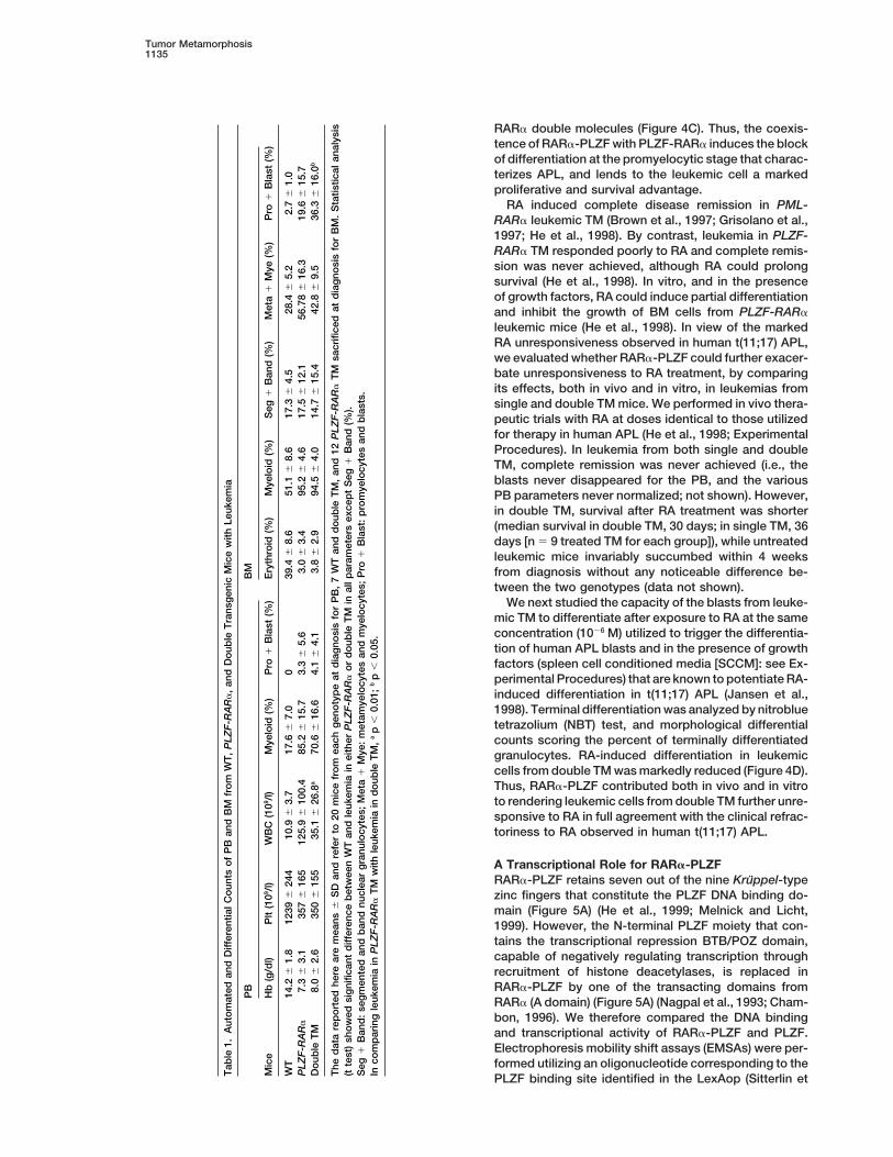
By contrast, the comparative analysis of the morpho-logical and hematological features of the leukemia indouble and single PLZF-RARa TM revealed striking dif-ferences (Table 1; Figures 3B and 3C). Leukemia inPLZF-RARa mice was characterized by leukocytosis inthe PB, and the infiltration in all organs by myeloid leuke-mic cells that fully retained the capacity to terminallymature, as revealed by differential counts on PB, BM,and spleen (Table 1; Figure 3B), but leukemia in doubleTM mice was characterized by a dramatic accumulationof immature blasts and of cells blocked at the promyelo-
Figure 2. Aberrant Hematopoiesis in RARa-PLZF TMcytic stage of differentiation (more than 50% of promye-
(A) Wright-Giemsa-stained BM and spleen cells from RARa-PLZFlocytes in BM in some cases) (Table 1; Figure 3B). Fur-TM and WT littermates. Differential counts and morphological analy-thermore, as in human APL, leukemia was characterizedsis showed a marked increase in myeloid components at variousby low platelet, anemia, and mildly increased whitestages of maturation in both BM and spleen of the TM. Magnification:
31000. blood cell (WBC) counts in the PB. Leukocytosis was(B) Splenomegaly in RARa-PLZF TM. The ratio between the spleen much less pronounced in double TM than in PLZF-RARaand body weight in the diseased RARa-PLZF TM was 0.85 6 0.06 TM. Flow-cytometric analysis revealed that leukemia in(n 5 9); WT mice, 0.30 6 0.01 (n 5 30).
double TM was characterized by a significant accumula-(C) Flow cytometrical analysis of BM and spleen (SP) cells fromtion of c-Kit1/CD341 and Gr-11/Mac-12 cells represent-RARa-PLZF TM and WT littermates. This analysis (n 5 9) confirmeding early myeloid progenitors in the BM and spleen (Fig-the accumulation of myeloid cells of various degree of maturation
(Gr-1, Mac-1, and Gr1/Mac1 positive) in both the BM and spleen of ure 3C) (BM cells from leukemic double TM [n 5 8] were:RARa-PLZF TM. c-Kit1/CD341, 37.17% 6 8.45%; Gr-11/Mac-12, 23.29% 6
9.71%; PLZF-RARa TM [n 5 8]: c-Kit1/CD341, 21.52% 615.77%; Gr-11/Mac-12, 15.08% 6 7.85%; WT [n 5 12]:c-Kit1/CD341, 2.05% 6 1.38%; Gr-11/Mac-12, 7.81% 6terminally differentiate toward mature granulocytes, as
shown by the fact that the differential counts in the BM 5.63%; comparable values were observed in the spleenof these mice), in keeping with the fact that myeloidand spleen did not reveal a consistent accumulation
of a specific immature cellular population, but instead differentiation is impaired in double TM leukemia, butcan proceed to term in PLZF-RARa leukemia. Subcuta-revealed progressive hyperplasia of the myeloid/granu-
locytic compartment in the absence of a block of differ- neous injection of leukemic BM and splenic cells intonude mice demonstrated that leukemias in both singleentiation (ratio of myeloid to lymphoid in BM: 4.99 6 2.7
in RARa-PLZF TM [n 5 9] versus 2.28 6 0.4 in wild type and double TM were transplantable (data not shown).Hematological and pathological follow-up of the double[WT; n 5 30]) (Figure 2A). The peripheral blood (PB) was

Molecular Cell1134
Figure 3. Characterization of PLZF-RARa/RARa-PLZF Double TM
(A) The interbreeding strategy and the respective phenotypes of the various transgenic lines are indicated.(B) Wright-Giemsa-stained PB and BM cells from PLZF-RARa and double leukemic mice. Magnification: 3400 for PB; 31000 for BM. Thenumber of WBC in PB is dramatically increased in the PLZF-RARa leukemic mice, while mildly increased in double TM. In the PLZF-RARa,leukemia myeloid cells at various stages of maturation replace the normal BM populations. In the double leukemia, a homogeneous populationof cells showing promyelocytic features infiltrates the BM.(C) Flow cytometric analysis on BM cells and splenocytes from PLZF-RARa, double leukemic mice, and control mice using antibodies againstthe cell surface markers Gr-1, Mac-1, c-Kit, and CD34. The green line shows the isotypic control for each sample. For each antibody, thepercentage of positive cells is given in the respective histogram.(D) Expression of PLZF-RARa and RARa-PLZF fusion proteins in double TM. Cell lysate was prepared from BM cells of double TM withleukemia (lane 1, a representative TM obtained by intercrossing line 5814 with line 6014; lane 2, a representative TM obtained by intercrossingline 6062 with line 5998) and WT mouse. 100 mg of protein were separated on 8% or 12% SDS-PAGE minigels and run in parallel forPLZF-RARa or RARa-PLZF, respectively. PLZF-RARa and RARa-PLZF were detected utilizing anti-RARa (RPa[F]) and anti-PLZF antibodies,respectively. b-actin revealed the amount of protein loaded.
TM revealed that, as for the PLZF-RARa and PML-RARa from double leukemic TM both in the presence or ab-sence of growth factors (Figure 4A). The clonogenicTM (He et al., 1997; He et al., 1998), leukemia was pre-capacity of normal hematopoietic precursors from bothceded by a myeloproliferative disorder characterized bysingle and double TM was markedly reduced, as shownthe accumulation in the BM and the spleen of immatureby the reduced number of colony-forming unit-granulo-myeloid cells and promyelocytes (data not shown).cyte, macrophage (CFU-GM), in agreement with the al-Thus, RARa-PLZF metamorphoses the CML-like leuke-most complete replacement of the normal hematopoi-mic phenotype observed in single PLZF-RARa TM intoetic precursors by the neoplastic population in the BMan APL-like phenotype.of the leukemic TM (Figure 4B; data not shown). Onthe contrary, the number of leukemic clusters (small
RARa-PLZF Modulates Proliferation, Apoptosis, colonies, 8–20 cells) characteristically observed in BMand Differentiating Response to RA cultures from AML patients (Moore et al., 1974) wasTreatment in Leukemic Cells markedly increased in cultures from leukemic cells fromTo further characterize the role of RARa-PLZF in leuke- double TM (Figure 4B). Both the overall rate of apoptosismogenesis in double TM we studied proliferation, apo- as well as growth factor deprivation-induced pro-ptosis, and differentiation in leukemic cells from single grammed cell death, as evaluated both by Annexin-V/and double TM. Thymidine incorporation assays in liquid PI and TUNEL stainings, were dramatically reduced in
leukemic cells harboring the RARa-PLZF and PLZF-cultures showed a higher proliferation rate in BM cells
Tumor Metamorphosis1135
RARa double molecules (Figure 4C). Thus, the coexis-tence of RARa-PLZF with PLZF-RARa induces the blockof differentiation at the promyelocytic stage that charac-terizes APL, and lends to the leukemic cell a markedproliferative and survival advantage.
RA induced complete disease remission in PML-RARa leukemic TM (Brown et al., 1997; Grisolano et al.,1997; He et al., 1998). By contrast, leukemia in PLZF-RARa TM responded poorly to RA and complete remis-sion was never achieved, although RA could prolongsurvival (He et al., 1998). In vitro, and in the presenceof growth factors, RA could induce partial differentiationand inhibit the growth of BM cells from PLZF-RARaleukemic mice (He et al., 1998). In view of the markedRA unresponsiveness observed in human t(11;17) APL,we evaluated whether RARa-PLZF could further exacer-bate unresponsiveness to RA treatment, by comparingits effects, both in vivo and in vitro, in leukemias fromsingle and double TM mice. We performed in vivo thera-peutic trials with RA at doses identical to those utilizedfor therapy in human APL (He et al., 1998; ExperimentalProcedures). In leukemia from both single and doubleTM, complete remission was never achieved (i.e., theblasts never disappeared for the PB, and the variousPB parameters never normalized; not shown). However,in double TM, survival after RA treatment was shorter(median survival in double TM, 30 days; in single TM, 36days [n 5 9 treated TM for each group]), while untreatedleukemic mice invariably succumbed within 4 weeksfrom diagnosis without any noticeable difference be-tween the two genotypes (data not shown).
We next studied the capacity of the blasts from leuke-mic TM to differentiate after exposure to RA at the sameconcentration (1026 M) utilized to trigger the differentia-tion of human APL blasts and in the presence of growthfactors (spleen cell conditioned media [SCCM]: see Ex-perimental Procedures) that are known to potentiate RA-induced differentiation in t(11;17) APL (Jansen et al.,1998). Terminal differentiation was analyzed by nitrobluetetrazolium (NBT) test, and morphological differentialcounts scoring the percent of terminally differentiatedgranulocytes. RA-induced differentiation in leukemiccells from double TM was markedly reduced (Figure 4D).Thus, RARa-PLZF contributed both in vivo and in vitroto rendering leukemic cells from double TM further unre-sponsive to RA in full agreement with the clinical refrac-toriness to RA observed in human t(11;17) APL.
A Transcriptional Role for RARa-PLZFRARa-PLZF retains seven out of the nine Kruppel-typezinc fingers that constitute the PLZF DNA binding do-main (Figure 5A) (He et al., 1999; Melnick and Licht,1999). However, the N-terminal PLZF moiety that con-tains the transcriptional repression BTB/POZ domain,capable of negatively regulating transcription throughrecruitment of histone deacetylases, is replaced inRARa-PLZF by one of the transacting domains fromRARa (A domain) (Figure 5A) (Nagpal et al., 1993; Cham-bon, 1996). We therefore compared the DNA bindingand transcriptional activity of RARa-PLZF and PLZF.Electrophoresis mobility shift assays (EMSAs) were per-formed utilizing an oligonucleotide corresponding to the
Tab
le1.
Aut
om
ated
and
Diff
eren
tialC
oun
tso
fP
Ban
dB
Mfr
om
WT
,P
LZF
-RA
Ra
,an
dD
oub
leT
rans
gen
icM
ice
with
Leuk
emia
PB
BM
Mic
eH
b(g
/dl)
Plt
(109 /
l)W
BC
(109 /
l)M
yelo
id(%
)P
ro1
Bla
st(%
)E
ryth
roid
(%)
Mye
loid
(%)
Seg
1B
and
(%)
Met
a1
Mye
(%)
Pro
1B
last
(%)
WT
14.2
61.
812
396
244
10.9
63.
717
.66
7.0
039
.46
8.6
51.1
68.
617
.36
4.5
28.4
65.
22.
76
1.0
PLZ
F-R
AR
a7.
36
3.1
357
616
512
5.9
610
0.4
85.2
615
.73.
36
5.6
3.0
63.
495
.26
4.6
17.5
612
.156
.78
616
.319
.66
15.7
Do
uble
TM
8.0
62.
635
06
155
35.1
626
.8a
70.6
616
.64.
16
4.1
3.8
62.
994
.56
4.0
14.7
615
.442
.86
9.5
36.3
616
.0b
The
dat
are
po
rted
here
are
mea
ns6
SD
and
refe
rto
20m
ice
fro
mea
chg
eno
typ
eat
dia
gno
sis
for
PB
,7
WT
and
do
uble
TM
,an
d12
PLZ
F-R
AR
aT
Msa
crifi
ced
atd
iag
nosi
sfo
rB
M.
Sta
tistic
alan
alys
is(t
test
)sh
ow
edsi
gni
fican
td
iffer
ence
bet
wee
nW
Tan
dle
ukem
iain
eith
erP
LZF
-RA
Ra
or
do
uble
TM
inal
lpar
amet
ers
exce
pt
Seg
1B
and
(%).
Seg
1B
and
:se
gm
ente
dan
db
and
nucl
ear
gra
nulo
cyte
s;M
eta
1M
ye:
met
amye
locy
tes
and
mye
locy
tes;
Pro
1B
last
:p
rom
yelo
cyte
san
db
last
s.In
com
par
ing
leuk
emia
inP
LZF
-RA
Ra
TM
with
leuk
emia
ind
oub
leT
M,
ap
,0.
01;
bp
,0.
05.
PLZF binding site identified in the LexAop (Sitterlin et

Molecular Cell1136
al., 1997). A DNA-protein complex was detected withwhole cell lysate from 293T cells transfected with eitherFLAGRARa-PLZF or PLZF (Figure 5B). The specificity ofthe LexAop-RARa-PLZF or LexAop-PLZF complexeswas demonstrated as they were completely competedaway by an excess of unlabeled LexAop oligo (Figure5B). Anti-PLZF and anti-FLAG antibodies (for RARa-PLZF) supershifted these complexes, demonstratingthat they contain PLZF and RARa-PLZF, respectively.We next transfected 293T cells with a luciferase reporterconstruct harboring the PLZF responsive element fromthe LexAop, and PLZF or RARa-PLZF expression vec-tors, and compared the ability of these proteins to re-press transcription. The basal activity of the reporterconstruct driven by an HSV-TK heterologous promoterwas repressed by PLZF in a dose-dependent manner(Figure 5C). RARa-PLZF completely lost the transcrip-tional repressive ability of PLZF (Figure 5C). Thus,RARa-PLZF may interfere with PLZF function at the tran-scriptional level through its ability to interact with PLZFresponsive elements.
The Functional Inactivation of PLZF Is a CriticalEvent in APL PathogenesisTo test genetically whether RARa-PLZF acts as a domi-nant negative inhibitor of PLZF function, we utilized micein which we had inactivated PLZF by homologous re-combination (Barna et al., 2000). We intercrossed PLZF-RARa TM with PLZF1/2 mice to obtain PLZF-RARa TMin the three distinct PLZF1/1, PLZF1/2 and PLZF2/2
backgrounds (n 5 20 per group), and characterized, ona comparative basis, leukemogenesis and hemopoiesisin littermates obtained from these crosses (Figure 6A).PLZF2/2 mice did not display defects in myeloid hemo-poiesis as evaluated by flow cytometric analysis, WBCcount, and differential counts performed in BM, PB, and
Figure 4. RARa-PLZF Controls Proliferation, Survival, and Differen- spleen in a 1-year follow up (not shown). PLZF-RARa/tiation of Leukemic Blasts PLZF1/1 mice obtained from these intercrosses devel-(A) RARa-PLZF increased the proliferative potential of leukemic oped “CML”-like leukemia, indistinguishable from thosecells. 3H-thymidine incorporation assays were performed on BM observed in our original PLZF-RARa TM (Tables 1 andcells from sex-age matched leukemic PLZF-RARa, double TM, and 2; Figures 3 and 6; He et al., 1998). Strikingly, however,WT littermates after a 24 hr incubation with 3H-thymidine. 1 3 106/
leukemia in PLZF-RARa/PLZF2/2 mice displayed identi-ml BM cells were incubated in DMEM medium supplemented withcal features to those in PLZF-RARa/RARa-PLZF double20% FBS, with or without 2% SCCM (GF1 and GF2). Means 6 SDTM, such as a dramatic accumulation of blast/promyelo-from triplicate values of one of three representative experiments are
shown. cytic cells in the BM and spleen and the absence of(B) RARa-PLZF enhances the formation of leukemic colonies in leukocytosis, as demonstrated by morphological andmethylcellulose colony forming assays. Colony formation assays flow cytometric analysis. Infiltrating cells were c-Kit, Gr-1,were performed on BM cells from sex-age matched leukemic PLZF-
Mac-1 positive, and displayed promyelocytic featuresRARa, double TM, and WT littermates. 3 3 104 cells were plated(PLZF-RARa/PLZF2/2 mice: c-Kit1, 34.17% 6 18.1%;per dish in 0.9% methylcellulose medium in triplicate. CFU-GM andGr-11/Mac-11, 59.4% 6 20.1%; PLZF-RARa/PLZF1/1leukemic clusters were scored at day 7. CFU-GM includes CFU-G,
M, and GM. Colony numbers (means 6 SD from triplicates) are from mice: c-Kit1, 15.2% 6 3.8%; Gr-11/Mac-11, 76.1% 6one of two experiments with similar results. 12.8%; data are means 6 standard deviation [SD] from(C) RARa-PLZF protects leukemic cells from apoptosis. BM cells n 5 3 mice for each group) (Table 2; Figures 6B andwere incubated in DMEM medium, supplemented with 20% FBS
6C). Moreover, leukemias in PLZF-RARa/PLZF1/2 miceand 2% SCCM, and apoptosis scored by Annexin V and TUNELdisplayed morphological and flow cytometric featuresstaining analyses after 48 hr of culture (GF1). Apoptosis upon
growth factor withdrawal was measured by culturing the cells withSCCM for 3 days as mentioned above and for 2 additional days inthe absence of factors (GF2). The percentages of viable cells(Annexin V2/PI2) (left panels; mean 6 SD from three experiments), (D) Differential response to RA in leukemic cells from double TM. BMand apoptosis as detected by in situ TUNEL staining (right panels; cells from PLZF-RARa and double TM with leukemia were incubatedpercent of positive cells: GF1: double TM, 5.2% 6 2%; single TM, with or without 1026M RA in the presence of 2% SCCM. Cytospins8.8% 6 1.6%; GF2: double TM, 34% 6 4%; single TM, 80% 6 from BM cells after a 4-day culture were stained with Wright-Giemsa.7.6%; means 6 SD of one of three experiments with similar results) Percentage (means 6 SD from two experiments) of mature (seg-are shown. mented) granulocytes and representative morphology are shown.

Tumor Metamorphosis1137
Figure 6. APL in PLZF-RARa/PLZF2/2 Mice
(A) The interbreeding strategy and the respective phenotypes of thetransgenic and knock-out lines are indicated.(B) Flow cytometry analysis on BM cells from PLZF-RARa/PLZF1/1
and PLZF-RARa/PLZF2/2 leukemic mice using a c-Kit antibody.The green line shows the isotypic control for each sample. Thepercentage of positive cells is given in the respective histogram.(C) Morphological characterization of PLZF-RARa/PLZF1/1 andPLZF-RARa/PLZF2/2 mice. Cytospins of BM cells from representa-tive PLZF-RARa/PLZF1/1 and PLZF-RARa/PLZF2/2 leukemic micewere stained with Wright-Giemsa. In the PLZF-RARa PLZF1/1 leuke-Figure 5. Aberrant Transcriptional Properties of the RARa-PLZF Fu-mia, myeloid cells at various stages of maturation replace the normalsion ProteinBM cells. In the PLZF-RARa/PLZF2/2 leukemia, a homogeneous
(A) Modular organization of RARa, PLZF, PLZF-RARa, RARa-PLZF population of cells showing promyelocytic features infiltrates thefusion proteins. RARa is subdivided into its conserved functional BM. Magnification: 31000.domains: C and E identify the DNA and ligand binding domains,respectively. D identifies the protein-protein interaction domain.PLZF contains nine zinc fingers, which mediate DNA binding, and
fected by the inactivation of PLZF, and mice in eachthe POZ domain, which allows protein-protein interaction and tran-scriptional repression activities. Black arrowheads indicate where group still developed leukemia after 6 months of latency.PLZF and RARa fuse to each other in APL. Thus, the progressive reduction of the PLZF dose meta-(B) EMSA shows binding of PLZF and FLAGRARa-PLZF to 32P-end- morphoses the CML phenotype observed in PLZF-labelled L4 probe (4x LexAop-derived probe). Proteins were ex- RARa leukemic mice into leukemia with classic APLpressed transiently in 293T cells. For the competition assays, a 100-
features.fold M excess of unlabelled L4 DNA (lanes 3 and 7) or 200-fold Mexcess of nonspecific CRE oligonucleotides (lanes 4 and 8) wasincluded in the binding reactions. For the antibody supershift experi- Discussionments, anti-PLZF (lane 5) or anti-FLAG (lane 9) monoclonal antibod-ies were used to confirm the presence of PLZF and FLAGRARa-PLZF,
Tumor Modifiers and Tumor Metamorphosersrespectively, in the retarded complexes. Arrowheads indicate theWe have demonstrated that two genetic events thatsuper-shifted complexes. One of three experiments is shown.occur simultaneously in human APL as a consequence(C) 293T cells were transiently transfected with L4-TKluc, indicated
amount of PLZF or RARa-PLZF expression plasmids, and 50 ng of a reciprocal chromosomal translocation are both nec-CMV-b2galactosidase as an internal control for transfection effi- essary, in combination, to cause the disease in its dis-ciency. Luciferase activity was measured 24 hr after transfection. tinctive form. Neither event is sufficient per se for theResults were normalized for b2galactosidase activity and are ex-
pathogenesis of a disease that would resemble APL.pressed as a percentage of reporter activity when transfected withLeukemia in double TM displayed features of classic300 ng vector pSG5 only (taken as 100%). Experiments were carriedAPL, closer to human APL than the leukemia observedout three times in duplicate; error bar represents SD.in PML-RARa TM (Grisolano et al., 1997; He et al., 1997).Several examples of multistep carcinogenesis havebeen described and elucidated (Kinzler and Vogelstein,in PB, BM, and spleen, which were intermediate to those
observed in PLZF1/1 and PLZF2/2 TM (not shown). Sur- 1998). These multiple events are required for full-blownmalignant transformation, and each event would be se-prisingly, as in double TM, leukemia onset was unaf-

Molecular Cell1138
lected because it favors cellular proliferation, survival,and tumor invasiveness. Mutations in strong tumor de-terminants, such as proto-oncogenes or tumor suppres-sors, would have catastrophic consequences inevitablyleading to tumorigenesis; while mutations in weaker de-terminants not able to transform on their own, the tumormodifiers, would accelerate the oncogenic process. Inthis model, neoplastic transformation is regarded as anadditive/quantitative process, as if there were a thresh-old value for neoplastic transformation that has to bereached by adding multiple transforming units. This sim-ple model is challenged by what is observed modelingt(11;17) APL in the mouse. In this case, one oncogenicevent, PLZF-RARa, already leads to leukemia at com-plete penetrance. In the aforementioned model for trans-formation, the PLZF-RARa provide several transformingunits that allow for the easy passage of the thresholdvalue through the accumulation of additional geneticevents in the 6-month latency period. However, the leu-kemia triggered by PLZF-RARa is not APL. RARa-PLZFis, therefore, a critical determinant of the leukemia phe-notype. In this process, RARa-PLZF acts neither as aclassic oncogene, since its activity is not leukemogenicper se, nor as a tumor modifier: RARa-PLZF metamor-phoses a CML-like phenotype into a leukemia similar tohuman APL, without affecting tumor onset. The mostrelevant implication of these findings is that they geneti-cally demonstrate the qualitative nature of the neoplas-tic process, an aspect that can be summarized by thealgorithm: (phenotype A, CML-like leukemia [PLZF-RARa] 1 phenotype B, myeloid hyperplasia [RARa-PLZF] 5 phenotype C, APL [PLZF-RARa 1 RARa-PLZF]). Phenotype C cannot simply be regarded as themere addition of the phenotypes observed in A and B.APL in double TM is the qualitatively novel biologicaloutcome of two concomitant aberrant activities on dis-tinctive pathways.
Furthermore, because PLZF-RARa does not requireRARa-PLZF for leukemogenesis, the formation of anaberrant RARa-PLZF fusion gene would not necessarilybe selected as a subsequent additional hit throughouttumor progression. Indeed, in APL, PLZF-RARa andRARa-PLZF are formed simultaneously as a conse-quence of a single reciprocal translocational event, andthe majority of t(11;17) APL patients are found to expressthe RARa-PLZF fusion gene, thus underscoring the rele-vance of the coexistence of these two products (Lichtet al., 1995; Licht et al., 1996; Grimwade et al., 1997). Itremains to be seen whether PLZF-RARa by itself couldcause a CML-like leukemia in humans as well, when notco-expressed with RARa-PLZF.
By contrast only 70% of t(15;17) APL expresses thereciprocal product (Alcalay et al., 1992; Chang et al.,1992; Grimwade et al., 1996). This is consistent with thephenotype observed in PML-RARa TM. In fact, PML-RARa can cause APL-like leukemia in mice in absenceof RARa-PML (Grisolano et al., 1997; He et al., 1997).However, the long latency period (more than 1 year) andthe low penetrance (10%–20% of TM) of these APL-likeleukemias suggest that additional events are requiredfor full-blown transformation. When expressed, RARa-PML could therefore act as a classic tumor modifierrequired for the acceleration of the leukemogenic pro-
Tab
le2.
Aut
om
ated
and
Diff
eren
tialC
oun
tso
fP
Ban
dB
Mfr
om
PLZ
F-R
AR
a/P
LZF
2/2
and
PLZ
F-R
AR
a/P
LZF
1/ 1
Mic
ew
ithLe
ukem
ia
PB
BM
Mic
eH
b(g
/dl)
Plt
(109 /
l)W
BC
(109 /
l)M
yelo
id(%
)P
ro1
Bla
st(%
)E
ryth
roid
(%)
Mye
loid
(%)
Seg
1B
and
(%)
Met
a1
Mye
(%)
Pro
1B
last
(%)
PLZ
F-R
AR
a/P
LZF
1/ 1
7.7
61.
756
26
320
147
611
6.4
77.8
65.
72.
26
1.1
3.1
62.
395
.36
6.2
22.2
615
.553
.66
13.2
19.4
67.
3P
LZF
-RA
Ra
/PLZ
F2
/ 212
.46
0.7
561
620
636
.16
39.3
a61
.86
12.4
4.3
63.
80.
46
0.1
95.3
62.
410
.36
1.1
48.1
610
.836
.86
6.4b
The
dat
are
po
rted
here
are
mea
ns6
SD
and
refe
rto
nine
and
six
for
PLZ
F-R
AR
a/P
LZF
1/ 1
and
PLZ
F-R
AR
a/P
LZF
2/ 2
mic
eat
dia
gno
sis,
resp
ectiv
ely.
Seg
1B
and
:se
gm
ente
dan
db
and
nucl
ear
gra
nulo
cyte
s;M
eta
1M
ye:
met
amye
locy
tes
and
mye
locy
tes;
Pro
1B
last
:p
rom
yelo
cyte
san
db
last
s.S
tatis
tical
anal
ysis
(tte
st):
ap
,0.
01;
bp
,0.
05.
cess. In fact, RARa-PML does not cause leukemia in

Tumor Metamorphosis1139
TM, but the co-expression of RARa-PML with PML- nick and Licht, 1999). The fact that RARa-PLZF can actas an aberrant version of PLZF at the transcription level,RARa increases the incidence and accelerates the onset
of leukemia (Pollock et al., 1999; our unpublished obser- and that its presence or the inactivation of PLZF causean APL-like phenotype in PLZF-RARa TM demonstratesvations).the importance of the blockade of PLZF function forAPL pathogenesis. On the other hand, these findingsThe Multiple Biological Functions of RARa-PLZFimply that if PLZF-RARa does interfere with the PLZFin APL Pathogenesispathway, this does not result in abrogation of PLZFWhen co-expressed with PLZF-RARa, RARa-PLZF canfunction. The concomitant interference of PLZF-RARainduce a block in myeloid/promyelocytic differentiation,and RARa-PLZF with the RARa/RXR pathways, respec-increase cellular proliferation, and enhance cell survival.tively, is therefore, necessary, albeit not sufficient, forEach of these functions is obviously critical for leukemo-APL pathogenesis.genesis. However, while the last two functions are com-
patible with the myeloid hyperplasia observed in RARa-Experimental ProceduresPLZF TM, the block of differentiation is not observed in
RARa-PLZF TM. Two possibilities can be entertainedConstruction of the Transgene
to reconcile this apparent paradox: (1) PLZF-RARa and The human RARa-PLZF cDNA (1 Kb) was cloned into the artificialRARa-PLZF could interfere with two distinct molecular SalI site of the hCG minigene expression vector, which was intro-pathways (see below), whose combined deregulation duced one base pair (bp) after the hCG ATG, which was previously
mutagenized. hCG minigene drives the expression of the fusionwould then lead to impaired myeloid differentiation; orcDNA, specifically in early stages of myeloid cell differentiation (He(2) an excessive proliferation rate of early myeloid pro-et al., 1997; He et al., 1998). A BamHI-NotI fragment from this con-genitors triggered by the combined activities of PLZF-struct was purified for egg injection as previously described (He et
RARa and RARa-PLZF could indirectly cause a block al., 1997; He et al., 1998).in promyelocytic differentiation, in agreement with thenotion that proliferation and differentiation of hemopoi- DNA, RNA, and Western Blot Analysesetic precursors are tightly and inversely interrelated (Liu Genomic tail DNA was digested with EcoRI or HindIII and hybridized
with probe CT, a 250-bp HindIII-XbaI hCG genomic fragment, andet al., 1996; Cheng et al., 2000).probe HS1, a 1-Kb hCG genomic fragment. A murine 2.3-Kb BamHIRARa-PLZF can also modulate response to RA.p53 cDNA probe was used to normalize for DNA loading and deter-t(11;17) APL patients do not respond to RA treatmentmine the transgene copy number. Total RNA was prepared from
(Licht et al., 1995). Leukemia in PLZF-RARa TM also mouse BM using Trizol reagents (Gibco BRL). Nested RT-PCR wasresponds poorly to RA treatment (He et al., 1998). How- performed using the primers shown in Figure 1A, which span cathep-ever, RA can induce partial response and prolong sur- sin G intron 1. For Northern blot analysis, denatured total RNA (20
mg) was hybridized with the human RARa-PLZF HpaII fragment asvival in PLZF-RARa leukemic TM. Furthermore, supra-a probe (RP probe, see Figure 1A) following standard procedures.pharmacological doses of RA (4-fold greater than theFor Western blot analysis, cell lysate was prepared from BM cellsone administered to human APL) can induce completeof leukemic mice and WT mice in E1A buffer as described (He et
albeit transient remission in PLZF-RARa leukemic TM as al., 1997; He et al., 1998). Total protein was then separated on 8%well (He et al., 1998). The further RA unresponsiveness or 12% sodium dodecyl sulfate polyacrylamide gel electrophoresisconferred by RARa-PLZF to the leukemic blasts both in (SDS-PAGE) minigels run in parallel for PLZF-RARa or RARa-PLZF,
respectively. PLZF-RARa was detected with the RPa(F) anti-RARavitro and in vivo not only explains the marked resistance(kindly provided by P. Chambon) and RARa-PLZF with an anti-PLZFto RA treatment observed in human t(11;17) APL, butantibody (Barna et al., 2000). The amount of protein loaded wasalso provides a further mechanism for the differentiationnormalized by reblotting the membranes with a b-actin antibody.
block observed in leukemia from double TM, in view ofthe important physiological role played by RA in control- Follow-Up of TMling normal myelopoiesis (Gudas et al., 1994; Chambon, Hematopoiesis in all five RARa-PLZF lines was studied over a 30-1996). By contrast, no apparent difference was observed month follow-up period. One group of mice was bled on a monthly
basis, together with age-matched littermate controls (60 TM and 30in RA sensitivity or clinical outcomes in t(15;17) APLcontrol mice). Automated and differential counts, as well as morpho-patients who do, or do not, harbor the RARa-PML fusionlogical analysis of PB cells, were performed on each sample. In atranscript (Grimwade et al., 1996; Li et al., 1997), or insecond group, four TM and four WT controls were sacrificed every
PML-RARa/RARa-PML double TM (Pollock et al., 1999). 2 months within the first year of life, and eight TM and four controlswere sacrificed every 2 months after the first year of life for grossand microscopic examination of all organs, and in particular PB,A Two Critical Hits for t(11;17) APLBM, spleen, liver, lymph nodes, and thymus. To obtain double TMPLZF-RARa can act as a dominant negative transcrip-we intercrossed two lines of PLZF-RARa (L5814 and L6062) withtional repressor of RARa, through aberrant nuclear co-three lines of RARa-PLZF (L5998, L6008 and L6014). Double TMrepressor/HDAC associations (Grignani et al., 1998;harboring PLZF-RARa, RARa-PLZF, and the single PLZF-RARa TM
Guidez et al., 1998; He et al., 1998; Lin et al., 1998). (over 100 for each genotype) were studied on a comparative basisPLZF-RARa can form, via its PLZF moiety, co-repressor as described above. To obtain PLZF-RARa TM in various PLZF
backgrounds, we intercrossed two PLZF-RARa TM lines (L5814 andcomplexes that are insensitive to RA. PLZF-RARa canL6062) with one PLZF1/2 line (Barna et al., 2000), and PLZF-RARa/also interfere with the function of the remaining normalPLZF1/1, PLZF-RARa/PLZF1/2, and PLZF-RARa/PLZF2/2 litter-RARa allele and other nuclear receptors through its abil-mates (20 for each genotype) were followed up on a comparativeity to homodimerize and heterodimerize with and se-basis.
quester RXR within the co-repressor/HDAC complex(Minucci et al., 2000; Salomoni and Pandolfi, 2000). Hematology, Histopathology, and ImmunohistochemistryPLZF-RARa can also physically interact with PLZF, pos- Mice were bled from the tail. WBC, hemoglobin (Hb) and platelet
(Plt) in PB were monitored utilizing an automated counter (Techniconsibly affecting its function as well (He et al., 1999; Mel-

Molecular Cell1140
H2). Differential counts of PB and BM were performed microscopi- Acknowledgmentscally on Wright-Giemsa-stained cytospin. For postmortem patholog-ical examination, sections from various organs were stained with We thank C. Cordon-Cardo, J.-H. Dong, J. Hung, T. Tolentino, and
L. Longo for advice, help with some experiments, and support; andhematoxylin and eosin. Myeloperoxidase (Dako), B220/CD45R, andCD3 antibodies (PharMingen) were used for immunohistochemical P. Chambon for the RARa antibody. L.-Z. H. was partially supported
by the Charles H. Revson Foundation. P. P. P. is a Scholar of thestaining on BM and spleen sections as described (He et al., 1997).The diagnosis of leukemia was made on the basis of the following Leukemia Society of America. This work was funded by grants from
the National Institutes of Health (CA 08748 and CA 74031, awardedconcomitant criteria: (1) WBC . 30,000/ml; (2) Hb , 10g/dl; and (3)Plt , 800,000/ml. Alternatively, when leukocytosis was absent the to P. P. P.).diagnosis was also made based on: (1) Hb , 6 g/dl and Plt ,
500,000/ml; (2) Hb , 3 g/dl; or (3) Plt , 300,000/ml. All these criteria Received May 10, 2000; revised September 15, 2000.were fulfilled in two consecutive weekly tests.
References
Flow Cytometry Alcalay, M., Zangrilli, D., Fagioli, M., Pandolfi, P.P., Mencarelli, A.,Cells were stained with fluorochrome-conjugated Mac-1 (CD11b), Lo Coco, F., Biondi, A., and Pelicci, P.G. (1992). Expression patternGr-1, CD34, c-kit, Sca-1, and CD3 as previously described (He et al., of the RARa/PML fusion gene in acute promyelocytic leukemia.1997; He et al., 1998). Fluorescein isothiocyanate or phycoerythrin Proc. Natl. Acad. Sci. USA 89, 4840–4844.conjugated isotypic antibodies were used as controls. All antibodies
Barna, M., Hawe, N.L., Niswander, and Pandolfi, P.P. (2000). Plzfwere obtained from PharMingen. Analysis was carried out using aregulates limb and axial skeletal patterning. Nature Genet. 25,FACScan flow cytometer (Becton-Dickinson).166–172.
Brown, D., Kogan, S., Lagasse, E., Weissman, I., Alcalay, M., Pelicci,P.G., Atwater, S., and Bishop, J.M. (1997). A PML/RARa transgeneEMSA and Transcriptional Assaysinitiates murine acute promyelocytic leukemia. Proc. Natl. Acad. Sci.PLZF and FLAGRARa-PLZF proteins were expressed transiently inUSA 94, 2551–2556.293T cells. Cellular extracts were incubated with a g32ATP-labeled
L4 probe, the 4x LexAop-derived sequence and electrophoresed Chambon, P. (1996). A decade of molecular biology of retinoic acidon a 4% polyacrylamide gel as described. 293T cells were transiently receptors. FASEB J. 10, 940–954.transfected using Superfect reagents (Qiagen). The total amount of
Chang, K.S., Stass, K.A., Chu, D.T., Deaven, L.L., Trujillo, J.M., andDNA transfected was 2 mg; the amount of expression vector (pSG5)
Freireich, E. (1992). Characterization of a fusion of cDNA (RARA/was kept constant in each transfection. Luciferase activity was mea-
myl) transcribed from the t(15;17) translocation breakpoint in acutesured 24 hr after transfection. Results were normalized for b-galac-
promyelocytic leukemia. Mol. Cell. Biol. 12, 800–810.tosidase activity.
Cheng, T., Rodrigues, N., Shen, H., Yang, Y., Dombkowski, D., Sykes,M., and Scadden, D.T. (2000). Hematopoietic stem cell quiescencemaintained by p21cip1/waf1. Science 287, 1804–1808.Proliferation, Apoptosis, and Clonogenic Assays
BM cells were cultured in Dulbecco’s modified Eagle’s medium Grignani, F., De Matteis, S., Nervi, C., Tomassoni, L., Gelmetti, V.,(DMEM) supplemented with 20% fetal bovine serum (FBS) with or Cioce, M., Fanelli, M., Ruthardt, M., Ferrara, F.F., Zamir, I., et al.without 2% pokeweed mitogen-stimulated SCCM (StemCell Tech- (1998). Fusion proteins of the retinoic acid receptor-alpha recruitnologies, Vancouver; containing IL3 and GM-CSF). Aliquots from histone deacetylase in promyelocytic leukaemia. Nature 391,each culture were harvested after 20 hr and 3H-thymidine was 815–818.added. After a further 24 hr incubation, incorporation of 3H-thymidine Grimwade, D., Howe, K., Langabeer, S., Davies, L., Oliver, F., Walker,was measured in a scintillation counter. Apoptosis analysis was H., Swirsky, D., Wheatley, K., Goldstone, A., Burnett, A., et al. (1996).performed after 48 hr of culture. Apoptosis upon growth factor with- Establishing the presence of the t(15;17) in suspected acute promy-drawal was scored after 3-days culture in the presence of SCCM elocytic leukaemia: cytogenetic, molecular and PML immunofluo-followed by 2 additional days of culture without SCCM. Cellular rescence assessment of patients entered into the M.R.C. ATRAapoptosis was analyzed by flow cytometry after propidium iodide trial. M.R.C. Adult Leukaemia Working Party. Br. J. Haematol. 94,(PI), an anti-Annexin V antibody (PharMingen) staining. Cytospin 557–573.cell preparations were also stained using a kit for TUNEL analysis
Grimwade, D., Gorman, P., Duprez, E., Howe, K., Langabeer, S.,(Boehringer Mannheim). 49, 6-diamidino-2-phenylindole was usedOliver, F., Walker, H., Culligan, D., Waters, J., Pomfret, M., et al.to reveal nuclei. At least 300 cells were scored under a fluorescence(1997). Characterization of cryptic rearrangements and variant trans-microscope for each sample. Clonogenic assays from BM cells werelocations in acute promyelocytic leukemia. Blood 90, 4876–4885.performed following the experimental conditions described in WangGrisolano, J.L., Wesselschmidt, R.L., Pelicci, P.G., and Ley, T.J.et al., 1998.(1997). Altered myeloid development and acute leukemia in trans-genic mice expressing PML-RARa under control of cathepsin Gregulatory sequences. Blood 89, 376–387.In Vivo RA Therapy
Preclinical trials with RA in PLZF-RARa/RARa-PLZF and PLZF- Gudas, L., Sporn, M., and Roberts, A. (1994). The retinoids: CellularRARa leukemic mice were carried out by administering RA per os biology and biochemistry of the retinoids. (New York: Raven Press),daily at a dose of 1.5 mg/g of mouse body weight. RA treatment was pp. 443–520.initiated at the onset of leukemia. Response to RA was monitored by Guidez, F., Ivins, S., Zhu, J., Soderstrom, M., Waxman, S., and Zel-performing weekly differential and automated counts on PB, in order ent, A. (1998). Reduced retinoic acid-sensitivities of nuclear receptorto monitor WBC, Plt, and red cell counts, and hb values, and to corepressor binding to PML- and PLZF-RARalpha underlie molecu-score for the presence of blasts in the PB. Untreated control groups lar pathogenesis and treatment of acute promyelocytic leukemia.consisted of leukemic mice from the same lines, which were also Blood 91, 2634–3642.bled once a week.
He, L.-Z., Tribioli, C., Rivi, R., Peruzzi, D., Pelicci, P.G., Soares,V., Cattoretti, G., and Pandolfi, P.P. (1997). Acute leukemia withpromyelocytic features in PML/RARa transgenic mice. Proc. Natl.In Vitro Differentiation Assays with RAAcad. Sci. USA 94, 5302–5307.Cells were cultured as for 3H-thymidine incorporation assays with
or without RA (1027M and 1026 M). At day 4, cells were harvested He, L.-Z., Guidez, F., Tribioli, C., Peruzzi, D., Ruthardt, M., Zelent,A., and Pandolfi, P.P. (1998). Distinct interactions of PML-RARalphafrom cultures and applied for morphological analysis, differential
count, and the NBT analyses as previously described (He et al., and PLZF-RARalpha with co-repressors determine differential re-sponses to RA in APL. Nature Genet. 18, 126–135.1997).

Tumor Metamorphosis1141
He, L.-Z., Merghoub, T., and Pandolfi, P.P. (1999). In vivo analysis Warrell, R.P., Jr., de The, H., Wang, Z.Y., and Degos, L. (1993). Acutepromyelocytic leukemia. New Engl. J. Med. 329, 177–189.of the molecular pathogenesis of Acute Promyelocytic Leukemia in
the mouse and its therapeutic implications. Oncogene 118, 5278–5292.
Jansen, J.H., de Ridder, M.C., Geertsma, W.M.C., Erpelinck, C.A.J.,van Lom, K., Smit, B., Slater, R. vd Reijden, B.A., de Greef, G.E.,Sonneveld, P., et al. (1998). Complete remission of t(11;17) positiveacute promyelocytic leukemia by all-trans retinoic acid and G-CSF.Blood 94, 39–45.
Kinzler, K.W., and Vogelstein, B. (1998). Familial cancer syndromes:The role of caretakers and gatekeepers. In The Genetic Basis ofHuman Cancer, B. Vogelstein and K.W. Kinzler, eds. (New York:McGraw-Hill), pp. 241–242.
Li, Y.P., Andersen, J., Zelent, A., Rao, S., Paietta, E., Tallman, M.S.,Wiernik, P.H., and Gallagher, R.E. (1997). RAR alpha1/RAR alpha2-PML mRNA expression in acute promyelocytic leukemia cells: amolecular and laboratory-clinical correlative study. Blood 90,306–312.
Licht, J.D., Chomienne, C., Goy, A., Chen, A., Scott, A., Head, D.R.,Michaux, J.L., Wu, Y., DeBlasio, A., Miller, W.H., Jr., et al. (1995).Clinical and molecular characterization of a rare syndrome of acutepromyelocytic leukemia associated with translocation (11;17). Blood85, 1083–1094.
Licht, J.D., Shaknovich, R., English, M.A., Melnick, A., Li, J.-Y.,Reddy, J.C., Dong, S., Chen, S.-J., Zelent, A., and Waxman, S. (1996).Reduced and altered DNA-binding and transcriptional properties ofthe PLZF-retinoic acid receptor-a chimera generated in t(11;17)-associated acute promyelocytic leukemia. Oncogene 12, 323–336.
Lin, R.J., Nagy, L., Inoue, S., Shao, W., Miller, W.H., Jr., and Evans,R.M. (1998). Role of the histone deacetylase complex in acute pro-myelocytic leukaemia. Nature 391, 811–814.
Liu, M. Lee, M.H., Cohen, M., Bommakanti, M., and Freedman, L.P.(1996). Transcriptional activation of the Cdk inhibitor p21 by vitaminD3 leads to the induced differentiation of the myelomonocytic cellline U937. Genes Dev. 10, 142–153.
Look, A.T. (1997). Oncogenic transcription factors in the humanacute leukemias. Science 278, 1059–1064.
Melnick, A., and Licht, J.D. (1999). Deconstructing a disease: RARal-pha, its fusion partners, and their roles in the pathogenesis of acutepromyelocytic leukemia. Blood 93, 3167–3215.
Melo, J.V., Gordon, D.E., Cross, N.C., and Goldman, J.M. (1993).The ABL-BCR fusion gene is expressed in chronic myeloid leukemia.Blood 81, 158–165.
Minucci, S., Maccarana, M. Cioce, M., De Luca, P., Gelmetti, V.,Segalla, S., Di Croce, L., Giavara, S., Matteucci, C., Gobbi, A., et al.(2000). Oligomerization of RAR and AML1 transcription factors asa novel mechanism of oncogenic activation. Mol. Cell 5, 811–820.
Moore, M.A., Spitzer, G., Williams, N., Metcalf, D., and Buckley, J.(1974). Agar culture studies in 127 cases of untreated acute leuke-mia: the prognostic value of reclassification of leukemia accordingto in vitro growth characteristics. Blood 44, 1–18.
Nagpal, S., Friant, S., Nakshatri, H., and Chambon, P. (1993). RARsand RXRs: evidence for two autonomous transactivation functions(AF-1 and AF-2) and heterodimerization in vivo. EMBO J. 12, 2349–2360.
Pollock, J.L., Westervelt, P., Kurichety, A.K., Pelicci, P.G., Grisolano,J.L., and Ley, T.J. (1999). A bcr-3 isoform of RARa-PML potentiatesthe development of PML-RARa-driven acute promyelocytic leuke-mia. Proc. Natl. Acad. Sci. USA 96, 15103–15108.
Salomoni, P., and Pandolfi, P.P. (2000). Transcription regulation ofcellular transformation. Nat. Med. 6, 742–744.
Sitterlin, D., Tiollais, P., and Transy, C. (1997). The RAR alpha-PLZFchimera associated with Acute Promyelocytic Leukemia has re-tained a sequence-specific DNA-binding domain. Oncogene 14,1067–1074.
Wang, Z.G., Delva, L., Gaboli, M., Zhang, H., Rivi, R., Cordon-Cardo,C., Grosveld, F., and Pandolfi, P.P. (1998). Role of PML in cell growthand the retinoic acid pathway. Science 279, 1547–1551.