tDCS over the left inferior frontal cortex improves speech production in aphasia
Upload
independentCategory
view
1download
0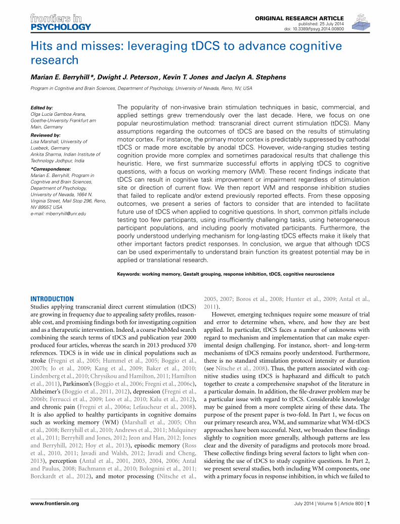
ORIGINAL RESEARCH ARTICLEpublished: 25 July 2014
doi: 10.3389/fpsyg.2014.00800
Hits and misses: leveraging tDCS to advance cognitiveresearchMarian E. Berryhill*, Dwight J. Peterson , Kevin T. Jones and Jaclyn A. Stephens
Program in Cognitive and Brain Sciences, Department of Psychology, University of Nevada, Reno, NV, USA
Edited by:
Olga Lucía Gamboa Arana,Goethe-University Frankfurt amMain, Germany
Reviewed by:
Lisa Marshall, University ofLuebeck, GermanyAnkita Sharma, Indian Institute ofTechnology Jodhpur, India
*Correspondence:
Marian E. Berryhill, Program inCognitive and Brain Sciences,Department of Psychology,University of Nevada, 1664 N.Virginia Street, Mail Stop 296, Reno,NV 89557, USAe-mail: [email protected]
The popularity of non-invasive brain stimulation techniques in basic, commercial, andapplied settings grew tremendously over the last decade. Here, we focus on onepopular neurostimulation method: transcranial direct current stimulation (tDCS). Manyassumptions regarding the outcomes of tDCS are based on the results of stimulatingmotor cortex. For instance, the primary motor cortex is predictably suppressed by cathodaltDCS or made more excitable by anodal tDCS. However, wide-ranging studies testingcognition provide more complex and sometimes paradoxical results that challenge thisheuristic. Here, we first summarize successful efforts in applying tDCS to cognitivequestions, with a focus on working memory (WM). These recent findings indicate thattDCS can result in cognitive task improvement or impairment regardless of stimulationsite or direction of current flow. We then report WM and response inhibition studiesthat failed to replicate and/or extend previously reported effects. From these opposingoutcomes, we present a series of factors to consider that are intended to facilitatefuture use of tDCS when applied to cognitive questions. In short, common pitfalls includetesting too few participants, using insufficiently challenging tasks, using heterogeneousparticipant populations, and including poorly motivated participants. Furthermore, thepoorly understood underlying mechanism for long-lasting tDCS effects make it likely thatother important factors predict responses. In conclusion, we argue that although tDCScan be used experimentally to understand brain function its greatest potential may be inapplied or translational research.
Keywords: working memory, Gestalt grouping, response inhibition, tDCS, cognitive neuroscience
INTRODUCTIONStudies applying transcranial direct current stimulation (tDCS)are growing in frequency due to appealing safety profiles, reason-able cost, and promising findings both for investigating cognitionand as a therapeutic intervention. Indeed, a coarse PubMed searchcombining the search terms of tDCS and publication year 2000produced four articles, whereas the search in 2013 produced 370references. TDCS is in wide use in clinical populations such asstroke (Fregni et al., 2005; Hummel et al., 2005; Boggio et al.,2007b; Jo et al., 2009; Kang et al., 2009; Baker et al., 2010;Lindenberg et al., 2010; Chrysikou and Hamilton, 2011; Hamiltonet al., 2011), Parkinson’s (Boggio et al., 2006; Fregni et al., 2006c),Alzheimer’s (Boggio et al., 2011, 2012), depression (Fregni et al.,2006b; Ferrucci et al., 2009; Loo et al., 2010; Kalu et al., 2012),and chronic pain (Fregni et al., 2006a; Lefaucheur et al., 2008).It is also applied to healthy participants in cognitive domainssuch as working memory (WM) (Marshall et al., 2005; Ohnet al., 2008; Berryhill et al., 2010; Andrews et al., 2011; Mulquineyet al., 2011; Berryhill and Jones, 2012; Jeon and Han, 2012; Jonesand Berryhill, 2012; Hoy et al., 2013), episodic memory (Rosset al., 2010, 2011; Javadi and Walsh, 2012; Javadi and Cheng,2013), perception (Antal et al., 2001, 2003, 2004, 2006; Antaland Paulus, 2008; Bachmann et al., 2010; Bolognini et al., 2011;Borckardt et al., 2012), and motor processing (Nitsche et al.,
2005, 2007; Boros et al., 2008; Hunter et al., 2009; Antal et al.,2011).
However, emerging techniques require some measure of trialand error to determine when, where, and how they are bestapplied. In particular, tDCS faces a number of unknowns withregard to mechanism and implementation that can make exper-imental design challenging. For instance, short- and long-termmechanisms of tDCS remains poorly understood. Furthermore,there is no standard stimulation protocol intensity or duration(see Nitsche et al., 2008). Thus, the pattern associated with cog-nitive studies using tDCS is haphazard and difficult to patchtogether to create a comprehensive snapshot of the literature ina particular domain. In addition, the file-drawer problem may bea particular issue with regard to tDCS. Considerable knowledgemay be gained from a more complete airing of these data. Thepurpose of the present paper is two-fold. In Part 1, we focus onour primary research area, WM, and summarize what WM-tDCSapproaches have been successful. Next, we broaden these findingsslightly to cognition more generally, although patterns are lessclear and the diversity of paradigms and protocols more broad.These collective findings bring several factors to light when con-sidering the use of tDCS to study cognitive questions. In Part 2,we present several studies, both including WM components, onewith a primary focus in response inhibition, in which we failed to
www.frontiersin.org July 2014 | Volume 5 | Article 800 | 1
Berryhill et al. tDCS and cognition
consider one or more of these factors and failed to observe signif-icant effects. The goal of this article is to facilitate tDCS researchin cognition in healthy individuals by sharing what has workedreliably, what has failed, and what lessons we have extracted.
PART 1: tDCS SUCCESSESWHAT WORKS IN WORKING MEMORY?WM plays a significant role in many cognitive tasks and iscontrolled by broad frontoparietal networks accessible to tDCS.These features have made it attractive to researchers interested inapplying tDCS to studies of cognition. This approach has beensuccessful. The most consistent set of WM data comes from theuse of n-back tasks paired with left dorsolateral prefrontal cor-tex (DLPFC) stimulation in healthy (Ohn et al., 2008; Andrewset al., 2011; Mulquiney et al., 2011; Zaehle et al., 2011) andspecial populations (Boggio et al., 2006; Jo et al., 2009); for a
recent meta-analysis see (Brunoni and Vanderhasselt, 2014); seeTable 1. In short, these findings reveal a robust pattern: anodaltDCS to the left DLPFC improves verbal n-back task performancewhen compared to either sham or cathodal tDCS. This pattern oftDCS-linked WM improvement remains constant across a varietyof stimulus intensities, durations, and participant populations.Indeed, recent meta-analysis looking at these studies identified areliable reaction time improvement during active tDCS in thosestudies using the n-back and stimulating the DLPFC (Brunoniand Vanderhasselt, 2014, but see also Jacobson et al., 2012).
This consistency highlights at least one notable exception.When we tested healthy older adults in a verbal and visual 2-backtask we replicated the anodal tDCS benefit, but only in those withmore education (Berryhill and Jones, 2012). Surprisingly, thosewith less than a college-degree received no benefit from tDCS.Instead their performance revealed a nearly equal and opposite
Table 1 | Peer-reviewed studies of WM paired with tDCS.
Authors Task N Site mA Dur (min) Comparison
IMPROVED WM AFTER ANODAL tDCS TO THE DLPFC: n-BACK TASK
Andrews et al.,2011
2-, 3-bk, verbal, digit span 10 L DLPFC, during orbefore task
1 10 During: digit span: A > S
Berryhill andJones, 2012
Visual, verbal 2-bk 12*, OA L, R DLPFC 1.5 10 L and R: A > S, in moreeducated
Boggio et al., 2006 Verbal 3-bk 18, PD L DLPFC, M1 1, 2 20 DLPFC: 2 mA: A > S
Fregni et al., 2005 Verbal 3-bk 15 L DLPFC, M1 1 10 L DLPFC: A > S
Jo et al., 2009 Verbal 2-bk 10, R. stroke L DLPFC 2 30 Pre/post differences: A >
S
Hoy et al., 2013 Verbal 2-, 3-bk 18 L DLPFC 1, 2 20 RT: A > S: 2-bk: 1 mAfaster than 2 mA
Kim et al., 2014 Verbal 3-bk 9/8* L DLPFC 1 20 A > S: N = 9 benefited,N = 8 no effect
Mulquiney et al.,2011
2-bk, Sternberg 10 L DLPFC 1 10 2-back: A > S
Ohn et al., 2008 3-bk, Korean letters 15 L DLPFC 1 30 Pre/post: A > S
Zaehle et al., 2011 Verbal 2-bk 16 L DLPFC 1 15 A > C
IMPROVED WM AFTER ANODAL tDCS: MIXED SITES, TASKS
Boggio et al., 2009 Visual recognition 10, AD L DLPFC, L temporal 2 30 L DLPFC: A > S
Jones andBerryhill, 2012
E1. WM Changedetection, sequential; E2.WM Change detection
E1: 10*; E2: 14* R PPC 1.5 10 E1: High WMC: A,C > SLow WMC; S > A,C; E2:High WMC: A,C > S
Tanoue et al., 2013 Pre-cue, retro-cue WM 23 R PPC, R PFC 1.5 10 Pre-cue: S > C, PPC =PFC; Retro-cue: S > C:PPC > PFC
Tseng et al., 2012 Change detection 10* R PPC 1.5 15 Low WMC: A > S
IMPAIRED WM AFTER tDCS: MIXED SITES, TASKS
Berryhill et al.,2010
Sequential WM,recognition, recall
11 R PPC 1.5 10 Recognition: S > C
Ferrucci et al.,2008
Sternberg 13 Cerebellum 2 15 S > A, C: Impairedpractice benefits
Marshall et al.,2005
Sternberg 12 L, R DLPFC 260 μA P15 RT: Stim slower thansham
Abbreviations: A, anodal; AD, Alzheimer’s disease; bk, back; C, cathodal; Dur, duration of stimulation in minutes; E, experiment; L, left; mA, tDCS strength in
milliamperes; N, number of participants; P, pulsed at 15 s on/15 s off, R, right; S, sham; OA, older adults; PD, Parkinson’s disease. All findings pertain to accuracy
unless reference to reaction time is noted. In cases where the between-subjects effects were significant, N refers to group size and is noted with an *.
Frontiers in Psychology | Cognitive Science July 2014 | Volume 5 | Article 800 | 2
Berryhill et al. tDCS and cognition
decrement in performance. Furthermore, regardless of whetherthe stimuli were verbal or visual and whether stimulation sitewere the left or the right the differences in education predictedwhether tDCS helped or hurt WM performance. This was the firstpaper, to our knowledge, showing that an inhomogeneous popu-lation significantly modulated tDCS effects in different directions.A recent paper has used current modeling to clarify that partici-pants only benefited on a verbal 3-back WM task when the tDCSapplying DLPFC during a verbal 3-back task current modelingindicates that those who benefited showed significant modula-tion in the DLPFC current, but those who did not improved didnot show significant DLPFC modulation (Kim et al., 2014). Theyattributed these data to morphological differences in brain struc-ture and where current flow went. This finding reveals the needfor refining stimulation targeting by registering an individual’sMRI scans with tDCS electrode placement as is the practice inTMS research. However, their data also showed that those whostarted with higher WM performance garnered greater benefitsfrom tDCS.
There are several other papers pairing tDCS with different WMtasks (e.g., old/new recognition, recall, change detection) andparietal stimulation sites. These data are less clear. For example,we found that anodal tDCS to the right posterior parietal cor-tex (PPC: P4) selectively interfered with WM probed by old/newrecognition, but not with WM probed by recall (Berryhill et al.,2010). We subsequently found that when participants performedtwo WM tasks of different difficulty levels in the same session,that tDCS effects were only apparent for the more challengingtask (Jones and Berryhill, 2012). Importantly, here, again, therewere significant and opposing patterns in the data such that youngadults with high WM span benefited but those with low WM spanperformance was impaired after cathodal or anodal tDCS to theright PPC. In contrast, a similar study also applying anodal tDCSto the right PPC reported that those with low WM performanceon the sham day performed better during the anodal session ona challenging change detection task (Tseng et al., 2012). However,this study lacked an independent measure of WM capacity to seg-ment their participants. Instead, behavioral performance duringthe sham session was used. Thus, the observed effects are contam-inated by regression to the mean because poor performers duringthe sham session were likely to perform better at another sessionregardless of tDCS presence. However, here, again are several datapoints indicating that population differences predict the directionand magnitude of tDCS effects on WM.
A second issue buried in these data is that effects are apparentwhen tasks are difficult. Apart from the WM papers just notedthere is at least one other analysis that has found that tDCS effectswere apparent only when the task demands were difficult. Morespecifically, in an associative memory task participants learnedface-name and place-name pairs and received left or right anodaltDCS. In younger and older adults, tDCS provided a performancebenefit only when the participants struggled to produce the cor-rect face or place name, as evidenced by long reaction times (Rosset al., 2010, 2011). One way to think about this in terms of tDCSis that the extra stimulation can serve as a tipping factor. This isconsistent with our understanding that tDCS induces changes thechanges in neuronal excitability—cells become more (anodal) or
less (cathodal) likely to fire action potentials. When tasks are easy,the outcome is clear and the addition of tDCS does not changeperformance. However, when tasks or even trials are very difficult,tDCS effects emerge. When designing a task to pair with tDCS, itis worth ensuring that the task demands are sufficiently challeng-ing for participants and/or that the more challenging trials can beisolated for separate analysis.
A third emerging issue that becomes more apparent whenreviewing the tDCS-WM papers is that the effect sizes tend to besmall and the studies are underpowered. For example, althoughearly studies report significant effects with 10 participants, morerecent papers tend to include 20 or more participants. One spec-ulation is that early studies tapped homogeneous populations,presumably available lab personnel, to participate and this meantresulted in more consistent performance and tDCS patterns.
A related concern is that as laboratories become more com-fortable with the tDCS technique they are subject to added noisefrom poorly motivated participants. Although this is a problemfor many experimental techniques, it may be particularly relevantif unmotivated participants do not engage during challengingtasks or during challenging trials. As mentioned above, the sub-tle response-shifts induced by tDCS may be particularly sensitiveto contamination from poorly motivated participants. However,this notion must be considered as speculative because there areno explicit data testing the role that motivation plays in tDCSdesigns, although we are currently testing this hypothesis.
WHAT WORKS IN COGNITION?Apart from WM, there is broad use of tDCS to investigate wide-ranging cognitive topics (for a recent review see Coffman et al.,2012). A real challenge is the diversity in experimental paradigmsand tasks makes it difficult to identify consistent patterns in thetDCS literature. Furthermore, a recent review paper highlighted“foundational” problems associated with tDCS and the impactof variability across participants, issues associated with cognitiveset and performance, the reliability of effects over time and cur-rent dynamics (Horvath et al., 2014; see also Lopez-Alonso et al.,2014). However, in a few areas of upper-level cognitive domainssome consistency is beginning to emerge. In Table 2, we providean admittedly incomplete survey of cognitive studies employingtDCS reporting significant effects in healthy adults. As in WM,the majority of cognitive studies target frontal stimulation sitesand thus, it may not be surprising that the papers describing sig-nificant effects of tDCS relate to upper-level cognitive tasks (e.g.,response inhibition, memory, decision making). From these scat-tered findings, it can be difficult to predict the direction of effectsin a tDCS study and it can be difficult to know why the effectsare as they are. As noted above, these are the studies with positivefindings and likely there are many other null findings that wouldbe informative for the research population. In the following sec-tion, we raise several points to consider when developing tDCSstudies of cognition.
CONSIDERATIONS WHEN APPLYING tDCS TO COGNITIVEQUESTIONSHere, we summarize some of our observations as a set of fac-tors to consider before using tDCS in a cognitive experiment.
www.frontiersin.org July 2014 | Volume 5 | Article 800 | 3
Berryhill et al. tDCS and cognition
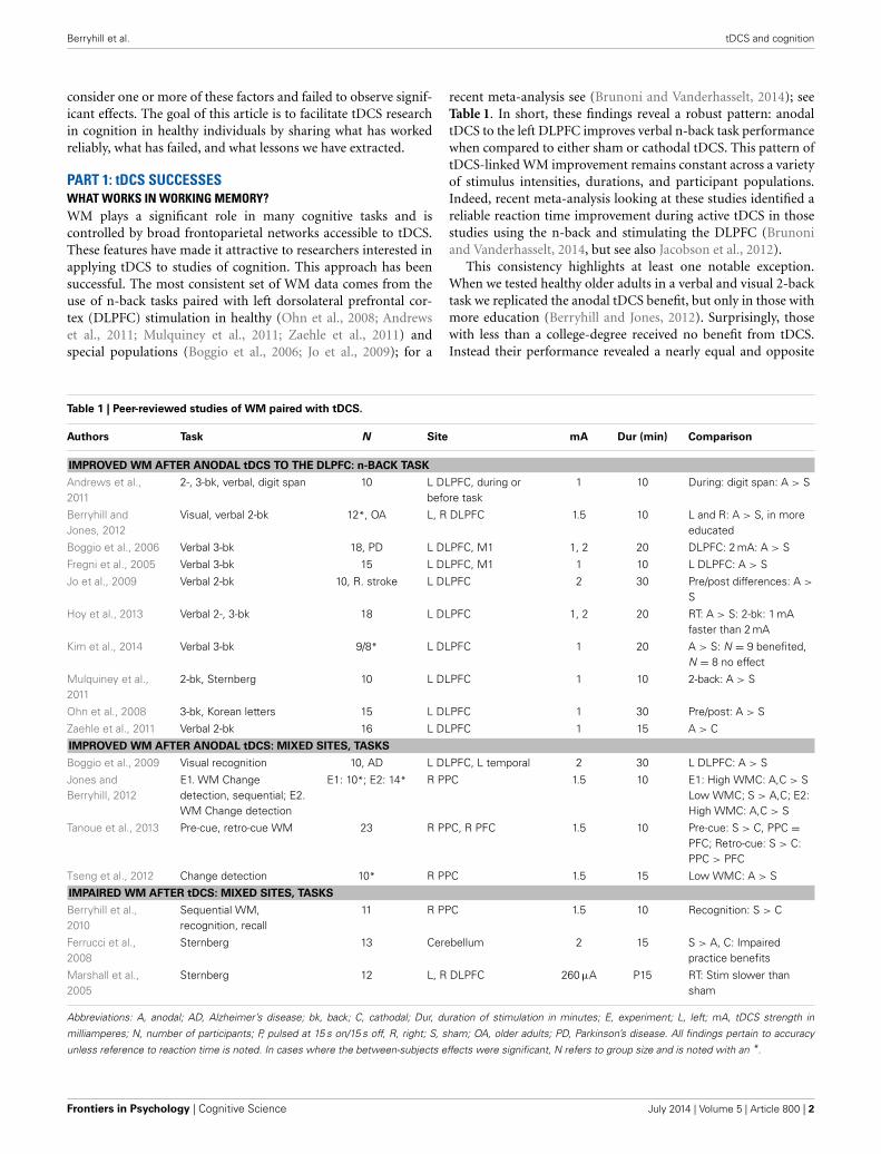
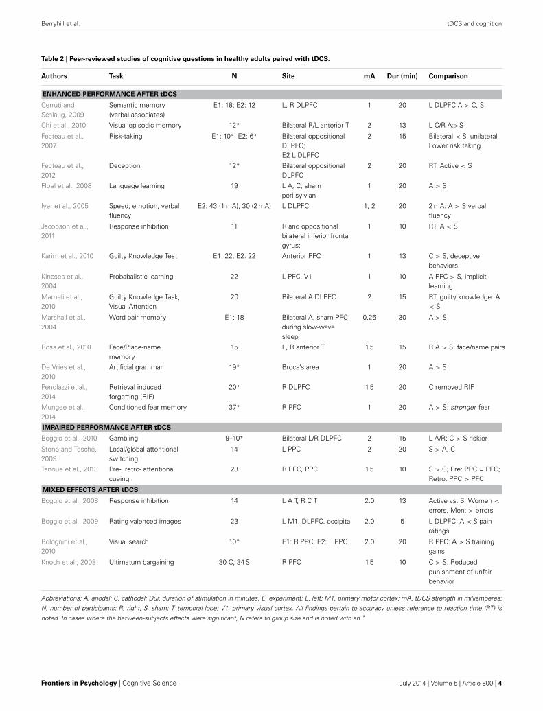
Table 2 | Peer-reviewed studies of cognitive questions in healthy adults paired with tDCS.
Authors Task N Site mA Dur (min) Comparison
ENHANCED PERFORMANCE AFTER tDCS
Cerruti andSchlaug, 2009
Semantic memory(verbal associates)
E1: 18; E2: 12 L, R DLPFC 1 20 L DLPFC A > C, S
Chi et al., 2010 Visual episodic memory 12* Bilateral R/L anterior T 2 13 L C/R A:>S
Fecteau et al.,2007
Risk-taking E1: 10*; E2: 6* Bilateral oppositionalDLPFC;E2 L DLPFC
2 15 Bilateral < S, unilateralLower risk taking
Fecteau et al.,2012
Deception 12* Bilateral oppositionalDLPFC
2 20 RT: Active < S
Floel et al., 2008 Language learning 19 L A, C, shamperi-sylvian
1 20 A > S
Iyer et al., 2005 Speed, emotion, verbalfluency
E2: 43 (1 mA), 30 (2 mA) L DLPFC 1, 2 20 2 mA: A > S verbalfluency
Jacobson et al.,2011
Response inhibition 11 R and oppositionalbilateral inferior frontalgyrus;
1 10 RT: A < S
Karim et al., 2010 Guilty Knowledge Test E1: 22; E2: 22 Anterior PFC 1 13 C > S, deceptivebehaviors
Kincses et al.,2004
Probabalistic learning 22 L PFC, V1 1 10 A PFC > S, implicitlearning
Mameli et al.,2010
Guilty Knowledge Task,Visual Attention
20 Bilateral A DLPFC 2 15 RT: guilty knowledge: A< S
Marshall et al.,2004
Word-pair memory E1: 18 Bilateral A, sham PFCduring slow-wavesleep
0.26 30 A > S
Ross et al., 2010 Face/Place-namememory
15 L, R anterior T 1.5 15 R A > S: face/name pairs
De Vries et al.,2010
Artificial grammar 19* Broca’s area 1 20 A > S
Penolazzi et al.,2014
Retrieval inducedforgetting (RIF)
20* R DLPFC 1.5 20 C removed RIF
Mungee et al.,2014
Conditioned fear memory 37* R PFC 1 20 A > S; stronger fear
IMPAIRED PERFORMANCE AFTER tDCS
Boggio et al., 2010 Gambling 9–10* Bilateral L/R DLPFC 2 15 L A/R: C > S riskier
Stone and Tesche,2009
Local/global attentionalswitching
14 L PPC 2 20 S > A, C
Tanoue et al., 2013 Pre-, retro- attentionalcueing
23 R PFC, PPC 1.5 10 S > C; Pre: PPC = PFC;Retro: PPC > PFC
MIXED EFFECTS AFTER tDCS
Boggio et al., 2008 Response inhibition 14 L A T, R C T 2.0 13 Active vs. S: Women <
errors, Men: > errors
Boggio et al., 2009 Rating valenced images 23 L M1, DLPFC, occipital 2.0 5 L DLPFC: A < S painratings
Bolognini et al.,2010
Visual search 10* E1: R PPC; E2: L PPC 2.0 20 R PPC: A > S traininggains
Knoch et al., 2008 Ultimatum bargaining 30 C, 34 S R PFC 1.5 10 C > S: Reducedpunishment of unfairbehavior
Abbreviations: A, anodal; C, cathodal; Dur, duration of stimulation in minutes; E, experiment; L, left; M1, primary motor cortex; mA, tDCS strength in milliamperes;
N, number of participants; R, right; S, sham; T, temporal lobe; V1, primary visual cortex. All findings pertain to accuracy unless reference to reaction time (RT) is
noted. In cases where the between-subjects effects were significant, N refers to group size and is noted with an *.
Frontiers in Psychology | Cognitive Science July 2014 | Volume 5 | Article 800 | 4
Berryhill et al. tDCS and cognition
We also provide some rationales for strategically violating theserecommendations while remaining successful.
HOMOGENEOUS POPULATIONSEqual and opposite effects in different populations can eas-ily obscure effects. Researchers interested in relating structure-function relationships can reduce the noise in their data bytargeting a homogeneous population. More practically, armedwith the knowledge that population differences are pertinent itis advisable to include measures of relevant factors such as WMcapacity in a WM study. This permits incorporating some demo-graphic or other factor in the analysis. This is particularly relevantfor applied and translational applications of tDCS in developmentfor general use. For this purpose, it is essential to identify popu-lation differences and use that information to predict who willgarner the greatest benefit from tDCS.
LOW POWERThe effect sizes in cognitive studies of tDCS are modest. Thus,it is important to counter low power by enrolling sufficientlylarge cohorts. Because population differences have been reportedshowing equal and opposite effects of tDCS, it is likely oth-ers exist. Such differences may be adding significant noise andobscuring positive findings.
CHALLENGING TASKSThe effects of tDCS are subtle. It is unlikely that tDCS could sig-nificantly influence supraliminal response patterns. As such, it isduring near-threshold events that tDCS effect become apparent—for instance, when the task is really difficult. Experimental designshould include tasks that are adaptive such that all participants areperforming an effortful task. Analyses should be designed to per-mit separation of easy trials (e.g., high accuracy, fast responses)from more difficult trials.
POOR MOTIVATIONThis issue is related to the use of challenging tasks. We suspect thattDCS is particularly sensitive to participants with low motivation.This may be a particular problem when testing freely availableundergraduate volunteers who value course credit more than theresearch. Low motivation may matter because tDCS effects aresubtle and seem to shape performance only over the range inthe heart of response functions where there is variability in theresponse outcome. In other words, tDCS will not change some-one’s response when it is 100%, but it may push responses from49% in one direction to 52%.
PART 2: PROVING THE POINTBelow we give examples for which we have concluded our criteriawere not adequately met. We offer them in the hopes that ourmissteps will permit others to avoid them.
GROUPING MECHANISMS IN VISUAL WMFault: poor motivationOne area of interest for tDCS relates to improving func-tion, in particular visual WM (VWM). Some reports suggestthat VWM can benefit from Gestalt principles of grouping(e.g., proximity, similarity, connectedness, common fate) as they
facilitate visual perception (Wertheimer, 1950; Palmer and Rock,1994). Specifically, incorporating similarity, proximity, com-mon fate, common region, or uniform connectedness improvesVWM performance in change detection tasks (Xu, 2002, 2006;Woodman et al., 2003; Xu and Chun, 2007, 2009; Brady andTenenbaum, 2013; Peterson and Berryhill, 2013; Luria and Vogel,2014). Moreover, a recent fMRI experiment found evidence thatgrouped arrays were associated with lower amplitude responses inthe BOLD signal corresponding to the intraparietal sulcus (IPS)during maintenance when compared to ungrouped items (Xu andChun, 2007). The inferior parietal regions that reflect increases inset size up to VWM capacity limits (e.g., Todd and Marois, 2004,2005; Xu and Chun, 2006) register grouped items as intermediatesteps rather than as full set size increases.
Consequently, we hypothesized that tDCS targeting the rightIPS would modulate grouping benefits associated with VWM per-formance. Our previous work had already identified VWM dis-ruption after cathodal tDCS (1.5 mA, 10 min) to this same pari-etal site (Berryhill et al., 2010; Tanoue et al., 2013). Specifically,we predicted that cathodal tDCS would interrupt VWM group-ing. We also anticipated that the interruption would be morepronounced in those with high WM capacity as preliminarydata showed that these individuals benefited from groupingmore than low WM capacity individuals. Furthermore, wehad previously identified enhanced tDCS effects for challeng-ing tasks in high WM capacity individuals (Jones and Berryhill,2012). Thus, we anticipated the possibility of observing dif-ferent patterns of effects as a function of high or low WMcapacity.
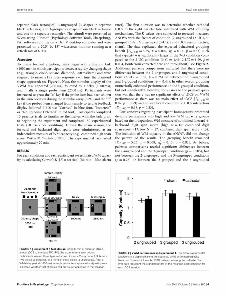
METHODParticipants and tDCS protocolThirty-three right-handed, neurologically intact graduate andundergraduate students with normal or corrected-to-normalvision participated in the current experiment (Mean age = 21.3,25 female) and received $15 per hour. On separate days, partic-ipants completed the VWM task after sham or cathodal tDCS(right posterior parietal scalp site: P4, 10 min, 1.5 mA; EldithMagstim GMbH, Ilmenau, Germany). During the sham ses-sion, current was ramped up and down for the first and last20 s of the stimulation interval to mimic sensations associatedwith current change. The anode was placed on the contralat-eral cheek. Session order was counterbalanced across partici-pants. Active/sham stimulation occurred prior to the task, whileparticipants completed practice trials. Electrodes were removedprior to the start of the task. All experimental protocols wereapproved by the Institutional Review Board of the University ofNevada.
StimuliThe task design slightly modified a paradigm previously pub-lished that showed significant VWM grouping benefits (Xuand Chun, 2007). We summarize these methods here. Stimuliwere gray, symmetrical novel shapes (2.6 × 2.6◦ visual angle)presented on black rectangles (6.9 × 18◦ of visual angle) againsta gray background. There were three experimental conditionsvarying the stimulus grouping: 2-ungrouped (2 shapes in
www.frontiersin.org July 2014 | Volume 5 | Article 800 | 5
Berryhill et al. tDCS and cognition
separate black rectangles), 3-ungrouped (3 shapes in separateblack rectangles), and 3-grouped (2 shapes in one black rectangleand one in a separate rectangle). The stimuli were presented at57 cm using EPrime® (Psychology Software Tools, Sharpsburg,PA) software running on a Dell © desktop computer and werepresented on a 20.5′′ by 13′′ widescreen monitor running at arefresh rate of 60 Hz.
ProcedureTo insure focused attention, trials began with a fixation task(1000 ms), in which participants viewed a rapidly changing shape(e.g., triangle, circle, square, diamond, 200 ms/item) and wererequired to make a key press response each time the diamondshape appeared; see Figure 1. Next, the stimulus display of theVWM task appeared (200 ms), followed by a delay (1000 ms),and finally a single probe item (2500 ms). Participants wereinstructed to press the “o” key if the probe item had been shownin the same location during the stimulus array (50%) and the “n”key if the probed item changed from sample to test. A feedbackdisplay followed (1300 ms: “Correct” in blue font, “Incorrect”or “No Response Detected” in red font). Participants completed15 practice trials to familiarize themselves with the task priorto beginning the experiment and completed 150 experimentaltrials (50 trials per condition). During the sham session, theforward and backward digit spans were administered as anindependent measure of WM capacity (e.g., combined digit spanscore; WAIS-IV, Wechsler, 2008). The experimental task lastedapproximately 20 min.
RESULTSFor each condition and each participant we estimated WM capac-ity by calculating Cowan’s K: [K = set size∗ (hit rate—false alarm
FIGURE 1 | Experiment 1 task design. After 10 min of sham or 1.0 mAanodal tDCS to the right PPC (P4), the experimental task began.Participants viewed three types of arrays: 2 items (2-ungrouped), 3 items intwo boxes (3-grouped), or 3 items in three boxes (3-ungrouped). After aWM delay period (1000 ms), a single probe item appeared and participantsindicated whether that stimulus had previously appeared in that location.
rate)]. The first question was to determine whether cathodaltDCS to the right parietal lobe interfered with WM groupingmechanisms. The K-values were subjected to repeated-measuresANOVA with the factors of condition [2-ungrouped (2-UG), 3-grouped (3-G), 3-ungrouped (3-UG)] and tDCS session (active,sham). The data replicated the expected behavioral groupingbenefit [F(2, 64) = 5.39, p = 0.007, η2
p = 0.14, β = 0.83] suchthat capacity was significantly larger in the 3-G condition com-pared to the 2-UG condition (3-G = 1.49, 2-UG = 1.29, p =0.004, Bonferroni corrected here and throughout); see Figure 2.Additional pairwise comparisons indicated that there were nodifferences between the 2-ungrouped and 3-ungrouped condi-tions (3-UG = 1.38, p = 0.26) or between the 3-ungroupedand 3-grouped conditions (p = 0.44). In other words, groupingnumerically enhanced performance on the 3-grouped condition,but not significantly. However, the answer to the primary ques-tion was that there was no significant effect of tDCS on VWMperformance as there was no main effect of tDCS [F(1, 32) =0.07, p = 0.79] and no significant condition × tDCS interaction[F(2, 64) = 0.18, p = 0.83].
Our concerns regarding participant homogeneity prompteddividing participants into high and low WM capacity groupsbased on the independent WM measure of combined forward +backward digit span scores (high N = 16: combined digitspan score >13; low N = 17: combined digit span score <13).The inclusion of WM capacity in the ANOVA did not changethe pattern of the results. The grouping benefit remained[F(2, 62) = 5.26, p = 0.008, η2
p = 0.15, β = 0.82]. As before,pairwise comparisons reveled significant differences betweenthe 2-ungrouped and the 3-grouped condition (p = 0.005), butnot between the 2-ungrouped and the 3-ungrouped conditions(p = 0.26) or between the 3-grouped and the 3-ungrouped
FIGURE 2 | VWM performance in Experiment 1. The three experimentalconditions are displayed along the abscissa, while estimated capacity(based on Cowan’s K formula, 2001) is depicted along the ordinate. Theerror bars represent the standard errors of the means in each condition foreach tDCS session.
Frontiers in Psychology | Cognitive Science July 2014 | Volume 5 | Article 800 | 6
Berryhill et al. tDCS and cognition
conditions (p = 0.47). As before, there was no main effect oftDCS [F(1, 31) = 0.05, p = 0.83], and no two- or three-way inter-actions (all p’s > 0.13). In summary, there was no difference inVWM performance in response to grouping via common regionbetween high and low WM capacity individuals. Additionally,diverging from our predictions, there was no effect of cathodaltDCS relative to sham tDCS.
DISCUSSIONHere, we tested the hypothesis that cathodal tDCS targeting pari-etal regions involved in grouping processes would disrupt theseprocesses. Contrary to our predictions, tDCS did not modulategrouping benefits in VWM. Furthermore, WM capacity did notcontribute to the current results as cathodal tDCS failed to inter-rupt grouping benefits to WM performance in either high orlow WM capacity participants. These findings were unexpectedbecause we previously interrupted WM using cathodal tDCS tothe right parietal lobe in a VWM recognition task (Berryhill et al.,2010). One possibility to account for these null results is that par-ticipants may not have been effortfully engaged in the task. Thisinterpretation is supported by low VWM performance (e.g., meanK-values for the 2-UG, 3-UG, 3-G conditions: 1.29, 1.38, 1.49items compared to the ∼1.9, 2.3, 2.6 items per condition reportedby Xu and Chun, 2007). Anecdotally, in our previous success-ful study, the participants were largely graduate students knownto the experimenter, rather than undergraduates interested inobtaining course extra-credit.
Alternatively, previous fMRI findings revealed bilateral IPSactivity during this task (Xu and Chun, 2007). Thus, some mightargue that a unilateral stimulation protocol might not have suffi-cient power to prevent some contralateral compensatory mecha-nism. However, we think that this is unlikely for several reasons.First, there is evidence supporting right IPL in attending to stim-uli across both visual hemifields (e.g., Sheremata et al., 2010;Szczepanski and Kastner, 2013), making it more likely to seedisrupted performance in the VWM task after right lateralizedstimulation. Second, and perhaps of greater relevance, we pre-viously interrupted VWM using the identical tDCS protocol butdifferent VWM tasks (Berryhill et al., 2010; Tanoue et al., 2013).Thus, we suspect that participants’ engagement was the mostimportant factor in this particular experiment.
REDUCING ADHD IMPULSIVITYFaults: low power, heterogeneous population, low task difficultyThe familiar symptoms of attention deficit hyperactivity dis-order (ADHD) include impulsivity, restlessness, and difficultyconcentrating (Faraone and Biederman, 2005). Recent findingssuggest that there is abnormal brain structure and function inthe pre-supplementary motor area (pre-SMA) in ADHD. Whenpeople with ADHD perform tasks requiring response inhibitionthey have smaller activations in the pre-SMA (Mulligan et al.,2011). These findings suggest that the pre-SMA is not sufficientlyactivated during response inhibition tasks. Recently, in healthyadults response inhibition was predictably modulated by tDCSto the pre-SMA: anodal improved performance whereas catho-dal tDCS impaired performance (Hsu et al., 2011). Other reportsshow tDCS-linked improvement in response inhibition tasks in
those with major depressive disorder (Boggio et al., 2007a) andstroke (Kang et al., 2009). Thus, we tested whether directinganodal tDCS to the pre-SMA would modulate performance ina response inhibition task, the Go/No-Go task. First, based onthe logic just described, we anticipated that anodal tDCS wouldimprove Go/No-Go task performance in young adults with lowor high ADHD symptomology, and thereby replicating the Hsuet al. findings in healthy young adults using the stop-signal task(Hsu et al., 2011). We anticipated an interaction such that thosewith high symptomology would garner greater tDCS benefitsthan the low symptomology group. We also completed two WMtasks: the operation span task and a spatial n-back task. Thesetasks were included to clarify the specificity of tDCS influences.Both WM tasks engage frontoparietal networks, but were notexpected to show modulation by tDCS to the pre-SMA. Here,we tested an unmedicated undergraduate population to look atADHD symptomology because we do not apply tDCS to peopletaking stimulants or anti-depressants (e.g., those prescribed forADHD).
METHODThe University Institutional Review Board approved all proto-cols. Volunteers completed the Adult ADHD Self-Report Scale(ASRS—v1.1; Kessler et al., 2005). This short screen was devel-oped to identify ADHD symptomology in adults and has beenvalidated in adult (Adler et al., 2006) and college-aged (Fuller-Killgore et al., 2013) populations. Scores were derived from thesix-screener questions in Part A of the ASRS, the most pre-dictive of ADHD. Questions probed the frequency with whichparticipants forgot appointments, completed tasks, or felt dis-tracted Responses were converted from verbal labels (e.g., “never,”“sometimes,” “rarely,” “often,” “very often”) to point values (1–5).Questions 1–3 required doubling the point value when answersof “sometimes,” “often,” or “very often” were recorded; questions4–6 required doubling the point value when answers “often” or“very often” were recorded. Four or more answers requiring dou-bling meet the criteria for “high likelihood” of ADHD (Kessleret al., 2005). To identify participants we recruited participantshigh (ASRS scores 39–58) and low (ASRS scores <19) in ADHDsymptomology. This high ADHD group met the criteria of highlikelihood of ADHD. Thirty-six right-handed, normal, neurotyp-ical participants were subsequently enrolled (age 18–37, 14 male).Participants were screened to ensure they were not taking medica-tions that modulate the excitability of the brain (e.g., stimulants).Participants completed anodal, cathodal and sham tDCS sessions,in counterbalanced order across 3 separate days with a minimumwashout period of 24 h. Current was administered using a com-mercial stimulator (Eldith Magstim GMbH, Ilmenau, Germany).To target the right pre-SMA, we placed the electrode 2 cm tothe right of FZ (10–20 system) and the reference electrode wasplaced on the contralateral cheek. Participants received 10 minof 1.5 mA tDCS during anodal and cathodal sessions and duringsham stimulation current was ramped up and down for the initialand final 20 s of the period. Participants completed practice tri-als of each task during stimulation and after the electrodes wereremoved, they began the experimental trials of the tasks describedbelow.
www.frontiersin.org July 2014 | Volume 5 | Article 800 | 7
Berryhill et al. tDCS and cognition
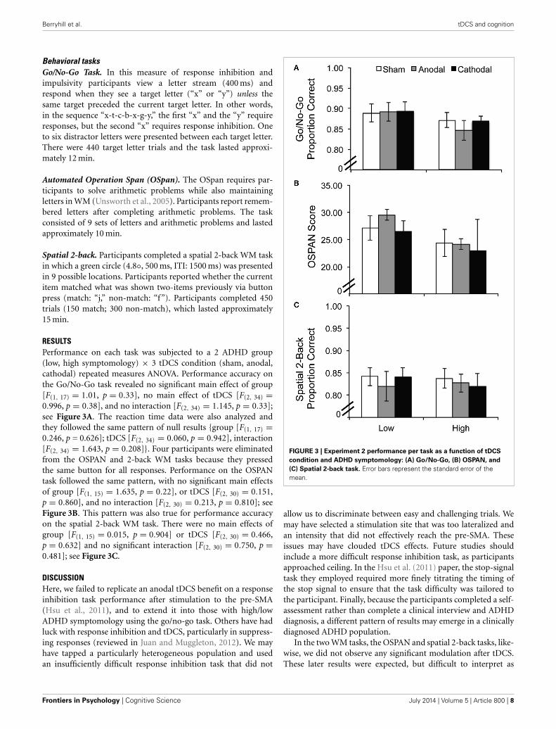
Behavioral tasksGo/No-Go Task. In this measure of response inhibition andimpulsivity participants view a letter stream (400 ms) andrespond when they see a target letter (“x” or “y”) unless thesame target preceded the current target letter. In other words,in the sequence “x-t-c-b-x-g-y,” the first “x” and the “y” requireresponses, but the second “x” requires response inhibition. Oneto six distractor letters were presented between each target letter.There were 440 target letter trials and the task lasted approxi-mately 12 min.
Automated Operation Span (OSpan). The OSpan requires par-ticipants to solve arithmetic problems while also maintainingletters in WM (Unsworth et al., 2005). Participants report remem-bered letters after completing arithmetic problems. The taskconsisted of 9 sets of letters and arithmetic problems and lastedapproximately 10 min.
Spatial 2-back. Participants completed a spatial 2-back WM taskin which a green circle (4.8◦, 500 ms, ITI: 1500 ms) was presentedin 9 possible locations. Participants reported whether the currentitem matched what was shown two-items previously via buttonpress (match: “j,” non-match: “f”). Participants completed 450trials (150 match; 300 non-match), which lasted approximately15 min.
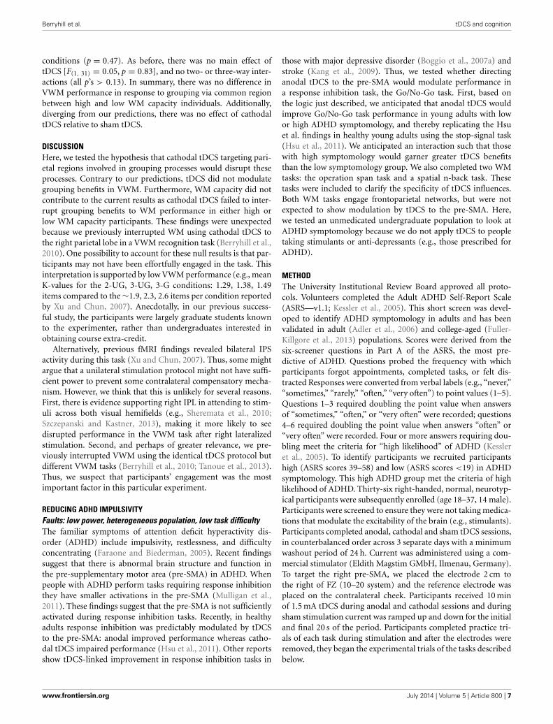
RESULTSPerformance on each task was subjected to a 2 ADHD group(low, high symptomology) × 3 tDCS condition (sham, anodal,cathodal) repeated measures ANOVA. Performance accuracy onthe Go/No-Go task revealed no significant main effect of group[F(1, 17) = 1.01, p = 0.33], no main effect of tDCS [F(2, 34) =0.996, p = 0.38], and no interaction [F(2, 34) = 1.145, p = 0.33];see Figure 3A. The reaction time data were also analyzed andthey followed the same pattern of null results {group [F(1, 17) =0.246, p = 0.626]; tDCS [F(2, 34) = 0.060, p = 0.942], interaction[F(2, 34) = 1.643, p = 0.208]}. Four participants were eliminatedfrom the OSPAN and 2-back WM tasks because they pressedthe same button for all responses. Performance on the OSPANtask followed the same pattern, with no significant main effectsof group [F(1, 15) = 1.635, p = 0.22], or tDCS [F(2, 30) = 0.151,p = 0.860], and no interaction [F(2, 30) = 0.213, p = 0.810]; seeFigure 3B. This pattern was also true for performance accuracyon the spatial 2-back WM task. There were no main effects ofgroup [F(1, 15) = 0.015, p = 0.904] or tDCS [F(2, 30) = 0.466,p = 0.632] and no significant interaction [F(2, 30) = 0.750, p =0.481]; see Figure 3C.
DISCUSSIONHere, we failed to replicate an anodal tDCS benefit on a responseinhibition task performance after stimulation to the pre-SMA(Hsu et al., 2011), and to extend it into those with high/lowADHD symptomology using the go/no-go task. Others have hadluck with response inhibition and tDCS, particularly in suppress-ing responses (reviewed in Juan and Muggleton, 2012). We mayhave tapped a particularly heterogeneous population and usedan insufficiently difficult response inhibition task that did not
FIGURE 3 | Experiment 2 performance per task as a function of tDCS
condition and ADHD symptomology: (A) Go/No-Go, (B) OSPAN, and
(C) Spatial 2-back task. Error bars represent the standard error of themean.
allow us to discriminate between easy and challenging trials. Wemay have selected a stimulation site that was too lateralized andan intensity that did not effectively reach the pre-SMA. Theseissues may have clouded tDCS effects. Future studies shouldinclude a more difficult response inhibition task, as participantsapproached ceiling. In the Hsu et al. (2011) paper, the stop-signaltask they employed required more finely titrating the timing ofthe stop signal to ensure that the task difficulty was tailored tothe participant. Finally, because the participants completed a self-assessment rather than complete a clinical interview and ADHDdiagnosis, a different pattern of results may emerge in a clinicallydiagnosed ADHD population.
In the two WM tasks, the OSPAN and spatial 2-back tasks, like-wise, we did not observe any significant modulation after tDCS.These later results were expected, but difficult to interpret as
Frontiers in Psychology | Cognitive Science July 2014 | Volume 5 | Article 800 | 8
Berryhill et al. tDCS and cognition
site-specific tDCS effects given the null findings in the primarytask of interest, the go/no-go task.
GENERAL DISCUSSIONNeurostimulation via tDCS is a useful tool for investigatingaspects of cognition. As with any tool there are some things thatwork better than others, and a set of appropriate situations fora given tool. Here, we have summarized some findings demon-strating successful applications of tDCS in studies of WM (seeTable 1) and other cognitive modalities (see Table 2). Althoughthese studies all report positive findings there is still consider-able variability in terms of the pattern of effects, paradigms usedand tDCS parameters. For instance, stimulus intensity, duration,tDCS electrode montage are inconsistent. The most consistentpattern in the published literature has been to report significantimprovements in WM tested in verbal n-back tasks and anodaltDCS to the left DLPFC. In other cognitive realms a patchworkof findings is emerging revealing consistent effects in memory,deception, and cognitive control. However, there are exceptionsand forays into different tasks, populations, and parameters haveproduced different patterns of results. We also note that the pro-liferation of neurostimulation effects (e.g., tACS, TRNS) will becertain to raise additional issues to maximize their experimentaland applied usefulness (for a recent review see Kuo and Nitsche,2012; see also Snowball et al., 2013).
We think the file-drawer problem is a particular challenge here.We offered several of our own missteps and several factors to con-sider when applying tDCS to cognitive questions. We believe thatappropriate consideration of these factors will facilitate futureexperimental design and serve to increase interpretable outcomes.Ideally, the following is available: a homogenous, highly moti-vated, large population engaging in a challenging cognitive taskthat permits selective identification and analysis of the most diffi-cult trials. When one or more of these factors is overlooked theremay be little to report.
WHAT DO THESE CONSIDERATIONS REVEAL ABOUT tDCS?The considerations we noted above point us toward an emerg-ing research question. Why do group differences matter? Theunderlying mechanism of tDCS remains unclear, although long-term neuroplasticity is indicated (Rosenkranz et al., 2000; Nitscheet al., 2003). Some pharmacological work in humans indicatesthat blocking sodium or calcium channels prevents longer-lastingeffects of anodal tDCS and antagonizing NMDA receptors pre-vents longer-lasting effects of anodal or cathodal tDCS, at leastin motor cortex (Nitsche et al., 2003). Furthermore, at a largerscale, the way current flows through a particular individual’s braincertainly varies and has tremendous implications for applied andexperimental use of tDCS (Bikson et al., 2013). Incorporatingother variables, including the structural and functional connec-tivity of stimulated networks and their temporal dynamics willalso improve experimental success when applying tDCS. We sus-pect that the answers to these questions will also clarify why somegroups benefit more than others and why homogeneous groupsprovide more consistent data. For an individualized approach totDCS application, the underlying molecular mechanism must befully clarified and this in turn will advance our understanding at
the network level. At this point, another way to counter these dif-ferences is simply through brute power and by running sufficientparticipants.
The role of task difficulty is important. As noted in the intro-duction, when a participant knows the answer definitively tDCSis unlikely to have an effect. This means tasks that are too easydo not show any effect of tDCS. It is only in the challenging, bor-derline cases where tDCS serves as a tipping factor. As we age, weencounter more of these near-threshold cases and this populationmay be the one to benefit most from tDCS-linked cognitive inter-ventions. In particular, in the realm of WM, small improvementscan benefit quality of life and are worth the effort to gain them.
CONCLUSIONStDCS is a safe, affordable neurostimulation technique that is well-tolerated in healthy and special populations. Regular tDCS isnot well-positioned to target carefully defined cortical regionsfor studies of structure-function relationships. The current arti-cle provided several considerations for review when preparing atDCS study. Because tDCS provides the greatest effect at thresh-old level decisions, challenging tasks are necessary. Participantsmust be sufficiently engaged and motivated. Finally, the natureof the participants also should be carefully considered as groupdifferences can predict nearly equal and opposite responses totDCS. Furthermore, whereas early tDCS studies often targetedmotor and visual regions, studies of cognition require sufficientpower and larger numbers of participants. When these consid-erations were sufficiently weighed, we have succeeded. We offerseveral cautionary tales detailing our failed efforts when theseconsiderations were ignored. Finally, in conclusion, the best useof tDCS may well be in translational and applied settings. Inthis arena, tDCS already shows promise as a way to maintainand/or restore cognitive function across various populations.Clearly, there is great interest in effective cognitive interventionsas the aging population grows and tDCS may be a part of theanswer.
ACKNOWLEDGMENTSThis work was supported by an Institutional Development Award(IDeA) from the National Institute of General Medical Sciencesof the NIH P20GM103650 (PI Michael Webster, Project LeaderMarian Berryhill), and NEI R15EY022775 (to Marian Berryhilland Gideon Caplovitz). The content is solely the responsibility ofthe authors and does not represent the official views of the NIH,the NIGMS, or the NEI.
REFERENCESAdler, L. A., Spencer, T., Faraone, S. V., Kessler, R. C., Howes, M. J., Biederman,
J., et al. (2006). Validity of pilot Adult ADHD Self- Report Scale (ASRS)to rate adult ADHD symptoms. Ann. Clin. psychiatry 18, 145–148. doi:10.1080/10401230600801077
Andrews, S. C., Hoy, K. E., Enticott, P. G., Daskalakis, Z. J., and Fitzgerald, P. B.(2011). Improving working memory: the effect of combining cognitive activ-ity and anodal transcranial direct current stimulation to the left dorsolateralprefrontal cortex. Brain Stimul. 4, 84–89. doi: 10.1016/j.brs.2010.06.004
Antal, A., Kincses, T. Z., Nitsche, M. A., and Paulus, W. (2003). Modulation ofmoving phosphene thresholds by transcranial direct current stimulation ofV1 in human. Neuropsychologia 41, 1802–1807. doi: 10.1016/S0028-3932(03)00181-7
www.frontiersin.org July 2014 | Volume 5 | Article 800 | 9

Berryhill et al. tDCS and cognition
Antal, A., Nitsche, M. A., Kruse, W., Kincses, T. Z., Hoffmann, K. P., and Paulus, W.(2004). Direct current stimulation over V5 enhances visuomotor coordinationby improving motion perception in humans. J. Cogn. Neurosci. 16, 521–527. doi:10.1162/089892904323057263
Antal, A., Nitsche, M. A., and Paulus, W. (2001). External modulation of visualperception in humans. Neuroreport 12, 3553–3555. doi: 10.1097/00001756-200111160-00036
Antal, A., Nitsche, M. A., and Paulus, W. (2006). Transcranial direct current stim-ulation and the visual cortex. Brain Res. Bull. 68, 459–463. doi: 10.1016/j.brainresbull.2005.10.006
Antal, A., and Paulus, W. (2008). Transcranial direct current stimulation and visualperception. Perception 37, 367–374. doi: 10.1068/p5872
Antal, A., Polania, R., Schmidt-Samoa, C., Dechent, P., and Paulus, W. (2011).Transcranial direct current stimulation over the primary motor cortex duringfMRI. Neuroimage 55, 590–596. doi: 10.1016/j.neuroimage.2010.11.085
Bachmann, C. G., Muschinsky, S., Nitsche, M. A., Rolke, R., Magerl, W., Treede,R. D., et al. (2010). Transcranial direct current stimulation of the motor cor-tex induces distinct changes in thermal and mechanical sensory percepts. Clin.Neurophysiol. 121, 2083–2089. doi: 10.1016/j.clinph.2010.05.005
Baker, J. M., Rorden, C., and Fridriksson, J. (2010). Using transcranial direct-current stimulation to treat stroke patients with aphasia. Stroke 41, 1229–1236.doi: 10.1161/STROKEAHA.109.576785
Berryhill, M. E., and Jones, K. T. (2012). tDCS selectively improves working mem-ory in older adults with more education. Neurosci. Lett. 521, 148–151. doi:10.1016/j.neulet.2012.05.074
Berryhill, M. E., Wencil, E. B., Coslett, H. B., and Olson, I. R. (2010). A selectiveworking memory impairment after transcranial direct current stimulation tothe right parietal lobe. Neurosci. Lett. 479, 312–316. doi: 10.1016/j.neulet.2010.05.087
Bikson, M., Name, A., and Rahman, A. (2013). Origins of specificity duringtDCS: anatomical, activity-selective, and input-bias mechanisms. Front. Hum.Neurosci. 7:688. doi: 10.3389/fnhum.2013.00688
Boggio, P. S., Bermpohl, F., Vergara, A. O., Muniz, A. L., Nahas, F. H., Leme,P. B., et al. (2007a). Go-no-go task performance improvement after anodaltranscranial DC stimulation of the left dorsolateral prefrontal cortex in majordepression. J. Affect. Disord. 101, 91–98. doi: 10.1016/j.jad.2006.10.026
Boggio, P. S., Campanha, C., Valasek, C. A., Fecteau, S., Pascual-Leone, A., andFregni, F. (2010). Modulation of decision-making in a gambling task in olderadults with transcranial direct current stimulation. Eur. J. Neurosci. 31, 593–597.doi: 10.1111/j.1460-9568.2010.07080.x
Boggio, P. S., Ferrucci, R., Mameli, F., Martins, D., Martins, O., Vergari, M., et al.(2012). Prolonged visual memory enhancement after direct current stimulationin Alzheimer’s disease. Brain Stimul. 5, 223–230. doi: 10.1016/j.brs.2011.06.006
Boggio, P. S., Ferrucci, R., Rigonatti, S. P., Covre, P., Nitsche, M., Pascual-Leone,A., et al. (2006). Effects of transcranial direct current stimulation on workingmemory in patients with Parkinson’s disease. J. Neurol. Sci. 249, 31–38. doi:10.1016/j.jns.2006.05.062
Boggio, P. S., Nunes, A., Rigonatti, S. P., Nitsche, M. A., Pascual-Leone, A., andFregni, F. (2007b). Repeated sessions of noninvasive brain DC stimulation isassociated with motor function improvement in stroke patients. Restor. Neurol.Neurosci. 25, 123–129.
Boggio, P. S., Rocha, R. R., Da Silva, M. T., and Fregni, F. (2008). Differentialmodulatory effects of transcranial direct current stimulation on a facial expres-sion go-no-go task in males and females. Neurosci. Lett. 447, 101–105. doi:10.1016/j.neulet.2008.10.009
Boggio, P. S., Valasek, C. A., Campanha, C., Giglio, A. C., Baptista, N. I., Lapenta,O. M., et al. (2011). Non-invasive brain stimulation to assess and modulateneuroplasticity in Alzheimer’s disease. Neuropsychol. Rehabil. 21, 703–716. doi:10.1080/09602011.2011.617943
Boggio, P. S., Zaghi, S., and Fregni, F. (2009). Modulation of emotions associatedwith images of human pain using anodal transcranial direct current stimulation(tDCS). Neuropsychologia 47, 212–217. doi: 10.1016/j.neuropsychologia.2008.07.022
Bolognini, N., Fregni, F., Casati, C., Olgiati, E., and Vallar, G. (2010). Brainpolarization of parietal cortex augments training-induced improvement ofvisual exploratory and attentional skills. Brain Res. 1349, 76–89. doi: 10.1016/j.brainres.2010.06.053
Bolognini, N., Rossetti, A., Casati, C., Mancini, F., and Vallar, G. (2011).Neuromodulation of multisensory perception: a tDCS study of the
sound-induced flash illusion. Neuropsychologia 49, 231–237. doi: 10.1016/j.neuropsychologia.2010.11.015
Borckardt, J. J., Bikson, M., Frohman, H., Reeves, S. T., Datta, A., Bansal, V., et al.(2012). A pilot study of the tolerability and effects of high-definition tran-scranial direct current stimulation (HD-tDCS) on pain perception. J. Pain 13,112–120. doi: 10.1016/j.jpain.2011.07.001
Boros, K., Poreisz, C., Munchau, A., Paulus, W., and Nitsche, M. A. (2008).Premotor transcranial direct current stimulation (tDCS) affects primary motorexcitability in humans. Eur. J. Neurosci. 27, 1292–1300. doi: 10.1111/j.1460-9568.2008.06090.x
Brady, T. F., and Tenenbaum, J. B. (2013). A probabilistic model of visual workingmemory: incorporating higher order regularities into working memory capacityestimates. Psychol. Rev. 120, 85–109. doi: 10.1037/a0030779
Brunoni, A. R., and Vanderhasselt, M. A. (2014). Working memory improve-ment with non-invasive brain stimulation of the dorsolateral prefrontalcortex: a systematic review and meta-analysis. Brain Cogn. 86C, 1–9. doi:10.1016/j.bandc.2014.01.008
Cerruti, C., and Schlaug, G. (2009). Anodal transcranial direct current stimulationof the prefrontal cortex enhances complex verbal associative thought. J. Cogn.Neurosci. 21, 1980–1987. doi: 10.1162/jocn.2008.21143
Chi, R. P., Fregni, F., and Snyder, A. W. (2010). Visual memory improvedby non-invasive brain stimulation. Brain Res. 1353, 168–175. doi:10.1016/j.brainres.2010.07.062
Chrysikou, E. G., and Hamilton, R. H. (2011). Noninvasive brain stimulation inthe treatment of aphasia: exploring interhemispheric relationships and theirimplications for neurorehabilitation. Restor. Neurol. Neurosci. 29, 375–394. doi:10.3233/RNN-2011-0610
Coffman, B. A., Trumbo, M. C., Flores, R. A., Garcia, C. M., Van DerMerwe, A. J., Wassermann, E. M., et al. (2012). Impact of tDCS on per-formance and learning of target detection: interaction with stimulus char-acteristics and experimental design. Neuropsychologia 50, 1594–1602. doi:10.1016/j.neuropsychologia.2012.03.012
De Vries, M. H., Barth, A. C., Maiworm, S., Knecht, S., Zwitserlood, P., andFloel, A. (2010). Electrical stimulation of Broca’s area enhances implicitlearning of an artificial grammar. J. Cogn. Neurosci. 22, 2427–2436. doi:10.1162/jocn.2009.21385
Faraone, S. V., and Biederman, J. (2005). What is the prevalence of adult ADHD?results of a population screen of 966 adults. J. Atten. Disord. 9, 384–391. doi:10.1177/1087054705281478
Fecteau, S., Boggio, P., Fregni, F., and Pascual-Leone, A. (2012). Modulation ofuntruthful responses with non-invasive brain stimulation. Front. Psychiatry3:97. doi: 10.3389/fpsyt.2012.00097
Fecteau, S., Pascual-Leone, A., Zald, D. H., Liguori, P., Theoret, H., Boggio, P. S.,et al. (2007). Activation of prefrontal cortex by transcranial direct current stim-ulation reduces appetite for risk during ambiguous decision making. J. Neurosci.27, 6212–6218. doi: 10.1523/JNEUROSCI.0314-07.2007
Ferrucci, R., Bortolomasi, M., Vergari, M., Tadini, L., Salvoro, B., Giacopuzzi, M.,et al. (2009). Transcranial direct current stimulation in severe, drug-resistantmajor depression. J. Affect. Disord. 118, 215–219. doi: 10.1016/j.jad.2009.02.015
Ferrucci, R., Mameli, F., Guidi, I., Mrakic-Sposta, S., Vergari, M., Marceglia, S., et al.(2008). Transcranial direct current stimulation improves recognition memoryin Alzheimer disease. Neurology 71, 493–498. doi: 10.1212/01.wnl.0000317060.43722.a3
Floel, A., Rosser, N., Michka, O., Knecht, S., and Breitenstein, C. (2008).Noninvasive brain stimulation improves language learning. J. Cogn. Neurosci.20, 1415–1422. doi: 10.1162/jocn.2008.20098
Fregni, F., Boggio, P. S., Lima, M. C., Ferreira, M. J., Wagner, T., Rigonatti, S. P.,et al. (2006a). A sham-controlled, phase II trial of transcranial direct currentstimulation for the treatment of central pain in traumatic spinal cord injury.Pain 122, 197–209. doi: 10.1016/j.pain.2006.02.023
Fregni, F., Boggio, P. S., Mansur, C. G., Wagner, T., Ferreira, M. J., Lima,M. C., et al. (2005). Transcranial direct current stimulation of the unaf-fected hemisphere in stroke patients. Neuroreport 16, 1551–1555. doi:10.1097/01.wnr.0000177010.44602.5e
Fregni, F., Boggio, P. S., Nitsche, M. A., Rigonatti, S. P., and Pascual-Leone, A.(2006b). Cognitive effects of repeated sessions of transcranial direct currentstimulation in patients with depression. Depress. Anxiety 23, 482–484. doi:10.1002/da.20201.
Frontiers in Psychology | Cognitive Science July 2014 | Volume 5 | Article 800 | 10

Berryhill et al. tDCS and cognition
Fregni, F., Boggio, P. S., Santos, M. C., Lima, M., Vieira, A. L., Rigonatti, S. P., et al.(2006c). Noninvasive cortical stimulation with transcranial direct current stim-ulation in Parkinson’s disease. Mov. Disord. 21, 1693–1702. doi: 10.1002/mds.21012
Fuller-Killgore, M. D., Burlison, J., and Dwyer, W. (2013). Comparison of threeADHD screening instruments in college students of varying cognitive ability.J. Atten. Disord. 17, 449–454. doi: 10.1177/1087054712438136
Hamilton, R. H., Chrysikou, E. G., and Coslett, B. (2011). Mechanisms of aphasiarecovery after stroke and the role of noninvasive brain stimulation. Brain Lang.118, 40–50. doi: 10.1016/j.bandl.2011.02.005
Horvath, J. C., Carter, O., and Forte, J. D. (2014). Transcranial direct current stim-ulation: five important issues we aren’t discussing (but probably should be).Front. Syst. Neurosci. 8:2. doi: 10.3389/fnsys.2014.00002
Hoy, K. E., Emonson, M. R., Arnold, S. L., Thomson, R. H., Daskalakis, Z. J., andFitzgerald, P. B. (2013). Testing the limits: investigating the effect of tDCS doseon working memory enhancement in healthy controls. Neuropsychologia 51,1777–1784. doi: 10.1016/j.neuropsychologia.2013.05.018
Hsu, T. Y., Tseng, L. Y., Yu, J. X., Kuo, W. J., Hung, D. L., Tzeng, O. J., et al. (2011).Modulating inhibitory control with direct current stimulation of the superiormedial frontal cortex. Neuroimage 56, 2249–2257. doi: 10.1016/j.neuroimage.2011.03.059
Hummel, F., Celnik, P., Giraux, P., Floel, A., Wu, W. H., Gerloff, C., et al. (2005).Effects of non-invasive cortical stimulation on skilled motor function in chronicstroke. Brain 128, 490–499. doi: 10.1093/brain/awh369
Hunter, T., Sacco, P., Nitsche, M. A., and Turner, D. L. (2009). Modulation ofinternal model formation during force field-induced motor learning by anodaltranscranial direct current stimulation of primary motor cortex. J. Physiol. 587,2949–2961. doi: 10.1113/jphysiol.2009.169284
Iyer, M. B., Mattu, U., Grafman, J., Lomarev, M., Sato, S., and Wassermann, E.M. (2005). Safety and cognitive effect of frontal DC brain polarization inhealthy individuals. Neurology 64, 872–875. doi: 10.1212/01.WNL.0000152986.07469.E9
Jacobson, L., Javitt, D. C., and Lavidor, M. (2011). Activation of inhibition: dimin-ishing impulsive behavior by direct current stimulation over the inferior frontalgyrus. J. Cogn. Neurosci. 23, 3380–3387. doi: 10.1162/jocn_a_00020
Jacobson, L., Koslowsky, M., and Lavidor, M. (2012). tDCS polarity effects in motorand cognitive domains: a meta-analytical review. Exp. Brain Res. 216, 1–10. doi:10.1007/s00221-011-2891-9
Javadi, A. H., and Cheng, P. (2013). Transcranial Direct Current Stimulation(tDCS) enhances reconsolidation of long-term memory. Brain Stimul. 6,668–674. doi: 10.1016/j.brs.2012.10.007
Javadi, A. H., and Walsh, V. (2012). Transcranial direct current stimulation (tDCS)of the left dorsolateral prefrontal cortex modulates declarative memory. BrainStimul. 5, 231–241. doi: 10.1016/j.brs.2011.06.007
Jeon, S. Y., and Han, S. J. (2012). Improvement of the working memory and namingby transcranial direct current stimulation. Ann. Rehabil. Med. 36, 585–595. doi:10.5535/arm.2012.36.5.585
Jo, J. M., Kim, Y.-H., Ko, M.-H., Ohn, S. H., Joen, B., and Lee, K. H. (2009).Enhancing the working memory of stroke patients using tDCS. Am. J. Phys.Med. Rehabil. 88, 404–409. doi: 10.1097/PHM.0b013e3181a0e4cb
Jones, K. T., and Berryhill, M. E. (2012). Parietal contributions to visualworking memory depend on task difficulty. Front. Psychiatry 3:81. doi:10.3389/fpsyt.2012.00081
Juan, C. H., and Muggleton, N. G. (2012). Brain stimulation and inhibitory control.Brain Stimul. 5, 63–69. doi: 10.1016/j.brs.2012.03.012
Kalu, U. G., Sexton, C. E., Loo, C. K., and Ebmeier, K. P. (2012). Transcranialdirect current stimulation in the treatment of major depression: a meta-analysis.Psychol. Med. 42, 1791–1800. doi: 10.1017/S0033291711003059
Kang, E. K., Baek, M. J., Kim, S., and Paik, N. J. (2009). Non-invasive corticalstimulation improves post-stroke attention decline. Restor. Neurol. Neurosci. 27,645–650. doi: 10.3233/RNN-2009-0514
Karim, A. A., Schneider, M., Lotze, M., Veit, R., Sauseng, P., Braun, C., et al.(2010). The truth about lying: inhibition of the anterior prefrontal corteximproves deceptive behavior. Cereb. Cortex 20, 205–213. doi: 10.1093/cercor/bhp090
Kessler, R. C., Adler, L., Ames, M., Demler, O., Faraone, S., Hiripi, E., et al. (2005).The World Health Organization Adult ADHD Self-Report Scale (ASRS): a shortscreening scale for use in the general population. Psychol. Med. 35, 245–256. doi:10.1017/S0033291704002892
Kim, J. H., Kim, D. W., Chang, W. H., Kim, Y. H., Kim, K., and Im, C. H. (2014).Inconsistent outcomes of transcranial direct current stimulation may originatefrom anatomical differences among individuals: electric field simulation usingindividual MRI data. Neurosci. Lett. 564, 6–10. doi: 10.1016/j.neulet.2014.01.054
Kincses, T. Z., Antal, A., Nitsche, M. A., Bartfai, O., and Paulus, W. (2004).Facilitation of probabilistic classification learning by transcranial direct cur-rent stimulation of the prefrontal cortex in the human. Neuropsychologia 42,113–117. doi: 10.1016/S0028-3932(03)00124-6
Knoch, D., Nitsche, M. A., Fischbacher, U., Eisenegger, C., Pascual-Leone, A., andFehr, E. (2008). Studying the neurobiology of social interaction with transcra-nial direct current stimulation–the example of punishing unfairness. Cereb.Cortex 18, 1987–1990. doi: 10.1093/cercor/bhm237
Kuo, M. F., and Nitsche, M. A. (2012). Effects of transcranial electrical stimulationon cognition. Clin. EEG Neurosci. 43, 192–199. doi: 10.1177/1550059412444975
Lefaucheur, J. P., Antal, A., Ahdab, R., Ciampi De Andrade, D., Fregni, F., Khedr, E.M., et al. (2008). The use of repetitive transcranial magnetic stimulation (rTMS)and transcranial direct current stimulation (tDCS) to relieve pain. Brain Stimul.1, 337–344. doi: 10.1016/j.brs.2008.07.003
Lindenberg, R., Renga, V., Zhu, L. L., Nair, D., and Schlaug, G. (2010).Bihemispheric brain stimulation facilitates motor recovery in chronic strokepatients. Neurology 75, 2176–2184. doi: 10.1212/WNL.0b013e318202013a
Loo, C. K., Sachdev, P., Martin, D., Pigot, M., Alonzo, A., Malhi, G. S., et al. (2010).A double-blind, sham-controlled trial of transcranial direct current stimulationfor the treatment of depression. Int. J. Neuropsychopharmacol. 13, 61–69. doi:10.1017/S1461145709990411
Lopez-Alonso, V., Cheeran, B., Rio-Rodriguez, D., and Fernandez-Del-Olmo, M.(2014). Inter-individual variability in response to non-invasive brain stimula-tion paradigms. Brain Stimul. 7, 372–380. doi: 10.1016/j.brs.2014.02.004
Luria, R., and Vogel, E. K. (2014). Come together, right now: dynamic overwritingof an object’s history through common fate. J. Cogn. Neurosci. 26, 1819–1828.doi: 10.1162/jocn_a_00584
Mameli, F., Mrakic-Sposta, S., Vergari, M., Fumagalli, M., Macis, M., Ferrucci, R.,et al. (2010). Dorsolateral prefrontal cortex specifically processes general - butnot personal - knowledge deception: multiple brain networks for lying. Behav.Brain Res. 211, 164–168. doi: 10.1016/j.bbr.2010.03.024
Marshall, L., Molle, M., Hallschmid, M., and Born, J. (2004). Transcranial directcurrent stimulation during sleep improves declarative memory. J. Neurosci. 24,9985–9992. doi: 10.1523/JNEUROSCI.2725-04.2004
Marshall, L., Molle, M., Siebner, H. R., and Born, J. (2005). Bifrontal transcranialdirect current stimulation slows reaction time in a working memory task. BMCNeurosci. 6:23. doi: 10.1186/1471-2202-6-23
Mulligan, R. C., Knopik, V. S., Sweet, L. H., Fischer, M., Seidenberg, M., andRao, S. M. (2011). Neural correlates of inhibitory control in adult atten-tion deficit/hyperactivity disorder: evidence from the Milwaukee longitudinalsample. Psychiatry Res. 194, 119–129. doi: 10.1016/j.pscychresns.2011.02.003
Mulquiney, P. G., Hoy, K. E., Daskalakis, Z. J., and Fitzgerald, P. B. (2011).Improving working memory: exploring the effect of transcranial ran-dom noise stimulation and transcranial direct current stimulation on thedorsolateral prefrontal cortex. Clin. Neurophysiol. 122, 2384–2389. doi:10.1016/j.clinph.2011.05.009
Mungee, A., Kazzer, P., Feeser, M., Nitsche, M. A., Schiller, D., and Bajbouj,M. (2014). Transcranial direct current stimulation of the prefrontal cor-tex: a means to modulate fear memories. Neuroreport 25, 480–484. doi:10.1097/WNR.0000000000000119
Nitsche, M. A., Fricke, K., Henschke, U., Schlitterlau, A., Liebetanz, D., Lang, N.,et al. (2003). Pharmacological modulation of cortical excitability shifts inducedby transcranial direct current stimulation in humans. J. Physiol. 553(Pt 1),293–301. doi: 10.1113/jphysiol.2003.049916
Nitsche, M. A., Cohen, L. G., Wassermann, E. M., Priori, A., Lang, N., Antal, A.,et al. (2008). Transcranial direct current stimulation: state of the art 2008. BrainStimul. 1, 206–223. doi: 10.1016/j.brs.2008.06.004
Nitsche, M. A., Roth, A., Kuo, M. F., Fischer, A. K., Liebetanz, D., Lang, N., et al.(2007). Timing-dependent modulation of associative plasticity by general net-work excitability in the human motor cortex. J. Neurosci. 27, 3807–3812. doi:10.1523/JNEUROSCI.5348-06.2007
Nitsche, M. A., Seeber, A., Frommann, K., Klein, C. C., Rochford, C., Nitsche, M. S.,et al. (2005). Modulating parameters of excitability during and after transcranialdirect current stimulation of the human motor cortex. J. Physiol. 568, 291–303.doi: 10.1113/jphysiol.2005.092429
www.frontiersin.org July 2014 | Volume 5 | Article 800 | 11

Berryhill et al. tDCS and cognition
Ohn, S. H., Park, C. I., Yoo, W. K., Ko, M. H., Choi, K. P., Kim, G. M.,et al. (2008). Time-dependent effect of transcranial direct current stimula-tion on the enhancement of working memory. Neuroreport 19, 43–47. doi:10.1097/WNR.0b013e3282f2adfd
Palmer, S., and Rock, I. (1994). Rethinking perceptual organization: the role ofuniform connectedness. Psychon. Bull. Rev. 1, 29–55. doi: 10.3758/BF03200760
Penolazzi, B., Stramaccia, D. F., Braga, M., Mondini, S., and Galfano, G. (2014).Human memory retrieval and inhibitory control in the brain: beyond corre-lational evidence. J. Neurosci. 34, 6606–6610. doi: 10.1523/JNEUROSCI.0349-14.2014
Peterson, D. J., and Berryhill, M. E. (2013). The Gestalt principle of similar-ity benefits visual working memory. Psychon. Bull. Rev. 20, 1282–1289. doi:10.3758/s13423-013-0460-x
Rosenkranz, K., Nitsche, M. A., Tergau, F., and Paulus, W. (2000). Diminutionof training-induced transient motor cortex plasticity by weak transcranialdirect current stimulation in the human. Neurosci. Lett. 296, 61–63. doi:10.1016/S0304-3940(00)01621-9
Ross, L. A., Mccoy, D., Coslett, H. B., Olson, I. R., and Wolk, D. A. (2011). Improvedproper name recall in aging after electrical stimulation of the anterior temporallobes. Front. Aging Neurosci. 3:16. doi: 10.3389/fnagi.2011.00016
Ross, L. A., Mccoy, D., Wolk, D. A., Coslett, H. B., and Olson, I. R. (2010). Improvedproper name recall by electrical stimulation of the anterior temporal lobes.Neuropsychologia 48, 3671–3674. doi: 10.1016/j.neuropsychologia.2010.07.024
Sheremata, S. L., Bettencourt, K. C., and Somers, D. C. (2010). Hemispheric asym-metry in visuotopic posterior parietal cortex emerges with visual short-termmemory load. J. Neurosci. 30, 12581–12588. doi: 10.1523/JNEUROSCI.2689-10.2010
Snowball, A., Tachtsidis, I., Popescu, T., Thompson, J., Delazer, M., Zamarian,L., et al. (2013). Long-term enhancement of brain function and cognitionusing cognitive training and brain stimulation. Curr. Biol. 23, 987–992. doi:10.1016/j.cub.2013.04.045
Stone, D. B., and Tesche, C. D. (2009). Transcranial direct current stimulationmodulates shifts in global/local attention. Neuroreport 20, 1115–1119. doi:10.1097/WNR.0b013e32832e9aa2
Szczepanski, S. M., and Kastner, S. (2013). Shifting attentional priorities: control ofspatial attention through hemispheric competition. J. Neurosci. 33, 5411–5421.doi: 10.1523/JNEUROSCI.4089-12.2013
Tanoue, R. T., Jones, K. T., Peterson, D. J., and Berryhill, M. E. (2013). Differentialfrontal involvement in shifts of internal and perceptual attention. Brain Stimul.6, 675–682. doi: 10.1016/j.brs.2012.11.003
Todd, J. J., and Marois, R. (2004). Capacity limit of visual short-termmemory in human posterior parietal cortex. Nature 428, 751–754. doi:10.1038/nature02466
Todd, J. J., and Marois, R. (2005). Posterior parietal cortex activity predicts indi-vidual differences in visual short-term memory capacity. Cogn. Affect. Behav.Neurosci. 5, 144–155. doi: 10.3758/CABN.5.2.144
Tseng, P., Hsu, T. Y., Chang, C. F., Tzeng, O. J., Hung, D. L., Muggleton, N. G.,et al. (2012). Unleashing potential: transcranial direct current stimulation over
the right posterior parietal cortex improves change detection in low-performingindividuals. J. Neurosci. 32, 10554–10561. doi: 10.1523/JNEUROSCI.0362-12.2012
Unsworth, N., Heitz, R. P., Schrock, J. C., and Engle, R. W. (2005). An auto-mated version of the operation span task. Behav. Res. Methods 37, 498–505. doi:10.3758/BF03192720
Wechsler, D. (2008). Wechsler Adult Intelligence Scale, 4th Edn. Administration andScoring Manual. San Antonio, TX: Psychological Corporation.
Wertheimer, M. (1950). “Gestalt theory,” in A Sourcebook of Gestalt Psychology, edW. D. Ellis (New York, NY: Humanities Press), 1–11. (Original work published1924).
Woodman, G. F., Vecera, S. P., and Luck, S. J. (2003). Perceptual organizationinfluences visual working memory. Psychon. Bull. Rev. 10, 80–87. doi: 10.3758/BF03196470
Xu, Y. (2002). Limitations of object-based feature encoding in visual short-termmemory. J. Exp. Psychol. Hum. Percept. Perform. 28, 458–468. doi: 10.1037/0096-1523.28.2.458
Xu, Y. (2006). Understanding the object benefit in visual short-term memory: theroles of feature proximity and connectedness. Percept. Psychophys. 68, 815–828.doi: 10.3758/BF03193704
Xu, Y., and Chun, M. M. (2006). Dissociable neural mechanisms supporting visualshort-term memory for objects. Nature 440, 91–95. doi: 10.1038/nature04262
Xu, Y., and Chun, M. M. (2007). Visual grouping in human parietal cor-tex. Proc. Natl. Acad. Sci. U.S.A. 104, 18766–18771. doi: 10.1073/pnas.0705618104
Xu, Y., and Chun, M. M. (2009). Selecting and perceiving multiple visual objects.Trends Cogn. Sci. 13, 167–174. doi: 10.1016/j.tics.2009.01.008
Zaehle, T., Sandmann, P., Thorne, J. D., Jancke, L., and Herrmann, C. S. (2011).Transcranial direct current stimulation of the prefrontal cortex modulatesworking memory performance: combined behavioural and electrophysiologicalevidence. BMC Neurosci. 12:2. doi: 10.1186/1471-2202-12-2
Conflict of Interest Statement: The authors declare that the research was con-ducted in the absence of any commercial or financial relationships that could beconstrued as a potential conflict of interest.
Received: 08 April 2014; accepted: 07 July 2014; published online: 25 July 2014.Citation: Berryhill ME, Peterson DJ, Jones KT and Stephens JA (2014) Hits and misses:leveraging tDCS to advance cognitive research. Front. Psychol. 5:800. doi: 10.3389/fpsyg.2014.00800This article was submitted to Cognitive Science, a section of the journal Frontiers inPsychology.Copyright © 2014 Berryhill, Peterson, Jones and Stephens. This is an open-accessarticle distributed under the terms of the Creative Commons Attribution License(CC BY). The use, distribution or reproduction in other forums is permitted, providedthe original author(s) or licensor are credited and that the original publication in thisjournal is cited, in accordance with accepted academic practice. No use, distribution orreproduction is permitted which does not comply with these terms.
Frontiers in Psychology | Cognitive Science July 2014 | Volume 5 | Article 800 | 12
Copyright © 2022 FDOKUMEN