
The Promyelocytic Leukemia Zinc Finger-MicroRNA-221/-222 Pathway Controls Melanoma Progression...
11
The Promyelocytic Leukemia Zinc Finger–MicroRNA-221/-222 Pathway Controls Melanoma Progression through Multiple Oncogenic Mechanisms Federica Felicetti, 1 M. Cristina Errico, 1 Lisabianca Bottero, 1 Patrizia Segnalini, 1 Antonella Stoppacciaro, 2 Mauro Biffoni, 1 Nadia Felli, 1 Gianfranco Mattia, 1 Marina Petrini, 1 Mario P. Colombo, 3 Cesare Peschle, 1 and Alessandra Care ` 1 1 Department of Hematology, Oncology, and Molecular Medicine, Istituto Superiore Sanita ` and 2 Department of Histopathology, II Faculty of Medicine and Surgery, Sant’Andrea Hospital, University ‘‘La Sapienza,’’ Rome, Italy, and 3 Immunotherapy and Gene Therapy Unit, Department of Experimental Oncology, Fondazione Istituti Di Ricovero e Cura a Carattere Scientifico Istituto Nazionale Tumori, Milan, Italy Abstract The incidence of cutaneous melanoma is steadily increasing. Although several molecular abnormalities have been associ- ated with melanoma progression, the mechanisms underlying the differential gene expression are still largely unknown and targeted therapies are not yet available. Noncoding small RNAs, termed microRNAs (miR), have been recently reported to play important roles in major cellular processes, including those involved in cancer development and progression. We have identified the promyelocytic leukemia zinc finger (PLZF) transcription factor as a repressor of miR-221 and miR-222 by direct binding to their putative regulatory region. Spe- cifically, PLZF silencing in melanomas unblocks miR-221 and miR-222, which in turn controls the progression of the neoplasia through down-modulation of p27Kip1/CDKN1B and c-KIT receptor, leading to enhanced proliferation and differentiation blockade of the melanoma cells, respectively. In vitro and in vivo functional studies, including the use of antisense ‘‘antagomir’’ oligonucleotides, confirmed the key role of miR-221/-222 in regulating the progression of human melanoma; this suggests that targeted therapies suppressing miR-221/-222 may prove beneficial in advanced melanoma. [Cancer Res 2008;68(8):2745–54] Introduction Cutaneous melanoma is an aggressive neoplasm refractory to traditional therapies, especially at the metastatic stage. Further- more, its incidence is continuously increasing during the last decade (1). Melanomas develop through a multistep process that from normal melanocytes proceeds to nevi and to radial and vertical growth phase tumors (2). Although several molecular abnormalities have been associated with melanoma progression, as the loss of AP-2 transcription factor (3) or the high mutation rate of the B-RAF oncogene (4), the mechanisms underlying the differential gene expression are still largely unknown and the conventional histologic classification remains the best prognostic factor (5). A new class of small regulatory RNA sequences, termed micro- RNAs (miR), has recently been identified. Although relatively few miR targets have been experimentally validated, growing evidence indicates that miRs play important roles in major cellular processes (e.g., proliferation and differentiation, apoptosis, and angiogenesis) and, as a consequence, their abnormal expression may contribute to cancer development/progression (6, 7). MiR-221 and miR-222 are clustered on the X chromosome and possibly transcribed in a common precursor suggestive of a coor- dinate functional role. They have been reported to be overex- pressed in pancreatic cancer (8), papillary thyroid carcinoma (9), glioblastoma (10, 11), and prostate carcinoma (12). Considering that, in some cases, miR-221 and miR-222 exert their function through c-KIT receptor (9, 13), in view also of c-KIT down- regulation in the majority of invasive and metastatic melanomas (14), we tested whether miR-221 and miR-222 might be directly involved in melanoma pathogenesis. We show that the promyelo- cytic leukemia zinc finger (PLZF), previously reported as a tumor suppressor down-modulated in melanomas (15), is an upstream negative regulator of miR-221 and miR-222 expression. Moreover, we provide evidences of miR-221 and miR-222 capabilities to regulate two distinct but functionally convergent pathways of melanocyte transformation through the cyclin-dependent kinase inhibitor 1B (p27Kip1/CDKN1B) on one side and c-KIT and its downstream genes on the other. Materials and Methods Cell lines culture and transduction. The human melanoma cell lines used in the current study were stabilized from surgical specimens obtained from primary or metastatic tumors at the Istituto Nazionale Tumori in Milan (Italy). Cell lines were characterized for growth in soft agar and, whenever possible, their metastatic potential was evaluated into athymic nude mice. We included in Supplementary Table S1 the stage and a reference for each analyzed cell line. Normal human epidermal melanocytes from the foreskin were obtained from Promocell. The PLZF cDNA encompassing its complete coding sequence was cloned into the retroviral vector LXSN as described (15). ‘‘Control’’ cell lines are always empty vector transduced. Overexpression of miR-221 and miR-222 was obtained in melanoma cells by using a lentiviral vector system according to standard techniques (13). MiR-221 and miR-222 silencing by antagomir treatment. Chemically modified antisense oligonucleotides (antagomir) have been used to inhibit miR expression in vitro and in vivo (16, 17). The sequences of antagomir- 221 and antagomir-222 used are as follows: 5¶P-GAAACCCAGCAGAC- AAUGUAGCU-3¶-Chl and 5¶P-GAGACCCAGUAGCCAGAUGUAGCU-3¶-Chl, respectively; all the bases were 2¶-OMe modified. Antagomir oligonucleo- tides, deprotected, desalted, and purified by high-performance liquid Note: Supplementary data for this article are available at Cancer Research Online (http://cancerres.aacrjournals.org/). Requests for reprints: Alessandra Care `, Department of Hematology, Oncology, and Molecular Medicine, Istituto Superiore Sanita `, Viale Regina Elena, 299-00161 Rome, Italy. Phone: 39-06-49902411; Fax: 39-06-49387087; E-mail: [email protected]. I2008 American Association for Cancer Research. doi:10.1158/0008-5472.CAN-07-2538 www.aacrjournals.org 2745 Cancer Res 2008; 68: (8). April 15, 2008 Research Article Research. on February 18, 2016. © 2008 American Association for Cancer cancerres.aacrjournals.org Downloaded from
-
Upload
independent -
Category
Documents
-
view
0 -
download
0
Transcript of The Promyelocytic Leukemia Zinc Finger-MicroRNA-221/-222 Pathway Controls Melanoma Progression...

The Promyelocytic Leukemia Zinc Finger–MicroRNA-221/-222
Pathway Controls Melanoma Progression through Multiple
Oncogenic Mechanisms
Federica Felicetti,1M. Cristina Errico,
1Lisabianca Bottero,
1Patrizia Segnalini,
1
Antonella Stoppacciaro,2Mauro Biffoni,
1Nadia Felli,
1Gianfranco Mattia,
1
Marina Petrini,1Mario P. Colombo,
3Cesare Peschle,
1and Alessandra Care
1
1Department of Hematology, Oncology, and Molecular Medicine, Istituto Superiore Sanita and 2Department of Histopathology,II Faculty of Medicine and Surgery, Sant’ Andrea Hospital, University ‘‘La Sapienza,’’ Rome, Italy, and 3Immunotherapy andGene Therapy Unit, Department of Experimental Oncology, Fondazione Istituti Di Ricovero e Cura a CarattereScientifico Istituto Nazionale Tumori, Milan, Italy
Abstract
The incidence of cutaneous melanoma is steadily increasing.Although several molecular abnormalities have been associ-ated with melanoma progression, the mechanisms underlyingthe differential gene expression are still largely unknown andtargeted therapies are not yet available. Noncoding smallRNAs, termed microRNAs (miR), have been recently reportedto play important roles in major cellular processes, includingthose involved in cancer development and progression. Wehave identified the promyelocytic leukemia zinc finger (PLZF)transcription factor as a repressor of miR-221 and miR-222by direct binding to their putative regulatory region. Spe-cifically, PLZF silencing in melanomas unblocks miR-221and miR-222, which in turn controls the progression of theneoplasia through down-modulation of p27Kip1/CDKN1Band c-KIT receptor, leading to enhanced proliferation anddifferentiation blockade of the melanoma cells, respectively.In vitro and in vivo functional studies, including the use ofantisense ‘‘antagomir’’ oligonucleotides, confirmed the keyrole of miR-221/-222 in regulating the progression of humanmelanoma; this suggests that targeted therapies suppressingmiR-221/-222 may prove beneficial in advanced melanoma.[Cancer Res 2008;68(8):2745–54]
Introduction
Cutaneous melanoma is an aggressive neoplasm refractory totraditional therapies, especially at the metastatic stage. Further-more, its incidence is continuously increasing during the lastdecade (1). Melanomas develop through a multistep process thatfrom normal melanocytes proceeds to nevi and to radial andvertical growth phase tumors (2). Although several molecularabnormalities have been associated with melanoma progression,as the loss of AP-2 transcription factor (3) or the high mutationrate of the B-RAF oncogene (4), the mechanisms underlying thedifferential gene expression are still largely unknown and theconventional histologic classification remains the best prognosticfactor (5).
A new class of small regulatory RNA sequences, termed micro-RNAs (miR), has recently been identified. Although relatively fewmiR targets have been experimentally validated, growing evidenceindicates that miRs play important roles in major cellular processes(e.g., proliferation and differentiation, apoptosis, and angiogenesis)and, as a consequence, their abnormal expression may contributeto cancer development/progression (6, 7).MiR-221 and miR-222 are clustered on the X chromosome and
possibly transcribed in a common precursor suggestive of a coor-dinate functional role. They have been reported to be overex-pressed in pancreatic cancer (8), papillary thyroid carcinoma (9),glioblastoma (10, 11), and prostate carcinoma (12). Consideringthat, in some cases, miR-221 and miR-222 exert their functionthrough c-KIT receptor (9, 13), in view also of c-KIT down-regulation in the majority of invasive and metastatic melanomas(14), we tested whether miR-221 and miR-222 might be directlyinvolved in melanoma pathogenesis. We show that the promyelo-cytic leukemia zinc finger (PLZF), previously reported as a tumorsuppressor down-modulated in melanomas (15), is an upstreamnegative regulator of miR-221 and miR-222 expression. Moreover,we provide evidences of miR-221 and miR-222 capabilities toregulate two distinct but functionally convergent pathways ofmelanocyte transformation through the cyclin-dependent kinaseinhibitor 1B (p27Kip1/CDKN1B) on one side and c-KIT and itsdownstream genes on the other.
Materials and Methods
Cell lines culture and transduction. The human melanoma cell lines
used in the current study were stabilized from surgical specimens obtained
from primary or metastatic tumors at the Istituto Nazionale Tumori inMilan (Italy). Cell lines were characterized for growth in soft agar and,
whenever possible, their metastatic potential was evaluated into athymic
nude mice. We included in Supplementary Table S1 the stage and a
reference for each analyzed cell line. Normal human epidermal melanocytesfrom the foreskin were obtained from Promocell.
The PLZF cDNA encompassing its complete coding sequence was cloned
into the retroviral vector LXSN as described (15). ‘‘Control’’ cell lines arealways empty vector transduced. Overexpression of miR-221 and miR-222
was obtained in melanoma cells by using a lentiviral vector system
according to standard techniques (13).
MiR-221 and miR-222 silencing by antagomir treatment. Chemicallymodified antisense oligonucleotides (antagomir) have been used to inhibit
miR expression in vitro and in vivo (16, 17). The sequences of antagomir-
221 and antagomir-222 used are as follows: 5¶P-GAAACCCAGCAGAC-AAUGUAGCU-3¶-Chl and 5¶P-GAGACCCAGUAGCCAGAUGUAGCU-3¶-Chl,respectively; all the bases were 2¶-OMe modified. Antagomir oligonucleo-
tides, deprotected, desalted, and purified by high-performance liquid
Note: Supplementary data for this article are available at Cancer Research Online(http://cancerres.aacrjournals.org/).
Requests for reprints: Alessandra Care, Department of Hematology, Oncology, andMolecular Medicine, Istituto Superiore Sanita, Viale Regina Elena, 299-00161 Rome,Italy. Phone: 39-06-49902411; Fax: 39-06-49387087; E-mail: [email protected].
I2008 American Association for Cancer Research.doi:10.1158/0008-5472.CAN-07-2538
www.aacrjournals.org 2745 Cancer Res 2008; 68: (8). April 15, 2008
Research Article
Research. on February 18, 2016. © 2008 American Association for Cancercancerres.aacrjournals.org Downloaded from

chromatography (Dharmacon), were transfected at doses ranging from50 to 250 nmol/L by using Lipofectamine 2000 (Invitrogen), according to themanufacturer’s procedures. As controls, an unrelated antagomir (specificallythe antagomir targeting miR-133a that is not expressed in melanomas) anda FITC-conjugated oligonucleotide targeting the luciferase sequence (FITColigonucleotide to antagomir ratio, 1:10) were transfected as well. Trans-fection efficiencies were analyzed by fluorescence-activated cell sorting. Cellgrowth was monitored on days 1, 2, and 3 after transfection, and RNAs andproteins were extracted for further analysis.
To study any possible miR-dependent effect on p27 protein stability, the
expression levels of p27 were analyzed through a cycloheximide treatment
(60 Ag/mL) in parental versus antagomir-transfected melanoma cells. Total
protein extracts were analyzed by Western blot at the indicated time points,and the normalized amounts were plotted in a regression curve.
p27 was specifically silenced by using small interfering RNA [ON-
TARGET plus small interfering RNA (siRNA); Dharmacon]. Briefly, 24 h after
plating, cells were transfected either with sip27 or with a siRNA control(200 nmol/L). On day 2, cells were subjected to a second round of trans-
fections with antagomir-133, as an irrelevant control, or with antagomir-
221+222 (150 nmol/L). The level of p27 was analyzed 72 h after the firsttransfection, and the proliferative rate was evaluated up to day 4.
Electrophoretic mobility shift assay. Nuclear extracts were prepared
from green fluorescent protein (GFP)- and PLZF-transduced 293FT cells.
In each sample, 20 Ag of nuclear extracts were incubated with 3.0 � 104 cpmof 32P-labeled double-stranded oligonucleotide in a binding buffer
containing 12% glycerol, 12 mmol/L HEPES (pH 7.9), 4 mmol/L Tris-HCl
(pH 8.0), 100 mmol/L KCl, 1 mmol/L EDTA, 1 mmol/L DTT, 5 mmol/L
MgCl2, poly(deoxyinosinic-deoxycytidylic acid), and bovine serum albumin.Reaction mixtures were incubated on ice for 45 min; the protein-DNA
complexes were resolved on a 5% polyacrylamide gel. The gel was dried
and exposed to a Typhoon Scanner (Amersham). For competition experi-ments, a 300- to 500-fold molar excess of unlabeled oligonucleotide was
added. For supershift analysis, an anti-PLZF monoclonal antibody was used
(Calbiochem).
As control of specificity, point mutations were inserted in the corebinding sequences for PLZF, ‘‘T(A/C)(A/C)AGT’’. The sequences of the
oligonucleotides are listed below; bold capital letters indicate the core
sequences, and lowercase letters indicate the mutated bases.
BS1 (wild-type) 5¶-ACTGAGGATAATACAGTTATTTTACCAAAC-3¶.BS1 (mutated) 5¶-ACTGAGGATAAatCtGTTATTTTACCAAAC-3¶.BS2 (wild-type) 5¶-GTGACATTAAATAAAGTGCCACATATTTTC-3¶.BS2 (mutated) 5¶-GTGACATTAAcTgcAGTGCCACATATTTTC-3¶.BS3 (wild-type) 5¶-GTAATTCAAGGTAAAGTTTTCATTATTAAAG-3¶.BS3 (mutated) 5¶-GTAATTCAAGcTgcAGTTTTCATTATTAAAG-3¶.
Chromatin immunoprecipitation assay. Cells (5 � 106) from control(LXSN transduced) HeLa or Me665/1 and the corresponding PLZFretrovirally transduced cell lines were fixed in 1% formaldehyde for10 min at room temperature. Cells were washed with ice-cold 1� PBS,scraped in 1� PBS plus protease inhibitors, and collected by centrifugation.
Cell pellets, resuspended in cell lysis buffer [50 mmol/L Tris-HCl (pH 8.0),10 mmol/L EDTA, and 1% SDS] plus protease inhibitors, were thensonicated. DNA-protein complexes were immunoprecipitated using 3 Ag ofthe following antibodies: anti-PLZF (Calbiochem) or, as an internal control,the unrelated anti–DVL-1 (Santa Cruz Biotechnology). DNA-protein cross-
links were reversed by heating at 65jC overnight. The recovered DNAswere then PCR amplified with the following primer set: DIR(�514) 5¶-CAGCATACATGATTCCTTGTGA-3¶ and REV(�260) 5¶-CTTTGGTGTTTGA-GATGTTTGG-3¶, corresponding to PLZF binding site at �490 (BS1);DIR(�262) 5¶-GGATCTACACTGGCTACTGAG-3¶ and REV(+80) 5¶-GTCA-CAAGGAATCATGTATGC-3¶, PLZF binding site at �182 (BS2); and DIR(+16)5¶-CCTAGAACTTGACTCTCTCC-3¶ and REV(+276) 5¶-GCTGCTGGAAGGTG-TAGGTA-3¶, PLZF binding site at +165 (BS3). Control amplification wascarried out on input chromatin (preserved before immunoprecipitation)and on DVL-1 (mock) immunoprecipitated chromatin. HeLa and Me665/1
control cell lines, which do not express endogenous PLZF, represent the
negative control. To confirm the specificity of the immunoprecipitatedproducts, glyceraldehyde-3-phosphate dehydrogenase (GAPDH) PCR were
also run.Luciferase assay. A DNA fragment containing the putative regulatory
region upstream to miR-222/-221 ( from �555 to +336 nt) was amplified and
cloned in pGL3basic (Promega). To further analyze the functional role of
different binding sites, shorter constructs ( from �555 to �55 nt and from�55 to +336 nt), encompassing the wild-type or mutated sequences,
were also made. 293FT cells were transfected with Lipofectamine 2000
(Invitrogen) and (a) 150 ng of pGL3basic or of pGL3 containing the abovegenomic fragments, (b) 300 ng of pCDNA or pCDNA/PLZF plasmid, and (c)
10 ng of Renilla. At 48 h, cells were lysed and their luciferase activity was
measured by using the FemtomasterFB 12 (Zylux). The wild-type pGL3b
(�555/+336) plasmid cotransfected with the control pCDNA was consideredas 100%. As controls of specificity, point mutations were inserted in the
wild-type core binding sequence for PLZF, T(A/C)(A/C)AGT, by using the
QuickChange site-directed mutagenesis kit (Stratagene). Mutated nucleo-
tides are the same as indicated in the Electrophoretic Mobility Shift Assaysection. Luciferase assays were also performed in Me665/1/LXSN and
Me665/1/PLZF melanoma cells after Lipofectamine 2000 (Invitrogen)
transfection with (a) 800 ng of pGL3basic or pGL3 containing the abovereported genomic fragments and (b) 20 ng of Renilla.
Target analysis. Bioinformatic analysis was performed by using these
specific programs: TargetScan,4 PicTar,5 and RNAhybrid.6
In vivo assay. For the in vivo assays, empty vector–transduced or miR-transduced Me1402/R cells in exponential growth phase were injected s.c. at
doses of 106 or 5 � 106 into adult athymic nude mice purchased from
Charles River (Calco) and maintained at the Istituto Nazionale Tumori
according to institutional guidelines. Tumor growths were monitored twicea week for at least 4 wk. Points represent the mean tumor volume (obtained
by multiplying two perpendicular diameters) FSD.
For antagomir treatment, athymic nude mice were s.c. injected with
5 � 106 cells of Me665/1 metastatic melanoma; after approximately 1 wk,when tumors became palpable, mice received intratumor antagomir-221
plus antagomir-222 (at doses ranging between 0.3 and 0.5 mg/mouse) or
control saline. Tumor growths were monitored twice a week accordingto the above standard criteria.
Statistical analysis. Statistical and frequency distribution analysis was
performed by Excel. Differences between two or three groups were com-
pared with Student’s t test. P < 0.05 or less was considered to be statisticallysignificant.
Results
The expression of miR-221 and miR-222 correlates withmelanoma progression. The progression of human melanomatoward the metastatic phenotype has been reported to be asso-ciated, among many other molecular abnormalities, with loss ofexpression of the tyrosine kinase receptor c-KIT; in addition, c-KITreexpression is able to inhibit tumor growth and metastasis innude mice (3).Because recent studies have shown that c-KIT receptor is a
major functional target of miR-221 and miR-222 (13), we evaluatedby Northern blot the endogenous levels of miR-221 and miR-222 ina panel of melanoma cell lines (see Supplementary Table S1 andref. 15), including primary vertical growth phase and metastaticmelanomas, in comparison with normal human melanocytes fromthe foreskin. MiR-221 and miR-222 were almost undetectable innormal human melanocytes and increasingly expressed throughouta stepwise transformation process (Fig. 1A). To rule out anypossible culture artifact, we performed in situ hybridization on
4 http://www.targetscan.org/5 http://pictar.bio.nyu.edu/6 http://bibiserv.techfak.uni-bielefeld.de/
Cancer Research
Cancer Res 2008; 68: (8). April 15, 2008 2746 www.aacrjournals.org
Research. on February 18, 2016. © 2008 American Association for Cancercancerres.aacrjournals.org Downloaded from
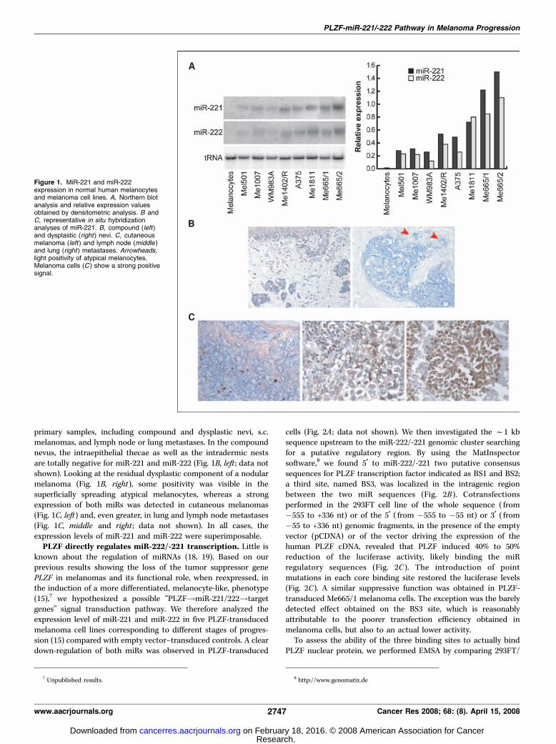
primary samples, including compound and dysplastic nevi, s.c.melanomas, and lymph node or lung metastases. In the compoundnevus, the intraepithelial thecae as well as the intradermic nestsare totally negative for miR-221 and miR-222 (Fig. 1B, left ; data notshown). Looking at the residual dysplastic component of a nodularmelanoma (Fig. 1B, right), some positivity was visible in thesuperficially spreading atypical melanocytes, whereas a strongexpression of both miRs was detected in cutaneous melanomas(Fig. 1C, left) and, even greater, in lung and lymph node metastases(Fig. 1C, middle and right ; data not shown). In all cases, theexpression levels of miR-221 and miR-222 were superimposable.PLZF directly regulates miR-222/-221 transcription. Little is
known about the regulation of miRNAs (18, 19). Based on ourprevious results showing the loss of the tumor suppressor genePLZF in melanomas and its functional role, when reexpressed, inthe induction of a more differentiated, melanocyte-like, phenotype(15),7 we hypothesized a possible ‘‘PLZF!miR-221/222!targetgenes’’ signal transduction pathway. We therefore analyzed theexpression level of miR-221 and miR-222 in five PLZF-transducedmelanoma cell lines corresponding to different stages of progres-sion (15) compared with empty vector–transduced controls. A cleardown-regulation of both miRs was observed in PLZF-transduced
cells (Fig. 2A ; data not shown). We then investigated the f1 kbsequence upstream to the miR-222/-221 genomic cluster searchingfor a putative regulatory region. By using the MatInspectorsoftware,8 we found 5¶ to miR-222/-221 two putative consensussequences for PLZF transcription factor indicated as BS1 and BS2;a third site, named BS3, was localized in the intragenic regionbetween the two miR sequences (Fig. 2B). Cotransfectionsperformed in the 293FT cell line of the whole sequence ( from�555 to +336 nt) or of the 5¶ ( from �555 to �55 nt) or 3¶ ( from�55 to +336 nt) genomic fragments, in the presence of the emptyvector (pCDNA) or of the vector driving the expression of thehuman PLZF cDNA, revealed that PLZF induced 40% to 50%reduction of the luciferase activity, likely binding the miRregulatory sequences (Fig. 2C ). The introduction of pointmutations in each core binding site restored the luciferase levels(Fig. 2C). A similar suppressive function was obtained in PLZF-transduced Me665/1 melanoma cells. The exception was the barelydetected effect obtained on the BS3 site, which is reasonablyattributable to the poorer transfection efficiency obtained inmelanoma cells, but also to an actual lower activity.To assess the ability of the three binding sites to actually bind
PLZF nuclear protein, we performed EMSA by comparing 293FT/
7 Unpublished results. 8 http://www.genomatix.de
Figure 1. MiR-221 and miR-222expression in normal human melanocytesand melanoma cell lines. A, Northern blotanalysis and relative expression valuesobtained by densitometric analysis. B andC, representative in situ hybridizationanalyses of miR-221. B, compound (left)and dysplastic (right ) nevi. C, cutaneousmelanoma (left ) and lymph node (middle )and lung (right ) metastases. Arrowheads,light positivity of atypical melanocytes.Melanoma cells (C ) show a strong positivesignal.
PLZF-miR-221/-222 Pathway in Melanoma Progression
www.aacrjournals.org 2747 Cancer Res 2008; 68: (8). April 15, 2008
Research. on February 18, 2016. © 2008 American Association for Cancercancerres.aacrjournals.org Downloaded from

GFP and 293FT/PLZF nuclear protein extracts. EMSA revealedDNA-PLZF–containing complexes on BS1 and BS2 sites and, withan apparent lower affinity, on BS3; the DNA-protein complexeswere competed out by wild-type oligomers and the specificity wasconfirmed by both supershift and site-specific mutations (seeSupplementary Fig. S1 and Materials and Methods). As an internalcontrol, we also included the PLZF binding site shown on the TpoRpromoter region (data not shown; ref. 20). Finally, the in vivointeraction between these putative cis-regulatory elements andPLZF was investigated by chromatin immunoprecipitation (ChIP)
assays performed in control HeLa and Me665/1 cell lines comparedwith the corresponding PLZF-transduced cells. PCR amplifica-tions of unsheared input genomic DNA and anti-PLZF antibody-mediated reaction gave PCR products of the expected sizes,whereas the same reactions, immunoprecipitated with an irrele-vant antibody, gave a barely detectable or undetectable amplifiedproduct (Fig. 2D), thus confirming direct and specific binding ofPLZF. In agreement with the transfection results, we did not obtainPLZF binding on BS3 site when ChIP was performed in Me665/1melanoma cell line. Accordingly, in melanomas, we always found
Figure 2. PLZF down-regulates miR-221and miR-222. A, Northern blot analysisof empty vector– or PLZF-transducedmelanoma cell lines. B, schematic of thegenomic region upstream to pre–miR-222/-221. BS1, BS2 , and BS3, PLZF putativebinding sites; horizontal arrows, directionof miR transcription. C, luciferase assaysobtained by cotransfecting differentgenomic fragments (as shown in B) in293FT cells, transfected or not withPLZF, and in Me665/1 control or PLZF-transduced melanoma cells. As controls,mutated binding sites are included.j, P < 0.05 pGL3/(�555/+336) fragmentversus basal activity; **, P < 0.001;*, P < 0.005 pGL3 constructs in PLZFversus empty vector–transduced cells.D, ChIP assays were performed with HeLaand PLZF/HeLa (left) and Me665/1 andPLZF/Me665/1 (right ), subsequentlyanalyzed by PCR. mut, mutated.
Cancer Research
Cancer Res 2008; 68: (8). April 15, 2008 2748 www.aacrjournals.org
Research. on February 18, 2016. © 2008 American Association for Cancercancerres.aacrjournals.org Downloaded from

an apparent coregulation of miR-221 and miR-222 (see Figs. 1Aand 2A).Overexpression of miR-221/-222 results in increased tumor-
igenesis. To directly test the functional role of miR-221/-222 ontumorigenesis, we used a lentiviral vector to transduce theMe1402/R melanoma cell line, selected on the basis of its lowbut detectable levels of miR-221/-222 and of its ability to producemelanin, a function often lost in more advanced melanomas.Northern blot analysis confirmed miR overexpression in miR-transduced versus empty vector–transduced control cells (Supple-mentary Fig. S2A).Melanoma cells overexpressing miR-221 or miR-222 showed an
increase in the proliferative rate, regardless of the serumconcentration (Supplementary Fig. S1B ; data not shown). Accord-ingly, by cell cycle analysis on hydroxyurea-synchronized cells, miR-transduced Me1402/R cells showed a decrease of G1 and acorresponding increase of the S and G2-M phases. At time 0, flowcytometric analysis of DNA showed 80% to 85% of the cells in theG1 phase; the analysis, successively performed at 2, 4, and 6 hoursafter hydroxyurea removal, revealed an earlier onset of DNAsynthesis induced by miR-221 and miR-222 paralleled by a fasterreduction of G1 cells, contributing to the proliferative advantage(Fig. 3A).As a next step, a Boyden chamber assay was used to measure the
effects of miR-221 and miR-222 overexpression on cellularmigration and invasion of these melanoma cells. In both assaysfor both miRs, we observed a significant increase in the invasiveand chemotactic capabilities (Fig. 3B). The effects of miR-221/-222overexpression were also evaluated on melanoma capacities offorming foci in agar semisolid medium. A significant enhancementof 10- and 5-fold of the number of foci was observed (Fig. 3C) formiR-221 and miR-222, respectively.Finally, to further confirm miR-221 and miR-222 functions on
melanoma tumorigenicity, we studied their role in an in vivo model.MiR- and empty vector–transduced Me1402/R cell lines wereinjected s.c. into athymic nude mice, and tumor growths followedthrough 4 weeks. Tumor volumes of miR-expressing melanoma
cells showed a significant increase when compared with controls atall time points (Fig. 3D ; data not shown).Suppression of endogenous miR-221 and miR-222 by
antagomir treatment. The functional effects of miR-221 andmiR-222 inhibitions were also analyzed in an advanced melanomain an attempt to reduce its malignancy. The Me665/1 metastaticcell line was treated either in vitro or in vivo with antagomir-221and/or antagomir-222 molecules. This new class of antisenseconsists of RNA oligonucleotides able to efficiently and stablyknock down specific miRs (16, 17). In vitro treatment reducedthe proliferation rate of 60% to 70% with respect to cells eitheruntreated or treated with an unrelated antagomir (i.e., the anti-sense sequence targeting miR-133a, whose expression is restrictedto heart and skeletal muscle; Fig. 4A, left ; ref. 17). The specificity ofthe down-regulation of miRs was confirmed by quantitative real-time reverse transcription-PCR (RT-PCR) and, when possible, byNorthern blot (Fig. 4A, right ; data not shown). As expected,considering the high level of homology, we observed a partial cross-reaction between antagomir-221 and antagomir-222. Moreover,Me665/1 cells transfected with antagomir-221 and/or antagomir-222 showed a decrease in both their invasion and migrationabilities (Fig. 4B) and in the number of foci outgrowing insemisolid medium, compared with control antagomir-miR-133a–treated cells (Fig. 4C). Interestingly, the few small colonies derivedfrom antagomir-221– and/or antagomir-222–transfected cellsdisplayed a flat, nontransformed morphology (Fig. 4C). Finally,in vivo , one bolus intratumor injection of antagomirs-221+222 intoathymic nude mice previously inoculated with parental Me665/1cells inhibited tumor progression at least for the successive 7 days,a time point in which antagomir-treated nodules showed largenecrotic regions (Fig. 4D ; data not shown). Altogether, the resultsindicate the capacity of antagomir-221 and antagomir-222 toinhibit melanoma progression both in vitro and in vivo .Analysis of miR-221 and miR-222 target genes: c-KIT
receptor. MiR-221 and miR-222 were already reported to targetc-KIT in normal human erythropoietic (13) and endothelial (21)cells; in addition, c-KIT is known to play several critical roles in the
Figure 3. MiR-221 and miR-222overexpression in Me1402/R: In vitro andin vivo functional studies. A, cell cycleanalysis in synchronized cells. B, invasionand chemotaxis assays. Columns, mean;bars, SD. C, representative results ofcell colony growth in semisolid medium.D, in vivo tumor growth into athymic nudemice. Tumor volumes were evaluated afters.c. injection of 5 � 106 cells. Data arerepresentative of at least two independentexperiments. Control, empty vector–transduced Me1402/R cell line. *, P < 0.05;**, P < 0.01.
PLZF-miR-221/-222 Pathway in Melanoma Progression
www.aacrjournals.org 2749 Cancer Res 2008; 68: (8). April 15, 2008
Research. on February 18, 2016. © 2008 American Association for Cancercancerres.aacrjournals.org Downloaded from

melanocytes physiology (14). Based on these findings, we analyzedmiR-221/-222 as putative upstream regulators of c-KIT receptor inthe tumorigenic transformation process of melanocytes. We founda striking inverse correlation between miR-221 and miR-222 andc-KIT expression levels during melanoma progression (Fig. 5A).Western blot analysis showed that c-KIT expression was reduced inmiR-221– and miR-222–transduced Me1402/R melanoma com-pared with control cell line (Fig. 5B, left), whereas c-KIT mRNA wasonly slightly modified (Fig. 5B, right). In the same cells, we observedthat MITF, a gene downstream to the c-KIT/mitogen-activatedprotein kinase pathway and known to exert a multifunctional rolein the complex network leading to melanogenesis (22), wasmoderately down-regulated (Supplementary Fig. S3; Fig. 5C). Thereduction of MITF activity, in turn, leads to decreased levels ofMITF-regulated enzymes, tyrosinase (TYR), and tyrosinase-relatedprotein-1 (TRP-1; Fig. 5C ; ref. 23). Also, by measuring theabsorbance of miR-221– or miR-222–transduced Me1402/R versuscontrol cell lysates, we observed a decrease in melanin pigmentsynthesis of 40% and 30%, respectively (data not shown). Notably,the reverse expression pattern was observed in PLZF-transducedMe1402/R (Fig. 5B and C) as well as in the antagomir-treatedMe665/1 melanoma cell lines (Fig. 5D). In both cases, down-regulation of miRs clearly induces the up-regulation of c-KIT and ofits downstream pathway, leading to melanogenesis (SupplementaryFig. S3).
Analysis of miR-221 and miR-222 target genes: p27/CDKN1B. We focused our attention on p27, already reported asa target of miR-221 and miR-222 in the context of prostatecarcinoma and glioblastoma (11, 12). To find out whether p27 wasan actual target of miR-221 and/or miR-222 in melanoma cells, weanalyzed the levels of p27 protein and mRNA in miR- or PLZF-transduced versus control empty vector–transduced Me1402/Rmelanoma cell line. The level of p27 was increased by PLZFoverexpression (Fig. 6A). On the contrary, the amount of thep27 protein was strongly decreased after miR transduction, withoutaffecting the accumulation of p27 mRNA (Fig. 6A) and con-firming the typical features of a miRNA target gene. In addition,we observed a strong up-regulation of p27 when the endoge-nously expressed miR-221 and miR-222 were inhibited byantagomir in Me665/1 metastatic melanoma cell line (Fig. 6B).Bioinformatics-based approaches predicted the presence offour naturally occurring putative binding sites at the p27 3¶untranslated region (UTR). To investigate the direct interactionbetween the miRs and p27 mRNA and the relative functionalities ofthe putative binding sites, we separately cloned the four ‘‘seeds’’downstream to the luciferase open reading frame. Interestingly,only the presence of wild-type ‘‘seed-1’’ caused a 70% inhibi-tion of the luciferase activity, whereas visible but not signifi-cant effects were observed for the other three sites (Fig. 6C ;data not shown). Mutations within the seed-1 sequence totally
Figure 4. MiR-221 and/or miR-222 silencing by antagomir treatment in Me665/1 metastatic cell line. A, cell proliferation (left ). Columns, mean; bars, SD;representative quantitative real-time PCR after antagomir treatment (right ; dose, 200 nmol/L). RNU6B was used as internal control. B, invasion and chemotaxisassays. Columns, mean; bars, SD. C, evaluation of the foci number obtained in semisolid medium and morphologic comparison between colonies. Columns, mean;bars, SD. D, in vivo effects of antagomir-221+222. Athymic nude mice, f1 wk after s.c. injection of Me665/1, received intratumor antagomirs-221+222 or control saline(n = 5 per group). Antagomir-133a was used as a control. Columns, mean of a minimum of two independent experiments; bars, SD. **, P < 0.001; *, P < 0.05.
Cancer Research
Cancer Res 2008; 68: (8). April 15, 2008 2750 www.aacrjournals.org
Research. on February 18, 2016. © 2008 American Association for Cancercancerres.aacrjournals.org Downloaded from

abolished this repression, indicating the specificity of the action(Fig. 6C).To rule out any miR-dependent effect on p27 protein stability,
the expression levels of p27 were analyzed through a cycloheximidetreatment in parental versus antagomir-transfected Me1402/R orMe665/1 melanoma cell lines. Although the starting amounts ofp27 were obviously different in the antagomir-treated versus theuntreated cells, we did not observe significant changes in the rateof p27 degradation (see Fig. 6D, top ; data not shown). Moreover,through p27-specific silencing, we confirmed that most, if not all,of miR-221/-222 effects on cell cycling are actually mediated bythis protein. Indeed, the reduction of the proliferative rate observedin antagomir-221/-222–transfected melanoma cells was abrogatedby the simultaneous down-regulation of p27 mRNA (see Fig. 6D,bottom).
Discussion
The multistep transformation process leading from melanocytesto metastatic melanoma cells is mainly characterized by uncon-trolled proliferation, loss of CDKN, and abnormal expression ofgrowth factors or growth factor receptors (24). Although geneexpression profiling revealed that numerous molecular changes areassociated with melanoma, the prognosis of this neoplasia is stillbased on histopathologic criteria, and molecular therapies are notyet available. To improve prognostic criteria and therapeutic tools,it is essential to study in depth the molecular oncogenic pathwaysimplicated in melanoma transformation and progression.This study shows that the transcription factor PLZF binds to and
transcriptionally inhibits miRNA-221 and miRNA-222. Thus, the lackof PLZF in melanomas unblocks miR-221 and miR-222, which areincreasingly expressed along with the disease progression and, by
Figure 5. Analysis of miR-221 and miR-222 targetgenes: c-KIT. A, miR-221 and miR-222 expressions areinversely correlated with c-KIT protein level in melanomaprogression. B, representative Western blot (WB ; left)and RT-PCR (right ) analyses of c-KIT in Me1402/Rcontrol versus miR-221– or miR-222–transduced cells.C, RT-PCR analysis of MITF, TYR , and TRP-1 genes inthe same cell lines. D, Western blot of c-KIT, MITF, TYR,and TRP-1 in antagomir-221 and/or antagomir-222 versusantagomir-133a–treated (control ) Me665/1 melanomametastatic cells. GAPDH and actin were the internalcontrols. Relative expression values are shown inSupplementary Fig. S3.
PLZF-miR-221/-222 Pathway in Melanoma Progression
www.aacrjournals.org 2751 Cancer Res 2008; 68: (8). April 15, 2008
Research. on February 18, 2016. © 2008 American Association for Cancercancerres.aacrjournals.org Downloaded from

inhibiting c-KIT and p27 translation, favors the induction of a malig-nant phenotype. Functional studies showed that the overexpressionof miR-221 or miR-222 increased the proliferative growth rate, theinvasion and migration capabilities, the anchorage-independentgrowth, and reduced differentiation and melanogenesis, all hallmarksof oncogenic progression. The induction of a more tumorigenic phe-notype by miR-221/-222 was confirmed in the athymic nude micemodel. More important, suppression of endogenous miR-221/-222 bytreatment with antagomir oligonucleotides strongly reduced cellgrowth, invasion, chemotaxis, and foci formation in vitro. In vivo ,melanoma xenotransplants showed impaired progression whentreated with antagomir-221/-222 with respect to controls, at least forthe week of observation before mice were killed for tumor histology.All these findings relate to our previous results (15),9 showing
the lack of PLZF in melanomas and the induction of a less
malignant phenotype upon its reexpression in vitro as well asin vivo . Gene expression profiling of PLZF-negative versus PLZF-positive melanomas showed that PLZF controls the expression ofseveral genes involved in tumor progression. In particular, it down-regulates tumor-promoting genes, as integrin avb3 and matrixmetalloproteinase (MMP)-9 , and conversely induces genes favoringmelanoma cell differentiation, as c-KIT and the downstream MITF,TYR , and TRP-1 . We here show that the expression of thesedifferentiation genes is miR-221/-222 dependent, shedding newlight on our previous hypothesis, suggesting that PLZF down-regulates an ‘‘unknown’’ repressor (15). In fact, PLZF–miR-221/-222pathway mediates the suppression of the differentiation-associatedgenes. Considering the down-regulation of MMP-2 and MMP-9 inPLZF-transduced melanomas, we searched for the involvementof these metalloproteases as one of the cause underlying theincreased invasiveness of miR-221/-222–overexpressing cells.RT-PCR analysis confirmed a 2- to 3-fold increase of both MMP-2and MMP-9 in miR-transduced Me1402/R and a parallel decrease
Figure 6. Analysis of miR-221 and miR-222 target genes:p27/CDKN1B. A, representative Western blot (left ) andquantitative RT-PCR (right ) analyses in Me1402/Rcontrol versus PLZF-, miR-221–, or 222–transduced cells.B, Western blot of antagomir-221– and/or antagomir-222–treated versus antagomir-133–treated (control )Me665/1 melanoma cells. Actin was used as internalcontrol. C, luciferase reporter assay (columns, meanof minimum of 5 experiments per group; bars, SD)performed by cotransfecting miR-221 and/or miR-222with a Luc reporter gene linked to p27 3¶ UTR; mutated 3¶UTR sequence and a nontargeting oligomer were alsoincluded. **, P < 0.001. D, top, Me665/1 cells weretransfected with either antagomir-133, as a control,or with antagomir-221+222 and treated with cycloheximide(CHX ). Protein p27 and actin levels were evaluatedat the indicated time points and the normalized amountsplotted as a regression plot. Bottom, Me665/1 cells weretransfected with either siControl or sip27; after 24 h, cellswere transfected again with antagomir-133, as a control,or with antagomir-221+222. Proteins from the differentcell populations were analyzed for p27 levels, and cellproliferation was evaluated. Wt, wild-type; Non-targ,nontargeting.
9 Unpublished.
Cancer Research
Cancer Res 2008; 68: (8). April 15, 2008 2752 www.aacrjournals.org
Research. on February 18, 2016. © 2008 American Association for Cancercancerres.aacrjournals.org Downloaded from
in antagomir-221+222–transfected Me665/1 cell line (data notshown).The enhanced expression of miR-221/-222 in melanomas acti-
vates at least two important pathways governing cell proliferationand melanogenesis through p27 and c-KIT receptor regulation,respectively (Supplementary Fig. S4; Figs. 5 and 6).p27 plays an important function in regulating progression
through the cell cycle, from G1 to S phase, by binding to CDK/CYCLIN complexes (25). Recently, cell cycle modulators have beenshown to have a predictive and prognostic value in a 10-yearmelanoma follow-up (26); accordingly, p27 expression is progres-sively lost during progression from benign nevi to metastatic cells,and its reduction is associated with a poor survival (25, 27, 28).Although p27 is a recognized tumor suppressor, inactivating pointmutations are rare and p27 protein levels are mostly regulatedat posttranscriptional/posttranslational level. The principal p27regulatory mechanisms include ubiquitin-dependent degradationby the proteasome (29), functional inactivation by mislocalizationinto the cytoplasm, or phosphorylation events controlling p27binding to its cellular targets (30, 31). We propose that the miR-221/-222–based mechanism, blocking p27 translation, mightrepresent an additional oncogenic mechanism underlying theabnormal cell cycle rate of advanced melanoma and of manyother tumors. Accordingly, the knockdown of miR-221 and/or miR-222 increases p27 in PC3 prostate and U87 glioblastoma cell lines(11, 12).The c-KIT receptor is a melanocytic multifunctional player
regulating melanogenesis, cell growth, migration, and survival (14).This receptor is directly targeted by miR-221/-222 in normalerythropoiesis (13) and neoangiogenesis (32), as well as in papillarythyroid carcinoma (9). In the progression of human melanoma, theloss of c-KIT is a crucial event; up to 70% of metastases lack thereceptor and can, as a consequence, escape SCF/c-KIT–triggeredapoptosis (33). Looking at the downstream transduction pathwayleading to melanogenesis and melanocytes differentiation, wefound, as a secondary target gene of miR-221 and miR-222, MITF, amaster lineage regulator in melanocyte maturation (23) controlling,among several other functions, the main melanogenic enzymessuch as TYR and TRP-1. Recent results showed that MITF isapparently able to play both antiproliferative and proproliferativefunctions, depending on its expression level (34). Specifically, moredifferentiated melanomas express higher levels of MITF and exhibitless aggressive phenotypes, whereas intermediate amounts ofMITF, as in melanomas analyzed here, induce proliferation (35–37).
Depletion of MITF has been recently reported to increase p27stability (37), whereas we observed a comparable expressionpattern of MITF and p27 in the analyzed melanoma cell lines.This apparent divergence might be reconciled, considering theslight miR-dependent modulation of MITF in miR-221/-222–expressing cells (see Supplementary Fig. S3) compared with theabrogation of MITF using siRNA (37). p27 expression level mightderive from a dynamic equilibrium that, at least in melanomacell lines, seems to favor p27 inhibition, possibly because of miR-221/-222 amounts and kinetics. Moreover, we did not find anyeffect on p27 protein stability due to miR-221/-222 and down-stream targets (including MITF). Finally, we point out that MITF is,on average, expressed at significantly lower level in melanomasthan in melanocytes and that increased levels of MITF seem toreduce melanoma proliferation even in the presence of mutatedB-RAF (38).In conclusion, we showed that PLZF negatively regulates the
expression of miR-221 and miR-222. In advanced melanomas, PLZFsilencing up-modulates these two miRs that, in turn, activate atleast two oncogenic pathways involved in melanoma progression,through p27 and c-KIT deregulation (Supplementary Fig. S4).In particular, p27 suppression operates in many tumors, such aspancreatic cancer, glioblastoma, thyroid carcinoma (8–11), andprostate cancer (12), whereas the tumorigenic action exertedthrough c-KIT down-regulation is shared by few other neoplasias(9). The most common mechanism of c-KIT–based oncogenesis isactually represented by activating mutations, as in gastrointestinalstromal tumors (39). The present studies may lead to newmolecular therapies in advanced melanoma, which still lackseffective treatments (40). Although PLZF reexpression mightrepresent a valuable tool in reducing the malignant phenotype(15), the intrinsic limitations of gene therapy render this approachdifficult. Based on in vitro and in vivo results, we propose theinhibition of miR-221/-222 by antagomir treatment as a moreattractive and safe approach for translation into the clinical setting.
Acknowledgments
Received 7/5/2007; revised 1/2/2008; accepted 2/7/2008.Grant support: Italian Ministry of Health (A. Care); Italy-USA miR Oncology
Program, Istituto Superiore di Sanita, Rome (C. Peschle); and Italian Association forCancer Research (M.P. Colombo).
The costs of publication of this article were defrayed in part by the payment of pagecharges. This article must therefore be hereby marked advertisement in accordancewith 18 U.S.C. Section 1734 solely to indicate this fact.
We thank G. Loreto for figure preparation and E. Meccia for technical support.
References
1. Gray-Schopfer V, Wellbrock C, Marais R. Melanomabiology and new target therapy. Nature 2007;445:851–7.
2. Miller AJ, Mihm MC. Melanoma. N Engl J Med 2006;355:51–65.
3. Huang S, Jean D, Luca M, Tainsky MA, Bar-Eli M. Lossof AP-2 results in downregulation of c-KIT andenhancement of melanoma tumorigenicity and metas-tasis. EMBO J 1998;17:4358–69.
4. Dhomen N, Marais R. New insight into BRAF mutationin cancer. Curr Opin Genet Dev 2007;17:31–9.
5. Clark WH, Elder DE, Guerry D IV, Epstein MN, GreeneMH, Van Horn M. A study of tumor progression: theprecursor lesion of superficial spreading and nodularmelanoma. Hum Pathol 1984;15:1147–65.
6. Calin GA, Croce CM. MicroRNA signatures in humancancers. Nat Rev Cancer 2006;6:857–66.
7. Esquela-Kerscher A, Slack FJ. Oncomirs-microRNAswith a role in cancer. Nat Rev Cancer 2006;6:259–69.
8. Lee EJ, Gusev Y, Jiang J, et al. Expression profilingidentifies microRNA signature in pancreatic cancer. Int JCancer 2007;120:1046–54.
9. He H, Jazdzewski K, Li W, et al. The role of microRNAgenes in papillary thyroid carcinoma. Proc Natl Acad SciU S A 2005;102:19075–80.
10. Ciafre SA, Galardi S, Mangiola A, et al. Exten-sive modulation of a set of microRNA in primaryglioblastoma. Biochem Biophys Res Commun 2005;334:1351–8.
11. le Sage C, Nagel R, Egan DA, et al. Regulation of thep27Kip1 tumor suppressor by miR-221 and miR-222promotes cancer cell proliferation. EMBO J 2007;26:3699–708.
12. Galardi S, Mercatelli N, Giorda E, et al. MiR-221 andmiR-222 expression affects the proliferation potencial of
human prostate carcinoma cell lines by targetingp27kip1. J Biol Chem 2007;282:23716–24.
13. Felli N, Fontana L, Pelosi E, et al. MicroRNAs 221 and222 inhibit normal erythropoiesis and erythroleukemiccell growth via kit receptor down-modulation. Proc NatlAcad Sci U S A 2005;102:18081–6.
14. Alexeev V, Yoon K. Distinctive role of the c-kitreceptor tyrosine kinase signaling in mammalianmelanocytes. J Invest Dermatol 2006;126:1102–10.
15. Felicetti F, Bottero L, Felli N, et al. Role of PLZF inmelanoma progression. Oncogene 2004;3:4567–76.
16. Krutzfeldt J, Rajewsky N, Braich R, et al. Silencing ofmicroRNAs in vivo with ‘‘antagomirs’’. Nature 2005;438:685–9.
17. Care A, Catalucci D, Felicetti F, et al. MicroRNA-133 controls cardiac hypertrophy. Nat Med 2007;13:613–8.
18. Zhou X, Ruan J, Wang G, Zhang W. Characterization
PLZF-miR-221/-222 Pathway in Melanoma Progression
www.aacrjournals.org 2753 Cancer Res 2008; 68: (8). April 15, 2008
Research. on February 18, 2016. © 2008 American Association for Cancercancerres.aacrjournals.org Downloaded from

Cancer Research
Cancer Res 2008; 68: (8). April 15, 2008 2754 www.aacrjournals.org
and identification of microRNA core promoters in fourmodel species. PLoS Comput Biol 2007;3:412–23.
19. Fazi F, Rosa A, Fatica A, et al. A minicircuitrycomprised of microRNA-223 and transcription factorsNFI-A and C/EBPa regulates human granulopoiesis. Cell2005;123:819–31.
20. Labbaye C, Quaranta MT, Pagliuca A, et al. PLZFinduces megakaryocytic development, activates Tporeceptor expression and interacts with GATA1 protein.Oncogene 2002;21:6669–79.
21. Poliseno L, Tuccoli A, Mariani L, et al. MicroRNAsmodulate the angiogenic properties of HUVECs. Blood2006;108:3068–71.
22. Hemesath TJ, Price ER, Takemoto C, Badalian T,Fisher DE. MAP kinase links the transcription factorMicrophthalmia to c-kit signalling in melanocytes.Nature 1998;391:298–301.
23. Levy C, Khaled M, Fisher DE. MITF: master regulatorof melanocyte development and melanoma oncogene.Trends Mol Med 2006;12:406–14.
24. Polsky D, Cordon-Cardo C. Oncogenes in melanoma.Oncogene 2003;22:3087–91.
25. Li W, Sanki A, Karim RZ, et al. The role of cell cycleregulatory proteins in the pathogenesis of melanoma.Pathology 2006;38:287–01.
26. Tchernev G, Orfanos CE. Downregulation of cell cycle
modulators p21, p27, p53, Rb and proapoptotic Bcl-2-related proteins Bax and Bak in cutaneous melanoma isassociated with worse patient prognosis: preliminaryfindings. J Cutan Pathol 2007;34:247–56.
27. Florenes VA, Maelandsmo GM, Kerbel RS, SlingerlandJM, Nesland JM, Holm R. Protein expression of the cell-cycle inihibitor p27Kip1 in malignant melanoma:inverse correlation with disease-free survival. Am JPathol 1998;153:305–12.
28. Ivan D, Diwan AH, Esteva FJ, Prieto VG. Expression ofcell cycle p27Kip1 and its inactivator Jab1 in melano-cytic lesions. Mod Pathol 2004;17:811–8.
29. Kotoshiba S, Kamura T, Hara T, Ishida N, NakayamaKI. Molecular dissection of the interaction between p27and Kip1 ubiquitylation-promoting complex, the ubiq-uitin ligase that regulates proteolysis of p27 in G1 phase.J Biol Chem 2005;280:17694–700.
30. Delmas C, Aragou N, Poussard S, Cottin P, DarbonJM, Manenti S. MAP kinase-dependent degradation ofp27Kip1 by calpains in choroidal melanoma cells.Requirement of p27Kip1 nuclear export. J Biol Chem2003;278:12443–51.
31. Koff A. How to decrease p27Kip1 levels during tumordevelopment. Cancer Cell 2006;9:75–6.
32. Suarez Y, Fernandez-Hernando C, Pober JS, SessaWC. Dicer dependent microRNAs regulate gene expres-
sion and functions in human endothelial cells. Circ Res2007;100:1164–73.
33. Hussein MR, Haemel AK, Wood GS. Apoptosis andmelanoma: molecular mechanisms. J Pathol 2003;199:275–88.
34. Goding C, Meyskens FL. Microphthalmic-associat-ed transcription factor integrates melanocyte biologyand melanoma progression. Clin Cancer Res 2006;12:1069–73.
35. Price ER, Ding H-F, Badalian T, et al. Lineage-specificsignaling in melanocytes. J Biol Chem 1998;273:17983–6.
36. Lekmine F, Chang CK, Sethakorn N, Das Gupta TK,Salti GI. Role of microphthalmia transcription factor(Mitf) in melanoma differentiation. Biochem BiophysRes Commun 2007;354:830–5.
37. Carreira S, Goodall J, Denat L, et al. Mitf regulation ofDia 1 controls melanoma proliferation and invasiveness.Genes Dev 2006;20:3426–39.
38. Wellbrock C, Marais R. Elevated expression of MITFcounteracts B-RAF-stimulated melanocyte and melano-ma cell proliferation. J Cell Biol 2005;170:703–8.
39. Fletcher JA, Rubin BP. KIT mutations in GIST. CurrOpin Genet Dev 2007;17:3–7.
40. Wolchok JD, Saenger YM. Current topics in melano-ma. Curr Opin Oncol 2007;19:116–20.
Research. on February 18, 2016. © 2008 American Association for Cancercancerres.aacrjournals.org Downloaded from

2008;68:2745-2754. Cancer Res Federica Felicetti, M. Cristina Errico, Lisabianca Bottero, et al. Oncogenic MechanismsPathway Controls Melanoma Progression through Multiple
MicroRNA-221/-222−The Promyelocytic Leukemia Zinc Finger
Updated version
http://cancerres.aacrjournals.org/content/68/8/2745
Access the most recent version of this article at:
Material
Supplementary
http://cancerres.aacrjournals.org/content/suppl/2008/04/11/68.8.2745.DC1.html
Access the most recent supplemental material at:
Cited articles
http://cancerres.aacrjournals.org/content/68/8/2745.full.html#ref-list-1
This article cites 40 articles, 12 of which you can access for free at:
Citing articles
http://cancerres.aacrjournals.org/content/68/8/2745.full.html#related-urls
This article has been cited by 29 HighWire-hosted articles. Access the articles at:
E-mail alerts related to this article or journal.Sign up to receive free email-alerts
Subscriptions
Reprints and
To order reprints of this article or to subscribe to the journal, contact the AACR Publications
Permissions
To request permission to re-use all or part of this article, contact the AACR Publications
Research. on February 18, 2016. © 2008 American Association for Cancercancerres.aacrjournals.org Downloaded from


![[2016] JMSC Civ. 221 - Supreme Court of Jamaica](https://static.fdokumen.com/doc/165x107/63245e343a06c6d45f0688d3/2016-jmsc-civ-221-supreme-court-of-jamaica.jpg)