Misguided Transcriptional Elongation Causes Mixed Lineage Leukemia
Upload
khangminh22Category
view
0download
0
FEMS Microbiology Reviews, fuaa003, 44, 2020, 208–218
doi: 10.1093/femsre/fuaa003Advance Access Publication Date: 3 February 2020Review Article
REVIEW ARTICLE
Translation elongation factor P (EF-P)Katherine R. Hummels and Daniel B. Kearns*
Department of Biology, Indiana University, Bloomington, IN USA∗Corresponding author: Jordan Hall, 469, 1001 E 3rd St, Bloomington, IN 47405. Tel: +(812) 856–2523; E-mail: [email protected]
One sentence summary: Here the authors review how models of elongation factor P have changed over time.
Editor: Franz Narberhaus
ABSTRACT
Translation elongation factor P (EF-P) is conserved in all three domains of life (called eIF5A and aIF5A in eukaryotes andarchaea, respectively) and functions to alleviate ribosome pausing during the translation of specific sequences, includingconsecutive proline residues. EF-P was identified in 1975 as a factor that stimulated the peptidyltransferase reaction in vitrobut its involvement in the translation of tandem proline residues was not uncovered until 2013. Throughout the fourdecades of EF-P research, perceptions of EF-P function have changed dramatically. In particular, while EF-P was thought topotentiate the formation of the first peptide bond in a protein, it is now broadly accepted to act throughout translationelongation. Further, EF-P was initially reported to be essential, but recent work has shown that the requirement of EF-P forgrowth is conditional. Finally, it is thought that post-translational modification of EF-P is strictly required for its functionbut recent studies suggest that EF-P modification may play a more nuanced role in EF-P activity. Here, we review the historyof EF-P research, with an emphasis on its initial isolation and characterization as well as the discoveries that altered ourperceptions of its function.
Keywords: EF-P; translation; ribosome pausing; proline; eIF5a
INTRODUCTION
Translation, or the process of decoding mRNA transcripts intoamino acid primary protein sequences, is central to all life.Peptide bond formation between amino acids is catalyzed bya highly conserved molecular machine called the ribosome(Roberts 1958). Ribosomes are processive enzymes capable ofsynthesizing long stretches of polypeptides at a high catalyticrate ranging from 6 (in the case of eukaryotes) to 20 (in the case ofbacteria) peptide bonds per second (Pederson 1984; Bostrom et al.1986; Liang et al. 2000; Ignolia, Lareau and Weissman 2011). Therate of translation elongation depends, at least in part, on thetemplate and can be slowed by the interaction of the ribosomewith particular mRNA sequences, the decoding of rare codons,and the translation of certain consecutive amino acids (Tanneret al. 2009; Ignolia, Lareau and Weissman 2011; Li, Oh and Weiss-man 2012; Charneski and Hurst 2013; Chevance, Le Guyon andHughes 2014; Dana and Tuller 2014; Gardin et al. 2014). Ratherthan being an unfortunate and unavoidable feature of the ribo-some, suboptimal translation efficiency has been implicated in
the regulation of protein copy number, folding, and secretion(Ude et al. 2013; Fluman et al. 2014; Qi et al. 2018).
The majority of the ribosome is made up of rRNA, whichcomprises approximately 60% of the structure and is directlyresponsible for the catalysis of peptide bond formation (Tissiereset al. 1959; Nissen et al. 2000). Ribosomes also contain ∼50 pro-teins that serve to stabilize the core rRNA structure as well asassimilate signals from other cellular pathways. In addition tothe canonical ribosome structural proteins, elongation factorsthat transiently interact with the ribosome during translationelongation are also ubiquitous. For example, translation elonga-tion factors EF-Tu and EF-G are responsible for delivering amino-acylated tRNAs to the ribosome and ensuring proper ribosometranslocation following peptide bond formation, respectively(Reviewed in Czworkowski and Moore 1996). While associationis transient, EF-Tu and EF-G nonetheless interact with the ribo-some and participate in each cycle of peptide bond formation.By contrast, another elongation factor, EF-P, only participates ina subset of peptide bond formation events.
Received: 28 August 2019; Accepted: 30 January 2020
C© The Author(s) 2020. Published by Oxford University Press on behalf of FEMS. All rights reserved. For permissions, please e-mail:[email protected]
208
Dow
nloaded from https://academ
ic.oup.com/fem
sre/article/44/2/208/5721369 by guest on 16 January 2022

Hummels and Kearns 209
EF-P
Paused Ribosome
Actively TranslatingRibosome
EF-P-MediatedPause Alleviation
Key
tRNA
ProlineOther Amino Acids
Figure 1. EF-P alleviates ribosome pausing at consecutive proline residues. Graphic depicting the current model of the function of EF-P in translation. Ribosomes pauseduring the translation of particular amino acid sequences such as consecutive proline residues, and pausing results in an empty E-site that allows the entry of EF-P.Upon association with a paused ribosome, EF-P interacts with the P-site tRNA and alleviates ribosome pausing.
EF-P is conserved in all domains of life, where it is calledeIF5A in eukaryotes and aIF5A in archaea and was recentlyshown to alleviate ribosome pausing during the translation ofconsecutive proline residues (Fig. 1) (Kyrpides and Woese 1998;Doerfel et al. 2013; Ude et al. 2013). Structural analysis indi-cates that consecutive proline residues impose an unfavorablepeptidyl-tRNA geometry that is counteracted by EF-P to pro-mote translation elongation (Huter et al. 2017). EF-P is post-translationally modified in all organisms studied to date andit is thought that post-translational modification is importantto alleviate ribosome pausing (Yanagisawa et al. 2010; Doerfelet al. 2013; Lassak et al. 2015; Yanagisawa et al 2016). While it isnow widely accepted that EF-P acts throughout translation elon-gation, understanding of its function was misled by the beliefthat it was primarily involved in the catalysis of the peptidebond between the first two amino acids and was thought to beabsolutely essential for growth. While EF-P is essential in manyorganisms, recent work has shown that the requirement of EF-Pfor growth in Escherichia coli is conditional and even dispensablein Bacillus subtilis, suggesting a more complex role for EF-P incellular viability (Rajkovic et al. 2016; Tollerson, Witzky and Ibba2018 ). Throughout the history of EF-P research, views of its func-tion have quickly arisen and are later called into question. Here,we review the history of EF-P research with a focus on how ourunderstanding of its function in the cell has changed over time.While the eukaryotic and archaeal orthologs eIF5A and aIF5A arenot the focus of this review (see Park and Wolff 2018 and Turpaev2018 for recent reviews), we will also highlight aspects of theirhistory when they influenced our understanding of EF-P func-tion in bacteria.
EF-P DISCOVERY
Early experiments aimed to interrogate the mechanism of trans-lation utilized relatively few model in vitro systems. In one, theability of factors to promote the synthesis of poly-phenylalaninepeptides was assayed. In these experiments, it was shown thatin addition to the ribosome, aminoacylated-tRNAs and an mRNAtemplate, several soluble factors (EF-G, EF-Tu and EF-Ts) wererequired (Lucas-Lenard and Lipmann 1966). EF-G, EF-Tu andEF-Ts were not required, however, for another model transla-tion system: the formation of a peptide bond between fMet-tRNAfMet and puromycin (Traut and Monro 1964; Madden, Trautand Monro 1968). Puromycin is an antibiotic that chemicallyresembles the 3’ end of aminoacylated tRNAs and, upon serv-ing as a substrate in the peptidyl-transferase reaction, it stim-ulates premature termination of translation (Yarmolinsky andde la Haba 1959; Morris and Schweet 1961). As such, deliveryof tRNA’s by EF-Tu/EF-Ts and ribosome translocation directedby EF-G are not required for the fMet-tRNAfMet-puromycin reac-tion. The puromycin model was instrumental in determining
the minimal requirements for the peptidyl-transferase reactionwhich were shown to be housed in the 50S subunit of the ribo-some (Traut and Monro 1964; Madden, Traut and Monro 1968).
While puromycin was critical to early studies of transla-tion, it is a relatively poor peptidyltransferase acceptor, resultingin slow peptide bond formation particularly when puromycinconcentrations are low (Glick and Ganoza 1975). Ribosome-mediated peptide bond formation between fMet-tRNAfMet andpuromycin, however, could be improved by adding fractions ofwhole cell lysates and sub-fractionation identified a soluble fac-tor that accelerated the reaction (Glick and Ganoza 1975). More-over, the stimulatory factor was not required for 70S ribosomeassembly or for binding of fMet-tRNAfMet and was inhibited byantibiotics that block the peptidyl-transferase center (Glick andGanoza 1975). Thus, it was proposed that this factor increasedthe ribosome’s inherent ability to catalyze peptide bond forma-tion and, due to its ability to stimulate the peptidyl-transferasereaction, it was named elongation factor P (EF-P) (Glick andGanoza 1975).
Reaction between fMet-tRNAfMet and puromycin, however,was insufficient to determine whether or not EF-P stimulatedthe peptidyl-transferase reaction throughout translation elon-gation or whether the stimulation was restricted to the first pep-tide bond. As such, the effect of EF-P on the translation of poly-phenylalanine and poly-lysine was assayed in a subsequentreport (Glick and Ganoza 1976). It was observed that addition ofEF-P increased poly-lysine synthesis ∼10-fold, consistent withit acting throughout translation elongation (Glick and Ganoza1976). While EF-P also stimulated poly-phenylalanine produc-tion, the degree to which it did so (∼2.5-fold) was lower than thatobserved for poly-lysine, indicating that the stimulatory effectwas perhaps sequence specific (Glick and Ganoza 1976).
To explore the apparent sequence specificity of EF-P, its abil-ity to promote peptide bond formation between fMet-tRNAfMet
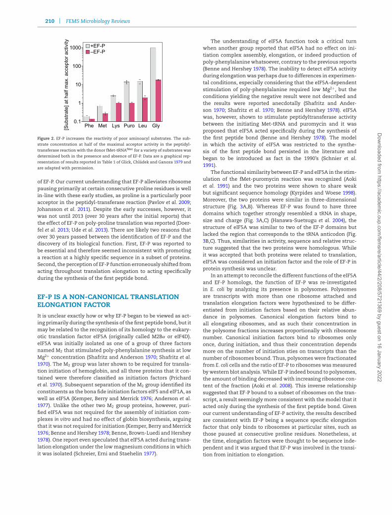
and diverse aminoacyl acceptors was tested (Glick, Chladekand Ganoza 1979). Phenylalanine is a high affinity substrate inthe peptidyl-transferase reaction and addition of EF-P had littleeffect on its reaction with fMet-tRNAfMet (Fig. 2) (Glick, Chladekand Ganoza 1979). Incorporation of lysine, however, proceededat a slower rate and increased 2.5-fold upon the addition of EF-P(Fig. 2) (Glick, Chladek and Ganoza 1979). The stimulatory effectof EF-P on peptide bond formation was even greater for aminoa-cyl accepters with less reactivity than lysine (namely leucine,puromycin and glycine) (Fig. 2) (Glick, Chladek and Ganoza 1979).Thus, EF-P stimulated peptide bond formation in a manner thatdepended on the efficiency of the acceptor and functioned toaccelerate polymerization of amino acids that would otherwisebe incorporated slowly into proteins (Glick, Chladek and Ganoza1979).
These remarkable early studies form the critical and defini-tive biochemical foundation for understanding the mechanism
Dow
nloaded from https://academ
ic.oup.com/fem
sre/article/44/2/208/5721369 by guest on 16 January 2022
210 FEMS Microbiology Reviews
Figure 2. EF-P increases the reactivity of poor aminoacyl substrates. The sub-
strate concentration at half of the maximal acceptor activity in the peptidyl-transferase reaction with the donor fMet-tRNAfMet for a variety of substrates wasdetermined both in the presence and absence of EF-P. Data are a graphical rep-
resentation of results reported in Table 1 of Glick, Chladek and Ganoza 1979 andare adapted with permission.
of EF-P. Our current understanding that EF-P alleviates ribosomepausing primarily at certain consecutive proline residues is wellin-line with these early studies, as proline is a particularly pooracceptor in the peptidyl-transferase reaction (Pavlov et al. 2009;Johansson et al. 2011). Despite the early successes, however, itwas not until 2013 (over 30 years after the initial reports) thatthe effect of EF-P on poly-proline translation was reported (Doer-fel et al. 2013; Ude et al. 2013). There are likely two reasons thatover 30 years passed between the identification of EF-P and thediscovery of its biological function. First, EF-P was reported tobe essential and therefore seemed inconsistent with promotinga reaction at a highly specific sequence in a subset of proteins.Second, the perception of EF-P function erroneously shifted fromacting throughout translation elongation to acting specificallyduring the synthesis of the first peptide bond.
EF-P IS A NON-CANONICAL TRANSLATIONELONGATION FACTOR
It is unclear exactly how or why EF-P began to be viewed as act-ing primarily during the synthesis of the first peptide bond, but itmay be related to the recognition of its homology to the eukary-otic translation factor eIF5A (originally called M2Bα or eIF4D).eIF5A was initially isolated as one of a group of three factorsnamed M2 that stimulated poly-phenylalanine synthesis at lowMg2+ concentration (Shafritz and Anderson 1970; Shafritz et al.1970). The M2 group was later shown to be required for transla-tion initiation of hemoglobin, and all three proteins that it con-tained were therefore classified as initiation factors (Prichardet al. 1970). Subsequent separation of the M2 group identified itsconstituents as the bona fide initiation factors eIF5 and eIF1A, aswell as eIF5A (Kemper, Berry and Merrick 1976; Anderson et al.1977). Unlike the other two M2 group proteins, however, puri-fied eIF5A was not required for the assembly of initiation com-plexes in vitro and had no effect of globin biosynthesis, arguingthat it was not required for initiation (Kemper, Berry and Merrick1976; Benne and Hershey 1978; Benne, Brown-Luedi and Hershey1978). One report even speculated that eIF5A acted during trans-lation elongation under the low magnesium conditions in whichit was isolated (Schreier, Erni and Staehelin 1977).
The understanding of eIF5A function took a critical turnwhen another group reported that eIF5A had no effect on ini-tiation complex assembly, elongation, or indeed production ofpoly-phenylalanine whatsoever, contrary to the previous reports(Benne and Hershey 1978). The inability to detect eIF5A activityduring elongation was perhaps due to differences in experimen-tal conditions, especially considering that the eIF5A-dependentstimulation of poly-phenylalanine required low Mg2+, but theconditions yielding the negative result were not described andthe results were reported anecdotally (Shafritz and Ander-son 1970; Shafritz et al. 1970; Benne and Hershey 1978). eIF5Awas, however, shown to stimulate peptidyltransferase activitybetween the initiating Met-tRNA and puromycin and it wasproposed that eIF5A acted specifically during the synthesis ofthe first peptide bond (Benne and Hershey 1978). The modelin which the activity of eIF5A was restricted to the synthe-sis of the first peptide bond persisted in the literature andbegan to be introduced as fact in the 1990’s (Schnier et al.1991).
The functional similarity between EF-P and eIF5A in the stim-ulation of the fMet-puromycin reaction was recognized (Aokiet al. 1991) and the two proteins were shown to share weakbut significant sequence homology (Kyrpides and Woese 1998).Moreover, the two proteins were similar in three-dimensionalstructure (Fig. 3A,B). Whereas EF-P was found to have threedomains which together strongly resembled a tRNA in shape,size and charge (Fig. 3A,C) (Hanawa-Suetsugu et al. 2004), thestructure of eIF5A was similar to two of the EF-P domains butlacked the region that corresponds to the tRNA anticodon (Fig.3B,C). Thus, similarities in activity, sequence and relative struc-ture suggested that the two proteins were homologous. Whileit was accepted that both proteins were related to translation,eIF5A was considered an initiation factor and the role of EF-P inprotein synthesis was unclear.
In an attempt to reconcile the different functions of the eIF5Aand EF-P homologs, the function of EF-P was re-investigatedin E. coli by analyzing its presence in polysomes. Polysomesare transcripts with more than one ribosome attached andtranslation elongation factors were hypothesized to be differ-entiated from initiation factors based on their relative abun-dance in polysomes. Canonical elongation factors bind toall elongating ribosomes, and as such their concentration inthe polysome fractions increases proportionally with ribosomenumber. Canonical initiation factors bind to ribosomes onlyonce, during initiation, and thus their concentration dependsmore on the number of initiation sties on transcripts than thenumber of ribosomes bound. Thus, polysomes were fractionatedfrom E. coli cells and the ratio of EF-P to ribosomes was measuredby western blot analysis. While EF-P indeed bound to polysomes,the amount of binding decreased with increasing ribosome con-tent of the fraction (Aoki et al. 2008). This inverse relationshipsuggested that EF-P bound to a subset of ribosomes on the tran-script, a result seemingly more consistent with the model that itacted only during the synthesis of the first peptide bond. Givenour current understanding of EF-P activity, the results describedare consistent with EF-P being a sequence specific elongationfactor that only binds to ribosomes at particular sites, such asthose paused at consecutive proline residues. Nonetheless, atthe time, elongation factors were thought to be sequence inde-pendent and it was argued that EF-P was involved in the transi-tion from initiation to elongation.
Dow
nloaded from https://academ
ic.oup.com/fem
sre/article/44/2/208/5721369 by guest on 16 January 2022

Hummels and Kearns 211
50S
30S
P A
eIF5A tRNAPheEF-P
EF-P
To scale
A B C DI
II
III
Figure 3. The three dimensional structures of EF-P, eIF5A, and tRNA are similar. A) Three dimensional structure of Thermus thermophilus EF-P (PDB ID 1UEB). Domains
I, II and III are labeled accordingly. B) Three dimensional structure of S. cerevisiae eIF5A (PDB ID 3ER0). C) Three dimensional structure of S. cerevisiae tRNAPhe (PDB ID1EVV). D) The location of EF-P (blue) bound to a ribosome (grey) stalled at a polyproline sequence solved by cryo-electron microscopy (PDB ID 6ENJ). The positions ofthe 50S subunit, 30S subunit, P-site tRNA (salmon) and A-site tRNA (green) are labeled accordingly.
EF-P AND eIF5A ACT THROUGHOUTTRANSLATION ELONGATION
The first in vivo evidence that eIF5A acted during translationelongation rather than initiation was provided by several stud-ies that characterized its activity in Saccharomyces cerevisiae (Jaoand Chen 2006; Zanelli et al. 2006; Saini et al. 2009). Immuno-preciptation of eIF5A indicated that it was in a complex withribosomes and elongation factors, but not initiation factors, con-sistent with it having a post-initiation function (Jao and Chen2006; Zanelli et al. 2006). Moreover polysomes, rather than mono-somes, accumulated at the non-permissive temperature in aneIF5A temperature-sensitive mutant, consistent with a defectin translation elongation (Zanelli et al. 2006; Saini et al. 2009).Finally, the role of eIF5A in elongation was solidified by com-paring the ribosome transit time in the presence and absenceof eIF5A (Saini et al. 2009). The ribosome transit time was deter-mined by radioactively labeling nascent proteins and determin-ing the amount of time elapsed between when radioactivity wasdetected in total proteins and when it was detected in completedproteins that had been released from the ribosome. Inactivationof eIF5A increased the ribosome transit time to twice that of wildtype, providing conclusive evidence that eIF5A promoted trans-lation at a step following initiation (Fig. 4) (Saini et al. 2009). Thus,it was concluded that eIF5A acted as an elongation factor and itwas speculated that EF-P likely functioned in similar manner.
Again, if eIF5A/EF-P functioned as elongation factors, theydid so in a manner that had not been previously attributed tothis class of protein. Specifically, depletion of eIF5A only resultedin a ∼30% decrease in the rate of protein synthesis in vivo whichwas not as severe as one would expected for canonical elonga-tion factors (Kang and Hershey 1994). Thus, it was concludedthat eIF5A likely only operated on a subset of transcripts. Con-sistent with conditional activity, the absence of EF-P was foundto result in a reduction in a subset of proteins in the outermembrane of Salmonella and in a subset of virulence factors ofAgrobacterium (Peng et al. 2001; Zou et al. 2012). Combined withthe ability of EF-P to stimulate a subset of peptidyl-transferasereactions (Glick, Chladek and Ganoza 1979), it seemed possiblethat EF-P and eIF5A activity was dependent on some as-yet-unknown sequence, either at the transcript or protein level.
In 2013, an aspect of the sequence-specificity of EF-P wasrevealed when two groups simultaneously reported that in E.coli, EF-P alleviated ribosome pausing at poly-proline motifs dur-ing translation elongation. In one of these seminal studies, amutant defective for EF-P activity was found to reduce expres-sion of the Cad acid resistance system (Ude et al. 2013). Exper-
Figure 4. eIF5A promotes a step in translation following initiation. Analysis of
[35S]Met incorporation into total peptides (closed symbols) or completed pep-tides (open symbols) in wild type S. cerevisiae or the eIF5A temperature sensitivemutant (eIF5Ats) at the non-permissive temperature. Ribosome transit times, ametric that is unaffected by translation initiation, are indicated. Red text and
lines added to original panels. Panel adapted with permission from Saini et al.
2009.
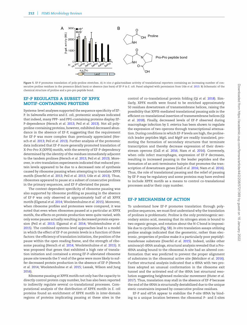
iments using translational reporters in vivo showed that EF-Ppromoted expression when the reporter gene was introduceddownstream of a PPP motif in CadC, the sensor protein respon-sible for activation of the Cad module (Ude et al. 2013). Moreover,reductions in the number of sequential prolines alleviated EF-Pdependency (Fig. 5A) (Ude et al. 2013). The other study revisitedthe early observations that EF-P promoted peptide bond forma-tion between poor amino-acyl acceptors and tested the reactiv-ity of an array of amino acids in the presence and absence ofEF-P (Doerfel et al. 2013). EF-P strongly stimulated peptide bondformation within PPP and PPG tripeptides in vitro (Doerfel et al.2013). Thus, EF-P promoted translation elongation through poly-proline-containing sequences and the observation was con-firmed for eIF5A in S. cerevisiae shortly thereafter (Gutierrez et al.2013).
Dow
nloaded from https://academ
ic.oup.com/fem
sre/article/44/2/208/5721369 by guest on 16 January 2022
212 FEMS Microbiology Reviews
NHOH
O
Proline
NH
O
N Pro-Pro peptide bond
O
OH
A B
Figure 5. EF-P promotes translation of poly-proline stretches. A) in vivo β-galactosidase activity of translational reporters containing an increasing number of con-secutive proline residues in the presence (black bars) or absence (tan bars) of EF-P in E. coli. Panel adapted with permission from Ude et al. 2013. B) Schematic of the
chemical structure of proline and a pro-pro peptide bond.
EF-P REGULATES A SUBSET OF XPPXMOTIF-CONTAINING PROTEINS
Systems-level analyses supported the sequence specificity of EF-P. In Salmonella enterica and E. coli, proteomic analyses indicatedthat indeed, many PPP- and PPG-containing proteins display EF-P-dependence (Hersch et al. 2013; Peil et al. 2013). Not all poly-proline containing proteins, however, exhibited decreased abun-dance in the absence of EF-P, suggesting that the requirementfor EF-P was more complex than previously appreciated (Her-sch et al. 2013; Peil et al. 2013). Further analysis of the proteomicdata indicated that EF-P more generally promoted translation ofX-Pro-Pro-X (XPPX) motifs, with the severity of EF-P-dependencydetermined by the identity of the residues immediately adjacentto the tandem prolines (Hersch et al. 2013; Peil et al. 2013). More-over, in vitro translation experiments indicated that reduced pro-tein levels appeared to be due to a decreased rate of synthesiscaused by ribosome pausing when attempting to translate XPPXmotifs (Doerfel et al. 2013; Peil et al. 2013; Ude et al. 2013). Thus,ribosomes appeared to pause at a subset of consecutive prolinesin the primary sequences, and EF-P alleviated the pause.
The context-dependent specificity of ribosome pausing wasalso supported by ribosome profiling as pausing in the absenceof EF-P was only observed at approximately half of all XPPXmotifs (Elgamal et al. 2014; Woolstenhulme et al. 2015). Moreover,when ribosome profiles and proteomes were compared, it wasnoted that even when ribosomes paused at a particular at XPPXmotifs, the effects on protein production were quite varied, withonly some pauses actually resulting in decreased protein expres-sion (Peil et al. 2013; Elgamal et al. 2014; Woolstenhulme et al.2015). The combined systems-level approaches lead to a modelin which the effect of EF-P on protein levels is a function of threefactors: the efficiency of translation initiation, the position of thepause within the open reading frame, and the strength of ribo-some pausing (Hersch et al. 2014; Woolstenhulme et al. 2015). Itwas proposed that genes that exhibited a high rate of transla-tion initiation and contained a strong EF-P-alleviated ribosomepause site towards the 5’ end of the gene were more likely to suf-fer decreased protein production in the absence of EF-P (Herschet al. 2014; Woolstenhulme et al. 2015; Lassak, Wilson and Jung2016).
Ribosome pausing at XPPX motifs not only has the capacity todirectly control protein copy number, but has also been reportedto indirectly regulate several co-translational processes. Com-putational analysis of the distribution of XPPX motifs in E. coliproteins found an enrichment of XPPX motifs in inter-domainregions of proteins implicating pausing at these sites in the
control of co-translational protein folding (Qi et al. 2018). Sim-ilarly, XPPX motifs were found to be enriched approximately50 residues downstream of transmembrane helices, raising thepossibility that XPPX-mediated translational pausing aids in theefficient co-translational insertion of transmembrane helices (Qiet al. 2018). Finally, decreased levels of EF-P observed duringmacrophage infection by S. enterica has been shown to regulatethe expression of two operons through transcriptional attenua-tion. During conditions in which EF-P levels are high, the proline-rich leader peptides MgtL and MgtP are readily translated, pro-moting the formation of secondary structures that terminatetranscription and thereby decrease expression of their down-stream operons (Gall et al. 2016; Nam et al. 2016). Conversely,when cells infect macrophages, expression of EF-P decreases,resulting in increased pausing in the leader peptides and theformation of an anti-terminator hairpin that promotes the tran-scription of downstream genes (Gall et al. 2016; Nam et al. 2016).Thus, the role of translational pausing and the relief of pausingby EF-P may be regulatory and some proteins may have evolvedto include XPPX motifs as a means to control co-translationalprocesses and/or their copy number.
EF-P MECHANISM OF ACTION
To understand how EF-P promotes translation through poly-proline sequences, we must first understand why the translationof prolines is problematic. Proline is the only proteinogenic sec-ondary amino acid, meaning that its nitrogen atom is bound totwo organic groups, and rotation about the N-C bond is impossi-ble due to cyclization (Fig. 5B). In vitro translation assays utilizingproline analogs indicated that the geometric, rather than elec-tronic, properties of proline make it a particularly poor peptidyl-transferase substrate (Doerfel et al. 2015). Indeed, unlike otheraminoacyl-tRNA analogs, structural analysis revealed that a Pro-tRNA analog bound to the ribosomal A-site had an altered con-formation that was predicted to prevent the proper alignmentof substrates in the ribosomal active site (Melnikov et al. 2016).Further structural analysis indicated that a tRNA with two pro-lines adopted an unusual conformation in the ribosome exittunnel and the activated end of the tRNA lost structural reso-lution suggesting heightened molecular movement (Huter et al.2017). Thus, translation may stall in the absence of EF-P becausethe end of the tRNA is structurally destabilized due to the uniquesteric constraints imposed by consecutive proline residues.
EF-P and eIF5A appear to stabilize the P-site tRNA by bind-ing to a unique location between the ribosomal P- and E-sites
Dow
nloaded from https://academ
ic.oup.com/fem
sre/article/44/2/208/5721369 by guest on 16 January 2022

Hummels and Kearns 213
(Fig. 3D) (Blaha, Stanley and Steitz 2009; Schmidt et al. 2016;Huter et al. 2017). While parts of EF-P/eIF5A extend toward thepeptidyl transferase center, the protein seems too remote to par-ticipate in the peptidyl transferase reaction directly (Blaha, Stan-ley and Steitz 2009; Schmidt et al. 2016). Instead, it appears thatEF-P/eIF5A interacts with the adjacent tRNA in the P-site (Fig. 3D)(Blaha, Stanley and Steitz 2009; Schmidt et al. 2016; Huter et al.2017). Interaction with the P-site tRNA reduces mobility of itsactivated end and the structural stabilization is thought to pro-mote peptide bond formation (Huter et al. 2017). In support ofthis model, calculation of free energy parameters between aproline analog and puromycin showed that EF-P reduced thefree energy of the reaction by altering the entropy rather thanenthalpy component (Doerfel et al. 2015). Thus, EF-P appears tobind near the E-site and restrict dynamic mobility of the P-sitetRNA to stabilize a conformation compatible with the next pep-tidyl transferase reaction. We note that the requirement of EF-P for growth in E. coli is alleviated at lower temperatures, per-haps consistent with a role of EF-P in reducing molecular motion(Tollerson, Witzky and Ibba 2018).
Recently, mutations that partially suppressed the need forEF-P in B. subtilis were reported, one of which perhaps supportthe idea that EF-P is involved in controlling space within theribosome active site (Hummels and Kearns 2019). The peptidyl-transferase reaction is a catalyzed by residues in the 23S rRNAand one residue near the active site (guanosine 2551) is methy-lated for mechanistically unknown reasons (Lovgren and Wik-strom 2001). A loss of function mutation in YacO, the enzymepredicted to add the methyl group, reduced the requirement forEF-P (Hummels and Kearns 2019). Thus, methylation of guano-sine 2551 may exacerbate constraints imposed on the poly-prolyl-tRNA and its absence may better accommodate the nextreaction. Other suppressor mutations that were identified areharder to explain. For example, change-of-function mutationswere identified in two ribosomal small subunit proteins, far fromwhere EF-P is thought to either bind or act (Hummels and Kearns2019). Still other suppressors indicated a role for proteins ofpoorly understood function but nonetheless are predicted to beribosome-associated (Hummels and Kearns 2019). At present,it is hard to tell which suppressors directly inform mechanismand which highlight as-yet-unappreciated consequences of EF-Pactivity.
Evidence suggests that EF-P may do more than alleviate ribo-some pausing at a subset of XPPX motifs. A recent study ana-lyzed E. coli EF-P binding dynamics in vivo using super-resolutionfluorescence microscopy and showed that EF-P interacted withribosomes much more often than the frequency of XPPX motifsin transcripts (Mohapatra et al. 2017). It was proposed that EF-Psamples empty ribosome E-sites during a majority of translationelongation cycles to minimize the time in which a stalled ribo-some awaits rescue (Mohapatra et al. 2017). Alternatively, EF-Pmay alleviate stalling at additional primary sequences as it doeswhen promoting translation of E. coli PoxB, a protein that doesnot contain any XPPX motifs (Hersch et al. 2013). EF-P also accel-erates the incorporation of non-standard amino acids, such ascertain D-amino acids and non-proteinogenic secondary aminoacids when they are ligated to tRNApro (Doerfel et al. 2015; Katohet al. 2016; Katoh, Iwane and Suga 2017). Beyond acceleratingtranslation, EF-P has been shown promote reading frame main-tenance at CC[C/U]-[C/U] mRNA sequences, particularly thoselocated at the second codon of an open reading frame (Gamperet al. 2015). Finally, suppressors of the efp mutant growth defectin Erwinia amylovora were identified to contain loss-of-function
mutations in the DEAH-box RNA helicase HrpA3, raising the pos-sibility that EF-P has previously unappreciated effects on mRNAprocessing (Klee et al. 2019).
Whatever the mechanism of EF-P, current thinking seemsto agree that its function is closely related to its structure, andthe size and shape of the EF-P protein resembles that of a tRNAmolecule (Fig. 3A,C). EF-P is composed of three domains. DomainIII of EF-P resembles the part of tRNA that contains the anti-codon (Fig. 3A,C). While domain III of EF-P is essential andmay recognize elements in the E-site codon, eIF5A lacks thisdomain (Fig. 3A,B) (Hanawa-Suetsugu et al. 2004; Huter et al.2017). Domain II structurally contorts EF-P into a right angleand recognizes the presence of tRNAPro by interaction with aunique structure in the tRNAPro D-loop not found in any othertRNA save tRNAfMet (Fig. 3A) (Katoh et al. 2016). Structural analy-sis of eIF5A does not support specific interactions with the D-loop tRNA, so while the two proteins appear to have similarfunctions, they appear to have different recognition elements,perhaps consistent with the ability of eIF5A to relieve pausingat non-proline containing peptides (Pelechano and Alepuz 2017;Schuller et al. 2017). The highest degree of conservation both atthe sequence and structural level between EF-P and e/aIF5A isobserved in domain I that resembles the activated end of thetRNA (Fig. 3A–C). When bound to ribosomes, domain I lies clos-est to the peptidyl-transferase center and may constitute the‘active site’ for the relief of translational pausing (Fig. 3D). Fur-ther, another conserved feature of domain I between EF-P, eIF5Aand aIF5A is the fact that a residue at the tip of domain I is post-translationally modified.
EF-P POST-TRANSLATIONAL MODIFICATION
In all organisms studied to date, EF-P is post-translationallymodified on a conserved lysine or arginine at the position anal-ogous to the aminoacylation site of tRNAs (Katz et al. 2013).Despite the ubiquity of post-translational modification (PTM) ofEF-P, eIF5A, and aIF5A, the chemical identity of the modifica-tion varies dramatically between species. To date, the followingmoieties have been identified as EF-P, eIF5A, and aIF5A PTMs: R-β-lysinyl-hydroxylysine (E. coli and S. enterica), rhamnose (Pseu-domonas aeruginosa, Neisseria meningitidis, and Shewanella onei-densis), hypusine (eukaryotes, and Sulfolobus solfataricus), deoxy-hypusine (Haloferax volcanii) and 5-aminopentanol (B. subtilis)(Cooper et al. 1983; Navarre et al. 2010; Yanagisawa et al. 2010;Peil et al. 2012; Lassak et al. 2015; Rajkovic et al. 2015; Prunettiet al. 2016; Rajkovic et al. 2016; Yanagisawa et al. 2016; Bassaniet al. 2019) (Fig. 6). In most cases, the enzymes responsible forconstructing the PTMs have been identified (reviewed in Las-sak, Wilson and Jung 2016 and Rajkovic and Ibba 2017), but theexact biosynthetic pathway for 5-aminopentanol constructionin B. subtilis as well as hypusine synthesis in archaea is unclear(Bassani et al. 2019; Witzky et al. 2018). Moreover, bioinformaticanalyses have indicated that while EF-P is ubiquitous, many bac-terial genomes lack homologs of the currently recognized EF-PPTM enzymes (Fig. 7) (Hummels et al. 2017; Rajkovic and Ibba2017). Thus, it is likely that additional as-yet-unknown moietiesare utilized to post-translationally modify EF-P.
Despite the chemical diversity of EF-P post-translationalmodification, the presence of the modification is thought toplay an integral role in the EF-P mechanism. A close connectionbetween modification and EF-P activity is supported by geneticanalyses indicating that, in many organisms, mutation of theenzymes responsible for PTM resulted in phenotypes similar to
Dow
nloaded from https://academ
ic.oup.com/fem
sre/article/44/2/208/5721369 by guest on 16 January 2022

214 FEMS Microbiology Reviews
Figure 6. EF-P, eIF5A and aIF5A are post-translationally modified. Chemical struc-tures of the moieties currently known to post-translationally modify EF-P, eIF5A
or aIF5A. The R-group of the conserved lysine or arginine residue that serves asthe site of post-translational modification is depicted in black and the modifica-tion group is depicted in green.
that observed upon mutation of EF-P (Yanagisawa et al. 2010;Zou et al. 2012; Ude et al. 2013; Lassak et al. 2015; Yanagisawaet al. 2016; Klee et al. 2019). Moreover, structural analysis of E.coli EF-P bound to the ribosome indicated that the R-β-lysinyl-hydroxylysine moiety extended toward the peptidyl-transferasecenter and interacted with the aminoacylated end of the P-sitetRNA to conformationally stabilize the nascent peptide chain(Huter et al. 2017). Thus, the modification group was in positionto participate in the alleviation of ribosome pausing, but howthe modification participates per se, and how chemically dis-tinct modification groups serve similar functions, is unclear.
In addition to mechanistically participating in EF-P/eIF5aactivity, some evidence suggests that the PTM may be regula-tory. Many PTM pathways involve maturation of the modifica-tion while it is bound to EF-P, resulting, at least transiently, in thepresence of modification intermediates. While the physiologi-cal consequences of these intermediates are still unclear, recentwork in Fusarium graminearum suggests that they have the abilityto alter eIF5A activity (Martinez-Rocha et al. 2016). F. graminearumeIF5A is modified by hypusine, and overexpression of deoxyhy-pusine synthase (DHS) resulted in a hypervirulent phenotypetowards maize and wheat (Martinez-Rocha et al. 2016). Con-versely, overexpression of the enzyme deoxyhypusine hydrolase(DOHH) dramatically reduced virulence (Martinez-Rocha et al.2016). Whereas DHS is capable of both ligating and removingdeoxyhypusine from eIF5A, the modification is only stabilizedupon its conversion to hypusine by DOHH (Park et al. 2003). Assuch, overexpression of DOHH resulted in the complete hypusi-nation of eIF5A, with no unmodified or deoxyhypusinated eIF5Adetected in cell extracts (Martinez-Rocha et al. 2016). Thus, itwas proposed that unmodified and/or partially modified eIF5Aplayed a previously unrecognized role in vivo to promote hyper-virulence.
Another regulatory consequence of partial modification wassuggested when mutations in the B. subtilis PTM machinery hadvaried phenotypic effects. Mutation of YmfI, the enzyme respon-sible for catalyzing the reduction of 5-aminopentanone in the
final step of 5-aminopentanol PTM biosynthesis, resulted in areduction in EF-P activity (Kearns et al. 2004; Hummels et al.2017). Activity could be enhanced in the absence of YmfI eitherby mutating synthesis enzymes earlier in the PTM pathway,thereby preventing the accumulation of 5-aminopentanone, orby mutating the post-translationally modified residue on EF-Pand abolishing modification altogether (Hummels et al. 2017;Witzky et al. 2018). It was therefore concluded that PTM was notexplicitly essential for EF-P activity in B. subtilis and that accu-mulation of one particular intermediate was instead inhibitory.Moreover, it has been recently shown in E. coli that either artifi-cially modifying EF-P with a rhamnose moiety rather than thenative R-β-lysinyl-hydroxylysine or simply overexpressing EF-P in a strain defective in PTM altogether was sufficient to pro-mote the translation of an EF-P-dependent protein (Volkweinet al. 2019). Thus, at least in E. coli, modification per se is moreimportant that the specific shape or chemistry of the modifica-tion group, and like that observed in B. subtilis, PTM is not strictlyrequired for EF-P function.
EF-P ESSENTIALITY
EF-P clearly plays a role in the optimal expression of a widevariety of proteins but the biological importance is unclear.Early studies indicated that EF-P was essential for viability inE. coli, consistent with broad effects on translation. To demon-strate essentiality, the gene encoding EF-P, efp, was supplied on atemperature-sensitive plasmid and the chromosomal copy of efpwas disrupted (Aoki et al. 1997). At the permissive temperature(33◦C), the plasmid was able to replicate, EF-P was expressed,and the cells grew normally (Aoki et al. 1997). Upon shifting to thenon-permissive temperature (44◦C), plasmid replication failed,EF-P was depleted, the rate of protein synthesis was reduced,and cell growth arrested (Aoki et al. 1997). During the construc-tion of large-scale deletion libraries of E. coli, however, efp dele-tion mutants were able to be isolated, although they impartedsignificant growth defects (Baba et al. 2006; Balibar, Iwanowiczand Dean 2013). Thus, EF-P appeared to promote growth but itsstrict essentiality seemed either inconsistent or conditional.
A recent study supported the conditional requirement forEF-P in the growth of E. coli (Tollerson, Witzky and Ibba 2018).It was found that propagating E. coli either at a lower temper-ature or in a minimal medium was sufficient to alleviate thegrowth defect of an EF-P mutant, likely by relieving the trans-lation load (Tollerson, Witzky and Ibba 2018). Consistent withtranslational load dependency, polysomes accumulated in theabsence of EF-P during growth at high but not low tempera-tures, and it was proposed that at higher temperatures (andhigher growth rates) ribosomes became sequestered at XPPXmotifs (Tollerson, Witzky and Ibba 2018). Thus, the conclusionthat EF-P was essential in early studies can perhaps be attributedto the temperature-dependent nature of the EF-P mutant used.When the culture was shifted to high temperatures to evict theplasmid, the translational burden was increased to levels thatrequired EF-P for the release of ribosomes from XPPX motifs andcatastrophic inability to relieve ribosome sequestration led toapparent lethality.
While the majority of reports indicate that EF-P is requiredfor growth, the severity of the requirement varies betweenorganisms. In many organisms, including the eukaryotic sys-tems of yeast, fruit flies and human cells lines, as well as the bac-terial pathogens N. meningitidis and Mycobacterium tuberculosis,eIF5A/EF-P has been shown, and is still considered, to be abso-lutely essential for cell viability (Schnier et al. 1991, Patel et al.
Dow
nloaded from https://academ
ic.oup.com/fem
sre/article/44/2/208/5721369 by guest on 16 January 2022

Hummels and Kearns 215
Figure 7. Distribution of the EF-P post-translational modification machinery. Presence of homologs to the EF-P post-translational modification enzymes that have beenidentified to date across the tree of life. Numbers indicate the following clades (1) Flavobacterium-Cytophaga-Bacteroides group, (2) Chlamydiales, (3) Planctomycetes,(4) Spirochaetes, (5) Actinobacteria, (6) Deinococcus-Thermus group and (7) Cyanobacteria. DHS, EarP and EpmA homologs, were identified as reported in Hummelset al. 2017. DOHH, YmfI, YaaO, GsaB and YnbB homologs were identified using a BLAST search (BLAST + version 2.6.0, Camacho et al. 2009) with an E-value threshold
of 1e-20. For each group of enzymes, the associated post-translational modification moiety is listed in parentheses. Where multiple enzyme groups are encoded inthe same genome, the bar is split accordingly. White space indicates the absence of homologs to any known sets of EF-P modification enzymes.
2009; Yanagisawa et al. 2016; and Sassetti, Boyd and Rubin 2003).Likewise, in the archaeal system S. solfataricus, deletion of aIF5Awas unsuccessful but knockdown of its expression resulted in asignificant growth rate decrease, highlighting its importance forgrowth (Bassani et al. 2019). In contrast, some bacteria such as A.tumefaciens, Acinetobacter baylyi, S. enterica, and P. aeruginosa donot require strictly EF-P but experience reduced growth rate inits absence, like that observed in E. coli (Peng et al. 2001; de Crecyet al. 2007; Zou et al. 2012; Balibar, Iwanowicz and Dean 2013).Finally, there is at least one example, the bacterium B. subtilis, inwhich mutation of efp has little to no effect on growth, even dur-ing conditions in which the translational burden is presumablyhigh (Rajkovic et al. 2016). Thus, based on the function of EF-P,it stands to reason that growth would most likely be impairedin its absence, but the connection between EF-P and growth isclearly not absolute.
Even if EF-P is required for growth, the reason it is requiredisn’t entirely clear and may be due to pleiotropic effects. Theobserved pleiotropy in the absence of EF-P may be the directresult of stalling within, and limiting expression of, multipleessential proteins. An example of direct limitation is the stallingthat occurs within E. coli ValS, the essential valine aminoacyl-tRNA synthetase (Starosta et al. 2014). Reduction of ValS levelsin the absence of EF-P limits the production of charged valine-tRNAs and results in indirect stalling on valine codons in addi-tion to poly-proline stretches, thereby exacerbating pleiotropy(Starosta et al. 2014, Woolstenhulme et al. 2015). Alternatively,
pleiotropy may be indirect in that widespread ribosome stallingcould sequester ribosomes and reduce overall ribosome avail-ability. Reduced ribosome sequestration may explain why B. sub-tilis mutants defective in EF-P experience strong ribosome paus-ing in numerous essential genes, including ValS, but experiencelittle effect on growth, as poly-prolines were found to be under-represented in the B. subtilis genome (Rajkovic and Ibba 2017;Hummels and Kearns 2019). Finally, eIF5A in S. cerevisiae hasrecently been shown to also promote translation terminationand thus, at least in this system, essentiality may be unrelatedto the relief of ribosome pausing (Pelechano and Alepuz 2017;Schuller et al. 2017).
Phenotypes that rely on protein complexes with specific sub-unit stoichiometries may be particularly sensitive to transla-tional pausing in the absence of EF-P. For example, B. subtilis cellsdefective in EF-P exhibited a reduced number of flagella due tostalling within one particular structural protein (Hummels andKearns 2019). Flagella are complex nanomachines and insuffi-cient production of even one component restricts motility at thephenotypic level (Kearns et al. 2004; Hummels and Kearns 2019).Consistent with defects in relative stoichiometry, the require-ment of EF-P for the activation of the E. coli Cad module wasattributed to copy number fluctuations in a complex of two pro-teins (Ude et al. 2013). Specifically, EF-P alleviates pausing in thetranscriptional activator CadC, such that in its absence, CadClevels decrease and become constitutively inhibited by directinteraction with its antagonist LysP (Neely, Dell and Olson 1994;
Dow
nloaded from https://academ
ic.oup.com/fem
sre/article/44/2/208/5721369 by guest on 16 January 2022

216 FEMS Microbiology Reviews
Ude et al. 2013). Thus, EF-P is necessary to maintain a productiveratio of the two proteins to respond to low pH and lysine. Finally,EF-P promotes translation of one component of the F1F0 ATPaseand a relative deficiency in this protein may result in a failureof the entire machine and contribute to growth rate reductionin E. coli (Elgamal et al. 2014; Hersch et al. 2014). It is difficultto evaluate the importance of EF-P in maintaining subunit sto-ichiometry within complexes or otherwise because at presentfew EF-P-related phenotypes have been directly attributedto a single target (Ude et al. 2013; Hummels and Kearns2019).
CONCLUDING REMARKS
EF-P was first discovered as an elongation factor that promotedpeptide bond formation between poor aminoacyl acceptors. Fora time, EF-P was misunderstood to be involved in the syn-thesis of the first peptide bond, but now we recognize it as abona fide elongation factor, albeit a very unusual one in thatit exhibits sequence specificity. Molecular-genetic and systems-level approaches show that the specificity requirement hangson improving the translation rate through primary sequencesencoding particularly poor peptidyl transfer substrates like con-secutive prolines. Thus, after nearly 4 decades of study, ourunderstanding of the activity of EF-P has come full circle andthe role of EF-P in translation proposed by Glick, Chladek andGanoza in 1979 appears to be accurate.
While we understand a great deal more about EF-P todaythan when it was discovered, many aspects of EF-P biologyremain mysterious. For example, EF-P promotes translationthrough many but not all XPPX motifs hinting at still furtherrules that govern its specificity. Moreover, not all proteins thatexperience XPPX-mediated stalling actually experience reducedprotein copy number (Elgamal et al. 2014; Woolstenhulme et al.2015). Exactly why some XPPX motifs are particularly difficult totranslate isn’t entirely clear, nor is the mechanism by which EF-Prelieves this problem. The function of post-translational modi-fication, once thought to be essential for EF-P activity, warrantsreconsideration as unmodified EF-P and variants modified witha foreign chemical moiety have been recently reported as func-tional (Hummels et al. 2017; Volkwein et al. 2019). If the modifi-cation is part of the mechanism by which EF-P relieves trans-lational pausing, how can so many diverse modifications allaccommodate a conserved function? Alternatively, if the modi-fication is regulatory, when and how is the modification addedor altered to change EF-P function?
Finally, and perhaps the biggest question, is: how does EF-P promote growth? The absence of EF-P results in reduced lev-els of a subset of proteins some of which are enzymes involvedin essential metabolism. Enzymes are rather resistant to fluctu-ation in abundance, however, and it is not clear why a reduc-tion in their level(s) would be growth limiting or even inhibitory.Moreover, recent work suggests that the requirement of EF-P, atleast in E. coli, can be conditionally relieved simply by inducingslower growth at lower temperatures (Tollerson, Witzky and Ibba2018). If slow growth and the corresponding reduction in transla-tion rate is sufficient to abolish the need for EF-P, then why doesEF-P appear to be essential in slow-growing organisms such asMycobacterium (Sassetti, Boyd and Rubin 2003)? It is clear that EF-P is conserved in both form and function in all organisms acrossevery domain of life and plays a specific but nonetheless impor-tant role in maintaining high rates of translation. Growth essen-tiality would be a strong selection to ensure such high conser-
vation, but if EF-P is not strictly required for growth, why is it sohighly conserved?
ACKNOWLEDGEMENTS
We thank Michael Ibba and Jake McKinlay for comments andhelpful suggestions. Work in the authors’ lab on this topicwas supported by the National Science Foundation GraduateResearch Fellowship 1342962 to KRH and National Institutes ofHealthgrant GM131783 to DBK.
Conflicts of interests. None declared.
REFERENCES
Anderson WF, Bosch L, Cohn WE et al. International symposiumon protein synthesis: summary of Fogarty Center-NIH work-shop held in Bathesda, Maryland, on 18–20 October, 1976.FEBS Lett 1977;76:1–10.
Aoki H, Adams S-L, Chung D-G et al. Cloning, sequencingand overexpression of the gene for prokaryotic factor EF-P involved in peptide bond synthesis. Nucleic Acid Res1991;19:6215–20.
Aoki H, Dekany K, Adams S-L et al. The gene encoding the elon-gation factor P protein is essential for viability and is requiredfor protein synthesis. J Biol Chem 1997;272:32254–9.
Aoki H, Xu J, Emili A et al. Interactions of elongation factor EF-Pwith the Escherichia coli ribosome. FEBS J 2008;275:671–81.
Baba T, Ara T, Hasegawa M et al. Construction of Escherichia coliK-12 in-frame, single-gene knockout mutants: the Keio col-lection. Mol Syst Bio 2006;2:2006.0008
Balibar CJ, Iwanowicz D, Dean CR. Elongation Factor P is dispens-able in Escherichia coli and Pseudomonas aeruginosa. Curr Micro-biol 2013;67:293–9.
Bassani F, Zink IA, Pribasnig T et al. Indications for a moonlight-ing function of translation factor aIF5A in the crenarchaeumSulfolobus solfataricus. RNA Biol 2019;16:675–85.
Benne R, Brown-Luedi ML, Hershey JWB. Purification and char-acterization of protein synthesis initiation factors eIF-1, eIF-4C, eIF-4D, and eIF-5 from rabbit reticulocytes. J Biol Chem1978;253:3070–7.
Benne R, Hershey JWB. The mechanism of action of protein syn-thesis initiation factors from rabbit reticulocytes. J Biol Chem1978;253:3078–87.
Blaha G, Stanley RE, Steitz TA. Formation of the first peptidebond: the structure of EF-P bound to the 70S ribosome. Sci-ence 2009;325:966–70.
Bostrom K, Wetteesten M, Boren J et al. Pulse-chase studies of thesynthesis and intracellular transport of apolipoprotein B-100in Hep G2 cells. J Biol Chem 1986;261:13800–6.
Camacho C, Coulouris G, Avagyan V et al. BLAST+: architectureand applications. BMC Bioinformatics 2009;10:421.
Charneski CA, Hurst LD. Positively charged residues arethe major determinants of ribosomal velocity. PLos Genet2013;11:e1001508.
Chevance FFV, Le Guyon S, Hughes KT. The effects ofcodon context on in vivo translation speed. PLOS Genet2014;10:e1004392.
Cooper HL, Park MH, Folk JE et al. Identification of the hypusine-containing protein Hy+ as translation initiation factor eIF-4D. Proc Natl Acad Sci 1983;80:1854–7.
de Crecy E, Metzgar D, Allen C et al. Development of a novel con-tinuous culture device for experimental evolution of bacte-rial populations. Appl Microbiol Biotechnol 2007;77:489–96.
Dow
nloaded from https://academ
ic.oup.com/fem
sre/article/44/2/208/5721369 by guest on 16 January 2022

Hummels and Kearns 217
Czworkowski J, Moore PB. The elongation phase of protein syn-thesis. Prog Nucleic Acid Res 1996;54:293–332.
Dana A, Tuller T. The effect of tRNA levels on decoding times ofmRNA codons. Nucleic Acids Res 2014;42:9171–81.
Doerfel LK, Wohlgemuth I, Kothe C et al. EF-P is essential forrapid synthesis of proteins containing conservative prolineresidues. Science 2013;339:85–8.
Doerfel LK, Wohlgemuth I, Kubyshkin V. Entropic contributionof elongation factor P to proline positioning at the catalyticcenter of the ribosome. J Am Chem Soc 2015;137:12997–13006.
Elgamal S, Katz A, Hersch SJ et al. EF-P dependent pauses inte-grate proximal and distal signals during translation. PLOSGenet 2014;10:e1004553.
Fluman N, Navon S, Bibi E et al. mRNA-programmed translationpauses in the targeting of E. coli membrane proteins. eLife2014;3:e03440.
Gall AR, Datsenk KA, Figueroa-Bossi N et al. Mg2+ regulates tran-scription of mgtA in Salmonella typhimurium via translationof proline codons during synthesis of the MgtL peptide. ProcNat Acad Sci 2016;113:15096–101.
Gamper HB, Masuda I, Frenkel-Morgenstern M et al. Mainte-nance of protein synthesis reading frame by EF-P and m1G37-tRNA. Nat Commun 2015;6:7266.
Gardin J, Yeasmin R, Yurovsky A et al. Measurement of aver-age decoding rates of the 61 sense codons in vivo. eLife2014;3:e03735.
Glick BR, Chladek S, Ganoza MC. Peptide bond formation stim-ulated by protein synthesis factor EF-P depends on theaminoacyl moiety of the acceptor. Eur J Biochem 1979;97:23–8.
Glick BR, Ganoza MC. Characterization and site of action of asoluble protein that stimulates peptide-bond synthesis. EurJ Biochem 1976;71:483–91.
Glick BR, Ganoza MC. Identification of a soluble proteinthat stimulates peptide bond synthesis. Proc Natl Acad Sci1975;72:4257–60.
Gutierrez E, Shin BS, Woolstenhulme CJ et al. eIF5A promotestranslation of polyproline motifs. Molec Cell 2013;51:35–45.
Hanawa-Suetsugu K, Sekine S, Sakai H et al. Crystal structure ofelongation factor P from Thermus thermophilus HB8. Proc NatAcad Sci 2004;101:9595–600.
Hersch SJ, Elgamal S, Katz A et al. Translation initiation ratedetermines the impact of ribosome stalling on bacterial pro-tein synthesis. J Biol Chem 2014;289:28160–71.
Hersch SJ, Wang M, Zou SB et al. Divergent protein motifsdirect elongation factor P-mediated translational regula-tion in Salmonella enterica and Escherichia coli. mBio 2013;4:e00180–13.
Hummels KR, Kearns DB. Suppressor mutations in ribosomalproteins and FliY restore Bacillus subtilis swarming motilityin the absence of EF-P. PLOS Genet 2019;15:e1008179.
Hummels KR, Witzky A, Rajkovic A et al. Carbonyl reduc-tion by YmfI in Bacillus subtilis prevents accumulationof an inhibitory EF-P modification state. Mol Microbiol2017;106:236–51.
Huter P, Arenz S, Bock LV et al. Structural basis for polyproline-mediated ribosome stalling and rescue by the translationelongation factor P EF-P. Mol Cell 2017;68:515–27.
Ignolia NT, Lareau LF, Weissman JS. Ribosome profiling of mouseembryonic stem cells reveals the complexity and dynamicsof mammalian proteomes. Cell 2011;147:789–802.
Jao D, Chen K. Tandem affinity purification revealed thehypusine-dependent binding of eukaryotic initiation factor5A to the translating 80S ribosomal complex. J Cell Biochem2006;97:583–98.
Johansson M, Ieong K-W, Trobro S et al. pH-sensitivity of the ribo-somal peptidyl transfer reaction dependent on the identity ofthe A-site aminoacyl-tRNA. Proc Nat Acad Sci 2011;108:79–84.
Kang HA, Hershey JWB. Effect of initiation factor eIF-5A deple-tion on protein synthesis and proliferation of Saccharomycescerevisiae. J Biol Chem 1994;269:3934–40.
Katoh T, Iwane Y, Suga H. Logical engineering of D-arm andT-stem of tRNA that enhances D-amino acid incorporation.Nucleic Acids Res 2017;45:12601–10.
Katoh T, Wohlgemuth I, Nagano M et al. Essential structural ele-ments in tRNAPro for EF-P-mediated alleviation of transla-tion stalling. Nat Commun 2016;7:11657.
Katz A, Solden L, Zou SB et al. Molecular evolution of protein-RNA mimicry as a mechanism for translation control. NucleicAcids Res 2013;42:3261–71.
Kearns DB, Chu F, Rudner R et al. Genes governing swarmingin Bacillus subtilis and evidence for a phase variation mech-anism controlling surface motility. Mol Microbiol 2004;52:357–69.
Kemper WM, Berry KW, Merrick WC. Purification and proper-ties of rabbit reticulocyte protein synthesis initiation factorsM2Bα and M2Bβ. J Biol Chem 1976;251:5551–7.
Klee SM, Sinn JP, Holmes AC et al. Extragenic suppression ofElongation Factor P gene mutant phenotypes in Erwiniaamylovora. J Bacteriol 2019;201:e00722–18.
Kyrpides NC, Woese CR. Universally conserved translation initi-ation factors. Proc Natl Acad Sci 1998;95:224–8.
Lassak J, Keilhauer EC, Furst M et al. Arginine-rhamnosylation asnew strategy to activate translation elongation factor P. NatChem Biol 2015;11:266–70.
Lassak J, Wilson DN, Jung K. Stall no more at polyprolinestretches with the translation elongation factors EF-P and IF-5A. Mol Microbiol 2016;99:219–35.
Liang ST, Xu YC, Dennis P et al. mRNA composition and controlof bacterial gene expression. J Bacteriol 2000;182:3037–44.
Li G, Oh E, Weissman JS. The anti-Shine-Dalgarno sequencedrives transaltional pausing and codon choice in bacteria.Nature 2012;484:538–41.
Lucas-Lenard J, Lipmann F. Separation of three microbial aminoacid polymerization factors. Proc Natl Acad Sci 1966;55:1562–6.
Lovgren JM, Wikstrom PM. The rlmB gene is essential for forma-tion of Gm2251 in 23S rRNA but not for ribosome maturationin Escherichia coli. J. Bacteriol 2001;183:6957–60.
Madden BEH, Traut RR, Monro RE. Ribosome-catalyzed pep-tidyl transfer: the polyphenylalanine system. J Mol Biol1968;35:333–45.
Martinez-Rocha AL, Woriedh M, Chemnitz J et al. Posttransla-tional hypusination of the eukaryotic translation initiationfactor-5A regulates Fusarium graminearum virulence. Sci Rep2016;6:24698.
Melnikov S, Mailliot J Shin B-S. Crystal structure of hypusine-containing translation factor eIF5A bound to a rotatedeukaryotic ribosome. J Mol Biol 2016;428:3570–6.
Mohapatra S, Choi H, Ge X et al. Spatial distribution andribosome-binding dynamics of EF-P in live Escherichia coli.mBio 2017;8:e0030–17.
Morris AJ, Schweet RS. Release of soluble protein from reticulo-cyte ribosomes. Biochim Biophys Acta 1961;47:415–6.
Nam D, Choi E, Shin D et al. tRNAPro-mediated downregulation ofelongation factor P is required for mgtCBR expression duringSalmonella infection. Mol Microbiol 2016. 102:221–32.
Navarre WW, Zou BS, Roy H et al. PoxA, YjeK, and Elongation Fac-tor P coordinately modulate virulence and drug resistance inSalmonella enterica. Mol Cell 2010;39:209–21.
Dow
nloaded from https://academ
ic.oup.com/fem
sre/article/44/2/208/5721369 by guest on 16 January 2022

218 FEMS Microbiology Reviews
Neely MN, Dell CL, Olson ER. Roles of LysP and CadC in mediatingthe lysine requirement for acid induction of the Escherichiacoli cad operon. J Bacteriol 1994;176:3278–85.
Nissen P, Hansen J, Ban N et al. The structural basis of ribosomeactivity in peptide bond formation. Science 2000;289:920–30.
Park J-H, Wolff EC, Folk JE et al. Reversal of the deoxyhypusinesynthesis reaction. J Biol Chem 2003;278:32683–91.
Park MH, Wolff E. Hypusine, a polyamine-derived amino acidcritical for eukaryotic translation. J Biol Chem 2018;293:18710–8.
Patel HP, Costa-Mattioli M, Schulze KL et al. The Drosophiladeoxyhupusine hydroxylase homologue nero and its targeteIF5A are required for cell growth and the regulation ofautophagy. J Cell Biol 2009;185:1181–94.
Pavlov MY, Watts RE, Tan Z et al. Slow peptide bond formationby proline and other N-alkylamino acids in translation. ProcNatl Acad Sci 2009;106:50–54.
Pederson S. Escherichia coli ribosomes translate in vivo with vari-able rate. EMBO J 1984;3:2895–8.
Peil L, Starosta AL, Lassak J et al. Distict XPPX sequence motifsinduce ribosome stalling, which is rescued by the translationelongation factor EF-P. Proc Natl Acad Sci 2013;110:15265–70.
Peil L, Starosta AL, Virumae K et al. Lys34 of translation elon-gation factor EF-P is hydroxylated by YfcM. Nat Chem Biol2012;8:695–7.
Pelechano V, Alepuz P. eIF5A facilitates translation termina-tion globally and promotes the elongation of many nonpolyproline-specific tripeptide sequences. Nucleic Acids Res2017;45:7326–38.
Peng W-T, Banta LM, Charles TC et al. The chvH locus of Agrobac-terium encodes a homologue of an elongation factor involvedin protein synthesis. J Bacteriol 2001;183:36–45.
Prichard PM, Gilbert JM, Shafritz DA et al. Factors for the initiationof haemoglobin synthesis by rabbit reticulocyte ribosomes.Nature 1970;226:511–4.
Prunetti L, Graf M, Blaby IK et al. Deciphering the translation ini-tiation factor 5A modification pathway in halophilic Archaea.Archaea 2016;2016:7316725.
Qi F, Motz M, Jung K et al. Evolutionary analysis of polyprolinemotifs in Escherichia coli reveals their regulatory role in trans-lation. PLoS Comput Biol 2018;14:e1005987.
Rajkovic A, Erickson S, Witzky A et al. Cyclic rhamnosylatedElongation factor P establishes antibiotic resistance in Pseu-domonas aeruginosa. MBio 2015;6:e00823–15.
Rajkovic A, Hummels KR, Witzky A et al. Translation con-trol of swarming proficiency in Bacillus subtilis by 5-amino-pentanolylated Elongation Factor P. J Biol Chem2016;291:10976–85.
Rajkovic A, Ibba M. Elongation factor P and the control of trans-lation elongation. Annu Rev Micriobiol 2017;71:117–31.
Roberts RB. Introduction. In: Roberts RB (ed). Microsomal particlesand protein synthesis; papers presented at the First Symposium ofthe Biophysical Society, at the Massachusetts Institute of Technol-ogy, Cambridge, February 5, 6, and 8, 1958. New York, Publishedon behalf of the Washington Academy of Sciences, Washing-ton, D.C., by Pergamon Press, 1958; vii–viii.
Saini P, Eyler DE, Green R et al. Hypusine-containing pro-tein eIF5A promotes translation elongation. Nature 2009;459:118–21.
Sassetti CM, Boyd DH, Rubin EJ. Genes required for mycobacte-rial growth defined by high density mutagenesis. Mol Micro-biol 2003;48:77–84.
Schmidt C, Becker T, Heuer A et al. Structure of hypusinylatedeukaryotic translation factor eIF-5a bound to the ribosome.Nucleic Acid Res 2016;44:1944–51.
Schnier J, Schwelberge HG, Smit-McBride Z et al. Translation ini-tiation factor 5A and its hypusine modification are essentialfor cell viability in the yeast Saccharomyces cerevisiae. Mol CellBiol 1991;11:3105–44.
Schreier MH, Erni B, Staehelin T. Initiation of mammalian pro-tein synthesis: I. Purification and characterization of seveninitiation factors. J Mol Biol 1977;116:727–53.
Schuller AP, Wu CC-C, Dever TE et al. eIF5A functions globallyin translation elongation and termination. Mol Cell 2017;66:1–12.
Shafritz DA, Anderson WF. Isolation and partial characteriza-tion of reticulocyte factors M1 and M2. J Biol Chem 1970;245:5553–9.
Shafritz DA, Prichard PM, Gilbert JM et al. Separation of two fac-tors M1 and M2, required for poly u dependent polypeptidesynthesis by rabbit reticulocyte ribosomes at low magne-sium ion concentration. Biochem Biophys Res Commun 1970;38:721–7.
Starosta AL, Lassak J, Peil L et al. A conserved proline triplet inVal-tRNA synthetase and the origin of elongation factor P.Cell Rep 2014;9:476–83.
Tanner DR, Cariello DA, Woolstenhulme CJ et al. Genetic identi-fication of nascent peptides that induce ribosome stalling. JBiol Chem 2009;284:34809–18.
Tissieres A, Watson JD, Schlessinger D et al. Ribonucleoproteinparticles from Escherichia coli. J Mol Biol 1959;1:221–33.
Tollerson R, Witzky A, Ibba M. Elongation factor P is requiredto maintain proteome homeostasis at high growth rate. ProcNatl Acad Sci 2018;115:11072–7.
Traut RR, Monro RE. The puromycin reaction and its relation toprotein synthesis. J Mol Biol 1964;10:63–72.
Turpaev KT. Translation Factor eIF5A, modification with hypu-sine and role in regulation of gene expression. eIF5Aas a target for pharmacolocial interventions. Biochemistry2018;83:863–73.
Ude S, Lassak J, Starosta AL et al. Translation elongation factorEF-P alleviates ribosome stalling at polyproline stretches. Sci-ence 2013;339:82–85.
Volkwein W, Krajczyk R, Jagtap PKA et al. Switching the post-translational modification of translation elongation factorEF-P. Front Microbiol 2019;10:1148.
Witzky A, Hummels KR, Tollerson R et al. EF-P posttranslationalmodification has variable impact on polyproline translationin Bacillus subtilis. mBio 2018;9:e00306–18.
Woolstenhulme CJ, Guydosh NR, Green R et al. High-precisionanalysis of translational pausing by ribosome profiling inbacteria lacking EFP. Cell Rep 2015;11:13–21.
Yanagisawa T, Sumida T, Ishii R et al. A paralog of lysyl-tRNA synthetase aminoacylates a conserved lysine residuein translation elongation factor P. Nat Struct Mol Bio2010;17:1136–43.
Yanagisawa T, Takahashi H, Suzuki T et al. Neisseria menin-gitidis translation elongation factor P and its active-sitearginine residue are essential for cell viability. PLoS One2016;11:e0147907.
Yarmolinsky MB, de la Haba G. Inhibition by puromycin of theamino acid incorporation into protein. Proc Natl Acad Sci1959;45:1721–9.
Zanelli CF, Maragno ALC, Gregio APB et al. eIF5A binds totranslational machinery components and affects trans-lation in yeast. Biochem Biophys Res Commun 2006;348:1358–66.
Zou SB, Hersch SJ, Roy H et al. Loss of Elongation Factor P dis-rupts bacterial outer membrane integrity. J Bacteriol 2012;194:413–5.
Dow
nloaded from https://academ
ic.oup.com/fem
sre/article/44/2/208/5721369 by guest on 16 January 2022
Copyright © 2022 FDOKUMEN