Structural Basis for Diversity of the EF-hand Calcium-binding ...
Upload
independentCategory
view
6download
0
doi:10.1016/j.jmb.2005.02.066 J. Mol. Biol. (2005) 348, 939–949
Structural Insights into Fusidic Acid Resistance andSensitivity in EF-G
Sebastian Hansson1, Ranvir Singh1, Anatoly T. Gudkov2, Anders Liljas1
and Derek T. Logan1*
1Department of MolecularBiophysics, Lund UniversityBox 124, S-221 00 Lund, Sweden
2Institute of Protein ResearchRussian Academy of Sciences142290 Pushchino, MoscowRegion, Russian Federation
0022-2836/$ - see front matter q 2005 E
Abbreviations used: FA, fusidic aelongation factor G and Tu, respectaminoacyl transfer RNA; MPD, meE-mail address of the correspond
Fusidic acid (FA) is a steroid antibiotic commonly used against Grampositive bacterial infections. It inhibits protein synthesis by stallingelongation factor G (EF-G) on the ribosome after translocation.A significant number of the mutations conferring strong FA resistancehave been mapped at the interfaces between domains G, III and Vof EF-G.However, direct information on how such mutations affect the structurehas hitherto not been available. Here we present the crystal structures oftwo mutants of Thermus thermophilus EF-G, G16V and T84A, which exhibitFA hypersensitivity and resistance in vitro, respectively. These mutants alsohave higher and lower affinity for GTP respectively than wild-type EF-G.The mutations cause significant conformational changes in the switch IIloop that have opposite effects on the position of a key residue, Phe90,which undergoes large conformational changes. This correlates with theimportance of Phe90 in FA sensitivity reported in previous studies. Thesestructures substantiate the importance of the domain G/domainIII/domain V interfaces as a key component of the FA binding site. Themutations also cause subtle changes in the environment of the “P-looplysine”, Lys25. This led us to examine the conformation of the equivalentresidue in all structures of translational GTPases, which revealed that EF-Gand eEF2 form a group separate from the others and suggested that the roleof Lys25 may be different in the two groups.
q 2005 Elsevier Ltd. All rights reserved.
Keywords: protein synthesis; elongation factor G; conformational change;fusidic acid resistance; crystal structures
*Corresponding authorIntroduction
The elongation phase of bacterial protein bio-synthesis on the ribosome is promoted by tworelated GTPases, elongation factors Tu and G (EF-Tuand EF-G) which interact sequentially with theribosome.1,2 EF-Tu in complex with GTP andaminoacyl transfer RNA (aa-tRNA) binds withhigh affinity to ribosomes with an empty acceptorsite (A-site), while the peptidyl site (P-site) isoccupied by peptidyl tRNA carrying the growingpolypeptide. Following codon–anticodonrecognition, GTP hydrolysis takes place and aconformational change in EF-Tu leads to a decreasein its affinity for aa-tRNA and the ribosome.3 As a
lsevier Ltd. All rights reserve
cid; EF-G, EF-Tu,ively; aa-tRNA,thane pentane diol.ing author:
result, EF-Tu:GDP leaves the ribosome. Thepeptidyl transferase reaction then occurs spon-taneously and the nascent polypeptide is therebyelongated by one amino acid. Subsequently EF-G incomplex with GTP catalyses the translocation step,in which the A-site tRNA carrying the nascentpolypeptide and the deacylated tRNA in the P-siteare moved to the P and exit (E) sites, respectively,with concomitant advance of the mRNA by onecodon.4 After GTP hydrolysis EF-G in complex withGDP dissociates from the ribosome, which is thenready for the next round of elongation. EF-G, likeEF-Tu, belongs to the GTPase superfamily.5 Bothfactors bind to overlapping sites on the ribosome ina sequential and mutually exclusive manner andtheir GTPase activities are both dramaticallyenhanced by ribosome binding.6,7
The source of all detailed structural informationon EF-G to date has been the protein from Thermusthermophilus, which contains 691 amino acidresidues and consists of six domains8 (Figure 1(a)).
d.
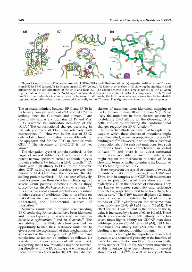
Figure 1. Comparison of EF-G structures with H573A, T84A and G16Vmutations. (a) Superimposition of the Ca tracesfromH573A EF-G (green), T84A (magenta) and G16V (yellow). (b) Zoom in on the box in (a) showing the significant localdifferences in the conformations of switch II and helix BIII. The colour scheme is the same as for (a). (c) An all-atomrepresentation of switch II in the “wild-type” conformation observed in mutant H573A. The importance of Phe90 andPro85 for the hydrophobic core can clearly be seen. In all panels, the GDP molecules are shown in a ball-and-stickrepresentation with carbon atoms coloured identically to the Ca traces. The Mg ions are shown as magenta spheres.
940 Fusidic Acid Sensitivity and Resistance in EF-G
The structural mimicry between EF-G and EF-Tu inits ternary complex with aa-tRNA and GDPNP isstriking, since the G-domain and domain II arestructurally similar and domains III, IV and V ofEF-G resemble the anticodon stem-loop of thetRNA.9 The conformational changes occurring inthe catalytic cycle of EF-Tu are relatively wellcharacterized.10,11 However, in the case of EF-G,detailed structural information is available only forthe apo form and for the EF-G in complex withGDP.8,12 The structure of EF-G:GTP is not yetknown.
The elongation cycle of protein synthesis is thetarget of several antibiotics. Fusidic acid (FA), apotent narrow spectrum steroid antibiotic, blocksprotein synthesis by inhibiting EF-G directly.13 FAbinds with high affinity to EF-G on the ribosomeafter GTP hydrolysis and therefore prevents therelease of EF-G:GDP from the ribosome, therebystalling protein synthesis.14 FA has been effectivelyused for more than three decades in clinics againstsevere Gram positive infections such as thosecaused by certain Staphylococcus aureus strains.15,16
It is an active agent against staphylococci resistantto other classes of antibiotics. Due to its effects onEF-G it has also been used as an effective tool tounderstand the fundamental aspects oftranslation.17
Numerous mutations in the fusA gene encodingEF-G conferring FA resistance have been identifiedand phenotypically characterized in vivo inSalmonella typhimurium18 and in S. aureus.19 Theavailability of the EF-G structure provided anopportunity to map these resistance mutations togive a plausible explanation of their mechanisms ofaction and of the binding site of FA.8,20 The mainconclusions so far can be outlined as follows. (1)Resistant mutations are spread all over EF-G,suggesting that a few mutations might be interact-ing directly with the FA binding site while most ofthem exert their effects indirectly. (2) Three distinct
clusters of mutations were identified, mapping tothe G domain, domain III and domain V. (3) Mostlikely the mutations in these clusters operate bymodulating EF-G affinity for the ribosome, FA orboth, and/or by restricting the conformationalchanges required for EF-G function.20,21
In our earlier efforts we have tried to explain theways in which these clusters of mutations mightexert their effect, as well as proposing a probable FAbinding site.20,22 However, in spite of the substantialinformation about FA resistant mutations, few suchmutations have been characterized in detailin vitro22–24 and there is no direct structuralinformation available on such mutants whichmight explain the mechanism of action of FA instructural terms or further illuminate the location ofthe FA binding site on EF-G.
Here we present the crystal structures of two suchmutants of EF-G from T. thermophilus, G16V andT84A, both in complex with GDP. Both mutants areactive in poly(U)-directed translation and theyhydrolyse GTP in the presence of ribosomes. Theyare known to confer sensitivity and resistancetowards FA, respectively, and have been character-ized in vitro.25 The G16V mutant displays a 20 timeslower Ki value for inhibition by FA of multiplerounds of GTP hydrolysis on the ribosome thandoes wild-type EF-G (0.4 mM versus 7.5 mM). Theeffect for the T84A mutant is the opposite: the Ki
value is increased by a factor of 60, to 440 mM. Theseeffects are correlated with GTP affinity: G16V hasseven times higher affinity for GDPNP than doeswild-type protein (13 mM versus 94 mM); T84A hasfour times less affinity (410 mM), while the GDPbinding is not affected in either mutant.
Our results highlight the importance of switch IIdynamics and structural plasticity at the interface ofthe G domain with domains III and V for sensitivityor resistance of EF-G to FA. Significant movementsat this interface have been observed in crystalstructures of EF-G21,22 as well as in cryo-electron

Fusidic Acid Sensitivity and Resistance in EF-G 941
microscopy (EM) studies of ribosomal com-plexes.26–28 Comparable studies of the eukaryotichomologue eEF2 and eukaryotic 80 S ribosomesalso show similar domain movements.29,30 We alsoprovide an explanation for the observed correlationbetween FA resistance and GTP affinity for theseEF-G mutants.
Results
The space group is P212121 for both mutantcrystals, as in all the other reported crystalstructures of EF-G.8,12,21,22 Two types of cell dimen-sions have been reported for EF-G. While the a and caxes are very similar, there is a large variation in thelength of the b axis. In the first group (wild-typeEF-G with or without GDP; PDB entries 1DAR,1EFG, 2EFG and 1ELO) b is z106 A while mutantH573A (PDB entry 1FNM) has a significantlyshorter b axis: bZ86.0 A (Table 1). This mutantalso exhibits a 108 rotation of domains III, IV and Vwith regard to domains G and II, around an axisbetween domains G and V, leading to a shift of 9 Aat the tip of domain IV.22 This more compactconformation of EF-G leads to an ordering ofdomain III, which is largely invisible in the othergroup of structures. In the following descriptionand discussion, the crystal structure of the mutantH573A22 will be used for comparison due to the factthat it contains an ordered domain III. Both themutant structures discussed here are overall moresimilar to H573A than to wild-type EF-G, in bothdomain orientation and crystal packing. The differ-ences are most likely due to the ordered binding ofMg2C to GDP in the crystal forms with the shorter
Table 1. Crystallographic data collection and structurerefinement statistics
A. ParametersEF-G mutant T84A G16VSpace group P212121Cell dimensions (A)a-axis 78.2 76.3b-axis 88.5 89.7c-axis 116.9 114.9Data resolution (A) 25–2.4 28–2.6No. observations 206, 752 213, 096No. unique reflections 27, 567 24, 149Completeness (overall/outer shell) (%)
99.6/87.9 98.2/97.2
I/sI (overall/outer shell) 19.1/4.1 14.0/3.2Rmerge (%) 5.9 (41.2) 6.4 (44.7)
B. RefinementRmodel 21.0 22.0Rfree 27.4 29.7No. of non-hydrogen atoms 5289 5149No. of water molecules 134 86
C. rms deviations from ideal geometryBond length (A) 0.013 0.012Bond angles (8) 1.52 1.43
D. Ramachandran plot (%)Most favoured 90.6 87.8Additionally allowed 8.9 10.7Generously allowed 0.5 1.1Disallowed 0 0.4
cell dimensions (see below). Another argument forthe relevance of the H573A structure is the strikingresemblance of its switch II conformation to the oneobserved in the crystal structure of the apo form ofthe eukaryotic homologue eEF2,31 where domain IIIis also fully ordered. The domain movementsobserved between the two forms of EF-G:GDP arenot in conflict with experiments on EF-G mobilityinvolving cross-linking.32 Besides, the H573Amutant is fully active in GTP hydrolysis andtranslocation.33 Taken together, this evidencesuggests that the structure of H573A:GDP is thebest one for comparison to the current mutantEF-Gs.An overall superposition of the three structures of
mutant H573A and mutants G16V and T84A isshown in Figure 1(a). The most pronouncedconformational differences between the three struc-tures are located in switch II and its surroundings.These local conformational differences cause smallshifts in the relative orientations of the domains.In H573A, switch II is located between the GDP
molecule in domain G and helix BIII in domain IIIand consists of a loop region (residues 84–90) and ahelix known as BG (residues 91–100; Figure 1(b)). Itis located at the interface between the P-loop, whichforms part of the nucleotide-binding site in the Gdomain, and domains II, III and V. The residuesGly16 and Thr84 are located on b-strand 1G and atthe beginning of the switch II loop, respectively.Their Ca atoms are only 6 A apart in space. Thr84makes an important stabilizing interaction throughits Og atom to the main-chain carbonyl oxygen atomof Ile17. At Gly16 there is no space for any largerresidue than a glycine. The tip of switch II isstabilized by a small hydrophobic core consisting ofthe side-chains from one side of helix BG (Phe90,Val94 and the aliphatic parts of Glu93 and Ser97)packing against Pro85 from the loop region(Figure 1(c)).Switch II is rather flexible in both mutants. In
SIGMAA-weighted 2mjFojKDjFcj maps for T84A,most of the model can easily be fitted into theelectron density, except for His87. This residue lacksdensity around its Ca and parts of the side-chain(Figure 2(a)). Density appears when the map iscontoured at 0.8s. Electron density for the backboneof switch II is clearly visible in the G16Vmutant andmost of the side-chains have density at a contourlevel of 1.0s, except for once more that of residueHis87 (Figure 2(b)). Again some density appears forthis residue at a contour level of 0.8s. However,His87 is not known as an essential residue inmodulating EF-G sensitivity/resistance towardsFA.
Comparison of the conformations of mutantsT84A and H573A in complex with GDP
Threonine 84 is part of the conserved DtpGhmotif in switch II.5 It makes two H-bonds from itsOg atom: one to the main-chain carbonyl of Ile17 atthe end of strand 1G of the central b-sheet (adjacent

Figure 2. Electron density maps for the T84A and G16Vmutants. Electron density for the switch II loop and helix(BG) in EF-G mutants (a) T84A and (b) G16V. All atoms ofthe switch II region are shown in a ball-and-stickrepresentation. The SIGMAA-weighted 2mjFojKDjFcjmaps from Refmac are shown as dark green meshesand are contoured at a level of 0.8s above the mean.
942 Fusidic Acid Sensitivity and Resistance in EF-G
to Gly16) and one to the side-chain of Lys25 in theP-loop. The loss of these interactions throughmutation of Thr84 to Ala makes no difference tothe overall conformation of either the P-loop orstrand 1G, except for a 1.5 A movement of the tip ofLys25 towards the core of domain G (see Discus-sion). In contrast, the mutation has a drastic effecton the conformation of the loop region of switch II(residues 84–91). In H573A, residues 87–90 form a
b-turn that projects the side-chains of His87 andVal88 towards helix CG (Figure 3(a) and (b), greenstructures). Phe90 protrudes in the oppositedirection and packs against Leu457 and Ile461 onhelix BIII of domain III. In contrast, in T84A Ala84flips upwards toward domain 3 by approximately1708 due to the loss of the H-bond to its Og atom andits Ca atom moves away from the central b-sheet by4.4 A compared to wild-type (Figure 3(c)). Thehydrophobic core of switch II is disrupted due tothe movement of Pro85 away from the core. The tipof switch II adopts a new conformation, inparticular giving a distinct new side-chain orien-tation to Phe90. Phe90 no longer forms part of theswitch II core but rather packs into a hydrophobicpocket formed by Leu457, His458, Ile92 and Val94,thus constituting a buttress between helix BG ofswitch II (Figure 3(a)) and domain III. Both Phe90and domain III have well-defined electron density,in contrast to mutant G16V, where domain III ismore poorly ordered (see below). Helix BG is largelyunaffected by the mutation.
Apart from these local conformational changes,the overall structure of T84A is well-conserved withrespect to H573A. The rms deviation between thetwo structures is 0.7 A for 643 Ca atoms and thereare no special domain movements except for thosein the loop part of switch II. The lack of domainrearrangements is most likely due in part to thenegligible influence of the mutation on helix BG.
Comparison of the conformations of mutantsG16V and H573A in complex with GDP
Glycine 16 is part of the conserved structuralscaffold of G-proteins, situated in strand 1G of thecentral b-sheet. In wild-type and H573A EF-G thereis no space for residues larger than Gly at thisposition. In other G-proteins very few confor-mational changes are observed between the GDPand GTP conformations in this region.34 Anextensive mesh of contacts stabilizes the structuresat this point. Residue 16 is surrounded by the side-chains of Thr84 and Leu101 of helix BG (the switch IIhelix). The local effect of the G16V mutation is tobreak some of the interactions responsible for theconformations at the beginning and end of switch IIin H573A (Figure 3(d)). In G16V Thr84 is pushedaway from the central b-sheet by the side-chain ofVal16, in the direction of domain III. This results inthe loss of the H-bond between the Og atom ofThr84 and the carbonyl group of Ile17, similarly tomutant T84A; nevertheless in G16V the same atommaintains an H-bond to the N3 atom of Lys25, dueto flexibility in the latter side-chain. Otherwise thereare only small differences in the P-loop that can beattributed to the mutation. The side-chain of His20moves by roughly 1 A and the tip of the side-chainof Lys25 moves by 1.5 A out of the protein core (seeDiscussion). The G16Vmutation also causes a hingemovement in helix BG. Val16 pushes away the side-chain of Leu101, whose Ca atom moves by 0.6 A.However, the movement is amplified towards the

Figure 3. Important interactions and conformational changes in switch II. (a) and (b) The substantially differentconformations of switch II, in particular Phe90, in EF-G mutants T84A (a), and G16V (b), which highlight the importanceof Phe90 for fusidic acid resistance or sensitivity. In both panels the H573A structure is shown as in a green cartoonrepresentation with all atoms drawn for the side-chains. Mutant T84A is drawn in magenta in (a) and mutant G16V isdrawn in yellow in (b). (c) and (d) The subtle conformational changes induced in the side-chain of Lys25 by the twomutations T84A (c) and G16V (d), which suggests an explanation of the different affinities of these mutants for GTP. Themolecular representations and colour coding are as for (a) and (b). In the T84A structure shown in (c) the interactionsbetween Thr84 to both the side-chain of Lys25 and the carbonyl oxygen atom on Ile17 are lost. Pro85 loses its interactionto the hydrophobic core of switch II and Asp83 moves to compensate the stabilization of Lys25. In G16V the interactionbetween Og of Thr84 and the carbonyl oxygen atom of Ile17 is lost.
Fusidic Acid Sensitivity and Resistance in EF-G 943
N-terminal end of helix BG, with a maximum Ca
displacement of 2.3 A at Glu95.The rest of switch II refolds to accommodate the
changes at the beginning and end caused by theadded bulk of Val16. The tip of switch II is stabilizedby a set of interactions quite different from H573Aor T84A. The hydrophobic core found in the H573Amutant structure is again broken, as the loop regionof switch II is pushed away from the central b-sheet,causing an outward movement of Pro85. Phe90 isrotated away from domain III towards domain V,where it makes hydrophobic interactions withSer668, Phe669 and Val670 (Figure 3(b)). Thisrepresents a third distinct conformation for Phe90,
which is diametrically opposed to the conformationin T84A when both are compared to the H573Astructure.Relative to the G domain the other domains
rearrange slightly. There is a hinge movement ofdomain II away from the central b-sheet thatfollows the hinge movement of helix BG. At theextremity of domain II the Ca atom of Glu295 movesby as much as 2.6 A. There is some rearrangementof the H-bonding pattern between residues Arg96and Arg99 on the helix BG following switch II andresidues 399–400 at the end of domain II (results notshown).The effects on domain III are more extensive. The
944 Fusidic Acid Sensitivity and Resistance in EF-G
new conformation of switch II appears to provideless stabilization of domain III than in the H573Astructure, as electron density for much of domain IIIis weaker in the G16V mutant despite the samecompact overall conformation of EF-G. In particularhelix BIII, (residues 456–467), which makes contactswith switch II, has indistinct side-chain electrondensity and has been built as poly(Ala). This helix
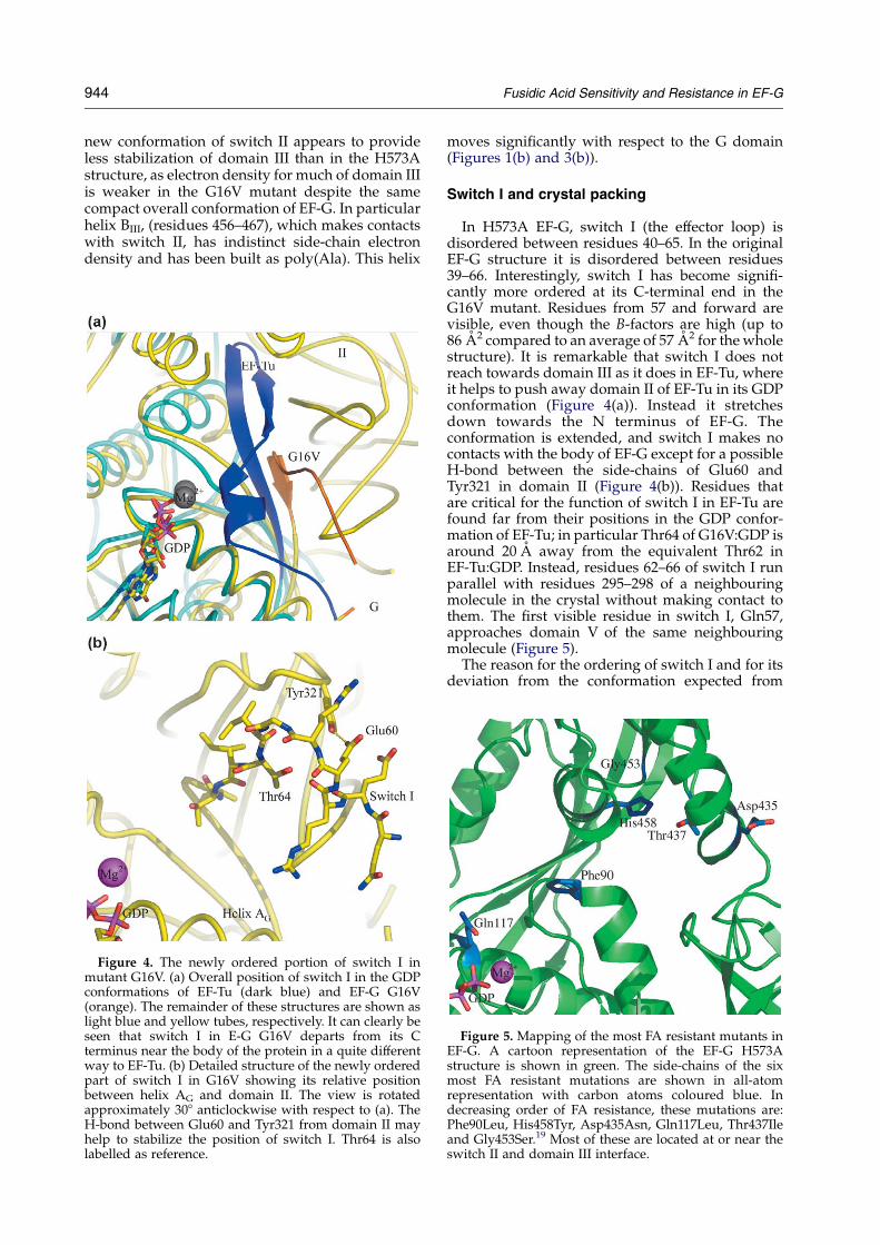
Figure 4. The newly ordered portion of switch I inmutant G16V. (a) Overall position of switch I in the GDPconformations of EF-Tu (dark blue) and EF-G G16V(orange). The remainder of these structures are shown aslight blue and yellow tubes, respectively. It can clearly beseen that switch I in E-G G16V departs from its Cterminus near the body of the protein in a quite differentway to EF-Tu. (b) Detailed structure of the newly orderedpart of switch I in G16V showing its relative positionbetween helix AG and domain II. The view is rotatedapproximately 308 anticlockwise with respect to (a). TheH-bond between Glu60 and Tyr321 from domain II mayhelp to stabilize the position of switch I. Thr64 is alsolabelled as reference.
moves significantly with respect to the G domain(Figures 1(b) and 3(b)).
Switch I and crystal packing
In H573A EF-G, switch I (the effector loop) isdisordered between residues 40–65. In the originalEF-G structure it is disordered between residues39–66. Interestingly, switch I has become signifi-cantly more ordered at its C-terminal end in theG16V mutant. Residues from 57 and forward arevisible, even though the B-factors are high (up to86 A2 compared to an average of 57 A2 for the wholestructure). It is remarkable that switch I does notreach towards domain III as it does in EF-Tu, whereit helps to push away domain II of EF-Tu in its GDPconformation (Figure 4(a)). Instead it stretchesdown towards the N terminus of EF-G. Theconformation is extended, and switch I makes nocontacts with the body of EF-G except for a possibleH-bond between the side-chains of Glu60 andTyr321 in domain II (Figure 4(b)). Residues thatare critical for the function of switch I in EF-Tu arefound far from their positions in the GDP confor-mation of EF-Tu; in particular Thr64 of G16V:GDP isaround 20 A away from the equivalent Thr62 inEF-Tu:GDP. Instead, residues 62–66 of switch I runparallel with residues 295–298 of a neighbouringmolecule in the crystal without making contact tothem. The first visible residue in switch I, Gln57,approaches domain V of the same neighbouringmolecule (Figure 5).
The reason for the ordering of switch I and for itsdeviation from the conformation expected from
Figure 5. Mapping of the most FA resistant mutants inEF-G. A cartoon representation of the EF-G H573Astructure is shown in green. The side-chains of the sixmost FA resistant mutations are shown in all-atomrepresentation with carbon atoms coloured blue. Indecreasing order of FA resistance, these mutations are:Phe90Leu, His458Tyr, Asp435Asn, Gln117Leu, Thr437Ileand Gly453Ser.19 Most of these are located at or near theswitch II and domain III interface.

Fusidic Acid Sensitivity and Resistance in EF-G 945
work on EF-Tu might be explained by the presenceof a crystal contact apparently induced by thepresence of a Mg ion associated with GDP in thecrystal structure. Domain II of a neighbouring EF-Gmolecule in the crystal comes close to the GDPmolecule and the side-chain of Glu295 makes anH-bond to one of the water molecules coordinatingthe Mg2C. In this conformation Tyr342 of domain IIin the neighbouring molecule also makes anH-bond directly to the 2 0-OH of the GDP riboseand the carbonyl oxygen atom of Gly347 H-bonds tothe 2-NH2 group of the base. Arg396 makes a saltbridge to Glu22 in the P-loop (not shown, seeLaurberg et al.22 for details). The resulting crystalpacking is quite different from that of wild-typeEF-G in complex with GDP, where ordered Mg2C
was not observed (PDB code 1ELO), even though itwas included in the crystallization experiments, butis very similar to that in mutant H573A (PDB code1FNM), where ordered Mg2C is observed.22
Thus, while we cannot exclude that the orderingof switch I in the G16V mutant structure representsa biologically relevant conformation, subject to ahigh degree of rearrangement upon binding to theribosome, we think it more likely that the confor-mation seen in the G16V mutant is due to thepresence of crystal contacts and crowding aroundthe GDP molecule, as proposed.22
The crystal packing in mutant T84A is verysimilar to that of G16V and H573A, with somesubtle differences. Glu295 of the neighbouringmolecule approaches the Mg2C more closely thanin G16V (3.0 A versus 4.8 A). Switch I is not visiblebetween residues 40 and 67, despite the similarity incrystal packing compared to G16V, although theelectron density can be seen to extend in the samedirection from the C terminus at lower contourlevels. The reasons for this difference are notapparent.
Figure 6. Comparison of the position of Phe90 inH573A, T84A and G16V. In H573A (green), Phe90 reachesinto the hydrophobic pocket of switch II and is presseddown by helix BIII from domain III. In T84A (magenta),Phe90 reaches into the interface of switch II and helix BIII
of domain III. Phe90 in G16V (yellow) makes a third,completely different set of interactions. It makes hydro-phobic interactions with Phe669 and Val670 in domain V.
Discussion
The two crystal structures presented hererepresent the first direct evidence of the significanteffects on EF-G tertiary structure that can be causedby simple point mutations. Although the structuralstudies have been performed on T. thermophilusEF-G, it is clear that they have direct relevance forEF-G in other organisms. For example, the doublemutation A66V/T84A (Val69 and Thr84 inT. thermophilus EF-G) confers strong FA resistanceupon Escherichia coli EF-G,24 which has 60%sequence identity to the T. thermophilus factor.Since T. thermophilus EF-G naturally contains theVal69 residue, the T84A mutant is a close mimic ofthe double mutant in E. coli. In S. typhimurium anFA resistant allele of fusA encoding EF-G with theP413L mutation confers a slow growing phenotypeand selection of resistance strains for fast growthresulted in internal revertants carrying anadditional mutation along with P413L. The G13Vrevertant in S. typhimurium EF-G (also having 60%
identity to T. thermophilus) reduces FA resistancesignificantly of the strain carrying the fusA genewith the P413L mutation.20 Gly16 in EF-Gfrom T. thermophilus corresponds to Gly13 inS. typhimurium EF-G. Together with the in vitroresults for the T. thermophilus factor,25 this evidenceindicates that EF-G structure and function in thedifferent organisms are highly related in theirbehaviour towards FA.
Mechanism of FA resistance
On the basis of structural mapping of FA resistantmutations, it has been suggested that the interfacebetween switch II, domain III and domain V is themost likely place where FA binds.20,22 The twomutants studied are close to the proposed FAbinding site and also to the nucleotide-bindingsite. Thr84 is part of the DxxG consensus motif inthe switch II region which is present in all Gproteins.5 This motif is generally involved in theconformational changes between the GTP and GDPstates of G proteins.35 Although it is of exclusivelystructural importance in EF-G, Gly16 is in closevicinity to the GTP binding site, with a distance of10 A between its Ca atom and the b-phosphate ofGDP in the crystal structures.The current work reveals two new, distinct switch
II conformations in the T84A and G16V mutantstructures when compared to the structure of theH753A mutant, which we use as a model for wild-type EF-G. These conformational changes result insubstantial movements of the Phe90 side-chain.Phe90 is part of the small hydrophobic core thatstabilizes the conformation of the tip of switch II in

946 Fusidic Acid Sensitivity and Resistance in EF-G
wild-type EF-G. The side-chain conformation ofPhe90 in the H573A mutant structure of EF-G liesbetween the two extreme conformations observedin the T84A and G16V mutant structures (Figure 6).The diametrically opposite positions of the Phe90side-chain in G16V and T84A thus appear to becorrelated to their ability to confer high resistanceand sensitivity towards FA, respectively. In thiscontext it merits attention that mutation of Phe90 toLeu in S. aureus confers 500-fold resistance towardsFAwhile mutations of the other two residues whichflank the cavity of the proposed FA binding site,Asp435 to Asn and His458 to Tyr, both in S. aureus,lead to 125 and 168-fold resistance, respectively.19,22
It is also noteworthy that the equivalent Phe111 inEF2 undergoes large conformational changesbetween the apo- and sordarin-boundconformations.29
The dramatic movement of Phe90 as observed inT84A and G16V structures is related to the“domino” effect which we proposed earlier,22
where we suggested a possible link between thenucleotide binding and the FA binding sites.Interestingly, residues outside switch II in thisdomino chain (e.g. Gln117, Leu457) can alsoinfluence FA resistance when mutated.18,22 Thisemphasizes the significant role of switch II and theinterface between the two parts of the molecule,namely domains G and II versus domains III, IV andV, in fusidic acid resistance.22 The two extreme anddiametrically opposite side-chain conformations ofPhe90 in the mutants represent almost the totalrange of available conformational space for thisresidue. Along with switch II, Phe90 thus appears torespond differentially to mutations, in this wayregulating the gating of the possible cavity for FAbinding and thus modulating the resistance of EF-Gtowards FA. Mutations far from this domain inter-face may exert their influence by relaying confor-mational changes to Phe90. Phe90 thus appears tobe an essential residue that can partition mutationsinto varying degrees of resistance by its ability toadopt discrete conformational states in between thetwo ends of the spectrum of conformational spacerepresented by G16V and T84A. Conformationalchanges associated with EF-G upon ribosomebinding may further regulate the domainreorganisation, with the Phe90 sensor readjustingitself to take into account those signals. Electronmicroscopy reconstructions of EF-G on the ribo-some in different functional states27,36 suggest thatthe influence of the ribosome is most likely indirect,by facilitating a conformation of EF-G which is lessfavourable in solution. In any case the flexibility ofPhe90 and the rest of switch II is highly likely to bean essential component of the conformationalchange blocked by FA.
The conformation of switch II in the originalcrystal structure of EF-G in complex with GDP21 isvery similar to that in the T84A mutant from Phe90and onwards, although residues 84–89 are verydifferent. This may appear to be in conflict with theinterpretation described above; however, since
domain III is largely disordered in this structure(in particular helix BIII is not visible) it is notpossible to describe how this would affect theinteractions of switch II with domain III. Never-theless, the similarity of switch II in these twostructures does confirm that this conformation ofthis part of switch II in T84A is an energeticallystable one.
A possible role for Lys25 in affinity for GDP andGTP
Several FA resistant mutants have reduced GTPaffinity and FA sensitive EF-G mutants haveincreased affinity for GTP.25 Mutants T84A andG16V in solution have been reported to have fourtimes less and three times higher affinity forGDPNP, respectively, as compared to wild-typeEF-G. There thus seems to be a correlation betweenthe FA resistance of EF-G and its affinity for GTP,though GDP binding is not affected.25 The crystalstructures of G16V and T84A presented here are incomplex with GDP, thus a direct interpretation ofthe correlation between FA resistance of EF-G andGTP affinity is not possible. However, the structuressuggest a possible explanation for this correlationthat can be investigated further.
We observe subtle changes in the position of theside-chain of the “P-loop lysine”, Lys25, due to themutations, which are, as previously noted, close inspace. In wild-type EF-G and in the H573A mutant,Thr84 makes an H-bond to Lys25, stabilizing itsside-chain. In G16V Lys25 retains its H-bond toThr84 but follows it towards its new position,resulting in a movement of the N3 atom of Lys25 by1.6 A out from the core of domain G towards thesupposed g-phosphate position (Figure 3(d)). InThr84 the H-bond between Lys25 and Thr84 is lostand is replaced by a new H-bond to the side-chainof Asp83. This has the effect that N3 of Lys25 movesaway from the supposed g-phosphate position by1.5 A towards the core of domain G (Figure 3(c)).This suggested to us that the dynamic behaviour ofLys25 may be important for the GTPase activity ofEF-G. It is known that at least one lysine residue isrequired for GTP binding in EF-G from E. coli.37 Ofthe 39 lysine residues present in EF-G fromT. thermophilus, Lys25 is almost completely con-served in all known GTPases, ATPases and even insome kinases.38,39 It is quite excluded from thesolvent in the wild-type EF-G structure and is in thevicinity of the nucleotide-binding site.
The movements observed in Lys25 and itsconservation led us to examine its behaviour inthe structures of other translational GTPases. EF-Tu,IF2/eIF5B, aIF2 and SelB have been characterizedstructurally in complex with GDP and GTPanalogues.40–44 A close comparison of these struc-tures with those of EF-G provides insights into thepossible role of the P-loop lysine (Lys24 in EF-Tu,Lys18 in IF2/eIF5B, Lys23 in aIF2 and Lys32 ineEF2). In EF-G, eEF-2 and IF2/eIF5B there is a polarresidue with H-bonding possibility (Thr or Ser) at

Fusidic Acid Sensitivity and Resistance in EF-G 947
the position equivalent to Thr84 in EF-G that caninteract with the P-loop lysine. In EF-G and eEF2 thelysine is indeed hydrogen-bonded to this residue,pointing straight towards switch II and unavailablefor interaction with GDP.31 In EF-Tu, its eukaryoticand mitochondrial homologues, SelB and aIF2,there is a hydrophobic residue (Cys, Met, Val) atthe position equivalent to Thr84. In the GDPcomplexes of these translational GTPases theP-loop lysine is bent towards the nucleotide andinteracts with the b-phosphate oxygen atom;41–45 inthe corresponding GDPNP complexes the samelysine residue interacts with both the b andg-phosphate oxygen atoms.41–43,46 In none of thelatter structures is there direct interactions betweenthe P-loop lysine and switch II except through awater molecule. In addition, despite the presence ofThr in IF2/eIF5B, the interaction visible in EF-Gbetween Lys25 and Thr84 is not seen between Lys18and Thr77 in either of the IF2/eIF5B structures.41 Itthus seems that an important interaction betweenthe P-loop lysine and the Thr or Ser of the DxxGmotif in switch II is conserved in eEF2 and EF-G,but not in the other translational GTPases. This maypoint to differences in the way that the nucleotide-binding site is influenced by the ribosome duringthe catalytic cycle. The differential affinities of EF-Gmutants T84A and G16V for GTP may thus beexplained by the direct effect of these mutations onthe environment of Lys25. However, this requiresadditional experiments to be clarified. Preliminaryexperiments indicate that the K25A mutant ofT. thermophilus EF-G has severely reduced GTPaseactivity (A.T.G., unpublished results).
In conclusion, the structural studies of the twomutants presented here show that the conformationof Phe90 parallels the affinity for fusidic acid, againsuggesting that the FA binding site is betweendomains G, III and V. In addition to the dominochain providing a signalling pathway between thenucleotide binding site and the presumed FAbinding site, switch II can directly influence theenvironment of the nucleotide, as seen in themovements of Lys25.
Materials and Methods
† http://www.pymol.org.
Cloning, expression and purification
The gene for EF-G from T. thermophilus was cut outfrom the original pET11c vector using NdeI and EcoRIand ligated into pET13a with the same restriction sites.The site-directed mutagenesis was performed usingQuickChange XL II (Stratagenew). The primers usedfor G16V were forward 5 0-CTCCGGAACATCGTCATCGCCGCCC and reverse 5 0-GGGCGGCGATGACGATGTTCCGGAG; for T84A the forward primer was5 0-AACATCATCGACGCCCCGGGCCACG and reverse5 0-CGTGGCCCGGGGCGTCGATGATGAA. Expressionand purification of both the mutants, were performed asdescribed47 with slight modifications. Heat denaturingtreatment of post-ribosomal supernatant was carried outat 70 8C for 15 minutes for coagulation of E. coli proteins.
Coagulated proteins were removed by centrifugation andthe supernatant was applied to a Source 15Q anionexchange column (AmershamBiosciencesw). EF-G waseluted with a gradient from 0 M to 1 M NaCl in a buffercontaining 60 mM Tris–HCl (pH 7.6), 5 mM b-mercapto-ethanol, 1 mM EDTA and 0.1 mM PMSF. Protein obtainedafter anion exchange chromatography was pure enoughto yield good quality crystals. All protein solutions wereconcentrated in crystallization buffer containing 5 mMTris–HCl, 10 mM MgCl2 (pH 7.6) to a concentration of3–4 mg/ml.
Crystallization
The crystallization buffer for T84A and G16V was5 mM Tris–HCl (pH 7.6), 10 mM MgCl2 and 5 mM GDP(Sigmaw). Crystals of T84A:GDP and G16V:GDP weregrown from hanging drops in vapour diffusion experi-ments at pH 7.3 against 17% (w/v) polyethylene glycol8000 (Flukaw), 100 mM Hepes and 46 mM Tris; 4 ml of a4 mg/ml protein solution was mixed with 4 ml ofreservoir solution. The G16V and T84A drops wereinitially streak seeded with wild-type EF-G crystals butin later experiments crystals of the respective mutantswere used. Good quality crystals of both mutantsappeared overnight and were of suitable size for datacollections (0.4 mm!0.4 mm!0.2 mm). Crystals werepassed rapidly through a drop with conditions identicalwith the crystallization drops but with the addition of49% methane pentane diol (MPD) as cryo-protectant,from which they were transferred directly into the liquidnitrogen cryo stream. The T84A:GDP and G16V:GDPcrystals appeared to be more fragile than wild-type EF-Gand the use of glycerol as cryo-protectant was notsuitable, therefore MPD was used instead.
Data collection and structure determination
Diffraction data for T84A:GDP were collected to 2.4 Aresolution on a 165 mm MarCCD detector at station I711of the Max-II synchrotron, Lund, Sweden and theG16V:GDP data were collected to 2.6 A resolution on anADSCQ4R CCD detector at station ID14-EH1 of the ESRF,Grenoble, France. All data were collected at 100 K. Thedata were integrated with XDS.48 All data were furtherprocessed with programs from the CCP4 suite49 using theCCP4i interface.50 The T84A:GDP structure was solvedusing the structure of the H573A mutant, Protein DataBank (PDB) entry 1FNM22 as search model in themolecular replacement program Molrep.51 TheG16V:GDP structure was solved using T84A:GDP assearch model. Both models were rebuilt using XtalView52
and refined with Refmac553 (Table 1). Figures were madeusing Pymol† except for Figure 3, which was made usingBobscript.54 Structural superimpositions were madeusing SwissPDB Viewer.
Coordinates
Coordinates and structure factors have been depositedin the RCSB Protein Data Bank55 with accession numbers2BM1 for the G16V mutant and 2BM0 for the T84Amutant.

948 Fusidic Acid Sensitivity and Resistance in EF-G
Acknowledgements
This work was supported by grants from theSwedish Research Council (to V.R.), the CrafoordFoundation, Carl Tryggers Foundation and theRoyal Swedish Academy of Sciences (to D.L.), agrant from VR and EU grant QLK2-CT-2002-00892(to A.L.), and in part by Russian grant 02-04-48953from RFBR (to A.T.G.). We thank staff at MAX-lab(Y. Cerenius) and beamline ID14 of the ESRF (inparticular C. Romao) for help with data collection.
References
1. Nishizuka, Y. & Lipmann, F. (1966). Comparison ofguanosine triphosphate split and polypeptide syn-thesis with a purified E. coli system. Proc. Natl Acad.Sci. USA, 55, 212–219.
2. Richman, N. & Bodley, J. W. (1972). Ribosomes cannotinteract simultaneously with elongation factors EF-Tuand EF-G. Proc. Natl Acad. Sci. USA, 69, 686–689.
3. Krab, I. M. & Parmeggiani, A. (1998). EF-Tu, a GTPaseodyssey. Biochim. Biophys. Acta, 1443, 1–22.
4. Rodnina, M. V., Savelsbergh, A. & Wintermeyer, W.(1999). Dynamics of translation on the ribosome:molecular mechanics of translocation. FEMSMicrobiol. Rev. 23, 317–333.
5. Bourne, H. R., Sanders, D. A. & McCormick, F. (1991).The GTPase superfamily: conserved structure andmolecular mechanism. Nature, 349, 117–127.
6. Richter, D. (1972). Inability of E. coli ribosomes tointeract simultaneously with the bacterial elongationfactors EF-Tu and EF-G. Biochem. Biophys. Res. Com-mun. 46, 1850–1856.
7. Miller, D. L. (1972). Elongation factors EF Tu and EF Ginteract at related sites on ribosomes. Proc. Natl Acad.Sci. USA, 69, 752–755.
8. Ævarsson, A., Brazhnikov, E., Garber, M.,Zheltonosova, J., Chirgadze, Y., Al-Karadaghi, S.et al. (1994). Three-dimensional structure of theribosomal translocase: elongation factor G fromThermus thermophilus. EMBO J. 13, 3669–3677.
9. Liljas, A. (1996). Imprinting through molecularmimicry. Protein synthesis. Curr. Biol. 6, 247–249.
10. Polekhina, G., Thirup, S., Kjeldgaard, M., Nissen, P.,Lippmann, C. & Nyborg, J. (1996). Helix unwindingin the effector region of elongation factor EF-Tu-GDP.Structure, 4, 1141–1151.
11. Abel, K., Yoder, M. D., Hilgenfeld, R. & Jurnak, F.(1996). An alpha to beta conformational switch inEF-Tu. Structure, 4, 1153–1159.
12. Czworkowski, J., Wang, J., Steitz, T. A. & Moore, P. B.(1994). The crystal structure of elongation factor Gcomplexed with GDP, at 2.7 A resolution. EMBO J. 13,3661–3668.
13. Tanaka, N., Kinoshita, T. & Masukawa, H. (1968).Mechanism of protein synthesis inhibition by fusidicacid and related antibiotics. Biochem. Biophys. Res.Commun. 30, 278–283.
14. Bodley, J. W., Zieve, F. J., Lin, L. & Zieve, S. T. (1969).Formation of the ribosome-G factor-GDP complex inthe presence of fusidic acid. Biochem. Biophys. Res.Commun. 37, 437–443.
15. Whitby, M. (1999). Fusidic acid in the treatment ofmethicillin-resistant Staphylococcus aureus. Int.J. Antimicrob. Agents, 12(Suppl 2), S67–S71.
16. Whitby, M. (1999). Fusidic acid in septicaemia andendocarditis. Int. J. Antimicrob. Agents, 12(Suppl 2),S17–S22.
17. Czworkowski, J. & Moore, P. B. (1996). The elongationphase of protein synthesis. Prog. Nucl. Acid Res. Mol.Biol. 54, 293–332.
18. Johanson, U. & Hughes, D. (1994). Fusidic acid-resistant mutants define three regions in elongationfactor G of Salmonella typhimurium. Gene, 143, 55–59.
19. Nagaev, I., Bjorkman, J., Andersson, D. I. & Hughes,D. (2001). Biological cost and compensatory evolutionin fusidic acid-resistant Staphylococcus aureus. Mol.Microbiol. 40, 433–439.
20. Johanson, U., Aevarsson, A., Liljas, A. & Hughes, D.(1996). The dynamic structure of EF-G studied byfusidic acid resistance and internal revertants. J. Mol.Biol. 258, 420–432.
21. Al-Karadaghi, S., Ævarsson, A., Garber, M.,Zheltonosova, J. & Liljas, A. (1996). The structure ofelongation factor G in complex with GDP: confor-mational flexibility and nucleotide exchange.Structure, 4, 555–565.
22. Laurberg, M., Kristensen, O., Martemyanov, K. A.,Gudkov, A. T., Nagaev, I., Hughes, D. & Liljas, A.(2000). Structure of a mutant EF-G reveals domain IIIand possibly the fusidic acid binding site. J. Mol. Biol.303, 593–603.
23. MacVanin, M., Johanson, U., Ehrenberg, M. &Hughes, D. (2000). Fusidic acid-resistant EF-G per-turbs the accumulation of ppGpp. Mol. Microbiol. 37,98–107.
24. Richter Dahlfors, A. A. & Kurland, C. G. (1990). Novelmutants of elongation factor G. J. Mol. Biol. 215,549–557.
25. Martemyanov, K. A., Liljas, A., Yarunin, A. S. &Gudkov, A. T. (2001). Mutations in the G-domain ofelongation factor G from Thermus thermophilus affectboth its interaction with GTP and fusidic acid. J. Biol.Chem. 276, 28774–28778.
26. Agrawal, R. K., Penczek, P., Grassucci, R. A. & Frank,J. (1998). Visualization of elongation factor G on theEscherichia coli 70 S ribosome: the mechanism oftranslocation. Proc. Natl Acad. Sci. USA, 95, 6134–6138.
27. Valle, M., Zavialov, A., Sengupta, J., Rawat, U.,Ehrenberg, M. & Frank, J. (2003). Locking andunlocking of ribosomal motions. Cell, 114, 123–134.
28. Stark, H., Rodnina, M. V., Wieden, H. J., van Heel, M.& Wintermeyer, W. (2000). Large-scale movement ofelongation factor G and extensive conformationalchange of the ribosome during translocation. Cell, 100,301–309.
29. Jorgensen, R., Ortiz, P. A., Carr-Schmid, A., Nissen, P.,Kinzy, T. G. & Andersen, G. R. (2003). Two crystalstructures demonstrate large coonformationalchanges in the eukaryotic ribosomal translocase.Nature, 10, 379–385.
30. Spahn, C. M., Gomez-Lorenzo, M. G., Grassucci,R. A., Jorgensen, R., Andersen, G. R., Beckmann, R.et al. (2004). Domain movements of elongation factoreEF2 and the eukaryotic 80 S ribosome facilitate tRNAtranslocation. EMBO J. 23, 1008–1019.
31. Jorgensen, R., Yates, S. P., Teal, D. J., Nilsson, J.,Prentice, G. A., Merrill, A. R. & Andersen, G. R.(2004). Crystal structure of ADP-ribosylated ribo-somal translocase from Saccharomyces cerevisiae. J. Biol.Chem. 279, 45919–45925.
32. Peske, F., Matassova, N. B., Savelsbergh, A., Rodnina,M. V. & Wintermeyer, W. (2000). Conformationally

Fusidic Acid Sensitivity and Resistance in EF-G 949
restricted elongation factor G retains GTPase activitybut is inactive in translocation on the ribosome. Mol.Cell, 6, 501–505.
33. Martemyanov, K. A., Yarunin, A. S., Liljas, A. &Gudkov, A. T. (1998). An intact conformation at the tipof elongation factor G domain IV is functionallyimportant. FEBS Letters, 434, 205–208.
34. Vetter, I. R. & Wittinghofer, A. (2001). The guaninenucleotide-binding switch in three dimensions.Science, 294, 1299–1304.
35. Kjeldgaard, M., Nyborg, J. & Clark, B. F. (1996). TheGTP binding motif: variations on a theme. FASEB J.10, 1347–1368.
36. Agrawal, R. K., Linde, J., Sengupta, J., Nierhaus, K. H.& Frank, J. (2001). Localization of L11 protein on theribosome and elucidation of its involvement in EF-G-dependent translocation. J. Mol. Biol. 311, 777–787.
37. Giovane, A., Balestrieri, C. & Gualerzi, C. (1982).Structure–function relationships in Escherichia colitranslational elongation factor G: modification oflysine residues by the site-specific reagent pyridoxalphosphate. Biochemistry, 21, 5224–5230.
38. Saraste, M., Sibbald, P. R. & Wittinghofer, A. (1990).The P-loop–a common motif in ATP- and GTP-binding proteins. Trends Biochem. Sci. 15, 430–434.
39. Leipe, D. D., Wolf, Y. I., Koonin, E. V. & Aravind, L.(2002). Classification and evolution of P-loop GTPasesand related ATPases. J. Mol. Biol. 317, 41–72.
40. Krab, I. M. & Parmeggiani, A. (2002). Mechanisms ofEF-Tu, a pioneer GTPase. Prog. Nucl. Acid Res. Mol.Biol. 71, 513–551.
41. Roll-Mecak, A., Cao, C., Dever, T. E. & Burley, S. K.(2000). X-ray structures of the universal translationinitiation factor IF2/eIF5B: conformational changeson GDP and GTP binding. Cell, 103, 781–792.
42. Schmitt, E., Blanquet, S. & Mechulam, Y. (2002). Thelarge subunit of initiation factor aIF2 is a closestructural homologue of elongation factors. EMBO J.21, 1821–1832.
43. Leibundgut, M., Frick, C., Thanbichler, M., Bock, A. &Ban, N. (2005). Selenocysteine tRNA-specificelongation factor SelB is a structural chimaera ofelongation and initiation factors. EMBO J. 24, 11–22.
44. Andersen, G. R., Thirup, S., Spremulli, L. L. &
Nyborg, J. (2000). High resolution crystal structureof bovine mitochondrial EF-Tu in complex with GDP.J. Mol. Biol. 297, 421–436.
45. Berchtold, H., Reshetnikova, L., Reiser, C. O.,Schirmer, N. K., Sprinzl, M. & Hilgenfeld, R. (1993).Crystal structure of active elongation factor Tu revealsmajor domain rearrangements. Nature, 365, 126–132.
46. Nissen, P., Kjeldgaard, M., Thirup, S., Polekhina, G.,Reshetnikova, L., Clark, B. F. & Nyborg, J. (1995).Crystal structure of the ternary complex of Phe-tRNAPhe, EF-Tu, and a GTP analog. Science, 270,1464–1472.
47. Martemyanov, K. A., Liljas, A. & Gudkov, A. T. (2000).Extremely thermostable elongation factor G fromAquifex aeolicus: cloning, expression, purification,and characterization in a heterologous translationsystem. Protein Expr. Purif. 18, 257–261.
48. Kabsch, W. (1993). Automatic processing of rotationdiffraction data from crystals of initially unknownsymmetry and cell constants. J. Appl. Crystallog. 26,795–800.
49. Collaborative Computational Project, No. 4. (1994).The CCP4 suite: programs for protein crystallo-graphy. Acta Crystallog. sect. D, 50, 760–763.
50. Potterton, E., Briggs, P., Turkenburg, M. & Dodson, E.(2003). A graphical user interface to the CCP4program suite. Acta Crystallog. sect. D: Biol. Crystallog.59, 1131–1137.
51. Vagin, A. & Teplyakov, A. (2000). An approach tomulti-copy search in molecular replacement. ActaCrystallog. sect. D: Biol. Crystallog. 56, 1622–1624.
52. McRee, D. E. (1999). XtalView/Xfit—a versatileprogram for manipulating atomic coordinates andelectron density. J. Struct. Biol. 125, 156–165.
53. Murshudov, G. N. (1997). Refinement of macro-molecular structures by the maximum-likelihoodmethod. Acta Crystallog. sect. D: Biol. Crystallog. 53,240–255.
54. Esnouf, R. M. (1997). An extensively modified versionof MolScript that includes greatly enhanced coloringcapabilities. J. Mol. Graph. Model. 15, 132–134 see alsopp. 112–113.
55. Berman, H. M., Westbrook, J., Feng, Z., Gilliland, G.,Bhat, T. N., Weissig, H. et al. (2000). The Protein DataBank. Nucl. Acids Res. 28, 235–242.
Edited by J. Doudna
(Received 23 December 2004; received in revised form 25 February 2005; accepted 27 February 2005)
Copyright © 2022 FDOKUMEN