Essential Role for ADAM19 in Cardiovascular Morphogenesis
9
MOLECULAR AND CELLULAR BIOLOGY, Jan. 2004, p. 96–104 Vol. 24, No. 1 0270-7306/04/$08.000 DOI: 10.1128/MCB.24.1.96–104.2004 Copyright © 2004, American Society for Microbiology. All Rights Reserved. Essential Role for ADAM19 in Cardiovascular Morphogenesis Hong-Ming Zhou, 1 Gisela Weskamp, 1 Vale ´rie Chesneau, 1 Umut Sahin, 1,2 Andrea Vortkamp, 3 Keisuke Horiuchi, 1 Riccardo Chiusaroli, 4 Rebecca Hahn, 5 David Wilkes, 5 Peter Fisher, 6 Roland Baron, 4 Katia Manova, 7 Craig T. Basson, 5 Barbara Hempstead, 8 and Carl P. Blobel 1 * Cell Biology Program, Sloan-Kettering Institute, 1 and Molecular Cytology Core Facility, Sloan-Kettering Institute, Memorial Sloan- Kettering Cancer Center, 7 and Molecular Cardiology Laboratory, Cardiology Division, Department of Cell and Developmental Biology, 5 and Hematology Division, 8 Department of Medicine, Weill Medical College of Cornell University, New York, New York 10021; Department of Molecular and Cellular Biology and Biochemistry, Brown University, Providence, Rhode Island 02912 2 ; Max Planck Institute for Molecular Genetics, Otto Warburg Laboratory, D14195 Berlin, Germany 3 ; Department of Orthopaedics and Cell Biology, Yale University School of Medicine, New Haven, Connecticut 06510 4 ; and Department of Anatomy and Cell Biology, Columbia University, New York, New York 10032 6 Received 18 July 2003/Returned for modification 16 September 2003/Accepted 8 October 2003 Congenital heart disease is the most common form of human birth defects, yet much remains to be learned about its underlying causes. Here we report that mice lacking functional ADAM19 (mnemonic for a disintegrin and metalloprotease 19) exhibit severe defects in cardiac morphogenesis, including a ventricular septal defect (VSD), abnormal formation of the aortic and pulmonic valves, leading to valvular stenosis, and abnormalities of the cardiac vasculature. During mouse development, ADAM19 is highly expressed in the conotruncus and the endocardial cushion, structures that give rise to the affected heart valves and the membranous ventricular septum. ADAM19 is also highly expressed in osteoblast-like cells in the bone, yet it does not appear to be essential for bone growth and skeletal development. Most adam19 / animals die perinatally, likely as a result of their cardiac defects. These findings raise the possibility that mutations in ADAM19 may contribute to human congenital heart valve and septal defects. ADAMs (mnemonic for a disintegrin and metalloprotease) are membrane-anchored glycoproteins with key roles in fertil- ization, neurogenesis, angiogenesis, Alzheimer’s disease, and the release of proteins such as epidermal growth factor (EGF) receptor ligands and tumor necrosis factor family members from the plasma membrane (3, 4, 17, 37, 39, 41). ADAM19 (also referred to as meltrin ) was initially identified in muscle cells and was later found to be expressed in several other tissues, most prominently in heart, lung, and bone (18, 27, 52), during dendritic cell differentiation (13) and Notch-induced T-cell maturation (9). The catalytic activity of ADAM19 to- wards candidate substrates has been explored by overexpres- sion in cells and by purifying recombinantly expressed soluble forms of the entire ectodomain or the pro- and metallopro- tease domains (7, 42, 49, 53). Overexpressed ADAM19 en- hances ectodomain shedding of two of several splice variants of neuregulin I- (42), a ligand for the ErbB family of receptor tyrosine kinases (11). Furthermore, overexpression of ADAM19 increases ectodomain release of tumor necrosis fac- tor-related activation-induced cytokine (TRANCE, also re- ferred to as osteoprotegerin-ligand [OPGL]) (7), a protein with important roles in osteoclast differentiation, dendritic cell survival, and mammary gland development (12, 25, 28). In light of the high expression of ADAM19 in heart and bone and its ability to cleave TRANCE as well as splice vari- ants of neuregulin I-, we were interested in evaluating the function of ADAM19 in mice, with an emphasis on its role in heart and bone development. Here we present an analysis of mice lacking functional ADAM19 (adam19 / mice). MATERIALS AND METHODS Generation of adam19 / mice. adam19 / mice were generated by the Sloan- Kettering Institute transgenic facility by following standard procedures using stem cells with a secretory gene trap insertion in ADAM19 (30). All mice evaluated in this study were of mixed genetic background (129Sv/C57BL6), and morphological and histological comparisons between wild-type and adam19 / mice were performed with littermates that resulted from matings of heterozygous parents. Genotyping was performed either by Southern blotting (50) or by PCR. For PCR analysis, two sets of primers were used simultaneously to detect both the wild-type and mutant alleles. The primers used to detect the wild-type allele were TTA CCA GGA ACA GTG CCA GC (sense primer) and ATC ACT GCT GTC TTT GAG ATC (antisense primer). The primers specific for the mutant allele were GCC CTG AAT GAA CTG CAG GAC G and CAC GGG TAG CCA ACG CTA TGT C. Enzymatic deglycosylation and Western blot analysis. Treatment of cell ly- sates with endoglycosidase H (EndoH; New England Biolabs) or peptide-N- glycosidase (PNGase; New England Biolabs) and Western blot analysis were performed as previously described (32). In situ hybridization. Timed matings were set up to generate embryos at different stages of gestation (embryonic day 11.5 [E11.5], E13.5, and E16.5) for an analysis of ADAM19 expression by mRNA in situ hybridization. Mouse embryos were fixed in 4% paraformaldehyde overnight at 4°C, and graded series of ethanol were subsequently used to dehydrate the fixed embryos. Dehydrated tissues were cleared with Histoclear, embedded in paraffin, sectioned, and mounted on Fisher Superfrost Plus slides. The appropriate linearized plasmids were used to prepare 33 P-labeled RNA probes with T7 or Sp6 RNA polymerases by using a ribonucleotide triphosphate mix with 12 M cold UTP and 4 M hot UTP. The RNA in situ hybridization procedure was performed essentially as described previously (33). * Corresponding author. Mailing address: Cell Biology Program, Sloan-Kettering Institute, Memorial Sloan-Kettering Cancer Center, Box 368, 1275 York Ave., New York, NY 10021. Phone: (212) 639- 2915. Fax: (212) 717-3047. E-mail: [email protected]. 96 on December 15, 2014 by guest http://mcb.asm.org/ Downloaded from
-
Upload
independent -
Category
Documents
-
view
0 -
download
0
Transcript of Essential Role for ADAM19 in Cardiovascular Morphogenesis
MOLECULAR AND CELLULAR BIOLOGY, Jan. 2004, p. 96–104 Vol. 24, No. 10270-7306/04/$08.00�0 DOI: 10.1128/MCB.24.1.96–104.2004Copyright © 2004, American Society for Microbiology. All Rights Reserved.
Essential Role for ADAM19 in Cardiovascular MorphogenesisHong-Ming Zhou,1 Gisela Weskamp,1 Valerie Chesneau,1 Umut Sahin,1,2 Andrea Vortkamp,3
Keisuke Horiuchi,1 Riccardo Chiusaroli,4 Rebecca Hahn,5 David Wilkes,5 Peter Fisher,6Roland Baron,4 Katia Manova,7 Craig T. Basson,5 Barbara Hempstead,8
and Carl P. Blobel1*Cell Biology Program, Sloan-Kettering Institute,1 and Molecular Cytology Core Facility, Sloan-Kettering Institute, Memorial Sloan-
Kettering Cancer Center,7 and Molecular Cardiology Laboratory, Cardiology Division, Department of Cell andDevelopmental Biology,5 and Hematology Division,8 Department of Medicine, Weill Medical College of Cornell
University, New York, New York 10021; Department of Molecular and Cellular Biology and Biochemistry,Brown University, Providence, Rhode Island 029122; Max Planck Institute for Molecular Genetics,
Otto Warburg Laboratory, D14195 Berlin, Germany3; Department of Orthopaedics and CellBiology, Yale University School of Medicine, New Haven, Connecticut 065104; and
Department of Anatomy and Cell Biology, Columbia University, New York, New York 100326
Received 18 July 2003/Returned for modification 16 September 2003/Accepted 8 October 2003
Congenital heart disease is the most common form of human birth defects, yet much remains to be learnedabout its underlying causes. Here we report that mice lacking functional ADAM19 (mnemonic for a disintegrinand metalloprotease 19) exhibit severe defects in cardiac morphogenesis, including a ventricular septal defect(VSD), abnormal formation of the aortic and pulmonic valves, leading to valvular stenosis, and abnormalitiesof the cardiac vasculature. During mouse development, ADAM19 is highly expressed in the conotruncus andthe endocardial cushion, structures that give rise to the affected heart valves and the membranous ventricularseptum. ADAM19 is also highly expressed in osteoblast-like cells in the bone, yet it does not appear to beessential for bone growth and skeletal development. Most adam19�/� animals die perinatally, likely as a resultof their cardiac defects. These findings raise the possibility that mutations in ADAM19 may contribute tohuman congenital heart valve and septal defects.
ADAMs (mnemonic for a disintegrin and metalloprotease)are membrane-anchored glycoproteins with key roles in fertil-ization, neurogenesis, angiogenesis, Alzheimer’s disease, andthe release of proteins such as epidermal growth factor (EGF)receptor ligands and tumor necrosis factor family membersfrom the plasma membrane (3, 4, 17, 37, 39, 41). ADAM19(also referred to as meltrin �) was initially identified in musclecells and was later found to be expressed in several othertissues, most prominently in heart, lung, and bone (18, 27, 52),during dendritic cell differentiation (13) and Notch-inducedT-cell maturation (9). The catalytic activity of ADAM19 to-wards candidate substrates has been explored by overexpres-sion in cells and by purifying recombinantly expressed solubleforms of the entire ectodomain or the pro- and metallopro-tease domains (7, 42, 49, 53). Overexpressed ADAM19 en-hances ectodomain shedding of two of several splice variants ofneuregulin I-� (42), a ligand for the ErbB family of receptortyrosine kinases (11). Furthermore, overexpression ofADAM19 increases ectodomain release of tumor necrosis fac-tor-related activation-induced cytokine (TRANCE, also re-ferred to as osteoprotegerin-ligand [OPGL]) (7), a proteinwith important roles in osteoclast differentiation, dendritic cellsurvival, and mammary gland development (12, 25, 28).
In light of the high expression of ADAM19 in heart andbone and its ability to cleave TRANCE as well as splice vari-
ants of neuregulin I-�, we were interested in evaluating thefunction of ADAM19 in mice, with an emphasis on its role inheart and bone development. Here we present an analysis ofmice lacking functional ADAM19 (adam19�/� mice).
MATERIALS AND METHODS
Generation of adam19�/� mice. adam19�/� mice were generated by the Sloan-Kettering Institute transgenic facility by following standard procedures usingstem cells with a secretory gene trap insertion in ADAM19 (30). All miceevaluated in this study were of mixed genetic background (129Sv/C57BL6), andmorphological and histological comparisons between wild-type and adam19�/�
mice were performed with littermates that resulted from matings of heterozygousparents. Genotyping was performed either by Southern blotting (50) or by PCR.For PCR analysis, two sets of primers were used simultaneously to detect boththe wild-type and mutant alleles. The primers used to detect the wild-type allelewere TTA CCA GGA ACA GTG CCA GC (sense primer) and ATC ACT GCTGTC TTT GAG ATC (antisense primer). The primers specific for the mutantallele were GCC CTG AAT GAA CTG CAG GAC G and CAC GGG TAGCCA ACG CTA TGT C.
Enzymatic deglycosylation and Western blot analysis. Treatment of cell ly-sates with endoglycosidase H (EndoH; New England Biolabs) or peptide-N-glycosidase (PNGase; New England Biolabs) and Western blot analysis wereperformed as previously described (32).
In situ hybridization. Timed matings were set up to generate embryos atdifferent stages of gestation (embryonic day 11.5 [E11.5], E13.5, and E16.5) foran analysis of ADAM19 expression by mRNA in situ hybridization. Mouseembryos were fixed in 4% paraformaldehyde overnight at 4°C, and graded seriesof ethanol were subsequently used to dehydrate the fixed embryos. Dehydratedtissues were cleared with Histoclear, embedded in paraffin, sectioned, andmounted on Fisher Superfrost Plus slides. The appropriate linearized plasmidswere used to prepare 33P-labeled RNA probes with T7 or Sp6 RNA polymerasesby using a ribonucleotide triphosphate mix with 12 �M cold UTP and 4 �M hotUTP. The RNA in situ hybridization procedure was performed essentially asdescribed previously (33).
* Corresponding author. Mailing address: Cell Biology Program,Sloan-Kettering Institute, Memorial Sloan-Kettering Cancer Center,Box 368, 1275 York Ave., New York, NY 10021. Phone: (212) 639-2915. Fax: (212) 717-3047. E-mail: [email protected].
96
on Decem
ber 15, 2014 by guesthttp://m
cb.asm.org/
Dow
nloaded from
LacZ staining. Embryos at E11.5 were fixed in 2% paraformaldehyde for 10min at 4°C, washed extensively in phosphate-buffered saline (PBS), and stainedovernight at 37°C in PBS–10 mM MgCl2–0.2 mg of X-Gal (5-bromo-4-chloro-3-indolyl-�-D-galactopyranoside)/ml. Embryos were refixed in 4% paraformalde-hyde for 30 min and then processed for sectioning as described above.
Whole-mount skeletal preparation. E18.5 embryos were eviscerated, fixed in95% ethanol overnight, and then stained overnight with 0.05% alcian blue 8GXin 95% ethanol and 5% acetic acid. After being rinsed and washed with 95%ethanol overnight, they were placed in 2% KOH until the bones became clearlyvisible. The bones were stained with 0.1% alizarin red in 1% KOH overnight andrinsed in 1% KOH–20% glycerol for 2 days. For storage, specimens were trans-ferred into a 1:1 mixture of 95% ethanol and glycerol.
Immunohistochemistry. The sections were prepared as described above, post-fixed with ice-cold acetone for 10 min, and immersed in 0.1% H2O2 to inactivatethe endogenous peroxidase. Following preincubation with PBS–10% normalgoat serum–2% bovine serum albumin for 30 min, the slides were incubated for3 h with antibodies against the platelet endothelial cell adhesion molecule 1(PECAM-1/CD31; Santa Cruz Biotech, Santa Cruz, Calif.) and then washed andincubated with biotin-conjugated goat anti-rabbit immunoglobulin G. Boundantibodies were visualized by using the avidin-biotin complex detection methodaccording to the manufacturer’s instructions (Vector Laboratories, Burlingame,Calif.). After development the sections were counterstained with hematoxylin.
TRANCE, HB-EGF, and neuregulin I-�1 and I-�2 expression constructs. Tofacilitate the detection of both precursor and shed forms of TRANCE, heparin-
binding (HB)-EGF, and neuregulin I-�1 and I-�2, all proteins were expressed asfusion proteins bearing an alkaline phosphatase (AP) module in their extracel-lular domain. pAPtag5-TRANCE and the HB-EGF-AP plasmid have been de-scribed previously (7, 46). The HB-EGF-AP plasmid was a gift from S. Higash-iyama (Osaka University Medical School). pEF-BOS-HA-neuregulin I-�1(kindly provided by A. Fujisawa-Sehara, Kyoto University [42]) was used as atemplate to amplify a portion of mouse neuregulin I-�1 sequence (nucleotides880 to 1210) bearing a 5� XhoI site and a 3� XbaI. The digested fragment was thensubcloned at the corresponding restriction sites of the pAPtag5 vector (Gen-hunter Corp.), yielding a protein with the alkaline phosphatase tag attached atthe N terminus of the EGF repeat. Human neuregulin I-�2 partial cDNA(nucleotides 977 to 2374; GenBank accession number NM_013957) was obtainedfrom the MDA-MB-231 cell line by reverse transcription-PCR using primerscarrying external XbaI sites (5�-GCTCTAGAAACCACTGGGACAAGCCATCTTG-3� and 5�-GCTCTAGAGTTATACAGCAATAGGGTCTTG-3�). Theidentity of the neuregulin I-�2 isoform was confirmed by sequencing, and theXbaI-digested fragment was subcloned at the corresponding site into thepAPtag5 vector.
Ectodomain shedding assays. COS-7 cells were cotransfected with eitherpAPtag5-TRANCE, HB-EGF-AP, pAPtag5-neuregulin I-�1, or pAPtag5-neu-regulin I-�2, together with full-length wild-type (pcDNA3-ADAM19) or E�Amutant (pcDNA3-ADAM19E/A) ADAM19 (7). Mouse embryonic fibroblasts(mEFs) were prepared from 13.5-day-old wild-type or ADAM19-deficient em-bryos and transfected with the indicated plasmids by using Lipofectamine2000
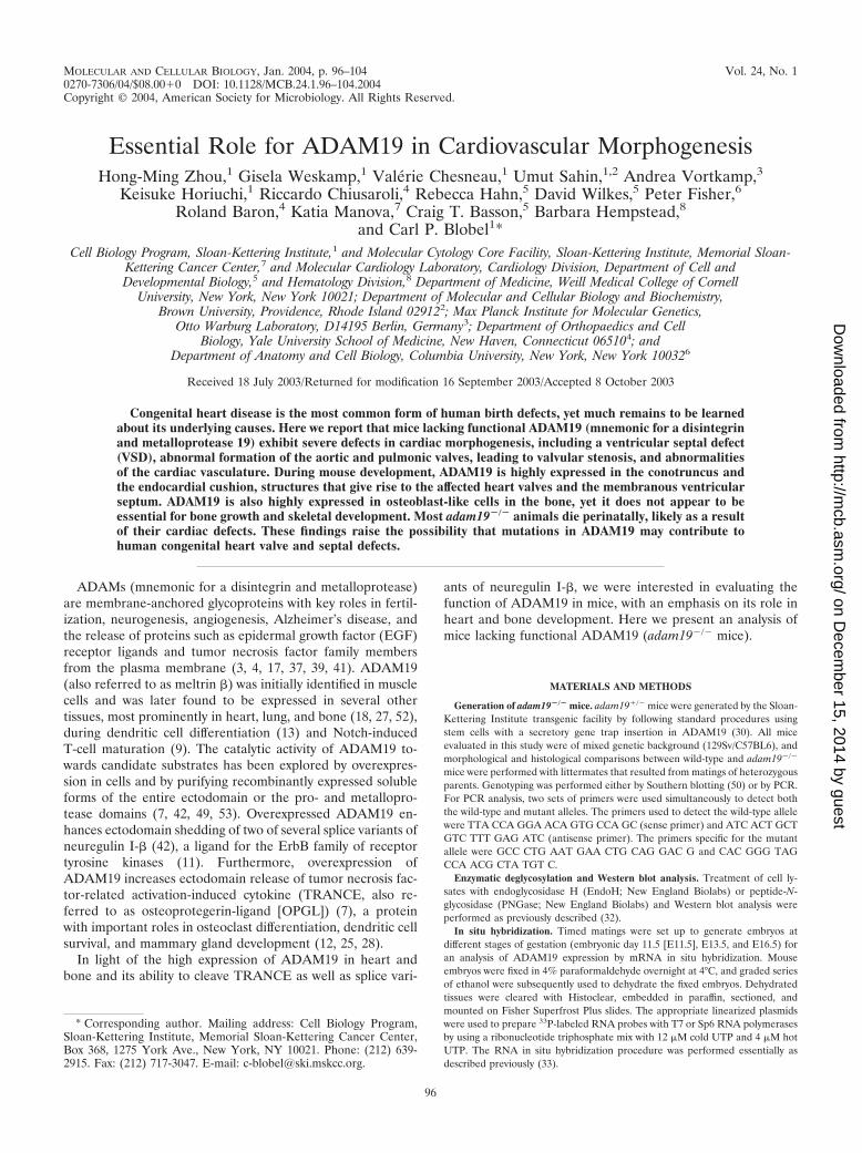
FIG. 1. ADAM19 gene trap construct. (a) Diagram of wild-type ADAM19 and the mutant form generated by a secretory gene trap insertion(30). (b) Results of PCR genotyping. (c) Western blots of brain extracts of wild-type (�/�), heterozygous (�/�), or homozygous (�/�) ADAM19mutant mice were analyzed as described previously (50) with an antibody raised against the ectodomain of ADAM19. (d and e) Western blots ofbrain extracts from adam19�/� mice (d) or wild-type mice (e) treated with EndoH or peptide-N-glycosidase (PNGase) were probed with anantibody against LacZ (d) or against the ectodomain of ADAM19 (e) (see also Fig. 1c). N-linked high mannose glycans are converted into complexcarbohydrates by passage through the medial-Golgi network. Complex carbohydrates are usually resistant to treatment with EndoH, whereastreatment with PNGase removes both high-mannose glycans and complex N-linked carbohydrate residues. Acquisition of EndoH resistancetherefore indicates that a glycoprotein has emerged from the ER and has passed through the medial-Golgi network. The arrows in panel d markthe position of the mutant ADAM19/LacZ/neo (�-geo) fusion protein.
VOL. 24, 2004 ANALYSIS OF MICE LACKING ADAM19 97
on Decem
ber 15, 2014 by guesthttp://m
cb.asm.org/
Dow
nloaded from
(Invitrogen) as previously described (7, 50). The day following transfection, eachwell was washed once in PBS and incubated for 1 h in Opti-MEM I (Invitrogen)(nonstimulated conditioned medium) and then for an additional hour in Opti-MEM I containing 25 ng of phorbol 12-myristate 13-acetate (PMA)/ml (stimu-lated conditioned medium). Conditioned media were collected, and cells werelysed in PBS containing 1% Triton X-100, 2 �g of leupeptin/ml, 10 �g of soybeantrypsin inhibitor/ml, 500 �M iodoacetamide, and 1 mM 1,10-phenanthroline.His-tagged shed forms of TRANCE-AP in the supernatant as well as membrane-anchored TRANCE-AP in cell lysate were concentrated by using Talon metalaffinity resin and analyzed by sodium dodecyl sulfate-polyacrylamide gel electro-phoresis as previously described (7). Shed HB-EGF-AP, neuregulin I-�1-AP,and neuregulin I-�2-AP present in the conditioned media were concentrated by
using concanavalin A-Sepharose (Amersham Biosciences) and analyzed by so-dium dodecyl sulfate-polyacrylamide gel electrophoresis as described previously(7). COS-7 cell lysates were assayed for the expression of wild-type or catalyti-cally inactive (E�A mutant) ADAM19 by Western blotting with anti-ADAM19polyclonal antibodies as described previously (7).
RESULTS AND DISCUSSION
Mice lacking functional ADAM19 were obtained from EScells with a secretory gene trap insertion in the ADAM19 gene(Fig. 1a; see Materials and Methods) (30). The progeny of
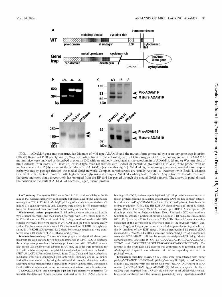
FIG. 2. Heart defects in adam19�/� mice. Ventral view of hearts from newborn wild-type (a) or adam19�/� mice (b). Distended blood-filled vascularstructures are indicated by arrowheads. (c, d, and e) CD31 staining confirms the vascular nature of the abnormal structures (shown by arrows in panelsd and e). (f through i) Cross-section through CD31-stained (f and g) and �-smooth muscle actin-stained (h and i) blood vessel within the septalmyocardium of wild-type (f), adam19�/� (h), and adam19�/� (g and i) animals. The sections shown in panels f and g are from newborn mice, whereasthe sections in panels h and i are from E17.5 embryos. Arrows indicate normal smooth muscle ensheathment in a wild-type mouse (f and h) and abnormalor apparently lacking smooth muscle cell ensheathment of vessels in an adam19�/� mouse (g and i). Note that �-smooth muscle-actin also stainsmyocardial cells surrounding smooth muscle cells in h and i. (j, k, and l) Electron micrographs of cross-sections through myocardial capillaries of awild-type (j) or adam19�/� (k and l) heart (the asterisks in k and l indicate perivascular edema). (m and n) Longitudinal section through a heart of anewborn wild-type (m) or adam19�/� (n) mouse stained with hematoxylin and eosin; the aortic valve is indicated by arrows; the aorta is overriding VSDin panel n. (o and p) Cross-sections through a heart of a newborn wild-type (o) or adam19�/� (p) mouse. In panels o and p, an arrow points to thetricuspid valve, which displays a lack of remodeling in the adam19�/� heart (p). An asterisk is placed next to the leaflets of the mitral valve. (q and r)Cross-sections of the pulmonic (q) and aortic valve (r) of a wild-type heart and an adam19�/� heart. Three comparable serial sections of 10 �m inthickness, spaced three sections (30 �m) apart, are shown for each valve. Abbreviations: RA, right atrium; LA, left atrium; RV, right ventricle; LV, leftventricle.
98 ZHOU ET AL. MOL. CELL. BIOL.
on Decem
ber 15, 2014 by guesthttp://m
cb.asm.org/
Dow
nloaded from
matings of heterozygous ADAM19 mutant mice had a Men-delian distribution of the targeted allele at birth (28.0% �/�,47.2% �/�, 24.8% �/�; n � 254). However, 80% of ho-mozygous adam19�/� mice died in the first few days afterbirth, while heterozygous ADAM19 mutant mice were healthyand fertile (distribution of offspring at day 21 after birth [P21],30.8% �/�, 64.9% �/�, 4.3% �/�; n � 487).
Western blot analysis of brain extracts confirmed that wild-type ADAM19 is absent in homozygous mutant mice (Fig. 1band c). Using an anti-�-galactosidase antibody to detect themutant form of ADAM19 (Fig. 1a), we observed that theN-linked carbohydrate residues never acquire resistance toendoglycosidase H (EndoH) (Fig. 1d), indicating that most orall of the mutant protein never reaches the medial Golgi com-partment. In contrast, mature wild-type ADAM19 acquires
resistance to EndoH and is cleaved by a proprotein convertasein the trans-Golgi network (Fig. 1e) (22). The gene trap inser-tion into the cysteine-rich domain of ADAM19 evidently pre-vents proper protein folding, resulting in retention of mutantADAM19 in the endoplasmic reticulum (ER) by chaperonesand subsequent degradation (45). Therefore, since no detect-able amounts of mutant ADAM19 emerge from the ER andadam19�/� mice have no evident pathological phenotype, theabnormal phenotypes in adam19�/� mice likely result from aloss of ADAM19 function and not from a dominant negativeeffect of the mutant protein.
In situ examination of the thoracic cavity of adam19�/� micerevealed abnormally distended blood-filled vascular structureson the anterior wall of the right ventricle (Fig. 2b, d, and e).Immunohistochemical evaluation of the right ventricle demon-
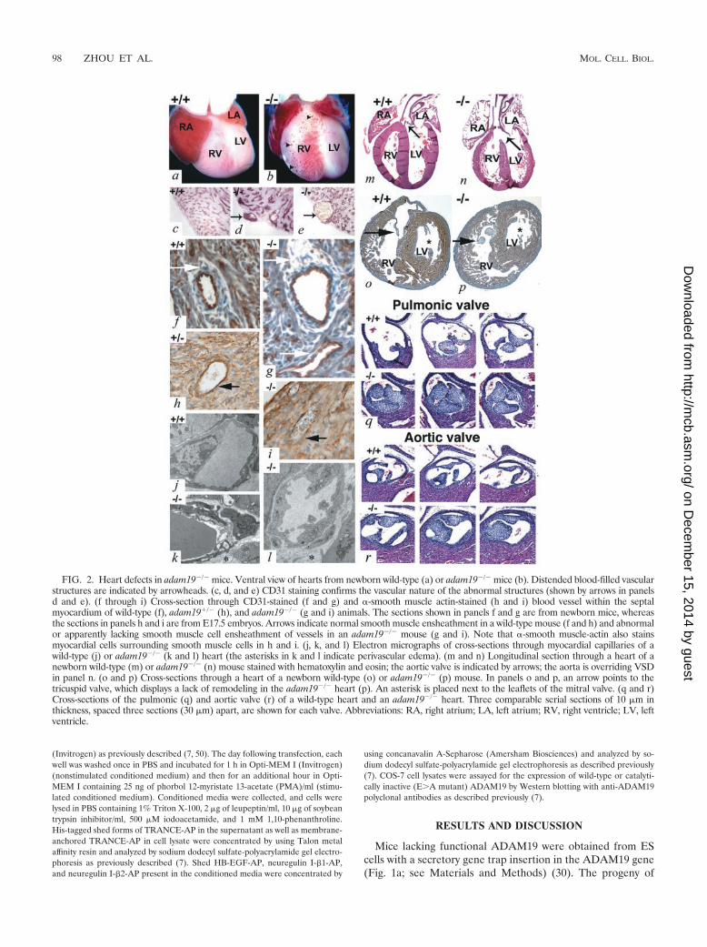
FIG. 3. Analysis of the expression of ADAM19 and neuregulin during heart and brain development by in situ mRNA hybridization andcomparison of endocardial cushion development in wild-type and adam19�/� embryos. Hearts of wild-type E10.5 (a through d) and E 12.5 embryos(e through h), were probed with antisense RNA probes for mouse ADAM19 (a, b, e, and f) or neuregulin (c, d, g, and h) (51). In panels b, d, f,and h, an asterisk marks the atrioventricular endocardial cushion. In panels b and d, a thick arrow points to the atrial endocardium, whereADAM19, but little neuregulin, is expressed. A thin arrow points to the ventricular endocardium, where there is some neuregulin expression, butlittle ADAM19 expression. The arrowhead (b and d) points towards a limited dorsal area of the conotruncal outflow tract where expression ofADAM19 and neuregulin appear to overlap. Panels a, c, e, and g are bright field; panels b, d, f, and h are dark field. (i through n) Sections ofwild-type (i and m), adam19�/� (k), or adam19�/� (j, l, and n) hearts at E10.5 (i and j), E11.5 (k and l), and E12.5 (m and n). Sections in panelsk and l are of an E11.5 conotruncal endocardial cushion of a heterozygous (k) heart or homozygous ADAM19 mutant heart (l) stained with X-Galin order to detect cells expressing lacZ under the ADAM19 promoter. Cells located below the gap indicated by an arrow in panels k and l are partof the conotruncal endocardial cushion. In panels m and n, the endocardial cushion is marked by an arrow. (o through r) Adjacent sections of theneuroepithelium of the fourth ventricle of E12.5 embryos (o and p) or of the developing spinal cord of E10.5 embryos (q and r) were probed withantisense RNA probes for mouse ADAM19 (o and q) or neuregulin (p and r) (51). In panels o and p, an arrow points to a region of enhancedcoexpression of ADAM19 and neuregulin in a defined area of the neuroepithelium of the fourth ventricle. In panels q and r, thick arrows markthe dorsal root ganglion and thin arrows mark the ventral horn of the spinal cord. Abbreviations: A, atrium; V, ventricle; CT, conotruncalendocardial cushion; EC, atrioventricular endocardial cushion.
VOL. 24, 2004 ANALYSIS OF MICE LACKING ADAM19 99
on Decem
ber 15, 2014 by guesthttp://m
cb.asm.org/
Dow
nloaded from
strated that the walls of these distended vessels are composedof endothelial cells (CD31 positive) (Fig. 2d and e). Further-more, blood vessels within the myocardium of adam19�/� miceappeared abnormal, with disrupted smooth muscle cell en-sheathment (Fig. 2g and i; wild-type controls are shown in Fig.2f and h). In electron micrographs of myocardial endothelialcells of adam19�/� mice, we frequently observed perivascularedema in capillaries, extensive vacuolization of endothelialcells (Fig. 2k and l; wild-type control is shown in Fig. 2j), andoccasional rupture of both capillaries and arterioles with re-lease of erythrocytes into the extravascular space (data notshown). These ultrastructural defects are consistent with thefocal intramyocardial hemorrhage observed in someadam19�/� hearts at the light microscopic level (data notshown).
A histological evaluation of adam19�/� hearts revealedmembranous ventricular septal defects (VSD) (Fig. 2n) as wellas stenotic aortic and pulmonic valves with abnormally thick-ened leaflets in all hearts examined at birth (12 of 12; Fig. 2qand r). adam19�/� neonates exhibited moderate malrotationof the cardiac outflow tracts, in some cases resulting in anoverriding aorta. These defects resemble conotruncal malfor-mations, such as Tetralogy of Fallot and double-outlet rightventricle, which are important forms of human congenitalheart disease with high morbidity. Thickened tricuspid valveswere found in about one-half of adam19�/� hearts (Fig. 2p),
and atrial septal defects were observed in one-third ofadam19�/� hearts (data not shown). The mitral valve ap-peared normal in all adam19�/� hearts examined (Fig. 2n andp), and septation of the outflow tract into pulmonary arteryand aorta was also unaffected (Fig. 2q and r).
The membranous aspect of the ventricular septum and theheart valves arising from the endocardial cushions betweenE10.5 and E13.5 (24, 29) prompted evaluation of ADAM19expression in these developmental structures. In situ mRNAhybridization revealed prominent ADAM19 expression in theatrioventricular and conotruncal endocardial cushions betweenE10.5 and E12.5 (Fig. 3a, b, e, and f; also data not shown). Thisexpression pattern is consistent with the observed defects inadam19�/� mice. The complete penetrance of proximalconotruncal defects (VSD and aortic and pulmonic valve de-fects) compared to the partial penetrance of atrioventriculardefects (ostium primum atrial septal and tricuspid valve de-fects) in adam19�/� mice demonstrates that the proximalconotruncal endocardial cushion is most sensitive to loss ofADAM19 activity.
Cardiac neural crest cells make important contributions tothe morphogenesis of the conotruncal endocardial cushion,suggesting that ADAM19 may have a role in neural crestmigration (24). Furthermore, the related ADAM13 has beenimplicated in neural crest migration in Xenopus laevis (1).However, in adam19�/� mice, other structures that depend on
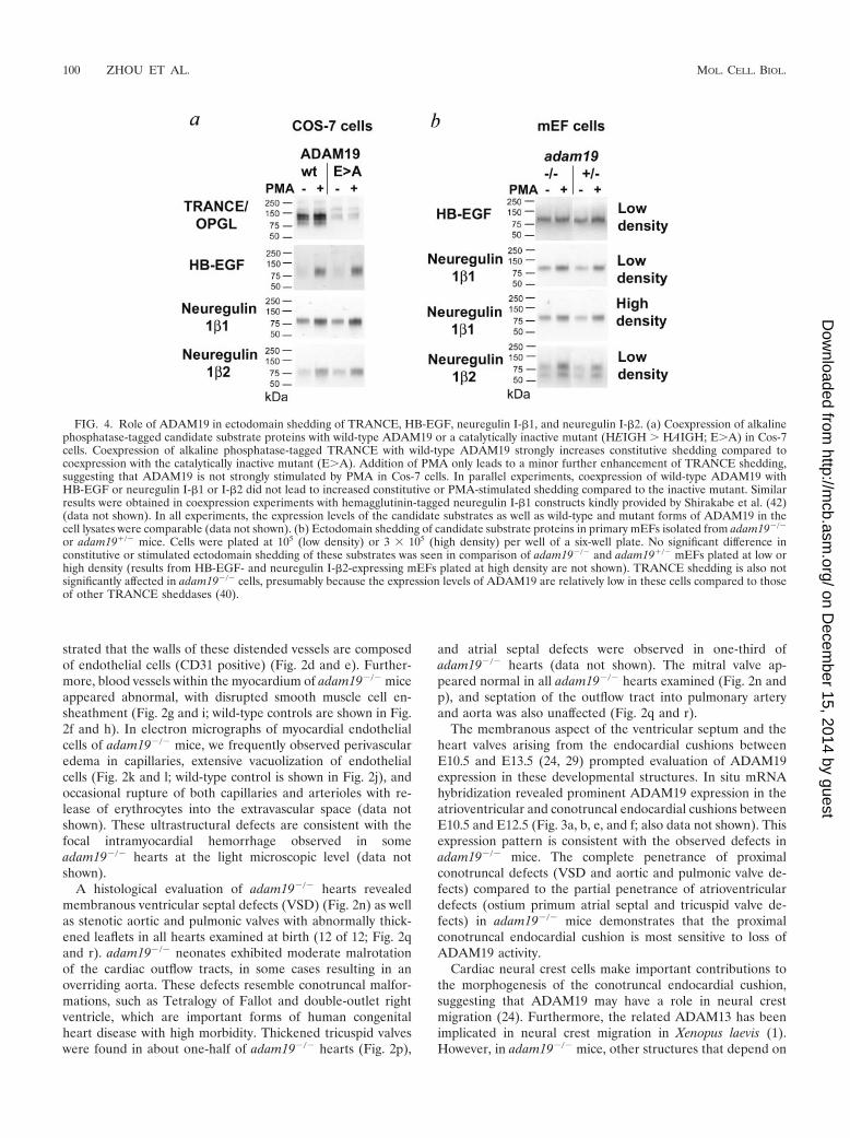
FIG. 4. Role of ADAM19 in ectodomain shedding of TRANCE, HB-EGF, neuregulin I-�1, and neuregulin I-�2. (a) Coexpression of alkalinephosphatase-tagged candidate substrate proteins with wild-type ADAM19 or a catalytically inactive mutant (HEIGH � HAIGH; E�A) in Cos-7cells. Coexpression of alkaline phosphatase-tagged TRANCE with wild-type ADAM19 strongly increases constitutive shedding compared tocoexpression with the catalytically inactive mutant (E�A). Addition of PMA only leads to a minor further enhancement of TRANCE shedding,suggesting that ADAM19 is not strongly stimulated by PMA in Cos-7 cells. In parallel experiments, coexpression of wild-type ADAM19 withHB-EGF or neuregulin I-�1 or I-�2 did not lead to increased constitutive or PMA-stimulated shedding compared to the inactive mutant. Similarresults were obtained in coexpression experiments with hemagglutinin-tagged neuregulin I-�1 constructs kindly provided by Shirakabe et al. (42)(data not shown). In all experiments, the expression levels of the candidate substrates as well as wild-type and mutant forms of ADAM19 in thecell lysates were comparable (data not shown). (b) Ectodomain shedding of candidate substrate proteins in primary mEFs isolated from adam19�/�
or adam19�/� mice. Cells were plated at 105 (low density) or 3 105 (high density) per well of a six-well plate. No significant difference inconstitutive or stimulated ectodomain shedding of these substrates was seen in comparison of adam19�/� and adam19�/� mEFs plated at low orhigh density (results from HB-EGF- and neuregulin I-�2-expressing mEFs plated at high density are not shown). TRANCE shedding is also notsignificantly affected in adam19�/� cells, presumably because the expression levels of ADAM19 are relatively low in these cells compared to thoseof other TRANCE sheddases (40).
100 ZHOU ET AL. MOL. CELL. BIOL.
on Decem
ber 15, 2014 by guesthttp://m
cb.asm.org/
Dow
nloaded from
migration of neural crest cells, such as the thymus, hyoid bone,larynx, and palate, appeared morphologically normal (data notshown). Moreover, adam19�/� and wild-type embryos had en-docardial cushions of similar shape and cellularity at E10.5 andE12.5, when neural crest migration to the region is complete(Fig. 3i, j, m, and n). Finally, when we stained heart sections ofheterozygous or homozygous mutant ADAM19 embryos withX-Gal to mark cells expressing lacZ under the ADAM19 pro-moter, we found no difference in the distribution of stainedcells at E11.5 (Fig. 3k and l). This result argues against a rolefor ADAM19 in the migration of ADAM19-expressing cellsinto the endocardial cushion. Instead, the heart defects seen innewborn adam19�/� mice, in particular the thickened andimproperly remodeled aortic, pulmonic, and tricuspid valves,are more consistent with a key role for ADAM19 regulatingmorphogenesis and remodeling of the endocardial cushion af-ter ADAM19-expressing cells have entered this structure.
Recent studies have demonstrated that mice lacking HB-EGF also have thickened aortic and pulmonic valves (19, 20).
Furthermore, mice lacking ADAM17, which has a critical rolein processing and presumably also activating HB-EGF (34, 44),phenocopy mice lacking HB-EGF with respect to the thick-ened aortic and pulmonic valves (20). In order to test whetherADAM19 might also contribute to HB-EGF shedding fromthe endocardium, we coexpressed both proteins in Cos-7 cellsand also compared HB-EGF shedding in adam19�/� andadam19�/� mEFs. In order to confirm that ADAM19 is activein coexpression experiments, we included TRANCE/OPGL,an osteoclast differentiation factor and dendritic cell survivalfactor that is a known substrate of ADAM19 (7), as a positivecontrol (Fig. 4a). When HB-EGF was coexpressed with wild-type ADAM19 or a catalytically inactive mutant (E�A), nodifference in constitutive or PMA-induced shedding was ob-served (Fig. 4a). Furthermore, we saw no difference in consti-tutive or PMA-stimulated shedding of HB-EGF fromadam19�/� mEF cells compared to heterozygous controls (Fig.4b) or wild-type controls (data not shown). Taken together,
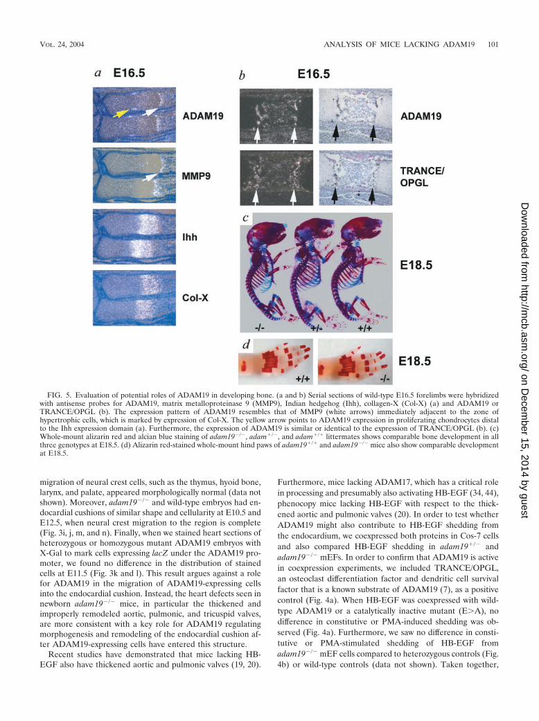
FIG. 5. Evaluation of potential roles of ADAM19 in developing bone. (a and b) Serial sections of wild-type E16.5 forelimbs were hybridizedwith antisense probes for ADAM19, matrix metalloproteinase 9 (MMP9), Indian hedgehog (Ihh), collagen-X (Col-X) (a) and ADAM19 orTRANCE/OPGL (b). The expression pattern of ADAM19 resembles that of MMP9 (white arrows) immediately adjacent to the zone ofhypertrophic cells, which is marked by expression of Col-X. The yellow arrow points to ADAM19 expression in proliferating chondrocytes distalto the Ihh expression domain (a). Furthermore, the expression of ADAM19 is similar or identical to the expression of TRANCE/OPGL (b). (c)Whole-mount alizarin red and alcian blue staining of adam19�/�, adam�/�, and adam�/� littermates shows comparable bone development in allthree genotypes at E18.5. (d) Alizarin red-stained whole-mount hind paws of adam19�/� and adam19�/� mice also show comparable developmentat E18.5.
VOL. 24, 2004 ANALYSIS OF MICE LACKING ADAM19 101
on Decem
ber 15, 2014 by guesthttp://m
cb.asm.org/
Dow
nloaded from
these experiments argue against a role of ADAM19 in HB-EGF shedding.
ADAM19 has also been implicated in ectodomain sheddingof neuregulin I-�, a protein that is essential for proper tra-beculation of the heart during early development (26, 36, 42).However, when we coexpressed neuregulin I-�1 or I-�2 withwild-type or mutant ADAM19 in Cos-7 cells, no difference inectodomain shedding was evident (Fig. 4a). Furthermore, nosignificant defect was seen in the constitutive or PMA-stimu-lated shedding of neuregulin I-�1 and I-�2 in sparsely ordensely seeded adam19�/� mEFs compared to heterozygousor wild-type controls (Fig. 4b and data not shown). When wecompared the expression pattern of ADAM19 and neuregulinin adjacent sections of E10.5 and E12.5 mouse embryos, wefound little, if any, coexpression in the endocardial cushion
(Fig. 3a through h). Neuregulin has also been implicated in thedevelopment of the cardiac conduction system (38), yet wefound no differences between the electrocardiograms of new-born adam19�/� mice and those of wild-type mice (data notshown). Taken together, these results suggest that the heartdefects seen in adam19�/� mice are not due to defects inprocessing neuregulin I-�1 or I-�2. It remains to be deter-mined whether ADAM19 has a role in processing other neu-regulin isoforms (11) in cells and tissues where both proteinsare coexpressed, such as in the endocardium overlying theendocardial cushion and a limited portion of the outflow tractof the developing heart at E10.5 (Fig. 3b and d), a defined areaof neuroepithelial cells within the fourth brain ventricle (Fig.3o and p), and part of the ventral horn of the spinal cord anddorsal root ganglia (Fig. 3q and r; see also reference 27).
In a previous study, it was demonstrated that ADAM19 ishighly expressed in bone (18). Because ADAM19 can cleavethe osteoclast differentiation factor TRANCE in Cos-7 cells(see above and reference 7), we evaluated whether ADAM19may have a role in bone development. In situ hybridizationshowed that ADAM19 is highly expressed in a subpopulationof cells residing immediately adjacent to the zone of collagen-X-expressing hypertrophic chondrocytes in the growth plate(Fig. 5a and b). The expression pattern of ADAM19 resemblesthat of MMP9 (48) (Fig. 5a), which is also found next to cellsexpressing collagen-X. Furthermore, at E16.5, TRANCE ap-pears to be coexpressed with ADAM19 (Fig. 5b), raising thepossibility that ADAM19 could participate in TRANCE shed-ding in this particular bone area. Finally, a distinct domain ofADAM19 expression is found in a subset of highly proliferat-ing chondrocytes adjacent to the perichondrium and distal toearly hypertrophic chondrocytes expressing Indian hedgehog.Whole-mount alizarin red and alcian blue staining as well asmorphometric analysis of bone sections from wild-type andadam19�/� mice did not uncover evident histopathologicaldifferences in bone development (Fig. 5c and d and data notshown). Further studies will be necessary to evaluate bones ofsurviving adult adam19�/� mice for more-subtle defects inbone remodeling that may not be apparent during bone devel-opment.
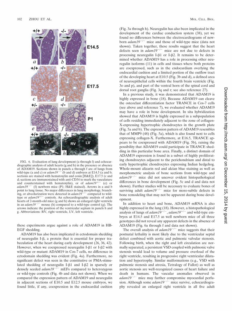
In addition to heart and bone, ADAM19 mRNA is alsohighly expressed in the lung (18). However, a histopathologicalanalysis of lungs of adam19�/�, adam19�/� and wild-type em-bryos at E14.5 and E17.5 as well newborn mice of all threegenotypes did not reveal any apparent defects in the absence ofADAM19 (Fig. 6a through f and data not shown).
The overall analysis of adam19�/� mice suggests that theirpostnatal lethality is most likely due to the ventricular septaldefect combined with aortic and pulmonic valvular stenosis.Following birth, when the right and left circulation are nor-mally separated, a persistent VSD coupled with pulmonic valvestenosis would lead to volume and pressure overload of theright ventricle, resulting in progressive right ventricular dilata-tion and hypertrophy. Similar malformations (e.g., VSD withpulmonic stenosis and atresia, Tetralogy of Fallot) as well asaortic stenosis are well-recognized causes of heart failure anddeath in humans. The vascular anomalies observed inadam19�/� mice may further compromise myocardial perfu-sion. Although some adam19�/� mice survive, echocardiogra-phy revealed an enlarged right ventricle in all five adult
FIG. 6. Evaluation of lung development (a through f) and echocar-diographic analysis of adult hearts (g and h) in the presence or absenceof ADAM19. Sections shown in panels a through f are of lungs fromwild-type (a and c) or adam19�/�(b and d) embryos at E14.5 (a and b;sections are stained with hematoxylin and eosin [H&E]), E17.5 (c andd; sections are immunostained with anti-CD34 to mark the vasculatureand counterstained with hematoxylin), or of adam19�/� (e) oradam19�/� (f) newborn mice (P1; H&E stained). Arrows in a and bpoint to lung tissue. No major differences in lung morphology, branch-ing, or alveolarization were detected in adam19�/� compared to wild-type or adam19�/� controls. An echocardiographic analysis of adulthearts of 2-month-old mice (g and h) shows an enlarged right ventriclein an adam19�/� mouse (h) compared to a wild-type control (g). Thearrows indicate the position of the ventricular septum in panels h andg. Abbreviations: RV, right ventricle, LV, left ventricle.
102 ZHOU ET AL. MOL. CELL. BIOL.
on Decem
ber 15, 2014 by guesthttp://m
cb.asm.org/
Dow
nloaded from
adam19�/� animals examined (Fig. 6g and h and data notshown). This is consistent with sublethal cardiac defects thatare still compatible with long-term but nonstressed survival.Adult adam19�/� mice appear healthy and are fertile, al-though four of five pregnant adam19�/� mice died late inpregnancy, likely due to increased circulatory demands. In-deed, human adults with similar forms of congenital heartdisease represent high-risk obstetrical patients.
Congenital heart disease is the most common form of birthdefects in humans, yet little is known about the underlyingmolecular causes (2, 8, 15, 29, 43, 47). In light of the criticalrole of mouse ADAM19 in heart development, it will be in-teresting to determine whether certain types of human con-genital heart defects are caused by mutations in ADAM19,which is located on human chromosome 5q33.3. To date, onlya few proteins on the cell surface or in the extracellular matrixhave been implicated in endocardial cushion development, in-cluding neurotrophin-3 (10), tumor growth factor � (5, 6) andthe related BMP6 and BMP7 (23), and most recently HB-EGFand ADAM17 (19, 20). It will now be interesting to furtherexplore potential functional connections between ADAM19and these proteins as well as other molecules that are impli-cated in endocardial cushion transformation and/or conotrun-cal defects, such as RXR�, smad6, and TBX1 (14, 16, 21, 31,35). It is also possible that ADAM19-dependent cell-cell in-teractions or signaling via its cytoplasmic domain contribute toits role in heart development (39). We anticipate that thediscovery of an essential role for ADAM19 in heart develop-ment in mice will lead to a better understanding of the causesunderlying human congenital heart disease.
ACKNOWLEDGMENTS
This work was supported by National Institutes of Health grant RO1GM64750 (to C.P.B.).
We thank Phil Leighton, Bill Skarnes, and Marc Tessier-Lavigne forgenerously providing embryonic stem cells carrying a gene trap inser-tion in ADAM19, Leona Cohen-Gould for providing electron micro-graphs, Thomas Ludwig, Willie Mark, Liz Lacy, Jay Edelberg, andDavid Christini for valuable advice, and Thadeous Kacmarczyk, DanHarrigan, Kevin Curran, Maria Kobi, Conny Kreschel, and membersof the Sloan-Kettering Institute transgenic facility for excellent tech-nical assistance.
REFERENCES
1. Alfandari, D., H. Cousin, A. Gaultier, K. Smith, J. M. White, T. Darribere,and D. W. DeSimone. 2001. Xenopus ADAM 13 is a metalloprotease re-quired for cranial neural crest-cell migration. Curr. Biol. 11:918–930.
2. Basson, C. T., and C. E. Seidman. 1998. Genetic studies of myocardial andvascular disease, p. 2429–2447. In E. J. Topol (ed.), Textbook of cardiovas-cular medicine. Lippincott-Raven, Philadelphia, Pa.
3. Black, R. A., and J. M. White. 1998. ADAMs: focus on the protease domain.Curr. Opin. Cell Biol. 10:654–659.
4. Blobel, C. P. 2000. Remarkable roles of proteolysis on and beyond the cellsurface. Curr. Opin. Cell Biol. 12:606–612.
5. Brown, C. B., A. S. Boyer, R. B. Runyan, and J. V. Barnett. 1999. Require-ment of type III TGF-beta receptor for endocardial cell transformation inthe heart. Science 283:2080–2082.
6. Camenisch, T. D., D. G. Molin, A. Person, R. B. Runyan, A. C. Gitten-berger-de Groot, J. A. McDonald, and S. E. Klewer. 2002. Temporal anddistinct TGFbeta ligand requirements during mouse and avian endocardialcushion morphogenesis. Dev. Biol. 248:170–181.
7. Chesneau, V., D. Becherer, Y. Zheng, H. Erdjument-Bromage, P. Tempst,and C. P. Blobel. 2003. Catalytic properties of ADAM19. J. Biol. Chem.278:22331–22340.
8. Chien, K. R. 2000. Genomic circuits and the integrative biology of cardiacdiseases. Nature 407:227–232.
9. Deftos, M. L., E. Huang, E. W. Ojala, K. A. Forbush, and M. J. Bevan. 2000.Notch1 signaling promotes the maturation of CD4 and CD8 SP thymocytes.Immunity 13:73–84.
10. Donovan, M. J., R. Hahn, L. Tessarollo, and B. L. Hempstead. 1996. Iden-tification of an essential nonneuronal function of neurotrophin 3 in mam-malian cardiac development. Nat. Genet. 14:210–213.
11. Falls, D. L. 2003. Neuregulins: functions, forms, and signaling strategies.Exp. Cell Res. 284:14–30.
12. Fata, J. E., Y. Y. Kong, J. Li, T. Sasaki, J. Irie-Sasaki, R. A. Moorehead, R.Elliott, S. Scully, E. B. Voura, D. L. Lacey, W. J. Boyle, R. Khokha, and J. M.Penninger. 2000. The osteoclast differentiation factor osteoprotegerin-ligandis essential for mammary gland development. Cell 103:41–50.
13. Fritsche, J., M. Moser, S. Faust, A. Peuker, R. Buttner, R. Andreesen, andM. Kreutz. 2000. Molecular cloning and characterization of a human met-alloprotease disintegrin–a novel marker for dendritic cell differentiation.Blood 96:732–739.
14. Galvin, K. M., M. J. Donovan, C. A. Lynch, R. I. Meyer, R. J. Paul, J. N.Lorenz, V. Fairchild-Huntress, K. L. Dixon, J. H. Dunmore, M. A. Gim-brone, Jr., D. Falb, and D. Huszar. 2000. A role for smad6 in developmentand homeostasis of the cardiovascular system. Nat. Genet. 24:171–174.
15. Goldmuntz, E., and B. S. Emanuel. 1997. Genetic disorders of cardiacmorphogenesis. The DiGeorge and velocardiofacial syndromes. Circ. Res.80:437–443.
16. Gruber, P. J., S. W. Kubalak, T. Pexieder, H. M. Sucov, R. M. Evans, andK. R. Chien. 1996. RXR alpha deficiency confers genetic susceptibility foraortic sac, conotruncal, atrioventricular cushion, and ventricular muscle de-fects in mice. J. Clin. Investig. 98:1332–1343.
17. Horiuchi, K., G. Weskamp, L. Lum, H. P. Hammes, H. Cai, T. A. Brodie, T.Ludwig, R. Chiusaroli, R. Baron, K. T. Preissner, K. Manova, and C. P.Blobel. 2003. Potential role for ADAM15 in pathological neovascularizationin mice. Mol. Cell. Biol. 23:5614–5624.
18. Inoue, D., M. Reid, L. Lum, J. Kratzschmar, G. Weskamp, Y. M. Myung, R.Baron, and C. P. Blobel. 1998. Cloning and initial characterization of mousemeltrin beta and analysis of the expression of four metalloprotease-disinte-grins in bone cells. J. Biol. Chem. 273:4180–4187.
19. Iwamoto, R., S. Yamazaki, M. Asakura, S. Takashima, H. Hasuwa, K.Miyado, S. Adachi, M. Kitakaze, K. Hashimoto, G. Raab, D. Nanba, S.Higashiyama, M. Hori, M. Klagsbrun, and E. Mekada. 2003. Heparin-binding EGF-like growth factor and ErbB signaling is essential for heartfunction. Proc. Natl. Acad. Sci. USA 100:3221–3226.
20. Jackson, L. F., T. H. Qiu, S. W. Sunnarborg, A. Chang, C. Zhang, C.Patterson, and D. C. Lee. 2003. Defective valvulogenesis in HB-EGF andTACE-null mice is associated with aberrant BMP signaling. EMBO J. 22:2704–2716.
21. Jerome, L. A., and V. E. Papaioannou. 2001. DiGeorge syndrome phenotypein mice mutant for the T-box gene, Tbx1. Nat. Genet. 27:286–291.
22. Kang, T., Y. G. Zhao, D. Pei, J. F. Sucic, and Q. X. Sang. 2002. Intracellularactivation of human adamalysin 19/disintegrin and metalloproteinase 19 byfurin occurs via one of the two consecutive recognition sites. J. Biol. Chem.277:25583–25591.
23. Kim, R. Y., E. J. Robertson, and M. J. Solloway. 2001. Bmp6 and Bmp7 arerequired for cushion formation and septation in the developing mouse heart.Dev. Biol. 235:449–466.
24. Kirby, M. L. 1999. Contribution of neural crest to heart and vessel morphol-ogy, p. 179–193. In R. P. Harvey and N. Rosenthal (ed.), Heart development.Academic Press, London, United Kingdom.
25. Kong, Y. Y., H. Yoshida, I. Sarosi, H. L. Tan, E. Timms, C. Capparelli, S.Morony, A. J. Oliveira-dos-Santos, G. Van, A. Itie, W. Khoo, A. Wakeham,C. R. Dunstan, D. L. Lacey, T. W. Mak, W. J. Boyle, and J. M. Penninger.1999. OPGL is a key regulator of osteoclastogenesis, lymphocyte develop-ment and lymph-node organogenesis. Nature 397:315–323.
26. Kramer, R., N. Bucay, D. J. Kane, L. E. Martin, J. E. Tarpley, and L. E.Theill. 1996. Neuregulins with an Ig-like domain are essential for mousemyocardial and neuronal development. Proc. Natl. Acad. Sci. USA 93:4833–4838.
27. Kurisaki, T., A. Masuda, N. Osumi, Y. Nabeshima, and A. Fujisawa-Sehara.1998. Spatially- and temporally-restricted expression of meltrin alpha(ADAM12) and beta (ADAM19) in mouse embryo. Mech. Dev. 73:211–215.
28. Lacey, D. L., E. Timms, H. L. Tan, M. J. Kelley, C. R. Dunstan, T. Burgess,R. Elliott, A. Colombero, G. Elliott, S. Scully, H. Hsu, J. Sullivan, N.Hawkins, E. Davy, C. Capparelli, A. Eli, Y. X. Qian, S. Kaufman, I. Sarosi,V. Shalhoub, G. Senaldi, J. Guo, J. Delaney, and W. J. Boyle. 1998. Osteo-protegerin ligand is a cytokine that regulates osteoclast differentiation andactivation. Cell 93:165–176.
29. Lamers, W. H., and A. F. Moorman. 2002. Cardiac septation: a late contri-bution of the embryonic primary myocardium to heart morphogenesis. Circ.Res. 91:93–103.
30. Leighton, P. A., K. J. Mitchell, L. V. Goodrich, X. Lu, K. Pinson, P. Scherz,W. C. Skarnes, and M. Tessier-Lavigne. 2001. Defining brain wiring patternsand mechanisms through gene trapping in mice. Nature 410:174–179.
31. Lindsay, E. A., F. Vitelli, H. Su, M. Morishima, T. Huynh, T. Pramparo, V.Jurecic, G. Ogunrinu, H. F. Sutherland, P. J. Scambler, A. Bradley, and A.Baldini. 2001. Tbx1 haploinsufficieny in the DiGeorge syndrome regioncauses aortic arch defects in mice. Nature 410:97–101.
32. Lum, L., and C. P. Blobel. 1997. Evidence for distinct serine protease activ-
VOL. 24, 2004 ANALYSIS OF MICE LACKING ADAM19 103
on Decem
ber 15, 2014 by guesthttp://m
cb.asm.org/
Dow
nloaded from
ities with a potential role in processing the sperm protein fertilin. Dev. Biol.191:131–145.
33. Manova, K., K. Nocka, P. Besmer, and R. F. Bachvarova. 1990. Gonadalexpression of c-kit encoded at the W locus of the mouse. Development110:1057–1069.
34. Merlos-Suarez, A., S. Ruiz-Paz, J. Baselga, and J. Arribas. 2001. Metallo-protease-dependent protransforming growth factor-alpha ectodomain shed-ding in the absence of tumor necrosis factor-alpha-converting enzyme.J. Biol. Chem. 276:48510–48517.
35. Merscher, S., B. Funke, J. A. Epstein, J. Heyer, A. Puech, M. M. Lu, R. J.Xavier, M. B. Demay, R. G. Russell, S. Factor, K. Tokooya, B. S. Jore, M.Lopez, R. K. Pandita, M. Lia, D. Carrion, H. Xu, H. Schorle, J. B. Kobler,P. Scambler, A. Wynshaw-Boris, A. I. Skoultchi, B. E. Morrow, and R.Kucherlapati. 2001. TBX1 is responsible for cardiovascular defects in velo-cardio-facial/DiGeorge syndrome. Cell 104:619–629.
36. Meyer, D., and C. Birchmeier. 1995. Multiple essential functions of neuregu-lin in development. Nature 378:386–390.
37. Primakoff, P., and D. G. Myles. 2000. The ADAM gene family: surfaceproteins with an adhesion and protease activity packed into a single mole-cule. Trends Genet. 16:83–87.
38. Rentschler, S., J. Zander, K. Meyers, D. France, R. Levine, G. Porter, S. A.Rivkees, G. E. Morley, and G. I. Fishman. 2002. Neuregulin-1 promotesformation of the murine cardiac conduction system. Proc. Natl. Acad. Sci.USA 99:10464–10469.
39. Schlondorff, J., and C. P. Blobel. 1999. Metalloprotease-disintegrins: mod-ular proteins capable of promoting cell-cell interactions and triggering sig-nals by protein ectodomain shedding. J. Cell Sci. 112:3603–3617.
40. Schlondorff, J. S., L. Lum, and C. P. Blobel. 2001. Biochemical and phar-macological criteria define two shedding activities for TRANCE/OPGL thatare distinct from the TNF alpha convertase (TACE). J. Biol. Chem. 276:14665–14674.
41. Seals, D. F., and S. A. Courtneidge. 2003. The ADAMs family of metallo-proteases: multidomain proteins with multiple functions. Genes Dev. 17:7–30.
42. Shirakabe, K., S. Wakatsuki, T. Kurisaki, and A. Fujisawa-Sehara. 2001.Roles of meltrin beta/ADAM19 in the processing of neuregulin. J. Biol.Chem. 276:9352–9358.
43. Srivastava, D., and E. N. Olson. 2000. A genetic blueprint for cardiac de-velopment. Nature 407:221–226.
44. Sunnarborg, S. W., C. L. Hinkle, M. Stevenson, W. E. Russell, C. S. Raska,J. J. Peschon, B. J. Castner, M. J. Gerhart, R. J. Paxton, R. A. Black, andD. C. Lee. 2002. Tumor necrosis factor-alpha converting enzyme (TACE)regulates epidermal growth factor receptor ligand availability. J. Biol. Chem.277:12838–12845.
45. Suzuki, T., Q. Yan, and W. J. Lennarz. 1998. Complex, two-way traffic ofmolecules across the membrane of the endoplasmic reticulum. J. Biol. Chem.273:10083–10086.
46. Tokumaru, S., S. Higashiyama, T. Endo, T. Nakagawa, J. Miyagawa, K.Yamamori, Y. Hanakawa, H. Ohmoto, K. Yoshino, Y. Shirakata, Y. Matsu-zawa, K. Hashimoto, and N. Taniguchi. 2000. Ectodomain shedding ofepidermal growth factor receptor ligands is required for keratinocyte migra-tion in cutaneous wound healing. J. Cell Biol. 151:209–220.
47. Vaughan, C. J., and C. T. Basson. 2000. Molecular determinants of atrial andventricular septal defects and patent ductus arteriosus. Am. J. Med. Genet.97:304–309.
48. Vu, T. H., J. M. Shipley, G. Bergers, J. E. Berger, J. A. Helms, D. Hanahan,S. D. Shapiro, R. M. Senior, and Z. Werb. 1998. MMP-9/gelatinase B is a keyregulator of growth plate angiogenesis and apoptosis of hypertrophic chon-drocytes. Cell 93:411–422.
49. Wei, P., Y. G. Zhao, L. Zhuang, S. Ruben, and Q. X. Sang. 2001. Expressionand enzymatic activity of human disintegrin and metalloproteinaseADAM19/meltrin beta. Biochem. Biophys. Res. Commun. 280:744–755.
50. Weskamp, G., H. Cai, T. A. Brodie, S. Higashyama, K. Manova, T. Ludwig,and C. P. Blobel. 2002. Mice lacking the metalloprotease-disintegrin MDC9(ADAM9) have no evident major abnormalities during development or adultlife. Mol. Cell. Biol. 22:1537–1544.
51. Wilkinson, D. G., J. A. Bailes, J. E. Champion, and A. P. McMahon. 1987. Amolecular analysis of mouse development from 8 to 10 days post coitumdetects changes only in embryonic globin expression. Development 99:493–500.
52. Yagami-Hiromasa, T., T. Sato, T. Kurisaki, K. Kamijo, Y. Nabeshima, andA. Fujisawa-Sehara. 1995. A metalloprotease-disintegrin participating inmyoblast fusion. Nature 377:652–656.
53. Zhao, Y. G., P. Wei, and Q. X. Sang. 2001. Inhibitory antibodies againstendopeptidase activity of human adamalysin 19. Biochem. Biophys. Res.Commun. 289:288–294.
104 ZHOU ET AL. MOL. CELL. BIOL.
on Decem
ber 15, 2014 by guesthttp://m
cb.asm.org/
Dow
nloaded from