Does Anybody Want an Injectable Rotavirus Vaccine ... - MDPI
Upload
uuniversidaddepanamCategory
view
4download
0
JOURNAL OF VIROLOGY, Jan. 2005, p. 184–192 Vol. 79, No. 10022-538X/05/$08.00�0 doi:10.1128/JVI.79.1.184–192.2005Copyright © 2005, American Society for Microbiology. All Rights Reserved.
Silencing the Morphogenesis of RotavirusTomas Lopez,† Minerva Camacho,† Margarita Zayas, Rebeca Najera, Rosana Sanchez,
Carlos F. Arias,* and Susana Lopez*Departamento de Genetica del Desarrollo y Fisiologıa Molecular, Instituto de Biotecnologıa,
Universidad Nacional Autonoma de Mexico, Cuernavaca, Morelos, Mexico
Received 4 June 2004/Accepted 27 July 2004
The morphogenesis of rotaviruses follows a unique pathway in which immature double-layered particles(DLPs) assembled in the cytoplasm bud across the membrane of the endoplasmic reticulum (ER), acquiringduring this process a transient lipid membrane which is modified with the ER resident viral glycoproteinsNSP4 and VP7; these enveloped particles also contain VP4. As the particles move towards the interior of theER cisternae, the transient lipid membrane and the nonstructural protein NSP4 are lost, while the virussurface proteins VP4 and VP7 rearrange to form the outermost virus protein layer, yielding mature infectioustriple-layered particles (TLPs). In this work, we have characterized the role of NSP4 and VP7 in rotavirusmorphogenesis by silencing the expression of both glycoproteins through RNA interference. Silencing theexpression of either NSP4 or VP7 reduced the yield of viral progeny by 75 to 80%, although the underlyingmechanism of this reduction was different in each case. Blocking the synthesis of NSP4 affected the intracel-lular accumulation and the cellular distribution of several viral proteins, and little or no virus particles(neither DLPs nor TLPs) were assembled. VP7 silencing, in contrast, did not affect the expression or distri-bution of other viral proteins, but in its absence, enveloped particles accumulated within the lumen of the ER,and no mature infectious virus was produced. Altogether, these results indicate that during a viral infection,NSP4 serves as a receptor for DLPs on the ER membrane and drives the budding of these particles into theER lumen, while VP7 is required for removing the lipid envelope during the final step of virus morphogenesis.
Rotaviruses are nonenveloped icosahedral viruses whosecapsid is formed by three concentric layers of protein. Theinnermost layer is formed by 60 dimers of VP2 that surroundsthe viral genome composed of 11 segments of double-strandedRNA and 12 copies of each VP1, the virus polymerase, andVP3, the virus capping enzyme. The second layer of protein isformed by 280 trimers of VP6, which sits on top of VP2 to formdouble-layered particles (DLPs). Finally, the addition of 280trimers of glycoprotein VP7 which constitute the outermostlayer of the virus and 60 dimeric spikes of the VP4 protein toDLPs form triple-layered particles (TLPs) that represent themature infectious virus (13).
Rotavirus morphogenesis occurs by an unusual processwhere DLPs, which are thought to assemble in cytoplasmicinclusions termed viroplasms, bud across the membrane of theendoplasmic reticulum (ER). During this process, the DLPsacquire a transient lipid envelope which is subsequently lost toyield the mature infectious TLPs (33). The ER membranethrough which DLPs bud is modified by two viral proteins, thevirion surface protein VP7 and the nonstructural polypeptideNSP4 (5, 7, 21). NSP4 has a large cytosolic domain that inter-acts with DLPs, and it has been proposed that this interactiondrives the translocation of the double-layered particles into thelumen of the ER (3, 35). NSP4 is also known to associate withthe lumen-oriented VP7 and with VP4, forming a heterotri-
meric complex that is thought to participate in the budding ofDLPs into the ER (22).
During the last step of rotavirus morphogenesis, the tran-sient lipid envelope is lost and the viral proteins are rearrangedso that VP4 and VP7 assemble to form the outer layer of thevirus, while NSP4 is excluded from the virus particle. Themechanisms underlying the loss of the lipid envelope and theviral protein NSP4 as well as the correct assembly of the outer-layer proteins are not understood, although it has been sug-gested that the membrane-destabilizing activities of NSP4and/or VP4 and the relatively high calcium concentrationpresent in the lumen of the ER could be implicated in theremoval of the lipid envelope (10, 30, 36). The produced ma-ture virus is then released either by cell lysis in MA104 cells(13) or after transport along an atypical trafficking pathwayfrom the ER to the plasma membrane in polarized epithelialCaco-2 cells (18). It has recently been shown that the expres-sion of rotavirus genes can be efficiently silenced by RNAinterference using small interfering RNAs (siRNAs) that havea sequence complementary to the viral gene to be silenced (1,9, 32). siRNA has proven to be a very useful tool to dissect thefunction of the viral genes in the context of a natural infection(1, 20). Silencing the expression of the rhesus rotavirus VP4gene showed that in the absence of VP4, the DLPs were stillable to bud into the ER and to lose the lipid envelope, yieldingTLPs that lack VP4 spikes. These results indicated that VP4 isrequired neither for the translocation of DLPs into the ER norfor the removal of the lipid membrane from the transientlyenveloped particles. In this work, we evaluated the role thatthe viral glycoproteins VP7 and NSP4 play in the morphogen-esis of rotavirus. We found that silencing the NSP4 gene af-fected the accumulation and the cellular distribution of several
* Corresponding author. Mailing address: Depto. Genetica del De-sarrollo y Fisiologıa Molecular, Instituto de Biotecnologıa, UNAM,Av. Universidad 2001, Col. Chamilpa, Cuernavaca, Morelos 62210,Mexico. Phone: (52) (777) 3114701. Fax: (52) (777) 3172388. E-mailfor Carlos F. Arias: [email protected]. E-mail for Susana Lopez:[email protected].
† T.L. and M.C. contributed equally to this work.
184

viral proteins and that little or no virus was assembled in itsabsence. VP7 silencing, in contrast, did not affect the expres-sion or the distribution of other viral proteins, but when it wasnot present, enveloped particles accumulated within the lumenof the ER, and very little, if any, mature infectious virus wasproduced.
MATERIALS AND METHODS
Cells and viruses. The rhesus monkey epithelial cell line MA104 was grown inEagle’s minimal essential medium (MEM) supplemented with 10% fetal bovineserum (FBS) and was used for all experiments carried out in this work. Rhesusrotavirus (RRV) was obtained from H. B. Greenberg (Stanford University,Stanford, Calif.), simian rotavirus SA11 was obtained from H. H. Malherbe, andporcine rotavirus YM was isolated in our laboratory. All rotavirus strains werepropagated in MA104 cells as described previously (26).
Antibodies. Monoclonal antibodies (MAbs) to VP2 (3A8), VP6 (255/60), VP7(60), and NSP4 (B4) were kindly provided by H. B. Greenberg. MAb HS2directed to VP4 (25), the rabbit anti-rotavirus polyclonal serum raised againstpurified RRV TLPs, and the rabbit anti-vimentin sera were produced in ourlaboratory. Rabbit polyclonal sera to NSP2, NSP3, and NSP5 have been de-scribed previously (16). Alexa-488- and Alexa-568-conjugated secondary anti-bodies and phalloidin-Alexa-546 were purchased from Molecular Probes (Eu-gene, Oreg.). Horseradish peroxidase-conjugated goat anti-rabbit polyclonalantibody was from Perkin-Elmer Life Sciences (Boston, Mass.), and horseradishperoxidase-conjugated rabbit anti-mouse immunoglobulin G and rabbit anti-tubulin antibody were from Zymed (San Francisco, Calif.).
siRNA transfection. The siRNAs were obtained from Dharmacon Research(Lafayette, Colo.) as annealed duplexes. The sequence and relative positions ofthe siRNAs (shown in Table 1) were chosen based on the rules proposed byElbashir et al. (12). As irrelevant controls, siRNAs with sequences from thecaveolin-1, lamin A/C, or green fluorescent protein gene were used during thiswork. None of these siRNAs affected the viral protein synthesis or viral infec-tivity, and no differences were found between them; thus, they are genericallyreferred to as control siRNAs (Table 1). Transfection of siRNAs was carried outin nearly confluent cell monolayers by using 2 �l of Lipofectamine (Invitrogen)per 100 �l of siRNAs at 600 pmol/ml in MEM without antibiotics. The trans-fection mixture was added to cells previously washed with MEM and incubatedfor 8 h at 37°C. After this time, the transfection mixture was removed and thecells were washed with MEM and kept in this medium for 48 h at 37°C beforevirus infection.
Infection of cells and titration of viral progeny. Transfected cell monolayers in24- or 48-well plates were infected with 3 virus focus-forming units per cell andthen incubated for 24 h at 37°C. At this time, the cells were then lysed by twofreeze-thaw cycles, and the lysates were treated with 10 �g of trypsin/ml for 30min at 37°C. The infectious titer of the viral preparations was obtained by animmunoperoxidase focus assay (26). Briefly, confluent MA104 cells in 96-wellplates were washed twice with phosphate-buffered saline (PBS), and serial dilu-tions (n-fold) of the above-mentioned viral lysate were adsorbed to the cells for60 min at 37°C. After the adsorption period, the virus inoculum was removed, thecells were washed once with PBS, MEM was added, and the infection was left to
proceed for 14 h at 37°C for RRV. RRV-infected cells were detected by animmunoperoxidase focus detection assay using a rabbit hyperimmune serum torotavirus as described previously (26). The focus-forming units were countedwith the help of a Visiolab 1000 station (Biocom, France), as has been previouslyreported (17).
Immunoblots. Cells were transfected with siRNAs and infected with rotavirusRRV as described above. Twelve hours postinfection (hpi), the cells were lysedwith Laemmli sample buffer, and the proteins were separated by sodium dodecylsulfate-polyacrylamide gel electrophoresis (SDS-PAGE) and then transferred tonitrocellulose membranes (Millipore, Bedford, Mass.). Membranes were blockedwith 5% nonfat dried milk in PBS and incubated at 4°C with the indicatedprimary antibodies in PBS containing 0.1% milk followed by an incubation withsecondary, species-specific, horseradish peroxidase-conjugated antibodies. Theperoxidase activity was developed by the Western Lightning ChemiluminescenceReagent Plus (Perkin-Elmer Life Sciences) according to the manufacturer’sinstructions.
Immunofluorescence. MA104 cells grown on glass coverslips were transfectedas mentioned above, and 48 h posttransfection, the cells were infected withrotavirus RRV at a multiplicity of infection (MOI) of 3. Eight hours postinfec-tion, the cells were fixed with 2% paraformaldehyde in PBS for 20 min at 37°C.After this time, the cells were washed twice with PBS containing 50 mM NH4Cland were permeabilized by incubation with PBS–0.5% Triton X-100–50 mMNH4Cl for 15 min at room temperature and washed twice with PBS with gentleswirling. The coverslips were then incubated for 1 h at room temperature withprimary antibodies diluted in blocking buffer (50 mM NH4Cl and 1% bovineserum albumin in PBS) and then rinsed four times with PBS. The coverslips werethen incubated with the appropriate Alexa-labeled secondary antibodies inblocking buffer for 1 h at room temperature. The cells were washed four timeswith PBS and mounted onto glass slides with Fluoprep (BioMerieux) and theantifading agent DABCO (100 mg/ml; Sigma, St. Louis, Mo.). The slides wereanalyzed with a Bio-Rad MRC-600 confocal microscope and CoMOS MPL-1000software or with a Nikon E600 epifluorescence microscope coupled to aDXM1200 digital still camera (Nikon). The images were then digitally capturedand prepared with Adobe Photoshop software version 7.0.
Radiolabeling, isolation, and analysis of viral particles. Cells grown in 48-wellplates were transfected with siRNAs and infected with rotavirus RRV as de-scribed above. At 4 hpi, the medium was replaced by MEM without methionine,supplemented with 50 �Ci of Easy Tag EXPRESS-35S labeling mix (Dupont,NEN)/ml, and incubated for 8 h; after this period, the cells were washed andlysed with Laemmli sample buffer. To prepare purified viral particles, cells grownin 75-cm2 flasks were transfected and infected as described previously and wereharvested until complete cytopathic effect was attained; the virus in the celllysates was pelleted by centrifugation for 1 h at 25,000 rpm at 4°C with an SW28rotor (Beckman). The virus pellet was resuspended in TNC buffer (10 mMTris-HCl [pH 7.5], 140 mM NaCl, 10 mM CaCl2), sonicated once for 20 s, andextracted with Genetron (trichloro-monofluoro-ethane). CsCl was then added toobtain a density of 1.36 g/cm3, the mixture was centrifuged for 18 h at 36,000 rpmwith an SW50.1 rotor, and the virus bands were collected by puncture. The CsClin the bands was removed by desalting-centrifugation on Sephadex G-25, and theprotein composition of each band was analyzed by SDS-PAGE and silver stain-ing.
TABLE 1. Sequences and nucleotide positions of the siRNAs for RRV VP7 and NSP4
siRNA Nucleotide positions Sense sequence Relative inhibitionb
VP7-1 317–337 AAU UCG UGG AAG GAU ACA CUC �VP7-2 390–410 AAU ACA CGG AUA UUG CUU CCU �VP7-3c 681–701 AAG UCG CUA CAG CUG AAA AAC ���VP7-4 751–771 AAC UGC UAC UUG CAC UAU CAG �VP7-5 217–237 AAU GGA CAC UGC AUA CGC UAA ��NSP4-1c 183–203 AAG GCC UCG GUU CCA ACC AUG ��NSP4-2 352–372 AAG AAA UGA GAC GUC AGC UGG �Controla
Lamin A/C AAC UGG ACU UCC AGA AGA ACA �Caveolin-1 AAG CCC AAC AAC AAG GCC AUG �GFP AAC UUA CCC UGA AGU UCA UCU �
a The sequences of the irrelevant controls used througouht this work are listed (they were purchased from Dharmacon). GFP, green fluorescent protein.b Relative inhibition of the production of viral progeny compared to cells transfected with an irrelevant siRNA. ���, �70%; ��, 50%; �, �25%; �, no effect.c The most effective siRNAs for silencing the expression of these genes were named siRNANSP4 and siRNAVP7 throughout the text.
VOL. 79, 2005 ROTAVIRUS MORPHOGENESIS 185
Electron microscopy. Cells grown in 75-cm2 flasks were transfected withsiRNAs and infected with rotavirus RRV at an MOI of 3 as described above.Eight hours postinfection, the cells were fixed in 2.5% glutaraldehyde–0.1 Mcacodylate (pH 7.2), postfixed with 1% osmium tetroxide, and embedded inEpon 812 resin. The ultrathin sections obtained were stained with 2% uranylacetate–1% lead citrate (Reynolds mix) (31). The grids were examined with aZeiss EM-900 electron microscope at 80 kV.
RESULTS
siRNAs directed to rotavirus NSP4 and VP7 genes inhibitthe synthesis of the encoded glycoproteins. To evaluate theeffect of siRNAs on the synthesis of viral glycoproteins NSP4and VP7, siRNAs corresponding to the sequences of the NSP4and VP7 genes of RRV were transfected into MA104 cells.Forty-eight hours after transfection, the cells were infectedwith rotavirus RRV, and 4 hpi, the proteins were metabolicallylabeled with EXPRESS-35S labeling mix for 8 h and analyzedby gel electrophoresis to evaluate the synthesis of viral pro-teins. Initially, five and two different siRNAs were tested forVP7 and NSP4, respectively. Their sequences and their relativeeffect on the specific inhibition of protein synthesis are shownin Table 1. The most effective siRNAs for silencing the expres-sion of these genes were named siRNANSP4 and siRNAVP7
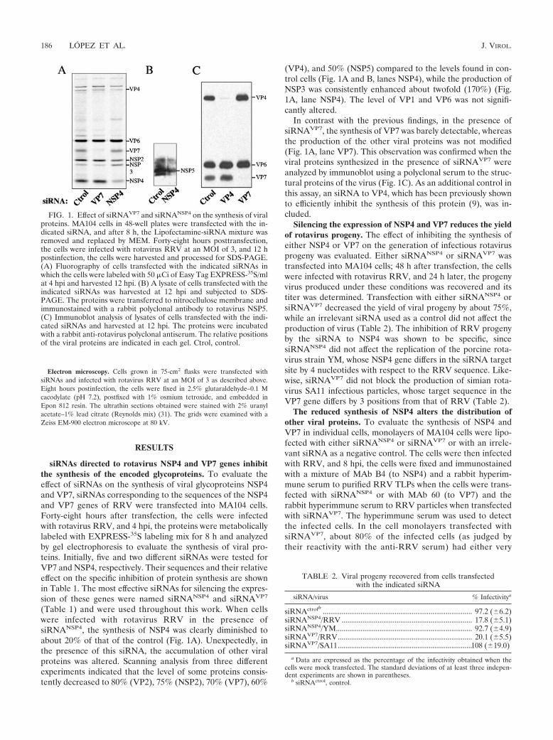
(Table 1) and were used throughout this work. When cellswere infected with rotavirus RRV in the presence ofsiRNANSP4, the synthesis of NSP4 was clearly diminished toabout 20% of that of the control (Fig. 1A). Unexpectedly, inthe presence of this siRNA, the accumulation of other viralproteins was altered. Scanning analysis from three differentexperiments indicated that the level of some proteins consis-tently decreased to 80% (VP2), 75% (NSP2), 70% (VP7), 60%
(VP4), and 50% (NSP5) compared to the levels found in con-trol cells (Fig. 1A and B, lanes NSP4), while the production ofNSP3 was consistently enhanced about twofold (170%) (Fig.1A, lane NSP4). The level of VP1 and VP6 was not signifi-cantly altered.
In contrast with the previous findings, in the presence ofsiRNAVP7, the synthesis of VP7 was barely detectable, whereasthe production of the other viral proteins was not modified(Fig. 1A, lane VP7). This observation was confirmed when theviral proteins synthesized in the presence of siRNAVP7 wereanalyzed by immunoblot using a polyclonal serum to the struc-tural proteins of the virus (Fig. 1C). As an additional control inthis assay, an siRNA to VP4, which has been previously shownto efficiently inhibit the synthesis of this protein (9), was in-cluded.
Silencing the expression of NSP4 and VP7 reduces the yieldof rotavirus progeny. The effect of inhibiting the synthesis ofeither NSP4 or VP7 on the generation of infectious rotavirusprogeny was evaluated. Either siRNANSP4 or siRNAVP7 wastransfected into MA104 cells; 48 h after transfection, the cellswere infected with rotavirus RRV, and 24 h later, the progenyvirus produced under these conditions was recovered and itstiter was determined. Transfection with either siRNANSP4 orsiRNAVP7 decreased the yield of viral progeny by about 75%,while an irrelevant siRNA used as a control did not affect theproduction of virus (Table 2). The inhibition of RRV progenyby the siRNA to NSP4 was shown to be specific, sincesiRNANSP4 did not affect the replication of the porcine rota-virus strain YM, whose NSP4 gene differs in the siRNA targetsite by 4 nucleotides with respect to the RRV sequence. Like-wise, siRNAVP7 did not block the production of simian rota-virus SA11 infectious particles, whose target sequence in theVP7 gene differs by 3 positions from that of RRV (Table 2).
The reduced synthesis of NSP4 alters the distribution ofother viral proteins. To evaluate the synthesis of NSP4 andVP7 in individual cells, monolayers of MA104 cells were lipo-fected with either siRNANSP4 or siRNAVP7 or with an irrele-vant siRNA as a negative control. The cells were then infectedwith RRV, and 8 hpi, the cells were fixed and immunostainedwith a mixture of MAb B4 (to NSP4) and a rabbit hyperim-mune serum to purified RRV TLPs when the cells were trans-fected with siRNANSP4 or with MAb 60 (to VP7) and therabbit hyperimmune serum to RRV particles when transfectedwith siRNAVP7. The hyperimmune serum was used to detectthe infected cells. In the cell monolayers transfected withsiRNAVP7, about 80% of the infected cells (as judged bytheir reactivity with the anti-RRV serum) had either very
FIG. 1. Effect of siRNAVP7 and siRNANSP4 on the synthesis of viralproteins. MA104 cells in 48-well plates were transfected with the in-dicated siRNA, and after 8 h, the Lipofectamine-siRNA mixture wasremoved and replaced by MEM. Forty-eight hours posttransfection,the cells were infected with rotavirus RRV at an MOI of 3, and 12 hpostinfection, the cells were harvested and processed for SDS-PAGE.(A) Fluorography of cells transfected with the indicated siRNAs inwhich the cells were labeled with 50 �Ci of Easy Tag EXPRESS-35S/mlat 4 hpi and harvested 12 hpi. (B) A lysate of cells transfected with theindicated siRNAs was harvested at 12 hpi and subjected to SDS-PAGE. The proteins were transferred to nitrocellulose membrane andimmunostained with a rabbit polyclonal antibody to rotavirus NSP5.(C) Immunoblot analysis of lysates of cells transfected with the indi-cated siRNAs and harvested at 12 hpi. The proteins were incubatedwith a rabbit anti-rotavirus polyclonal antiserum. The relative positionsof the viral proteins are indicated in each gel. Ctrol, control.
TABLE 2. Viral progeny recovered from cells transfectedwith the indicated siRNA
siRNA/virus % Infectivitya
siRNActrolb ............................................................................... 97.2 (�6.2)siRNANSP4/RRV ..................................................................... 17.8 (�5.1)siRNANSP4/YM........................................................................ 92.7 (�4.9)siRNAVP7/RRV....................................................................... 20.1 (�5.5)siRNAVP7/SA11.......................................................................108 (�19.0)
a Data are expressed as the percentage of the infectivity obtained when thecells were mock transfected. The standard deviations of at least three indepen-dent experiments are shown in parentheses.
b siRNActrol, control.
186 LOPEZ ET AL. J. VIROL.

low or undetectable levels of VP7. Similarly, transfectionwith siRNANSP4 caused a large number of infected cells tohave barely detectable levels of NSP4 (Fig. 2).
Of interest, when the polyclonal serum to RRV was used tostain the virus-infected, siRNANSP4-transfected cells, a strikingfilamentous pattern which was not present in cells transfectedwith either siRNAVP7 or the irrelevant siRNA was observed(Fig. 2). To find out which viral protein(s) was responsible forthis filamentous signal, a detailed analysis of the distribution ofthe individual proteins was carried out by using MAbs to VP2,VP4, VP6, VP7, and NSP4 and monospecific polyclonal sera toNSP2, NSP3, and NSP5. This analysis showed that the intra-cellular distribution of several viral proteins was altered insiRNANSP4-transfected cells. VP6 was found to extensively re-distribute, forming filaments that appear to extend to the pe-riphery of the cell instead of being localized to viroplasms, as
was observed in cells transfected with siRNAVP7 or with thesiRNA control (Fig. 3). These filaments are not the result of anassociation of VP6 with tubulin, actin, or vimentin, since thesignal of none of these proteins, as detected with specific an-tibodies or with phalloidin in the case of actin, colocalized withVP6 (data not shown).
VP2 was found to distribute homogeneously in the cyto-plasm rather than in viroplasms (Fig. 3). The mixed pattern ofVP4 that has been previously reported (15, 24), which consistsof perinuclear ring-like or semicircular structures closely asso-ciated to viroplasms (15) and a filamentous array due to itsassociation with microtubules (24), was observed in cells trans-fected with the control siRNA and siRNAVP7; however, in thepresence of siRNANSP4, most of the perinuclear structuresdisappeared while the filamentous signal of VP4 remained andwas even more evident than that of control cells (Fig. 3). In
FIG. 2. Effect of siRNAs to NSP4 and VP7 on the expression of the corresponding protein in infected cells. MA104 cells transfected with theindicated siRNAs were infected 48 h posttransfection, and at 8 hpi, the cells were fixed and processed for immunofluorescence. The expressionlevels of VP7 and NSP4 were monitored by using MAbs 60 (�-VP7) and B4 (�-NSP4), respectively, and the cell infection by RRV was monitoredby using a rabbit polyclonal antiserum to rotavirus (�-RRV) followed by anti-mouse Alexa-568 and anti-rabbit Alexa-488 antibodies, respectively.
VOL. 79, 2005 ROTAVIRUS MORPHOGENESIS 187
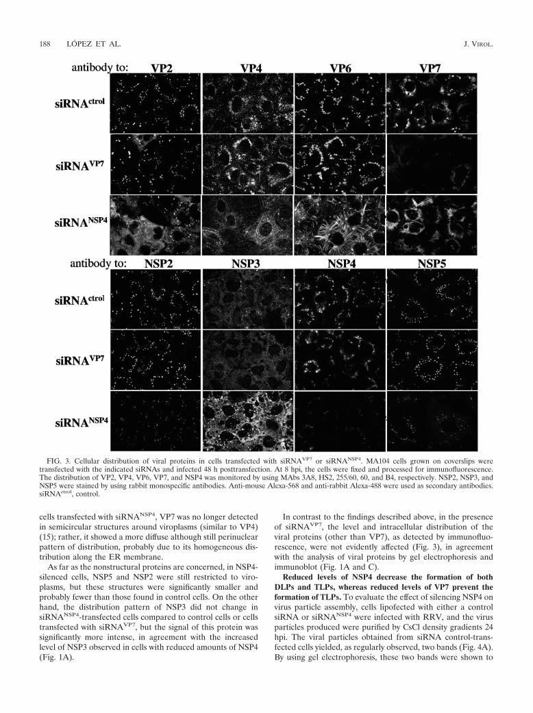
cells transfected with siRNANSP4, VP7 was no longer detectedin semicircular structures around viroplasms (similar to VP4)(15); rather, it showed a more diffuse although still perinuclearpattern of distribution, probably due to its homogeneous dis-tribution along the ER membrane.
As far as the nonstructural proteins are concerned, in NSP4-silenced cells, NSP5 and NSP2 were still restricted to viro-plasms, but these structures were significantly smaller andprobably fewer than those found in control cells. On the otherhand, the distribution pattern of NSP3 did not change insiRNANSP4-transfected cells compared to control cells or cellstransfected with siRNAVP7, but the signal of this protein wassignificantly more intense, in agreement with the increasedlevel of NSP3 observed in cells with reduced amounts of NSP4(Fig. 1A).
In contrast to the findings described above, in the presenceof siRNAVP7, the level and intracellular distribution of theviral proteins (other than VP7), as detected by immunofluo-rescence, were not evidently affected (Fig. 3), in agreementwith the analysis of viral proteins by gel electrophoresis andimmunoblot (Fig. 1A and C).
Reduced levels of NSP4 decrease the formation of bothDLPs and TLPs, whereas reduced levels of VP7 prevent theformation of TLPs. To evaluate the effect of silencing NSP4 onvirus particle assembly, cells lipofected with either a controlsiRNA or siRNANSP4 were infected with RRV, and the virusparticles produced were purified by CsCl density gradients 24hpi. The viral particles obtained from siRNA control-trans-fected cells yielded, as regularly observed, two bands (Fig. 4A).By using gel electrophoresis, these two bands were shown to
FIG. 3. Cellular distribution of viral proteins in cells transfected with siRNAVP7 or siRNANSP4. MA104 cells grown on coverslips weretransfected with the indicated siRNAs and infected 48 h posttransfection. At 8 hpi, the cells were fixed and processed for immunofluorescence.The distribution of VP2, VP4, VP6, VP7, and NSP4 was monitored by using MAbs 3A8, HS2, 255/60, 60, and B4, respectively. NSP2, NSP3, andNSP5 were stained by using rabbit monospecific antibodies. Anti-mouse Alexa-568 and anti-rabbit Alexa-488 were used as secondary antibodies.siRNActrol, control.
188 LOPEZ ET AL. J. VIROL.
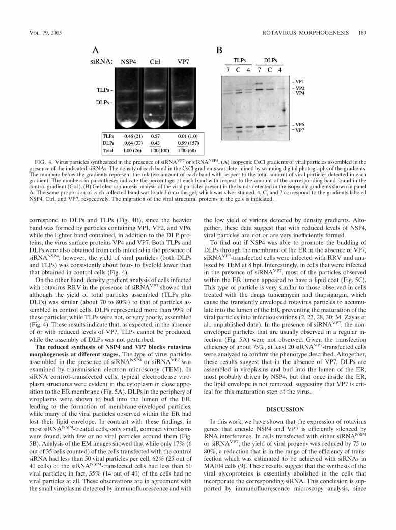
correspond to DLPs and TLPs (Fig. 4B), since the heavierband was formed by particles containing VP1, VP2, and VP6,while the lighter band contained, in addition to the DLP pro-teins, the virus surface proteins VP4 and VP7. Both TLPs andDLPs were also obtained from cells infected in the presence ofsiRNANSP4; however, the yield of viral particles (both DLPsand TLPs) was consistently about four- to fivefold lower thanthat obtained in control cells (Fig. 4).
On the other hand, density gradient analysis of cells infectedwith rotavirus RRV in the presence of siRNAVP7 showed thatalthough the yield of total particles assembled (TLPs plusDLPs) was similar (about 70 to 80%) to that of particles as-sembled in control cells, DLPs represented more than 99% ofthese particles, while TLPs were not, or very poorly, assembled(Fig. 4). These results indicate that, as expected, in the absenceof or with reduced levels of VP7, TLPs cannot be produced,while the assembly of DLPs was not perturbed.
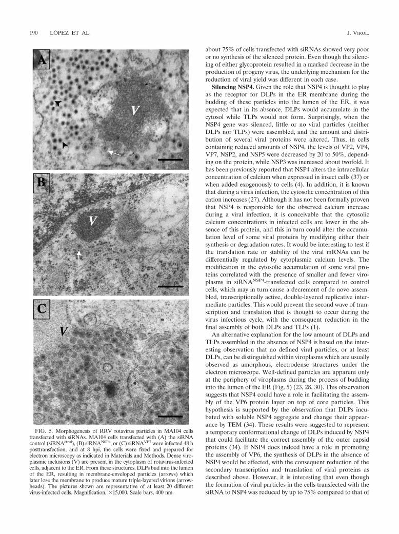
The reduced synthesis of NSP4 and VP7 blocks rotavirusmorphogenesis at different stages. The type of virus particlesassembled in the presence of siRNANSP4 or siRNAVP7 wasexamined by transmission electron microscopy (TEM). InsiRNA control-transfected cells, typical electrodense viro-plasm structures were evident in the cytoplasm in close appo-sition to the ER membrane (Fig. 5A). DLPs in the periphery ofviroplasms were shown to bud into the lumen of the ER,leading to the formation of membrane-enveloped particles,while many of the viral particles observed within the ER hadlost their lipid envelope. In contrast with these findings, inmost siRNANSP4-treated cells, only small, compact viroplasmswere found, with few or no viral particles around them (Fig.5B). Analysis of the EM images showed that while only 17% (6out of 35 cells counted) of the cells transfected with the controlsiRNA had less than 50 viral particles per cell, 62% (25 out of40 cells) of the siRNANSP4-transfected cells had less than 50viral particles; in fact, 35% (14 out of 40) of the cells had noviral particles at all. These observations are in agreement withthe small viroplasms detected by immunofluorescence and with
the low yield of virions detected by density gradients. Alto-gether, these data suggest that with reduced levels of NSP4,viral particles are not or are very inefficiently formed.
To find out if NSP4 was able to promote the budding ofDLPs through the membrane of the ER in the absence of VP7,siRNAVP7-transfected cells were infected with RRV and ana-lyzed by TEM at 8 hpi. Interestingly, in cells that were infectedin the presence of siRNAVP7, most of the particles observedwithin the ER lumen appeared to have a lipid coat (Fig. 5C).This type of particle is very similar to those observed in cellstreated with the drugs tunicamycin and thapsigargin, whichcause the transiently enveloped rotavirus particles to accumu-late into the lumen of the ER, preventing the maturation of theviral particles into infectious virions (2, 23, 28, 30; M. Zayas etal., unpublished data). In the presence of siRNAVP7, the non-enveloped particles that are usually observed in a regular in-fection (Fig. 5A) were not observed. Given the transfectionefficiency of about 75%, at least 20 siRNAVP7-transfected cellswere analyzed to confirm the phenotype described. Altogether,these results suggest that in the absence of VP7, DLPs areassembled in viroplasms and bud into the lumen of the ER,most probably driven by NSP4, but that once inside the ER,the lipid envelope is not removed, suggesting that VP7 is crit-ical for this maturation step of the virus.
DISCUSSION
In this work, we have shown that the expression of rotavirusgenes that encode NSP4 and VP7 is efficiently silenced byRNA interference. In cells transfected with either siRNANSP4
or siRNAVP7, the yield of viral progeny was reduced by 75 to80%, a reduction that is in the range of the efficiency of trans-fection which was estimated to be achieved with siRNAs inMA104 cells (9). These results suggest that the synthesis of theviral glycoproteins is essentially abolished in the cells thatincorporate the corresponding siRNA. This conclusion is sup-ported by immunofluorescence microscopy analysis, since
FIG. 4. Virus particles synthesized in the presence of siRNAVP7 or siRNANSP4. (A) Isopycnic CsCl gradients of viral particles assembled in thepresence of the indicated siRNAs. The density of each band in the CsCl gradients was determined by scanning digital photographs of the gradients.The numbers below the gradients represent the relative amount of each band with respect to the total amount of viral particles detected in eachgradient. The numbers in parentheses indicate the percentage of each band with respect to the amount of the corresponding band found in thecontrol gradient (Ctrl). (B) Gel electrophoresis analysis of the viral particles present in the bands detected in the isopycnic gradients shown in panelA. The same proportion of each collected band was loaded onto the gel, which was silver stained. 4, C, and 7 correspond to the gradients labeledNSP4, Ctrl, and VP7, respectively. The migration of the viral structural proteins in the gels is indicated.
VOL. 79, 2005 ROTAVIRUS MORPHOGENESIS 189
about 75% of cells transfected with siRNAs showed very pooror no synthesis of the silenced protein. Even though the silenc-ing of either glycoprotein resulted in a marked decrease in theproduction of progeny virus, the underlying mechanism for thereduction of viral yield was different in each case.
Silencing NSP4. Given the role that NSP4 is thought to playas the receptor for DLPs in the ER membrane during thebudding of these particles into the lumen of the ER, it wasexpected that in its absence, DLPs would accumulate in thecytosol while TLPs would not form. Surprisingly, when theNSP4 gene was silenced, little or no viral particles (neitherDLPs nor TLPs) were assembled, and the amount and distri-bution of several viral proteins were altered. Thus, in cellscontaining reduced amounts of NSP4, the levels of VP2, VP4,VP7, NSP2, and NSP5 were decreased by 20 to 50%, depend-ing on the protein, while NSP3 was increased about twofold. Ithas been previously reported that NSP4 alters the intracellularconcentration of calcium when expressed in insect cells (37) orwhen added exogenously to cells (4). In addition, it is knownthat during a virus infection, the cytosolic concentration of thiscation increases (27). Although it has not been formally proventhat NSP4 is responsible for the observed calcium increaseduring a viral infection, it is conceivable that the cytosoliccalcium concentrations in infected cells are lower in the ab-sence of this protein, and this in turn could alter the accumu-lation level of some viral proteins by modifying either theirsynthesis or degradation rates. It would be interesting to test ifthe translation rate or stability of the viral mRNAs can bedifferentially regulated by cytoplasmic calcium levels. Themodification in the cytosolic accumulation of some viral pro-teins correlated with the presence of smaller and fewer viro-plasms in siRNANSP4-transfected cells compared to controlcells, which may in turn cause a decrement of de novo assem-bled, transcriptionally active, double-layered replicative inter-mediate particles. This would prevent the second wave of tran-scription and translation that is thought to occur during thevirus infectious cycle, with the consequent reduction in thefinal assembly of both DLPs and TLPs (1).
An alternative explanation for the low amount of DLPs andTLPs assembled in the absence of NSP4 is based on the inter-esting observation that no defined viral particles, or at leastDLPs, can be distinguished within viroplasms which are usuallyobserved as amorphous, electrodense structures under theelectron microscope. Well-defined particles are apparent onlyat the periphery of viroplasms during the process of buddinginto the lumen of the ER (Fig. 5) (23, 28, 30). This observationsuggests that NSP4 could have a role in facilitating the assem-bly of the VP6 protein layer on top of core particles. Thishypothesis is supported by the observation that DLPs incu-bated with soluble NSP4 aggregate and change their appear-ance by TEM (34). These results were suggested to representa temporary conformational change of DLPs induced by NSP4that could facilitate the correct assembly of the outer capsidproteins (34). If NSP4 does indeed have a role in promotingthe assembly of VP6, the synthesis of DLPs in the absence ofNSP4 would be affected, with the consequent reduction of thesecondary transcription and translation of viral proteins asdescribed above. However, it is interesting that even thoughthe formation of viral particles in the cells transfected with thesiRNA to NSP4 was reduced by up to 75% compared to that of
FIG. 5. Morphogenesis of RRV rotavirus particles in MA104 cellstransfected with siRNAs. MA104 cells transfected with (A) the siRNAcontrol (siRNActrol), (B) siRNANSP4, or (C) siRNAVP7 were infected 48 hposttransfection, and at 8 hpi, the cells were fixed and prepared forelectron microscopy as indicated in Materials and Methods. Dense viro-plasmic inclusions (V) are present in the cytoplasm of rotavirus-infectedcells, adjacent to the ER. From these structures, DLPs bud into the lumenof the ER, resulting in membrane-enveloped particles (arrows) whichlater lose the membrane to produce mature triple-layered virions (arrow-heads). The pictures shown are representative of at least 20 differentvirus-infected cells. Magnification, �15,000. Scale bars, 400 nm.
190 LOPEZ ET AL. J. VIROL.
control cells, the cytosolic accumulation of the viral proteinsdid not decreased accordingly. This observation suggests thateither the half-life of the mRNAs synthesized by the enteringvirus particle is long enough to direct the synthesis of viralproteins up to 12 h postinfection (time at which the cells wereharvested for protein analysis) (Fig. 1A) or the viral particleresponsible for the second round of mRNA synthesis is not theDLP per se, as has been suggested previously (13), but rathera subviral replication intermediate particle that contains littleor no VP6.
In addition to the modification of the level of synthesis ofseveral viral proteins, the silencing of NSP4 also caused amarked intracellular redistribution of some of these proteins.The relocalization of VP7 in the ER membrane from discretesites around viroplasms (15) to a more diffuse pattern (Fig. 3)is most likely the direct result of the absence of NSP4 sinceVP7 (an ER lumen-oriented protein) has been shown to in-teract with NSP4 (22), and this protein, which has domainslocated at both sides of the ER membrane (33), is the only linkof VP7 with the rest of the viral proteins in the cytoplasm. Thelarge decrease in the signal of VP4 around viroplasms, as wellas the relocalization of VP2 to a more homogenous distribu-tion in the cytosol, and of VP6 from viroplasms to fibers couldalso be the direct result of the absence of NSP4, since thecytoplasmic carboxy-terminal domain of this protein is knownto interact with VP4 and VP6 (and VP6 in turn interacts withVP2). Alternatively, the reduced size and number of viro-plasms in NSP4-silenced cells might be the cause of VP2 andVP6 not being present in these structures. This could alsoexplain the redistribution of VP4, since this protein has beendescribed to be closely associated to viroplasms (15). The fil-aments formed by VP6 in the absence of NSP4 are not theresult of its interaction with tubulin microtubules, actin fila-ments, or vimentin intermediate filaments, since no colocaliza-tion was observed by immunofluorescence microscopy (datanot shown). Apparently, the VP6 filamentous structures do notresult from the self-polymerization of the protein since theyare substantially longer and thicker than the VP6 tubes thathave been described previously (14, 19). In addition, these VP6fibers could not be observed by TEM. Thus, it seems that VP6probably associates with some type of cellular fibers that havenot yet been identified. Since a similar redistribution of viralproteins was observed when gene 11 of RRV, which encodesNSP5 and NSP6 proteins, was silenced (Lopez et al., unpub-lished), it is not possible to determine whether the altereddistribution of the viral proteins is the direct effect of theabsence of NSP4 or the indirect effect of the decreased syn-thesis of NSP5 and NSP2 as a result of the silencing of NSP4.
Silencing VP7. When the cells were transfected withsiRNAVP7, the amount of viral proteins synthesized and theirintracellular distribution were not affected, and the viral cycleproceeded apparently undisturbed until the morphogeneticprocess was arrested within the lumen of the ER, where en-veloped particles that failed to produce mature infectious virusaccumulated. Similar results were obtained by Silvestri et al.,who found that silencing the expression of the VP7 gene ofrotavirus strain DXRRV led to the accumulation of DLPs andthe inhibition of the production of infectious virus (32). Thelack of formation of TLPs in the absence of VP7 was expectedsince this protein forms the outermost protein layer of the
virus. However, it was surprising to find that the lipid envelopewas not lost from the usually transient enveloped particles inthe absence of VP7. VP4 and NSP4 have both been demon-strated to have membrane-destabilizing activities (6, 10, 36),and they have both been suggested as candidates for removingthe lipid envelope acquired during the budding of DLPs intothe lumen of the ER. However, the fact that the lipid envelopeis retained in the absence of VP7, even when NSP4 is present(Fig. 5C), strongly suggests that VP7 is essential for this mat-uration step of rotavirus morphogenesis. The central role ofVP7 in the morphogenetic process of the virus is supported bythe fact that the lipid envelope was removed in the absence ofVP4 and spikeless TLPs were assembled in which the structureof the VP7 surface layer was indistinguishable by cryoelectronmicroscopy from the VP7 layer present in complete infectiousvirus (B. V. V. Prasad et al., unpublished data).
It has been previously reported that VP7 is arranged on thesurface of the virion as trimers (8, 38), and more recently, itwas more recently shown that its trimerization is dependent onthe concentration of calcium (11). It is tempting to proposethat soon after the budding of DLPs into the lumen of the ER,VP7, which is embedded in the envelope surrounding the in-termediary particles, assembles in a calcium-dependent pro-cess into a tight protein layer that could exclude the lipidenvelope from the virion as well as NSP4 and other nonstruc-tural proteins that have been shown to be present in smallamounts in these intermediary particles (29), yielding the ma-ture infectious virus. The role of VP7 in removing the lipidmembrane from the enveloped particles is supported by thefact that virus morphogenesis is also arrested at the envelopedstage of the particles, when the cells are treated with thapsi-gargin (23, 28) or the calcium ionophore A23187 (30), whichdecreases the concentration of calcium in the ER, or withtunicamycin that blocks the glycosylation of VP7 and NSP4 (2,30). As mentioned above, it cannot be excluded, however, thatthe glycosylated form of NSP4 or an N-glycosylated cellularprotein participates in this step, since an SA11 rotavirus variant(clone 28) that has a nonglycosylated VP7 is also arrested atthe enveloped intermediate stage when treated with tunicamy-cin (28).
It is also important that in the absence of VP7 (this work) aswell as in the absence of VP4 (9), viral DLPs are still able tobud into the lumen of the ER, indicating that NSP4, and notthe heterotrimer VP4/VP7/NSP4, is sufficient to serve as areceptor for DLPs on the ER membrane and to drive thebudding of these particles into the ER lumen during the latesteps of virus morphogenesis.
Based on the data described in this work, together withprevious observations, we propose the following working mod-el for the final stages of rotavirus morphogenesis. DLPs assem-ble at the periphery of viroplasms and bud across the mem-brane of the ER with the help of NSP4. During the processof budding, VP4 (that interacts with the cytosolic domain ofNSP4), NSP4, and VP7 are incorporated into the intermediaryenveloped particles. Once the particles are inside the ER, thehigh-calcium environment of the lumen triggers the lateralinteraction of VP7 molecules (located on the outside of thelipid envelope) promoting the surface protein layer to tightenand making it interact with the VP6 layer, excluding the lipidsand NSP4 during this process. This model does not rule out,
VOL. 79, 2005 ROTAVIRUS MORPHOGENESIS 191

however, the possibility that NSP4 or cellular proteins might beneeded in this process. The exclusion of lipids and NSP4 fromthe intermediary particles by an as-yet-undetermined mecha-nism would leave the VP4 protein in place, yielding the matureinfectious virus. This model should serve as a framework todesign experiments to elucidate the complex morphogeneticprocess of rotaviruses.
ACKNOWLEDGMENTS
We are grateful to Rafaela Espinosa and Pedro Romero for theirexcellent technical assistance with the cell cultures and virus purifica-tion, respectively, and to Xochitl Alvarado for her help with the con-focal microscope.
This work was partially supported by grants 55000573 and 55000613from the Howard Hughes Medical Institute and G37621N fromConacyt Mexico.
REFERENCES
1. Arias, C. F., M. A. Dector, L. Segovia, T. Lopez, M. Camacho, P. Isa, R.Espinosa, and S. Lopez. 2004. RNA silencing of rotavirus gene expression.Virus Res. 102:43–51.
2. Arias, C. F., S. Lopez, and R. T. Espejo. 1982. Gene protein products ofSA11 simian rotavirus genome. J. Virol. 41:42–50.
3. Au, K. S., W. K. Chan, J. W. Burns, and M. K. Estes. 1989. Receptor activityof rotavirus nonstructural glycoprotein NS28. J. Virol. 63:4553–4562.
4. Ball, J. M., P. Tian, C. Q. Zeng, A. P. Morris, and M. K. Estes. 1996.Age-dependent diarrhea induced by a rotaviral nonstructural glycoprotein.Science 272:101–104.
5. Bergmann, C. C., D. Maass, M. S. Poruchynsky, P. H. Atkinson, and A. R.Bellamy. 1989. Topology of the non-structural rotavirus receptor glycopro-tein NS28 in the rough endoplasmic reticulum. EMBO J. 8:1695–1703.
6. Browne, E. P., A. R. Bellamy, and J. A. Taylor. 2000. Membrane-destabilizingactivity of rotavirus NSP4 is mediated by a membrane-proximal amphipathicdomain. J. Gen. Virol. 81:1955–1959.
7. Chan, W. K., K. S. Au, and M. K. Estes. 1988. Topography of the simianrotavirus nonstructural glycoprotein (NS28) in the endoplasmic reticulummembrane. Virology 164:435–442.
8. Crawford, S. E., S. K. Mukherjee, M. K. Estes, J. A. Lawton, A. L. Shaw,R. F. Ramig, and B. V. V. Prasad. 2001. Trypsin cleavage stabilizes therotavirus VP4 spike. J. Virol. 75:6052–6061.
9. Dector, M. A., P. Romero, S. Lopez, and C. F. Arias. 2002. Rotavirus genesilencing by small interfering RNAs. EMBO Rep. 3:1175–1180.
10. Denisova, E., W. Dowling, R. LaMonica, R. Shaw, S. Scarlata, F. Ruggeri,and E. R. Mackow. 1999. Rotavirus capsid protein VP5* permeabilizesmembranes. J. Virol. 73:3147–3153.
11. Dormitzer, P. R., H. B. Greenberg, and S. C. Harrison. 2000. Purifiedrecombinant rotavirus VP7 forms soluble, calcium-dependent trimers. Vi-rology 277:420–428.
12. Elbashir, S. M., J. Harborth, K. Weber, and T. Tuschl. 2002. Analysis ofgene function in somatic mammalian cells using small interfering RNAs.Methods 26:199–213.
13. Estes, M. K. 2001. Rotaviruses and their replication, p. 1747–1786. In D. M.Knipe, P. M. Howley, D. E. Griffin, M. A. Martin, R. A. Lamb, B. Roizman,and S. E. Straus (ed.), Fields virology, vol. 2. Lippincott Williams & Wilkins,Philadelphia, Pa.
14. Estes, M. K., S. E. Crawford, M. E. Penaranda, B. L. Petrie, J. W. Burns,W. K. Chan, B. Ericson, G. E. Smith, and M. D. Summers. 1987. Synthesisand immunogenicity of the rotavirus major capsid antigen using a baculovi-rus expression system. J. Virol. 61:1488–1494.
15. Gonzalez, R. A., R. Espinosa, P. Romero, S. Lopez, and C. F. Arias. 2000.Relative localization of viroplasmic and endoplasmic reticulum-resident ro-tavirus proteins in infected cells. Arch. Virol. 145:1963–1973.
16. Gonzalez, R. A., M. A. Torres-Vega, S. Lopez, and C. F. Arias. 1998. In vivo
interactions among rotavirus nonstructural proteins. Arch. Virol. 143:981–996.
17. Guerrero, C. A., S. Zarate, G. Corkidi, S. Lopez, and C. F. Arias. 2000.Biochemical characterization of rotavirus receptors in MA104 cells. J. Virol.74:9362–9371.
18. Jourdan, N., M. Maurice, D. Delautier, A. M. Quero, A. L. Servin, and G.Trugnan. 1997. Rotavirus is released from the apical surface of culturedhuman intestinal cells through nonconventional vesicular transport that by-passes the Golgi apparatus. J. Virol. 71:8268–8278.
19. Lepault, J., I. Petitpas, I. Erk, J. Navaza, D. Bigot, M. Dona, P. Vachette, J.Cohen, and F. A. Rey. 2001. Structural polymorphism of the major capsidprotein of rotavirus. EMBO J. 20:1498–1507.
20. Lopez, S., and C. F. Arias. 2004. Preface. Virus gene silencing by RNAinterference. Virus Res. 102:1–2.
21. Maass, D. R., and P. H. Atkinson. 1994. Retention by the endoplasmicreticulum of rotavirus VP7 is controlled by three adjacent amino-terminalresidues. J. Virol. 68:366–378.
22. Maass, D. R., and P. H. Atkinson. 1990. Rotavirus proteins VP7, NS28, andVP4 form oligomeric structures. J. Virol. 64:2632–2641.
23. Michelangeli, F., F. Liprandi, M. E. Chemello, M. Ciarlet, and M. C. Ruiz.1995. Selective depletion of stored calcium by thapsigargin blocks rotavirusmaturation but not the cytopathic effect. J. Virol. 69:3838–3847.
24. Nejmeddine, M., G. Trugnan, C. Sapin, E. Kohli, L. Svensson, S. Lopez, andJ. Cohen. 2000. Rotavirus spike protein VP4 is present at the plasma mem-brane and is associated with microtubules in infected cells. J. Virol. 74:3313–3320.
25. Padilla-Noriega, L., R. Werner-Eckert, E. R. Mackow, M. Gorziglia, G. Lar-ralde, K. Taniguchi, and H. B. Greenberg. 1993. Serologic analysis of humanrotavirus serotypes P1A and P2 by using monoclonal antibodies. J. Clin.Microbiol. 31:622–628.
26. Pando, V., P. Isa, C. F. Arias, and S. Lopez. 2002. Influence of calcium on theearly steps of rotavirus infection. Virology 295:190–200.
27. Perez, J. F., M. C. Ruiz, M. E. Chemello, and F. Michelangeli. 1999. Char-acterization of a membrane calcium pathway induced by rotavirus infectionin cultured cells. J. Virol. 73:2481–2490.
28. Petrie, B. L., M. K. Estes, and D. Y. Graham. 1983. Effects of tunicamycin onrotavirus morphogenesis and infectivity. J. Virol. 46:270–274.
29. Poruchynsky, M. S., and P. H. Atkinson. 1991. Rotavirus protein rearrange-ments in purified membrane-enveloped intermediate particles. J. Virol. 65:4720–4727.
30. Poruchynsky, M. S., D. R. Maass, and P. H. Atkinson. 1991. Calcium de-pletion blocks the maturation of rotavirus by altering the oligomerization ofvirus-encoded proteins in the ER. J. Cell Biol. 114:651–656.
31. Reynolds, E. S. 1963. The use of lead citrate at high pH as an electron-opaque stain in electron microscopy. J. Cell Biol. 17:208.
32. Silvestri, L. S., Z. F. Taraporewala, and J. T. Patton. 2004. Rotavirus rep-lication: plus-sense templates for double-stranded RNA synthesis are madein viroplasms. J. Virol. 78:7763–7774.
33. Taylor, J. A., and A. R. Bellamy. 2003. Interaction of the rotavirus nonstruc-tural glycoprotein NSP4 with the viral and cellular components, p. 225–235.In U. Desselberger and J. Gray (ed.), Viral gastroenteritis. Elsevier Science,Amsterdam, The Netherlands.
34. Taylor, J. A., J. A. O’Brien, V. J. Lord, J. C. Meyer, and A. R. Bellamy. 1993.The RER-localized rotavirus intracellular receptor: a truncated purifiedsoluble form is multivalent and binds virus particles. Virology 194:807–814.
35. Taylor, J. A., J. A. O’Brien, and M. Yeager. 1996. The cytoplasmic tail ofNSP4, the endoplasmic reticulum-localized non-structural glycoprotein ofrotavirus, contains distinct virus binding and coiled coil domains. EMBO J.15:4469–4476.
36. Tian, P., J. M. Ball, C. Q. Zeng, and M. K. Estes. 1996. The rotavirusnonstructural glycoprotein NSP4 possesses membrane destabilization activ-ity. J. Virol. 70:6973–6981.
37. Tian, P., M. K. Estes, Y. Hu, J. M. Ball, C. Q.-Y. Zeng, and W. P. Schilling.1995. The rotavirus nonstructural glycoprotein NSP4 mobilizes Ca2� fromthe endoplasmic reticulum. J. Virol. 69:5763–5772.
38. Yeager, M., K. A. Dryden, N. H. Olson, H. B. Greenberg, and T. S. Baker.1990. Three-dimensional structure of rhesus rotavirus by cryoelectron mi-croscopy and image reconstruction. J. Cell Biol. 110:2133–2144.
192 LOPEZ ET AL. J. VIROL.
Copyright © 2022 FDOKUMEN