Trypsin Activation Pathway of Rotavirus Infectivity
9
1996, 70(9):5832. J. Virol. C F Arias, P Romero, V Alvarez and S López infectivity. Trypsin activation pathway of rotavirus http://jvi.asm.org/content/70/9/5832 Updated information and services can be found at: These include: CONTENT ALERTS more» cite this article), Receive: RSS Feeds, eTOCs, free email alerts (when new articles http://journals.asm.org/site/misc/reprints.xhtml Information about commercial reprint orders: http://journals.asm.org/site/subscriptions/ To subscribe to to another ASM Journal go to: on May 30, 2014 by guest http://jvi.asm.org/ Downloaded from on May 30, 2014 by guest http://jvi.asm.org/ Downloaded from
-
Upload
independent -
Category
Documents
-
view
1 -
download
0
Transcript of Trypsin Activation Pathway of Rotavirus Infectivity

1996, 70(9):5832. J. Virol.
C F Arias, P Romero, V Alvarez and S López infectivity.Trypsin activation pathway of rotavirus
http://jvi.asm.org/content/70/9/5832Updated information and services can be found at:
These include:
CONTENT ALERTS more»cite this article),
Receive: RSS Feeds, eTOCs, free email alerts (when new articles
http://journals.asm.org/site/misc/reprints.xhtmlInformation about commercial reprint orders: http://journals.asm.org/site/subscriptions/To subscribe to to another ASM Journal go to:
on May 30, 2014 by guest
http://jvi.asm.org/
Dow
nloaded from
on May 30, 2014 by guest
http://jvi.asm.org/
Dow
nloaded from

JOURNAL OF VIROLOGY, Sept. 1996, p. 5832–5839 Vol. 70, No. 90022-538X/96/$04.0010Copyright q 1996, American Society for Microbiology
Trypsin Activation Pathway of Rotavirus InfectivityCARLOS F. ARIAS,* PEDRO ROMERO, VIRGINIA ALVAREZ, AND SUSANA LOPEZ
Departamento de Genetica y Fisiologıa Molecular, Instituto de Biotecnologıa, Universidad Nacional Autonoma deMexico, Colonia Miraval, Cuernavaca, Morelos 62250, Mexico
Received 25 March 1996/Accepted 30 May 1996
The infectivity of rotaviruses is increased by and most probably is dependent on trypsin treatment of thevirus. This proteolytic treatment specifically cleaves VP4, the protein that forms the spikes on the surface ofthe virions, to polypeptides VP5 and VP8. This cleavage has been reported to occur in rotavirus SA114fM attwo conserved, closely spaced arginine residues located at VP4 amino acids 241 and 247. In this work, we havecharacterized the VP4 cleavage products of rotavirus SA114S generated by in vitro treatment of the virus withincreasing concentrations of trypsin and with proteases AspN and a-chymotrypsin. The VP8 and VP5 polypep-tides were analyzed by gel electrophoresis and by Western blotting (immunoblotting) with antibodies raised tosynthetic peptides that mimic the terminal regions of VP4 generated by the trypsin cleavage. It was shown thatin addition to arginine residues 241 and 247, VP4 is cleaved at arginine residue 231. These three sites werefound to have different susceptibilities to trypsin, Arg-241 > Arg-231 > Arg-247, with the enhancement ofinfectivity correlating with cleavage at Arg-247 rather than at Arg-231 or Arg-241. Proteases AspN anda-chymotrypsin cleaved VP4 at Asp-242 and Tyr-246, respectively, with no significant enhancement of infec-tivity, although this enhancement could be achieved by further treatment of the virus with trypsin. The VP4 endproducts of trypsin treatment were a homogeneous VP8 polypeptide comprising VP4 amino acids 1 to 231 anda heterogeneous VP5, which is formed by two polypeptide species (present at a ratio of approximately 1:5) asa result of cleavage at either Arg-241 or Arg-247. A pathway for the trypsin activation of rotavirus infectivityis proposed.
Rotaviruses are the single most important etiologic agents ofsevere dehydrating infantile gastroenteritis in developed anddeveloping countries (18). These viruses are formed by threeconcentric layers of proteins that enclose a genome of 11segments of double-stranded RNA (36). The outermost layeris formed by two proteins, VP4 (776 amino acids) and VP7 (aglycoprotein of 326 amino acids) (12). VP7 forms the smoothexternal surface of mature virions, while dimers of VP4 formspikes that extend from this surface (2, 31). VP4 has essentialfunctions in the viral life cycle, including receptor binding andcell penetration (12). The properties of this protein are there-fore important determinants of host range, virulence, and in-duction of protective immunity.It has been known for some time that the infectivity of
rotaviruses is increased by, and most probably is dependent on,trypsin treatment of the virus (1, 5, 7, 37), and it has beenshown that this proteolytic treatment results in the specificcleavage of VP4 to polypeptides VP8 (28 kDa) and VP5 (60kDa) (8, 11, 13), which represent the amino- and carboxy-terminal regions of the protein, respectively (22). The cleavageof VP4 proceeds with a concomitant enhancement of viralinfectivity (11, 13). However, it does not affect cell binding (8,16, 17); rather, it has been associated with entry of the virus bydirect cell membrane penetration (17, 28, 35).We have previously shown by direct amino acid sequence
analysis that during activation of the infectivity of rotavirus
SA114fM by trypsin, the VP4 protein is cleaved at arginines241 and 247, generating two VP5 polypeptide species thatdiffer by 6 amino acids at their amino termini and are presentin a 1:5 (Arg-241/Arg-247) ratio in what appear to be the endproducts of the digestion (22). These two arginine residues areconserved in all rotavirus VP4 sequences analyzed, although insome rotavirus strains the arginine at position 247 is eitherreplaced by a lysine residue or located at position 246 (23, 25).In addition to these two arginines, an arginine residue at po-sition 231 of VP4 is conserved in the great majority of therotavirus strains studied (25). This arginine represents a po-tential cleavage site for trypsin that would not have been iden-tified by the NH2-terminal sequence of rotavirus SA114fMVP5, since cleavage at arginines 241 and 247 would have re-moved the potential NH2 terminus generated by cleavage atArg-231 (3, 24).The mechanism of activation of rotavirus infectivity that
leads to virus penetration is not known, although it is believedthat the penetration of the virus may be triggered by the ter-minal regions newly generated by the trypsin cleavage of VP4or by a possible conformational change in the VP4 moleculeresulting from this cleavage (22). To further the understandingof this mechanism, it is important to identify all sites in VP4that are cleaved by trypsin and to determine which of thesesites is (are) directly associated to the enhancement of infec-tivity. In this work we have shown that in addition to arginines241 and 247, Arg-231 is cleaved during trypsin activation ofthree different simian rotavirus strains, and we have found thatthe three cleavage sites have a different susceptibility to theprotease. We have also shown that in rotavirus SA114S, thecleavage at Arg-241 is not sufficient for the enhancement ofinfectivity, which, instead, seems to correlate with cleavage atArg-247.
* Corresponding author. Mailing address: Departamento deGenetica y Fisiologıa Molecular, Instituto de Biotecnologıa,Universidad Nacional Autonoma de Mexico, Apartado Postal 510-3,Colonia Miraval, Cuernavaca, Morelos 62250, Mexico. Phone: (52-73)29-1661. Fax: (52-73) 17-2388. Electronic mail address: [email protected].
5832
on May 30, 2014 by guest
http://jvi.asm.org/
Dow
nloaded from

MATERIALS AND METHODS
Viruses and cells. Rotavirus RRV was obtained from H. B. Greenberg, Stan-ford University, Stanford, Calif.; rotavirus SA114S (clone 3) was provided by M.K. Estes, Baylor College of Medicine, Houston, Tex.; rotavirus SA114fM wasoriginally obtained from H. H. Malherbe, University of Texas. These threerotavirus strains are of simian origin; however, strain SA114fM has a VP4 genethat is more closely related to that of the bovine rotavirus Nebraska calf diarrheavirus (29). All rotavirus strains were propagated in MA104 cells as describedpreviously (10).Virus labeling and purification. The viruses were labeled with [35S]methionine
and purified by CsCl isopycnic centrifugation as described previously (11). Toobtain unlabeled virus for Western blot (immunoblot) analysis, the virus-infectedcells were harvested after the full cytopathic effect had been attained, the celllysate was extracted with Freon, and the virus was pelleted through a 1-mlcushion of 30% sucrose in 20 mM Tris-HCl (pH 8.2)–1 mM MgCl2–150 mMNaCl–10 mM CaCl2 by centrifugation for 30 min at 80,000 rpm at 48C in theTLA100.4 rotor of the tabletop Beckman Optima ultracentrifuge. The viruspellet was resuspended in the same buffer as described above and kept at 48Cuntil use. To obtain trypsin uncleaved labeled or unlabeled virus, the cells werewashed four times with phosphate-buffered saline (pH 7.2) after adsorption ofthe trypsin-treated virus, and the virus preparation was kept on ice at all timesduring harvesting.Polyacrylamide gel electrophoresis. The different species of VP5 generated by
trypsin treatment of [35S]methionine-labeled virus were resolved by electro-phoresis in sodium dodecyl sulfate (SDS)–6% polyacrylamide gels with 4%cross-linkage (acrylamide-to-bisacrylamide ratio, 24:1 [wt/wt]), using the discon-tinuous buffer system of Laemmli as described previously (21). After electro-phoresis, the gels were treated for fluorography and exposed to film at 2708C.Protease treatments. Aliquots of the unlabeled virus were digested with var-
ious concentrations of diphenylcarbamyl chloride-treated trypsin (Sigma Chem-ical Co.) or AspN (Boehringer Mannheim; sequencing grade) for 30 min at 378C.Each aliquot was then split into six portions. Five of these portions were used forWestern blot analysis, and the sixth was used to determine the infectivity of thevirus, as described previously (26). In some cases, the AspN-digested viruseswere further digested with trypsin for 30 min at 378C, as indicated. The [35S]me-thionine-labeled viruses were digested with trypsin, AspN, or a-chymotrypsin(Boehringer Mannheim; sequencing grade) under the same conditions describedabove.Peptides and production of antipeptide sera. Peptides were obtained from
Research Genetics, Huntsville, Ala., and coupled to maileimide-activated key-hole limpet hemocyanin (KLH) (Pierce, Rockford, Ill.) as directed by the man-ufacturer. Four peptides were synthesized, representing amino acids 232 to 241(CNVVPLSLTAR; peptide 1), 242 to 258 (CDVIHYRAQANEDIVISK; pep-tide 2), 248 to 265 (AQANEDIVISKTSLWKEMC; peptide 3), and 216 to 231(CTEYINNGLPPIQNTR; peptide 4) of SA114S VP4 (see Fig. 3A). Peptideswere synthesized with an extra cysteine (underlined in the peptide sequencesabove) at the amino (peptides 1 and 2) or carboxy (peptide 3) terminus forcoupling to KLH. For peptide 4, Cys-216 originally present in the VP4 sequencewas used for coupling.For the primary immunization, rotavirus-negative BALB/c mice, 7 to 9 weeks
of age, were immunized intraperitoneally with 100 mg of KLH-coupled peptidein Freund’s complete adjuvant. Three booster injections were given subcutane-ously, at 2-week intervals, with the same amount of KLH-coupled peptide emul-sified in Freund’s incomplete adjuvant. The mice were bled after the third andfourth immunizations.Western blot analysis. For immunoblot analysis, the viral proteins were sep-
arated by electrophoresis in SDS–11% polyacrylamide gels (acrylamide-to-bisac-rylamide ratio, 30:0.8 [wt/wt]), transferred to nitrocellulose, and incubated withthe indicated dilutions of mouse antipeptide sera or monoclonal antibody (MAb)HS2 (30), kindly provided by L. Padilla-Noriega, Universidad Nacional Au-tonoma de Mexico, Cuernavaca, Mexico, as previously described (4). The secondantibody was peroxidase-labeled goat anti-mouse immunoglobulin G (IgG; Boe-hringer Mannheim) diluted 1:1,000. Antigen-antibody complexes were devel-oped by using a chemiluminescent assay system (Amersham) and quantified bydensitometry.
RESULTS
Analysis of the VP4 trypsin cleavage products by high-cross-linkage gels. During the activation of infectivity of rotavirusSA114fM by trypsin, VP4, the spike protein of the virus, iscleaved at two conserved, closely spaced arginine residues lo-cated at positions 241 and 247 (22). To determine if VP4 is alsocleaved at a conserved arginine residue located at position 231,[35S]methionine-labeled rotavirus SA114fM, produced underconditions where most VP4 molecules are not cleaved, wastreated in vitro with increasing concentrations of trypsin andthe viral proteins were analyzed by gel electrophoresis withhigh-cross-linkage gels (6% polyacrylamide with 4% cross-link-
age). This type of gel had been previously used by Liu et al. toresolve high-molecular-weight rotavirus proteins (21), andtheir experiments suggested that the VP5 species resultingfrom cleavage of VP4 at different arginines could also be re-solved.When the virus was treated with 0.2 mg of trypsin per ml,
three bands in the region of the gel where VP5 is expected tomigrate were observed (Fig. 1); upon treatment with increasingconcentrations of the protease, the more slowly migrating banddisappeared while the other two bands remained even at tryp-sin concentrations as high as 100 mg/ml. These three bandswere tentatively identified as the VP5 products generated bycleavage of VP4 at arginines 231, 241, and 247. The ratio of theVP5 protein bands that putatively correspond to VP4 cleavageat arginines 241 and 247 was about 1:5 in virus treated with 1mg of trypsin per ml or higher. This is the ratio previouslyfound for these two VP5 products present in virus treated with100 mg of trypsin per ml (22). These observations suggest thatthe two faster-migrating bands might indeed correspond toVP5-241 and VP5-247.The putative different species of VP5 are most probably
separated in the gel because of differential residual secondarystructure of the proteins rather than because of differences intheir molecular weight, since their migration does not corre-spond to that expected from their molecular weights. In thistype of gel, the VP8 polypeptide runs off the gel.It has been previously reported that treatment of rotavirus
SA114fM with a-chymotrypsin generates two VP4 cleavageproducts that are very similar to those produced by trypsintreatment (11). Analysis of the amino acid sequence aroundthe trypsin cleavage region (TCR) of SA114fM VP4 shows thatthere is only one amino acid in this region, a tyrosine residueat position 245, susceptible to cleavage by a-chymotrypsin (Fig.2A); therefore, treatment of SA114fM with this enzyme shouldproduce a single VP5 product, the result of cleavage at Tyr-245. As expected, when the virus was treated with a-chymo-trypsin, a single band which migrated between the putativeVP5-241 and VP5-247 species generated by trypsin was ob-served in the VP5 region of the gel (Fig. 2B).
FIG. 1. Analysis of the VP5 polypeptides generated by treatment of rotavirusSA114fM with trypsin. [35S]methionine-labeled purified virus particles weretreated for 30 min at 378C with the indicated concentrations of trypsin, and theproteins were separated by electrophoresis in 6% polyacrylamide gels with 4%cross-linkage. After electrophoresis, the gels were treated by fluorography andexposed to film at2708C. The positions of the viral proteins are indicated on theleft. The three putative VP5 species, the result of cleavage of VP4 at arginineresidues 231, 241, and 247, are labeled with dots.
VOL. 70, 1996 TRYPSIN ACTIVATION OF ROTAVIRUS INFECTIVITY 5833
on May 30, 2014 by guest
http://jvi.asm.org/
Dow
nloaded from
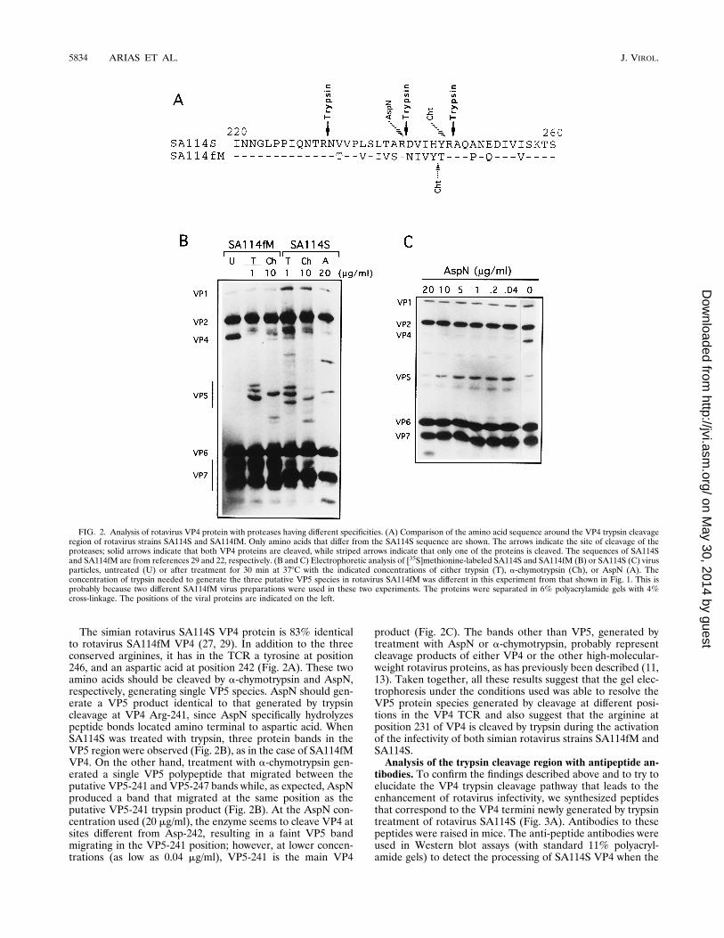
The simian rotavirus SA114S VP4 protein is 83% identicalto rotavirus SA114fM VP4 (27, 29). In addition to the threeconserved arginines, it has in the TCR a tyrosine at position246, and an aspartic acid at position 242 (Fig. 2A). These twoamino acids should be cleaved by a-chymotrypsin and AspN,respectively, generating single VP5 species. AspN should gen-erate a VP5 product identical to that generated by trypsincleavage at VP4 Arg-241, since AspN specifically hydrolyzespeptide bonds located amino terminal to aspartic acid. WhenSA114S was treated with trypsin, three protein bands in theVP5 region were observed (Fig. 2B), as in the case of SA114fMVP4. On the other hand, treatment with a-chymotrypsin gen-erated a single VP5 polypeptide that migrated between theputative VP5-241 and VP5-247 bands while, as expected, AspNproduced a band that migrated at the same position as theputative VP5-241 trypsin product (Fig. 2B). At the AspN con-centration used (20 mg/ml), the enzyme seems to cleave VP4 atsites different from Asp-242, resulting in a faint VP5 bandmigrating in the VP5-241 position; however, at lower concen-trations (as low as 0.04 mg/ml), VP5-241 is the main VP4
product (Fig. 2C). The bands other than VP5, generated bytreatment with AspN or a-chymotrypsin, probably representcleavage products of either VP4 or the other high-molecular-weight rotavirus proteins, as has previously been described (11,13). Taken together, all these results suggest that the gel elec-trophoresis under the conditions used was able to resolve theVP5 protein species generated by cleavage at different posi-tions in the VP4 TCR and also suggest that the arginine atposition 231 of VP4 is cleaved by trypsin during the activationof the infectivity of both simian rotavirus strains SA114fM andSA114S.Analysis of the trypsin cleavage region with antipeptide an-
tibodies. To confirm the findings described above and to try toelucidate the VP4 trypsin cleavage pathway that leads to theenhancement of rotavirus infectivity, we synthesized peptidesthat correspond to the VP4 termini newly generated by trypsintreatment of rotavirus SA114S (Fig. 3A). Antibodies to thesepeptides were raised in mice. The anti-peptide antibodies wereused in Western blot assays (with standard 11% polyacryl-amide gels) to detect the processing of SA114S VP4 when the
FIG. 2. Analysis of rotavirus VP4 protein with proteases having different specificities. (A) Comparison of the amino acid sequence around the VP4 trypsin cleavageregion of rotavirus strains SA114S and SA114fM. Only amino acids that differ from the SA114S sequence are shown. The arrows indicate the site of cleavage of theproteases; solid arrows indicate that both VP4 proteins are cleaved, while striped arrows indicate that only one of the proteins is cleaved. The sequences of SA114Sand SA114fM are from references 29 and 22, respectively. (B and C) Electrophoretic analysis of [35S]methionine-labeled SA114S and SA114fM (B) or SA114S (C) virusparticles, untreated (U) or after treatment for 30 min at 378C with the indicated concentrations of either trypsin (T), a-chymotrypsin (Ch), or AspN (A). Theconcentration of trypsin needed to generate the three putative VP5 species in rotavirus SA114fM was different in this experiment from that shown in Fig. 1. This isprobably because two different SA114fM virus preparations were used in these two experiments. The proteins were separated in 6% polyacrylamide gels with 4%cross-linkage. The positions of the viral proteins are indicated on the left.
5834 ARIAS ET AL. J. VIROL.
on May 30, 2014 by guest
http://jvi.asm.org/
Dow
nloaded from
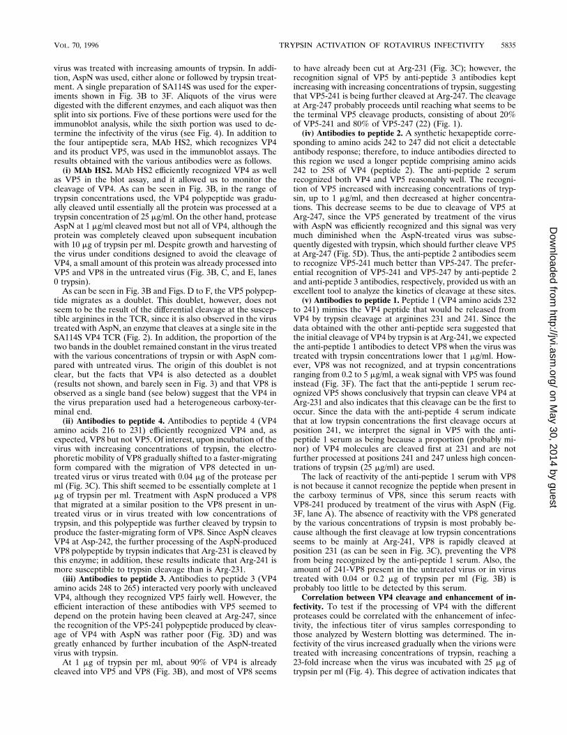
virus was treated with increasing amounts of trypsin. In addi-tion, AspN was used, either alone or followed by trypsin treat-ment. A single preparation of SA114S was used for the exper-iments shown in Fig. 3B to 3F. Aliquots of the virus weredigested with the different enzymes, and each aliquot was thensplit into six portions. Five of these portions were used for theimmunoblot analysis, while the sixth portion was used to de-termine the infectivity of the virus (see Fig. 4). In addition tothe four antipeptide sera, MAb HS2, which recognizes VP4and its product VP5, was used in the immunoblot assays. Theresults obtained with the various antibodies were as follows.(i) MAb HS2. MAb HS2 efficiently recognized VP4 as well
as VP5 in the blot assay, and it allowed us to monitor thecleavage of VP4. As can be seen in Fig. 3B, in the range oftrypsin concentrations used, the VP4 polypeptide was gradu-ally cleaved until essentially all the protein was processed at atrypsin concentration of 25 mg/ml. On the other hand, proteaseAspN at 1 mg/ml cleaved most but not all of VP4, although theprotein was completely cleaved upon subsequent incubationwith 10 mg of trypsin per ml. Despite growth and harvesting ofthe virus under conditions designed to avoid the cleavage ofVP4, a small amount of this protein was already processed intoVP5 and VP8 in the untreated virus (Fig. 3B, C, and E, lanes0 trypsin).As can be seen in Fig. 3B and Figs. D to F, the VP5 polypep-
tide migrates as a doublet. This doublet, however, does notseem to be the result of the differential cleavage at the suscep-tible arginines in the TCR, since it is also observed in the virustreated with AspN, an enzyme that cleaves at a single site in theSA114S VP4 TCR (Fig. 2). In addition, the proportion of thetwo bands in the doublet remained constant in the virus treatedwith the various concentrations of trypsin or with AspN com-pared with untreated virus. The origin of this doublet is notclear, but the facts that VP4 is also detected as a doublet(results not shown, and barely seen in Fig. 3) and that VP8 isobserved as a single band (see below) suggest that the VP4 inthe virus preparation used had a heterogeneous carboxy-ter-minal end.(ii) Antibodies to peptide 4. Antibodies to peptide 4 (VP4
amino acids 216 to 231) efficiently recognized VP4 and, asexpected, VP8 but not VP5. Of interest, upon incubation of thevirus with increasing concentrations of trypsin, the electro-phoretic mobility of VP8 gradually shifted to a faster-migratingform compared with the migration of VP8 detected in un-treated virus or virus treated with 0.04 mg of the protease perml (Fig. 3C). This shift seemed to be essentially complete at 1mg of trypsin per ml. Treatment with AspN produced a VP8that migrated at a similar position to the VP8 present in un-treated virus or in virus treated with low concentrations oftrypsin, and this polypeptide was further cleaved by trypsin toproduce the faster-migrating form of VP8. Since AspN cleavesVP4 at Asp-242, the further processing of the AspN-producedVP8 polypeptide by trypsin indicates that Arg-231 is cleaved bythis enzyme; in addition, these results indicate that Arg-241 ismore susceptible to trypsin cleavage than is Arg-231.(iii) Antibodies to peptide 3. Antibodies to peptide 3 (VP4
amino acids 248 to 265) interacted very poorly with uncleavedVP4, although they recognized VP5 fairly well. However, theefficient interaction of these antibodies with VP5 seemed todepend on the protein having been cleaved at Arg-247, sincethe recognition of the VP5-241 polypeptide produced by cleav-age of VP4 with AspN was rather poor (Fig. 3D) and wasgreatly enhanced by further incubation of the AspN-treatedvirus with trypsin.At 1 mg of trypsin per ml, about 90% of VP4 is already
cleaved into VP5 and VP8 (Fig. 3B), and most of VP8 seems
to have already been cut at Arg-231 (Fig. 3C); however, therecognition signal of VP5 by anti-peptide 3 antibodies keptincreasing with increasing concentrations of trypsin, suggestingthat VP5-241 is being further cleaved at Arg-247. The cleavageat Arg-247 probably proceeds until reaching what seems to bethe terminal VP5 cleavage products, consisting of about 20%of VP5-241 and 80% of VP5-247 (22) (Fig. 1).(iv) Antibodies to peptide 2. A synthetic hexapeptide corre-
sponding to amino acids 242 to 247 did not elicit a detectableantibody response; therefore, to induce antibodies directed tothis region we used a longer peptide comprising amino acids242 to 258 of VP4 (peptide 2). The anti-peptide 2 serumrecognized both VP4 and VP5 reasonably well. The recogni-tion of VP5 increased with increasing concentrations of tryp-sin, up to 1 mg/ml, and then decreased at higher concentra-tions. This decrease seems to be due to cleavage of VP5 atArg-247, since the VP5 generated by treatment of the viruswith AspN was efficiently recognized and this signal was verymuch diminished when the AspN-treated virus was subse-quently digested with trypsin, which should further cleave VP5at Arg-247 (Fig. 5D). Thus, the anti-peptide 2 antibodies seemto recognize VP5-241 much better than VP5-247. The prefer-ential recognition of VP5-241 and VP5-247 by anti-peptide 2and anti-peptide 3 antibodies, respectively, provided us with anexcellent tool to analyze the kinetics of cleavage at these sites.(v) Antibodies to peptide 1. Peptide 1 (VP4 amino acids 232
to 241) mimics the VP4 peptide that would be released fromVP4 by trypsin cleavage at arginines 231 and 241. Since thedata obtained with the other anti-peptide sera suggested thatthe initial cleavage of VP4 by trypsin is at Arg-241, we expectedthe anti-peptide 1 antibodies to detect VP8 when the virus wastreated with trypsin concentrations lower that 1 mg/ml. How-ever, VP8 was not recognized, and at trypsin concentrationsranging from 0.2 to 5 mg/ml, a weak signal with VP5 was foundinstead (Fig. 3F). The fact that the anti-peptide 1 serum rec-ognized VP5 shows conclusively that trypsin can cleave VP4 atArg-231 and also indicates that this cleavage can be the first tooccur. Since the data with the anti-peptide 4 serum indicatethat at low trypsin concentrations the first cleavage occurs atposition 241, we interpret the signal in VP5 with the anti-peptide 1 serum as being because a proportion (probably mi-nor) of VP4 molecules are cleaved first at 231 and are notfurther processed at positions 241 and 247 unless high concen-trations of trypsin (25 mg/ml) are used.The lack of reactivity of the anti-peptide 1 serum with VP8
is not because it cannot recognize the peptide when present inthe carboxy terminus of VP8, since this serum reacts withVP8-241 produced by treatment of the virus with AspN (Fig.3F, lane A). The absence of reactivity with the VP8 generatedby the various concentrations of trypsin is most probably be-cause although the first cleavage at low trypsin concentrationsseems to be mainly at Arg-241, VP8 is rapidly cleaved atposition 231 (as can be seen in Fig. 3C), preventing the VP8from being recognized by the anti-peptide 1 serum. Also, theamount of 241-VP8 present in the untreated virus or in virustreated with 0.04 or 0.2 mg of trypsin per ml (Fig. 3B) isprobably too little to be detected by this serum.Correlation between VP4 cleavage and enhancement of in-
fectivity. To test if the processing of VP4 with the differentproteases could be correlated with the enhancement of infec-tivity, the infectious titer of virus samples corresponding tothose analyzed by Western blotting was determined. The in-fectivity of the virus increased gradually when the virions weretreated with increasing concentrations of trypsin, reaching a23-fold increase when the virus was incubated with 25 mg oftrypsin per ml (Fig. 4). This degree of activation indicates that
VOL. 70, 1996 TRYPSIN ACTIVATION OF ROTAVIRUS INFECTIVITY 5835
on May 30, 2014 by guest
http://jvi.asm.org/
Dow
nloaded from

FIG. 3. Immunoblot analysis of rotavirus SA114S VP4 cleavage products. (A) Schematic representation of the trypsin cleavage region of rotavirus SA114S. Thethree conserved arginines are shown. The synthetic peptides that mimic the termini newly generated by trypsin treatment of the virus are indicated by open bars; thecircle at one end of the bar indicates the position of the cysteine that was added for coupling the peptide to KLH. Antibodies to these peptides were produced in mice.(B to F) A single preparation of SA114S was used. Aliquots of the virus were digested for 30 min at 378C with the indicated concentrations of trypsin (T) or 1 mg ofAspN per ml (A). In addition, a portion of the virus treated with AspN was further digested with 10 mg of trypsin per ml for 30 min at 378C. Each aliquot was thensplit into six equal portions; five of these portions were used for the immunoblot analysis with the antipeptide (a-pep) sera and the VP5 MAb HS2, as indicated, whilethe sixth portion was used to determine the infectivity of the virus (Fig. 4). Equal amounts of virus proteins were loaded in each gel; the different signals obtained forVP4, VP5, and VP8 are due to the differential recognition of these polypeptides by the various antibodies used. The virus proteins were separated by electrophoresisin standard SDS–11% polyacrylamide gels and transferred to nitrocellulose paper. The transferred proteins were incubated with serum to either peptide 1 (diluted1:100), peptide 2 (diluted 1:500), peptide 3 (dilute 1:300), peptide 4 (diluted 1:1,000), or MAb HS2 (diluted 1:1,000), and the bound antibody was identified with a1:1,000 dilution of a peroxidase-labeled goat anti-mouse immunoglobulin G, using a chemiluminescent substrate. The positions of the VP4 protein and its cleavageproducts VP5 and VP8 are indicated on the left. The weak band labeled with a dot in panels C to F corresponds to background binding to VP6, the most abundantprotein of the virus.
5836
on May 30, 2014 by guest
http://jvi.asm.org/
Dow
nloaded from

at least 95% of the activable virions were not infectious beforethe cleavage of VP4. The infectivity of SA114S was not signif-icantly enhanced by treatment with 1 mg of protease AspN perml (twofold increase); however, cleavage with this protease didnot preclude that the infectivity of the virus was augmented bysubsequent treatment with trypsin (Fig. 4). In a separate ex-periment, the infectivity of SA114S was increased only 1.5-foldby treatment with 2 mg of a-chymotrypsin per ml; however, theinfectivity of the chymotrypsin-treated virus was enhanced 12-fold by further treatment with 10 mg of trypsin per ml (data notshown). These results indicate that the additional cleavagesobserved in VP4, or the other viral proteins, in virus treatedwith either AspN or a-chymotrypsin (Fig. 2) do not inactivatethe virus. The fact that AspN only marginally increased theinfectivity of the virus indicates that cleavage at Arg-241 is notdirectly responsible for the 23-fold full enhancement of infec-tivity achieved with trypsin.To determine whether cleavage of VP4 at one or more of the
conserved arginines in the TCR could be correlated with theincrease in infectivity, we carried out a densitometric analysisof the VP8 and VP5 products identified with the various anti-peptide sera. The data obtained were normalized and plottedtogether with the fold increase in infectivity induced by thevarious trypsin concentrations (Fig. 4). For the interpretationof these data, we assumed that the antipeptide 2 and antipep-tide 3 sera recognize mainly VP5-241 and VP5-247, respec-tively. This assumption seems to be justified, since by densi-tometry, each of these two VP5 polypeptides was recognized bythe corresponding serum with an efficiency 7- to 10-fold higherthan that achieved with the other serum. For the antipeptide 4serum, the densitometric analysis was done on VP8-231. Ascan be seen, the level of VP8-231 reaches a maximum aftertreatment with trypsin 1 mg/ml and remains constant thereafterwhereas the level of VP5-241 also reaches a maximum at thisconcentration but then declines steadily at higher trypsin con-centrations. On the other hand, the level of VP5-247 keepsincreasing with the increasing concentrations of trypsin, as the
infectivity of the virus does. As shown by the reactivity withMAb HS2, it is clear that essentially all VP4 has been cleavedat 5 mg of trypsin per ml (Fig. 3B), although the infectivity stillincreases at 25 mg/ml (Fig. 4). These data strongly suggest thatthe cleavage directly responsible for the activation of infectivityis the one that occurs at Arg-247.VP4 cleavage of rotaviruses RRV and SA114fM. To test if
the VP4 trypsin cleavage pattern observed for SA114S wassimilar for other rotavirus strains, we analyzed rotavirusesSA114fM and RRV by immunoblot analysis with the antipep-tide 4 serum and MAb HS2. In both strains, Arg-241 alsoseems to be the initial trypsin cleavage site, since the electro-phoretic mobility of VP8 changed when the viruses weretreated with increasing concentrations of trypsin (Fig. 5).For rotavirus SA114fM, we could not detect any cleavage
products of VP4 in the untreated virus, and the pattern ofappearance of VP8 was very much like that of SA114S. Asingle band was observed at 0.04 mg of trypsin per ml, and thenit shifted to a faster-migrating form, such that at 1 mg/ml theshift was complete. In the case of RRV, the mobility of VP8decreased upon incubation of the virus with higher concentra-tions of trypsin compared with that of the VP8 band present inuntreated virus or virus treated with 0.04 mg of trypsin per ml,which probably reflects a change in the secondary structure ofthe protein. In this virus, the sequential cleavage of VP4 atarginines 231 and 241 seems more evident than in SA114S,since at 0.04 mg of trypsin per ml, essentially all VP4 has beencleaved whereas only about 10% of VP8 seems to have beenprocessed at Arg-231.VP5 was observed as a doublet in both SA114fM and RRV;
however, in this case, the doublet seems to be the result oftrypsin cleavage, since (i) the relative abundance of the twobands changed with increasing concentrations of trypsin inboth viruses; and (ii) for SA114fM, digestion with a-chymo-trypsin (which cleaves at Tyr-245) of the same virus prepara-tion shown in Fig. 5 produced a VP5 polypeptide that migratedas a single band (results not shown). A VP5 band with a slowermobility (putatively 241-VP5) appeared first and then shifted
FIG. 4. Correlation between trypsin cleavage of VP4 and infectivity of rota-virus SA114S. The sixth sample of each aliquot, obtained as described in thelegend to Fig. 3, was used to determine the infectivity of the virus by an immu-noperoxidase focus assay (26). The vertical bars represent the fold increase ininfectivity obtained with the various protease treatments. The basal infectivity(no trypsin treatment) used as the reference unit was 1.35 3 107 focus-formingunits/ml. Also shown in this graph are the results of the densitometric analysis ofthe VP5 and VP8 bands obtained by incubation with the antipeptide 2, 3, and 4sera in the immunoblot experiments shown in Fig. 3. The optical density (O.D.)was determined and normalized in each case to the highest signal obtained witha given antipeptide serum, and it was plotted against the trypsin concentrationused to digest the virus. Symbols: E, O.D. of antipeptide 4; Ç, O.D. of antipep-tide 3; h, O.D. of antipeptide 2.
FIG. 5. Immunoblot analysis of the VP4 cleavage products of rotavirusstrains SA114fM (A) and RRV (B). Virus particles were digested for 30 min at378C with the indicated concentrations of trypsin, and the virus proteins wereseparated by electrophoresis in standard SDS–11% polyacrylamide gels andtransferred to nitrocellulose paper. The transferred proteins were incubated witha mixture of a 1,000-fold dilution of each antipeptide 4 serum andMAb HS2, andthe bound antibody was identified as described in the legend to Fig. 3. Thepositions of the VP4 protein and its cleavage products VP5 and VP8 are indi-cated on the left.
VOL. 70, 1996 TRYPSIN ACTIVATION OF ROTAVIRUS INFECTIVITY 5837
on May 30, 2014 by guest
http://jvi.asm.org/
Dow
nloaded from

to a faster-migrating form, which presumably represents 247-VP5. As mentioned above, the sequential cleavage at arginines241 and 247 seems more evident for RRV VP4.
DISCUSSION
In this study, we have shown that during the enhancement ofinfectivity of rotavirus SA114S by trypsin, the VP4 protein iscleaved at three conserved arginines, located at positions 231,241, and 247, extending our previous findings that SA114fMVP4 was cleaved at arginines 241 and 247 and identifyingArg-231 as a new trypsin-susceptible site. The cleavage at thethree arginines appears to occur in an ordered sequence, withthe most susceptible site being Arg-241. We propose that thisis the preferred site for the first cleavage, followed by cleavageat Arg-231 and then by cleavage at Arg-247. Apparently, theinitial cleavage can also occur at Arg-231, which seems to bethe second most susceptible site. A model for the major trypsincleavage pathway of rotavirus SA114S VP4 is shown in Fig. 6.The terminal VP4 cleavage products generated by treatment
of rotaviruses SA114S and SA114fM with trypsin concentra-tions higher than 1 mg/ml were a VP8 polypeptide with ahomogeneous carboxy terminus (Arg-231) and a VP5 proteinhaving a heterogeneous amino terminus, with about 20 and80% of the molecules resulting from cleavage at VP4 arginines241 and 247, respectively (22) (Fig. 1). Complete cleavage atArg-247 could not be obtained even after treatment with tryp-sin concentrations as high as 100 mg/ml (not shown forSA114S). The structural bases that prevent all the VP4 mole-cules from being cleaved by trypsin at Arg-247 are not known;however, these results confirm our previous findings forSA114fM (22). The relevance of this 20% VP5-241 species forthe infectivity of the virus, if any, remains to be determined.It had been previously shown that the cleavage of VP4 by
trypsin correlates with the increase in infectivity of rotavirusSA114S (11, 13). In this work, we have shown that the treat-ment of this virus with AspN, which cleaves VP4 at the peptidebond between Arg-241 and Asp-242 (cleavage analog to thatcaused by trypsin at Arg-241), does not significantly increasethe infectivity of the virus, indicating that the single cleavage atthis site is not sufficient for and is not directly involved in theenhancement of infectivity. This enhancement, which, rather,
seems to correlate with cleavage at Arg-247, must be precise,since treatment of SA114S with a-chymotrypsin, which cleavesVP4 at Tyr-246, enhanced only marginally (this work) or didnot enhance (13) the virus infectivity. The fact that the increasein infectivity correlates with cleavage at Arg-247 does not nec-essarily mean that activation of the virus is not dependent oncleavage at Arg-231 (see also below).Neither of the four antipeptide sera produced had neutral-
ization activity against rotavirus SA114S, although an enzyme-linked immunosorbent assay (ELISA) showed that only theantipeptide 2 antibodies bound to the virus (data not shown).These results are consistent with our previous findings byELISA that antibodies to peptides comprising amino acids 220to 233 and 258 to 271 of rotavirus SA114fM VP4 did not reactwith the virus and did not have neutralization activity (3).Further experiments have to be carried out to determine to
which VP5 species (VP5-241 or VP5-247) the antipeptide 2antibodies bind in the virus particle; however, the fact thatthese antibodies did not neutralize the virus despite binding toit suggests that the termini newly generated by trypsin diges-tion are not involved in a direct interaction with the cell (al-though it cannot be ruled out that the carboxy terminus of VP8might play a role in the enhancement of infectivity) and thatinstead, the cleavage of VP4 induces a conformational changein VP5 or in VP8 through the interaction with VP5 (since thecleavage at Arg-247 seems to be critical to activate the infec-tivity) that is necessary for the virus to become infectious. Thisproposed structural change could occur immediately aftercleavage at Arg-247, or the cleavage at this position might onlyloosen the structure of VP4 such that the structural changeoccurs when the virus interacts with the cell receptor. Thelatter possibility seems more likely, since the difference in thestructure of VP4 between trypsin-treated and untreated virus,as determined by cryoelectron microscopy, does not seem to bevery striking (32). Changes in the virus surface proteins uponinteraction of the virus with the cell receptor on the cell surfaceor after endocytosis of the receptor-virus complex have beenwell documented for other viruses such as poliovirus (15),influenza virus (6), human immunodeficiency virus (9), tick-borne encephalitis virus (33), reovirus (14), and paramyxovirus(20). In this context, the cleavage of VP4 by trypsin might beneeded to release a lock and make the virus particle unstable,
FIG. 6. Model for the major trypsin cleavage activation pathway of rotavirus SA114S infectivity. The open bars represent the uncleaved protein and its intermediatetrypsin cleavage products. The arginines at positions 231, 241, and 247 are indicated by vertical lines. The first cleavage is proposed to be at Arg-241, although a smallproportion of VP4 molecules could be first cleaved at Arg-231 (striped arrow). The second cleavage site is proposed to be Arg-231, while the last site to be cleavedis Arg-247. The shadowed bars are the terminal VP4 cleavage products that remain associated with the viral particles, while the hatched boxes represent the connectingpeptides 232 to 241 and 242 to 247, whose fate is unknown. The numbers inside the bars represent the amino acid residue position at the amino or carboxy terminusof the corresponding polypeptide. The gradient arrow at the left indicates that increasing concentrations of trypsin are needed to cleave the least susceptible sites.
5838 ARIAS ET AL. J. VIROL.
on May 30, 2014 by guest
http://jvi.asm.org/
Dow
nloaded from
such that upon interaction with the cell surface, the virion isable to uncoat efficiently, helping to reverse the assembly stepof the surface proteins that should occur efficiently with theuncleaved VP4 during morphogenesis of the virus. In addition,the cleavage of VP4 and the proposed subsequent structuralchange of the protein upon contact of the virion with the cellreceptor might provide the virus with a mechanism to avoid theimmune selection pressure of the host against a conserved andessential domain of VP4 (needed, for example, for a secondaryvirus-receptor interaction [26]) that might not be exposed inthe protein before the specific virus-cell interaction. This strat-egy to avoid the host immune system surveillance might becomplementary or alternative to that proposed in the canyonhypothesis (34). The activation of virus infectivity by proteo-lytic cleavage of the viral spike proteins is also a strategycommonly used by enveloped viruses like paramyxoviruses,orthomyxoviruses, and retroviruses, including human immuno-deficiency virus (19).If the cleavage at Arg-247 is relevant for activation of virus
infectivity, why are the three arginine residues in the TCRconserved in the great majority of the VP4 proteins sequencedso far? We propose that the cleavage at Arg-241, although notdirectly involved in enhancement of infectivity, is a prerequi-site for the protein to be cleaved, at least efficiently, at Arg-247.In addition, cleavage at Arg-231 might be needed for an effi-cient cleavage at Arg-247, or the VP8-231 polypeptide could berequired for the structural change, proposed to be induced bycleavage at Arg-247, to occur. This hypothesis is testable, andfurther experiments should be done to confirm it. The cleavageat the three VP4 conserved arginines was also found to occurin rotavirus strains SA114fM and RRV, and the most suscep-tible site also appeared to be Arg-241, suggesting that theproposed trypsin cleavage pathway might be the same for theseviruses. It remains to be determined, however, if cleavage atArg-247 is also directly associated with the enhancement oftheir infectivity and if this pathway of activation is also true forrotavirus strains isolated from different animal species, includ-ing humans.
ACKNOWLEDGMENTS
This work was partially supported by grants 75191-527101 from theHoward Hughes Medical Institute, 3901 from the National Council forScience and Technology—Mexico, and IN207793 from DGAPA-UNAM.
REFERENCES
1. Almeida, J. D., T. Hall, J. E. Batnavala, B. M. Toterdell, and I. L. Chrystie.1978. The effect of trypsin on the growth of rotavirus. J. Gen. Virol. 40:213–218.
2. Anthony, I. D., S. Bullivant, S. Dayal, A. R. Bellamy, and J. A. Berriman.1991. Rotavirus spike structure and polypeptide composition. J. Virol. 65:4334–4340.
3. Arias, C. F., G. Garcıa, and S. Lopez. 1989. Priming for rotavirus neutralizingantibodies by a VP4 protein-derived synthetic peptide. J. Virol. 63:5393–5398.
4. Arias, C. F., M. Lizano, and S. Lopez. 1987. Synthesis in Escherichia coli andimmunological characterization of a polypeptide containing the cleavagesites associated with trypsin enhancement of rotavirus SA11 infectivity.J. Gen. Virol. 68:633–642.
5. Babiuk, L. A., K. Mohammed, L. Spence, M. Fauvel, and R. Petro. 1977.Rotavirus isolation and cultivation in the presence of trypsin. J. Clin. Mi-crobiol. 6:610–617.
6. Carr, C. M., and P. S. Kim. 1993. A spring-loaded mechanism for theconformational change of influenza hemagglutinin. Cell 73:823–832.
7. Clark, S. M., B. B. Barnett, and R. S. Spendlove. 1979. Production ofhigh-titer bovine rotavirus with trypsin. J. Clin. Microbiol. 9:413–417.
8. Clark, S. M., J. R. Roth, M. L. Clark, B. B. Barnett, and R. S. Spendlove.1981. Trypsin enhancement of rotavirus infectivity: mechanism of enhance-ment. J. Virol. 39:816–822.
9. Ellens, H., and C. Larsen. 1993. CD4-induced change in gp120/41 confor-mation and its potential relationship to fusion, p. 291–312. In J. Bentz (ed.),Viral fusion mechanisms. CRC Press, Inc., Boca Raton, Fla.
10. Espejo, R., E. Martınez, S. Lopez, and O. Munoz. 1980. Different polypep-tide composition of two human rotavirus types. Infect. Immun. 28:230–237.
11. Espejo, R. T., S. Lopez, and C. Arias. 1981. Structural polypeptides of simianrotavirus SA11 and the effect of trypsin. J. Virol. 37:156–160.
12. Estes, M. K., and J. Cohen. 1989. Rotavirus gene structure and function.Microbiol. Rev. 53:410–449.
13. Estes, M. K., D. Y. Graham, and B. B. Mason. 1981. Proteolytic enhance-ment of rotavirus infectivity: molecular mechanisms. J. Virol. 39:879–888.
14. Fernandes, J., D. Tang, G. Leone, and P. W. K. Lee. 1994. Binding ofreovirus to receptor leads to conformational changes in viral capsid proteinsthat are reversible upon virus detachment. J. Biol. Chem. 269:17043–17047.
15. Fricks, C. E., and J. M. Hogle. 1990. Cell-induced conformational change inpoliovirus: externalization of amino terminus of VP1 is responsible for lipo-some binding. J. Virol. 64:1934–1945.
16. Fukuhara, N., O. Yoshie, S. Kitaoka, and T. Konno. 1988. Role of VP3 inhuman rotavirus internalization after target cell attachment via VP7. J. Virol.62:2209–2218.
17. Kaljot, K. T., R. D. Shaw, D. H. Rubin, and H. B. Greenberg. 1988. Infectiousrotavirus enters cells by direct cell membrane penetration, not by endocyto-sis. J. Virol. 62:1136–1144.
18. Kapikian, A. Z., and R. M. Chanock. 1996. Rotaviruses, p. 1657–1708. InB. N. Fields et al. (ed.), Virology. Raven Press, New York.
19. Klenk, H.-D., and W. Garten. 1994. Activation cleavage of viral spike pro-teins by host proteases, p. 241–280. In E. Wimmer (ed.), Cellular receptorsfor animal viruses. Cold Spring Harbor Laboratory Press, Cold Spring Har-bor, N.Y.
20. Lamb, R. A. 1993. Paramyxovirus fusion: a hypothesis for changes. Virology197:1–11.
21. Liu, M., P. A. Offit, and M. K. Estes. 1988. Identification of the simianrotavirus SA11 genome segment 3 product. Virology 163:26–32.
22. Lopez, S., C. F. Arias, J. R. Bell, J. H. Strauss, and R. T. Espejo. 1985.Primary structure of the cleavage site associated with trypsin enhancement ofrotavirus SA11 infectivity. Virology 144:11–19.
23. Lopez, S., C. F. Arias, E. Mendez, and R. T. Espejo. 1986. Conservation inrotaviruses of the protein region containing the two sites associated withtrypsin enhancement of infectivity. Virology 154:224–227.
24. Mackow, E. R., R. D. Shaw, S. M. Matsui, P. T. Vo, M. N. Dang, and H. B.Greenberg. 1988. The rhesus rotavirus gene encoding protein VP3: locationof amino acids involved in homologous and heterologous rotavirus neutral-ization and identification of a putative fusion region. Proc. Natl. Acad. Sci.USA 85:645–649.
25. Mattion, N. M., J. Cohen, and M. K. Estes. 1994. The rotavirus proteins, p.169–250. In A. Z. Kapikian (ed.), Viral infections of the gastrointestinaltract. Marcel Dekker, Inc., New York.
26. Mendez, E., C. F. Arias, and S. Lopez. 1993. Binding to sialic acids is not aessential step for the entry of animal rotaviruses to epithelial cells in culture.J. Virol. 67:5253–5259.
27. Mitchell, D. B., and G. W. Both. 1989. Complete nucleotide sequence of thesimian rotavirus SA11 VP4 gene. Nucleic Acids Res. 17:2122.
28. Nandi, P., A. Charpilienne, and J. Cohen. 1992. Interaction of rotavirusparticles with liposomes. J. Virol. 66:3363–3367.
29. Nishikawa, K., K. Taniguchi, A. Torres, Y. Hoshino, K. Green, A. Z.Kapikian, R. M. Chanock, and M. Gorziglia. 1988. Comparative analysis ofthe VP3 gene of divergent strains of the rotaviruses simian SA11 and bovineNebraska calf diarrhea virus. J. Virol. 62:4022–4026.
30. Padilla-Noriega, L., S. J. Dunn, S. Lopez, H. B. Greenberg, and C. F. Arias.1995. Identification of two independent neutralization domains on the VP4trypsin cleavage products VP5* and VP8* of human rotavirus ST3. Virology206:148–154.
31. Prasad, B. V. V., J. W. Burns, E. Marietta, M. K. Estes, and W. Chiu. 1990.Localization of VP4 neutralization sites in rotavirus by three-dimensionalcryoelectron microscopy. Nature (London) 343:476–479.
32. Ramig, R. F., A. Shaw, D. Chen, M. K. Estes, W. Chiu, and B. V. V. Prasad.1993. Rotavirus protein interactions, phenotypes, and virion structure, abstr.W21-3, p. 43. In IXth International Congress of Virology.
33. Rey, F. A., F. X. Heinz, C. Mandl, C. Kunz, and S. C. Harrison. 1995. Theenvelope glycoprotein from tick-borne encephalitis virus at 2 A resolution.Nature (London) 375:291–298.
34. Rossmann, M. G. 1989. The canyon hypothesis. J. Biol. Chem. 264:14587–14590.
35. Ruiz, M. C., S. R. Alonso-Torre, A. Charpilienne, M. Vasseur, F. Michelan-geli, J. Cohen, and F. Alvarado. 1994. Rotavirus interaction with isolatedmembrane vesicles. J. Virol. 68:4009–4016.
36. Shaw, A. L., R. Rothnagel, D. Chen, R. F. Ramig, W. Chiu, and B. V. V.Prasad. 1993. Three-dimensional visualization of the rotavirus hemaggluti-nin structure. Cell 74:693–701.
37. Theil, K. W., E. H. Bohl, and A. G. Agnes. 1977. Cell culture propagation ofporcine rotavirus (reovirus-like agent). Am. J. Vet. Res. 38:1765–1768.
VOL. 70, 1996 TRYPSIN ACTIVATION OF ROTAVIRUS INFECTIVITY 5839
on May 30, 2014 by guest
http://jvi.asm.org/
Dow
nloaded from