Full Genome-Based Classification of Rotaviruses Reveals a Common Origin between Human WaLike and...
16
JOURNAL OF VIROLOGY, Apr. 2008, p. 3204–3219 Vol. 82, No. 7 0022-538X/08/$08.000 doi:10.1128/JVI.02257-07 Copyright © 2008, American Society for Microbiology. All Rights Reserved. Full Genome-Based Classification of Rotaviruses Reveals a Common Origin between Human Wa-Like and Porcine Rotavirus Strains and Human DS-1-Like and Bovine Rotavirus Strains † Jelle Matthijnssens, 1 * Max Ciarlet, 2 Erica Heiman, 3 Ingrid Arijs, 1 Thomas Delbeke, 1 Sarah M. McDonald, 3 Enzo A. Palombo, 4 Miren Iturriza-Go ´mara, 5 Piet Maes, 1 John T. Patton, 3 Mustafizur Rahman, 1,6 and Marc Van Ranst 1 Laboratory of Clinical and Epidemiological Virology, Department of Microbiology and Immunology, Rega Institute for Medical Research, University of Leuven, Leuven, Belgium 1 ; Vaccine and Biologics—Clinical Research, Merck and Co. Inc., North Wales, Pennsylvania 19454 2 ; Laboratory of Infectious Diseases, National Institutes of Allergy and Infectious Diseases, National Institutes of Health, Bethesda, Maryland 20892 3 ; Environment and Biotechnology Centre, Faculty of Life and Social Sciences, Swinburne University of Technology, Hawthorn, Victoria, Australia 4 ; Enteric Virus Unit, Virus Reference Department, Centre for Infections, Health Protection Agency, London, United Kingdom 5 ; and Laboratory of Virology, ICDDR,B: Mohakhali, Dhaka 1212, Bangladesh 6 Received 18 October 2007/Accepted 8 January 2008 Group A rotavirus classification is currently based on the molecular properties of the two outer layer proteins, VP7 and VP4, and the middle layer protein, VP6. As reassortment of all the 11 rotavirus gene segments plays a key role in generating rotavirus diversity in nature, a classification system that is based on all the rotavirus gene segments is desirable for determining which genes influence rotavirus host range restriction, replication, and virulence, as well as for studying rotavirus epidemiology and evolution. Toward establishing such a classification system, gene sequences encoding VP1 to VP3, VP6, and NSP1 to NSP5 were determined for human and animal rotavirus strains belonging to different G and P genotypes in addition to those available in databases, and they were used to define phylogenetic relationships among all rotavirus genes. Based on these phylogenetic analyses, appropriate identity cutoff values were determined for each gene. For the VP4 gene, a nucleotide identity cutoff value of 80% completely correlated with the 27 established P genotypes. For the VP7 gene, a nucleotide identity cutoff value of 80% largely coincided with the established G genotypes but identified four additional distinct genotypes comprised of murine or avian rotavirus strains. Phylogenetic analyses of the VP1 to VP3, VP6, and NSP1 to NSP5 genes showed the existence of 4, 5, 6, 11, 14, 5, 7, 11, and 6 genotypes, respectively, based on nucleotide identity cutoff values of 83%, 84%, 81%, 85%, 79%, 85%, 85%, 85%, and 91%, respectively. In accordance with these data, a revised nomenclature of rotavirus strains is proposed. The novel classification system allows the identification of (i) distinct genotypes, which probably followed separate evolutionary paths; (ii) interspecies transmissions and a plethora of reassortment events; and (iii) certain gene constellations that revealed (a) a common origin between human Wa-like rotavirus strains and porcine rotavirus strains and (b) a common origin between human DS-1-like rotavirus strains and bovine rotaviruses. These close evolutionary links between human and animal rotaviruses emphasize the need for close simultaneous monitoring of rotaviruses in animals and humans. Group A rotaviruses are major pathogens associated with acute gastroenteritis in humans and animals. Rotaviruses form a genus in the Reoviridae family and are characterized by a segmented double-stranded RNA genome (3, 16). The rotavi- rus genome is enclosed in a triple-layered icosahedral capsid and consists of 11 segments, encoding six viral structural pro- teins (VP1 to VP4, VP6, and VP7) and six nonstructural pro- teins (NSP1 to NSP6) (16). Each genome segment, with the exception of gene 11 that encodes two proteins (NSP5 and NSP6), codes for a single viral protein. The inner layer of the rotavirus virion is mainly composed of VP2, which encases VP1, the viral RNA-dependent RNA polymerase, and VP3, the viral capping enzyme (16). The middle layer of the virion is composed entirely of VP6 trimers, which determine rotavirus groups. For group A rotaviruses four different subgroups (SGs) have been recognized, based on the immunoreactivity of SG-specific monoclonal antibodies (MAbs) (19). VP7 and VP4 comprise the outer layer of the virion and are the basis of a binary classification system defining G types (glycoprotein) and P types (protease sensitive), respectively. VP7 and VP4 are capable of independently eliciting neutralizing antibodies, ini- tially used to define rotavirus G and P serotypes (16). The nonstructural proteins are variously involved in replication (NSP1 to NSP3, NSP5, and NSP6) and morphogenesis (NSP4), and with the exception of NSP4, all interact with nucleic acids (16). Due to the lack of proper immunological reagents and the increasing ease of sequencing, serotyping is being comple- * Corresponding author. Mailing address: Laboratory of Clinical and Epidemiological Virology, Department of Microbiology and Im- munology, Rega Institute for Medical Research, Minderbroedersstraat 10, B-3000 Leuven, Belgium. Phone: 32 16 332166. Fax: 32 16 332131. E-mail: [email protected]. † Supplemental material for this article may be found at http://jvi .asm.org/. Published ahead of print on 23 January 2008. 3204
Transcript of Full Genome-Based Classification of Rotaviruses Reveals a Common Origin between Human WaLike and...
JOURNAL OF VIROLOGY, Apr. 2008, p. 3204–3219 Vol. 82, No. 70022-538X/08/$08.00�0 doi:10.1128/JVI.02257-07Copyright © 2008, American Society for Microbiology. All Rights Reserved.
Full Genome-Based Classification of Rotaviruses Reveals a CommonOrigin between Human Wa-Like and Porcine Rotavirus Strains and
Human DS-1-Like and Bovine Rotavirus Strains�†Jelle Matthijnssens,1* Max Ciarlet,2 Erica Heiman,3 Ingrid Arijs,1 Thomas Delbeke,1
Sarah M. McDonald,3 Enzo A. Palombo,4 Miren Iturriza-Gomara,5 Piet Maes,1John T. Patton,3 Mustafizur Rahman,1,6 and Marc Van Ranst1
Laboratory of Clinical and Epidemiological Virology, Department of Microbiology and Immunology, Rega Institute for Medical Research,University of Leuven, Leuven, Belgium1; Vaccine and Biologics—Clinical Research, Merck and Co. Inc., North Wales,
Pennsylvania 194542; Laboratory of Infectious Diseases, National Institutes of Allergy and Infectious Diseases,National Institutes of Health, Bethesda, Maryland 208923; Environment and Biotechnology Centre, Faculty of
Life and Social Sciences, Swinburne University of Technology, Hawthorn, Victoria, Australia4;Enteric Virus Unit, Virus Reference Department, Centre for Infections, Health Protection Agency,
London, United Kingdom5; and Laboratory of Virology, ICDDR,B:Mohakhali, Dhaka 1212, Bangladesh6
Received 18 October 2007/Accepted 8 January 2008
Group A rotavirus classification is currently based on the molecular properties of the two outer layerproteins, VP7 and VP4, and the middle layer protein, VP6. As reassortment of all the 11 rotavirus genesegments plays a key role in generating rotavirus diversity in nature, a classification system that is based onall the rotavirus gene segments is desirable for determining which genes influence rotavirus host rangerestriction, replication, and virulence, as well as for studying rotavirus epidemiology and evolution. Towardestablishing such a classification system, gene sequences encoding VP1 to VP3, VP6, and NSP1 to NSP5 weredetermined for human and animal rotavirus strains belonging to different G and P genotypes in addition tothose available in databases, and they were used to define phylogenetic relationships among all rotavirus genes.Based on these phylogenetic analyses, appropriate identity cutoff values were determined for each gene. For theVP4 gene, a nucleotide identity cutoff value of 80% completely correlated with the 27 established P genotypes.For the VP7 gene, a nucleotide identity cutoff value of 80% largely coincided with the established G genotypesbut identified four additional distinct genotypes comprised of murine or avian rotavirus strains. Phylogeneticanalyses of the VP1 to VP3, VP6, and NSP1 to NSP5 genes showed the existence of 4, 5, 6, 11, 14, 5, 7, 11, and6 genotypes, respectively, based on nucleotide identity cutoff values of 83%, 84%, 81%, 85%, 79%, 85%, 85%,85%, and 91%, respectively. In accordance with these data, a revised nomenclature of rotavirus strains isproposed. The novel classification system allows the identification of (i) distinct genotypes, which probablyfollowed separate evolutionary paths; (ii) interspecies transmissions and a plethora of reassortment events;and (iii) certain gene constellations that revealed (a) a common origin between human Wa-like rotavirusstrains and porcine rotavirus strains and (b) a common origin between human DS-1-like rotavirus strains andbovine rotaviruses. These close evolutionary links between human and animal rotaviruses emphasize the needfor close simultaneous monitoring of rotaviruses in animals and humans.
Group A rotaviruses are major pathogens associated withacute gastroenteritis in humans and animals. Rotaviruses forma genus in the Reoviridae family and are characterized by asegmented double-stranded RNA genome (3, 16). The rotavi-rus genome is enclosed in a triple-layered icosahedral capsidand consists of 11 segments, encoding six viral structural pro-teins (VP1 to VP4, VP6, and VP7) and six nonstructural pro-teins (NSP1 to NSP6) (16). Each genome segment, with theexception of gene 11 that encodes two proteins (NSP5 andNSP6), codes for a single viral protein. The inner layer of the
rotavirus virion is mainly composed of VP2, which encasesVP1, the viral RNA-dependent RNA polymerase, and VP3,the viral capping enzyme (16). The middle layer of the virion iscomposed entirely of VP6 trimers, which determine rotavirusgroups. For group A rotaviruses four different subgroups(SGs) have been recognized, based on the immunoreactivity ofSG-specific monoclonal antibodies (MAbs) (19). VP7 and VP4comprise the outer layer of the virion and are the basis of abinary classification system defining G types (glycoprotein) andP types (protease sensitive), respectively. VP7 and VP4 arecapable of independently eliciting neutralizing antibodies, ini-tially used to define rotavirus G and P serotypes (16). Thenonstructural proteins are variously involved in replication(NSP1 to NSP3, NSP5, and NSP6) and morphogenesis (NSP4),and with the exception of NSP4, all interact with nucleic acids(16).
Due to the lack of proper immunological reagents and theincreasing ease of sequencing, serotyping is being comple-
* Corresponding author. Mailing address: Laboratory of Clinicaland Epidemiological Virology, Department of Microbiology and Im-munology, Rega Institute for Medical Research, Minderbroedersstraat10, B-3000 Leuven, Belgium. Phone: 32 16 332166. Fax: 32 16 332131.E-mail: [email protected].
† Supplemental material for this article may be found at http://jvi.asm.org/.
� Published ahead of print on 23 January 2008.
3204

mented with genotyping, which is based on identities betweensequences of cognate rotavirus gene segments. So far, 15 Ggenotypes (14 G serotypes) have been identified, and out of 27different P genotypes, 14 P serotypes (1A, 1B, and 2 to 14) havebeen identified with available VP4-specific antibodies (5, 30,34–37, 42, 62, 68). Traditionally, a cutoff value of 89% VP7amino acid sequence identity has been used to classify G ge-notypes, yielding a nearly complete concordance with the dif-ferent G serotypes (16, 57). In contrast, the 89% amino acididentity cutoff value for VP4, established by Gorziglia andcolleagues (18), does not result in an absolute concordancebetween different P genotypes and P serotypes. Specifically, Pserotypes have not been defined for approximately half of theP genotypes, which are designated by an Arabic numeral be-tween square brackets (16). Molecular analyses of VP6 is lim-ited to only a 379-bp fragment of VP6, which results in twobroad genogroups that do not correlate with the SG specific-ities (27). The classification of rotavirus nonstructural proteinsis limited to NSP4, and six genotypes (A to F) have beenrecognized based on clustering patterns in amino-acid-basedphylogenetic dendrograms (9, 22, 26, 46). To date, no classifi-cation for VP1 to VP3, NSP1 to NSP3, or NSP5 has beendescribed.
RNA-RNA hybridization assays have been used to analyzeand compare complete genomes of group A rotaviruses. Forhuman strains, three genogroups have been established: twomajor genogroups represented by the reference strains Wa andDS-1 and one minor genogroup represented by referencestrain AU-1 (51). Similar genogroups also have been estab-lished for several animal rotavirus strains although they aremore complex (50). The hybridization technique has proven tobe useful to investigate possible reassortment events betweenhuman strains belonging to different genogroups (49, 76) orbetween human and animal strains (48). Currently, rotavirusstrains are being analyzed and compared to one another bypartial or complete sequencing of all 11 gene segments as thisapproach allows direct determination of genetic relationships(38–40, 63, 67). In addition, sequencing of rotavirus genomes iscritical to the understanding of phylogenetic analyses and tothe elucidation of the patterns of virus evolution. One methodthat is used to study typical evolutionary distances betweenvirus strains is pairwise sequence identity profiles (2). Specifi-cally, this method illustrates virus genotypes as well-resolvedpeaks, providing the basis for classification systems. To prop-erly study the evolution of rotaviruses, the establishment of aclassification system in which individual genes fall into definedclusters/genotypes based on reliable percentage identity cutoffvalues is fundamental. Such a classification system could be animportant tool to elucidate how rotaviruses evolve over time.In addition, for viruses with segmented genomes, this type ofclassification system could also be used to determine whetheror not certain rotavirus genes cosegregate during reassortmentevents (gene linkage) or whether certain gene constellationsplay a role in rotavirus host range restriction or virulence.
In this study, phylogenetic analyses and pairwise sequenceidentity profiles were constructed for each of the gene seg-ments of group A rotaviruses to develop a uniform classifica-tion and nomenclature system for all 11 rotavirus genomesegments in a similar fashion to that established for VP4 andVP7. Based on percentage identity cutoff values, this novel
classification system illustrates phylogenetic relationships of all11 rotavirus genome segments and allows the identification ofdistinct genotypes (which likely followed separate evolutionarypaths) and reassortment events. In addition, the comprehen-sive classification system revealed genetic relationships amongrotaviruses from different host species, including evidence thathuman rotaviruses belonging to the Wa-like genogroup have acommon origin with porcine rotaviruses while those belongingto the DS-1-like genogroup have a common origin with bovinerotaviruses.
MATERIALS AND METHODS
Rotavirus strains. The human rotavirus strains Wa (G1P1A[8]), D (G1P1A[8]),DS-1 (G2P1B[4]), S2 (G2P1B[4]), AU-1 (G3P3[9]), YO (G3P1A[8]), P (G3P1A[8]),Hosokawa (G4P1A[8]), ST3 (G4P2A[6]), IAL28 (G5P1A[8]), SE584 (G6P3[9]), 69M (G8P4[10]), WI61 (G9P1A[8]), A64 (G10P11[14]), and L26 (G12P1B[4]); thebovine strains NCDV (G6P6[1]), BRV033 (G6P6[1]), and WC3 (G6P7[5]); and theporcine strains A131 (G3P9[7]), A253 (G11P9[7]), and A411 (G3P9[7]) arestandard strains that have been adapted to growth in cell culture (serotypes aregiven in parentheses). The human rotavirus strain B3458 (G9P1A[8]) was iso-lated in Belgium during the 2003 to 2004 season (unpublished data).
RNA extraction, reverse transcription-PCR (RT-PCR), and sequencing ofhuman rotavirus strains Wa, DS-1, S2, AU-1, YO, Hosokawa, and B3458 andbovine rotavirus strain NCDV. For each strain 140 �l of cell culture supernatant(or fecal suspension in the case of strain B3458) was used to extract viral RNAusing a QIAamp Viral RNA mini kit (Qiagen/Westburg, Leusden, The Nether-lands) according to the manufacturer’s instructions.
Ten microliters of extracted RNA was denatured at 97°C for 3 min andRT-PCR was carried out using a Qiagen One Step RT-PCR kit (Qiagen/West-burg). The primers used are shown in Data S1 in the supplemental material. TheRT-PCR was carried out with an initial reverse transcription step at 45°C for 30min; PCR activation was at 95°C for 15 min, followed by 40 cycles of amplifica-tion (45 s at 94°C, 45 s at 45°C, and 6 min at 68°C), with a final extension of 7 minat 72°C in a GeneAmp PCR System 9700 thermal cycler (Applied Biosystems,Foster City, CA).
The PCR amplicons were purified with a QIA Quick PCR purification kit(Qiagen/Westburg) and sequenced using the dideoxynucleotide chain termina-tion method with an ABI Prism BigDye Terminator Cycle Sequencing Reactionkit (Perkin-Elmer) on an ABI Prism 3100 automated sequencer (Perkin-Elmer).The sequencing was performed with the forward and reverse primers used for theRT-PCR. In addition, primer walking sequencing was performed to cover thecomplete sequence of a segment on both strands.
Determination of the 5� and 3� terminal sequences of human rotavirus strainsWa, DS-1, S2, AU-1, YO, Hosokawa, and B3458 and bovine rotavirus strainNCDV. To obtain the complete nucleotide sequence of each segment, the 5� and3� terminal sequences of the 11 gene segments were determined using a modifiedversion of the single-primer amplification method, as described previously (38).
RNA and protein sequence analyses of human rotavirus strains Wa, DS-1, S2,AU-1, YO, Hosokawa, and B3458 and bovine rotavirus strain NCDV. The chro-matogram sequencing files were analyzed using Chromas 2.23 (Technelysium,Queensland, Australia), and contigs were prepared using SeqMan II (DNAstar,Madison, WI). Nucleotide and protein sequence identity searches were per-formed using the National Center for Biotechnology Information (NationalInstitutes of Health, Bethesda, MD) BLAST (Basic Local Alignment SearchTool) server on GenBank database release 154.0 (1). Multiple sequence align-ments were calculated using ClustalX, version 1.81 (71). Sequences were man-ually edited in the GeneDoc, version 2.6.002, alignment editor (55).
In vitro transcription, PCR, cloning, and sequencing of the bovine rotavirusstrains BRV033 and WC3 and porcine rotavirus strains A131, A411, and A253.Single-stranded RNA transcripts were prepared from purified double-layeredBRV033, WC3, A131, A411, or A253 rotavirus particles as described previously(10). Briefly, reverse transcription was used to generate gene 1, 2, 3, 5, 7, 8, or 11complementary cDNA(s) using the corresponding primers complementary to the3� end as indicated in Data S1 in the supplemental material. Immediately aftersynthesis of the first cDNA strand, samples were heated to 95°C for 10 min, andPCR was carried out with the corresponding 3� end primer and a primer com-plementary to the 5� end (see Data S1 in the supplemental material); PCR wascarried out with 40 cycles of amplification (55 s at 94°C, 1 min at 50°C, and 7 minat 68°C) and a final extension of 10 min at 72°C. Amplified DNA was immedi-ately cloned into the TA3pCR2.1 vector (Invitrogen Corp., San Diego, CA)
VOL. 82, 2008 ROTAVIRUS ORIGIN AND CLASSIFICATION 3205
according to the manufacturer’s instructions. For accuracy in sequence determi-nation, two to four independent clones of VP1 to VP3, NSP1 to NSP3, orNSP5/NSP6 from individual PCRs using strains BRV033, WC3, A131, A411, andA253 were sequenced by the dideoxynucleotide chain termination method. Con-firmation of the DNA sequence was performed by sequencing both DNA strandsof each of the different clones using the M13 sense and antisense standardprimers and additional primers (available upon request). The 5� and 3� endsequences of the clones are identical or complementary to the corresponding 5�or 3� end PCR primers, respectively.
RNA extraction, RT-PCR, and sequencing of the genomes of human rotavirusstrains D, DS-1, P, ST3, IAL28, SE584, 69 M, W161, A64, and L26 and primaterotavirus strains TUCH and RRV were performed as described elsewhere (E.Heiman, S. M. McDonald, M. Barro, Z. F. Taraporewala, T. Bar-Magen, andJ. T. Patton, unpublished data; also H. Yang, Z. F. Taraporewala, K. Kerstak,and J. T. Patton, unpublished data).
Phylogenetic analysis. Phylogenetic and molecular evolutionary analyses wereconducted both at the nucleotide and amino acid levels using MEGA, version2.1, software. Genetic distances were calculated using the Poisson correctionparameter at the amino acid level and the Kimura-2 correction parameter at thenucleotide level. The dendrograms were constructed using the neighbor-joiningmethod (33).
Construction of pairwise identity frequency graphs. To obtain suitable cutoffvalues for evolution-based classification of each rotavirus genome segment, thepercentages of nucleotide and amino acid identities between the complete openreading frames of a large number of completely sequenced rotavirus genomesegments in GenBank as well as the sequences determined in this study werecalculated using the pairwise distances program of the MEGA program, version2.1 (33). The use of pairwise identity frequency graphs for the classification ofviruses has been approved by the International Committee on Taxonomy ofViruses (2), and the pairwise identity frequency graphs were constructed byplotting all the calculated pairwise identities in a graph with the percentageidentities in the abscissa (x axis) and the frequency of each of the calculatedpairwise identities in the ordinate (y axis).
Determination of the most appropriate nucleotide/amino acid identity cutoffpercentages. For each phylogenetic dendrogram, there were several alternativesto assign certain clusters as genotypes. To determine the most appropriate cutoffvalue for classification, several possible alternatives were investigated. For eachalternative, the nucleotide/amino acid identities between strains belonging to thesame cluster/genotype were designated the intragenotype identities, while thenucleotide/amino acid identities between strains belonging to different clusters/genotypes were designated the intergenotype identities. In the ideal case, theinter- and intragenotype identities should not overlap, and the cutoff percentageis defined as the percentage separating the inter- and the intragenotype identi-ties. In practice, the inter- and intragenotype identities do partially overlap insome cases, and the most appropriate percentage cutoff value was chosen as thepercentage at which the ratio of the intergenotype identity and the intragenotypeidentity (intergenotype identity/intragenotype identity) dropped below 1.
Nucleotide sequence accession numbers. The nucleotide sequence data re-ported in this paper were deposited in GenBank under the accession numbersfound in Data S2 in the supplemental material.
RESULTS
The nucleotide and deduced amino acid sequences of genesegments of several human rotavirus strains (Wa, DS-1, AU-1,YO, S2, Hosokawa, B3458, D, P, ST3, IAL28, SE584, 69 M,WI61, A64, and L26); the bovine rotavirus strains NCDV,WC3, and BRV033; and the porcine rotavirus strains A131,A253, and A411 were determined (see Data S2 in the supple-mental material) (Heiman et al., unpublished; also Yang et al.,unpublished). Multiple sequence alignments for the VP1 to 4,VP6, VP7, and NSP1 to NSP5 cognate gene segments wereconducted using both the sequences available in GenBankdatabases and the sequences determined in this study. Nucle-otide and amino acid sequence-based identity frequencygraphs were constructed according to the different options todetermine the most appropriate percentage identity cutoff val-ues. Subsequently, the deduced complete amino acid se-quences and the coding regions of the nucleotide alignments
were used for further analyses. Based on the alignments, sev-eral phylogenetic dendrograms were constructed. For VP4,VP7, and NSP4, the established genotyping system was reeval-uated, and for VP1 to VP3, VP6, NSP1 to NSP3, and NSP5,several alternative options to classify them into appropriateclusters/genotypes were investigated.
The results of the analyses conducted both at the nucleotideand amino acid levels were in complete accordance, resultingin identical genotypes/clusters (data not shown). However,there were several reasons favoring the use of the nucleotide-based classification over the amino acid-based classification. (i)The resolution of the different “peaks” in the nucleotide-basedfrequency-identity graphs was generally much better than thatin the amino acid-based frequency-identity graphs, resulting inlittle or no overlap between the peaks of the inter- and in-tragenotype identities, which resulted in a limited number ofdeviations from the proposed cutoff values. (ii) Since several ofthe rotavirus proteins are rather conserved (VP1, VP2, VP6,and NSP5), the resulting cutoff values based on amino acididentity were very high (up to 96%). The high level of relat-edness of these proteins also makes it difficult to classify anddifferentiate new strains based on (partial) amino acid se-quences, since there is only a very limited amount of sequencediversity. (iii) The phylogenetic dendrograms and the differentgenetic clusters which were used to define the different geno-types were supported by higher bootstrap values in the nucle-otide-based phylogenetic dendrograms than in the amino acid-based phylogenetic dendrograms. (iv) The genetic diversityobserved between different rotavirus strains was more evenlydistributed across the gene at the nucleotide level than at theamino acid level. This implies that genotyping of a partial genesequence, using the proposed cutoff values, would be muchmore reliable at the nucleotide level than at the amino acidlevel.
In addition to the use of pairwise distances for the construc-tion of the frequency identity graphs, several other more so-phisticated models to estimate the genetic distance betweenhomologous sequences, such as Jukes-Cantor, Kimura-2, andTamura-Nei (54), were evaluated. These different modelsyielded slightly lower cutoff percentage values, but, most im-portantly, the resulting phylogenetic dendrograms demon-strated identical sets of genotypes for all 11 genes, regardlessof which of the mathematical genetic distance models was used(data not shown).
VP4. More than 190 complete nucleotide and deducedamino acid sequences for VP4 were used in the analysis. Thedendrograms based on the nucleotide and amino acid se-quences are shown in Fig. 1A and B, respectively. The verticaldashed lines represent the division into different P genotypesas currently established, and the two resulting identity fre-quency graphs are shown in Fig. 1C and D. From these graphs,it was not obvious how to determine accurate nucleotide cutoffvalues or how to confirm that the established cutoff value of89% at the amino acid level was appropriate since there wasconsiderable overlap between the inter- and intragenotypeidentities (Fig. 1C and D). To further analyze the overlapbetween the inter- and intragenotypic identities, details of theboxed areas in Fig. 1C and D are shown magnified in Fig. 1Eand H. In addition, Fig. 1F and I show the intergenotypeidentities between rotaviruses belonging to genotypes P[4] and
3206 MATTHIJNSSENS ET AL. J. VIROL.

FIG. 1. Phylogenetic dendrograms of VP4 at the nucleotide (nt) level (A) and the amino acid (aa) level (B) Bootstrap values (1,000 replicates)are shown. Certain clusters are replaced by triangles, in which the height of the triangle represents the number of sequences, and the widthrepresents the genetic diversity inside that cluster. The dashed lines indicate the current division into P genotypes. Panels C and D show therespective identity frequency graphs. Panels E, F and G show details of the boxed regions of panel C, and panels H, I, and J show details of theboxed regions of panel D. In panels F and I, the identities between rotavirus strains belonging to the P[4] and P[8] genotypes are shown. In panelsG and J, the identities between rotavirus strains belonging to the P[4] and P[8] genotypes are omitted to show that the 80% nucleotide and 89%amino acid cutoff values are the most suited cutoff values.
VOL. 82, 2008 ROTAVIRUS ORIGIN AND CLASSIFICATION 3207

3208 MATTHIJNSSENS ET AL. J. VIROL.

P[8]; then in Fig. 1G and J, the identities between P[4] and P[8]were omitted to fully interpret the analyses. Figure 1G and Jclearly show that an 80% cutoff value at the nucleotide leveland the 89% cutoff value at the amino acid level, as currentlyestablished, were accurately suited to distinguish between dif-ferent VP4 genotypes as they may have evolved. Among all Pgenotypes evaluated, only the identities between the P[4] andP[8] genotypes were not separated by the 80% nucleotidecutoff value (range, 84 to 89% nucleotide identity) or the 89%amino acid cutoff values (range, 85 to 93% amino acid iden-tity), confirming the observation that these P genotypes areclosely related. This close relationship between P[4] and P[8]should be kept in mind when newly sequenced VP4 gene seg-ments are analyzed for their P genotype.
When the analyses were repeated for the VP8* region only(nucleotides 10 to 887 of VP4), a nearly identical result wasfound, with a 79% amino acid cutoff percentage (instead of80%) completely separating the established 27 P genotypes,except P[4] and P[8], for which the identities ranged from 83%to 88% (data not shown).
VP7. More than 1,000 VP7-encoding nucleotide and de-duced amino acid sequences, representing the 15 established Ggenotypes, were used for the analysis. The recently describedequine strain ERV99, identified as a new G genotype (21), wasrevealed to be a G6 rotavirus strain with a high level of identityto NCDV (data not shown). The nucleotide- and amino acid-based phylogenetic dendrograms are shown in Fig. 2A and B,respectively. Identity frequency graphs were constructed ac-cording to the 15 G genotypes, as they are currently defined,and the 89% amino acid cutoff value is shown as a vertical linein Fig. 2B. At the nucleotide level, an 80% cutoff value seemedthe most appropriate based on the data shown in Fig. 2C.Details of Fig. 2C and D (boxed regions) are depicted in Fig.2E and I and show that a considerable degree of intraspeciesdiversity was found below the nucleotide and amino acid cutoffpercentages, and a lower degree of interspecies identity wasobserved above the indicated cutoff percentages. Figure 2Fand J show that a large proportion of the intragenotype iden-tities, below the proposed cutoff values, were the result ofidentities between murine rotaviruses and G3 rotaviruses iso-lated from other species. Their sequence identities rangedfrom 73% to 79% (Fig. 2F, dark green bars) at the nucleotidelevel, completely below the 80% nucleotide identity cutoffvalue, and ranged from 80% to 92% at the amino acid level,which is almost entirely below the 89% amino acid identitycutoff value. This result strongly suggests that the murine ro-tavirus strains should be assigned to a different G genotype,tentatively designated G16. The detailed phylogenetic relation-ship between G3 rotavirus strains and the murine rotavirusstrains is available in Data S3 in the supplemental material.
Likewise, it is also clear in Fig. 2F and J (dark blue bars) thatthe identities among the avian rotavirus strains, all previouslyclassified as genotype G7, ranged from 67% to 99% at bothnucleotide and amino acid levels. According to the newly pro-posed 80% nucleotide and 89% amino acid identity cutoffvalues, there should be four different G genotypes comprisedof avian or avian-like rotavirus strains. Since the avian rotavi-rus strain Ch-2 (56) was the first G7 rotavirus to be character-ized and sequenced, it should remain as the prototype geno-type G7 avian strain, along with the avian strain Ty-3 (�99%nucleotide identity with Ch-2). Avian rotavirus strain Ty-1 (69to 78% nucleotide identity with other avian strain) can tenta-tively be assigned to a new genotype, G17 (32). The pigeonrotavirus strain PO-13 and the avian-like bovine rotavirusstrain 993/83 (65) (�95% nucleotide identity with PO-13) maybe assigned to new genotype G18, while avian strain Ch-1 (47)(67 to 74% nucleotide identity with other avian strain) may beassigned to new genotype G19. The detailed phylogenetic re-lationship between the avian rotavirus strains is available inData S4 in the supplemental material.
Some of the intergenotype identities above the cutoff values(Fig. 2F and J) were caused by identities between G5 and G11rotavirus strains (red bars), ranging from 80% to 85% at thenucleotide level (completely above the 80% cutoff value) andfrom 82% to 92% at the amino acid level (partially above the89% cutoff value). This suggests that strains assigned to G5 andG11 could be considered a single genotype, especially whenanalyzed at the nucleotide level. The detailed phylogeneticrelationship between G5 and G11 rotavirus strains is availablein Data S5 in the supplemental material. Figure 2G and Kshow that most of the remaining intragenotype identities belowthe cutoff values were due to identities between rotavirusesbelonging to the G3 genotype (light green bars). Also theremaining intergenotype identities above the cutoff valueswere mostly caused by identities between G3 and G14 rotavi-ruses (turquoise bars). Figure 2H and L show that, after theomission of the above-mentioned identities, an 80% identitycutoff value at the nucleotide level and an 89% identity cutoffvalue at the amino acid level were the most appropriate. Thesevalues allowed the identities between strains belonging to thesame genotypes to be mostly above the proposed cutoff valuesand identities between strains that belong to different geno-types to be mostly below the proposed cutoff values. The verylast remaining intergenotype identities above 89% (Fig. 2L)were due to identities between rotavirus strains belonging tothe G3 and G9 genotypes. Finally, the remaining intragenotypeidentities below 89% (Fig. 2L) were due to a high level ofintragenotype diversity among rotavirus strains belonging togenotypes G1, G2, G4, and G6.
FIG. 2. Phylogenetic dendrograms of VP7 at the nucleotide (nt) level (A) and the amino acid (aa) level (B). Bootstrap values (1,000 replicates)are shown. Certain clusters are replaced by triangles, in which the height of the triangle represents the number of sequences, and the widthrepresents the genetic diversity inside that cluster. The dashed lines indicate the current division into G genotypes. Panels C and D show therespective identity frequency graphs. Panels E, F, G, and H show details of the boxed regions of panel C, and panels I, J, K, and L show detailsof the boxed regions of panel D. In panels F and J, identities between rotavirus strains belonging to the murine G3 strains and the remaining G3strains (dark green), between rotavirus strains belonging to the G5 and G11 strains (red), and among rotavirus strains belonging to the G7 genotype(dark blue) are shown. In panels G and K identities among rotavirus strains belonging to the G3 genotype (light green) and between rotavirusstrains belonging to the G3 and G14 genotypes (turquoise) are shown.
VOL. 82, 2008 ROTAVIRUS ORIGIN AND CLASSIFICATION 3209
FIG. 3. Phylogenetic dendrogram of VP6 at the nucleotide (nt) level (A). Bootstrap values (2,000 replicates) are shown. Designations of speciesof origin are as follows: Bo, bovine; Hu, human; Rh, rhesus; Eq, equine; Po, porcine; Ov, ovine; La, lapine; Si, simian; Mu, murine; Av, avian. Thedashed line indicates the best option to divide the dendrogram into appropriate genotypes. The closing braces on the right side of the dendrograms
3210 MATTHIJNSSENS ET AL. J. VIROL.
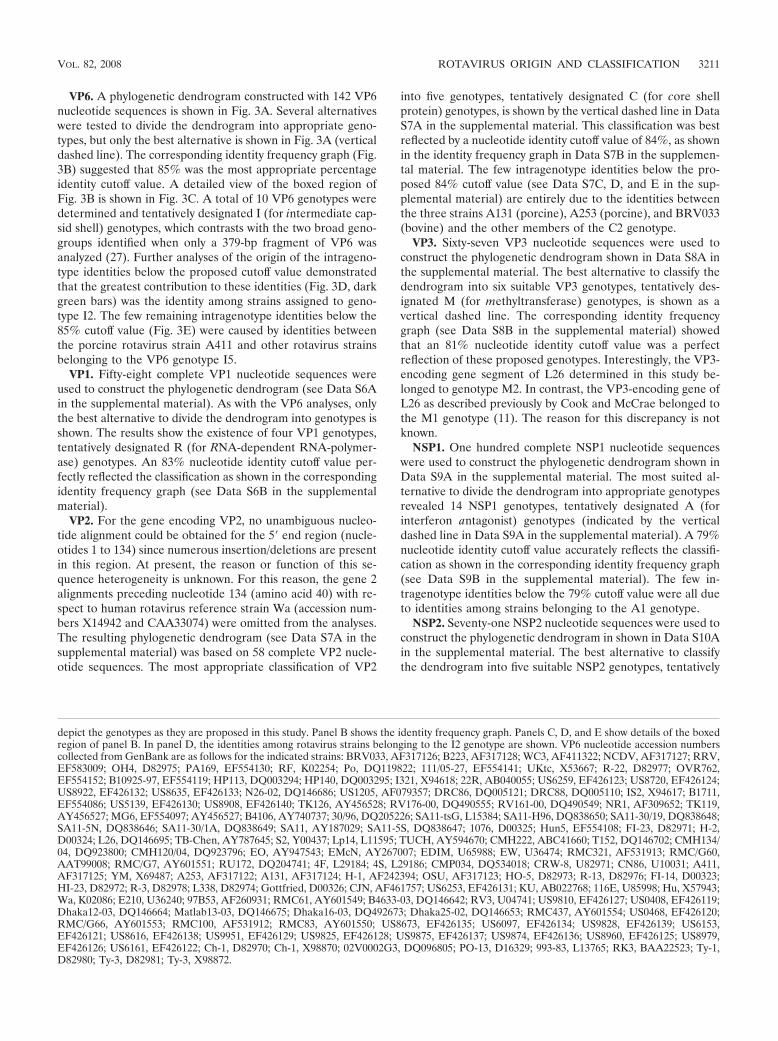
VP6. A phylogenetic dendrogram constructed with 142 VP6nucleotide sequences is shown in Fig. 3A. Several alternativeswere tested to divide the dendrogram into appropriate geno-types, but only the best alternative is shown in Fig. 3A (verticaldashed line). The corresponding identity frequency graph (Fig.3B) suggested that 85% was the most appropriate percentageidentity cutoff value. A detailed view of the boxed region ofFig. 3B is shown in Fig. 3C. A total of 10 VP6 genotypes weredetermined and tentatively designated I (for intermediate cap-sid shell) genotypes, which contrasts with the two broad geno-groups identified when only a 379-bp fragment of VP6 wasanalyzed (27). Further analyses of the origin of the intrageno-type identities below the proposed cutoff value demonstratedthat the greatest contribution to these identities (Fig. 3D, darkgreen bars) was the identity among strains assigned to geno-type I2. The few remaining intragenotype identities below the85% cutoff value (Fig. 3E) were caused by identities betweenthe porcine rotavirus strain A411 and other rotavirus strainsbelonging to the VP6 genotype I5.
VP1. Fifty-eight complete VP1 nucleotide sequences wereused to construct the phylogenetic dendrogram (see Data S6Ain the supplemental material). As with the VP6 analyses, onlythe best alternative to divide the dendrogram into genotypes isshown. The results show the existence of four VP1 genotypes,tentatively designated R (for RNA-dependent RNA-polymer-ase) genotypes. An 83% nucleotide identity cutoff value per-fectly reflected the classification as shown in the correspondingidentity frequency graph (see Data S6B in the supplementalmaterial).
VP2. For the gene encoding VP2, no unambiguous nucleo-tide alignment could be obtained for the 5� end region (nucle-otides 1 to 134) since numerous insertion/deletions are presentin this region. At present, the reason or function of this se-quence heterogeneity is unknown. For this reason, the gene 2alignments preceding nucleotide 134 (amino acid 40) with re-spect to human rotavirus reference strain Wa (accession num-bers X14942 and CAA33074) were omitted from the analyses.The resulting phylogenetic dendrogram (see Data S7A in thesupplemental material) was based on 58 complete VP2 nucle-otide sequences. The most appropriate classification of VP2
into five genotypes, tentatively designated C (for core shellprotein) genotypes, is shown by the vertical dashed line in DataS7A in the supplemental material. This classification was bestreflected by a nucleotide identity cutoff value of 84%, as shownin the identity frequency graph in Data S7B in the supplemen-tal material. The few intragenotype identities below the pro-posed 84% cutoff value (see Data S7C, D, and E in the sup-plemental material) are entirely due to the identities betweenthe three strains A131 (porcine), A253 (porcine), and BRV033(bovine) and the other members of the C2 genotype.
VP3. Sixty-seven VP3 nucleotide sequences were used toconstruct the phylogenetic dendrogram shown in Data S8A inthe supplemental material. The best alternative to classify thedendrogram into six suitable VP3 genotypes, tentatively des-ignated M (for methyltransferase) genotypes, is shown as avertical dashed line. The corresponding identity frequencygraph (see Data S8B in the supplemental material) showedthat an 81% nucleotide identity cutoff value was a perfectreflection of these proposed genotypes. Interestingly, the VP3-encoding gene segment of L26 determined in this study be-longed to genotype M2. In contrast, the VP3-encoding gene ofL26 as described previously by Cook and McCrae belonged tothe M1 genotype (11). The reason for this discrepancy is notknown.
NSP1. One hundred complete NSP1 nucleotide sequenceswere used to construct the phylogenetic dendrogram shown inData S9A in the supplemental material. The most suited al-ternative to divide the dendrogram into appropriate genotypesrevealed 14 NSP1 genotypes, tentatively designated A (forinterferon antagonist) genotypes (indicated by the verticaldashed line in Data S9A in the supplemental material). A 79%nucleotide identity cutoff value accurately reflects the classifi-cation as shown in the corresponding identity frequency graph(see Data S9B in the supplemental material). The few in-tragenotype identities below the 79% cutoff value were all dueto identities among strains belonging to the A1 genotype.
NSP2. Seventy-one NSP2 nucleotide sequences were used toconstruct the phylogenetic dendrogram in shown in Data S10Ain the supplemental material. The best alternative to classifythe dendrogram into five suitable NSP2 genotypes, tentatively
depict the genotypes as they are proposed in this study. Panel B shows the identity frequency graph. Panels C, D, and E show details of the boxedregion of panel B. In panel D, the identities among rotavirus strains belonging to the I2 genotype are shown. VP6 nucleotide accession numberscollected from GenBank are as follows for the indicated strains: BRV033, AF317126; B223, AF317128; WC3, AF411322; NCDV, AF317127; RRV,EF583009; OH4, D82975; PA169, EF554130; RF, K02254; Po, DQ119822; 111/05-27, EF554141; UKtc, X53667; R-22, D82977; OVR762,EF554152; B10925-97, EF554119; HP113, DQ003294; HP140, DQ003295; I321, X94618; 22R, AB040055; US6259, EF426123; US8720, EF426124;US8922, EF426132; US8635, EF426133; N26-02, DQ146686; US1205, AF079357; DRC86, DQ005121; DRC88, DQ005110; IS2, X94617; B1711,EF554086; US5139, EF426130; US8908, EF426140; TK126, AY456528; RV176-00, DQ490555; RV161-00, DQ490549; NR1, AF309652; TK119,AY456527; MG6, EF554097; AY456527; B4106, AY740737; 30/96, DQ205226; SA11-tsG, L15384; SA11-H96, DQ838650; SA11-30/19, DQ838648;SA11-5N, DQ838646; SA11-30/1A, DQ838649; SA11, AY187029; SA11-5S, DQ838647; 1076, D00325; Hun5, EF554108; FI-23, D82971; H-2,D00324; L26, DQ146695; TB-Chen, AY787645; S2, Y00437; Lp14, L11595; TUCH, AY594670; CMH222, ABC41660; T152, DQ146702; CMH134/04, DQ923800; CMH120/04, DQ923796; EO, AY947543; EMcN, AY267007; EDIM, U65988; EW, U36474; RMC321, AF531913; RMC/G60,AAT99008; RMC/G7, AY601551; RU172, DQ204741; 4F, L29184; 4S, L29186; CMP034, DQ534018; CRW-8, U82971; CN86, U10031; A411,AF317125; YM, X69487; A253, AF317122; A131, AF317124; H-1, AF242394; OSU, AF317123; HO-5, D82973; R-13, D82976; FI-14, D00323;HI-23, D82972; R-3, D82978; L338, D82974; Gottfried, D00326; CJN, AF461757; US6253, EF426131; KU, AB022768; 116E, U85998; Hu, X57943;Wa, K02086; E210, U36240; 97B53, AF260931; RMC61, AY601549; B4633-03, DQ146642; RV3, U04741; US9810, EF426127; US0408, EF426119;Dhaka12-03, DQ146664; Matlab13-03, DQ146675; Dhaka16-03, DQ492673; Dhaka25-02, DQ146653; RMC437, AY601554; US0468, EF426120;RMC/G66, AY601553; RMC100, AF531912; RMC83, AY601550; US8673, EF426135; US6097, EF426134; US9828, EF426139; US6153,EF426121; US8616, EF426138; US9951, EF426129; US9825, EF426128; US9875, EF426137; US9874, EF426136; US8960, EF426125; US8979,EF426126; US6161, EF426122; Ch-1, D82970; Ch-1, X98870; 02V0002G3, DQ096805; PO-13, D16329; 993-83, L13765; RK3, BAA22523; Ty-1,D82980; Ty-3, D82981; Ty-3, X98872.
VOL. 82, 2008 ROTAVIRUS ORIGIN AND CLASSIFICATION 3211
3212 MATTHIJNSSENS ET AL. J. VIROL.
designated N (for NTPase activity) genotypes, is shown as avertical dashed line (see Data S10A in the supplemental ma-terial). The corresponding identity frequency graph (see DataS10B in the supplemental material) showed that an 85% nu-cleotide identity cutoff value was a good reflection of theseproposed genotypes. The few intragenotype identities belowthe 85% the cutoff value were mainly due to identities amongstrains belonging to the N1 genotype, and the few intergeno-type identities above the cutoff value are due to identitiesbetween N1 and N2 (see Data S10B in the supplemental ma-terial).
NSP3. Seventy-seven NSP3 nucleotide sequences were usedto construct the phylogenetic dendrogram in shown in DataS11A in the supplemental material. The best alternative toclassify the dendrogram into seven suitable NSP3 genotypes,tentatively designated T (for translation enhancer) genotypes,is shown as a vertical dashed line (see Data S11A in thesupplemental material), and the corresponding identity fre-quency graph (see Data S11B in the supplemental material)showed that an 85% nucleotide identity cutoff value was themost appropriate for these proposed genotypes. The few in-tragenotype identities below the 85% cutoff value are due toidentities among strains belonging to the T1 genotype.
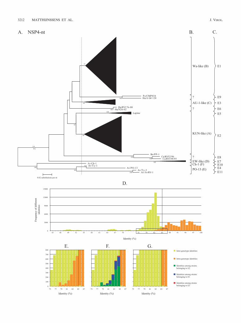
NSP4. For NSP4, a classification into six genotypes (A to F)is currently used, based on the clustering into amino acid-basedphylogenetic dendrograms using approximately 100 sequences(9, 22, 26, 46). In our analysis, a phylogenetic dendrogram wasconstructed with 430 available NSP4 nucleotide sequences(Fig. 4A). Large clusters have been replaced by triangles, butthe full dendrogram is shown in Data S12 in the supplementalmaterial. Several alternatives to divide the dendrogram intoappropriate genotypes were analyzed, but only the best alter-native is shown in Fig. 4A (vertical dashed line). The corre-sponding identity frequency graph (Fig. 4D) suggested that85% was the most appropriate percentage identity cutoff valuealthough a considerable number of intragenotype identitieswere found below this cutoff value. Although six NSP4 geno-types have been described to date, a total of 11 NSP4 geno-types, largely corresponding with the existing classification,were determined and tentatively designated E (for entero-toxin) genotypes when all available sequences were utilized.NSP4 genotype E1 corresponded completely with the Wa-like(genotype B) NSP4 genotype; genotype E2 largely corre-sponded to the KUN-like (genotype A) NSP4 genotype andwas the most diverse genotype, as the majority of the rotavirusstrains isolated from several species of origin (human, bovine,equine, simian, and ovine) clustered together, although thelapine or lapine-like human rotavirus strains were not includ-ed; genotype E3 corresponded completely to the AU-1-like(genotype C) NSP4 genotype; genotype E4 (previously NSP4
genotype E) was restricted to avian rotavirus strains (PO-13and Ty-1); genotype E5 was composed of lapine and a humanlapine-like rotavirus strains, previously belonging to the KUN-like (genotype A) NSP4 genotype. The assignment of the lap-ine NSP4 sequences into a single distinct genotype is alsosupported by previous observations showing a rather low de-gree of identity between lapine rotavirus strains and the re-maining NSP4 genotype A strains (9); genotype E6 was re-stricted to newly described unusual human G12 strains(N26-02 and RV176-00) (63); genotype E7 (previously NSP4genotype D) was composed entirely of murine rotavirusstrains; genotype E8 contained a bovine strain (PP-1, whichwas originally proposed to belong to the NSP4 genotype B)(15) and two canine strains (RV52/96 and RV198/95); geno-type E9 contained a human (A_G4_120) and a porcine(CMP034) strain that were described recently (30), while ge-notype E10 (previously NSP4 genotype F) contained the avianstrain Ch-1 only; and genotype E11 was composed of the avianrotavirus strains Ty-3 and AvRV-1. The majority of the newlyadded NSP4 genotypes are due to the inclusion of new NSP4sequences found in the GenBank database. A detail of theboxed region of Fig. 4D is shown in Fig. 4E and reveals thatthe greatest contribution to the intragenotype identities belowthe proposed cutoff value was generated by the identitiesamong strains assigned to the three largest genotypes, E2, E1,and E3 (Fig. 4F), in descending order.
NSP5. A phylogenetic dendrogram constructed with 113NSP5 nucleotide sequences is shown in Data S13A in thesupplemental material. Several alternatives to divide the den-drogram into appropriate genotypes were tested, but only thebest alternative is shown (see Data S13A in the supplementalmaterial; vertical dashed line). The corresponding identity fre-quency graph (see Data S13B in the supplemental material)suggested that 91% was the most appropriate percentage iden-tity cutoff value. A total of six NSP5 genotypes were deter-mined and tentatively designated H (for phosphoprotein) ge-notypes. A detail of the boxed region of Data S13B is shown inData S13C in the supplemental material. Further analyses ofthe origin of the intragenotype identities below the proposedcutoff value (91%) demonstrated that the greatest contributionto these identities (see Data S13D in the supplemental mate-rial; green, blue, and red bars) was the identity among strainsassigned to genotypes H2, H1, and H3, in descending order.
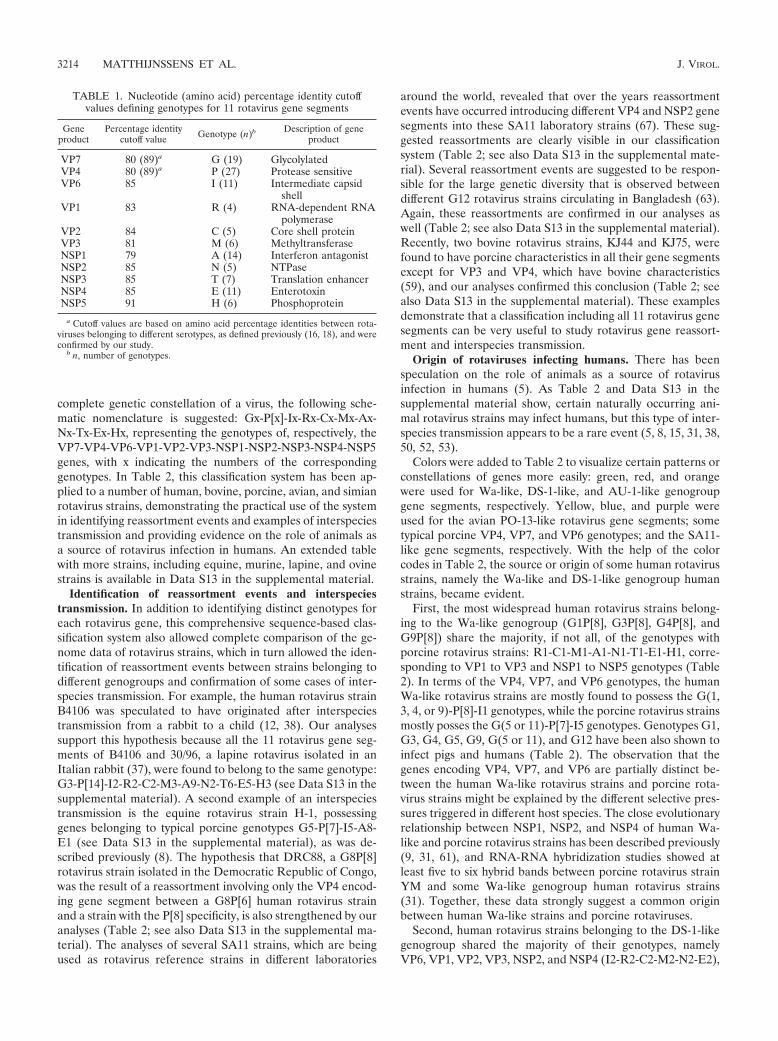
Recommendations for a rotavirus classification system ofall 11 genome segments. A summary of all 11 calculated cutoffvalues for the rotavirus genes segments is shown in Table 1.With these nucleotide cutoff percentages and the resultingappropriate genotypes for all the rotavirus proteins, a com-plete nomenclature and classification system can be proposedbased on the evidence provided in this study. To designate the
FIG. 4. Phylogenetic dendrogram of NSP4 at the nucleotide (nt) level (A). Bootstrap values (2,000 replicates) above 50 are shown. Designationsof species of origin are as follows: Bo, bovine; Hu, human; Po, porcine; Ca, canine; Av, avian. The dashed line indicates the best option to dividethe dendrogram into appropriate genotypes. Certain clusters are replaced by triangles, in which the height of the triangle represents the numberof sequences, and the width represents the genetic diversity inside that cluster. The full NSP4 phylogenetic dendrogram is provided in Data S6 inthe supplemental material as well as the accession numbers used to construct the dendrogram. The closing braces on the right side of thedendrograms depict the 11 E genotypes as they are proposed in this study (C). Panel B depicts the genotypes as they were previously used. PanelD shows the identity frequency graph. Panels E, F, and G show details of the boxed region of panel D. In panel F, the identities among rotavirusstrains belonging to the E3, E1, and E2 genotypes are shown. In panel G, the above-mentioned bars are omitted.
VOL. 82, 2008 ROTAVIRUS ORIGIN AND CLASSIFICATION 3213
complete genetic constellation of a virus, the following sche-matic nomenclature is suggested: Gx-P[x]-Ix-Rx-Cx-Mx-Ax-Nx-Tx-Ex-Hx, representing the genotypes of, respectively, theVP7-VP4-VP6-VP1-VP2-VP3-NSP1-NSP2-NSP3-NSP4-NSP5genes, with x indicating the numbers of the correspondinggenotypes. In Table 2, this classification system has been ap-plied to a number of human, bovine, porcine, avian, and simianrotavirus strains, demonstrating the practical use of the systemin identifying reassortment events and examples of interspeciestransmission and providing evidence on the role of animals asa source of rotavirus infection in humans. An extended tablewith more strains, including equine, murine, lapine, and ovinestrains is available in Data S13 in the supplemental material.
Identification of reassortment events and interspeciestransmission. In addition to identifying distinct genotypes foreach rotavirus gene, this comprehensive sequence-based clas-sification system also allowed complete comparison of the ge-nome data of rotavirus strains, which in turn allowed the iden-tification of reassortment events between strains belonging todifferent genogroups and confirmation of some cases of inter-species transmission. For example, the human rotavirus strainB4106 was speculated to have originated after interspeciestransmission from a rabbit to a child (12, 38). Our analysessupport this hypothesis because all the 11 rotavirus gene seg-ments of B4106 and 30/96, a lapine rotavirus isolated in anItalian rabbit (37), were found to belong to the same genotype:G3-P[14]-I2-R2-C2-M3-A9-N2-T6-E5-H3 (see Data S13 in thesupplemental material). A second example of an interspeciestransmission is the equine rotavirus strain H-1, possessinggenes belonging to typical porcine genotypes G5-P[7]-I5-A8-E1 (see Data S13 in the supplemental material), as was de-scribed previously (8). The hypothesis that DRC88, a G8P[8]rotavirus strain isolated in the Democratic Republic of Congo,was the result of a reassortment involving only the VP4 encod-ing gene segment between a G8P[6] human rotavirus strainand a strain with the P[8] specificity, is also strengthened by ouranalyses (Table 2; see also Data S13 in the supplemental ma-terial). The analyses of several SA11 strains, which are beingused as rotavirus reference strains in different laboratories
around the world, revealed that over the years reassortmentevents have occurred introducing different VP4 and NSP2 genesegments into these SA11 laboratory strains (67). These sug-gested reassortments are clearly visible in our classificationsystem (Table 2; see also Data S13 in the supplemental mate-rial). Several reassortment events are suggested to be respon-sible for the large genetic diversity that is observed betweendifferent G12 rotavirus strains circulating in Bangladesh (63).Again, these reassortments are confirmed in our analyses aswell (Table 2; see also Data S13 in the supplemental material).Recently, two bovine rotavirus strains, KJ44 and KJ75, werefound to have porcine characteristics in all their gene segmentsexcept for VP3 and VP4, which have bovine characteristics(59), and our analyses confirmed this conclusion (Table 2; seealso Data S13 in the supplemental material). These examplesdemonstrate that a classification including all 11 rotavirus genesegments can be very useful to study rotavirus gene reassort-ment and interspecies transmission.
Origin of rotaviruses infecting humans. There has beenspeculation on the role of animals as a source of rotavirusinfection in humans (5). As Table 2 and Data S13 in thesupplemental material show, certain naturally occurring ani-mal rotavirus strains may infect humans, but this type of inter-species transmission appears to be a rare event (5, 8, 15, 31, 38,50, 52, 53).
Colors were added to Table 2 to visualize certain patterns orconstellations of genes more easily: green, red, and orangewere used for Wa-like, DS-1-like, and AU-1-like genogroupgene segments, respectively. Yellow, blue, and purple wereused for the avian PO-13-like rotavirus gene segments; sometypical porcine VP4, VP7, and VP6 genotypes; and the SA11-like gene segments, respectively. With the help of the colorcodes in Table 2, the source or origin of some human rotavirusstrains, namely the Wa-like and DS-1-like genogroup humanstrains, became evident.
First, the most widespread human rotavirus strains belong-ing to the Wa-like genogroup (G1P[8], G3P[8], G4P[8], andG9P[8]) share the majority, if not all, of the genotypes withporcine rotavirus strains: R1-C1-M1-A1-N1-T1-E1-H1, corre-sponding to VP1 to VP3 and NSP1 to NSP5 genotypes (Table2). In terms of the VP4, VP7, and VP6 genotypes, the humanWa-like rotavirus strains are mostly found to possess the G(1,3, 4, or 9)-P[8]-I1 genotypes, while the porcine rotavirus strainsmostly posses the G(5 or 11)-P[7]-I5 genotypes. Genotypes G1,G3, G4, G5, G9, G(5 or 11), and G12 have been also shown toinfect pigs and humans (Table 2). The observation that thegenes encoding VP4, VP7, and VP6 are partially distinct be-tween the human Wa-like rotavirus strains and porcine rota-virus strains might be explained by the different selective pres-sures triggered in different host species. The close evolutionaryrelationship between NSP1, NSP2, and NSP4 of human Wa-like and porcine rotavirus strains has been described previously(9, 31, 61), and RNA-RNA hybridization studies showed atleast five to six hybrid bands between porcine rotavirus strainYM and some Wa-like genogroup human rotavirus strains(31). Together, these data strongly suggest a common originbetween human Wa-like strains and porcine rotaviruses.
Second, human rotavirus strains belonging to the DS-1-likegenogroup shared the majority of their genotypes, namelyVP6, VP1, VP2, VP3, NSP2, and NSP4 (I2-R2-C2-M2-N2-E2),
TABLE 1. Nucleotide (amino acid) percentage identity cutoffvalues defining genotypes for 11 rotavirus gene segments
Geneproduct
Percentage identitycutoff value Genotype (n)b Description of gene
product
VP7 80 (89)a G (19) GlycolylatedVP4 80 (89)a P (27) Protease sensitiveVP6 85 I (11) Intermediate capsid
shellVP1 83 R (4) RNA-dependent RNA
polymeraseVP2 84 C (5) Core shell proteinVP3 81 M (6) MethyltransferaseNSP1 79 A (14) Interferon antagonistNSP2 85 N (5) NTPaseNSP3 85 T (7) Translation enhancerNSP4 85 E (11) EnterotoxinNSP5 91 H (6) Phosphoprotein
a Cutoff values are based on amino acid percentage identities between rota-viruses belonging to different serotypes, as defined previously (16, 18), and wereconfirmed by our study.
b n, number of genotypes.
3214 MATTHIJNSSENS ET AL. J. VIROL.
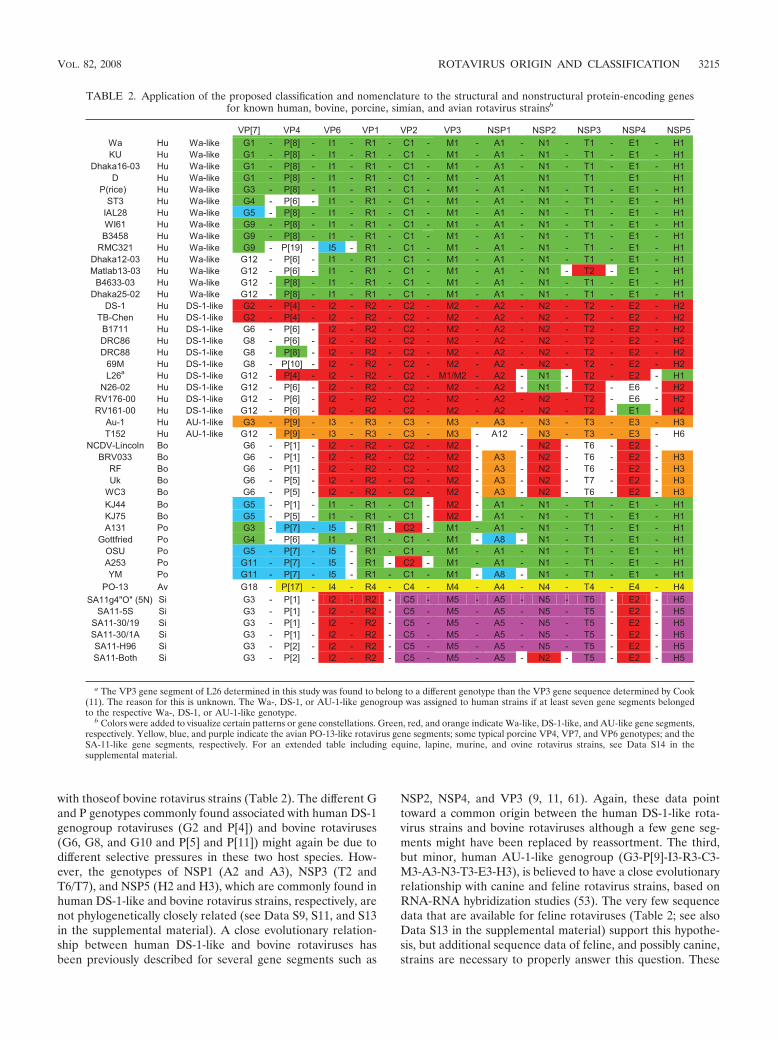
with thoseof bovine rotavirus strains (Table 2). The different Gand P genotypes commonly found associated with human DS-1genogroup rotaviruses (G2 and P[4]) and bovine rotaviruses(G6, G8, and G10 and P[5] and P[11]) might again be due todifferent selective pressures in these two host species. How-ever, the genotypes of NSP1 (A2 and A3), NSP3 (T2 andT6/T7), and NSP5 (H2 and H3), which are commonly found inhuman DS-1-like and bovine rotavirus strains, respectively, arenot phylogenetically closely related (see Data S9, S11, and S13in the supplemental material). A close evolutionary relation-ship between human DS-1-like and bovine rotaviruses hasbeen previously described for several gene segments such as
NSP2, NSP4, and VP3 (9, 11, 61). Again, these data pointtoward a common origin between the human DS-1-like rota-virus strains and bovine rotaviruses although a few gene seg-ments might have been replaced by reassortment. The third,but minor, human AU-1-like genogroup (G3-P[9]-I3-R3-C3-M3-A3-N3-T3-E3-H3), is believed to have a close evolutionaryrelationship with canine and feline rotavirus strains, based onRNA-RNA hybridization studies (53). The very few sequencedata that are available for feline rotaviruses (Table 2; see alsoData S13 in the supplemental material) support this hypothe-sis, but additional sequence data of feline, and possibly canine,strains are necessary to properly answer this question. These
TABLE 2. Application of the proposed classification and nomenclature to the structural and nonstructural protein-encoding genesfor known human, bovine, porcine, simian, and avian rotavirus strainsb
VP[7] VP4 VP6 VP1 VP2 VP3 NSP1 NSP2 NSP3 NSP4 NSP5 Wa Hu Wa-like G1 - P[8] - I1 - R1 - C1 - M1 - A1 - N1 - T1 - E1 - H1KU Hu Wa-like G1 - P[8] - I1 - R1 - C1 - M1 - A1 - N1 - T1 - E1 - H1
Dhaka16-03 Hu Wa-like G1 - P[8] - I1 - R1 - C1 - M1 - A1 - N1 - T1 - E1 - H1D Hu Wa-like G1 - P[8] - I1 - R1 - C1 - M1 - A1 N1 T1 E1 H1
P(rice) Hu Wa-like G3 - P[8] - I1 - R1 - C1 - M1 - A1 - N1 - T1 - E1 - H1ST3 Hu Wa-like G4 - P[6] - I1 - R1 - C1 - M1 - A1 - N1 - T1 - E1 - H1
IAL28 Hu Wa-like G5 - P[8] - I1 - R1 - C1 - M1 - A1 - N1 - T1 - E1 - H1WI61 Hu Wa-like G9 - P[8] - I1 - R1 - C1 - M1 - A1 - N1 - T1 - E1 - H1B3458 Hu Wa-like G9 - P[8] - I1 - R1 - C1 - M1 - A1 - N1 - T1 - E1 - H1
RMC321 Hu Wa-like G9 - P[19] - I5 - R1 - C1 - M1 - A1 - N1 - T1 - E1 - H1Dhaka12-03 Hu Wa-like G12 - P[6] - I1 - R1 - C1 - M1 - A1 - N1 - T1 - E1 - H1Matlab13-03 Hu Wa-like G12 - P[6] - I1 - R1 - C1 - M1 - A1 - N1 - T2 - E1 - H1
B4633-03 Hu Wa-like G12 - P[8] - I1 - R1 - C1 - M1 - A1 - N1 - T1 - E1 - H1Dhaka25-02 Hu Wa-like G12 - P[8] - I1 - R1 - C1 - M1 - A1 - N1 - T1 - E1 - H1
DS-1 Hu DS-1-like G2 - P[4] - I2 - R2 - C2 - M2 - A2 - N2 - T2 - E2 - H2TB-Chen Hu DS-1-like G2 - P[4] - I2 - R2 - C2 - M2 - A2 - N2 - T2 - E2 - H2B1711 Hu DS-1-like G6 - P[6] - I2 - R2 - C2 - M2 - A2 - N2 - T2 - E2 - H2DRC86 Hu DS-1-like G8 - P[6] - I2 - R2 - C2 - M2 - A2 - N2 - T2 - E2 - H2DRC88 Hu DS-1-like G8 - P[8] - I2 - R2 - C2 - M2 - A2 - N2 - T2 - E2 - H2
69M Hu DS-1-like G8 - P[10] - I2 - R2 - C2 - M2 - A2 - N2 - T2 - E2 - H2L26a Hu DS-1-like G12 - P[4] - I2 - R2 - C2 - M1/M2 - A2 - N1 - T2 - E2 - H1
N26-02 Hu DS-1-like G12 - P[6] - I2 - R2 - C2 - M2 - A2 - N1 - T2 - E6 - H2RV176-00 Hu DS-1-like G12 - P[6] - I2 - R2 - C2 - M2 - A2 - N2 - T2 - E6 - H2RV161-00 Hu DS-1-like G12 - P[6] - I2 - R2 - C2 - M2 - A2 - N2 - T2 - E1 - H2
Au-1 Hu AU-1-like G3 - P[9] - I3 - R3 - C3 - M3 - A3 - N3 - T3 - E3 - H3T152 Hu AU-1-like G12 - P[9] - I3 - R3 - C3 - M3 - A12 - N3 - T3 - E3 - H6
NCDV-Lincoln Bo G6 - P[1] - I2 - R2 - C2 - M2 - - N2 - T6 - E2 - BRV033 Bo G6 - P[1] - I2 - R2 - C2 - M2 - A3 - N2 - T6 - E2 - H3
RF Bo G6 - P[1] - I2 - R2 - C2 - M2 - A3 - N2 - T6 - E2 - H3Uk Bo G6 - P[5] - I2 - R2 - C2 - M2 - A3 - N2 - T7 - E2 - H3
WC3 Bo G6 - P[5] - I2 - R2 - C2 - M2 - A3 - N2 - T6 - E2 - H3KJ44 Bo G5 - P[1] - I1 - R1 - C1 - M2 - A1 - N1 - T1 - E1 - H1KJ75 Bo G5 - P[5] - I1 - R1 - C1 - M2 - A1 - N1 - T1 - E1 - H1A131 Po G3 - P[7] - I5 - R1 - C2 - M1 - A1 - N1 - T1 - E1 - H1
Gottfried Po G4 - P[6] - I1 - R1 - C1 - M1 - A8 - N1 - T1 - E1 - H1OSU Po G5 - P[7] - I5 - R1 - C1 - M1 - A1 - N1 - T1 - E1 - H1A253 Po G11 - P[7] - I5 - R1 - C2 - M1 - A1 - N1 - T1 - E1 - H1YM Po G11 - P[7] - I5 - R1 - C1 - M1 - A8 - N1 - T1 - E1 - H1
PO-13 Av G18 - P[17] - I4 - R4 - C4 - M4 - A4 - N4 - T4 - E4 - H4SA11g4"O" (5N Si G3 - P[1] - I2 - R2 - C5 - M5 - A5 - N5 - T5 - E2 - H5
SA11-5S Si G3 - P[1] - I2 - R2 - C5 - M5 - A5 - N5 - T5 - E2 - H5SA11-30/19 Si G3 - P[1] - I2 - R2 - C5 - M5 - A5 - N5 - T5 - E2 - H5SA11-30/1A Si G3 - P[1] - I2 - R2 - C5 - M5 - A5 - N5 - T5 - E2 - H5SA11-H96 Si G3 - P[2] - I2 - R2 - C5 - M5 - A5 - N5 - T5 - E2 - H5SA11-Both Si G3 - P[2] - I2 - R2 - C5 - M5 - A5 - N2 - T5 - E2 - H5
)
a The VP3 gene segment of L26 determined in this study was found to belong to a different genotype than the VP3 gene sequence determined by Cook(11). The reason for this is unknown. The Wa-, DS-1, or AU-1-like genogroup was assigned to human strains if at least seven gene segments belongedto the respective Wa-, DS-1, or AU-1-like genotype.
b Colors were added to visualize certain patterns or gene constellations. Green, red, and orange indicate Wa-like, DS-1-like, and AU-like gene segments,respectively. Yellow, blue, and purple indicate the avian PO-13-like rotavirus gene segments; some typical porcine VP4, VP7, and VP6 genotypes; and theSA-11-like gene segments, respectively. For an extended table including equine, lapine, murine, and ovine rotavirus strains, see Data S14 in thesupplemental material.
VOL. 82, 2008 ROTAVIRUS ORIGIN AND CLASSIFICATION 3215

results suggest that human and animal rotavirus strains arelinked very closely and emphasize the importance of monitor-ing rotaviruses in animal populations, as well as following un-common rotavirus strains in the human population veryclosely.
DISCUSSION
VP7, VP4, and NSP4 are the only rotavirus proteins forwhich a genotype-based classification system currently exists(9, 16, 18, 22, 26, 46). A serological classification exists for VP6,based on SG I- and II-specific MAbs, but by molecular andphylogenetic analyses of a 379-bp fragment of VP6, two geno-groups have been identified (27). Serological classification ofthe nonstructural proteins NSP1 to NSP3 and NSP5 and viralcore proteins VP1 and VP3 have not been carried out, andwhile the reactivity pattern of a MAb (YO-60) directed againstVP2 suggested the possible presence of VP2 subgroups inhuman rotavirus strains (70), the extent of this finding wasnever investigated further. Therefore, sequence analyses wouldprovide useful information to identify major clusters or geno-types in each of the rotavirus proteins. A similar approach hasalready been used for the determination of appropriate cutoffpercentages for the classification of astroviruses (58), sapovi-ruses (66), noroviruses (78), and papillomaviruses (13). Here,we propose a rotavirus classification system for all the rotavirusgenes that recognizes phylogenetic relationships and definesgenotypes based on percentage identity cutoff values for eachof the genes, which may have followed separate evolutionarypaths. Subsequently, the novel rotavirus classification systemwas used to identify reassortment events and gene constella-tions shared by human and animal rotaviruses.
Our phylogenetic classification of VP4 into different P ge-notypes showed that an 80% identity cutoff value was in ac-cordance with the established P genotypes. However, identitiesbetween P[8] and P[4] ranged from 84% to 89%, completelyabove the 80% cutoff value, reinforcing the idea that P[8] andP[4] are not only subtypes P1A and P1B of one serotype (16,18, 23) but also subtypes of one genotype. The three recentlydescribed novel porcine P genotypes, represented by strains344/04-1, CMP034, and P21-5, isolated in Italy, Thailand, andSlovenia, respectively (30, 35, 68), were all classified as distantmembers of the P[27] genotype, as was noted before (60).
The phylogenetic analyses of the VP7 gene sequences re-vealed that 80% and 89% identity cutoff values at the nucleo-tide and amino acid levels, respectively, provided the mostappropriate classification from an evolutionary perspective.However, the classification proposed herein does not fully con-cur with the current VP7 classification scheme, as outlinedbelow.
(i) Until now, all the VP7 genes of rotaviruses isolated frommice have been designated G3 or G3-like (14, 41, 74). Ourphylogenetic analyses indicate that the murine rotavirusescould be assigned to a new G genotype, tentatively designatedG16. Serological analyses of five murine strains, EW (alsoknown as EDIM), EB, EC, EL, and EHP, have revealed thatstrains EW and EHP are not recognized by G3-specific sera orG3-specific MAbs (6, 14, 20). Among the murine rotavirusstrains, only strain EB meets traditional criteria as serotypeG3, while murine strain EC, and probably EL, are recognized
by most, but not all, G3 serotype-specific MAbs (14). TheAmerican murine rotavirus strain EMcN showed a diversepattern of reactivity with G3-specific hyperimmune sera, andthe Japanese murine rotavirus strain YR-1 did not react withG3 specific MAbs at all (41, 74). We recognize that somemurine strains may exhibit a dual VP7 serotype similar tostrains MDR-13 (G3 and G5) and IAL28 (G5 and G11) andescape mutants of strain A253 (G11 and G3) (7, 16, 72).
(ii) All avian rotaviruses have been classified as serotype G7even though there are several conflicting serotyping reports inthe literature (25, 43, 65). The traditional avian genotype G7and the three proposed novel avian genotypes (G17, G18, andG19) show less than 78% and 84% identity at the nucleotideand amino acid levels, respectively, which are differences verysimilar to those observed between different mammalian rota-virus genotypes (data not shown).
(iii) As reported previously (7, 72), G5 and G11 rotavirusstrains are genotypically closely related. A human rotavirusstrain, IAL-28, exhibits both G5 and the G11 serotype speci-ficities (72), further strengthening the observation that G5 andG11 are closely related. With a few exceptions (8, 64, 73), bothG5 and G11 rotaviruses have been mainly isolated from pigs.Since genotype G11 is closer to genotype G5 than to genotypeG3, it is interesting that escape mutants of porcine strain A253(G11) exhibited both G11 and G3 serotype specificities insteadof G11 and G5 serotype specificities (7). Until the antigenicrelationships between G5 and G11 rotavirus strains are fullyunderstood, we propose to keep these two genotypes separateas they are clearly antigenically distinct.
(iv) Finally, the large sequence identity range found for G3rotaviruses (Fig. 2G, light green bars) is not to be ignored.Given that G3 rotaviruses have the largest spectrum of hostspecies and a higher degree of intergenotype sequence diver-sity than other genotypes (57), it does raise the level of com-plexity of the phylogenetic analyses (see discussion of murinerotavirus strains above) (29). In addition G3 and G14 rotavirusstrains are more closely related to each other serologically thandifferent genotypes usually are (10).
The high level of intragenotype sequence diversity within theVP2 and VP6 genotype 2 (C2 and I2, respectively), togetherwith the broad range of host species within these genotypes,resembles features observed in the VP7 serotype G3 (29, 57).A remarkable observation is that except for VP6 genotypes I2and I5, all the remaining eight VP6 genotypes contain rotavi-ruses isolated from one or two host species. No correlation wasfound between our VP6 genotyping system and VP6 SG spec-ificity (data not shown) as several of the VP6 genotypes containstrains with different SG specificities (17, 27, 41, 69).
The identification of distinct genotypes for each rotavirusgenome segment yielded multiple examples of related strains(Wa and KU, DS-1 and TB-Chen, and Dhaka25-02 and B4633-03), which cluster closely together in all 11 phylogenetic den-drograms, suggesting similar patterns of evolution. Several ofthe phylogenetic dendrograms, such as those for VP1, VP2,VP3, VP6, NSP2, and NSP3, show similar branching patternsfor most of the established genotypes.
Our classification system concurs with the main group Arotavirus genogroups (Wa, DS-1, and AU-1), as establishedthrough RNA-RNA hybridization analyses (51). In line withthe established human genogroups Wa, DS-1, and AU-1, the
3216 MATTHIJNSSENS ET AL. J. VIROL.
genotypes for each genome segment of these reference rota-virus strains were assigned to genotypes 1, 2, and 3, respec-tively, for consistency. All genome segments of the avian rota-virus PO-13 strain were assigned to genotype 4. For thegenome segments encoding NSP1, NSP2, NSP3, VP2, andVP3, genotype 5 was assigned to the cluster containing theSA11 clones. The remaining genotypes of the structural andnonstructural protein-encoding genes were assigned randomly,but systematically.
The demonstration that Wa-like human rotaviruses possesssimilar constellations of genes and, hence, a close genetic re-lationship to porcine rotaviruses while the DS-1-like humanrotaviruses have a close relationship with bovine rotavirusessuggests provocative ideas about the role of animals as a sourceof rotavirus infection in humans. The fact that each of thethree main human rotavirus families may have a different an-imal origin would be instrumental in obtaining a better under-standing of rotavirus host-range restriction and rotavirus ecol-ogy in nature. It is of interest that the only animals that can beproductively infected experimentally by human rotavirusstrains are piglets and calves (4). Piglets are monogastric ani-mals with intestinal physiology that resembles that of humansand are susceptible to infection and severe disease by thehuman rotavirus Wa strain (75) but not the DS-1 strain (24).On the other hand, human DS-1-like rotaviruses have beenshown to successfully infect cattle (45, 77), and the humanWa-like strain D can also infect and induce mild disease incalves (44). So far, dogs have been experimentally infected onlywith canine rotavirus strains (28).
To date a total of 35 human rotavirus strains have beencompletely sequenced, while only five and four porcine andbovine strains, respectively, have been completely sequenced(16, 38, 39, 63; also this report). Additional porcine and bovinerotavirus strains will be fully sequenced, allowing a furtherinvestigation of whether pigs and cows act as reservoirs forWa-like and DS-1-like human rotavirus strains, respectively.Likewise, we are currently sequencing all the genome segmentsof feline and canine strains to determine if these domesticanimals act as reservoirs for AU-1-like rotavirus strains inhumans.
A further advantage of the establishment of this novel geno-typing classification system is that a more systematic approachcan be used to investigate possible genetic linkages amongrotavirus genome segments. A comprehensive evaluation toinvestigate the possible genetic linkage between (sets of) genesfor all 11 genes is being currently conducted (unpublisheddata).
In summary, we calculated nucleotide percentage identitycutoff values to define genotypes for the gene segments encod-ing the 11 proteins of group A rotaviruses. Appropriate geno-types were defined, and a novel nomenclature system is beingproposed, allowing a rational comparison between differentrotavirus strains and international standardization. The appli-cation of this classification system to a large number of rota-virus strains identified distinct genotypes in all genes, whichprobably follow separate evolutionary paths, and allow theidentification of multiple reassortment and interspecies trans-mission events. The system also revealed the possible animalorigin of the most common human rotavirus strains. The data
reemphasize the rationale for simultaneous analysis of animaland human rotavirus strains.
ACKNOWLEDGMENTS
A special word of our most sincere gratitude goes to Ulrich Dessel-berger for insightful suggestions and critical revisions of the manu-script. We also thank all the colleagues of the Laboratory of Clinicaland Epidemiological Virology, Department of Microbiology and Im-munology, Rega Institute for Medical Research, University of Leuven,Belgium, for their helpful comments and discussions.
J.M. was supported by the Institute for the Promotion of Innovationthrough Science and Technology in Flanders (IWT Vlaanderen).J.T.P. and S.M.M. were supported by the Intramural Research Pro-gram of the National Institute of Allergy and Infectious Diseases,NIH. E.H. was supported by an appointment to the Oak Ridge Asso-ciated Universities Research Associates/Specialists Program at theNIH.
REFERENCES
1. Altschul, S. F., W. Gish, W. Miller, E. W. Myers, and D. J. Lipman. 1990.Basic local alignment search tool. J. Mol. Biol. 215:403–410.
2. Ball, L. A. 2005. The universal taxonomy of viruses in theory and practice, p.3–8. In C. M. Fauquet, M. A. Mayo, J. Maniloff, U. Desselberger, and L. A.Ball (ed.), Virus taxonomy. Eighth report of the International Committee onTaxonomy of Viruses. Elsevier, Amsterdam, The Netherlands.
3. Both, G. W., A. R. Bellamy, and D. B. Mitchell. 1994. Rotavirus proteinstructure and function. Curr. Top. Microbiol. Immunol. 185:67–105.
4. Ciarlet, M., M. E. Conner, and M. K. Estes. 2003. The rat model of rotavirusinfection, p. 291–306. In J. Gray and U. Desselberger (ed.), Perspectives inmedical virology: viral gastroenteritis. Elsevier Science BV, Amsterdam, TheNetherlands.
5. Ciarlet, M., and M. K. Estes. 2002. Rotaviruses: basic biology, epidemiologyand methodologies, p. 2573–2773. In G. Britton (ed.), Encyclopedia of en-vironmental microbiology. John Wiley and Sons, New York, NY.
6. Ciarlet, M., M. Hidalgo, and F. Liprandi. 1996. Cross-reactive, serotype- andmonotype-specific neutralization epitopes on VP7 of serotype G3 and G5porcine rotavirus strains. Arch. Virol. 141:601–614.
7. Ciarlet, M., Y. Hoshino, and F. Liprandi. 1997. Single point mutations mayaffect the serotype reactivity of serotype G11 porcine rotavirus strains: awidening spectrum? J. Virol. 71:8213–8220.
8. Ciarlet, M., P. Isa, M. E. Conner, and F. Liprandi. 2001. Antigenic andmolecular analyses reveal that the equine rotavirus strain H-1 is closelyrelated to porcine, but not equine, rotaviruses: interspecies transmissionfrom pigs to horses? Virus Genes 22:5–20.
9. Ciarlet, M., F. Liprandi, M. E. Conner, and M. K. Estes. 2000. Speciesspecificity and interspecies relatedness of NSP4 genetic groups by compar-ative NSP4 sequence analyses of animal rotaviruses. Arch. Virol. 145:371–383.
10. Ciarlet, M., F. Reggeti, C. I. Pina, and F. Liprandi. 1994. Equine rotaviruseswith G14 serotype specificity circulate among Venezuelan horses. J. Clin.Microbiol. 32:2609–2612.
11. Cook, J. P., and M. A. McCrae. 2004. Sequence analysis of the guanylyl-transferase (VP3) of group A rotaviruses. J. Gen. Virol. 85:929–932.
12. De Leener, K., M. Rahman, J. Matthijnssens, L. Van Hoovels, T. Goegebuer,I. van der Donck, and M. Van Ranst. 2004. Human infection with a P[14], G3lapine rotavirus. Virology 325:11–17.
13. de Villiers, E. M., C. Fauquet, T. R. Broker, H. U. Bernard, and H. zurHausen. 2004. Classification of papillomaviruses. Virology 324:17–27.
14. Dunn, S. J., J. W. Burns, T. L. Cross, P. T. Vo, R. L. Ward, M. Bremont, andH. B. Greenberg. 1994. Comparison of VP4 and VP7 of five murine rotavirusstrains. Virology 203:250–259.
15. El-Attar, L., W. Dhaliwal, C. R. Howard, and J. C. Bridger. 2001. Rotaviruscross-species pathogenicity: molecular characterization of a bovine rotaviruspathogenic for pigs. Virology 291:172–182.
16. Estes, M. K., and A. Z. Kapikian. 2007. Rotaviruses and their replication, p.1917–1974. In B. N. Fields, D. M. Knipe, P. M. Howley, D. E. Griffin, R. A.Lamb, M. A. Martin, B. Roizman, and S. E. Straus (ed.), Fields virology, 5thed. Lippincott, Williams and Wilkins, Philadelphia, PA.
17. Gonzalez, S. A., L. Tomasini, M. A. Tortorici, and J. L. Affranchino. 1995.VP6 from porcine rotavirus strain CN86: amino acid sequence divergencewith conservation of subgroup II specificity. J. Gen. Virol. 76:221–224.
18. Gorziglia, M., G. Larralde, A. Z. Kapikian, and R. M. Chanock. 1990.Antigenic relationships among human rotaviruses as determined by outercapsid protein VP4. Proc. Natl. Acad. Sci. USA 87:7155–7159.
19. Greenberg, H., V. McAuliffe, J. Valdesuso, R. Wyatt, J. Flores, A. Kalica, Y.Hoshino, and N. Singh. 1983. Serological analysis of the subgroup protein ofrotavirus, using monoclonal antibodies. Infect. Immun. 39:91–99.
20. Greenberg, H. B., P. T. Vo, and R. Jones. 1986. Cultivation and character-ization of three strains of murine rotavirus. J. Virol. 57:585–590.
VOL. 82, 2008 ROTAVIRUS ORIGIN AND CLASSIFICATION 3217

21. Gulati, B. R., R. Deepa, B. K. Singh, and C. D. Rao. 2007. Diversity in Indianequine rotaviruses: identification of genotype G10,P6[1] and G1 strains anda new VP7 genotype (G16) strain in specimens from diarrheic foals in India.J. Clin. Microbiol. 45:972–978.
22. Horie, Y., O. Masamune, and O. Nakagomi. 1997. Three major alleles ofrotavirus NSP4 proteins identified by sequence analysis. J. Gen. Virol. 78:2341–2346.
23. Hoshino, Y., R. W. Jones, R. M. Chanock, and A. Z. Kapikian. 2002. Gen-eration and characterization of six single VP4 gene substitution reassortantrotavirus vaccine candidates: each bears a single human rotavirus VP4 geneencoding P serotype 1A[8] or 1B[4] and the remaining 10 genes of rhesusmonkey rotavirus MMU18006 or bovine rotavirus UK. Vaccine 20:3576–3584.
24. Hoshino, Y., L. J. Saif, S. Y. Kang, M. M. Sereno, W. K. Chen, and A. Z.Kapikian. 1995. Identification of group A rotavirus genes associated withvirulence of a porcine rotavirus and host range restriction of a humanrotavirus in the gnotobiotic piglet model. Virology 209:274–280.
25. Hoshino, Y., R. G. Wyatt, H. B. Greenberg, J. Flores, and A. Z. Kapikian.1984. Serotypic similarity and diversity of rotaviruses of mammalian andavian origin as studied by plaque-reduction neutralization. J. Infect. Dis.149:694–702.
26. Ito, H., M. Sugiyama, K. Masubuchi, Y. Mori, and N. Minamoto. 2001.Complete nucleotide sequence of a group A avian rotavirus genome and acomparison with its counterparts of mammalian rotaviruses. Virus Res. 75:123–138.
27. Iturriza-Gomara, M., C. Wong, S. Blome, U. Desselberger, and J. Gray.2002. Molecular characterization of VP6 genes of human rotavirus isolates:correlation of genogroups with subgroups and evidence of independentsegregation. J. Virol. 76:6596–6601.
28. Johnson, C. A., R. W. Fulton, W. G. Henk, and T. G. Snider, III. 1983.Inoculation of neonatal gnotobiotic dogs with a canine rotavirus. Am. J. Vet.Res. 44:1682–1686.
29. Kapikian, A. Z., Y. Hoshino, and R. M. Chanock. 2001. Rotaviruses, p.1787–1834. In D. M. Knipe, P. M. Howley, D. E. Griffin, R. A. Lamb, M. A.Martin, B. Roizman, and S. E. Straus (ed.), Fields virology, 4th ed. Lippin-cott Williams and Wilkins, Philadelphia, PA.
30. Khamrin, P., N. Maneekarn, S. Peerakome, W. Chan-It, F. Yagyu, S. Okitsu,and H. Ushijima. 2007. Novel porcine rotavirus of genotype P[27] shares newphylogenetic lineage with G2 porcine rotavirus strain. Virology 361:243–252.
31. Kojima, K., K. Taniguchi, and N. Kobayashi. 1996. Species-specific andinterspecies relatedness of NSP1 sequences in human, porcine, bovine, fe-line, and equine rotavirus strains. Arch. Virol. 141:1–12.
32. Kool, D. A., and I. H. Holmes. 1993. The avian rotavirus Ty-1 Vp7 nucleotideand deduced amino acid sequences differ significantly from those of Ch-2rotavirus. Arch. Virol. 129:227–234.
33. Kumar, S., K. Tamura, I. B. Jakobsen, and M. Nei. 2001. MEGA2: molec-ular evolutionary genetics analysis software. Bioinformatics 17:1244–1245.
34. Liprandi, F., M. Gerder, Z. Bastidas, J. A. Lopez, F. H. Pujol, J. E. Ludert,D. B. Joelsson, and M. Ciarlet. 2003. A novel type of VP4 carried by aporcine rotavirus strain. Virology 315:373–380.
35. Martella, V., M. Ciarlet, K. Banyai, E. Lorusso, S. Arista, A. Lavazza, G.Pezzotti, N. Decaro, A. Cavalli, M. S. Lucente, M. Corrente, G. Elia, M.Camero, M. Tempesta, and C. Buonavoglia. 2007. Identification of group Aporcine rotavirus strains bearing a novel VP4 (P) genotype in Italian swineherds. J. Clin. Microbiol. 45:577–580.
36. Martella, V., M. Ciarlet, K. Banyai, E. Lorusso, A. Cavalli, M. Corrente, G.Elia, S. Arista, M. Camero, C. Desario, N. Decaro, A. Lavazza, and C.Buonavoglia. 2006. Identification of a novel VP4 genotype carried by aserotype G5 porcine rotavirus strain. Virology 346:301–311.
37. Martella, V., M. Ciarlet, A. Camarda, A. Pratelli, M. Tempesta, G. Greco, A.Cavalli, G. Elia, N. Decaro, V. Terio, G. Bozzo, M. Camero, and C.Buonavoglia. 2003. Molecular characterization of the VP4, VP6, VP7, andNSP4 genes of lapine rotaviruses identified in Italy: emergence of a novelVP4 genotype. Virology 314:358–370.
38. Matthijnssens, J., M. Rahman, V. Martella, Y. Xuelei, S. De Vos, K. DeLeener, M. Ciarlet, C. Buonavoglia, and M. Van Ranst. 2006. Full genomicanalysis of human rotavirus strain B4106 and lapine rotavirus strain 30/96provides evidence for interspecies transmission. J. Virol. 80:3801–3810.
39. Matthijnssens, J., M. Rahman, X. Yang, T. Delbeke, I. Arijs, J. P. Kabue,J. J. Muyembe, and M. Van Ranst. 2006. G8 rotavirus strains isolated in theDemocratic Republic of Congo belong to the DS-1-like genogroup. J. Clin.Microbiol. 44:1801–1809.
40. Maunula, L., and C. H. von Bonsdorff. 2002. Frequent reassortments mayexplain the genetic heterogeneity of rotaviruses: analysis of Finnish rotavirusstrains. J. Virol. 76:11793–11800.
41. McNeal, M. M., J. Belli, M. Basu, A. H. Choi, and R. L. Ward. 2004.Discovery of a new strain of murine rotavirus that is consistently shed in largequantities after oral inoculation of adult mice. Virology 320:1–11.
42. McNeal, M. M., K. Sestak, A. H. Choi, M. Basu, M. J. Cole, P. P. Aye, R. P.Bohm, and R. L. Ward. 2005. Development of a rotavirus-shedding model inrhesus macaques, using a homologous wild-type rotavirus of a new P geno-type. J. Virol. 79:944–954.
43. McNulty, M. S., G. M. Allan, D. Todd, J. B. McFerran, E. R. McKillop, D. S.Collins, and R. M. McCracken. 2006. Isolation of rotaviruses from turkeysand chickens: demonstration of distinct serotypes and RNA electrophero-types. Avian Pathol. 9:363–375.
44. Mebus, C., N. Stair, N. Underdahl, and M. Twiehaus. 1971. Pathology ofneonatal calf diarrhea induced by a reo-like virus. Vet. Pathol. 8:490–505.
45. Mebus, C. A., R. G. Wyatt, R. L. Sharpee, M. M. Sereno, A. R. Kalica, A. Z.Kapikian, and M. J. Twiehaus. 1976. Diarrhea in gnotobiotic calves causedby the reovirus-like agent of human infantile gastroenteritis. Infect. Immun.14:471–474.
46. Mori, Y., M. A. Borgan, N. Ito, M. Sugiyama, and N. Minamoto. 2002.Sequential analysis of nonstructural protein NSP4s derived from group Aavian rotaviruses. Virus Res. 89:145–151.
47. Mori, Y., M. A. Borgan, M. Takayama, N. Ito, M. Sugiyama, and N.Minamoto. 2003. Roles of outer capsid proteins as determinants of patho-genicity and host range restriction of avian rotaviruses in a suckling mousemodel. Virology 316:126–134.
48. Nakagomi, O., and T. Nakagomi. 2002. Genomic relationships among rota-viruses recovered from various animal species as revealed by RNA-RNAhybridization assays. Res. Vet. Sci. 73:207–214.
49. Nakagomi, O., and T. Nakagomi. 1991. Molecular evidence for naturallyoccurring single VP7 gene substitution reassortant between human rotavi-ruses belonging to two different genogroups. Arch. Virol. 119:67–81.
50. Nakagomi, O., and T. Nakagomi. 1991. Genetic diversity and similarityamong mammalian rotaviruses in relation to interspecies transmission ofrotavirus. Arch. Virol. 120:43–55.
51. Nakagomi, O., T. Nakagomi, K. Akatani, and N. Ikegami. 1989. Identifica-tion of rotavirus genogroups by RNA-RNA hybridization. Mol. Cell Probes3:251–261.
52. Nakagomi, O., A. Ohshima, Y. Aboudy, I. Shif, M. Mochizuki, T. Nakagomi,and T. Gotlieb-Stematsky. 1990. Molecular identification by RNA-RNAhybridization of a human rotavirus that is closely related to rotaviruses offeline and canine origin. J. Clin. Microbiol. 28:1198–1203.
53. Nakagomi, T., and O. Nakagomi. 1989. RNA-RNA hybridization identifies ahuman rotavirus that is genetically related to feline rotavirus. J. Virol. 63:1431–1434.
54. Nei, M., and S. Kumar. 2000. Molecular evolution and phylogenetics, p.33–50. Oxford University Press, New York, NY.
55. Nicholas, K. B., H. B. Nicholas, and D. W. Deerfield. 1997. GeneDoc:analysis and visualization of genetic variation. EMBnet News 4:14.
56. Nishikawa, K., Y. Hoshino, and M. Gorziglia. 1991. Sequence of the VP7gene of chicken rotavirus Ch2 strain of serotype 7 rotavirus. Virology 185:853–856.
57. Nishikawa, K., Y. Hoshino, K. Taniguchi, K. Y. Green, H. B. Greenberg,A. Z. Kapikian, R. M. Chanock, and M. Gorziglia. 1989. Rotavirus VP7neutralization epitopes of serotype 3 strains. Virology 171:503–515.
58. Noel, J. S., T. W. Lee, J. B. Kurtz, R. I. Glass, and S. S. Monroe. 1995. Typingof human astroviruses from clinical isolates by enzyme immunoassay andnucleotide sequencing. J. Clin. Microbiol. 33:797–801.
59. Park, S. H., L. J. Saif, C. Jeong, G. K. Lim, S. I. Park, H. H. Kim, S. J. Park, Y. J.Kim, J. H. Jeong, M. I. Kang, and K. O. Cho. 2006. Molecular characterizationof novel G5 bovine rotavirus strains. J. Clin. Microbiol. 44:4101–4112.
60. Parra, G. I., M. E. Galeano, and J. Arbiza. 2007. Genetic relationshipbetween porcine rotavirus strains bearing a new P-type. Vet. Microbiol.125:193–195.
61. Patton, J. T., L. Salter-Cid, A. Kalbach, E. A. Mansell, and M. Kattoura.1993. Nucleotide and amino acid sequence analysis of the rotavirus non-structural RNA-binding protein NS35. Virology 192:438–446.
62. Rahman, M., J. Matthijnssens, S. Nahar, G. Podder, D. A. Sack, T. Azim,and M. Van Ranst. 2005. Characterization of a novel P[25],G11 humangroup a rotavirus. J. Clin. Microbiol. 43:3208–3212.
63. Rahman, M., J. Matthijnssens, X. Yang, T. Delbeke, I. Arijs, K. Taniguchi,M. Iturriza-Gomara, N. Iftekharuddin, T. Azim, and M. Van Ranst. 2007.Evolutionary history and global spread of the emerging G12 human rotavi-ruses. J. Virol. 81:2382–2390.
64. Rahman, M., R. Sultana, G. Ahmed, S. Nahar, Z. M. Hassan, F. Saiada, G.Podder, A. S. Faruque, A. K. Siddique, D. A. Sack, J. Matthijnssens, M. VanRanst, and T. Azim. 2007. Prevalence of G2P[4] and G12P[6] rotavirus,Bangladesh. Emerg. Infect. Dis. 13:18–24.
65. Rohwedder, A., H. Hotop, N. Minamoto, H. Ito, O. Nakagomi, and H.Brussow. 1997. Bovine rotavirus 993/83 shows a third subtype of avian VP7protein. Virus Genes 14:147–151.
66. Schuffenecker, I., T. Ando, D. Thouvenot, B. Lina, and M. Aymard. 2001.Genetic classification of “Sapporo-like viruses.” Arch. Virol. 146:2115–2132.
67. Small, C., M. Barro, T. L. Brown, and J. T. Patton. 2007. Genome hetero-geneity of SA11 rotavirus due to reassortment with “O” agent. Virology359:415–424.
68. Steyer, A., M. Poljsak-Prijatelj, D. Barlic-Maganja, U. Jamnikar, J. Z. Mi-jovski, and J. Marin. 2007. Molecular characterization of a new porcinerotavirus P genotype found in an asymptomatic pig in Slovenia. Virology359:275–282.
69. Tang, B., J. M. Gilbert, S. M. Matsui, and H. B. Greenberg. 1997. Compar-
3218 MATTHIJNSSENS ET AL. J. VIROL.

ison of the rotavirus gene 6 from different species by sequence analysis andlocalization of subgroup-specific epitopes using site-directed mutagenesis.Virology 237:89–96.
70. Taniguchi, K., T. Urasawa, and S. Urasawa. 1986. Reactivity patterns tohuman rotavirus strains of a monoclonal antibody against VP2, a componentof the inner capsid of rotavirus. Brief report. Arch. Virol. 87:135–141.
71. Thompson, J. D., T. J. Gibson, F. Plewniak, F. Jeanmougin, and D. G.Higgins. 1997. The CLUSTAL_X windows interface: flexible strategies formultiple sequence alignment aided by quality analysis tools. Nucleic AcidsRes. 25:4876–4882.
72. Timenetsky, M. C., V. Gouvea, N. Santos, R. C. Carmona, and Y. Hoshino.1997. A novel human rotavirus serotype with dual G5-G11 specificity. J. Gen.Virol. 78:1373–1378.
73. Uchida, R., B. D. Pandey, J. B. Sherchand, K. Ahmed, M. Yokoo, T.Nakagomi, L. E. Cuevas, N. A. Cunliffe, C. A. Hart, and O. Nakagomi. 2006.Molecular epidemiology of rotavirus diarrhea among children and adults inNepal: detection of G12 strains with P[6] or P[8] and a G11P[25] strain.J. Clin. Microbiol. 44:3499–3505.
74. Ushijima, H., S. Morikawa, A. Mukoyama, and O. Nishio. 1995. Character-ization of VP4 and VP7 of a murine rotavirus (YR-1) isolated in Japan. Jpn.J. Med. Sci. Biol. 48:237–247.
75. Ward, L. A., B. I. Rosen, L. Yuan, and L. J. Saif. 1996. Pathogenesis of anattenuated and a virulent strain of group A human rotavirus in neonatalgnotobiotic pigs. J. Gen. Virol. 77:1431–1441.
76. Ward, R. L., O. Nakagomi, D. R. Knowlton, M. M. McNeal, T. Nakagomi,J. D. Clemens, D. A. Sack, and G. M. Schiff. 1990. Evidence for naturalreassortants of human rotaviruses belonging to different genogroups. J. Vi-rol. 64:3219–3225.
77. Wyatt, R. G., C. A. Mebus, R. H. Yolken, A. R. Kalica, H. D. James, Jr., A. Z.Kapikian, and R. M. Chanock. 1979. Rotaviral immunity in gnotobioticcalves: heterologous resistance to human virus induced by bovine virus.Science 203:548–550.
78. Zheng, D. P., T. Ando, R. L. Fankhauser, R. S. Beard, R. I. Glass, and S. S.Monroe. 2006. Norovirus classification and proposed strain nomenclature.Virology 346:312–323.
VOL. 82, 2008 ROTAVIRUS ORIGIN AND CLASSIFICATION 3219
![Human G9P[8] rotavirus strains circulating in Cameroon, 1999–2000: Genetic relationships with other G9 strains and detection of a new G9 subtype](https://static.fdokumen.com/doc/165x107/6332a59cf008040551049234/human-g9p8-rotavirus-strains-circulating-in-cameroon-19992000-genetic-relationships.jpg)