Influence of Calcium on the Early Steps of Rotavirus Infection
11
Influence of Calcium on the Early Steps of Rotavirus Infection Victoria Pando, Pavel Is ˇa, Carlos F. Arias, and Susana Lo ´ pez 1 Departamento de Gene ´ tica y Fisiologı ´a Molecular, Instituto de Biotecnologı ´a, Universidad Nacional Auto ´noma de Me ´ xico, Cuernavaca, Morelos 62250, Mexico Received June 13, 2001; accepted December 18, 2001 The structure of rotaviruses and many steps of their replication cycle depend on the concentration of calcium in the microenvironment. In this work, to learn about the role of calcium during the early steps of the infection, we characterized the effect of increasing the calcium concentration in the medium on the infectivity of rotaviruses. We found that a fivefold increase in the calcium concentration of the cell culture medium results in an increased viral titer in all rotavirus strains tested. The effect of this divalent ion seems to be mainly on the viral particle and not on the surface of the cell. Analysis of the intrinsic fluorescence spectra of purified triple-layered particles revealed that changes in the environment of tryptophan residues occurred as calcium concentration increased, suggesting that conformational changes in the viral particle might be responsible for the effect of this ion on the viral infectivity. © 2002 Elsevier Science (USA) Key Words: rotavirus; rotavirus infectivity; viral entry; calcium; intrinsic fluorescence; conformational changes. INTRODUCTION Rotaviruses are the major cause of severe acute diar- rhea of infants and young children under 2 years of age and are important pathogens in the young of many avian and mammalian species. These agents, members of the family Reoviridae, are nonenveloped icosahedric viruses consisting of three concentric layers of protein that sur- round the genome composed of 11 segments of double- stranded RNA. The outermost layer is composed of two proteins, VP4 and VP7, which are responsible for the initial interactions of the virus with the host cell. The smooth external surface of the virus is made up of 780 copies of glycoprotein VP7 arranged in trimers and of 120 copies of protein VP4, which form 60 dimeric spike-like structures that extend from the surface of the particle (Estes, 1996). VP4 has essential functions in the early virus–cell interactions, including receptor binding and cell penetra- tion (Crawford et al., 1994; Ludert et al., 1996; Mendez et al., 1996; Zarate et al., 2000). This protein is cleaved by trypsin into subunits VP5 and VP8, and it has been shown that this proteolytic treatment results in an en- hancement of rotavirus infectivity (Arias et al., 1996; Es- tes et al., 1981; Lopez et al., 1985). The role of VP7 during the early interactions of the virus with the cell has not been defined, but it has been proposed that it may modulate some functions of VP4 (Beisner et al., 1998; Mendez et al., 1996; Xu and Woode, 1994) and interact with cell surface molecules after the initial attachment of the virus through the VP4 protein (Coulson et al., 1997; Mendez et al., 1999). Recently, it has been established that the interaction of rotavirus with its host cell is a multistep process in which sequential contacts of the viral capsid with the cell sur- face take place (Mendez et al., 1999). Accordingly, differ- ent cellular molecules have been described as possible rotavirus receptors and coreceptors; among them gan- gliosides GM1 and GM3 (Delorme et al., 2001; Guo et al., 1999; Rolsma et al., 1998) and integrins 21, 41, and v3 (Coulson et al., 1997; Guerrero et al., 2000a; Hew- ish et al., 2000) have been found to play an important role during virus binding to, and penetration into, the cell. The replication cycle and the structure of rotaviruses are strongly dependent on the concentration of the ion calcium in the environment (Gajardo et al., 1997; Ruiz et al., 2000). During the replication cycle, the viral particles travel through different cellular compartments, each characterized by a different calcium concentration that is determinant for each step of the virus cycle. During cell entry, the triple-layered particle (TLP) loses the outer layer proteins, VP4 and VP7, and the double-layered particle (DLP), active in transcription, reaches the cyto- plasm. Although still controversial, the low-calcium envi- ronment of the cell has been proposed to be needed for this uncoating process (Cuadras et al., 1997; Ludert et al., 1987). During virus assembly, the double-layered particle buds from the cytoplasm into the lumen of the endoplas- mic reticulum (ER), which has a high-calcium environ- ment, acquiring during the process a transient lipid en- 1 To whom correspondence and reprint requests should be ad- dressed at Departamento de Ge ´ netica y Fisiologı ´a Molecular, Instituto de Biotecnologı ´a, Universidad Nacional Auto ´noma de Me ´ xico, Apar- tado Postal 510-3, Cuernavaca, Morelos 62250, Mexico. Fax: (52) (73) 172388. E-mail: [email protected]. Virology 295, 190–200 (2002) doi:10.1006/viro.2001.1337, available online at http://www.idealibrary.com on 0042-6822/02 $35.00 © 2002 Elsevier Science (USA) All rights reserved. 190
-
Upload
uuniversidaddepanam -
Category
Documents
-
view
1 -
download
0
Transcript of Influence of Calcium on the Early Steps of Rotavirus Infection

Virology 295, 190–200 (2002)
Influence of Calcium on the Early Steps of Rotavirus Infection
Victoria Pando, Pavel Isa, Carlos F. Arias, and Susana Lopez1
Departamento de Genetica y Fisiologıa Molecular, Instituto de Biotecnologıa, Universidad Nacional Autonoma de Mexico,Cuernavaca, Morelos 62250, Mexico
Received June 13, 2001; accepted December 18, 2001
The structure of rotaviruses and many steps of their replication cycle depend on the concentration of calcium in themicroenvironment. In this work, to learn about the role of calcium during the early steps of the infection, we characterizedthe effect of increasing the calcium concentration in the medium on the infectivity of rotaviruses. We found that a fivefoldincrease in the calcium concentration of the cell culture medium results in an increased viral titer in all rotavirus strainstested. The effect of this divalent ion seems to be mainly on the viral particle and not on the surface of the cell. Analysis ofthe intrinsic fluorescence spectra of purified triple-layered particles revealed that changes in the environment of tryptophanresidues occurred as calcium concentration increased, suggesting that conformational changes in the viral particle might be
INTRODUCTION
Rotaviruses are the major cause of severe acute diar-rhea of infants and young children under 2 years of ageand are important pathogens in the young of many avianand mammalian species. These agents, members of thefamily Reoviridae, are nonenveloped icosahedric virusesconsisting of three concentric layers of protein that sur-round the genome composed of 11 segments of double-stranded RNA. The outermost layer is composed of twoproteins, VP4 and VP7, which are responsible for theinitial interactions of the virus with the host cell. Thesmooth external surface of the virus is made up of 780copies of glycoprotein VP7 arranged in trimers and of 120copies of protein VP4, which form 60 dimeric spike-likestructures that extend from the surface of the particle(Estes, 1996).
VP4 has essential functions in the early virus–cellinteractions, including receptor binding and cell penetra-tion (Crawford et al., 1994; Ludert et al., 1996; Mendez etal., 1996; Zarate et al., 2000). This protein is cleaved bytrypsin into subunits VP5 and VP8, and it has beenshown that this proteolytic treatment results in an en-hancement of rotavirus infectivity (Arias et al., 1996; Es-tes et al., 1981; Lopez et al., 1985). The role of VP7 duringthe early interactions of the virus with the cell has notbeen defined, but it has been proposed that it may
1 To whom correspondence and reprint requests should be ad-dressed at Departamento de Genetica y Fisiologıa Molecular, Institutode Biotecnologıa, Universidad Nacional Autonoma de Mexico, Apar-
© 2002 Elsevier Science (USA)All rights reserved.
190
modulate some functions of VP4 (Beisner et al., 1998;Mendez et al., 1996; Xu and Woode, 1994) and interactwith cell surface molecules after the initial attachment ofthe virus through the VP4 protein (Coulson et al., 1997;Mendez et al., 1999).
Recently, it has been established that the interaction ofrotavirus with its host cell is a multistep process in whichsequential contacts of the viral capsid with the cell sur-face take place (Mendez et al., 1999). Accordingly, differ-ent cellular molecules have been described as possiblerotavirus receptors and coreceptors; among them gan-gliosides GM1 and GM3 (Delorme et al., 2001; Guo et al.,1999; Rolsma et al., 1998) and integrins �2�1, �4�1, and�v�3 (Coulson et al., 1997; Guerrero et al., 2000a; Hew-ish et al., 2000) have been found to play an important roleduring virus binding to, and penetration into, the cell.
The replication cycle and the structure of rotavirusesare strongly dependent on the concentration of the ioncalcium in the environment (Gajardo et al., 1997; Ruiz etal., 2000). During the replication cycle, the viral particlestravel through different cellular compartments, eachcharacterized by a different calcium concentration that isdeterminant for each step of the virus cycle. During cellentry, the triple-layered particle (TLP) loses the outerlayer proteins, VP4 and VP7, and the double-layeredparticle (DLP), active in transcription, reaches the cyto-plasm. Although still controversial, the low-calcium envi-ronment of the cell has been proposed to be needed forthis uncoating process (Cuadras et al., 1997; Ludert et al.,1987). During virus assembly, the double-layered particlebuds from the cytoplasm into the lumen of the endoplas-
responsible for the effect of this ion on the viral infectivity.Key Words: rotavirus; rotavirus infectivity; viral entry; ca
tado Postal 510-3, Cuernavaca, Morelos 62250, Mexico. Fax: (52) (73)172388. E-mail: [email protected].
doi:10.1006/viro.2001.1337, available online at http://www.idealibrary.com
0042-6822/02 $35.00
2 Elsevier Science (USA)
ntrinsic fluorescence; conformational changes.
mic reticulum (ER), which has a high-calcium environ-ment, acquiring during the process a transient lipid en-
© 200
lcium; i
on

velope. The formation of mature virus, in which the tran-sient envelope is replaced by the outer layer proteinsVP4 and VP7, is strictly dependent on the concentrationof calcium present in the ER lumen (Sharabari and Lee,1987). Compounds that alter the concentration of this ionin the ER, such as tapsigargin, which depletes the cal-cium from the ER lumen, block the maturation of rotavi-ruses at the enveloped stage, and the mature triple-layered particle is not formed (Poruchynsky et al., 1991).Also, the synthesis of the viral proteins alters the calciumhomeostasis of the cell, which favors the morphogenesisof the virus, and induces cell death (Dong et al., 1997).
The structure of the viral particle is also dependent onthe calcium concentration. The outermost layer of rota-virus contains calcium (Sharabari and Lee, 1987). In vitro,when the viral particles are treated with calcium chela-tors, the virus uncoats, releasing the transcriptionallyactive double-layered particle (Cohen et al., 1979). Thesensitivity of the viral particle to low concentrations ofcalcium segregates with VP7 (Gajardo et al., 1997), andmore recently it has been shown that calcium bindswithin VP7 trimers and that these calcium-stabilized tri-mers are the basic building block of the outer layer of therotavirus particle. Also, treatment of double-layered par-ticles with high concentrations of calcium (�1 M) causesthe intermediate layer of the particle, formed by VP6, todisassemble (Lepault et al., 2001).
In this work we characterized the effect of increasingthe calcium concentration in the cell culture medium onthe infectivity of rotaviruses. We found that a fivefoldincrease over the basal calcium concentration of the cell
culture medium resulted in an increased viral titer in allrotavirus strains tested. The effect of this divalent ionwas found to be mainly on the viral particle, and not onthe molecules present on the surface of the cell. Analysisof the intrinsic fluorescence spectra of purified TLPssuggests that conformational changes in the viral parti-cle might explain the effect of this ion on the enhance-ment of viral infectivity.
RESULTS
The infectivity of rhesus rotavirus RRV is increasedby high concentrations of calcium
Since many steps of the viral cycle of rotaviruses aremodulated by the concentration of calcium in the micro-environment, the effect of this ion on the infectivity ofrhesus rotavirus RRV was tested. In this assay, MA104cells were infected with RRV rotavirus suspended inEagle’s minimal medium (MEM) containing increasingconcentrations of CaCl2, starting from 1.8 mM, which isthe regular concentration of CaCl2 in the MEM. Theincrease in calcium concentration in the medium duringthe incubation of the virus with the cells resulted in anenhancement of viral infectivity, reaching a maximumtiter of virus at 14.4 mM CaCl2, which represented afourfold increase in the infectivity compared to the virustiter obtained in 1.8 mM CaCl2 (Fig. 1). It was not possibleto test higher concentrations of calcium in the mediumsince the viability of the MA104 cells was affected (datanot shown). Based on these data, to characterize theeffect of Ca2� on the infectivity of rotaviruses, we used inthe following assays 10 mM CaCl2 as the highest con-centration and 2 mM CaCl2 as the standard condition.
To analyze whether the effect of calcium on the viralinfectivity was specific to this ion, the effect of otherdivalent cations (Ba2�, Mg2�, Mn2�, Sr2�, and Zn2�) onrotavirus infectivity was tested. We found that in additionto Ca2�, the only other divalent ion that caused an in-crease in the infectivity of RRV was Sr2� (Table 1). Thiswas not surprising, since in addition to having an ionicradius very similar to that of calcium, Sr2� has beenreported to share some functional features with calcium,such as binding to calmodulin (Missiaen et al., 1999;
TABLE 1
Effect of Different Divalent Ions on the Infectivity of RRV Rotavirus
Divalent ion Infectivitya
BaCl2 0.95 � 0.04CaCl2 5.65 � 0.21SrCl2 3.70 � 0.56MgCl2 0.85 � 0.07
a Expressed as fold increase over control (viral infectivity in 2 mMCaCl2). The arithmetic mean � standard deviation of three independentexperiments performed in duplicate is shown.
FIG. 1. Effect of calcium concentration on the infectivity of rotavirusRRV. MA104 cells in 96-well plates were infected with trypsin-activatedvirus in MEM containing the indicated calcium concentrations. After 1 hof adsorption at 37°C, the virus inoculum was removed and fresh MEM(1.8 mM CaCl2) was added. The infected cells were incubated for 14 hat 37°C; after this time, the cells were fixed and stained by an immu-noperoxidase assay as described under Materials and Methods. Dataare expressed as fold increases over control (virus titer in 1.8 mMCaCl2 � 2 � 107 ffu/ml). The arithmetic mean � standard deviation ofthree independent experiments performed in duplicate is shown.
191EFFECT OF CALCIUM IN ROTAVIRUS ENTRY

Yamamoto et al., 1987). Strontium has also been reportedto be the only other divalent cation important for thestability of rotaviral particles (Shirley et al., 1981). Mag-nesium and barium did not have an effect on the viraltiter at a concentration of 10 mM; at this same concen-tration, manganese and zinc were toxic to the cells; at 4mM, which was the highest concentration tolerated bythe cells, these two ions did not affect the infectivity ofthe virus (not shown).
A high calcium concentration enhances the infectivityof several rotavirus strains
To study whether the effect of the calcium ions wasgeneral for rotavirus strains isolated from different ani-mal species, we infected MA104 cells with different ro-tavirus strains in medium containing either 2 or 10 mMCaCl2, and the titer of each virus was determined. Table2 shows that the infectivity of all rotavirus strains testedincreased in 10 mM Ca2�, although the magnitude of theincrement was different depending on the rotavirusstrain; for example, the titer of the human strain DS1increased about 50-fold over its control in 2 mM calcium,while the infectivity of the bovine strain NCDV increasedby only a factor of 2. It is noteworthy that in general theviruses that had the lowest titers in the control mediumhad the largest increments when their infectivity wasassayed in medium containing 10 mM calcium.
To determine the specificity of this effect, the infectivityof two other nonenveloped viruses, poliovirus and reovi-rus, in 2 and 10 mM CaCl2 was assayed. We found thatthe increase in the calcium concentration did not alterthe infectious titer of either of these two viruses, indicat-ing that the effect of calcium on the infectivity was spe-cific for rotaviruses.
Calcium affects the early interactions of rotaviruswith the cell surface
To determine whether the calcium increase in theculture medium favored the binding or the entry of rota-virus to the cell, we took advantage of the fact thatrotaviruses attach to the cell at 4°C, but penetrate only at37°C. In these assays, twofold dilutions of trypsin-acti-vated RRV virus, in MEM containing either 2 or 10 mMCaCl2, were adsorbed to monolayers of MA104 cells for30 min at 4°C; the unbound virus was removed, andwarm medium containing 10 or 2 mM CaCl2 was addedto the cells for 30 min at 37°C. After this incubationperiod the cells were washed, MEM containing 2 mMCaCl2 was added, and the infection was left to proceedfor 14 h at 37°C, at which time the cells were fixed andimmunostained. Figure 2 shows that the viral infectivityincreased only when 10 mM CaCl2 was present duringthe adsorption period, but not when the medium contain-ing 10 mM calcium was added after the virus was bound
in 2 mM calcium, suggesting that the effect of this ioncould be during the binding step.
To discover whether the increase in viral titer at 10 mMcalcium was due to an improved attachment of the viralparticles to the cell surface, the amount of virus bound tothe cells was determined. For this, purified TLPs in me-dium containing 2 or 10 mM CaCl2 were incubated onmonolayers of MA104 cells in 48 well-plates, for 1 h at4°C. After the unbound virus was removed, the cellswere washed extensively with MEM containing 2 mMcalcium and then lysed by two rounds of freeze–thaw.The cell-bound virus present in the lysate was deter-mined by an enzyme-linked immunosorbent assay(ELISA), as previously described (Zarate et al., 2000), andits infectious titer was determined. We found no differ-ence in the amount of viral particles bound to the cells inmedium containing 2 or 10 mM CaCl2, as determined bythe ELISA (Fig. 3A). However, when the titer of thesesame preparations was analyzed, we found that the virus
TABLE 2
Effect of Calcium on the Infectivity of Different Rotavirus Strains
Virus Origin
Virus titera in
Fold increaseb2 mM CaCl2 10 mM CaCl2
DS-1c Hu 6.2 � 104 3.5 � 106 56.5 � 19.0ST3d Hu 2.1 � 104 8.7 � 105 41.3 � 16.0TY-1c Av 3.5 � 105 6.6 � 106 18.9 � 5.0CH-2c Av 5.2 � 105 7.0 � 106 13.5 � 0.2nar3e Si 6.7 � 105 7.0 � 106 10.4 � 1.1Wa f Hu 3.9 � 106 2.9 � 107 7.4 � 0.3UKd Bo 1.2 � 106 8.5 � 106 7.1 � 0.2L338d Eq 9.4 � 106 5.8 � 107 6.2 � 3.1B223c Bo 5.7 � 105 2.7 � 106 4.8 � 0.1RFg Bo 4.0 � 106 1.8 � 107 4.7 � 0.4YMe Po 3.9 � 106 1.7 � 107 4.3 � 0.5H2d Eq 1.4 � 106 5.6 � 106 4.0 � 1.769Mh Hu 5.8 � 106 2.0 � 107 3.4 � 0.3RRV f Si 1.1 � 107 5.0 � 107 4.8 � 1.3SA11 4S i Si 4.3 � 107 1.1 � 108 2.5 � 0.2NCDVd Bo 6.9 � 107 1.6 � 108 2.3 � 0.6Poliovirus j Hu 4.0 � 104 4.0 � 104 1.0 � 0.2Reovirusk Hu 2.7 � 105 3.0 � 105 1.1 � 0.2
a The arithmetic mean of at least three independent experimentsperformed in duplicate is shown.
b Fold increase over control (infectivity in 2 mM CaCl2). The arith-metic mean � standard deviation of at least three independent exper-iments is shown.
c Y. Hoshino, National Institutes of Health, Bethesda, MD.d D. R. Snodgrass, Morendun Research Institute, Edinburgh, UK.e This laboratory IBT-UNAM, Mexico.f H. B. Greenberg, Stanford University, Palo Alto, CA.g J. Cohen, Institut National de la Recherche Agronomique, Cedex,
France.h G. N. Woode, College of Veterinary Medicine. Texas A&M Univer-
sity, Canyon, TX.i M. K. Estes, Baylor College of Medicine, Houston, TX.j R. M. del Angel, CINVESTAV-IPN, Mexico.k C. Ramos, CISEI, Instituto Nacional de Salud Publica, Mexico.
192 PANDO ET AL.
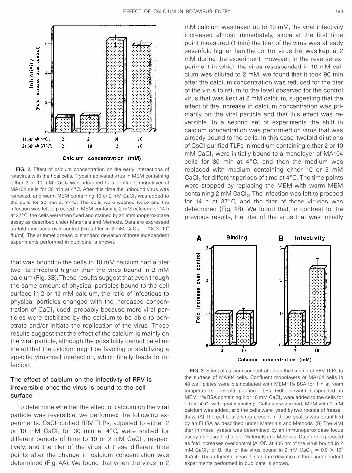
that was bound to the cells in 10 mM calcium had a titertwo- to threefold higher than the virus bound in 2 mMcalcium (Fig. 3B). These results suggest that even thoughthe same amount of physical particles bound to the cellsurface in 2 or 10 mM calcium, the ratio of infectious tophysical particles changed with the increased concen-tration of CaCl2 used, probably because more viral par-ticles were stabilized by the calcium to be able to pen-etrate and/or initiate the replication of the virus. Theseresults suggest that the effect of the calcium is mainly onthe viral particle, although the possibility cannot be elim-inated that the calcium might be favoring or stabilizing aspecific virus–cell interaction, which finally leads to in-fection.
The effect of calcium on the infectivity of RRV isirreversible once the virus is bound to the cellsurface
To determine whether the effect of calcium on the viralparticle was reversible, we performed the following ex-periments. CsCl-purified RRV TLPs, adjusted to either 2or 10 mM CaCl2 for 30 min at 4°C, were shifted fordifferent periods of time to 10 or 2 mM CaCl2, respec-tively, and the titer of the virus at these different timepoints after the change in calcium concentration wasdetermined (Fig. 4A). We found that when the virus in 2
mM calcium was taken up to 10 mM, the viral infectivityincreased almost immediately, since at the first timepoint measured (1 min) the titer of the virus was alreadysevenfold higher than the control virus that was kept at 2mM during the experiment. However, in the reverse ex-periment in which the virus resuspended in 10 mM cal-cium was diluted to 2 mM, we found that it took 90 minafter the calcium concentration was reduced for the titerof the virus to return to the level observed for the controlvirus that was kept at 2 mM calcium, suggesting that theeffect of the increase in calcium concentration was pri-marily on the viral particle and that this effect was re-versible. In a second set of experiments the shift incalcium concentration was performed on virus that wasalready bound to the cells. In this case, twofold dilutionsof CsCl-purified TLPs in medium containing either 2 or 10mM CaCl2 were initially bound to a monolayer of MA104cells for 30 min at 4°C, and then the medium wasreplaced with medium containing either 10 or 2 mMCaCl2 for different periods of time at 4°C. The time pointswere stopped by replacing the MEM with warm MEMcontaining 2 mM CaCl2. The infection was left to proceedfor 14 h at 37°C, and the titer of these viruses wasdetermined (Fig. 4B). We found that, in contrast to theprevious results, the titer of the virus that was initially
FIG. 3. Effect of calcium concentration on the binding of RRV TLPs tothe surface of MA104 cells. Confluent monolayers of MA104 cells in48-well plates were preincubated with MEM–1% BSA for 1 h at roomtemperature. Ice-cold purified TLPs (500 ng/well) suspended inMEM–1% BSA containing 2 or 10 mM CaCl2 were added to the cells for1 h at 4°C, with gentle shaking. Cells were washed, MEM with 2 mMcalcium was added, and the cells were lysed by two rounds of freeze–thaw. (A) The cell-bound virus present in these lysates was quantifiedby an ELISA as described under Materials and Methods. (B) The viraltiter in these lysates was determined by an immunoperoxidase focusassay, as described under Materials and Methods. Data are expressedas fold increases over control (A, OD at 405 nm of the virus bound in 2mM CaCl2; or B, titer of the virus bound in 2 mM CaCl2 � 0.9 � 106
ffu/ml). The arithmetic mean � standard deviation of three independentexperiments performed in duplicate is shown.
FIG. 2. Effect of calcium concentration on the early interactions ofrotavirus with the host cells. Trypsin-activated virus in MEM containingeither 2 or 10 mM CaCl2 was adsorbed to a confluent monolayer ofMA104 cells for 30 min at 4°C. After this time the unbound virus wasremoved, and warm MEM containing 10 or 2 mM CaCl2 was added tothe cells for 30 min at 37°C. The cells were washed twice and theinfection was left to proceed in MEM containing 2 mM calcium for 14 hat 37°C, the cells were then fixed and stained by an immunoperoxidaseassay as described under Materials and Methods. Data are expressedas fold increases over control (virus titer in 2 mM CaCl2 � 1.8 � 106
ffu/ml). The arithmetic mean � standard deviation of three independentexperiments performed in duplicate is shown.
193EFFECT OF CALCIUM IN ROTAVIRUS ENTRY
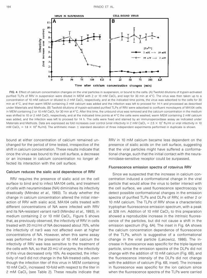
bound at either concentration of calcium remained un-changed for the period of time tested, irrespective of theshift in calcium concentration. These results indicate thatonce the virus was bound to the cell surface, a decreaseor an increase in calcium concentration no longer af-fected its interaction with the cell surface.
Calcium reduces the sialic acid dependence of RRV
RRV requires the presence of sialic acid on the cellsurface to bind and to infect MA104 cells, and treatmentof cells with neuraminidase (NA) diminishes its infectivitydrastically (Mendez et al., 1993). To study whether thechange in calcium concentration altered the initial inter-action of RRV with sialic acid, MA104 cells treated withdifferent concentrations of NA were infected with RRVand its NA-resistant variant nar3 (Mendez et al., 1993), inmedium containing 2 or 10 mM CaCl2. Figure 5 showsthat, as previously observed, the infectivity of RRV in cellstreated with 20 mU/ml of NA decreased about 75%, whilethe infectivity of nar3 was not affected even at higherconcentrations of NA. However, when this same assaywas performed in the presence of 10 mM calcium theinfectivity of RRV was less sensitive to the treatment ofthe cells with NA, so that 20 mU of enzyme, the infectivityof this virus decreased only 15%. As expected, the infec-tivity of nar3 did not change in the NA-treated cells, eventhough the infection titer of this virus in MEM containing10 mM CaCl2 increased 10-fold with respect to the titer in2 mM CaCl2 (see Table 2). These results indicate that
RRV in 10 mM calcium became less dependent on thepresence of sialic acids on the cell surface, suggestingthat the viral particles might have suffered a conforma-tional change, such that the initial contact with the neura-minidase-sensitive receptor could be surpassed.
Fluorescence emission spectra of rotavirus RRV
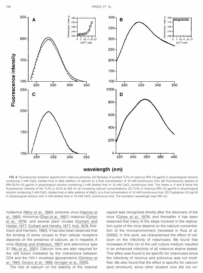
Since we suspected that the increase in calcium con-centration induced a conformational change in the viralparticle that would allow the virus to better interact withthe cell surface, we used fluorescence spectroscopy todetect possible conformational changes in the emissionspectra of purified TLPs and DLPs of RRV, in either 2 or10 mM calcium. The TLPs of RRV show a characteristictryptophan fluorescence emission spectrum, with a peakat 328 nm. Addition of 10 mM CaCl2 to this preparationshowed a reproducible increase in the intrinsic fluores-cence of the particles, but did not cause a shift in theemission spectrum (Fig. 6A). The inset in Fig. 6A showsthe calcium concentration dependence of fluorescenceof the TLPs, which is suggestive of a conformationalchange in the viral particle (Lakowicz, 1986). The in-crease in fluorescence was specific for the triple-layeredparticle, since the emission spectra of the DLPs did notchange with the addition of 10 mM calcium (Fig. 6B), andthe fluorescence intensity of the DLPs did not changewith the addition of calcium (Fig. 6B, inset). The increasein fluorescence was specific for the ion calcium sincewhen the fluorescence spectra of the TLPs were carried
FIG. 4. Effect of calcium concentration changes on the viral particles in suspension, or bound to the cells. (A) Twofold dilutions of trypsin-activatedpurified TLPs of RRV in suspension were diluted in MEM with 2 or 10 mM CaCl2 and kept for 30 min at 4°C. The virus was then taken up to aconcentration of 10 mM calcium or diluted to 2 mM CaCl2 respectively, and at the indicated time points, the virus was adsorbed to the cells for 30min at 4°C, and then warm MEM containing 2 mM calcium was added and the infection was left to proceed for 14 h and processed as describedunder Materials and Methods. (B) Twofold dilutions of trypsin-activated purified TLPs of RRV were adsorbed to confluent monolayers of MA104 cellsin MEM containing 2 or 10 mM CaCl2 for 30 min at 4°C. After this time, the unbound virus was removed and the calcium concentration in the mediumwas shifted to 10 or 2 mM CaCl2 respectively, and at the indicated time points at 4°C the cells were washed, warm MEM containing 2 mM calciumwas added, and the infection was left to proceed for 14 h. The cells were fixed and stained by an immunoperoxidase assay as indicated underMaterials and Methods. Data are expressed as fold increases over control (viral infectivity in 2 mM CaCl2 � 2.5 � 107 ffu/ml or viral infectivity in 10mM CaCl2 � 1.8 � 108 ffu/ml). The arithmetic mean � standard deviation of three independent experiments performed in duplicate is shown.
194 PANDO ET AL.

out using 10 mM MgCl2, a shift in the fluorescenceintensity of the viral particle was not detected (Fig. 6C), inagreement with the fact that magnesium did not have aneffect on the viral titer. A solution of tryptophan, used asa control, did not change its fluorescence when 10 mMcalcium was added (Fig. 6D). Addition of 10 mM calciumto purified TLPs of nar3 and Wa rotaviruses had the sameeffect on their emission spectra (data not shown). Takentogether, these results suggest that the increase in theintrinsic fluorescence of the rotavirus TLPs might reflecta change in the exposure and/or environment of thetryptophan residues of the viral particle upon addition ofcalcium, which in turn might indicate a conformationalchange in the rotavirus TLPs. Since the fluorescencespectra of the DLPs did not vary under the differentconcentrations of calcium tested, the conformationalchanges induced by Ca2� most likely occur in the pro-teins VP4 and VP7, which compose the outer layer of thevirion.
Determination of the tryptophan residues exposed inrotavirus particles
Aqueous fluorescence quenchers such as KI andacrylamide measure the exposure of tryptophan resi-dues to the aqueous environment. To determine whetherthe change in the fluorescence spectra observed whenthe TLPs were incubated with 10 mM CaCl2 was due toan increase in the tryptophan residues exposed to thesolvent, fluorescence quenching studies using KI werecarried out, and the degree of quenching was calculatedby the modified Stern–Volmer equation (see Materialsand Methods). Table 3 shows that there was a higher
degree of exposure of tryptophan residues when theTLPs of RRV and nar3 viruses were in 10 mM CaCl2,compared to the TLPs in 2 mM calcium, indicating thatthe increase in calcium concentration induced a confor-mational change in the viral particles that resulted in ahigher level of exposure of tryptophan residues to theaqueous environment.
DISCUSSION
A growing number of examples show that for a virus toenter its host cell, it must establish several sequentialinteractions with cell receptors and coreceptors, and ithas been shown that as a consequence of these inter-actions the viral particle may undergo conformationalchanges. These changes can be influenced by the pres-ence of divalent ions, such as calcium. For example, thision plays an important role in the stability of the viralcapsids of SV40 (Liddington et al., 1991), black beetle
TABLE 3
Determination of Tryptophan Residues Exposedin Rotavirus Particlesa
Virus 2 mM CaCl2 10 mM CaCl2
RRV 35.5% � 2.18 45.77% � 4.21nar3 22.43% � 0.96 39.88% � 2.31
a Data are expressed as the percentage of tryptophan residuesexposed in the viral particles at the indicated calcium concentration.These results represent the arithmetic mean � standard deviation ofthree independent experiments.
FIG. 5. Effect of calcium concentration on the infectivity of rotavirus in neuraminidase-treated cells. MA104 cells in 96-well plates were treated withthe indicated concentrations of NA from A. ureafaciens for 1 h at 37°C, washed, and subsequently infected with 1000 ffu/well of trypsin-activatedrotaviruses RRV or nar3, in MEM containing 2 or 10 mM CaCl2 for 1 h at 37°C. The virus inoculum was removed, fresh MEM with 2 mM calcium wasadded, and the infection was left to proceed for 14 h at 37°C. The cells were fixed and stained by an immunoperoxidase assay as described underMaterials and Methods. Data are expressed as the percentage of infectivity in control, untreated cells in 2 CaCl2 or 10 mM CaCl2. The arithmeticmean � standard deviation of three independent experiments performed in duplicate is shown.
195EFFECT OF CALCIUM IN ROTAVIRUS ENTRY
nodavirus (Wery et al., 1994), polyoma virus (Haynes etal., 1993), rhinovirus (Zhao et al., 1997), rotavirus (Cohenet al., 1979), and several plant viruses (Durham andHaidar, 1977; Durham and Hendry, 1977; Hull, 1978; Rob-inson and Harrison, 1982). It has also been observed thatthe binding of some viruses to their cellular receptorsdepends on the presence of calcium, as in hepatitis Avirus (Bishop and Anderson, 1997) and adenovirus type37 (Wu et al., 2001). Calcium ions are also required forthe cell fusion mediated by the interactions betweenCD4 and the HIV-1 envelope glycoproteins (Dimitrov etal., 1993; Doranz et al., 1999; Jernigan et al., 2000).
The role of calcium on the stability of the rotaviral
capsid was recognized shortly after the discovery of thevirus (Cohen et al., 1979), and thereafter it has beenobserved that many of the steps involved in the replica-tion cycle of the virus depend on the calcium concentra-tion of the microenvironment [reviewed in Ruiz et al.(2000)]. In this work, we characterized the effect of cal-cium on the infectivity of rotaviruses. We found thatincreases of this ion in the cell culture medium resultedin an enhanced infectivity of all rotavirus strains tested.This effect was found to be specific for rotaviruses sincethe infectivity of reovirus and poliovirus was not modi-fied. We also found that the effect is specific for calcium(and strontium), since other divalent ions did not en-
FIG. 6. Fluorescence emission spectra from rotavirus particles. (A) Samples of purified TLPs of rotavirus RRV (10 �g/ml) in physiological solutioncontaining 2 mM CaCl2 (dotted line) or after addition of calcium to a final concentration of 10 mM (continuous line). (B) Fluorescence spectra ofRRV-DLPs (10 �g/ml) in physiological solution containing 2 mM (dotted line) or 10 mM CaCl2 (continuous line). The insets in A and B show thefluorescence intensity of the TLPs or DLPs at 328 nm at increasing calcium concentrations. (C) TLPs of rotavirus RRV (10 �g/ml) in physiologicalsolution containing 2 mM CaCl2 (dotted line) or after addition of MgCl2 to a final concentration of 10 mM (continuous line). (D) Tryptophan (10 ng/ml)in physiological solution with 2 mM (dotted line) or 10 mM CaCl2 (continuous line). The excitation wavelength was 295 nm.
196 PANDO ET AL.
hance the viral infectivity, suggesting that the increase ininfectivity is due to the calcium ion per se, and not onlydue to the positive charges of this cation.
The results obtained in this work suggest that theeffect of calcium is on the viral particle, since the infec-tivity was increased when the virus was either bound tocells or preincubated in suspension in 10 mM calcium;this enhancement of infectivity was maintained even ifthe calcium concentration was reduced to 2 mM after thevirus was bound to the cells or after the preincubation ofthe virus in suspension.
We found that even though the same amount of viralparticles bound to the cell surface in both 2 and 10 mMcalcium, as determined by an ELISA, the infectious titerof the particles bound to cells in 10 mM calcium washigher than that of the viral particles bound in 2 mMcalcium. This observation suggests that when adsorbedto the cell surface in 10 mM calcium, more viral particleswere competent to proceed with the infection. It is knownthat of the total amount of virus produced as a result ofan infection, there is a high proportion of viral particlesthat are noninfectious. In rotavirus the infectious to phys-ical particle ratio is low, varying between different strainsof rotavirus. For example, for rotavirus RRV it has beenestimated that there is one infectious particle (IP) forevery 100–300 physical, noninfectious particles (PP); andin the case of the human strain Wa this ratio is evenlower, with one IP per 1–4 � 104 PP (Mendez et al., 1999).The reason why the physical particles are noninfectiousis not known, but the fact that the infectious titer of a viralstock is enhanced in the presence of 10 mM calciumsuggests that this cation induces a change in the con-formation of the viral capsid that makes the particlesprobably more stable and able to initiate a productiveinfection. Recently, Dormitzer et al. (2000) reported thatthe trimerization of VP7 depends on the presence ofcalcium, and they suggested that dissociation of thesetrimers might be the biochemical basis for the EDTA-induced uncoating of rotavirus particles.
We have proposed that the entry of rotavirus to its hostcell is a multistep process, in which the viral proteinsinteract with at least three different cellular molecules ina sequential manner (Mendez et al., 1999). We hypothe-sized that the virus could interact with these differentmolecules by experimenting conformational changesthat allow the virion to expose the protein domains re-sponsible for each of these interactions, although atpresent there has not been a direct observation of any ofthese putative conformational changes in the viral parti-cle. In this work, we found that rotavirus RRV, whichrequires sialic acid to bind to and to infect cells, becameless dependent at 10 mM calcium on the presence ofsialic acid on the surface of the cells. This observationsuggests that incubation of the virus in this calciumconcentration favors a conformational change in the RRVparticle that allows the virus to interact with the cell
surface, in a neuraminidase-resistant manner, surpass-ing the initial interaction of this virus with a sialic acid-containing receptor. The fact that other rotavirus strains,like nar3, which is a neuraminidase-resistant variant ofRRV, and human strains Wa and DS1, which are naturallyresistant to the neuraminidase treatment of the cells,also increased their titer upon addition of calcium sug-gests that the favored virus–cell interaction is sharedamong these strains. The precise virus–cell contact im-proved by the high calcium concentration needs to bedefined.
In this work we used the intrinsic tryptophan fluores-cence of purified TLPs to monitor the conformationalchanges in the viral particles induced by calcium. Tryp-tophan fluorescence is strongly influenced by the envi-ronment of its indole side chain and has thus proved tobe a useful tool for studying conformational changes inproteins, protein–protein interactions, and protein–mem-brane interactions (Carneiro et al., 2001; Lakowicz, 1986).
The intrinsic fluorescence studies on the TLPs of RRV,nar3, and Wa (shown only for RRV) revealed that changesin the solvent exposure of the tryptophan residues oc-curred as calcium increased, which is an indication of aconformational change in the viral particle. The quench-ing assays with KI further confirmed this observation,since it was determined that the percentage of trypto-phan residues exposed in the nar3 and RRV TLPs in thepresence of 10 mM calcium was higher than when theTLPs were suspended in 2 mM calcium.
Recently, it was suggested that the formation of theouter layer during viral assembly and the loss of theouter layer during the entry of the virus into the cell aremediated by a calcium-dependent conformationalchange in VP7 (Dormitzer et al., 2000). In this work wefound that the calcium concentration induced conforma-tional changes in the complete triple-layered viral parti-cle, which might be contributed in part by the changesthat occur in VP7, although the contribution of VP4 cannotbe neglected.
Rotaviruses have a very specific cell tropism, infectingonly the enterocytes on the tip of intestinal villi (Kapikianand Chanock, 1996). The digestive tract is exposed to awide variation in the concentrations of calcium; in thisregard, it is interesting to note that the young of therotavirus-susceptible mammalian species are prone todevelop the rotavirus disease, at an age where the prin-cipal component of the diet is milk, which contains amean calcium content of 10 mM (Mataloun and Leone,2000; Meschy, 2000), thus providing an excellent micro-environment for the virus infection. From the practicalpoint of view, the observations made in this work mightprove to be useful for the growth of those rotavirusstrains that typically are fastidious to work with, giventheir low infectious titer in tissue culture.
197EFFECT OF CALCIUM IN ROTAVIRUS ENTRY
MATERIALS AND METHODS
Cells and viruses
MA 104 cells, L929 (L) cells, and HeLa cells weregrown in Eagle’s MEM supplemented with 10% fetalbovine serum (FBS). Throughout this work, the MEMemployed for the calcium assays was initially preparedwithout calcium and was supplemented with the indi-cated concentrations of CaCl2.
Rotavirus strains DS-1, 69M, Wa, ST3, B223, NCDV, RF,UK, MDR-13, YM, H2, L338, SA114S, RRV, nar3, TY-1, andCH-2, reovirus type 1, and poliovirus type 3 (Leon strain)were obtained from different laboratories as indicated inTable 2. All rotavirus strains were propagated in MA104cells, in MEM containing 1.8 mM CaCl2 (Espejo et al.,1980); poliovirus and reovirus were grown in HeLa, and Lcells, respectively.
To prepare purified virus, virus-infected cells wereharvested after complete cytopathic effect was attained,the cell lysate was frozen and thawed twice, and thevirus was pelleted by centrifugation for 60 min at 25,000rpm at 4°C in an SW28 rotor (Beckman). The virus pelletwas resuspended in TNC buffer [10 mM Tris–HCl (pH7.5), 140 mM NaCl, 10 mM CaCl2], extracted with Freon,and subjected to isopycnic centrifugation in cesium chlo-ride gradients as previously described (Espejo et al.,1981). DLPs were prepared by treatment of purified TLPswith 50 mM EDTA for 30 min at 37°C and then purified byequilibrium centrifugation in CsCl gradients as de-scribed previously. The opalescent band correspondingto DLPs was collected and rebanded in a second CsClgradient. The protein composition of purified DLPs andTLPs was verified by SDS–PAGE, and their protein con-tent was determined by the Bradford protein assay (Bio-Rad).
Infectivity assays
Confluent monolayers of MA104 cells in 96-well tissueculture plates were infected for 1 h at 37°C with twofolddilutions of trypsin-activated rotavirus (10 �g of trypsinper milliliter for 30 min at 37°C). After this time, theexcess virus was removed; the cells were washed twiceand then incubated for 14 h at 37°C in MEM. The virus-infected cells were determined by an immunoperoxidasefocus assay, using a rabbit hyperimmune serum to por-cine rotavirus YM, as previously described (Arias et al.,1987). This assay measures the expression of viral anti-gens during the first round of infection. The virus titerwas expressed as focus-forming units (ffu) per milliliter.The focus-forming units were counted with the help of aVisiolab semiautomatic system as described previously(Guerrero et al., 2000b).
For the infectivity assays in the presence of differentdivalent ions, the MEM was supplemented with CaCl2,MgCl2, SrCl2, BaCl2, ZnCl2, or MnCl2 (Sigma Chemical
Co.) to achieve a final concentration of 10 mM for eachcation.
Neuraminidase treatment
MA 104 cells in 96-well plates were treated with dif-ferent concentrations of NA from Arthrobacter ureafa-ciens (Sigma Chemical Co.) for 1 h at 37°C, as previouslydescribed (Mendez et al., 1993). After two washes withMEM, the cells were infected with 1000 ffu/well of RRV ornar3 lysates, which were previously titrated in MEMcontaining either 2 or 10 mM CaCl2, and processed asdescribed above.
Binding assays
Confluent monolayers of MA104 cells in 48-well plateswere incubated with MEM containing 1% bovine serumalbumin (BSA) for 1 h at room temperature. Purified TLPs(500 ng/well) in ice-cold MEM–1% BSA with either 2 or 10mM CaCl2 in a final volume of 500 �l were added to thecells for 1 h on ice with gentle shaking, and then theunbound virus was removed and the cells were washedthree times with cold MEM. Finally, 120 �l of MEM wasadded to each well, the cells were subjected to tworounds of freeze–thaw, and the lysed cells were stored at�70°C. The cell-bound virus present in these lysateswas detected by an ELISA as described previously(Zarate et al., 2000), and its viral titer was determined byan immunoperoxidase focus assay as described above.
Fluorescence measurements
Fluorescence spectra were obtained using a Perkin–Elmer luminescence spectrometer (Model LS-50B), at25°C in 1-cm quartz cells (Perkin–Elmer) with a magneticstirrer. Intrinsic fluorescence was measured by excitingthe samples at 295 nm and collecting the emissionbetween 300 and 400 nm, at 1500 nm/min. The intrinsictryptophan fluorescence of purified viral particles (10�g/ml) was measured in physiological solution [130 mMNaCl, 3 mM KCl, 2 mM MgCl2, 1 mM NaHCO3, 0.5 mMNaHPO4, 5 mM HEPES–Na, 5 mM glucose (pH 7.4)](Santi et al., 1998), containing 2 or 10 mM CaCl2. Fluo-rescence quenching studies were carried out by addingaliquots of freshly prepared 5 mM KI (Yang and Teng,1998) to virus suspensions in either 2 or 10 mM calcium.The degree of quenching was calculated by the modifiedStern–Volmer equation (Lakowicz, 1986; Lehrer, 1971)
F0/�F0 � F� � ��1/Fa� � ��1/FaKQ��,
where F 0 and F are fluorescence intensities of the pro-tein at an appropriate wavelength in the absence and inthe presence of the quencher, respectively; F a is thefraction of fluorescence groups accessible to thequencher; K is the Stern–Volmer quenching constant;and Q is the concentration of quencher. A plot of F 0/
198 PANDO ET AL.
(F 0 � F) versus 1/[Q] will yield a straight line with aslope of 1/(F aK) and an intercept of 1/F a.
ACKNOWLEDGMENTS
We are grateful to Rafaela Espinosa and Pedro Romero for theirexcellent technical assistance. This work was partially supported byGrants 75197-527106 and 55000613 from the Howard Hughes MedicalInstitute, G0012-N9607 from the National Council for Science andTechnology–Mexico, and IN201399 from DGAPA-UNAM.
REFERENCES
Arias, C. F., Lizano, M., and Lopez, S. (1987). Synthesis in Escherichiacoli and immunological characterization of a polypeptide containingthe cleavage site associated with trypsin enhancement of rotavirusSA11 infectivity. J. Gen. Virol. 68, 633–642.
Arias, C. F., Romero, P., Alvarez, V., and Lopez, S. (1996). Trypsinactivation pathway of rotavirus infectivity. J. Virol. 70(9), 5832–5839.
Beisner, B., Kool, D., Marich, A., and Holmes, I. H. (1998). Characteri-sation of G serotype dependent non-antibody inhibitors of rotavirusin normal mouse serum. Arch. Virol. 143(7), 1277–1294.
Bishop, N. E., and Anderson, D. A. (1997). Early interaction of hepatitisA with cultured cells: Viral elution and the effect of pH and calcicumions. Arch. Virol. 142, 2161–2178.
Carneiro, F. A., Ferradosa, A. S., and Da Poian, A. T. (2001). LowpH-induced conformational changes in vesicular stomatitis virusglycoprotein involve dramatic structure reorganization. J. Biol. Chem.276, 62–67.
Cohen, J., Laporte, J., Charpilienne, A., and Scherer, R. (1979). Activationof rotavirus RNA polymerase by calcium chelation. Arch. Virol. 60,177–182.
Coulson, B. S., Londrigan, S. L., and Lee, D. J. (1997). Rotavirus containsintegrin ligand sequences and a disintegrin-like domain that areimplicated in virus entry into cells. Proc. Natl. Acad. Sci. USA 94(10),5389–5394.
Crawford, S. E., Labbe, M., Cohen, J., Burroughs, M. H., Zhou, Y. J., andEstes, M. K. (1994). Characterization of virus-like particles producedby the expression of rotavirus capsid proteins in insect cells. J. Virol.68(9), 5945–5952.
Cuadras, M. A., Arias, C. F., and Lopez, S. (1997). Rotaviruses induce anearly membrane permeabilization of MA104 cells and do not requirea low intracellular Ca2� concentration to initiate their replicationcycle. J. Virol. 71(12), 9065–9074.
Delorme, C., Brussow, H., Sidoti, J., Roche, N., Karlsson, K.-A., Neeser,J., and Teneberg, S. (2001). Glycosphingolipid binding specificities ofrotavirus: Identification of a sialic acid-binding epitope. J. Virol. 75(5),2276–2287.
Dimitrov, D., Broder, C., Berger, E., and Blumenthal, R. (1993). Calciumions required for cell fusion mediated by CD4–human immunodefi-ciency virus type I envelope glycoprotein interaction. J. Virol. 67(3),1647–1652.
Dong, Y., Zeng, C. Q. Y., Ball, J., Estes, M. K., and Morris, A. (1997). Therotavirus enterotoxin NSP4 mobilizes intracellular calcium in humanintestinal cells by stimulating phospholipase C-mediated inositol1,4,5-trisphosphate production. Proc. Natl. Acad. Sci. USA 94, 3960–3965.
Doranz, B. J., Baik, S. S., and Doms, R. W. (1999). Use of gp120 bindingassay to dissect the requirements and kinetics of human immuno-deficiency virus fusion events. J. Virol. 73(12), 10346–10358.
Dormitzer, P., Greenberg, H., and Harrison, S. C. (2000). Purified recom-binant rotavirus VP7 forms soluble, calcium-dependent trimers. Vi-rology 277, 420–428.
Durham, A., and Haidar, M. (1977). Cation binding by tobacco rattlevirus. Virology 77, 520–523.
Durham, A., and Hendry, D. (1977). Cation binding by tobacco mosaicvirus. Virology 77, 510–519.
Espejo, R., Lopez, S., and Arias, C. F. (1981). Structural polypeptides ofsimian rotavirus SA11 and the effect of trypsin. J. Virol. 37, 156–160.
Espejo, R., Martinez, E., Lopez, S., and Munoz, O. (1980). Differentpolypeptide composition of two human rotavirus types. Infect. Im-mun. 28, 230–237.
Estes, M. K. (1996). Rotaviruses and their replication. In “Virology” (N.Fields, D. M. Knipe, and P. M. Howley, Eds.), 3rd ed., pp. 1625–1655.Raven Press, New York.
Estes, M. K., Graham, D. Y., and Mason, B. B. (1981). Proteolytic en-hancement of rotavirus infectivity: Molecular mechanisms. J. Virol.39(3), 879–888.
Gajardo, R., Vende, P., Poncet, D., and Cohen, J. (1997). Two prolineresidues are essential in the calcium-binding activity of rotavirus VP7outer capsid protein. J. Virol. 71(3), 2211–2216.
Guerrero, C. A., Mendez, E., Zarate, S., Isa, P., Lopez, S., and Arias, C. F.(2000a). Integrin mediates rotavirus cell entry. Proc. Natl. Acad. Sci.USA 97, 14644–14649.
Guerrero, C. A., Zarate, S., Corkidi, G., Lopez, S., and Arias, C. F. (2000b).Biochemical characterization of rotavirus receptors in MA104 cells.J. Virol. 74(20), 9362–9371.
Guo, C., Nakagomi, O., Mochizuki, M., Ishida, H., Kiso, M., Ohta, Y.,Suzuki, T., Miyamoto, D., Jwa Hidari, K. I., and Suzuki, Y. (1999).Ganglioside GM1a on the cell surface is involved in the infection byhuman rotavirus KUN and MO strains 1. J. Biochem. 126, 683–688.
Haynes, J. I., II, Deching, C., and Consigli, R. A. (1993). Mutations in theputative calcium-binding domain of polyomavirus VP1 affect capsidassembly. J. Virol. 67, 2486–2495.
Hewish, M. J., Takada, Y., and Coulson, B. S. (2000). Integrins a2b1 anda4b1 can mediate SA11 rotavirus attachment and entry into cells.J. Virol. 74(1), 228–236.
Hull, R. (1978). The stabilization of the particles of turnip rosettevirus. III.Divalent cation. Virology 89, 418–422.
Jernigan, K., Blumenthal, R., and Puri, A. (2000). Varying effects oftemperature, Ca2� and cytochalasin on fusion activity mediated byimmunodeficiency virus type 1 and type 2 glycoproteins. FEBS Lett.474, 246–251.
Kapikian, A. Z., and Chanock, R. M. (1996). Rotaviruses. In “Virology” (N.Fields, D. N. Knipe, R. M. Howley, J. L. Chanock, J. L. Melnick, T. P.Monath, B. Roizman, and S. E. Straus, Eds.), 3rd ed., pp. 1657–1708.Raven Press, New York.
Lakowicz, J. R. (1986). “Principles of Fluorescence Spectroscopy,” 3rded. Plenum, New York.
Lehrer, S. S. (1971). Solute perturbation of protein fluorescence. Thequenching of the tryptophyl fluorescence of model compounds andof lysozyme by iodide ion. Biochem. J. 10, 3254–3263.
Lepault, J., Petitpas, I., Erk, I., Navaza, J., Bigot, D., Dona, M., Vachette,P., Cohen, J., and Rey, F. A. (2001). Structural polymorphism of themajor capsid protein of rotavirus. EMBO J. 20(7), 1498–1507.
Liddington, R., Yan, Y., Moulai, J., Sahli, R., Benjamin, T., and Harrison, S.(1991). Structure of simian virus 40 at 3.8-Å resolution. Nature 354,278–284.
Lopez, S., Arias, C. F., Bell, J. R., Strauss, J. H., and Espejo, R. T. (1985).Primary structure of the cleavage site associated with trypsin en-hancement of rotavirus SA11 infectivity. Virology 144, 11–19.
Ludert, J. E., Feng, N., Yu, J. H., Broome, R. L., Hoshino, Y., and Green-berg, H. B. (1996). Genetic mapping indicates that VP4 is the rotaviruscell attachment protein in vitro and in vivo. J. Virol. 70(1), 487–493.
Ludert, J. E., Michelangeli, F., Gil, F., Liprandi, F., and Esparza, J. (1987).Penetration and uncoating of rotaviruses in cultured cells. Intervirol-ogy 27(2), 95–101.
Mataloun, M. M., and Leone, C. R. (2000). Human milk mineral intakeand serum concentrations of calcium and phosphorus in newbornterm infants: Influence of a intrauterine growth restriction. Acta Pae-diatr. 89, 1093–1097.
Mendez, E., Arias, C. F., and Lopez, S. (1996). Interactions between the
199EFFECT OF CALCIUM IN ROTAVIRUS ENTRY
two surface proteins of rotavirus may alter the receptor-bindingspecificity of the virus. J. Virol. 70(2), 1218–1222.
Mendez, E., Arias, C. F., and Lopez, S. (1993). Binding to sialic acids isnot an essential step for the entry of animal rotaviruses to epithelialcells in culture. J. Virol. 67(9), 5253–5259.
Mendez, E., Lopez, S., Cuadras, M. A., Romero, P., and Arias, C. F.(1999). Entry of rotaviruses is a multistep process. Virology 263,450–459.
Meschy, F. (2000). Recent progress in the assessment of mineralrequirements of goats. Livestock Prod. Sci. 64, 9–14.
Missiaen, L., Parys, J., Weidema, A., Sipma, H., Vanlingen, S., De Smet,P., Callewaert, G., and De Smedt, H. (1999). The bell-shaped Ca2�
dependence of the inositol 1,4,5-triphosphate-induced Ca2� releaseis modulated by Ca2�/calmodulin. J. Biol. Chem. 274(20), 13748–13751.
Poruchynsky, M., Maass, D., and Atkinson, P. (1991). Calcium depletionblocks the maturation of rotavirus by altering the oligomerization ofvirus-encoded proteins in the ER. J. Cell. Biol. 114(4), 651–661.
Robinson, I., and Harrison, S. (1982). Structure of expanded state oftomato bushy stunt virus. Nature 297, 563–568.
Rolsma, M. D., Kuhlenschmidt, T. B., Gelberg, H. B., and Kuhlenschmidt,M. S. (1998). Structure and function of a ganglioside receptor forporcine rotavirus. J. Virol. 72(11), 9079–9091.
Ruiz, M. C., Cohen, J., and Michelangeli, F. (2000). Role of Ca2� in thereplication and pathogenisis of rotavirus and other viral infectious.Cell Calcium 3, 137–149.
Santi, C. M., Santos, T., Hernandez-Cruz, A., and Darszon, A. (1998).Properties of a novel pH-dependent Ca2� permeation pathway
present in male germ cells with possible roles in spermatogenesisand mature sperm function. J. Gen. Physiol. 112(1), 33–53.
Sharabari, M. S., and Lee, P. W. (1987). Bovine rotavirus maturation is acalcium dependent process. Virology 158, 103–111.
Shirley, J. A., Beards, G. M., Thouless, M. E., and Flewett, T. H. (1981).The influence of divalent cations on the stability of human rotavirus.Arch. Virol. 67, 1–9.
Wery, J. P., Reddy, V. S., Hosur, M. V., and Johnson, J. E. (1994). Therefined three-dimensional structure of an insect virus at 2.8 Å reso-lution. J. Mol. Biol. 235, 565–586.
Wu, E., Fernandez, J., Fleck, S., Von Seggern, D., Huang, S., and Nem-erow, G. (2001). A 50-kDa membrane protein mediates sialic acid-independent binding and infection of conjunctival cells by adenovi-rus type 37. Virology 279, 78–89.
Xu, Z., and Woode, G. (1994). Studies on the influence of the VP7 geneon rotavirus replication. Virology 198, 394–398.
Yamamoto, K., Nakayama, H., Nunoi, K., and Fujishima, M. (1987).Interaction of calmodulin I and the troponin–tropomyosin–actin com-plex effect of Ca� and Sr� ions. Biochem. J. 241, 905–909.
Yang, Y.-W., and Teng, C.-C. (1998). Circular dichroism and fluorescencestudies of polyomavirus major capsid protein VP1. J. Protein Chem.17(1), 61–71.
Zarate, S., Espinosa, R., Romero, P., Mendez, E., Arias, C. F., and Lopez,S. (2000). The VP5 domain of VP4 can mediate attachment of rota-viruses to cells. J. Virol. 74, 593–599.
Zhao, R., Hadfield, A. T., Kremer, M. J., and Rossmann, M. G. (1997).Cations in human rhinoviruses. Virology 227, 13–23.
200 PANDO ET AL.