Dehydration of natural gas using membranes. Part I: Composite membranes
Upload
khangminh22Category
view
0download
0
Proc. Indian Acad. Sci. (Anim. Sci.), Vol. 93, No.5, September 1984,pp. 463-483.© Printed in India.
Morphogenesis of the foetal membranes and placentation in the bat,Miniopterus schreibersii fu/iginosus (HODGSON)
G C CHARI and A GOPALAKRISHNADepartment of Zoology, Institute of Science, Nagpur 440001, India
MS received 12 September 1983; revised 5 April 1984
Abstract. The development of foetal membranes and the changes in the structure of theplacenta in the bat Minioprerus schreibersii fuliqinosus is described. The study reveals thatM. schreibersii fuliqinosus exhibit developmental characters not matched by any othermammal let alone any other bat.
Keywords. .M iniopterus schreibersii fuliqinosus; morphogenesis; foetal membrane;placentation
1. Introduction
The information concerning the embryology of Miniopterus schreibersii is restricted toan erroneous account of the development of the amnion (Da Costa 1920) and briefdescriptions of a part of the placenta during mid-pregnancy (Branca 1927; Grosser1927; Kempermann 1929). Even the more recent information on the structure of the'placenta of M. schreibersii (Malassine 1970) refers to a part of the placenta at aboutmid-gestation. These descriptions have failed to present the uniqueness of thedevelopment and the structure of the placenta of this bat. The present study hasrevealed that M. schreibersiifuliqinosus exhibits developmental characters not matchedby any other mammal, let alone any other bat. The peculiarities of the reproductivebehaviour and the early development and the process ofimplantation ofthe blastocystin this species have been described elsewhere (Chari and Gopalakrishna 1981).
2. Material and methods
The specimens of M. schreibersii fuliginosus were collected from the Robbers' cave,about 8 km from Mahabaleshwar, Maharashtra, India at frequent intervals fromJanuary to June 1978. Altogether 352 specimens at different stages of pregnancy wereexamined.
The female genitalia were dissected after killing the specimens with chloroform andfixed in various ways such as in neutral formalin, Bouin's, Carnoy's, Rossman's andZenker's fixatives. In cases of advanced pregnancy the uterine wall was slit open toenable penetration ofthe fixative. After fixation for 24 hr the genitalia were preserved in70%ethanol where necessary. The tissues were dehydrated by passing through gradedethanol, cleared in xylol, embedded in paraffin and sectioned serially at a thickness of3to 8 J.L. For routine histological study the sections were stained with Ehrlich'shaematoxylin and counterstained with eosin. A few selected sections from each series
463

464 G C Chari and A Gopalakrishna
were stained by the periodic acid-Schiff (PAS) procedure (Pearse 1968), some byHeidenhein's Azan technique and some by Mallory triple procedure.
3. Observations
The early formation of the primitive amniotic cavity has already been described (Chariand Gopalakrishna 1981). Further development of the foetal membranes and thechanges in the structure of the placenta are described here.
3.1 Neural groove stage
The following descriptions refer to the structure of the gravid uterus containing anembryo in the early neural groove stage ofdevelopment. Figures 1and 6 illustrate the TS
of the gravid uterus. The embryo wasin contact with the uterine endometrium on all itssides, thus obliterating the uterine lumen at the nidation level.The embryonic platewith a shallow neural groove in its centre was oriented towards the antimesometrialside of the uterus. Definitive amniotic folds had grown dorsally for a short distance
Figure 1. Semischematic diagram to illustrate the TS of the gravid uterus containing anembryo in the early neural groove stage of development (bi. om: bilaminar omphalopleure;ch-v. pi: chorio-vitelline placenta; exo: excocoelom; mes; mesometrium; u.gl: uterine gland; y-s.e: yolk-sac cavity).

Morphogenesis offoetal membranes in a bat
bi·om
465
Figure 2. Semischematic drawing of a sagittal section of the gravid uterus containing anembryo at the early limb-bud stage ofdevelopment (all.v:allantoic vesicle; am: amnion; pri. pi:primary chorioallantoic placenta; sec. pi: secondary chorioallantoic placenta. The otherlegends are as in figure 1).
from the margins of the embryonic disc and exocoelom had extended into these folds.The amniotic cavity was widely open and was roofed over by the superficial layer ofuterine endometrium. Mesoderm had extended into the yolk-sac wall on the lateralsides converting this segment of the yolk-sac wall into a trilaminar condition. Vitellinevessels had extended to about half the distance on the lateral sides of the trilaminaromphalopleure. Hence, there was a well formed chorio-vitelline placenta in relation tothe proximal half and a non-vascular yolk-sac placenta in the distal half of the lateralwall of the yolk sac. A part of the chorio-vitelline placenta adjacent to the embryonicdisc was, however, abolished due to the formation of the exocoelom which hadseparated the yolk-sac splanchnopleure from the chorion.
The trophoblast had penetrated 1/3 the thickness of the endometrium on the lateralwall of the uterus. In these regions the trophoblast could be distinguished into a basallayer of cytotrophoblast and a penetrating mantle of syncytiotrophoblast. Manymaternal blood capillaries were markedly enlarged near the foetal border of theplacenta on the lateral sides of the uterus and occurred in the form of large capillaryloops surrounded by syncytiotrophoblast (figure 7). In many places along the foetalborder of the chorio-vitelline placenta the cytotrophoblastic layer had pushed into thesyncytiotrophoblastic zone in the form ofnumerous hollow inpushings, and extraembryonic mesoderm had entered into these hollows. Hence, the foetal surface of theplacenta had numerous indentations-the chorionic villi-between which lay the

466 G C Chari and A Gopalakrishna
prl.pl
Figure 3. Semischematic diagram of the sagittal section of the gravid uterus to illustrate thearrangement of the foetal membranes at the late limb-bud stage ofdevelopment of the embryo.(Legends as in previous figures).
enlarged maternal blood capillary loops surrounded by syncytiotrophoblast mentioned above. The abembryonic region of the yolk-sac wall remained bilaminarthroughout gestation since mesoderm did not extend to this region. The trophoblasticlayer in this region remained in contact with the uterine endometrial tissue but did notinvade the endometrium.
At a slightly more advanced neural groove stage of development of the embryo(figure8) the tail fold of the amnion had grown to about half the distance on the dorsalside of the embryonic plate, and the head fold had grown for a short distance.Mesoderm and exocoelom had entered the amniotic folds thus separating the amnionfrom the chorion. The trophoblastic layer ofthe chorion had invaded the endometriumand had formed a narrow zone of syncytiotrophoblast surrounding enlarged loops ofmaternal blood capillaries. Thus, a temporary chorionic placenta was established inthese regions. The only change in the structure of the yolk sac was the extension ofvitelline vessels further towards the distal region of the lateral walls of the yolk sacresulting in the establishment of the chorio-vitelline placenta on the entire lateral wallof the uterus.
3.2 Early limb-bud stage
The general arrangement of the foetal membranes at this stage ofdevelopment is givenin figure 2. The foetus had developed one pair of limb-buds, and wassurrounded by a

Morphogenesis offoetal membranes in a bat
prt.pl
b1·om
467
Figure 4. Semischematic diagram of the sagittal section of a gravid uterus to illustrate thearrangement ofthe foetal membranes at about mid-pregnancy. (ter.pl:tertiary chorio-allantoicplacenta. Other legends are as in previous figures).
thin bilaminar amnion. The structure of the amnion did not change during the rest ofgestation except that it expanded progressively as pregnancy advanced to accommodate the growing foetus.
The enlargement of the amnion and the extension of the exocoelom had separated apart of the vascular splanchnopleure from its association with the placenta on theproximal part of the yolk sac. Hence, the chorio-vitelline placenta was abolished fromthis region. However, it was present in relation to the distal halfofthe lateral sides of theyolk sac which was in contact with the uterine wall.The endodermal cells ofthe yolk sacwere cubical and contained spherical nuclei in the regions where vitelline vessels hadextended, while they were squamous in the abembryonic bilaminar omphalopleure.
The allantois had grown across the exocoelom carrying foetal blood vessels to thealready existing chorionic placenta on the antimesometrial side of the uterus thusconverting this into the chorio-allantoic placenta. There was a large allantoic vesiclewhich had expanded on the foetal surface of the placenta. This stage was characterisedby the development of two caruncle-like protuberances (figure9),which bulged into theexocoelom on the foetal side of the placental cup, one on the cranial and the other onthe caudal side (with respect to the maternal uterus) at the margins of the chorioallantoic placenta and between the chorio-allantoic and chorio-vitelline placentae.

468 G C Chari and A Gopalakrishna
Figure 5. Semischematic diagram of the sagittal section of the gravid uterus to illustrate thearrangement of the foetal membranes at full term. (y-s. spl: yolk-sac splanchnopleure. Otherlegends as in previous figures).
The placenta at this stage was cup-shaped and can be recognised into three types (a)an extensive primary chorio-allantoic placenta on the antimesometrial side of theuterus, (b) the secondary allantoic placenta in the form of two caruncle-like structuresmentioned above, and (c) the chorio-vitelline placenta on the lateral sides of thegestation sac. Whereas the foetal mesenchyme and foetal blood vessels entered the
Figures 6-10. 6. TS of the uterus containing an embryo in the early neural groove stage ofdevelopment. Arrow points to the exocoelom and the region between the arrowheadsconstitute thechorio-vitelline placenta ( x SO). 7. Part of the choriovitelline placenta at neuralgroove stage ofdevelopment ofthe embryo. Note the large cut ends (arrow) ofmaternal bloodcapillary loops surrounded by trophoblast adjacent to the region of the chorio-vitellineplacenta (x 145). 8. Sagittal section of the uterus containing an embryo at very advancedneural groove stage ofdevelopment. Note the anuiiotic folds growing from the margin of theembryonic disc. The tail fold (arrow) has grown to about half the distance and the head fold(arrowhead) to a short distance. The exocoelom has separated the amniotic folds from thechorion ( x 52).9. Dissected uterus to expose the foetal surface ofthe antimesometrial side ofthe uterus containingan embryo in the early limb-bud stage ofdevelopment. Note the presenceof two secondary placental discs (arrow) (x 6). 10. Part of the primary chorioallantoicplacenta on the antimesometrial side ofthe uterus at early limb-bud stage. Note the presence ofcut ends ofnumerous primary placental tubules which are all nearly of the same diameter. Theplacental tubules lie within a mass of allantoic mesenchyme containing foetal capillaries(arrow) (x 120).

Morphogenesis offoetal membranes in a bat 469
k- 5

470 G C Chari and A Gopalakrishna
placental complex in the primary chorio-allantoic placenta, they remained on the foetalsurface in the secondary chorio-allantoic placenta and the chorio-vitelline placenta.
Histologically, the primary chorio-allantoic placenta was composed of a largenumber of simple placental tubules, which lay in a mass of foetal mesenchyme in whichfoetal blood capillaries occurred (figure 10). Each placental tubule consisted of anenlarged maternal blood capillary surrounded by two sheaths of trophoblast-an innersyncytiotrophoblast and an outer cytotrophoblast (figure 11). The maternal bloodcapillary had a distinct endothelial lining with squamous cells with darkly stainingnuclei. The endothelial cells of the maternal capillaries within the placental tubulesbecame progressively sparse as the tubules reached the foetal borders of the placenta.The syncytiotrophoblast had basophilic, darkly staining cytoplasm and irregularlyscattered nuclei, whereas the cytotrophoblastic layer consisted of a regular row ofcellswith lightly staining cytoplasm and dark nuclei. A distinct PAs-positive membraneoccurred between the endothelial lining and the syncytiotrophoblast. Examination ofserial sections revealed that a large number of maternal blood capillaries emerged outof the endometrial tissue and became surrounded by the trophoblastic sheaths as theypassed into the placental zone. While most of the capillaries remained unbranched, afew of them broke into one or two branches. Hence, most of the placental tubules werevery nearly of the same calibre. The capillaries within the placental tubules returned tothe endometrium after passing through the mass offoetal mesenchyme and joined largematernal channels in the endometrium. Hence, the placenta appeared to be composedof numerous capillary loops surrounded by trophoblastic layers and embedded infoetal mesenchyme. Some of the nuclei of the syncytiotrophoblast in a few placentaltubules were in the process of disintegration, and the disintegrated nuclei appeared instained sections of the placental tubules as a band of darkly staining row of granulesembedded in the syncytiotrophoblastic lamina in line with the circle of the intact nucleiof~e syncytiotrophoblastic layer.
The two small caruncle-like structures mentioned earlier were the forerunners of thesecondary chorio-allantoic placental discs which were formed by the hyperplasia of theperiplacental syncytiotrophoblast and the underlying endometrial stromal tissue. Thecaruncles bulged towards the foetal side of the placental cup (figure 12). The cells or"theendometrium had also undergone hypertrophy and vacuolation and" there werenumerous small fluid-filled intercellular spaces in this region. The layer of cytotrophoblast, which was in continuation with the cytotrophoblastic layer of the primary placenta"andthe chorio-vitelline placenta, formed the superficial coat on the foetal surface of thecaruncle. There was a mass of allantoic mesenchyme with a few foetal blood capillarieson the foetal surface of the caruncle (figure 13).
3.3 Late limb-bud stage
The arrangement of the foetal membranes at this stage is illustrated in figures 3 and 14.The foetus had developed two pairs of limb-buds. Exocoelom had expanded andextended to the lateral sides of the yolk-sac wall resulting in the complete separation ofthe vascular splanchnopleure from the chorion resulting in the abolition of the choriovitelline placenta. The separated yolk-sac splanchnopleure had undergone partialcollapse and was pushed towards the abembryonic bilaminer omphalopleure. Thehistoiogical structure of the yolk-sac wall had~riot undergone any noticeable change

Morphogenesis offoetal membranes in a bat 471
13
Figures 11-15. 11. A fewof the primary placental tubules at early limb-bud stage to showthe presence of maternal endothelial cells (arrowhead), syncytiotrophoblast (thick arrow) andcytotrophoblast (thin arrow) ( x 550). 12. TS oftile gravid uterus containing an embryo in theearly limb-bud stage of development.The choriovitelline placenta (arrow) occurs on the lateralsides of the gestation sac. Arrowhead points to the secondary chorio-allantoic placentalcaruncle. (x 43). 13. TS of the uterus ' containing an embryo in late limb-bud stage ofdevelopment. Arrow points to the primary placenta and the arrowhead to !.he secondarychorio-allantoic placenta. ( x 20). 14. The secondary placental caruncle at early limb-budstage. Note the presence of foetal mesenchyme and allantoiccapillaries lying on the foetal sideof the secondary placental bulge. ( x 84). 15. Foetal surface of the dissected uterus at latelimb-bud 'stage to show the presence of two secondary placental caruncles at late limb-budstage of development ( x 4'5) .

472 G C Chari and A Gopalakrishna

Morphogenesis offoetal membranes in a bat 473
over the previous stage. In the bilaminer segment of the yolk-sac wall the cells of thetrophoblast, which were in close apposition with the uterine endometrial tissue, hadhypertrophied and had become cubical to columner.
The allantoic mesenchyme carrying foetal blood vessels had spread on the entirefoetal surface of the placenta thereby converting the entire placenta into the chorioallantoic placenta. In sectional views the allantoic vesicle appeared like a crescenticspace in the allantoic mesenchyme between the amnion and the placenta.
The chorio-allantoic placenta could be recognised into two types (a) the bowl-shapedprimary chorio-allantoic placenta occupying a wide area of the uterine wall on theantimesometrial side of the uterus, and (b) two secondary chorio-allantoic placentaldiscs, one on each side ofthe primary allantoic placenta (figure 15). The structure of theprimary placenta was similar to that in the previous stage.
The secondary placental caruncles had enlarged and had become discoid. One or twoafferent maternal blood vessels coming from the uterine endometrium passed throughthe entire thickness of the disc and, after reaching the foetal border of the disc, broke upinto several branches which ran in all directions along the foetal border of the disc. Allalong their length they opened into small capillaries which returned the blood to largeveins deep in the endometrium. The cytotrophoblastic layer had been pushed.from thefoetal surface of the placental disc deep into the syncytiotrophoblastic zone of the discin the form ofnumerous hollow villi into which foetal mesenchyme had entered (figure16).
3.4 Mid-pregnancy
The general topography of the foetal membranes at this stage is illustrated in figure 4.The yolk sac had undergone further collapse and the endodermal cells of the vascularsplanchnopleure had hypertrophied and had become cubical, each with a large centrallyplaced nucleus. The mesodermal cellsalso had hypertrophied, but to a lesser extent, andits wall on the foetal side was adhering to the amnion in some places.
The chorio-allantoic placenta could be recognised into three kinds at this stage (a)theprimary placenta on the antimesometrial side of the uterus, (b) two secondary placentaldiscs, and (c)a tertiary placenta embedded within each ofthe secondary placental discs.The primary and the secondary placentae bore the same topographical relationships as
Figures 16-21. 16. Part of the foetal surface of the secondary placentaldiscat late limb-budstage of development. Note the invasion of foetal mesenchyme into the disc (x 280).17. Sagittal section of the antimesometrial part of the uterus at mid-pregnancy to show theprimary placenta (arrow) and two secondary placental discs on either side of it (x 10).18. Foetal surface ofthe placenta at about mid-pregnancy. Note the two secondary placentaldiscs (arrow) (x 4'5). 19. Primary placental tubules at mid-pregnancy. Note the absence ofthe endothelial lining and the disintegration of most of the nucleiof syncytiotrophoblast whichoccur in the form of numerous darkly staining granules forming a dark band (arrow).Arrowhead points to the layer of cytotrophoblast (x 550). 20. The junction between thesecondary placental disc and the remnants of the uterine endometrium at mid-pregnancy toshow the formation of the tertiary placenta in the form oflarge lacunae (arrow).The secondaryplacenta is composed ofa network of tubules (x 120).21. Part of the secondary placenta atmid-pregnancy. Note the presence of maternal endothelial cells (arrowhead) inside theplacental tubules ( x 160).

474 G C Chari and A Gopa/akrishna

Morphogenesis offoetal membranes in a bat 475
in the previous stage except that the secondary placental discs had enlarged and hadpartly encroached on the primary placenta (figures 17 and 18).The beginnings of thetertiary placenta were noticeable deep inside the secondary placental discs in thejunctional zone between the placental discs and the uterine endometrium.
The only apparent change in the histology of the primary placenta was thedisintegration of most of the nuclei of the syncytiotrophoblast to form darkly staininggranules, which, in stained sections of most of the tubules, occur as a dark band formedby the juxtaposition of these granules in the center of the cytoplasm of thesyncytiotrophoblast with almost no nucleus or only a few nuclei spaced far from oneanother (figure 19). The cytotrophoblastic sheath had also become considerablyattenuated in most of the tubules and occurred in some places as a layer of lightlystained widely separated cells surrounding the tubules.
The histology of the secondary placental discs had undergone considerable changeover the previous stage. Numerous chorionic villi,which had penetrated deep into themass of the disc from the foetal surface of the disc, had widened and had becomebranched, and these branches had become inter-connected (figure 20). Consequently,the intervening maternal channels invested by syncytiotrophoblast had becomecompressed into tubules-the placental tubules of the secondary placenta-which hadbecome invested by an outer sheath of cytotrophoblast belonging to the invadingchorionic villi. Thus, was formed a three-dimensional network ofplacental tubules eachhaving a central maternal blood capillary surrounded by an inner syncytiotrophoblastic and an outer cytotrophoblastic sheath. Endothelial cells were present in thematernal capillaries within the placental tubules but they were reduced in number andwere placed far apart (figure 21).The meshes of the network of placental tubules wereoccupied by foetal mesenchyme and foetal blood capillaries.
The syncytiotrophoblastic mantle on the maternal border of the secondary placentaldiscs had undergone hyperplasia and had formed a large mass ofcytoplasm in whichdarkly stained clusters of nuclei occurred randomly (figure 22). Numerous maternalblood capillaries with distinct endothelial lining of flat cells with dark fusiform nucleioccurred in the syncytiotrophoblastic mass (figure 23). This entire complex was theforerunner of the tertiary placenta. The blood capillaries in the tertiary placenta werecontinuous with the maternal capillaries in the placental tubules in the secondaryplacental discs.
At a slightly more advanced stage of pregnancy the enlargement .of the secondaryplacental discs had reduced the extent of the primary placenta considerably (figure 24).
Figures 22-27. 22. Part of the utero-placental junction in the region of the secondarychorio-allantoic placenta at mid-pregnancy. Note the presence of large masses of syncytiotrophoblast (arrow) at the maternal border of the secondary placenta. Note also largelacunae (arrowhead) of the tertiary chorio-allantoic placenta (x 112). 23. Part of thesyncytial area at the margin of the secondary placental disc at mid-pregnancy. Note the largepools of maternal blood and the presence offlat endotholial cells (arrow)bordering the lacunae(x 550). 24. Foetal surface of the chorio-allantoic placenta at an advanced stage ofpregnancy. Note the enlarged secondary placental discs ( x 5).25. - A fewdegenerating tubulesof the primary chorio-allantoic placenta at advanced pregnancy. See text for description.Arrow points to the cytotrophoblastic layer ( x 5(0). 26. Section of the uterus showing therelationship between the secondary placental discand the uterine wallat advanced pregnancy.Arrow points to the chorionic layer which has deeply undercut the placental disc. (x 22).27. Part of the tertiary chorio-allantoic placenta at advanced pregnancy. (x tOO).

476 G C Chari and A Gopalakrishna
The primary placental tubules had undergone rapid degeneration, and most of the cellsof the maternal capillary endothelial lining, and nearly all the nuclei of thesyncytiotrophoblast, had disappeared leaving only the cytotrophoblastic layer withvesicular nuclei (figure 25). An endothelial cell, however, could be located in a fewof the tubules. However, in all the primary placental tubules there was a prominentPAs~positive lamina lining the lumina of the tubules.
Each secondary placental disc was deeply undercut by a layer of chorion carryingfoetal capillaries from all the sides. Hence, in stained sections the disc appeared to beattached to the uterine wall by a wide peduncle (figure 26).There was no histologicalchange in the secondary placenta except for the attenuation of the cytotrophoblasticlayer covering the placental tubules. This layer was absent from many of the tubules.
There Was a marked change in the histology of the tertiary placenta. The enlargementand confluence of adjacent maternal capillaries had resulted in the formation of aninterconnected labyrinth of large blood lacunae (figure 27) surrounded by an innersheath of syncytiotrophoblast and an outer sheath of cytotrophoblast (figure 28), thelatter being continuous with the trophoblastic layer of the chorion which undercut theplacental disc. Due to the expansion of the maternal blood spaces the syncytiotrophoblastic layer was compressed to form a sheath to the blood spaces. Theendothelial cells had disappeared from most of these blood channels, and a thick layerofhomogeneous eosinophilic cytoplasm formed the inner lining of these channels. In afew lacunae the endothelial cells were present on the maternal border of theeosinophilic cytoplasmic layer. A PAs-positive dark scarlet discontinuous membrane lay.embedded within the thick cytoplasmic layer in the wall of all the lacunae. Allantoicmesenchyme carrying foetal capillaries invaded the tertiary placenta from the chorion,which undercut the secondary placental discs, and occurred between the walls ofadjacent lacunae, thus effecting the foetal vascularisation of the tertiary placenta.
3.5 Full term
The general topography of the foetal membranes is illustrated in figure 5. The amnionclosely adhered to the body of the foetus except in the region of the cervical flexurewhere a small fluid-filled amniotic cavity persisted.
The yolk sac occurred on the mesometrial side in the form of a collapsed bag withhighly folded splanchnopleure resulting in the yolk-sac lumen becoming reduced tonarrow spaces. Apart from the general folding of the splanchnopleure, the mesodermallayer was thrown into numerous secondary folds which projected into the exocoelom(figure 29). The endodermal layer was composed of flat cells with darkly stainingfusiform nuclei in these regions whereas the mesodermal cells were large, polygonal,vacuolated and contained spherical nuclei. In the abembryonic region, where thebilaminer omphalopleure was in contact with the endometrium, the trophoblast layerwas composed of tall columner cells.
The chorio-allantoic placenta consisted of two large secondary placental discs lyingone behind the other in the cranio-caudal axis of the uterus, and these were separated bya narrow gap which was all that remained of the primary placenta. The umbilical cordwas inserted in the region of the primary placenta (figure 30).There was no allantoicvasicle.
The primary placenta (figure 31) was composed ofa few placental tubules at different

Morphogenesis offoetal membranes in a bat 477
Figures 28--32. 28. Part of the tertiary placenta at advanced pregnancy. Note the absence ofmaternal capillary wall within the lacunae . Arrowhead points to syncytiotrophoblast and thinarrow points to cytotrophoblast. thick short arrow points to the homogeneous cytoplasmiclayer (x 550).29. Part of the free yolk-sac splanchnopleure at full term. Note the secondaryfolds of the mesodermal layer (arrow) which projects into the exocoelorn, Arrowhead points tothe cut end ofa vitelline blood capillary (x 140).30. Foetal surface of the placenta at full termto low the large secondary placental discs. The large dark nearly circular patch in the middle ofeach disc indicate the presence of the tertiary placenta in the deeper regions of the secondaryplacenta (x 4). 31. Part of the primary placenta occurring between the secondary placentaldiscs (arrow) at fullterm. Note the degenerating tubules of the primary placenta. (x 80).32: Cut ends of two oritie partially degenerated tubules of the primary placenta at full term.Note the thick dark membrane (arrow) forming the inner membrane of the tubule . Arrowheadpoints to a nucleus of the cytotrophoblast (PAS staining) ( x 550).

478 G C Chari and A Gopalakrishna
stages of degeneration. The less degenerated tubules had on their maternal border athick eosinophilic, PAs-positive, homogeneous, discontinuous membrane surroundedby a sheath of cytotrophoblast consisting of large vacuolated cells containing darklystained irregularly shaped nuclei (figure 32). The eosinophilic PAs-positive membranerepresented the remnants of the maternal endothelium and the syncytiotrophoblasticlayer of the placental tubules. In the tubules, which were in an advanced stage ofdegeneration, the wall was made of a layer of cytotrophoblast composed of largevacuolated cells. The inter-tubular areas were occupied by foetal mesenchyme andfoetal blood capillaries.
Each secondary placental disc (figure 33) was made up of a three-dimensionalnetwork of closely arranged placental tubules (figure 34) each composed of a centralmaternal blood capillary in which the endothelial cells occurred as widely separatedfusiform cells with small dark nuclei (figure 35) except in the large vessels at thematernal border of the disc where the endothelial cells were large and close together.Each tubule was surrounded by a layer of syncytiotrophoblast, which occurred as ahomogeneous zone of cytoplasm with irregularly scattered lightly staining nuclei. Theinter-tubular areas were occupied by foetal mesenchyme and foetal capillaries.
There were two or three large pools of maternal blood surrounded by a thinendothelial lining and syncytiotrophoblast at the junction between each secondaryplacental disc and the tertiary placenta embedded in it (figure 36). Maternal bloodcapillaries in the placental tubules of the secondary placenta opened into these largepools which, in their turn, opened into the lacunar system of the tertiary placenta.
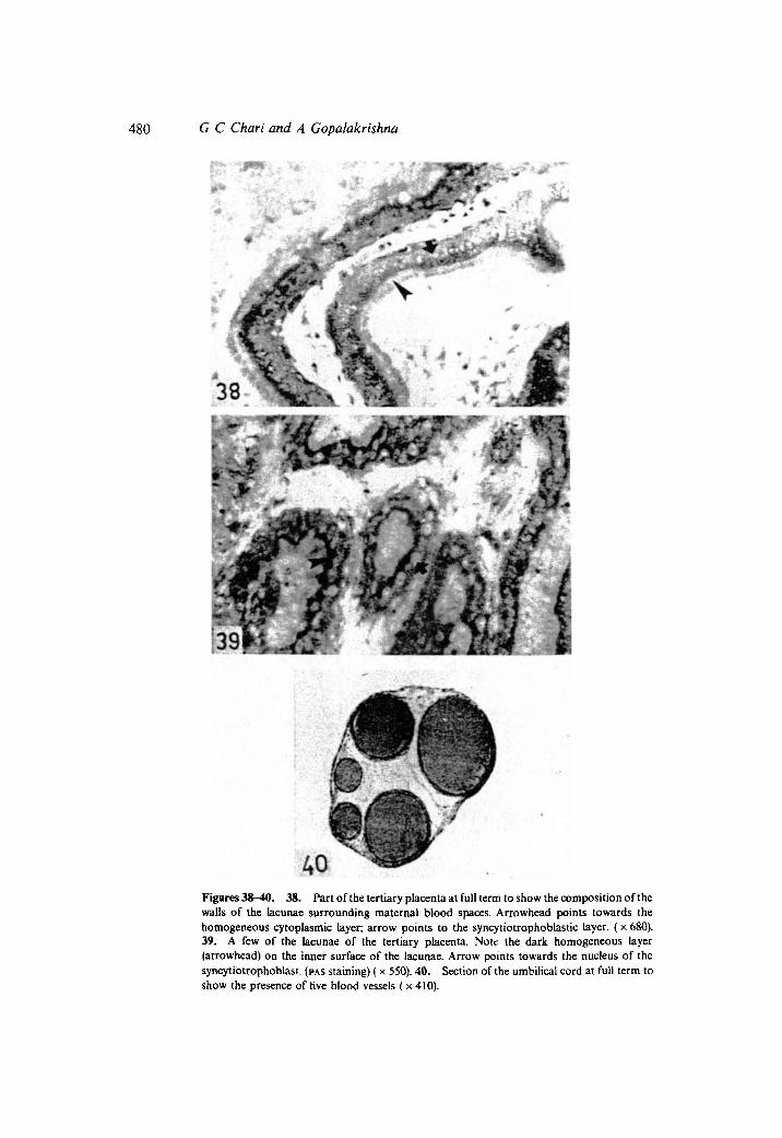
The tertiary placenta lay embedded deep within each secondary placental disc near itsmyometrial border, and consisted of a labyrinth of inter-eonnected large lacunaecoritaining maternal blood (figures 33 and 37). The lacunae were surrounded by thickwalls composed of a layer of darkly staining syncytiotrophoblast with large sphericalnuclei arranged close together in a row (figure 38). the cytotrophoblastic layer wasabsent from the walls of the lacunae. A few lightly staining cells abutting against theouter surface of the syncytiotrophoblastic sheath between the placenta and the thinremnants of endometrium at the utero-placental junction, and which were continuouswith the cells of the periplacental layer of trophoblast, were the only remnants ofcytotrophoblast. However, no cytotrophoblast was present in the lacunar system of thetertiary placenta. Foetal mesenchyme and foetal blood capillaries occurred in theregions between the walls ofthe adjacent lacunae. In sections stained by PAs-procedure,a dark, thick, scarlet, homogeneous layer became evident in the inner surface of thelacunae (figure 39). This layer is also intensely eosinophilic.
The umbilical cord was about 2 em long and was gently twisted two or three timesalong its length. It was inserted to the region of the primary placenta and broke up intotwo main branches, one to each secondary placental disc, and to a few small branchesirrigating the remnants of the primary placenta. Sections of the umbilical cord (figure40) revealed that there was no remnant of the allantoic duct. There were five bloodvessels comprising of a vitelline artery, a vitelline vein, two allantoic arteries and anallantoic vein, the last being the widest vessel in the umbilical cord.
Four to five large maternal blood vessels coming from the uterine wall pass throughthe entire thickness of each secondary placental disc and after reaching the foetalborder of the disc, broke into several branches which raidated in all directions along thefoetal surface of the placental disc. Blood from the radial vessels trickled into theplacental tubules through fine ostia all along their length. Blood from the placental

Morphogenesis offoetal membranes in a bat 479
Figures 33-37. 33. Section showing the relationship between the secondary placental discand the tertiary placenta at full term. Please see text for description ( x 34).34. Part of thesecondary placenta at full term to show its labyrinthine nature ( x 60).35. Cut ends of a fewtubules of secondary placenta at full term. Arrow points to the endothelial cellsof the maternalcapillary inside the tubule (x 210).36. Part of the secondary and tertiary placentae at fullterm. Arrow points to a large pool of maternal blood at the junction between the secondaryand tertiary placentae ( x 100). 37. Part of tertiary placenta at full term. Please see text fordescription (x 130).
480 G C Chari and A Gopalakrishna
40Figures 3s-40. 38. Part of the tertiary placenta at full term to show the composition of thewalls of the lacunae surrounding maternal blood spaces. Arrowhead points towards thehomogeneous cytoplasmic layer; arrow points to the syncytiotrophoblastic layer. (x 680).39. A few of the lacunae of the tertiary placenta. Note the dark homogeneous layer(arrowhead) on the inner surface of the lacunae. Arrow points towards the nucleus of thesyncytiotrophoblast. (PAS staining) ( x 550).40. Section of the umbilical cord at full term toshow the presence of five blood vessels (x 410).

Morphogenesis offoetal membranes in a bat 481
tubules collected into large pools (mentioned earlier) in the junction between thesecondary placenta and the tertiary placenta and then passed into the lacunar system ofthe tertiary placenta. Blood from the tertiary placenta returned to efferent maternalveins in the junctional zone between the placenta and the myometrium. It was evidentthat there were two places in the placental disc where maternal blood accumulatedbefore it returned to the maternal circulation. Perhaps, the formation of these twopools of maternal blood in the placenta is an adaptation in this bat to retain maternalblood in the placenta as long as possible so that maximum physiological exchange cantake place.
The parietal layer ofchorion, which lay closely abutting against the lateral wall of theuterus, consisted of hypertrophied tall columnar cells with lightly staining sphericalnuclei and a thin layer of extra-embryonic mesoderm on its foetal surface. Thetrophoblastic layer of the parietal chorion was in continuity with the trophoblasticlayer of the abembryonic bilaminar omphalopleure and also with the periplacentaltrophoblastic layer between the placenta and the myometrium.
4. Discussion
The foregoing study has revealed that Miniopterus schreibersii fuliginosus presentssome unique embryological features. It differs from all other members of the familyVespertilionidae (in which Miniopterus has been included so far) in the circumferentialattachment of the embryo to the uterine wall resulting in the obliteration of the uterinelumen at the level of gestation, whereas in all other vespertilionids so far studied theembryo is attached to the antimesometrial side ofthe uterus leaving a part of the uterinelumen persisting on the mesometrial side (Duval 1894-1896; Ramaswami 1933;Wimsatt 1944, 1945; Gopalakrishna 1949, 1950; Phansalkar 1972; Gopalkrishna andSapkal 1974; Luckett 1979).
The literature on the development of the amnion in bats has been reviewed by severalworkers (Gopalakrishna 1949; Wimsatt 1954; Gopalakrishna and Khaparde 1978;Gopalakrishna and Karim 1979, 1980; Jeevaji 1979). Amniogenesis in M. schreibersiifuliginosus appears to be unusual. While the primitive amniotic cavity arises as a smallspace within the embryonic mass (Chari and Gopalakrishna 1981)as in all other bats(Wimsatt 1944;Gopalakrishna 1949; Bhiwgade 1976;Gopalakrishna and Karim 1979,1980; Jeevaji 1979; Luckett 1979) the rapid expansion of the embryonic disc tears theroof of the primitive amniotic cavity, and, since the primitive amniotic cavity is roofedby the trophoblastic layer overlying the embryonic disc. the precocious loss of this layerbefore the trophoblast proliferates and invades the endometrium results in exposingthe embryonic plate to the superficial layer of uterine endometrium, the uterineepithelium having disappeared earlier. The amniotic cavity, therefore, becomesconfluent with the potential uterine lumen until the definitive amniotic folds developand fuse on the dorsal aspect of the embryonic plate. A parallel situation has not beennoticed in any other bat except perhaps in Eumops sp (Hamlett 1947) in which theamniotic cavity. was reported to be roofed over by the superficial layer of uterineendometrium for a short period.
The most unique feature of the embryology of this bat is the mode of developmentand the ultimate structure of the chorio-allantoic placenta. The early stages ofdevelopment ofthe placenta are similar to those ofother bats so far described (Wimsatt
A-1

482 G C Chari and A Gopalakrishna
1945, 1954, 1958; Gopalakrishna 1950, 1958; Gopalakrishna and Moghe 1960;Phansalkar 1972; Sapkal 1973). But the later development of the chorio-allantoicplacenta with the development of the secondary and the tertiary placentae in an almostchronological sequence and their ultimate structure are unmatched by any mammal, letalone any bat. The primary chorio-allantoic placenta is progressively replaced by twosecondary placental discs, and the tertiary placenta is developed within each of thesecondary placental discs. The secondary chorio-allantoic placenta is typicallylabyrinthine, and endotheliochorial (Vasochorial) and the tertiary chorio-allantoicplacenta, which has no parallel in any other mammal, should be considered ashaemomomochorial since only the syncytiotrophoblast is present in the walls of thelacunar system of the tertiary placenta.
The placenta of Miniopterus schreibersii of Europe was described by Branca (1927),Grosser (1927), Kempermann (1929) and more recently by Mallassine (1970). Fromthese descriptions it is evident that each author described only the histology of thechorio-allantoic placenta at one or two stages of development of this animal. None ofthe authors, however, gavea complete description of the gestation sac they studied nordid any earlier worker describe the full term placenta of this animal. From thedescriptions of Branca (1927) and Kempermann (1929) it is evident that they weredescribing the pregnant uterus a little earlier than mid-gestation. Apparently,Kempermann's (1929) "Haupt Placenta" refers to the secondary placenta describedhere. Malassine's (1970) description also appears to refer to a stage corresponding toabout mid-pregnancy before the development of the tertiary placenta. His descriptionspertain only to the histological structures of the secondary placenta (which he called"Disque placentarie") corresponding to a stage a little before mid-gestation. None ofthe authors knew about the development of the tertiary placenta. It is most unlikelythat the structure of the placenta of Miniopterus schreibersii of Europe can be sodifferent from that ofthe Indian sub-species ofM iniopterus schreibersii. The differencesbetween the descriptions of the earlier authors and those of the present are due to thefact that the earlier descriptions were based on the examination of the placenta eitherbefore or at about mid-pregnancy. Hence, the earlier authors missed to notice theuniqueness of the development and the histological structure of the definitive placentaof Miniopterus schreibersii.
References
Bhiwgade D A 1976Observations on some early stages ofthe development and implantation ofthe blastocystof Rhinolophus rouxi [Temminck); Proc. Indian Acad. Sci. (Anini. ScL) 84 201-209
Branca A 1927 Recherche sur la placentation des Chairopteres; Arch. Zool. Exp. 'Gen. 66 291-450Chari G C and Gopalakrishna A 1981 Transuterine migration of the embryo and implantation in the
vespertilionid bat, Miniopterus schreibersiifuliqinosus (Hodgson); Proc. First Eur. Symp. on bat Research3-7
Da Costa A C 1920Sur la formation de l'Amnios chez les cheiropteres (Miniopterus schreibersii)et en generalchez les mammaferes; Mem. Soc. Port.; Sci. Nat. Bioi. Ser. 3 1-51
Duval M 1894 Etudes sur I'Embryologie des Cheiropteres; J. Anat. Physiol. 31 93-160 and 427-474Duval M 1895 Etudes sur I'Embryologie des Cheiropteres; J. Anat. Physiol. 32 105--164,420-464Duval M 18% Etudes sur l'Embryologie des Cheiropteres. J Anat. Physiol. 331-31Gopalakrishna A 1949 Studies on the embryology of Microchiroptera-Part IV-an analysis of
implantation and early development in Scotophilus wroughtoni (Thomas); Proc. Indian Acad. Sci. (Anim.Sci.) 30 226-242
Morphogenesis offoetal membranes in a bat 483
Gopalakrishna A 1950 Studies on the embryology of Microchiroptera-Part V-placentation in thevespertilionid bat, Scotophi/us wroughtoni (Thomas); Proc. Indian Acad. Sci. (Anim. Sci.) 31 235--251
Gopalakrishna A 1958 Foetal membranes in some Indian Microchiroptera; J. Morphol. 102 157-197Gopalakrishna A and Karim K B 1979 Foetal membranes and placentation in Chiroptera; J. Reprod. Fertil.
56 417-429Gopalakrishna A and Karim K B 1980Female genital anatomy and the morphogenesis offoetal membranes
of Chiroptera and their bearing on the phylogenetic relationships of the group; Natl. Acad. Sci. India;Golden Jubilee Comm. Vol. 1980379-428
Gopalakrishna A and Khaparde M S 1978 Development of the foetal membranes and placentation in theIndian false vampire bat Megadarma lyra lyra (Geoffroy); Proc.Indian Acad. Sci. (Anim. Sci.) 87 179-194
Gopalakrishna A and Moghe M A 1960 Development of the foetal membranes in the Indian leaf-nosed bat,Hipposideros bicolor pallidus; Z. Anat. Enwickelungsgesch. 122137-149
Gopalakrishna A and Sapkal V M 1974 Foetal membranes in the Indian pipstrelle, Pipstrellus dormeri(Dobson) (Mammalia Chiroptera); J. Zool. Soc. India 26 1-9
Grosser D 1927 Fruhentwicklug, Eihautbildung and Placentation des Menschen und der Saugetiere."Deutsch Frauenheilk", von z. Opitz, Bergmann, Munchen 5
Hamlett G W D 1947 Embryology of the molossid bat Eumops sp; Anat. Rec. 97 304-341Jeevaji I H 1979 Early development and implantation of the blastocyst in the bat, Hipposideros speoris
(Schneider)-Hipposideridae-Microchiroptera; Proc. Indian Acad. Sci. (Anim. Sci.) 88 287-297Kempermann C T 1929 Die Placenta der FIedermaus Miniopterus Schreibersii und ihre functionel1e
Bedeutung; Z. Anat. Entwicklungsgesch. 91 292-303Luckett W P 1979The use of foetal membrane data in assessing Chiropteran phylogeny Proc. Fifth lnt., Bat
Res. Congr. 245--265Malassine A 1970 Etude histologique et ultra-structurale due disque placentaire de Minioptere, Miniopterus
schreibersii (Chiroptere); Sa Nature endotheliochoriale, Son carectere endocrine; Arch. Anat. Microsc.Morphol. Exp. 5999-112
Pearse AGE 1968 Histochemistry-Theoretical and applied (London: J and A Churchill Ltd)Phansalkar R B 1972 Early development and placentation in the vespertilionid bat, pipistrellus ceylonicus
chrysothrix (Ktoughton) Unpublished Ph.D. thesis, Nagpur University, NagpurRamaswami L S 1933 Some stages of the placentation in Vesperugo leisleri (Kuhl); J. Mysore Univ. 7 1-41Sapkal V M 1973Early development, development ofthefoetal membranes andplacentation in the vespertiliollid
bat, Pipstrellus dormeri (Dobson) Unpublished Ph.D. thesis, Nagpur University, NagpurWimsattW A 1944An analysis ofimplantation in the bat, Myotislucifuguslucifugus;Am. J. Anat. 74355--411Wimsatt W A 1945The placentation in the vespertilionid bat, Myotia lucifugus lucifuqus. Am. J. Anat. 771-51Wimsatt W A 1954 The foetal membranes and placentation of the tropical American vampire bat, Desmodus
rotundusmurinus; Acta A.MI. 11 285-341Wimsatt W A 1958 The allantoic placental barrier in Chiroptera. A new concept of its organisation and
histochemistry; Acta AMt. 32 141-186
Copyright © 2022 FDOKUMEN