A tension-induced mechanotransduction pathway promotes epithelial morphogenesis
7
LETTER doi:10.1038/nature09765 A tension-induced mechanotransduction pathway promotes epithelial morphogenesis Huimin Zhang 1 , Fre ´de ´ric Landmann 1 {*, Hala Zahreddine 1 *, David Rodriguez 1 , Marc Koch 2 & Michel Labouesse 1 Mechanotransduction refers to the transformation of physical forces into chemical signals. It generally involves stretch-sensitive channels or conformational change of cytoskeleton-associated proteins 1 . Mechanotransduction is crucial for the physiology of several organs and for cell migration 2,3 . The extent to which mech- anical inputs contribute to development, and how they do this, remains poorly defined. Here we show that a mechanotransduction pathway operates between the body-wall muscles of Caenorhabditis elegans and the epidermis. This pathway involves, in addition to a Rac GTPase, three signalling proteins found at the hemidesmo- some: p21-activated kinase (PAK-1), the adaptor GIT-1 and its partner PIX-1. The phosphorylation of intermediate filaments is one output of this pathway. Tension exerted by adjacent muscles or externally exerted mechanical pressure maintains GIT-1 at hemi- desmosomes and stimulates PAK-1 activity through PIX-1 and Rac. This pathway promotes the maturation of a hemidesmosome into a junction that can resist mechanical stress and contributes to co- ordinating the morphogenesis of epidermal and muscle tissues. Our findings suggest that the C. elegans hemidesmosome is not only an attachment structure, but also a mechanosensor that responds to tension by triggering signalling processes. We suggest that similar pathways could promote epithelial morphogenesis or wound healing in other organisms in which epithelial cells adhere to tension-generating contractile cells. Most organs and complex tissues contain several cell types, all of which can contribute to define organ or tissue shape. The way in which different cell types communicate during morphogenesis is poorly understood. In C. elegans, the epidermis and muscles both guide embryonic elongation 4 . The role of epidermal cells in elongation is well defined 4 . By contrast, our understanding of how muscles affect elongation and interact with the epidermis remains vague. Mutants with defective muscles arrest midway through elongation at a stage known as two-fold, and this phenotype is called Pat (paralysed at two- fold) 5 . Communication between muscles and the epidermis could be channelled through junctions that attach the epidermis to the extra- cellular matrix at the muscle–epidermis interface. These junctions fasten muscles to the exoskeleton and are essential for elongation 6 (Fig. 1a). Each junction includes two hemidesmosome-like units at the apical and basal epidermis plasma membranes, with intermediate filaments in between 6 (Supplementary Fig. 1a). Hereafter, we refer to hemidesmosome-like junctions as CeHDs (C. elegans hemidesmo- somes). The physiological role of CeHDs led us to consider whether muscles could signal to the epidermis through a mechanical input. We thus examined whether muscle contractions mechanically modify the epidermis, and we searched for CeHD proteins that respond to this mechanical change. If muscles deform the epidermis, their contractions should modify the relative positions of two points within the epidermis. We tested this possibility using the actin bundles anchored to the plasma membrane 7 as spatial landmarks, measuring the distance between bundles when muscles become active (Fig. 1b–d). Kymographs show that muscle contractions reduced this distance by about 50%, because the reduc- tion in distance was abolished in muscle-defective embryos (Fig. 1c, d and Supplementary Movies 1 and 2). Thus, muscle contractions laterally stretch and squeeze the epidermis, a process comparable to the stretching of cultured cells grown on elastic membranes 8,9 . Disruption of the CeHD core component, VAB-10A 10 (a plectin and BPAG1e homologue), also strongly compromised this process (Fig. 1c, d and Supplementary Movie 3), outlining the crucial role of CeHDs in transmitting muscle tension. Moreover, consistent with earlier find- ings suggesting that muscles help the patterning of CeHDs 5,11 , muscles promoted the maturation of CeHDs from an initial punctate distri- bution (Fig. 1e, f) to short parallel circumferential stripes (Fig. 1g, h), co-localizing with epidermal actin bundles (Supplementary Fig. 1d). CeHD structure was initially normal in embryos with defective myo- filaments, but the reorganization of CeHDs was abnormal in the absence of muscle tension (Supplementary Fig. 1b, c). To identify epidermal proteins that are activated by tension, we relied on a recent genetic screen that identified 14 genes whose knock- down—combined with a weak mutation in vab-10, called vab- 10a(e698) (Fig. 2a)—affects CeHD biogenesis 12 . Among these genes, we focused on the signalling molecule PAK-1 (Fig. 2b), because its mammalian homologues control the cytoskeleton and can relay changes in arterial pressure to activate downstream signalling 13,14 . We found that PAK-1 distribution coincides with intermediate- filament proteins at all stages of development, reorganizing into short parallel stripes typical of CeHDs (Fig. 2c–e and Supplementary Fig. 2a–c). PAK-1 was enriched at basal CeHDs marked by LET-805 (also known as myotactin), although it was also present at apical CeHDs (Supplementary Fig. 2d–g). Lack of PAK-1 function affected embryonic elongation, reducing body length by 19% (Supplementary Fig. 2h, i, l). Consistent with PAK-1 presence at CeHDs, the kinase-domain dele- tion mutant pak-1(ok448) (Fig. 2b), combined with the weak viable mutation vab-10A(e698), affected CeHD integrity. In these vab- 10A(e698); pak-1(ok448) double mutants, staining for VAB-10A showed a failure to form stripes in many areas (arrow in Fig. 2n) or less staining where muscles had detached from the body wall (arrowhead in Fig. 2n). As a result, more than 60% of these double mutants showed muscle detachment, which was associated with elongation arrest (Fig. 2k and Supplementary Table 1), and this was not seen in either single mutant (Fig. 2f, g, i, j and Supplementary Table 1). VAB-10A distribution became abnormal in vab-10A(e698); pak-1(ok448) double mutants after the 1.7-fold stage (Fig. 2h), when muscles start to contract, suggesting that CeHDs cannot maintain their integrity when exposed to muscle-induced tension. Taken together, these findings indicate that PAK-1 functions with VAB-10A to strengthen CeHD stability. Next, we investigated how PAK-1 helps the assembly of CeHDs. In vitro studies established that vertebrate PAK1 phosphorylates the *These authors contributed equally to this work. 1 Development and Stem Cells Program, IGBMC, CNRS (UMR7104), INSERM (U964), Universite ´ de Strasbourg, 1 rue Laurent Fries, BP10142, 67400 Illkirch, France. 2 Imaging Centre, IGBMC, CNRS (UMR7104), INSERM (U964), Universite ´ de Strasbourg, 1 rue Laurent Fries, BP10142, 67400 Illkirch, France. {Present address: MCBD Department, University of California, Santa Cruz, California 95064, USA. 3 MARCH 2011 | VOL 471 | NATURE | 99 Macmillan Publishers Limited. All rights reserved ©2011
-
Upload
independent -
Category
Documents
-
view
1 -
download
0
Transcript of A tension-induced mechanotransduction pathway promotes epithelial morphogenesis

LETTERdoi:10.1038/nature09765
A tension-induced mechanotransduction pathwaypromotes epithelial morphogenesisHuimin Zhang1, Frederic Landmann1{*, Hala Zahreddine1*, David Rodriguez1, Marc Koch2 & Michel Labouesse1
Mechanotransduction refers to the transformation of physicalforces into chemical signals. It generally involves stretch-sensitivechannels or conformational change of cytoskeleton-associatedproteins1. Mechanotransduction is crucial for the physiology ofseveral organs and for cell migration2,3. The extent to which mech-anical inputs contribute to development, and how they do this,remains poorly defined. Here we show that a mechanotransductionpathway operates between the body-wall muscles of Caenorhabditiselegans and the epidermis. This pathway involves, in addition to aRac GTPase, three signalling proteins found at the hemidesmo-some: p21-activated kinase (PAK-1), the adaptor GIT-1 and itspartner PIX-1. The phosphorylation of intermediate filaments isone output of this pathway. Tension exerted by adjacent musclesor externally exerted mechanical pressure maintains GIT-1 at hemi-desmosomes and stimulates PAK-1 activity through PIX-1 and Rac.This pathway promotes the maturation of a hemidesmosome into ajunction that can resist mechanical stress and contributes to co-ordinating the morphogenesis of epidermal and muscle tissues.Our findings suggest that the C. elegans hemidesmosome is notonly an attachment structure, but also a mechanosensor thatresponds to tension by triggering signalling processes. We suggestthat similar pathways could promote epithelial morphogenesis orwound healing in other organisms in which epithelial cells adhere totension-generating contractile cells.
Most organs and complex tissues contain several cell types, all ofwhich can contribute to define organ or tissue shape. The way in whichdifferent cell types communicate during morphogenesis is poorlyunderstood. In C. elegans, the epidermis and muscles both guideembryonic elongation4. The role of epidermal cells in elongation iswell defined4. By contrast, our understanding of how muscles affectelongation and interact with the epidermis remains vague. Mutantswith defective muscles arrest midway through elongation at a stageknown as two-fold, and this phenotype is called Pat (paralysed at two-fold)5. Communication between muscles and the epidermis could bechannelled through junctions that attach the epidermis to the extra-cellular matrix at the muscle–epidermis interface. These junctionsfasten muscles to the exoskeleton and are essential for elongation6
(Fig. 1a). Each junction includes two hemidesmosome-like units atthe apical and basal epidermis plasma membranes, with intermediatefilaments in between6 (Supplementary Fig. 1a). Hereafter, we refer tohemidesmosome-like junctions as CeHDs (C. elegans hemidesmo-somes). The physiological role of CeHDs led us to consider whethermuscles could signal to the epidermis through a mechanical input. Wethus examined whether muscle contractions mechanically modify theepidermis, and we searched for CeHD proteins that respond to thismechanical change.
If muscles deform the epidermis, their contractions should modifythe relative positions of two points within the epidermis. We tested thispossibility using the actin bundles anchored to the plasma membrane7
as spatial landmarks, measuring the distance between bundles whenmuscles become active (Fig. 1b–d). Kymographs show that musclecontractions reduced this distance by about 50%, because the reduc-tion in distance was abolished in muscle-defective embryos (Fig. 1c, dand Supplementary Movies 1 and 2). Thus, muscle contractionslaterally stretch and squeeze the epidermis, a process comparable tothe stretching of cultured cells grown on elastic membranes8,9.Disruption of the CeHD core component, VAB-10A10 (a plectin andBPAG1e homologue), also strongly compromised this process (Fig. 1c,d and Supplementary Movie 3), outlining the crucial role of CeHDs intransmitting muscle tension. Moreover, consistent with earlier find-ings suggesting that muscles help the patterning of CeHDs5,11, musclespromoted the maturation of CeHDs from an initial punctate distri-bution (Fig. 1e, f) to short parallel circumferential stripes (Fig. 1g, h),co-localizing with epidermal actin bundles (Supplementary Fig. 1d).CeHD structure was initially normal in embryos with defective myo-filaments, but the reorganization of CeHDs was abnormal in theabsence of muscle tension (Supplementary Fig. 1b, c).
To identify epidermal proteins that are activated by tension, werelied on a recent genetic screen that identified 14 genes whose knock-down—combined with a weak mutation in vab-10, called vab-10a(e698) (Fig. 2a)—affects CeHD biogenesis12. Among these genes,we focused on the signalling molecule PAK-1 (Fig. 2b), because itsmammalian homologues control the cytoskeleton and can relaychanges in arterial pressure to activate downstream signalling13,14.We found that PAK-1 distribution coincides with intermediate-filament proteins at all stages of development, reorganizing into shortparallel stripes typical of CeHDs (Fig. 2c–e and Supplementary Fig.2a–c). PAK-1 was enriched at basal CeHDs marked by LET-805 (alsoknown as myotactin), although it was also present at apical CeHDs(Supplementary Fig. 2d–g). Lack of PAK-1 function affected embryonicelongation, reducing body length by 19% (Supplementary Fig. 2h, i, l).
Consistent with PAK-1 presence at CeHDs, the kinase-domain dele-tion mutant pak-1(ok448) (Fig. 2b), combined with the weak viablemutation vab-10A(e698), affected CeHD integrity. In these vab-10A(e698); pak-1(ok448) double mutants, staining for VAB-10Ashowed a failure to form stripes in many areas (arrow in Fig. 2n) or lessstaining where muscles had detached from the body wall (arrowhead inFig. 2n). As a result, more than 60% of these double mutants showedmuscle detachment, which was associated with elongation arrest(Fig. 2k and Supplementary Table 1), and this was not seen in eithersingle mutant (Fig. 2f, g, i, j and Supplementary Table 1). VAB-10Adistribution became abnormal in vab-10A(e698); pak-1(ok448) doublemutants after the 1.7-fold stage (Fig. 2h), when muscles start to contract,suggesting that CeHDs cannot maintain their integrity when exposed tomuscle-induced tension. Taken together, these findings indicate thatPAK-1 functions with VAB-10A to strengthen CeHD stability.
Next, we investigated how PAK-1 helps the assembly of CeHDs. Invitro studies established that vertebrate PAK1 phosphorylates the
*These authors contributed equally to this work.
1Development and Stem Cells Program, IGBMC, CNRS (UMR7104), INSERM (U964), Universite de Strasbourg, 1 rue Laurent Fries, BP10142, 67400 Illkirch, France. 2Imaging Centre, IGBMC, CNRS(UMR7104), INSERM (U964), Universite de Strasbourg, 1 rue Laurent Fries, BP10142, 67400 Illkirch, France. {Present address: MCBD Department, University of California, Santa Cruz, California 95064,USA.
3 M A R C H 2 0 1 1 | V O L 4 7 1 | N A T U R E | 9 9
Macmillan Publishers Limited. All rights reserved©2011

intermediate-filament protein vimentin15. Hence, C. elegans PAK-1might also phosphorylate epidermal intermediate filaments (Sup-plementary Fig. 1a). We directly tested this hypothesis in two ways.First, we used two-dimensional gel analysis of embryonic extractsfollowed by immunoblotting with MH4 monoclonal antibody, whichrecognizes the CeHD proteins IFA-2 (also known as MUA-6) andIFA-3, as well as the non-epidermal protein IFA-1 (ref. 16). Thisrevealed the presence of two major intermediate-filament isoelectricspots, which were not present after phosphatase treatment of extracts(Fig. 3a, arrows) or in pak-1(ok448) extracts (Fig. 3b, arrows). Second,tagging IFA-3, the major intermediate-filament protein in the embry-onic epidermis, with Myc showed that PAK-1 specifically affects phos-phorylation of IFA-3 (Supplementary Fig. 3c). We therefore concludethat PAK-1 indeed affects the phosphorylation of an epidermal inter-mediate-filament protein.
We next assessed the effect of phosphorylation on intermediate-filament organization. Staining vab-10A(e698); pak-1(ok448) double
mutants with the MH4 monoclonal antibody revealed that abnormal,ectopic, intermediate-filament bundles were present outside CeHDs(Fig. 3g, arrow). We also observed this phenotype in combination withthe CRIB domain deletion allele pak-1(tm403) (that is, in vab-10A(e698); pak-1(tm403) double mutants) but not in vab-10A(e698),pak-1(ok448) or pak-1(tm403) single mutants (Fig. 3d–f and Sup-plementary Fig. 4a, b). The ectopic intermediate-filament bundlesseem to result from defective anchoring of intermediate filaments tothe mutant VAB-10A in CeHDs, because tagging the IFA-2/3 hetero-dimer partner IFB-1 with green fluorescent protein (GFP) resulted inthe same ectopic intermediate-filament stripes and muscle detach-ment phenotypes as observed in vab-10A(e698); pak-1(ok448) mutants(Supplementary Fig. 5a–d and Supplementary Table 1). Furthermore,we identified the S470 residue of IFA-3 as an important regulatory site.Changing this serine residue to an alanine abolished IFA-3 phosphor-ylation and disrupted the localization of IFA-3 to CeHDs in the vab-10A(e698) background (Supplementary Fig. 5e–k). Together, thesedata suggest that lack of IFA-3 phosphorylation reduces the recruit-ment of this protein to CeHDs and alters CeHD strength in vab-10A(e698) mutants.
Having established PAK-1 as a functionally important CeHD kinase,we next showed that muscle contractility triggers PAK-1 activity. Weused intermediate-filament phosphorylation and organization as read-outs. We examined two classes of muscle-defective embryo: one lack-ing EGL-19, a Ca21-activated channel that is required for muscle
WT
a D
V
A P
b
Hemidesmosomes
Intermediate
filaments
Muscles
Actin bundles
Adherens
junction
Embryonic sheath/exoskeleton
1
2
3
4
Co
ntr
actio
n (C
/R,
%)
2
vab-10A mutant
WT
unc-112 mutant
Time (s)
10
60
40
20
0
80
3 4
dDistance
Tim
e
unc-112mutant
c
WT
vab-10Amutant
1
2
Dorsal
epidermis
Lateral
epidermis
Ventral
epidermis
4
e
WT early WT early WT late WT late
f g h
VAB-10/Muscles
3
Figure 1 | Muscle tension promotes C. elegans hemidesmosomematuration. a, Schemes showing a C. elegans embryo (top) and a cross-sectionof the embryo (at the level of red lines; bottom) and its hemidesmosomes(CeHDs), numbered 1–4. Three epidermal cell types are found around thecircumference: dorsal and ventral (which uniquely express elt-3); and lateral. A,anterior; D, dorsal; P, posterior; V, ventral. b, Actin bundles (white) in WTembryo imaged by following an actin-binding domain labelled with GFP20. Thedashed box shows the region selected for the kymograph in c. Scale bar, 10mm.c, Kymographs showing the distance change between actin bundles (white) inWT embryos, unc-112(RNAi) mutant embryos (which are muscle deficient)and vab-10A(RNAi) embryos (which are CeHD deficient) (see alsoSupplementary Movies 1–3). Red circles indicate actin-anchoring pointsdisplaced by muscle contractions. C, contracted distance (orange); R, relaxeddistance (green). d, Quantification of tension changes in terms of distance(contracted divided by relaxed) and time span per contraction. Individual datapoints (n 5 15) and mean 6 s.e.m. (black crosses) are shown.e–h, Immunostaining of WT embryos at the 1.5–2-fold stage of development(early; e, f) or the 3–4-fold stage (late; g, h): muscles (red) and VAB-10A(green). Dashed boxes in e and g demarcate the regions shown in f andh, respectively.
VAB-10A/Muscles
vab-10A late pak-1 late
pak-1 late
vab-10A; pak-1 late
vab-10A; pak-1 late
i j k
l m n
f g h
vab-10A early pak-1 early vab-10A; pak-1 early
c d e
PAK-1 IFA Merge
a
CRIB domain Kinase domainPAK-1
tm403 ok448
572 amino acids
GFP
ABDVAB-10A Plakin domain
3436 amino acids
e698
bPlectin repeats
vab-10A late
Figure 2 | PAK-1 function is required for CeHD maturation.a, b, Conserved domains of VAB-10A and PAK-1 proteins. The missensemutation e698 maps to the region predicted to bind to intermediate filaments10.The deletions tm403 and ok448 remove the PAK-1 CRIB domain and kinasedomain, respectively. ABD, actin-binding domain. c–e, Co-localization ofPAK-1 with IFA-2 and IFA-3 in a WT larva, as determined byimmunofluorescence: PAK-1 (green) and IFA (red). Scale bar, 10mm.f–n, Immunostaining for muscle (red) and VAB-10A (green) of vab-10A(e698)(f, i, l), pak-1(ok448) (g, j, m) and vab-10A(e698); pak-1(ok448) (h, k, n) mutantembryos at early or late stages of development. Dashed boxes indicate areashown in panel below. Dashed line in k shows where muscles should be. Arrowin n shows area with muscles still attached. Arrowheads in h and n show areaswith muscles detached. Scale bar, 10mm.
RESEARCH LETTER
1 0 0 | N A T U R E | V O L 4 7 1 | 3 M A R C H 2 0 1 1
Macmillan Publishers Limited. All rights reserved©2011
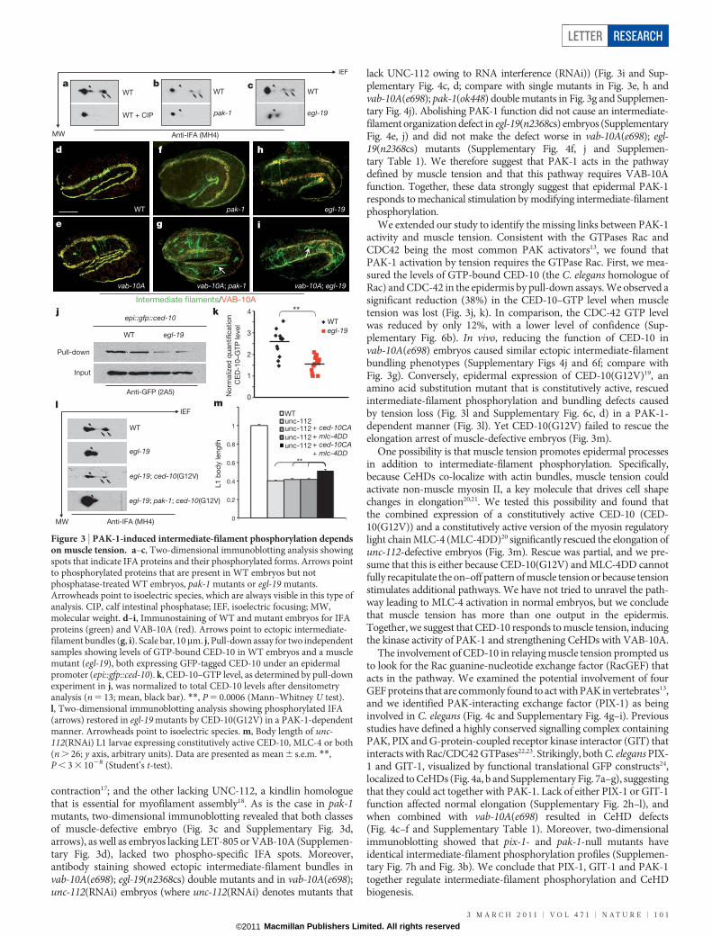
contraction17; and the other lacking UNC-112, a kindlin homologuethat is essential for myofilament assembly18. As is the case in pak-1mutants, two-dimensional immunoblotting revealed that both classesof muscle-defective embryo (Fig. 3c and Supplementary Fig. 3d,arrows), as well as embryos lacking LET-805 or VAB-10A (Supplemen-tary Fig. 3d), lacked two phospho-specific IFA spots. Moreover,antibody staining showed ectopic intermediate-filament bundles invab-10A(e698); egl-19(n2368cs) double mutants and in vab-10A(e698);unc-112(RNAi) embryos (where unc-112(RNAi) denotes mutants that
lack UNC-112 owing to RNA interference (RNAi)) (Fig. 3i and Sup-plementary Fig. 4c, d; compare with single mutants in Fig. 3e, h andvab-10A(e698); pak-1(ok448) double mutants in Fig. 3g and Supplemen-tary Fig. 4j). Abolishing PAK-1 function did not cause an intermediate-filament organization defect in egl-19(n2368cs) embryos (SupplementaryFig. 4e, j) and did not make the defect worse in vab-10A(e698); egl-19(n2368cs) mutants (Supplementary Fig. 4f, j and Supplemen-tary Table 1). We therefore suggest that PAK-1 acts in the pathwaydefined by muscle tension and that this pathway requires VAB-10Afunction. Together, these data strongly suggest that epidermal PAK-1responds to mechanical stimulation by modifying intermediate-filamentphosphorylation.
We extended our study to identify the missing links between PAK-1activity and muscle tension. Consistent with the GTPases Rac andCDC42 being the most common PAK activators13, we found thatPAK-1 activation by tension requires the GTPase Rac. First, we mea-sured the levels of GTP-bound CED-10 (the C. elegans homologue ofRac) and CDC-42 in the epidermis by pull-down assays. We observed asignificant reduction (38%) in the CED-10–GTP level when muscletension was lost (Fig. 3j, k). In comparison, the CDC-42 GTP levelwas reduced by only 12%, with a lower level of confidence (Sup-plementary Fig. 6b). In vivo, reducing the function of CED-10 invab-10A(e698) embryos caused similar ectopic intermediate-filamentbundling phenotypes (Supplementary Figs 4j and 6f; compare withFig. 3g). Conversely, epidermal expression of CED-10(G12V)19, anamino acid substitution mutant that is constitutively active, rescuedintermediate-filament phosphorylation and bundling defects causedby tension loss (Fig. 3l and Supplementary Fig. 6c, d) in a PAK-1-dependent manner (Fig. 3l). Yet CED-10(G12V) failed to rescue theelongation arrest of muscle-defective embryos (Fig. 3m).
One possibility is that muscle tension promotes epidermal processesin addition to intermediate-filament phosphorylation. Specifically,because CeHDs co-localize with actin bundles, muscle tension couldactivate non-muscle myosin II, a key molecule that drives cell shapechanges in elongation20,21. We tested this possibility and found thatthe combined expression of a constitutively active CED-10 (CED-10(G12V)) and a constitutively active version of the myosin regulatorylight chain MLC-4 (MLC-4DD)20 significantly rescued the elongation ofunc-112-defective embryos (Fig. 3m). Rescue was partial, and we pre-sume that this is either because CED-10(G12V) and MLC-4DD cannotfully recapitulate the on–off pattern of muscle tension or because tensionstimulates additional pathways. We have not tried to unravel the path-way leading to MLC-4 activation in normal embryos, but we concludethat muscle tension has more than one output in the epidermis.Together, we suggest that CED-10 responds to muscle tension, inducingthe kinase activity of PAK-1 and strengthening CeHDs with VAB-10A.
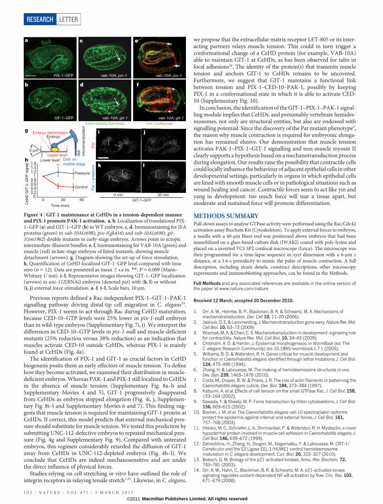
The involvement of CED-10 in relaying muscle tension prompted usto look for the Rac guanine-nucleotide exchange factor (RacGEF) thatacts in the pathway. We examined the potential involvement of fourGEF proteins that are commonly found to act with PAK in vertebrates13,and we identified PAK-interacting exchange factor (PIX-1) as beinginvolved in C. elegans (Fig. 4c and Supplementary Fig. 4g–i). Previousstudies have defined a highly conserved signalling complex containingPAK, PIX and G-protein-coupled receptor kinase interactor (GIT) thatinteracts with Rac/CDC42 GTPases22,23. Strikingly, both C. elegans PIX-1 and GIT-1, visualized by functional translational GFP constructs24,localized to CeHDs (Fig. 4a, b and Supplementary Fig. 7a–g), suggestingthat they could act together with PAK-1. Lack of either PIX-1 or GIT-1function affected normal elongation (Supplementary Fig. 2h–l), andwhen combined with vab-10A(e698) resulted in CeHD defects(Fig. 4c–f and Supplementary Table 1). Moreover, two-dimensionalimmunoblotting showed that pix-1- and pak-1-null mutants haveidentical intermediate-filament phosphorylation profiles (Supplemen-tary Fig. 7h and Fig. 3b). We conclude that PIX-1, GIT-1 and PAK-1together regulate intermediate-filament phosphorylation and CeHDbiogenesis.
0
1
2
3
4
WT
egl-19
IEF
MW
egl-19; pak-1; ced-10(G12V)
egl-19
WT
egl-19; ced-10(G12V)
Anti-GFP (2A5)
epi::gfp::ced-10
Pull-down
Input
WT egl-19
No
rmaliz
ed
quantificatio
n
CE
D-1
0–G
TP
level
j k
l
Intermediate filaments/VAB-10A
d f h
e
WT pak-1 egl-19
vab-10A vab-10A; pak-1 vab-10A; egl-19
IEF
MW
WT
pak-1
bWT
egl-19
WT
WT + CIP
a c
Anti-IFA (MH4)
**
m
L1 b
od
y leng
th
0
0.2
0.4
0.6
0.8
1
**
Anti-IFA (MH4)
g i
WT
unc-112 + ced-10CAunc-112
unc-112 + mlc-4DDunc-112 + ced-10CA
+ mlc-4DD
Figure 3 | PAK-1-induced intermediate-filament phosphorylation dependson muscle tension. a–c, Two-dimensional immunoblotting analysis showingspots that indicate IFA proteins and their phosphorylated forms. Arrows pointto phosphorylated proteins that are present in WT embryos but notphosphatase-treated WT embryos, pak-1 mutants or egl-19 mutants.Arrowheads point to isoelectric species, which are always visible in this type ofanalysis. CIP, calf intestinal phosphatase; IEF, isoelectric focusing; MW,molecular weight. d–i, Immunostaining of WT and mutant embryos for IFAproteins (green) and VAB-10A (red). Arrows point to ectopic intermediate-filament bundles (g, i). Scale bar, 10mm. j, Pull-down assay for two independentsamples showing levels of GTP-bound CED-10 in WT embryos and a musclemutant (egl-19), both expressing GFP-tagged CED-10 under an epidermalpromoter (epi::gfp::ced-10). k, CED-10–GTP level, as determined by pull-downexperiment in j, was normalized to total CED-10 levels after densitometryanalysis (n 5 13; mean, black bar). **, P 5 0.0006 (Mann–Whitney U test).l, Two-dimensional immunoblotting analysis showing phosphorylated IFA(arrows) restored in egl-19 mutants by CED-10(G12V) in a PAK-1-dependentmanner. Arrowheads point to isoelectric species. m, Body length of unc-112(RNAi) L1 larvae expressing constitutively active CED-10, MLC-4 or both(n . 26; y axis, arbitrary units). Data are presented as mean 6 s.e.m. **,P , 3 3 1028 (Student’s t-test).
LETTER RESEARCH
3 M A R C H 2 0 1 1 | V O L 4 7 1 | N A T U R E | 1 0 1
Macmillan Publishers Limited. All rights reserved©2011
Previous reports defined a Rac-independent PIX-1–GIT-1–PAK-1signalling pathway driving distal-tip cell migration in C. elegans24.However, PIX-1 seems to act through Rac during CeHD maturation,because CED-10–GTP levels were 25% lower in pix-1-null embryosthan in wild-type embryos (Supplementary Fig. 7i, j). We interpret thedifferences in CED-10–GTP levels in pix-1-null and muscle-deficientmutants (25% reduction versus 38% reduction) as an indication thatmuscles activate CED-10 outside CeHDs, whereas PIX-1 is mainlyfound at CeHDs (Fig. 4a).
The identification of PIX-1 and GIT-1 as crucial factors in CeHDbiogenesis posits them as early effectors of muscle tension. To definehow they become activated, we examined their distribution in muscle-deficient embryos. Whereas PAK-1 and PIX-1 still localized to CeHDsin the absence of muscle tension (Supplementary Fig. 8a–h andSupplementary Movies 4 and 5), GIT-1 progressively disappearedfrom CeHDs as embryos stopped elongation (Fig. 4i, j, Supplemen-tary Fig. 8i–l and Supplementary Movies 6 and 7). This finding sug-gests that muscle tension is required for maintaining GIT-1 protein atCeHDs. If correct, this model predicts that external mechanical pres-sure should substitute for muscle tension. We tested this prediction bysubmitting UNC-112-defective embryos to repeated mechanical pres-sure (Fig. 4g and Supplementary Fig. 9). Compared with untreatedembryos, this regimen considerably retarded the diffusion of GIT-1away from CeHDs in UNC-112-depleted embryos (Fig. 4h–l). Weconclude that CeHDs are indeed mechanosensitive and are underthe direct influence of physical forces.
Studies relying on cell stretching in vitro have outlined the role ofintegrin receptors in relaying tensile stretch1,25. Likewise, in C. elegans,
we propose that the extracellular matrix receptor LET-805 or its inter-acting partners relays muscle tension. This could in turn trigger aconformational change of a CeHD protein (for example, VAB-10A)able to maintain GIT-1 at CeHDs, as has been observed for talin infocal adhesions26. The identity of the protein(s) that transmits muscletension and anchors GIT-1 to CeHDs remains to be uncovered.Furthermore, we suggest that GIT-1 maintains a functional linkbetween tension and PIX-1–CED-10–PAK-1, possibly by keepingPIX-1 in a conformational state in which it is able to activate CED-10 (Supplementary Fig. 10).
In conclusion, the identification of the GIT-1–PIX-1–PAK-1 signal-ling module implies that CeHDs, and presumably vertebrate hemides-mosomes, not only are structural entities, but also are endowed withsignalling potential. Since the discovery of the Pat mutant phenotype5,the reason why muscle contraction is required for embryonic elonga-tion has remained elusive. Our demonstration that muscle tensionactivates PAK-1–PIX-1–GIT-1 signalling and non-muscle myosin IIclearly supports a hypothesis based on a mechanotransduction processduring elongation. Our results raise the possibility that contractile cellscould locally influence the behaviour of adjacent epithelial cells in otherdevelopmental settings, particularly in organs in which epithelial cellsare lined with smooth muscle cells or in pathological situations such aswound healing and cancer. Contractile forces seem to act like yin andyang in development: too much force will tear a tissue apart, butmoderate and sustained force will promote differentiation.
METHODS SUMMARYPull-down assays to analyse GTPase activity were performed using the Rac/Cdc42activation assay Biochem Kit (Cytoskeleton). To apply external forces to embryos,a needle with a 40-mm blunt end was positioned above embryos that had beenimmobilized on a glass-based culture dish (IWAKI) coated with poly-lysine andplaced on a inverted TCS SP2 confocal microscope (Leica). The microscope wasthen programmed for a time-lapse sequence in xyzt dimension with a 6-mm zdistance, at a 1.6-s periodicity to mimic the pulse of muscle contraction. A fulldescription, including strain details, construct descriptions, other microscopyexperiments and immunoblotting approaches, can be found in the Methods.
Full Methods and any associated references are available in the online version ofthe paper at www.nature.com/nature.
Received 12 March; accepted 20 December 2010.
1. Orr, A. W., Helmke, B. P., Blackman, B. R. & Schwartz, M. A. Mechanisms ofmechanotransduction. Dev. Cell 10, 11–20 (2006).
2. Jaalouk, D. E. & Lammerding, J. Mechanotransduction gone awry. Nature Rev. Mol.Cell Biol. 10, 63–73 (2009).
3. Wozniak, M. A. & Chen, C. S. Mechanotransduction in development: a growing rolefor contractility. Nature Rev. Mol. Cell Biol. 10, 34–43 (2009).
4. Chisholm, A. D. & Hardin, J. Epidermal morphogenesis. in WormBook (ed. TheC. elegans Research Community) doi:10.1895/wormbook.1.7.1 (2005).
5. Williams, B. D. & Waterston, R. H. Genes critical for muscle development andfunction in Caenorhabditis elegans identified through lethal mutations. J. Cell Biol.124, 475–490 (1994).
6. Zhang, H. & Labouesse, M. The making of hemidesmosome structures in vivo.Dev. Dyn. 239, 1465–1476 (2010).
7. Costa, M., Draper, B. W. & Priess, J. R. The role of actin filaments in patterning theCaenorhabditis elegans cuticle. Dev. Biol. 184, 373–384 (1997).
8. Katsumi, A. et al. Effects of cell tension on the small GTPase Rac. J. Cell Biol. 158,153–164 (2002).
9. Sawada, Y. & Sheetz, M. P. Force transduction by triton cytoskeletons. J. Cell Biol.156, 609–615 (2002).
10. Bosher, J. M. et al. The Caenorhabditis elegans vab-10 spectraplakin isoformsprotect the epidermis against internal and external forces. J. Cell Biol. 161,757–768 (2003).
11. Hresko, M. C., Schriefer, L. A., Shrimankar, P. & Waterston, R. H. Myotactin, a novelhypodermal protein involved in muscle-cell adhesion in Caenorhabditis elegans. J.Cell Biol. 146, 659–672 (1999).
12. Zahreddine, H., Zhang, H., Diogon, M., Nagamatsu, Y. & Labouesse, M. CRT-1/Calreticulin and the E3 Ligase EEL-1/HUWE1 control hemidesmosomematuration in C. elegans development. Curr. Biol. 20, 322–327 (2010).
13. Bokoch, G. M. Biology of the p21-activated kinases. Annu. Rev. Biochem. 72,743–781 (2003).
14. Orr, A. W., Hahn, C., Blackman, B. R. & Schwartz, M. A. p21-activated kinasesignaling regulates oxidant-dependent NF-kB activation by flow. Circ. Res. 103,671–679 (2008).
00.20.40.60.8
11.21.41.6
PIX-1–GFP vab-10A; pix-1
GIT-1–GFP
Intermediate filaments VAB-10A/Muscles
vab-10A; pix-1
vab-10A; git-1 vab-10A; git-1
a c e
b d f
Pat
+ p
ressure
Pat
0 min 30 min
GIT-1–GFP
i j
k l
6 μm
CeH
D G
IT-1
::GFP
sig
nal
g
h
Pressure
Control
0 30 60
Time (min)
**
Lower level
Upper level
Embryo deformationEmbryo
Still
mic
rom
anip
ula
tor
Dish on mobile stage
Figure 4 | GIT-1 maintenance at CeHDs in a tension-dependent mannerand PIX-1 promote PAK-1 activation. a, b, Localization of translational PIX-1–GFP (a) and GIT-1–GFP (b) in WT embryos. c, d, Immunostaining for IFAproteins (green) in vab-10A(e698); pix-1(gk416) and vab-10A(e698); git-1(tm1962) double mutants in early-stage embryos. Arrows point to ectopicintermediate-filament bundles. e, f, Immunostaining for VAB-10A (green) andmuscle (red) in late-stage embryos of listed mutants, showing muscledetachment (arrows). g, Diagram showing the set-up of force stimulation.h, Quantification of CeHD-localized GIT-1–GFP level compared with timezero (n 5 12). Data are presented as mean 6 s.e.m. **, P 5 0.009 (Mann–Whitney U test). i–l, Representative images showing GIT-1–GFP localization(arrows) in unc-112(RNAi) embryos (denoted pat) with (k, l) or without(i, j) external force stimulation. a–f, i–l, Scale bars, 10mm.
RESEARCH LETTER
1 0 2 | N A T U R E | V O L 4 7 1 | 3 M A R C H 2 0 1 1
Macmillan Publishers Limited. All rights reserved©2011
15. Goto, H. et al. Phosphorylation and reorganization of vimentin by p21-activatedkinase (PAK). Genes Cells 7, 91–97 (2002).
16. Woo, W. M., Goncharov, A., Jin, Y. & Chisholm, A. D. Intermediate filaments arerequired for C. elegans epidermal elongation. Dev. Biol. 267, 216–229 (2004).
17. Lee, R. Y., Lobel, L., Hengartner, M., Horvitz, H. R. & Avery, L. Mutations in the a1subunit of an L-type voltage-activated Ca21 channel cause myotonia inCaenorhabditis elegans. EMBO J. 16, 6066–6076 (1997).
18. Rogalski, T. M., Mullen, G. P., Gilbert, M. M., Williams, B. D. & Moerman, D. G. Theunc-112 gene in Caenorhabditis elegans encodes a novel component of cell-matrixadhesion structures required for integrin localization in the muscle cellmembrane. J. Cell Biol. 150, 253–264 (2000).
19. Bourne, H. R., Sanders, D. A. & McCormick, F. The GTPase superfamily: conservedstructure and molecular mechanism. Nature 349, 117–127 (1991).
20. Gally, C. et al. Myosin II regulation during C. elegans embryonic elongation: LET-502/ROCK, MRCK-1 and PAK-1, three kinases with different roles. Development136, 3109–3119 (2009).
21. Lecuit, T. & Lenne, P. F. Cell surface mechanics and the control of cell shape, tissuepatterns and morphogenesis. Nature Rev. Mol. Cell Biol. 8, 633–644 (2007).
22. Zhao, Z. S., Manser, E., Loo, T. H. & Lim, L. Coupling of PAK-interacting exchangefactor PIX to GIT1 promotes focal complex disassembly. Mol. Cell. Biol. 20,6354–6363 (2000).
23. Zhang, H., Webb, D. J., Asmussen, H., Niu, S. & Horwitz, A. F. A GIT1/PIX/Rac/PAKsignaling module regulates spine morphogenesis and synapse formation throughMLC. J. Neurosci. 25, 3379–3388 (2005).
24. Lucanic, M. & Cheng, H. J. A RAC/CDC-42-independent GIT/PIX/PAK signalingpathway mediates cell migration in C. elegans. PLoS Genet. 4, e1000269 (2008).
25. Giannone, G. & Sheetz, M. P. Substrate rigidity and force define form throughtyrosine phosphatase and kinase pathways. Trends Cell Biol. 16, 213–223 (2006).
26. del Rio, A. et al. Stretching single talin rod molecules activates vinculin binding.Science 323, 638–641 (2009).
Supplementary Information is linked to the online version of the paper atwww.nature.com/nature.
Acknowledgements We are grateful to H.-J. Cheng, the CGC and NBP-Japan, L. Lim,J. Nance, C. Gally and L. Broday for reagents, as well as M. Argentini for technical advice.We thank J. Ahringer for a discussion, and O. Pourquie, J.-L. Bessereau, S. Jarriault andmembers of the Labouesse laboratory (C. Gally, I. Kolotueva, N. Osmani and S. Quintin)for critical reading of the manuscript. This work was supported by grants from the ANR,ARC and EU (STREP-FP6 programme) (M.L.) and by institutional funds from the CNRSand INSERM.
Author Contributions H. Zhang and M.L. designed the study, analysed the data andwrote the paper. H. Zhang conducted most of the experiments. F.L. and H. Zahreddinemade some initial observations (tension-change modification of the epidermis, andPAK-1distributionandmutantphenotype) thatproved tobeessential fordesigning thestudy. D.R. provided technical help. M.K. helped to design and analyse the pressingexperiment.
Author Information Reprints and permissions information is available atwww.nature.com/reprints. The authors declare no competing financial interests.Readers are welcome to comment on the online version of this article atwww.nature.com/nature. Correspondence and requests for materials should beaddressed to M.L. ([email protected]).
LETTER RESEARCH
3 M A R C H 2 0 1 1 | V O L 4 7 1 | N A T U R E | 1 0 3
Macmillan Publishers Limited. All rights reserved©2011
METHODSStrains and genetic methods. Control N2 (Bristol) and other strains of C. eleganswere propagated as described previously27 at 20 uC. Mothers were shifted to 15 uCor 12 uC before egg-laying, when indicated. Alleles used in this study are vab-10A(e698), pak-1(ok448), pak-1(tm403), egl-19(n2368cs), ced-10(n3246), pix-1(gk416) and git-1(tm1962). The actin-binding-domain–GFP constructmcIs51[lin-26p::ABDvab-10::GFP, myo-2p::GFP] is driven by an epidermal pro-moter and reveals only actin filaments present in the epidermis20. egl-19 encodesthe a-subunit of the voltage-gated Ca21 channel; it is expressed in muscles andneurons but not in the epidermis17. The missense allele egl-19(n2368cs) leads tomild muscle defects at 20 uC and to a Pat phenotype at 12 uC5. The allele pak-1(ok448) encodes a protein with a deleted kinase domain; pak-1(tm403) encodes aprotein with a deleted CRIB domain20,24. vab-10A(e698) is a viable mutation caus-ing animals to have a bent head10,28. The null allele pix-1(gk416) encodes a proteinwith the entire SH3 domain deleted, causing a premature frameshift24. The strongloss-of-function allele git-1(tm1962) encodes a protein that lacks the second GITdomain, which is presumably required for binding to PIX-1 (ref. 24). The loss-of-function allele ced-10(n3246) is a G-to-A missense mutation resulting in a G60Rsubstitution29. The tvIs41[ifb-1::GFP, rol-6(su1006)] integrated strain was pro-vided by L. Broday30.RNAi. RNAi was induced either by bacterial feeding using specific clones from theMRC feeding RNAi library, after verifying the sequence identity of the corres-ponding insert12,31, or by microinjecting double-stranded RNA corresponding tothe relevant gene. Bacterial feeding RNAi was used for all experiments involvingunc-112 and for additional biochemical tests involving let-805 and vab-10A knock-down. Mothers were fed with double-stranded RNA corresponding to those genesfrom the L3 stage, generating 80–100% of embryos arrested at about the two-foldstage. RNAi by microinjection was used to test whether pix-1, vav-1, unc-73 or sos-1induced an intermediate-filament bundling phenotype in the vab-10A(e698) back-ground. The following primers were used for generating double-stranded RNA:pix-1, 59-taatacgactcactataggggatttgtgtgaaacccttcg, 39-taatacgactcactataggcatgaaaacactcacttcttcg; and sos-1, 59-taatacgactcactatagggaaaacggaaagattcgtct, 39- taatac-gactcactatagggacccattgattgatgacac. Double-stranded RNA for vav-1 and unc-73were generated using plasmids from the MRC RNAi library as templates31.Molecular biology and transgenesis. The translational PAK-1–GFP fusion wasgenerated by PCR cloning of the pak-1 coding sequence upstream of the GFP codingsequence in the vector pPD95.75, using a primer located 4 kilobases (kb) upstreamof the pak-1 start codon. Translational PIX-1–GFP and GIT-1–GFP fusion con-structs were provided by H.-J. Cheng. To drive gfp::ced-10 and gfp::cdc-42 in theepidermis, wild-type ced-10 and cdc-42 cDNAs were PCR amplified from totalembryonic RNA and cloned in-frame downstream of the GFP coding sequenceunder the control of a 432-base-pair dpy-7 promoter fragment32 (pPD95.75 back-bone). Sequence of the constitutively active CED-10(G12V) construct (provided byJ. Nance) was extracted by PCR from the plasmid pDA80 (ref. 33) and inserted intoa pPD95.75 derivative lacking the GFP coding sequence; the same dpy-7 promoterpiece was added. The constitutively active MLC-4(T17DS18D) form was generatedfrom the plasmid Pmlc-4::gfp::mlc-4(T17DS18D)20; the mlc-4 promoter wasreplaced by the elt-3 promoter, which is active only in dorsal and ventral epidermalcells in contact with muscles34. Pelt-3::gfp::mlc-4(T17DS18D) is referred to in thetext as MLC-4DD. The ifa-3::myc fusion construct was generated by using 6-kb ifa-3genomic sequence, including 3-kb upstream promoter, with the tag inserted justbefore the ifa-3 stop codon. Mutagenesis was carried out using a mutagenesis kit(Stratagene). Transgenes were injected at a concentration of 10 ngml21 for Ppak-1::pak-1::gfp, Ppix-1::pix-1::gfp, Pgit-1::git-1::gfp, Pdpy-7::gfp::ced-10 and Pdpy-7::gfp::cdc-42; 1ngml21 for Pdpy-7::ced-10(G12V) and ifa-3::myc; and 2 ngml21 forPelt-3::mlc-4(T17DS18D). For each transgene, two lines were selected for furtheranalysis.Elongation/body-length measurement. To measure the elongation defects ofpak-1, pix-1 and git-1 mutants, wild-type and mutant mothers were bleached,and eggs were left to hatch without bacteria at 20 uC for 20 h. DIC images of newlyhatched L1 larvae were taken under 310 magnification, and the body length ofeach larva was measured using ImageJ software (http://rsb.info.nih.gov/ij/). Totest whether expression of the constitutively active proteins CED-10(G12V) andMLC-4DD rescues the elongation of muscle-defective (Pat) embryos, strainscarrying Pdpy-7::ced-10(G12V), Pelt-3::mlc-4DD or both transgenes were fed onbacteria containing unc-112(RNAi) for 48 h. Paralysed larvae were obtained by 3-hegg laying followed by 24-h incubation at 20 uC. DIC images and fluorescentimages (for visualizing the presence of the transgene by co-injection markers) ofnewly hatched L1 larvae were taken under 320 magnification, and the body lengthof each larva was measured using ImageJ. Comparisons were made betweentransgene-negative and transgene-positive larvae produced from the samemothers. Statistical analysis was carried out by using Student’s t-test, and signifi-cance was accepted at P , 0.01.
Immunostaining and fluorescence microscopy. Embryos were fixed and stainedby indirect immunofluorescence as described elsewhere10. Dilution factors forprimary antibodies were anti-VAB-10A (4F2)10, 1/1000; anti-PAK-1 (providedby L. Lim)35, 1/200; anti-intermediate filament (MH4)36 and anti-LET-805(MH46)11, 1/500; uncharacterized muscle antigen37 (NE8/4C6, MRC), 1/50;anti-GFP (2A3, IGBMC antibody lab), 1/500; and anti-Myc (M6, IGBMCantibody lab), 1/1000. MH monoclonal antibodies were purchased from theDSHB (Iowa University). The MH4 monoclonal antibody recognizes three IFAintermediate filaments, IFA-1, IFA-2 and IFA-3, all of which can form heterodi-mers with IFB-1 (refs 16, 33, 38, 39). IFA-1 is present in the pharynx, vulva andseveral neurons; IFA-2 and IFA-3 are both present in the epidermis16,38,40. Geneticanalysis established that loss of IFA-3 function results in embryonic elongation andCeHD phenotypes comparable to those observed when IFB-1, VAB-10A or LET-805 are missing10,11,16. By contrast, IFA-2 acts during larval development16,38,40.
For still images of immunostained embryos and translational GFP-fusionembryos, stacks of images were captured every 0.3mm using a TCS SP5 confocalmicroscope (Leica); generally, 20 confocal sections were projected with maximumintensity and processed using ImageJ. Translational GFP-fusion strains carryingthe cryosensitive egl-19(n2368cs) allele were grown and kept at 12 uC beforeimaging.
Time-lapse movies were taken using a DMI6000 spinning-disk set-up (AndorRevolution/Leica). Images of the actin-binding–GFP line20 were captured every125 ms, using five stacks of images with 0.2-mm spacing. Kymograph analysis wasperformed using MetaMorph software (Universal Imaging). Movies of PAK-1–GFP and GIT-1–GFP in elongating or paralysed embryos were recorded every5 min, using ten stacks of images with 0.3-mm spacing for about 1 h.
To quantify ectopic intermediate-filament stripes, images of 7–15 MH4-immunostained embryos were taken for each genotype. Images were then shuffledand genotype blinded. An investigator who was not previously involved in thestudy counted the number of ectopic intermediate-filament stripes for eachembryo. Results are shown as the mean number of ectopic intermediate-filamentstripes present in each embryo for each genotype.Two-dimensional gel electrophoresis and immunoblotting. Two-dimensionalgel electrophoresis was carried out using 11-cm ReadyStrip IPG strips pH 5–8 (forMH4 antibody) or pH 3–6 (for anti-Myc antibody) in a PROTEAN IEF cell (Bio-Rad) according to the manufacturer’s protocol. C. elegans embryos at 1.5/3-foldstage were obtained by 2-h egg laying followed by growth for 6 h at 20 uC or 16 h at12 uC. Embryonic protein extracts were prepared by homogenization in a rehy-dration buffer containing 8 M urea, 3% CHAPS, 50 mM dithiothreitol and 0.2%Bio-Lyte Ampholyte. Proteins were transferred and immunoblotted with MH4(anti-intermediate filament) antibody or anti-Myc (M6) monoclonal antibodyusing standard protocols. The major spots in each sample were positioned atthe same distance from the anode.GTPase pull-down assay. The pull-down assay to analyse CED-10 and CDC-42activity was performed using a Rac/Cdc42 activation assay Biochem Kit(Cytoskeleton) according to the manufacturer’s protocol. To ensure that we wouldmeasure only the amount of GTP-bound and GDP-bound Rac/CDC-42 present inthe epidermis, extracts were prepared from animals carrying a GFP-taggedGTPase transgene under the control of the epidermis-specific promoter dpy-7(ref. 32) (see above). Embryonic protein extracts were prepared by homogenizing1.5/3-fold stage C. elegans embryos (see previous section) in cell-lysis buffer(CLB01, Cytoskeleton) at 4 uC. Two to three pairs of samples were processedtogether each time to ensure quick processing. The compatibility of the Rac/Cdc42 activation assay kit with the C. elegans system was tested and confirmedby GTP-c-loaded or GDP-loaded CED-10 or CDC-42 in embryo lysates before theanalysis, as recommended by the manufacturer’s protocol. After pull-down, theamount of GTP-bound GTPase was analysed by immunoblotting against the GFPtag. Densitometry analysis was performed using ImageJ. For all quantificationexperiments, statistical analysis was carried out by using the non-parametricMann–Whitney U test, and significance was accepted for P , 0.01.External mechanical stimulation of C. elegans embryos. To apply externalforces to embryos lacking internal muscle tension, microfilament needles wereproduced from glass capillaries using a DMZ universal puller. The needle tip wasmelted using a heater scope to create a blunt end of about 40mm in diameter. Theblunt-ended needle was installed onto an NK2 micromanipulator (Eppendorf)next to a TCS SP2 confocal microscope (Leica). Pat embryos carrying a GIT-1–GFP translational reporter were obtained by unc-112(RNAi) feeding. Embryoswere placed on a 12-mm glass-based culture dish (IWAKI) coated with poly-lysine. The culture dish was filled with M9 buffer at 1/2 dilution. After mountingthe culture dish containing embryos onto the microscope, the glass needle tip wascarefully placed on top of the embryo so that it just touched the eggshell. Theconfocal microscope was programmed for a time-lapse sequence in xyzt dimen-sion, with a z distance of 6mm, such that each upward movement of the stage
RESEARCH LETTER
Macmillan Publishers Limited. All rights reserved©2011
towards the needle tip squeezed the embryo in between. Pressing was done every1.6 s, at a rhythm that approximately mimicked the pulse of muscle contractions.Confocal images were taken before pressing at about the 1.4-fold stage, 30 min and60 min after pressing. Individual stacks acquired with the confocal microscopewere processed using ImageJ and a three-dimensional median filter followed by amaximum intensity projection41. The GIT-1–GFP levels at CeHDs were deter-mined by subtracting the background levels immediately adjacent to the CeHDs(Supplementary Fig. 7c, d). All final values were presented as the ratio against timezero. Statistical analysis was carried out with the non-parametric Mann–WhitneyU test, and significance was accepted for P , 0.01.
27. Brenner, S. The genetics of Caenorhabditis elegans. Genetics 77, 71–94 (1974).28. Hodgkin, J. Male phenotypes and mating efficiency in Caenorhabditis elegans.
Genetics 103, 43–64 (1983).29. Reddien, P. W. & Horvitz, H. R. CED-2/CrkII and CED-10/Rac control phagocytosis
and cell migration in Caenorhabditis elegans. Nature Cell Biol. 2, 131–136 (2000).30. Kaminsky, R. et al. SUMO regulates the assembly and function of a cytoplasmic
intermediate filament protein in C. elegans. Dev. Cell 17, 724–735 (2009).31. Kamath, R. S. et al. Systematic functional analysis of the Caenorhabditis elegans
genome using RNAi. Nature 421, 231–237 (2003).32. Gilleard, J. S., Barry, J. D. & Johnstone, I. L. cis regulatory requirements for
hypodermal cell-specific expression of the Caenorhabditis elegans cuticle collagengene dpy-7. Mol. Cell. Biol. 17, 2301–2311 (1997).
33. Anderson, D. C., Gill, J. S., Cinalli, R. M. & Nance, J. Polarization of the C. elegansembryo by RhoGAP-mediated exclusion of PAR-6 from cell contacts. Science 320,1771–1774 (2008).
34. Gilleard, J. S., Shafi, Y., Barry, J. D. & McGhee, J. D. ELT-3: a Caenorhabditis elegansGATA factor expressed in the embryonic epidermis during morphogenesis. Dev.Biol. 208, 265–280 (1999).
35. Chen, W., Chen, S., Yap, S. F. & Lim, L. The Caenorhabditis elegans p21-activatedkinase (CePAK) colocalizes with CeRac1 and CDC42Ce at hypodermal cellboundaries during embryo elongation. J. Biol. Chem. 271, 26362–26368 (1996).
36. Francis, R. & Waterston, R. H. Muscle cell attachment in Caenorhabditis elegans. J.Cell Biol. 114, 465–479 (1991).
37. Schnabel, R. Duels without obvious sense: counteracting inductions involved inbody wall muscle development in the Caenorhabditis elegans embryo.Development 121, 2219–2232 (1995).
38. Karabinos, A., Schmidt, H., Harborth, J., Schnabel, R. & Weber, K. Essential roles forfour cytoplasmic intermediate filament proteins in Caenorhabditis elegansdevelopment. Proc. Natl Acad. Sci. USA 98, 7863–7868 (2001).
39. Karabinos, A., Schulze, E., Schunemann, J., Parry, D. A. & Weber, K. In vivo and invitro evidence that the four essential intermediate filament (IF) proteins A1, A2, A3and B1 of the nematode Caenorhabditis elegans form an obligate heteropolymericIF system. J. Mol. Biol. 333, 307–319 (2003).
40. Hapiak, V.et al. mua-6, a gene required for tissue integrity inCaenorhabditis elegans,encodes a cytoplasmic intermediate filament. Dev. Biol. 263, 330–342 (2003).
41. Iannuccelli, E. et al. NEMO: a tool for analyzing gene and chromosome territorydistributions from 3D-FISH experiments. Bioinformatics 26, 696–697 (2010).
LETTER RESEARCH
Macmillan Publishers Limited. All rights reserved©2011