Cardiovascular Diabetology - Springer LINK
27
BioMed Central Page1of27 (page number not for citation purposes) Cardiovascular Diabetology Open Access Cardiovascular Diabetology 2002, 1 x Review Intimal redox stress: Accelerated atherosclerosis in metabolic syndrome and type 2 diabetes mellitus. Atheroscleropathy Melvin R Hayden* 1 and Suresh C Tyagi 2 Address: 1 Adjunct Assistant Professor Department of Family and Community Medicine University of Missouri Columbia, Missouri, USA and 2 Assistant Professor Department of Physiology and Biophysics University of Mississippi Medical Center Jackson, Mississippi 39216-4505, USA E-mail: Melvin R Hayden* - [email protected]; Suresh C Tyagi - [email protected] *Corresponding author Keywords: Atherosclerosis, Atheroscleropathy, Oxidative stress, ROS (reactive oxygen spe- cies), RNS (reactive nitrogen species), Reductive stress Abstract Metabolic syndrome, insulin resistance, prediabetes, and overt type 2 diabetes mellitus are associated with an accelerated atherosclerosis (atheroscleropathy). This quartet is also associated with multiple metabolic toxicities resulting in the production of reactive oxygen species. The redox stress associated with these reactive oxygen species contribute to the development, progression, and the final fate of the arterial vessel wall in prediabetic and diabetic atheroscleropathy. The prevention of morbidity and mortality of these intersecting metabolic diseases can be approached through comprehensive global risk reduction. Introduction Metabolic syndrome (MS), insulin resistance (IR), predia- betes (PD), and overt type 2 diabetes mellitus (T2DM) amplifies and accelerates the risk of atherosclerosis with its associated effect on morbidity and mortality. The multiple toxicities of this quartet: MS, IR, PD which includes impaired glucose tolerance (IGT) and impaired fasting glucose (IFG), and overt T2DM result in accelerat- ed atherosclerosis (macrovascular disease) or atheroscle- ropathy in addition to microvascular disease. It is appropriate to set forth definitions for this discussion. Definition Working definition of atherosclerosis: Atherosclerosis is a systemic dysfunctional endothelial, fo- cal occurring, chronic inflammatory, fibroprolifertive, an- giogenic, prothrombotic, multifactorial disease of the arterial intima caused by the retention of modified low density lipoproteins, hemodynamic, and redox stress [1– 4] (figure 1) (figure 2). Working definition of atheroscleropathy: Atheroscleropathy: The term used to describe the unique ac- celerated atherosclerosis observed in and associated with MS, IR, PD, and overt T2DM. Henceforth, in this review the term atheroscleropathy shall be used to describe accelerated atherosclerosis associated with MS, IR, PD, and overt T2DM. Three quarters of a century ago, a quote from Elliott P Jos- lin's presentation to the American College of Physicians in 1927 seems appropriate. Published: 27 September 2002 Cardiovascular Diabetology 2002, 1:3 Received: 18 August 2002 Accepted: 27 September 2002 This article is available from: http://www.cardiab.com/content/1/1/3 © 2002 Hayden and Tyagi; licensee BioMed Central Ltd. This article is published in Open Access: verbatim copying and redistribution of this article are permitted in all media for any purpose, provided this notice is preserved along with the article's original URL.
-
Upload
khangminh22 -
Category
Documents
-
view
0 -
download
0
Transcript of Cardiovascular Diabetology - Springer LINK
BioMed CentralCardiovascular Diabetology
ss
Open AcceCardiovascular Diabetology 2002, 1 xReviewIntimal redox stress: Accelerated atherosclerosis in metabolic syndrome and type 2 diabetes mellitus. AtheroscleropathyMelvin R Hayden*1 and Suresh C Tyagi2Address: 1Adjunct Assistant Professor Department of Family and Community Medicine University of Missouri Columbia, Missouri, USA and 2Assistant Professor Department of Physiology and Biophysics University of Mississippi Medical Center Jackson, Mississippi 39216-4505, USA
E-mail: Melvin R Hayden* - [email protected]; Suresh C Tyagi - [email protected]
*Corresponding author
Keywords: Atherosclerosis, Atheroscleropathy, Oxidative stress, ROS (reactive oxygen spe-cies), RNS (reactive nitrogen species), Reductive stress
AbstractMetabolic syndrome, insulin resistance, prediabetes, and overt type 2 diabetes mellitus areassociated with an accelerated atherosclerosis (atheroscleropathy). This quartet is also associatedwith multiple metabolic toxicities resulting in the production of reactive oxygen species. The redoxstress associated with these reactive oxygen species contribute to the development, progression,and the final fate of the arterial vessel wall in prediabetic and diabetic atheroscleropathy. Theprevention of morbidity and mortality of these intersecting metabolic diseases can be approachedthrough comprehensive global risk reduction.
IntroductionMetabolic syndrome (MS), insulin resistance (IR), predia-betes (PD), and overt type 2 diabetes mellitus (T2DM)amplifies and accelerates the risk of atherosclerosis withits associated effect on morbidity and mortality.
The multiple toxicities of this quartet: MS, IR, PD whichincludes impaired glucose tolerance (IGT) and impairedfasting glucose (IFG), and overt T2DM result in accelerat-ed atherosclerosis (macrovascular disease) or atheroscle-ropathy in addition to microvascular disease. It isappropriate to set forth definitions for this discussion.
DefinitionWorking definition of atherosclerosis:
Atherosclerosis is a systemic dysfunctional endothelial, fo-cal occurring, chronic inflammatory, fibroprolifertive, an-
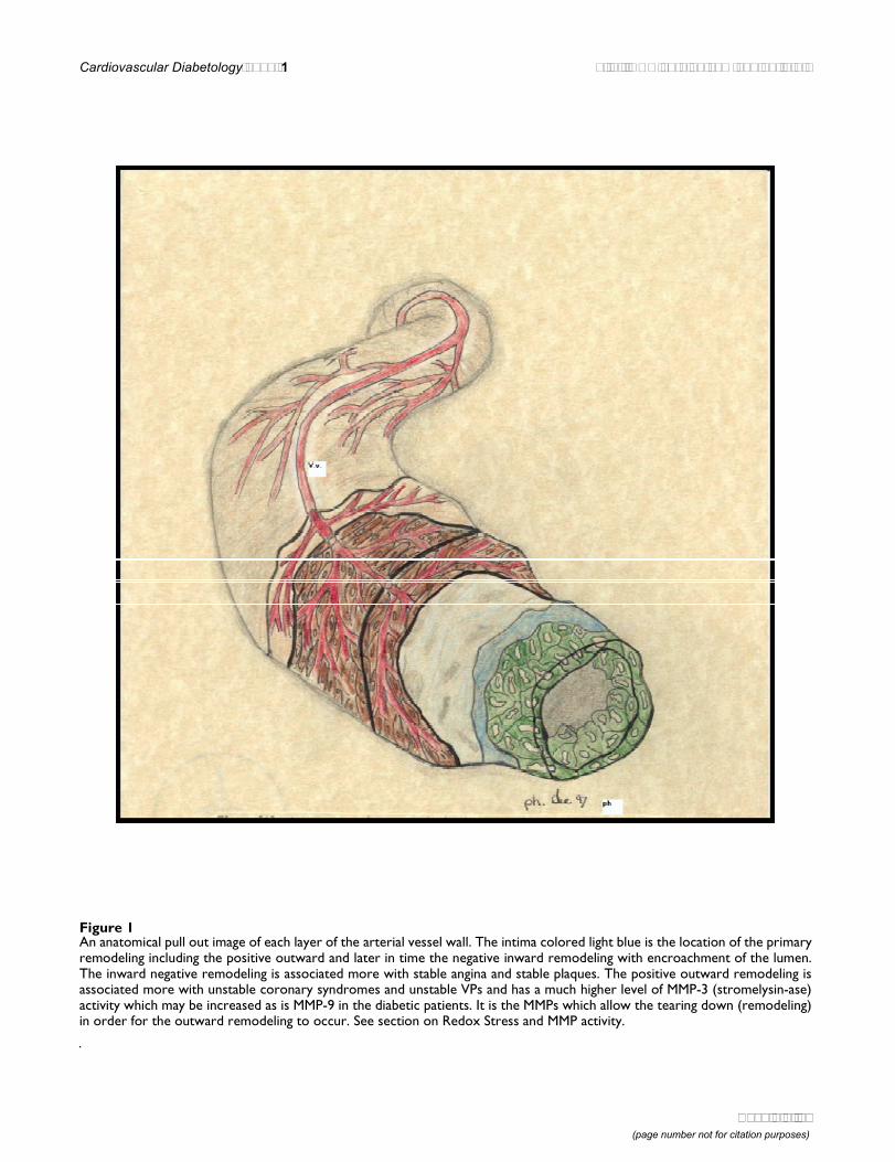
giogenic, prothrombotic, multifactorial disease of thearterial intima caused by the retention of modified lowdensity lipoproteins, hemodynamic, and redox stress [1–4] (figure 1) (figure 2).
Working definition of atheroscleropathy:
Atheroscleropathy: The term used to describe the unique ac-celerated atherosclerosis observed in and associated withMS, IR, PD, and overt T2DM.
Henceforth, in this review the term atheroscleropathy shallbe used to describe accelerated atherosclerosis associatedwith MS, IR, PD, and overt T2DM.
Three quarters of a century ago, a quote from Elliott P Jos-lin's presentation to the American College of Physicians in1927 seems appropriate.
Published: 27 September 2002
Cardiovascular Diabetology 2002, 1:3
Received: 18 August 2002Accepted: 27 September 2002
This article is available from: http://www.cardiab.com/content/1/1/3
© 2002 Hayden and Tyagi; licensee BioMed Central Ltd. This article is published in Open Access: verbatim copying and redistribution of this article are permitted in all media for any purpose, provided this notice is preserved along with the article's original URL.
Page 1 of 27(page number not for citation purposes)
Cardiovascular Diabetology 2002, 1 http://www.cardiab.com/content/1/1/3
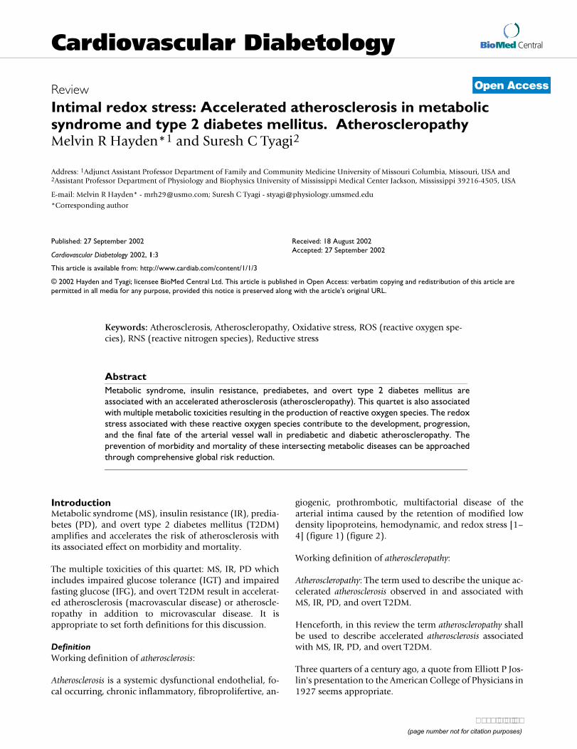
Figure 1An anatomical pull out image of each layer of the arterial vessel wall. The intima colored light blue is the location of the primaryremodeling including the positive outward and later in time the negative inward remodeling with encroachment of the lumen.The inward negative remodeling is associated more with stable angina and stable plaques. The positive outward remodeling isassociated more with unstable coronary syndromes and unstable VPs and has a much higher level of MMP-3 (stromelysin-ase)activity which may be increased as is MMP-9 in the diabetic patients. It is the MMPs which allow the tearing down (remodeling)in order for the outward remodeling to occur. See section on Redox Stress and MMP activity.
Page 2 of 27(page number not for citation purposes)
Cardiovascular Diabetology 2002, 1 http://www.cardiab.com/content/1/1/3
Figure 2This image portrays the anatomical relationships of the endothelium, intima, media, and adventitia. Each of these layers play animportant role in the development of atheroscleropathy. Since the discovery of the essential role of an intact endothelium forthe vasomotor control of musculo-elastic arteries by Furchgott and Zawadski in 1980, and the discovery of the NOSs in 1989,the endothelium has been found to play a central role in the maintenance of healthy arteries and found to be placed in a centralrole for the development and progression of atherogenesis and subsequent atheroscleropathy. The endothelium is five timesthe weight of the heart and equal to the weight of the liver. This organ is placed at a critical location as an interface with nutri-ents and toxic products not only at its luminal side of musculo-eleastic arteries but also at the endothelial extracellular matrixinterface at the site of capillaries. This exciting monolayer of unique cells is responsible for the production of a gas NO thatacts to modulate blood flow and is a naturally occurring interfacing antioxidant capable of scavenging ROS. The intima, sand-wiched between the internal elastic lamina of the medial smooth muscle cell layer and the endothelium is the site of atheroscle-rosis, intimopathy, and the atheroscleropathy associated with MS, IR, PD, and T2DM. The injurious stimuli depicted on theluminal side of the endothelial cell (including redox and oxidative stress with ROS) result in the adaptive changes in which weare familiar: Remodeling of the arterial vessel wall: From Atheroma to Atherosclerosis, to Atheroscleropathy: A MALIGNANTTRANSFORMATION.
Page 3 of 27(page number not for citation purposes)
Cardiovascular Diabetology 2002, 1 http://www.cardiab.com/content/1/1/3
"I believe the chief cause of premature development ofarteriosclerosis in diabetes, save for advancing age, isdue to an excess of fat, an excess of fat in the body,obesity, an excess of fat in the diet, and an excess of fatin the blood. With an excess of fat diabetes begins andfrom an excess of fat diabetics die, formerly of coma, re-cently of arteriosclerosis."[5] Refer to A-FLIGHT toxici-ties sections (F), (L), (T).
This master clinician of diabetes was one of the first phy-sicians to make the association regarding the double jeop-ardy of type 2 diabetes mellitus and atheroscleropathywith its associated morbidity and mortality in cardiovas-cular disease.
Recognition of the prophetic view of Joslin has now beenfulfilled in the 2001 National Cholesterol Education Pro-gram Adult Treatment Panel III guidelines. Diabetes (bothT1DM and T2DM) is now considered a coronary riskequivalent and the metabolic syndrome is included in themultiple risks factors for the development of atheroscle-ropathy. [6]
For today's atherosclerologists the history of atherosclerosisis rich and the theories are legion. Even today, knowledgein this field of study is expanding exponentially. In this re-view, we will try to remain focused on intimal redox stressand how this interacts with the manifold toxicities of IR,MS, PD, and T2DM to result in a unique acceleratedatherosclerosis which shall be called atheroscleropathy.
Redox homeostasis, redox stress, and oxidative stressCellular respiration (the transference of electrons betweenoxygen species) allows each of us to survive on this planetnot only at the cellular level but also as an organism.Homeostasis is a key element to all healthy physiologic
functions throughout the body and when there is loss ofhomeostasis, there is usually disease.
Redox homeostasis describes the normal physiologicprocess of reduction and oxidation in order to re-pair un-stable, damaging, reduced, reactive oxygen species (ROS)which will include the following oxygen free radicals (O2'– superoxide, H2O2 – hydrogen peroxide, -OH' hydroxylradical, and singlet oxygen) and organic analogues whichinclude reactive nitrogen species (RNS) primarily perox-ynitrite ONOO'.
This homeostatic balance between ROS and antioxidantcapacity is in contrast to redox stress (redox imbalance)which implies a loss of this unique homeostasis resultingin an excess production of ROS (tables 1 and 2) eitherthrough the process of reduction or oxidation.
Oxidative Stress implies a loss of redox homeostasis (im-balance) with an excess of ROS by the singular process ofoxidation. Both redox and oxidative stress may be associ-ated with an impairment of antioxidant defensive capacityas well as an overproduction of ROS.
It has been known for some time that ROS are detrimentaland toxic to cells and tissues as a result of injury to lipids,nucleic acids, and proteins: (A). Lipid peroxidation ofmembranes (loss of membrane function and increasedpermeability) and generation of lipid autoperoxidationreactions. (B). DNA damage leading to mutation anddeath. (C). Cross linking or vulcanization of sulfhydrylrich proteins (leading to stiff aged proteins specificallycollagen of the extracellular matrix). [7]
The evolutionary process of redox homeostasis allows hu-mans to survive in an atmosphere of high oxygen content.In addition our bodies have become "hard wired" to uti
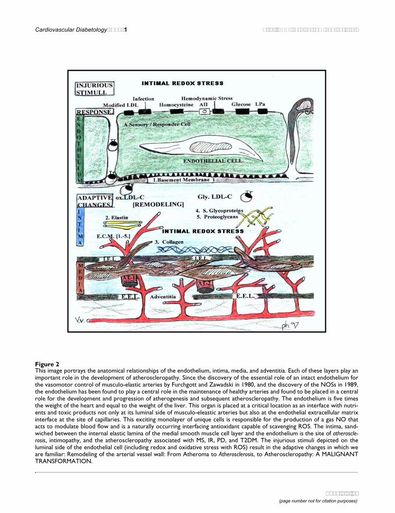
Table 1: Courtesy [9] origins of reactive oxygen species (ROS) which produce redox stress
I Excess O2 (oxygen therapy)II Absorption of radiant energy (ultraviolet light) or ionizing radiation (radiotherapy)III Exposure to toxins: carbon tetrachloride, dioxin, alloxan and streptozotocin to name just a fewIV Reduction-oxidation (redox) reactions during normal physiologic processes (cellular respiration)
A. Respiratory chain enzymes and oxygenB. Xanthine oxidaseC. Cytochrome P450 monooxygenase activityD. NAD(P)H / NADH oxidaseE. Fenton reaction: Fe++ + H2O2 � Fe+++ + OH + OH-
F. Haber-Weiss Reaction H2O2 + O2- � -OH- + O2 +OH-
V Ischemia – Ischemia reperfusion injuryVI Inflammatory processes. Acute and chronicVII Once free ROS radicals form, they can react with membrane lipids, proteins and nucleic acid to initiate auto-catalytic
reactions (ROS beget ROS) [9]
Page 4 of 27(page number not for citation purposes)
Cardiovascular Diabetology 2002, 1 http://www.cardiab.com/content/1/1/3
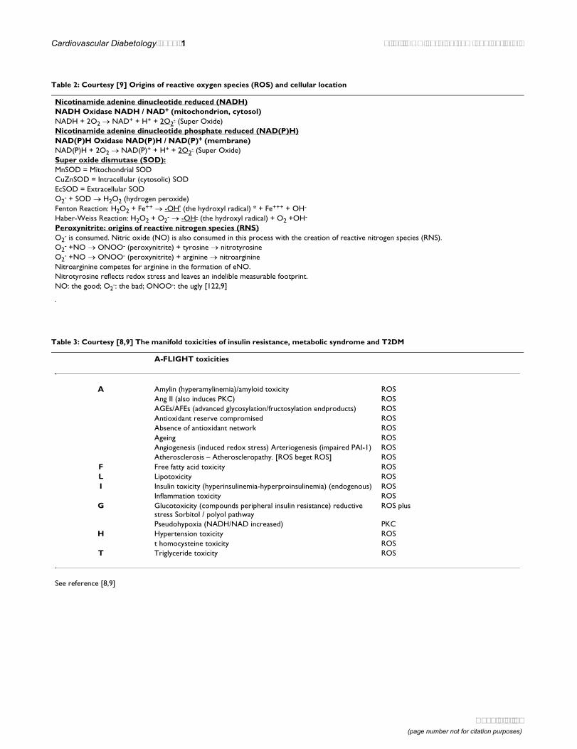
Table 2: Courtesy [9] Origins of reactive oxygen species (ROS) and cellular location
Nicotinamide adenine dinucleotide reduced (NADH)NADH Oxidase NADH / NAD+ (mitochondrion, cytosol)NADH + 2O2 � NAD+ + H+ + 2O2
- (Super Oxide)Nicotinamide adenine dinucleotide phosphate reduced (NAD(P)H)NAD(P)H Oxidase NAD(P)H / NAD(P)+ (membrane)NAD(P)H + 2O2 � NAD(P)+ + H+ + 2O2
- (Super Oxide)Super oxide dismutase (SOD):MnSOD = Mitochondrial SODCuZnSOD = Intracellular (cytosolic) SODEcSOD = Extracellular SODO2
- + SOD � H2O2 (hydrogen peroxide)Fenton Reaction: H2O2 + Fe++ � -OH' (the hydroxyl radical) * + Fe+++ + OH-
Haber-Weiss Reaction: H2O2 + O2- � -OH- (the hydroxyl radical) + O2 +OH-
Peroxynitrite: origins of reactive nitrogen species (RNS)O2
- is consumed. Nitric oxide (NO) is also consumed in this process with the creation of reactive nitrogen species (RNS).O2
- +NO � ONOO- (peroxynitrite) + tyrosine � nitrotyrosineO2
- +NO � ONOO- (peroxynitrite) + arginine � nitroarginineNitroarginine competes for arginine in the formation of eNO.Nitrotyrosine reflects redox stress and leaves an indelible measurable footprint.NO: the good; O2
-: the bad; ONOO-: the ugly [122,9]
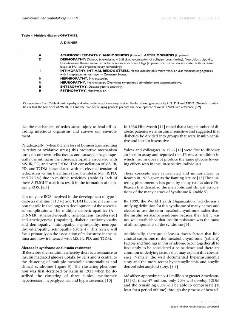
Table 3: Courtesy [8,9] The manifold toxicities of insulin resistance, metabolic syndrome and T2DM
A-FLIGHT toxicities
A Amylin (hyperamylinemia)/amyloid toxicity ROSAng II (also induces PKC) ROSAGEs/AFEs (advanced glycosylation/fructosylation endproducts) ROSAntioxidant reserve compromised ROSAbsence of antioxidant network ROSAgeing ROSAngiogenesis (induced redox stress) Arteriogenesis (impaired PAI-1) ROSAtherosclerosis – Atheroscleropathy. [ROS beget ROS] ROS
F Free fatty acid toxicity ROSL Lipotoxicity ROSI Insulin toxicity (hyperinsulinemia-hyperproinsulinemia) (endogenous) ROS
Inflammation toxicity ROSG Glucotoxicity (compounds peripheral insulin resistance) reductive
stress Sorbitol / polyol pathwayROS plus
Pseudohypoxia (NADH/NAD increased) PKCH Hypertension toxicity ROS
t homocysteine toxicity ROST Triglyceride toxicity ROS
See reference [8,9]
Page 5 of 27(page number not for citation purposes)
Cardiovascular Diabetology 2002, 1 http://www.cardiab.com/content/1/1/3
lize the mechanism of redox stress injury to fend off in-vading infectious organisms and survive our environ-ment.
Paradoxically, (when there is loss of homeostasis resultingin redox or oxidative stress) this protective mechanismturns on our own cells; tissues and causes damage, espe-cially the intima in the atheroscleropathy associated withMS, IR, PD, and overt T2DM. This constellation of MS, IR,PD, and T2DM is associated with an elevated tension ofredox stress within the intima (also the islet in MS, IR, PD,and T2DM) due to multiple toxicities. (table 3) Each ofthese A-FLIGHT toxicities result in the formation of dam-aging ROS. [8,9]
Not only are ROS involved in the development of type 1diabetes mellitus (T1DM) and T2DM but also play an im-portant role in the long-term development of the associat-ed complications: The multiple diabetic-opathies (A –DINNER: atheroscleropathy, angiogenesis (accelerated)and arteriogenesis (impaired), diabetic cardiomyopathyand dermopathy, intimopathy, nephropathy, neuropa-thy, enteropathy, retinopathy (table 4). This review willfocus primarily on the association of redox stress in the in-tima and how it interacts with MS, IR, PD, and T2DM.
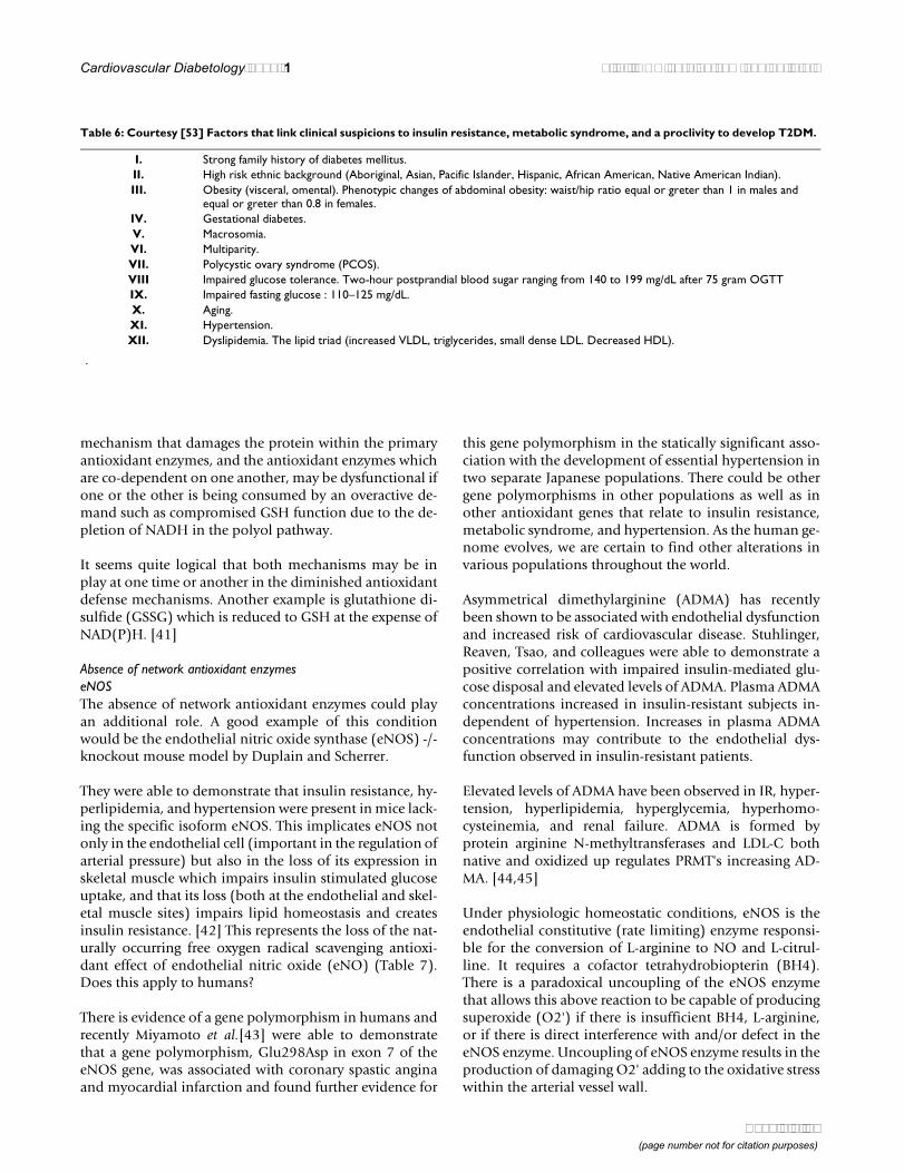
Metabolic syndrome and insulin resistanceIR describes the condition whereby there is a resistance toinsulin mediated glucose uptake by cells and is central tothe clustering of multiple metabolic abnormalities andclinical syndromes (figure 3). The clustering phenome-non was first described by Kylin in 1923 when he de-scribed the clustering of three clinical syndromes:hypertension, hyperglycemia, and hyperuricemia. [10]
In 1936 Himsworth [11] noted that a large number of di-abetic patients were insulin insensitive and suggested thatdiabetics be divided into groups that were insulin sensi-tive and insulin insensitive.
Yalow and colleagues in 1965 [12] were first to discoveran insulin assay and reported that IR was a condition inwhich insulin does not produce the same glucose lower-ing effects seen in insulin-sensitive individuals.
These concepts were rejuvenated and immortalized byReaven in 1988 given as the Banting lecture.[13] The clus-tering phenomenon has gone by many names since Dr.Reaven first described the metabolic and clinical associa-tions of the many names of Syndrome X. (table 5)
By 1999, the World Health Organization had chosen aunifying definition for this syndrome of many names andelected to use the term metabolic syndrome rather thanthe insulin resistance syndrome because they felt it wasnot well established that insulin resistance was the causeof all components of the syndrome.[14]
Additionally, there are at least a dozen factors that linkclinical suspicions to the metabolic syndrome. (table 6)Factors and findings in this syndrome occur together all tofrequently to be considered a coincidence and there arecommon underlying factors that may explain this coexist-ence. Namely, the well documented hyperinsulinemiastory and the more recent hyperamylinemia and amylinderived islet amyloid story. [8,9]
MS affects approximately 47 million or greater Americans.[15] Of these 47 million, only 20% will develop T2DMand the remaining 80% will be able to compensate (atleast for a period of time) through the process of beta cell
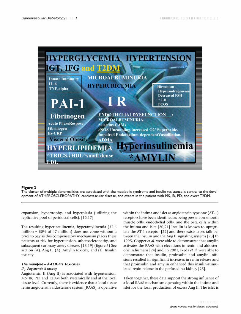
Table 4: Multiple diabetic-OPATHIES.
A-DINNER
A ATHEROSCLEROPATHY. ANGIOGENESIS (induced). ARTERIOGENESIS (impaired).D DERMOPATHY: Diabetic Scleroderma – Stiff skin, vulcanization of collagen (cross-linking). Necrobiosis Lipoidica
Diabeticorum. Brown sunken atrophic scars anterior shin of legs (impaired scar formation associated with increased levels of PAI-I and impaired injury remodeling).
I INTIMOPATHY. INTIMAL REDOX STRESS. Macro vascular plus micro vascular vasa vasorum angiogenesis with intraplaque hemorrhage � Coronary Events.
N NEPHROPATHY. Microvascular.N NEUROPATHY. Microvascular. Overriding sympathetic stimulation pro vasoconstrictive.E ENTEROPATHY. Delayed gastric emptying.R RETINOPATHY. Microvascular.
Observations from Table 4: Intimopathy and atheroscleropathy are very similar. Similar shared glucotoxicity in T1DM and T2DM. Dissimilar toxici-ties in that the toxicities of MS, IR, PD and the role of the aging process predate the development of overt T2DM. See reference [8,9]
Page 6 of 27(page number not for citation purposes)
Cardiovascular Diabetology 2002, 1 http://www.cardiab.com/content/1/1/3
expansion, hypertrophy, and hyperplasia (utilizing thereplicative pool of periductal cells). [16,17]
The resulting hyperinsulinemia, hyperamylinemia (37.6million = 80% of 47 million) does not come without aprice to pay as this compensatory mechanism places thesepatients at risk for hypertension, atheroscleropathy, andsubsequent coronary artery disease. [18,19] (figure 3) Seesection (A). Ang II, (A). Amylin toxicity, and (I). Insulintoxicity.
The manifold – A-FLIGHT toxicities(A). Angiotensin II toxicityAngiotensin II (Ang II) is associated with hypertension,MS, IR, PD, and T2DM both systemically and at the localtissue level. Currently, there is evidence that a local tissuerenin angiotensin aldosterone system (RAAS) is operative
within the intima and islet as angiotensin type one (AT-1)receptors have been identified as being present on smoothmuscle cells, endothelial cells, and the beta cells withinthe intima and islet [20,21] Insulin is known to upregu-late the AT-1 receptor [22] and there exists cross talk be-tween the insulin and the Ang II signaling systems [23] In1995, Copper et al. were able to demonstrate that amylinactivates the RAAS with elevations in renin and aldoster-one in humans [24] and, in 2001, Ikeda et al. were able todemonstrate that insulin, proinsulin and amylin infu-sions resulted in significant increases in renin release andthat proinsulin and amylin enhanced this insulin-stimu-lated renin release in the perfused rat kidney [25].
Taken together, these data support the strong influence ofa local RAAS mechanism operating within the intima andislet for the local production of excess Ang II. The islet is
Figure 3The cluster of multiple abnormalities are associated with the metabolic syndrome and insulin resistance is central to the devel-opment of ATHEROSCLEROPATHY, cardiovascular disease, and events in the patient with MS, IR, PD, and overt T2DM.
Hyperinsulinemia*AMYLIN
HYPERGLYCEMIAIGT, IFG and T2DM
I R
HYPERTENSION
Visceral Obesity
HYPERLIPIDEMIA^TRIGS. HDL^small denseLDL
PAI-1Fibrinogen
HirsuitismHyperandrogenemiaDecreased FSH^ LHPCOS
HYPERURICEMIAMICROALBUMINURIA
ENDOTHELIALDYSFUNCTION :MICROALBUMINURIA.Selectins CAMs eNOS-Uncoupling.Increased O2’ Superoxide.Impaired Endothelium-dependentVasodilation.ADMA
Innate ImmunityIL-6TNF-alpha
Acute PhaseResposeFibrinogenHs-CRP
Page 7 of 27(page number not for citation purposes)

Cardiovascular Diabetology 2002, 1 http://www.cardiab.com/content/1/1/3
quite vascular with an abundant supply of intra islet cap-illaries and endothelial cells and the intima is lined by acontinuous monocellular layer of endothelial cells (addi-tionally, the arterial vessel wall becomes highly vascularthrough the process of plaque angiogenesis as the vulner-able plaque evolves during the process of atheroscleropa-thy).
This allows the vascular NAD(P)H oxidase enzyme tocome into play. Ang II is one of the most potent endog-enous stimuli for the generation of superoxide O2
- via theactivation of vascular NAD(P)H oxidase. [26,27]
The interruption of this mechanism by the angiotensinconverting enzyme inhibitor (ACEi) ramipril in the HeartOutcomes Prevention Evaluation (HOPE) study may helpto explain the 32% risk reduction for developing T2DM aswell as the dramatic reduction in cardiovascular events.[28]
A special reference to Griendling and Harrison seems ap-propriate: "Out, damned DOT! Out I say" (where thedamned DOT represents the unpaired dots on Lewis dia-grams). [29] One of the best ways to prevent these dotsfrom forming is to prevent excess substrates (table 3)which cause the multiple toxicities and the multiplicativeeffect of the A-FLIGHT toxicities associated with MS, IR,PD, and T2DM.
In MS, IR, PD, and T2DM the intima and islet milieu willbe laden with the necessary substrates (hyperinsulinemia,hyperproinsulinemia and hyperamylinemia) to activatethe damaging cascading mechanism of Ang II, NAD(P)Hoxidase, superoxide (O2
-) and peroxynitrite (ONOO-)production while consuming the natural endogenouslyproduced antioxidant nitric oxide (NO) within the vul-nerable intima and islet.
(A). Advanced glycosylation endproducts: AGEAdvanced glycosylation endproducts (AGEs) are formedas a result of the non-enzymatic damaging protein glyca-tion due to an excess of glucose (hyperglycemia) presentin both T1DM, PD, and T2DM. AGEs are initially formedthrough the process of a glucose nucleophilic addition re-action with proteins forming a Schiff base followed by theformation of an Amadori compound which undergoesfurther reactions, rearrangements, dehydrations andcleavage resulting in brown insoluble, cross linked com-plexes called AGEs. This process is thought to liberateH2O2 through two pathways: the first is the 1,2-enoliza-tion pathway which leads to 3-deoxyglucosone formingH2O2 and glucosone; the second pathway is the 2,3-eno-lization pathway leading to 1-deoxyglucosone and puta-tive 1,4-deoxyglucosone. Under oxidative conditions, the2,3-enediol is thought to generate H2O2 and car-boxymethyllysine. 3-deoxyglucosones are known to beboth highly reactive intermediates in non-enzymatic glyc-osylation and also potent cross-linkers which are respon-sible for the polymerization of proteins to AGEs. Thesehighly cross-linked proteins, especially collagen, cause astiffening within the vessel which results in decreasedcompliance of the arterial vessel wall and may well playan important role in the development of diabetic diastolicdysfunction, diabetic cardiomyopathy, and the diastolicdysfunction of the arterial vessel wall. Furthermore, thereare advanced fructosylation endproducts (AFEs), whichactually have a greater affinity binding to proteins thanglucose and follow a similar pattern in the production ofthe ROS. [30–33] An excellent in depth review of AGE canbe found in an article by Aronson and Rayfield where theydiscuss how hyperglycemia promotes atherosclerosis [34].
The multiligand immunoglobulin superfamily cell surfacereceptor: the receptor for advanced glycation endproducts(RAGE) is up-regulated by the presence of AGE and resultsin the signal transduction of nuclear factor kappa B(NFkappa B) which then results in a chronically active in-flammatory state and links this section to section (I). In-flammation Toxicity and atheroscleropathy. [35,36]
(A). Antioxidant enzymesAntioxidant reserve compromisedIn addition to the excess generation of the ROS seen in di-abetes, there exists an impaired generation of endogenousantioxidants. Superoxide dismutase (SOD), [37] glutath-ione reduced (GSH), [38] and ascorbic acid (Vitamin C)[39] are all decreased and associated with atheroscleropa-thy in diabetes. Moreover, there is evidence of the dimin-ished capacity of other antioxidants such as uric acid andvitamin E with a reduced activity of catalase and glutath-ione peroxidase (GPx). (Table 6) [40] The exact mecha-nisms of impairment are still not completely understoodbut two explanations exist. Protein glycation may be a
Table 5: Courtesy [8,9] The myriad names of the metabolic syndrome
I. The insulin resistance syndromeII. Syndrome XIII. Reaven syndromeIV. Metabolic syndrome (preferred term by
WHO)V. Metabolic syndrome XVI. Multiple metabolic syndromeVII. Plurimetabolic syndromeVIII. Dysmetabolic syndromeIX. Cardiovascular dysmetabolic syndromeX. Cardiometabolic syndromeXI. The "H" phenomenonXII. The "Deadly quartet"
Page 8 of 27(page number not for citation purposes)
Cardiovascular Diabetology 2002, 1 http://www.cardiab.com/content/1/1/3
mechanism that damages the protein within the primaryantioxidant enzymes, and the antioxidant enzymes whichare co-dependent on one another, may be dysfunctional ifone or the other is being consumed by an overactive de-mand such as compromised GSH function due to the de-pletion of NADH in the polyol pathway.
It seems quite logical that both mechanisms may be inplay at one time or another in the diminished antioxidantdefense mechanisms. Another example is glutathione di-sulfide (GSSG) which is reduced to GSH at the expense ofNAD(P)H. [41]
Absence of network antioxidant enzymeseNOSThe absence of network antioxidant enzymes could playan additional role. A good example of this conditionwould be the endothelial nitric oxide synthase (eNOS) -/-knockout mouse model by Duplain and Scherrer.
They were able to demonstrate that insulin resistance, hy-perlipidemia, and hypertension were present in mice lack-ing the specific isoform eNOS. This implicates eNOS notonly in the endothelial cell (important in the regulation ofarterial pressure) but also in the loss of its expression inskeletal muscle which impairs insulin stimulated glucoseuptake, and that its loss (both at the endothelial and skel-etal muscle sites) impairs lipid homeostasis and createsinsulin resistance. [42] This represents the loss of the nat-urally occurring free oxygen radical scavenging antioxi-dant effect of endothelial nitric oxide (eNO) (Table 7).Does this apply to humans?
There is evidence of a gene polymorphism in humans andrecently Miyamoto et al.[43] were able to demonstratethat a gene polymorphism, Glu298Asp in exon 7 of theeNOS gene, was associated with coronary spastic anginaand myocardial infarction and found further evidence for
this gene polymorphism in the statically significant asso-ciation with the development of essential hypertension intwo separate Japanese populations. There could be othergene polymorphisms in other populations as well as inother antioxidant genes that relate to insulin resistance,metabolic syndrome, and hypertension. As the human ge-nome evolves, we are certain to find other alterations invarious populations throughout the world.
Asymmetrical dimethylarginine (ADMA) has recentlybeen shown to be associated with endothelial dysfunctionand increased risk of cardiovascular disease. Stuhlinger,Reaven, Tsao, and colleagues were able to demonstrate apositive correlation with impaired insulin-mediated glu-cose disposal and elevated levels of ADMA. Plasma ADMAconcentrations increased in insulin-resistant subjects in-dependent of hypertension. Increases in plasma ADMAconcentrations may contribute to the endothelial dys-function observed in insulin-resistant patients.
Elevated levels of ADMA have been observed in IR, hyper-tension, hyperlipidemia, hyperglycemia, hyperhomo-cysteinemia, and renal failure. ADMA is formed byprotein arginine N-methyltransferases and LDL-C bothnative and oxidized up regulates PRMT's increasing AD-MA. [44,45]
Under physiologic homeostatic conditions, eNOS is theendothelial constitutive (rate limiting) enzyme responsi-ble for the conversion of L-arginine to NO and L-citrul-line. It requires a cofactor tetrahydrobiopterin (BH4).There is a paradoxical uncoupling of the eNOS enzymethat allows this above reaction to be capable of producingsuperoxide (O2') if there is insufficient BH4, L-arginine,or if there is direct interference with and/or defect in theeNOS enzyme. Uncoupling of eNOS enzyme results in theproduction of damaging O2' adding to the oxidative stresswithin the arterial vessel wall.
Table 6: Courtesy [53] Factors that link clinical suspicions to insulin resistance, metabolic syndrome, and a proclivity to develop T2DM.
I. Strong family history of diabetes mellitus.II. High risk ethnic background (Aboriginal, Asian, Pacific Islander, Hispanic, African American, Native American Indian).III. Obesity (visceral, omental). Phenotypic changes of abdominal obesity: waist/hip ratio equal or greter than 1 in males and
equal or greter than 0.8 in females.IV. Gestational diabetes.V. Macrosomia.VI. Multiparity.VII. Polycystic ovary syndrome (PCOS).VIII Impaired glucose tolerance. Two-hour postprandial blood sugar ranging from 140 to 199 mg/dL after 75 gram OGTTIX. Impaired fasting glucose : 110–125 mg/dL.X. Aging.XI. Hypertension.XII. Dyslipidemia. The lipid triad (increased VLDL, triglycerides, small dense LDL. Decreased HDL).
Page 9 of 27(page number not for citation purposes)
Cardiovascular Diabetology 2002, 1 http://www.cardiab.com/content/1/1/3
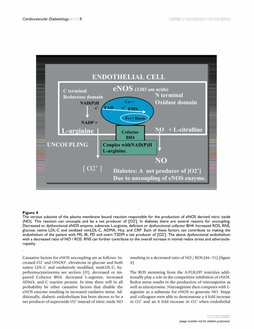
Causative factors for eNOS uncoupling are as follows: In-creased O2' and ONOO', elevations in glucose and bothnative LDL-C and oxidatively modified, mmLDL-C, hy-perhomocysteinemia see section (H), decreased or im-paired Cofactor BH4, decreased L-arginine, increasedADMA, and C reactive protein. In time there will in allprobability be other causative factors that disable theeNOS enzyme resulting in increased oxidative stress. Ad-ditionally, diabetic endothelium has been shown to be anet producer of superoxide O2' instead of nitric oxide NO
resulting in a decreased ratio of NO / ROS [46–51] (figure4)
The ROS stemming from the A-FLIGHT toxicities addi-tionally play a role in the competitive inhibition of eNOS.Redox stress results in the production of nitroarginine aswell as nitrotyrosine. Nitroarginine then competes with L-arginine as a substrate for eNOS to generate NO. Steppand colleagues were able to demonstrate a 4 fold increasein O2' and an 8 fold increase in O2' when endothelial
Figure 4The various subunits of the plasma membrane bound reaction responsible for the production of eNOS derived nitric oxide(NO). This reaction can uncouple and be a net producer of [O2']. In diabetes there are several reasons for uncoupling.Decreased or dysfunctional eNOS enzyme, substrate L-arginine, deficient or dysfunctional cofactor BH4. Increased ROS, RNS,glucose, native LDL-C and oxidized mmLDL-C, ADMA, Hcy, and CRP. Each of these factors can contribute to making theendothelium of the patient with MS, IR, PD and overt T2DM a net producer of [O2']. The above dysfunctional endotheliumwith a decreased ratio of NO / ROS. RNS can further contribute to the overall increase in intimal redox stress and atheroscle-ropathy.
NAD(P)H
NADP +
eNOS (1203 am acids)C terminalReductase domain N terminal
Oxidase domainFAD FMN
CofactorBH4
Ca++
Fe++ Haem
e’ e’
L-arginine NO + L-citrulline
NO
UNCOUPLING
[ O2’ ]
ENDOTHELIAL CELL
Diabetes: A net producer of [O2’]Due to uncoupling of eNOS enzyme.
Couples withNAD(P)HL-arginine.
Page 10 of 27(page number not for citation purposes)
Cardiovascular Diabetology 2002, 1 http://www.cardiab.com/content/1/1/3
cells were exposed to native LDL-C and mmLDL-C respec-tively. This uncoupling of eNOS plays an important rolein endothelial cell dysfunction and increased oxidativestress. [47] Hyperglycemia and peroxynitrite (ONOO')also induce eNOS uncoupling with increases in O2' pro-duction. [48] Just published, Verma S and colleagues re-ported that CRP caused a marked down regulation ofeNOS mRNA and protein expression with subsequentlower eNO production. The authors point out that CRPmay not just be a marker of atherosclerosis and increasedrisk of acute coronary events, but may also be a mediatorof this disease process. Strategies designed to lower CRPmay reduce cardiovascular risk by directly improving bio-availability of NO and endothelial function. [49] See sec-tion (I). Inflammation.
There are undoubtedly many more scenarios in whicheNOS can be impaired with resulting decreased NO but atthis point in time it is certainly interesting to see a tightconnection of impaired eNOS and the MS, IR, PD, andT2DM. Additionally, the synergistic importance regardingelevations of both glucose and native LDL-C or mmLDLwhich result in elevations of the detrimental superoxide(O2') can uncouple the eNOS enzyme leading to even fur-ther increases in O2'. The importance of treating LDL-C,HbA1c, and hypertension to goal are therefore paramountin reducing the oxidative damage and endothelial cell dys-function. (table 9) (figure 6) These examples onlystrengthen the statement that ROS beget ROS.
The synergism and the vicious cycle of redox and oxidativestress to the arterial vessel wall from ROS produced by vas-cular cells, especially the endothelium, as a result of the A-FLIGHT toxicities necessitates an aggressive global ap-proach. Wong T.Y. and colleagues for the AtherosclerosisRisk In Communities (ARIC) Investigators were able toshow that retinal arteriolar narrowing was independentlyassociated with the risk of developing future diabetes. Thissupports a microvascular role in the development of clin-ical diabetes and provides clinical evidence to support ahypothesis that eNOS and endothelial dysfunction maybe implicated in the pathogenesis of diabetes. This newclinical information, of arteriolar narrowing preceding theclinical onset of diabetes and implicating endothelial celldysfunction (including an eNOS defect) could play a ma-jor important role in the development of this polygenic –multifactorial disease of MS, IR, PD, overt T2DM andatheroscleropathy. [52]
This leads to an interesting Hypothesis:
Could it be that T2DM is really a cardiovasculardisease (evolving around a primary eNOS enzyme dys-function or defect with an effect on MS and IR) with a latemanifestation of glucose elevation i.e. PD and overt
T2DM? This would certainly tie the natural history ofT2DM and atheroscleropathy together [53].
Other antioxidant enzymesIf any one of the antioxidant enzymes (table 7) is missingor impaired or any combination of them are impaired,then we would expect to see a similar event as in theknockout mouse model. It would not have to be a com-plete knockout of the enzyme, as discussed above, as var-ious gene polymorphisms could exist which could resultin a decreased antioxidant reserve.
(A). AgeingAgeing has been shown to be associated with an increasedrisk of developing T2DM and atheroscleropathy. Ageingallows the multiplicative effects of the A-FLIGHT toxicitiesto become manifest. Advanced ageing leads to impairedendothelial nitric oxide synthesis and also enhanced en-dothelial apoptosis.
In addition, aged cells have a significantly enhanced con-centration (more than 3 fold) of oxidized low density li-poprotein, TNFalpha and caspase-3 activity as comparedto young cells. The decrease in eNO associated with agedcells creates a deficiency of the naturally occurring antioxi-dant eNO. [54]
Similarly, excess redox stress is felt to contribute to ageing.Information on the relationship of redox stress and ageingand inflammation (see section "(I). Inflammation Toxici-ty") is rapidly increasing and gaining wider recognition.[55,56]
(A). Amylin toxicityAmylin, also termed islet amyloid polypeptide (IAPP) is a37 amino acid polypeptide co-synthesized, co-packaged,and co-secreted by the islet beta cell with insulin. It maybe considered insulin's a fraternal twin.
Amylin parallels insulin synthesis, secretion, and excre-tion so that whenever you have hyperinsulinemia youhave hyperamylinemia and, in the same way, when insu-lin levels decline amylin levels decline.
Amylin stimulates lipolysis in vivo and may be a possiblemediator of induced insulin resistance. Ye et al., were ableto demonstrate that amylin infusion (5 nmol/h for 4 h)conscious rats that fasted for 5–7 hours resulted in an ele-vation of insulin, lactate and glucose (P < 0.05 vs. con-trol).
Despite the rise in insulin, plasma non-esterified fatty acidand glycerol were also elevated (P < 0.001). Although theplasma triglyceride content was unaltered, the triglyceridecontent in the liver was increased by 28% (P < 0.001) with
Page 11 of 27(page number not for citation purposes)
Cardiovascular Diabetology 2002, 1 http://www.cardiab.com/content/1/1/3
a similar tendency in muscle (18%, P = 0.1). These effectswere blocked by the rat amylin antagonist amylin-(8–37)and also by the anti-lipolytic agent acipimox. The authorsconcluded that amylin could exert a lipolytic-like actionin vivo. [57]
This elevation in amylin would correspond to the insulinresistant state with associated elevation in amylin in hu-mans. These data indicate that amylin may play a role byelevating free fatty acids which would aggravate or induce
the underlying insulin resistance and provide a mecha-nism for increasing the free fatty acid substrate for in-creased redox stress, cytotoxicity and intimal remodelingassociated with atheroscleropathy.
There are amylin binding sites within the renal cortex andamylin activates the RAAS with elevations in renin and al-dosterone [24]
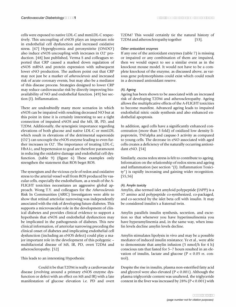
Figure 5Vulnerable plaques are proinflammatory, profibrotic, prothrombotic, proangiogenic, lipid ladened and recently found to beacidic. The activated endothelium is associated with endothelial dysfunction due to eNOS dysfunction and eNOS uncouplingwith resultant overproduction of superoxide [O2']. Diabetes is associated with a dysfunctional endothelium not only at sites ofvulnerable plaque but have been shown to be a systemic net producer of [O2']. The inflammatory cells and intimal remodelingare depicted in this image as well as an intraplaque hemorrhage (IPH) from the angiogenic vasavasorum vessels originating fromthe adventitia of this lipid ladened vulnerable plaque. Redox stress and ROS play a prominent role within the intima. The profi-brotic arm of the unstable VP is responsible for the positive outward and negative inward remodeling.
7/26/02 70
IPH MM
VULNERABLE -HOT PLAQUES: PLAQUES“ON FIRE”Double Jeopardy : T2DM and ATHEROSCLEROPATHY.
Activated Endothelium.eNOS Dys.eNOS Uncoupling.O2’ increased.phdecreased.REDOX STRESS.
Angiogenic InflammmedHOT
ProthromboticLipid Ladened
ACIDIC
REDOX STRESS
Page 12 of 27(page number not for citation purposes)
Cardiovascular Diabetology 2002, 1 http://www.cardiab.com/content/1/1/3
These findings suggest that glucotoxicity resulting in AGEformation both promotes and accelerates redox stress.[58]
Janson et al. have found that intermediate sized toxic amy-loid particles (ISTAPs) have been found to be cytotoxic tobeta cells inducing apoptosis by membrane disruption.[59]
(A). Angiogenesis (accelerated): Arteriogenesis (impaired): Vasculari-zation Paradox In T2DMThe process of Angiogenesis starts with capillaries and ends with more capillariesAs the atheroma matures there is an associated intenseplaque angiogenesis arising primarily from the adventitial
vasa vasorum (atherosclerotic intimopathy). These vesselsinvade the arterial vessel wall in a malignant like fashion.
This plaque vascularization (angiogenesis) corresponds tothe presence of the inflammatory infiltrate at the shoulderof these lesions, the development of the large lipid core,the thin fibrous cap, and the decrease in SMCs within thefibrous cap, to form, what we now term the vulnerableplaque. [1–4]
Extensions of the vasa vasorum (the vessels within a ves-sel) act as a custom delivery system within the vessel walls'vulnerable shoulder region supplying: 1. substrates of theRAAS; 2. substrates of native LDL-C; 3. the second wave ofinflammatory cells; 4. inflammatory mediators (variouscytokines and growth factors); and 5. provide an addition-
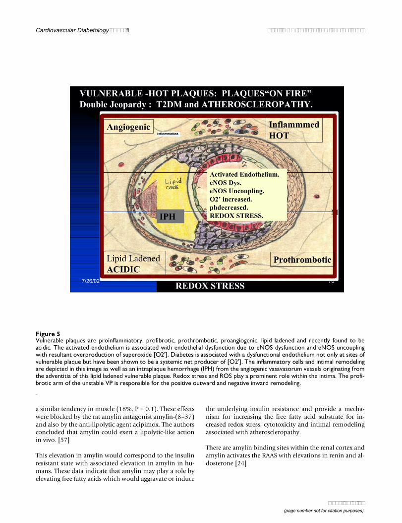
Figure 6Venn diagrams revealing the multiple intersects of this quartet of MS, IR, PD, and overt T2DM. The morbid – mortal intersec-tion of T2DM and Accelerated Atherosclerosis (ATHEROSCLEROPATHY) are a result of the interweaving threads that weavethis complicated mosaic fabric.
8/8/02 5
T2DM AtherosclerosisTwo
Common
Diseases
ATHEROSCLEROPATHY
INTIMOPATHY
RedoxStress Inflammation
Two
Interweaving
Threads
Complex
Mosaic
Fabric
A-FLIGHTTOXICITIES
MSIR
IGTIFG
Prediabetes
Page 13 of 27(page number not for citation purposes)

Cardiovascular Diabetology 2002, 1 http://www.cardiab.com/content/1/1/3
al conduit and source of redox stress at the endothelial cell– extracellular matrix interface within these vulnerableplaques.
This process is accelerated in the MS, IR, PD, and T2DM aswell as the T1DM patient. Vasa vasorum derived fragilecapillary-like vessels are prone to rupture and create intraplaque hemorrhages (IPH) which destabilize the plaqueand promote the possibility of being prone to rupturewith ensuing cardiovascular events. The pseudohypoxia(increased ratio of NADH/NAD+ discussed by William-son and Kilo see section (G) glucotoxicity.) in the polyol-sorbitol pathway as well as the true hypoxia induced bythe increasing intima media thickness may act to inducethe nuclear hypoxia inducible factor (hif-1) within thesmooth muscle and endothelial cells which result in theincreased expression of vascular endothelial growth factorVEGF which is so central and vital to the angiogenic proc-ess. This diabetic atherosclerotic intimopathy (plaque an-giogenesis) would be akin to the diabetic retinopathy.[3,60–62]
The process of arteriogenesis starts with small arterioles and ends with larger arteriolesIn contrast the vascularization process of arteriogenesis isimpaired. Even though patients with diabetes (bothT1DM and T2DM) have a much higher number of athero-sclerotic diseased arteries, mean coronary collateral ves-sels (CCV) are significantly decreased. [63]
Elevations in plasminogen activator inhibitor-1 (PAI-1)are present in the MS, IR, PD, and T2DM patient. Remod-eling collateralization (arteriogenesis) is stimulated by thetissue and urokinase plasminogen activators (tPA anduPA). PAI-1 elevations decrease the conversion of plas-minogen to plasmin because it inhibits tPA and uPA. Asplasmin is impaired there is a reduction of the conversionof inactive or pro-MMP-1 to active MMP-1 with inhibitionof ECM turnover with resultant impaired CCV formation.This paradox in diabetes vascularization contributes great-ly to the known poor outcomes associated with cardiovas-cular events in the diabetic. [3]
(A). Atherosclerosis and atheroscleropathyOnce atherosclerosis and atheroscleropathy has been ini-tiated and sustained, this process is self – perpetuating
Table 7: Courtesy [9] Antioxidants: catalytic/enzymatic inactivation of free radicals
Enzymatic antioxidantsSUPER OXIDE DISMUTASE (SOD) – Location: mitochondrion[O2
- + SOD � H2O2 + O2]ecSOD (extracellular)MnSOD (mitochondrial)CuZnSOD (intracellular)CATALASE – Location: peroxisome[2H2O2 + catalase � 2 H2O + O2]GLUTATHIONE PEROXIDASE – Location: mitochondrion/cytosol(Glutamyl-cysteinyl-glycine tripeptide) glutathione reduced -SH to the oxidized disulfide GSSG.(Glutathione peroxidase) [GSH + 2H2O2 � GSSG + H2O + O2](Glutathione reductase) [GSSG � GSH] at the expense of [NADH � NAD+] and/or [NAD(P)H � NAD(P)+]*NOS (nitric oxide synthase). – Location: membraneIsoforms:(e) NOS (endothelial): good (importance of eNOS uncoupling) LDL native and oxidized.(n)NOS (neuronal): good(i)NOS (inducible-inflammatory): good in host defense. BAD in chronic inflammation.O2
- and nitric oxide (NO) are consumed in this process with the creation of reactive nitrogen species (RNS).O2
- + NO � ONOO- (peroxynitrite) + tyrosine � nitrotyrosine. (also causes eNOS uncoupling)Nitrotyrosine reflects redox stress and leaves a measurable footprint.NO: the good; O2
-: the bad; ONOO-: the ugly [122]eNOS uncoupling causes the generation of O2' instead of NO induced by LDL-C, Glucose, O2', and ONOO'.Nonenzymatic antioxidantsURIC ACIDVITAMIN AVITAMIN CVITAMIN ETHIOLSAPOPROTEINS: Ceruloplasmin and transferrin. Bind copper and iron in forms which cannot participate in the Fenton reaction. [9]
Page 14 of 27(page number not for citation purposes)
Cardiovascular Diabetology 2002, 1 http://www.cardiab.com/content/1/1/3
(self sustaining) and the vicious cycle of ROS begettingROS comes into play with all of its inherent complica-tions. It's as if the multiple A-FLIGHT toxicities polymer-ize with redox stress and its associated ROS acting as anaccelerant. The associated ischemic cardiomyopathy andthe distinct diabetic cardiomyopathy are not within thescope of this review.
(F). Free fatty acidsFree fatty acid (FFA) elevation is known to be associatedwith IR, MS, PD, and T2DM. The metabolically activeform of FFAs are cytosolic long-chain acyl-CoA esters(LCACoA) and are responsible for cytosolic neutral trig-lyceride deposition in adipose and non-adipose tissues.
In 2001, McGarry gave an excellent presentation at theAmerican Diabetes Association meeting (ADA 2001 Ban-ting Lecture), discussing in detail how toxic FFA and LCA-CoA may be important in the development of insulinresistance, progressive beta cell dysfunction and death as-sociated with T2DM. [64]
Central obesity is associated with increased cytosolic neu-tral fat triglyceride stores in adipose and non-adipose tis-sues such as muscle (skeletal and cardiac), the liver,pancreatic beta cells and, possibly, endothelial cells.[64,65]
Intra-myocellular lipid was found to be more highly cor-related with insulin resistance than any other commonlymeasured indices such as body mass index, waist-to-hipratio or total body fat. Low insulin sensitivity was accom-panied by a marked increase in intra-myocellular lipid.Bakker et al.[65] proposed that the chronic low-grade pro-duction of ROS produced by respiring mitochondria is en-hanced by excessive cytosolic triglyceride stores andLCACoA esters in non-adipose tissue.
They proposed that LCACoA esters exert an inhibitory ef-fect on the adenosine nucleotide translocator with a re-sultant decrease in the ADP available. This decrease inADP slows the flow of electrons along the electron transferchain and increases the possibility of having single un-paired electrons to create the superoxide anion (O2
-) in-creasing oxidative mitochondrial stress, thus resulting in adysfunctional cell. Moreover, they suggest that these phe-nomena not only accelerate the atherosclerotic processbut also induce endothelial dysfunction and microalbu-minuria prior to the development of T2DM and possiblybeta cell dysfunction and failure. [65]
It is difficult to completely separate FFA toxicity from thesections which follow on lipoprotein toxicity and triglyc-eride toxicity as there is a dynamic relationship betweenthese three in the A-FLIGHT toxicities. In fact, FFAs are
transported by the protein fraction, albumin, and lipasesare constantly removing the long chain fatty acids fromthe glycerol backbone of triglycerides at the interface ofthe capillary endothelial cells creating free fatty acidswhich can freely move into cells throughout the body. In-tracellularly, the FFAs are then added to the glycerol back-bone in order to form cytosolic triglycerides stored asneutral fat, or are oxidized for fuel and energy generatingATP. If mitochondrial beta oxidation is over utilized ordysfunctional, the excess may then undergo the toxic non-beta non-mitochondrial pathway generating toxic FFAs orceramide (see section "(L). Lipotoxicity – Specific").
L). Lipotoxicity – generalizedLipotoxicity promotes oxidative stress and is associatedwith MS, IR, PD, and T2DM. There is an associated defectof lipoprotein metabolism frequently referred to as the"lipid triad". Elevated VLDL or triglycerides, atherogenicsmall dense LDL, and decreased HDL comprise this triadwhich is associated with atheroscleropathy and coronaryheart disease as well as increased redox stress. [66–68]
The increased VLDL, triglycerides, atherogenic smalldense LDL cholesterol and the diminished amount of theanti-atherogenic, antioxidant anti-inflammatory highdensity lipoprotein cholesterol would reduce the naturalantioxidant reserve. This combination supports an in-crease in redox stress in addition to the previously dis-cussed FFA toxicity. This also tends to support theoxidation, glycation and glycoxidation of existing lipo-proteins (modification) which results in increased ROSand redox stress.
Lipoproteins have the function of transporting lipidsthroughout the body. Low density lipoproteins are re-sponsible primarily for the transport of cholesterol withthe protein moiety involved: apolipoprotein (Apo) B 100.Very low density lipoproteins are responsible for thetransport of triglycerides with the protein moiety in-volved: Apo E. High density lipoproteins are responsiblefor reverse cholesterol transport and play an importantrole in being a naturally occurring potent anti-inflamma-tory and antioxidant agent with the protein moiety in-volved: Apo A. It is the protein moiety of the lipoproteinsthat is modified by the processes of oxidation, glycation,and glycoxidation with a resultant increase in redox stressand the production of ROS. Furthermore, the modifica-tion of the protein moiety is responsible for their reten-tion within the intima, inducing atherogenesis and thusatheroscleropathy. [69,70]
(L). Lipotoxicity – specificLipotoxicity is also associated with MS, IR, PD, andT2DM. Unger et al.[71–76] feel this specific lipotoxicity isattributed to products of the excessive non-beta- (non-mi-
Page 15 of 27(page number not for citation purposes)
Cardiovascular Diabetology 2002, 1 http://www.cardiab.com/content/1/1/3
tochondrial) oxidative metabolism of FFA in the skeletaland the myocardial muscle, the liver and the pancreatic is-lets.
In addition, these toxic metabolic products are thought tocause the complications of MS, IR, PD, and T2DM by cre-ating cellular dysfunction and, in time, promoting pro-grammed cellular death (lipoapoptosis). [74,75] In thenormal state, FFA delivery to non-adipose tissue is closelyregulated to its need for fuel. FFAs normally rise during ex-ercise and fasting in order to meet metabolic requirementsand thus, homeostasis is maintained. However, as a resultof over-nutrition (western diet), the FFA influx may ex-ceed FFA usage and FFA non-beta oxidation ensues.
These non-mitochondrial FFA metabolites, which are re-sponsible for injuring cells, result in lipoapoptosis, in-clude triglycerides, ceramide, and products of lipidperoxidation. Ceramide (an amino alcohol with a LCA-CoA attached to the amino group) has been implicated forsome time in the apoptotic pathway of the T1DM autoim-mune destruction of beta cells by sphingomyelin degrada-tion. [77]
Ceramide can be formed in these cells by direct de novosynthesis from FFAs. [72] Ceramide is responsible for theinduction and activation of NFkappa B. [78]
In the process of developing T2DM, only those beta cellswith the highest fat content give way to the ceramide cas-cade thus leaving enough functioning beta cells to main-tain insulin independence but not enough to compensatefor the co-existing insulin resistance with the subsequentdevelopment of impaired glucose tolerance, impairedfasting glucose and the development of overt T2DM. Thisentire process is magnified and progresses due to an in-tense redox (oxidative stress within the islet and intimawhich incorporates and implicates the multiplicativemanifold A-FLIGHT toxicities).
I). Insulin toxicityInsulin toxicity (hyperinsulinemia, hyperproinsulinemia,and hyperamylinemia) is associated with MS, IR, PD, andearly T2DM. In late T2DM as beta cell failure developsthere is no longer insulin toxicity. Insulin is known to up-regulate the number of AT-1 receptors, activate the RAAS,and be capable of cross talking with the AT-1 receptor. Re-cently, AT-1 receptors have been identified on the isletbeta cell and the islet endothelial cell.
Thus, hyperinsulinemia can be linked back to the section"(A). Angiotensin II" with resultant increased redox stresssystemically as well as within the intima and islet as insu-lin, proinsulin and amylin are all three elevated within theintima and islet milieu. [20–25]
Endogenous Hyperinsulinemia (eHI) is associated withMR, IR, PD, and early T2DM. Additionally, eHI is associ-ated with hypertension and atheroscleropathy (coronaryartery disease). eHI is also associated with elevated FFA,plasminogen activator inhibitor-1 (PAI-1), elevated sym-pathetic tone and activity, increased sodium and water re-absorption leading to volume expansion which leads toand supports hypertension in the clustering phenomenonof MS relating to section (H). Hypertension toxicity. Pre-viously discussed, insulin, proinsulin, and amylin havebeen noted to contribute to the elevation of Ang II with in-creases in renin and aldosterone.
Amylin the fraternal twin of insulin has been shown to in-duce lipolysis which elevates FFA and links to the sectionson (F), (L), and (T). The reader will note that the varioussections within the A-FLIGHT toxicities interact and playoff one another creating a vicious cycle of promoting re-dox stress within the intima and islet.
Additionally, it is important to note that increased proin-sulin concentrations predict death and morbidity causedby coronary heart disease independent of other major car-diovascular risk factors. [79,80]
(I). Inflammation toxicityA new insight into the study of atherosclerotic plaques hasevolved over the past decade and now the accepted role ofinflammation in vulnerable plaque pathology has beenwidely accepted in the field of atherosclerology.
Currently, chronic inflammation is gaining momentumas a prelude to MS, IR, PD, and T2DM.
Increasingly, this quartet in the continuum of the naturalhistory of diabetes is being accepted by diabetologists andresearchers as a chronic inflammatory disease. [9,81–92]
The four "cardinal signs" of inflammation described byAulus C. Celsus in De re medicina in 30 A.D. are: rubor, cal-or, dolor, and tumor and currently there is a large body ofinformation that atherosclerotic vulnerable plaques (VP)fit the above criteria.
RuborVPs have a unique increase in angiogenesis of the vasa va-sorum which act as a FedEx delivery system and thus in-crease flow for inflammatory cells and injurious substratesto promote vulnerability at the endothelial extracellularmatrix interface.
CalorRecently, these VPs have been shown to possess a highercore temperature.
Page 16 of 27(page number not for citation purposes)
Cardiovascular Diabetology 2002, 1 http://www.cardiab.com/content/1/1/3
DolorThere is no direct pain associated with the VP, however,once it is ruptured the cardiovascular event is quite pain-ful and the fixed stenotic lesions of atheroscleropathy cre-ate the painful syndrome of angina pectoris.
TumorThere is no doubt that there is swelling of even the athero-ma (outward positive remodeling) as well as encroach-ment upon the lumen with negative remodeling resultingin a stenotic lesion which entertains the fifth sign of in-flammation, functio laesa, inhibited or lost function. Re-cently, Naghavi M et al. were able to show that these VPswere more acidic which may be an asset in detecting theirpresence in vivo. [93–96] (figure 5)
Inflammation toxicity (with increased redox stress and cy-tokines) is associated with MS, IR, PD, and early as well aslate T2DM.
The innate inflammatory mediators, TNFalpha and inter-leukin 6 (IL-6), are tightly associated with central (visceral– omental) obesity, MS, IR, PD, and T2DM. [97][98,99]This innate immune system (IL-6 and TNF alpha whichactivates the acute phase response) is more ancient anddoes not require a previous antigenic stimulus as doestheacquired antigen – antibody related immune system.
Downstream from IL-6 and TNFalpha, elevated whiteblood cell count, sialic acid, orosomucoids and the acutephase reactants: highly sensitive C reactive protein (hs-CRP), fibrinogen, and serum amyloid A are associatedwith the development of T2DM and atheroscleropathy.Factor VIII, von Willebrand factor (indicating endothelialcell activation) and activated partial thromboplastin timehave also been implicated in the development of T2DM.[100]
NFkappa B is associated with redox stress and the isoforminducible iNOS in the apoptosis of the beta cell in bothT1DM and T2DM. Both NFkappa B and TNFalpha are in-duced by ROS [60].
The adhesion of the leukocytes to the post-capillaryvenule is an important step in the inflammatory processand the adhesion of the leukocytes to the endothelial cellsis induced by ROS. This effect is abolished by catalase butnot SOD, suggesting that H2O2 and the OH- radical butnot super oxide is involved. ROS treatment of endothelialcells induce the focal adhesion kinase pp 125 PAK, a cy-tosolic tyrosine kinase which has been implicated in theoxidant-mediated adhesion process. [101]
This section and the section (A). Ageing are closely relatedas ROS and RNS are widely implicated in the inflammato-ry and ageing process. [102]
Recently, Syad MA, Pietropaolo M, and colleagues [103]published a paper entitled: Is type 2 diabetes a chronic in-flammatory / autoimmune disease? They were able to de-tect a subset of patients with T2DM in which an acutephase response seemed to be associated with islet cell au-toimmunity. They were able to demonstrate that 12 % ofpatients age 65 and older had islet cell autoantibodies(ICA) and GAD. They also were able to detect a significantincrease in fibrinogen (P= 0.005) and C-reactive proteinlevels (P= 0.025) in patients with high levels of GAD 65and/or IA-2 autoantibodies as compared with antibodynegative patients and control subjects. [104]
This group of T2DM has been referred to by Zimmet andothers as latent autoimmune diabetes mellitus in adults(LADA) [105]
This information points to the presence of the acquiredimmune (humoral islet cell autoimmunity) system beingin play in a subset of older as well as younger patients withT2DM and that this system is significantly associated withthe downstream acute phase reactants of the innate im-mune system: C-reactive protein and fibrinogen. Thissame delicate interplay of the two immune systems maywell be in play in the development of atheroscleropathy aswe know there are autoantibodies to oxidized LDL-C. Asfurther knowledge emerges regarding these two immunesystems and how they interact we may have an even betterunderstanding of the complex mechanism of MS, IR, PD,T2DM, and atheroscleropathy.
The current medical literature has provided us with agrowing body of knowledge in studies of basic science, ep-idemiology, animal, and even human clinical trials thatimplicate inflammation in the pathogenesis of MS, IR,PD, T2DM and atheroscleropathy with their morbid,deadly intersection.
The above is information is incomplete and just a smallportion of information was presented to set the stage re-garding the role of inflammation in the intima and islet.In summary, there are two common threads that weavethese two diseases (T2DM and accelerated atherosclero-sis) together, resulting in the complex mosaic fabric ofatheroscleropathy:
Redox stress and inflamation. (figure 6)It is interesting to note that both HMG CoA Reductase in-hibitors (statins) and ACE inhibitors and ARBs are havingsuch a profound effect on non diabetic atherosclerosisand hypertension and an equal if not greater reduction in
Page 17 of 27(page number not for citation purposes)
Cardiovascular Diabetology 2002, 1 http://www.cardiab.com/content/1/1/3
events in the treatment of diabetic associated hyperten-sion, atheroscleropathy, and even delaying or preventingthe development of overt T2DM. (table 9) Note that thethree drug classes all have a direct or indirect positive ef-fect on inflammation and redox stress. Utilizing the RAASacronym may have a positive effect on event outcomes atthe morbid mortal intersection associated with the inter-weaving threads of redox stress and inflammation whichresult in atheroscleropathy.
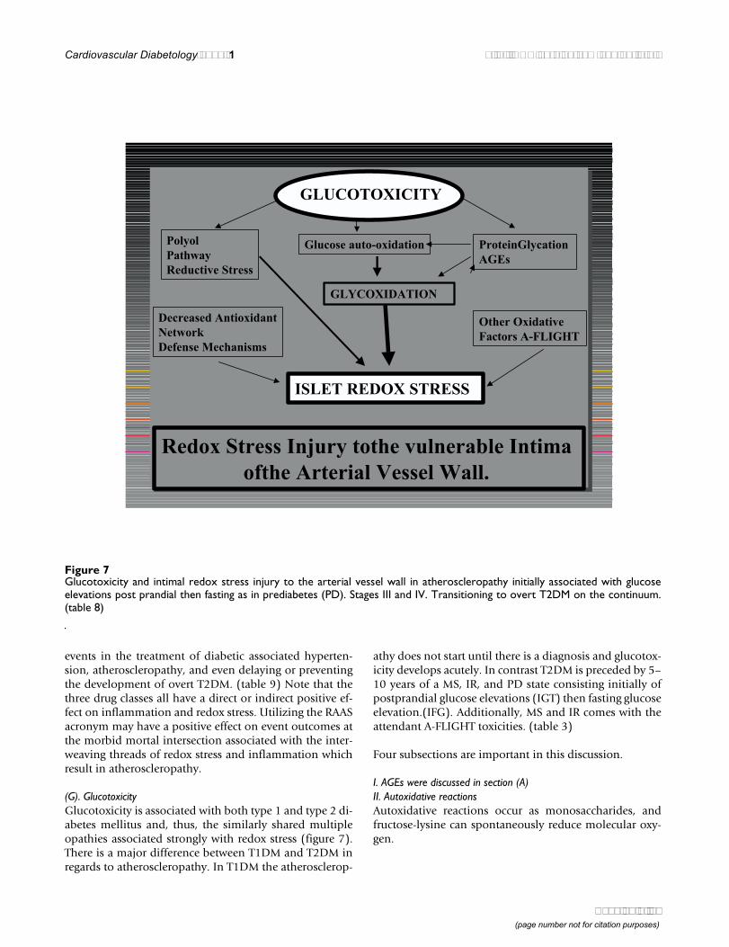
(G). GlucotoxicityGlucotoxicity is associated with both type 1 and type 2 di-abetes mellitus and, thus, the similarly shared multipleopathies associated strongly with redox stress (figure 7).There is a major difference between T1DM and T2DM inregards to atheroscleropathy. In T1DM the atherosclerop-
athy does not start until there is a diagnosis and glucotox-icity develops acutely. In contrast T2DM is preceded by 5–10 years of a MS, IR, and PD state consisting initially ofpostprandial glucose elevations (IGT) then fasting glucoseelevation.(IFG). Additionally, MS and IR comes with theattendant A-FLIGHT toxicities. (table 3)
Four subsections are important in this discussion.
I. AGEs were discussed in section (A)II. Autoxidative reactionsAutoxidative reactions occur as monosaccharides, andfructose-lysine can spontaneously reduce molecular oxy-gen.
Figure 7Glucotoxicity and intimal redox stress injury to the arterial vessel wall in atheroscleropathy initially associated with glucoseelevations post prandial then fasting as in prediabetes (PD). Stages III and IV. Transitioning to overt T2DM on the continuum.(table 8)
PolyolPathwayReductive Stress
Glucose auto-oxidation ProteinGlycationAGEs
Decreased AntioxidantNetworkDefense Mechanisms
Other OxidativeFactors A-FLIGHT
ISLET REDOX STRESS
Redox Stress Injury tothe vulnerable Intimaofthe Arterial Vessel Wall.
GLYCOXIDATION
GLUCOTOXICITY
Page 18 of 27(page number not for citation purposes)
Cardiovascular Diabetology 2002, 1 http://www.cardiab.com/content/1/1/3
The reduced oxygen products formed are O2-, OH-, and
H2O2. Each of these ROS can contribute to damaging lip-ids and proteins through cross-linking and fragmentation.[106–108]
The process of combined autoxidation and glycation isfrequently referred to as glycoxidation which is anothercommon process of protein modification. The ROS fromthese reactions serve not only as the source for autoxida-tion but also fuel the cycle of AGE formation (ROS begetROS).
Autoxidation occurs at the site of the protein componentembedded within the LDL cholesterol particle resulting inglycated LDL and glycoxidated LDL cholesterol whichcontribute to its retention just as oxidized LDL is retainedwithin the intima which initiates and sustains atherogen-esis and subsequent atheroscleropathy.
Native LDL is not atherogenic and is not retained withinthe intima; however, if it becomes modified by oxidation,glycation, glycoxidation or homocysteinated, it becomesmodified and retained (trapped to adjacent gly-cosaminoglycans and structural glycoproteins) to initiate,maintain, and accelerate the atherogenic process withinthe intima.
III. The Polyol – sorbitol pathwayThe polyol – sorbitol pathway is also driven by an excessproduction of glucose. Glucose is converted to sorbitol byaldose reductase at the expense of NADH/NAD(P)H be-ing converted to NAD+/NAD(P)+. Sorbitol is then con-verted to fructose by sorbitol dehydrogenase at theexpense of NAD+ NAD(P)+ being converted to NADH/NAD(P)H. [60–62]
This reductive stress (pseudohypoxia) of the polyol –sorbitol pathway thus amplifies the redox stress withinthe islet milieu. This singular pathway is of great impor-tance as it is the major pathway for supplying unpairedunstable electrons through the process of reduction. Thisreductive stress is dependent upon hyperglycemia associ-ated with both T1DM and T2DM. Postprandial hypergly-cemia results in reductive stress even before overt T2DMhas developed.
Were it not for the importance of this singular source of re-ductive stress, this review could have been entitled "Inti-mal Oxidative Stress".
IV. Glucose scavenging of nitric oxideEndothelial dysfunction is strongly associated with bothT1DM and T2DM. Brodsky et al. have recently been ableto demonstrate that glucose is capable of directly scaveng-ing NO resulting in the chemical inactivation of NO. They
were able to conclude that the glucose-mediated NO lossmay directly contribute to hypertension and endothelialdysfunction in diabetic patients. [109]
The authors were also able to show a glucose-mediateddecline in the lifetime of NO. These findings may have adirect, deleterious effect of decreasing the naturally occur-ring antioxidant capability of NO.
Glucotoxicity increases oxidative stress as demonstratedby increased 8-hydroxy-2'-deoxyguanosine (8-OhdG, amarker for oxidative stress) found in the urine and mono-nuclear cells from blood in T2DM patients.
Ihara et al. found higher levels of 8-OHdG and 4-hydroxy-2-nonenal- (HNE)-modified proteins in pancreatic beta-cells of GK rats (a model of non-obese type 2 diabetes)than in control Wistar rats. These levels increased propor-tionally with age. [110]
Section "(F). Free Fatty Acids" would lead one to believethat a lipocentric view is of extreme importance and maybe playing the dominant role in beta cell dysfunction andinsulin resistance.
Poitout and Robertson [111] have recently pointed out(with strong supporting data) that glucotoxicity is a pre-requisite for lipotoxicity. They propose that chronic hy-perglycemia (independent of hyperlipidemia) is toxic forbeta-cell function, whereas chronic hyperlipidemia is del-eterious only in the context of concomitant hyperglyc-emia. With time, both glucotoxicity and lipotoxicitycontribute to the progressive deterioration of glucosehomeostasis and beta cell dysfunction. Seldom do eitherof these two toxicities exist alone in the postprandial clin-ical setting of MS, IR, PD, and T2DM, and both contributeto the excess redox stress associated with the other A-FLIGHT toxicities, having an overall multiplicative effectwithin the intima and islet. [111]
Before leaving this section on glucotoxicity it is importantto note that cytosolic production of superoxide [O2'] re-sults in the activation of protein kinase C, increased for-mation of glucose-derived advanced glycation products,and an increased flux through the polyol – sorbitol path-way. Nishikawa T et al was able to show nicely that byblocking mitochondrial derived O2' with manganese su-peroxide dismutase or uncoupling mitochondrial oxida-tive phosphorylation they were able to prevent the abovecytosolic perturbations. [112] Additionally, Pennathur Set al were able to demonstrate in the Cynomologus monkeythat streptozotocin induced diabetes resulted in a hy-droxyl radical-like species which oxidized artery wall pro-teins. The oxidative products, ortho-tyrosine and meta-tyrosine correlated strongly with serum levels of glycated
Page 19 of 27(page number not for citation purposes)
Cardiovascular Diabetology 2002, 1 http://www.cardiab.com/content/1/1/3
hemoglobin. In these early lesions 3-nitrotyrosine was notcorrelated to the glycated hemoglobin. [113]
(H). Hypertension toxicityHypertension is associated with increased redox stress andROS activity. Furthermore, hypertension is associatedwith ROS mediated vascular damage and is closely associ-ated with the activation of Ang II (see section (A). Angi-otensin II) and its effect on the vascular NAD(P)H oxidasesuperoxide (O2
-) generating enzyme. [26]
Cellular sources of vascular superoxide production are theendothelial cell, vascular smooth muscle cell and adventi-tial fibroblasts. The major enzymatic sources areNAD(P)H oxidase, xanthine oxidase and, paradoxically,the eNOS enzyme (in the presence of oxidative stress ordeficiency of L-arginine or tetrahydrobiopterin).[114]
It is important to note that glucotoxicity is closely associ-ated with activation of the RAAS at the local, interstitialand tissue levels.
Recently, amylin has been implicated as being elevated inpatients who have a positive family history associatedwith hypertension and is elevated prior to the onset of hy-pertension when insulin remains at the normal level.Thus, amylin levels may become a screening tool for thedevelopment of future essential hypertension. [115]
Hypertension is associated with the clustering phenome-non of the metabolic syndrome and its importance to theoverall picture of redox stress is not to be underestimatedas it contributes significantly to the overall morbidity andmortality associated with T2DM. [116,117]
(H). Homocysteine toxicityThe general population of diabetics (T1DM and T2DM)will, in all probability, have the same amount of gene pol-ymorphism of the folate-dependent methylene tetrahy-drofolate reductase gene with subsequent mild tomoderate hyperhomocysteinemia (hHcy) which occurs in10–15% of the general population. [118–120]
This gene polymorphism is especially important in thoseindividuals with a decrease in dietary folate. hHcy can beimproved with folate supplementation and can improveendothelial-dependent endothelial cell dysfunction.[121]
Homocysteine (Hcy) is not usually elevated as a direct re-sult of diabetes unless there is an associated developmentof impaired renal function. As nephropathy develops,there is an associated elevation of total Hcy associatedwith a decline in glomerular filtration rate. [122]
This plays an extremely important role for those diabeticpatients on dialysis. [123] hHcy is thought to induce anoxidative inactivation of endothelial nitric oxide, in partby inhibiting or consuming the expression of cellular glu-tathione peroxidase (GPx). In heterozygous cystathioninebeta-synthase deficient -/+ mice, Weiss et al. were able torestore endothelial cell function by increasing cellular thi-ol and reducing glutathione pools and increasing GPx ac-tivity with restoration of the endothelial function. [124]
The ensuing cellular redox stress is magnified and totalHcy consumes NO by the indirect process of O2
- convert-ing NO to toxic peroxynitrite (ONOO-).
In addition to ONOO- formation, NO in conjunctionwith thiols and oxygen radicals generate nitrotyrosine andnitroarginine which compete for the substrate eNOS in afeedback mechanism, limiting further NO generation.[125–127] As a result, there is endothelial cell dysfunc-tion, endothelial cell toxicity and endothelial cell loss, in-creased ROS, increased ONOO- and decreased NOassociated with hHcy. [128] hHcy is multiplicative in na-ture and even though its effects may occur later in T2DMthan the other associated toxicities, it has a devastating ef-fect on endothelial cell function.
Presently, we know there are other toxicities operatingwithin the renal glomerulus producing microalbuminuria(reflecting endothelial cell dysfunction and damage) at astage prior to the declining glomerular filtration rate re-sponsible for hHcy.
A recent clinical study by Maejima et al.[129] revealed sig-nificant elevated levels of ONOO- peroxynitrite (by Griessmethod) in 126 T2DM patients as compared to 76 non-diabetic controls. ONOO- levels were related to the pres-ence of hypertension and advanced microvascular com-plications. In addition, ONOO- correlated positively withelevations in AGEs and serum lipid peroxide.
These data support the hypothesis that decreased en-dothelium-dependent vasodilatation in diabetic subjectsis associated with the impaired action of NO secondary toits consumption from redox stress rather than decreasedNO production from vascular endothelium. Clinically,abnormal NO metabolism is related to advanced diabeticmicrovascular complications. Zhang et al.[130] were ableto demonstrate that increased concentrations of Hcy re-sulted in a decreased NO response to bradykinin and L-ar-ginine.
They were able to show that Hcy stimulated the formationof superoxide anions and peroxynitrite with increased lev-els of nitrotyrosine. The addition of 5-methyltetrahydro-folate restored NO responses to bradykinin and L-arginine
Page 20 of 27(page number not for citation purposes)
Cardiovascular Diabetology 2002, 1 http://www.cardiab.com/content/1/1/3
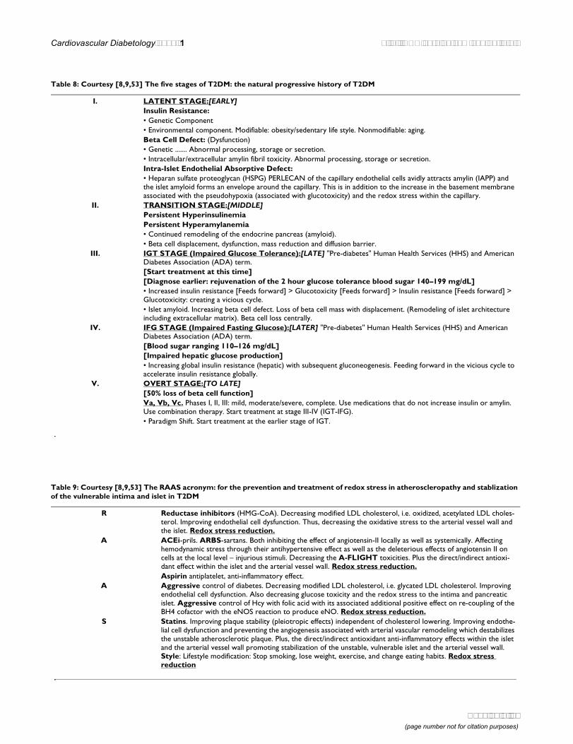
Table 8: Courtesy [8,9,53] The five stages of T2DM: the natural progressive history of T2DM
I. LATENT STAGE:[EARLY]Insulin Resistance:• Genetic Component• Environmental component. Modifiable: obesity/sedentary life style. Nonmodifiable: aging.Beta Cell Defect: (Dysfunction)• Genetic ....... Abnormal processing, storage or secretion.• Intracellular/extracellular amylin fibril toxicity. Abnormal processing, storage or secretion.Intra-Islet Endothelial Absorptive Defect:• Heparan sulfate proteoglycan (HSPG) PERLECAN of the capillary endothelial cells avidly attracts amylin (IAPP) and the islet amyloid forms an envelope around the capillary. This is in addition to the increase in the basement membrane associated with the pseudohypoxia (associated with glucotoxicity) and the redox stress within the capillary.
II. TRANSITION STAGE:[MIDDLE]Persistent HyperinsulinemiaPersistent Hyperamylanemia• Continued remodeling of the endocrine pancreas (amyloid).• Beta cell displacement, dysfunction, mass reduction and diffusion barrier.
III. IGT STAGE (Impaired Glucose Tolerance):[LATE] "Pre-diabetes" Human Health Services (HHS) and American Diabetes Association (ADA) term.[Start treatment at this time][Diagnose earlier: rejuvenation of the 2 hour glucose tolerance blood sugar 140–199 mg/dL]• Increased insulin resistance [Feeds forward] > Glucotoxicity [Feeds forward] > Insulin resistance [Feeds forward] > Glucotoxicity: creating a vicious cycle.• Islet amyloid. Increasing beta cell defect. Loss of beta cell mass with displacement. (Remodeling of islet architecture including extracellular matrix). Beta cell loss centrally.
IV. IFG STAGE (Impaired Fasting Glucose):[LATER] "Pre-diabetes" Human Health Services (HHS) and American Diabetes Association (ADA) term.[Blood sugar ranging 110–126 mg/dL][Impaired hepatic glucose production]• Increasing global insulin resistance (hepatic) with subsequent gluconeogenesis. Feeding forward in the vicious cycle to accelerate insulin resistance globally.
V. OVERT STAGE:[TO LATE][50% loss of beta cell function]Va, Vb, Vc. Phases I, II, III: mild, moderate/severe, complete. Use medications that do not increase insulin or amylin. Use combination therapy. Start treatment at stage III-IV (IGT-IFG).• Paradigm Shift. Start treatment at the earlier stage of IGT.
Table 9: Courtesy [8,9,53] The RAAS acronym: for the prevention and treatment of redox stress in atheroscleropathy and stablization of the vulnerable intima and islet in T2DM
R Reductase inhibitors (HMG-CoA). Decreasing modified LDL cholesterol, i.e. oxidized, acetylated LDL choles-terol. Improving endothelial cell dysfunction. Thus, decreasing the oxidative stress to the arterial vessel wall and the islet. Redox stress reduction.
A ACEi-prils. ARBS-sartans. Both inhibiting the effect of angiotensin-II locally as well as systemically. Affecting hemodynamic stress through their antihypertensive effect as well as the deleterious effects of angiotensin II on cells at the local level – injurious stimuli. Decreasing the A-FLIGHT toxicities. Plus the direct/indirect antioxi-dant effect within the islet and the arterial vessel wall. Redox stress reduction.Aspirin antiplatelet, anti-inflammatory effect.
A Aggressive control of diabetes. Decreasing modified LDL cholesterol, i.e. glycated LDL cholesterol. Improving endothelial cell dysfunction. Also decreasing glucose toxicity and the redox stress to the intima and pancreatic islet. Aggressive control of Hcy with folic acid with its associated additional positive effect on re-coupling of the BH4 cofactor with the eNOS reaction to produce eNO. Redox stress reduction.
S Statins. Improving plaque stability (pleiotropic effects) independent of cholesterol lowering. Improving endothe-lial cell dysfunction and preventing the angiogenesis associated with arterial vascular remodeling which destabilizes the unstable atherosclerotic plaque. Plus, the direct/indirect antioxidant anti-inflammatory effects within the islet and the arterial vessel wall promoting stabilization of the unstable, vulnerable islet and the arterial vessel wall. Style: Lifestyle modification: Stop smoking, lose weight, exercise, and change eating habits. Redox stress reduction
Page 21 of 27(page number not for citation purposes)
Cardiovascular Diabetology 2002, 1 http://www.cardiab.com/content/1/1/3
agonists. In addition, scavengers of peroxynitrite and SODmimetics reversed the Hcy-induced suppression of NOproduction by endothelial cells. Concentrations of Hcygreater than 20 �M produced a significant indirect sup-pression of eNOS activity without any discernible effectson its expression.
Li et al. just published an article showing an unexpectedeffect of Hcy-induced oxidative stress resulting in an in-crease of 3-hydroxy-3-methylglutaryl coenzyme A reduct-ase in vascular endothelial cells, as well as decreasingendothelial NO.
They were also able to demonstrate that "statins" (Table7) were able to increase NO as well as decreasing cellularcholesterol. [131]
Stuhlinger MC et al were able to demonstrate that Hcy im-pairs the nitric oxide synthase pathway. Homocysteine in-hibits dimethylarginine dimethylaminohydrolase(DDAH) which is responsible for degrading ADMA.
This effect of Hcy causes the endogenous inhibitor of ni-tric oxide synthase, ADMA to accumulate and inhibit ni-tric oxide synthesis. This effect helps to explain thedeleterious effect of Hcy on the endothelial cells ability topromote vasodilatation and associated endothelial celldysfunction with decreased NO synthesis. [132]
(T). Triglyceride toxicityMultiple lipases (intestinal, muscular – both skeletal andcardiac-, adipose, and hepatic) are responsible for the dy-namic flux between the long chain fatty acids (LCACoAesters) and the glycerol molecular backbone of the triglyc-erides (see section (F). Free Fatty Acids").
Hypertriglyceridemia certainly plays a role in toxicity re-garding the development of redox stress, not only its rolein lipotoxicity and FFA toxicity discussed previously, butindependently as its own marker of toxicity. There is aclose association of hypertriglyceridemia and the athero-genic small dense LDL cholesterol particles which aremore likely to be oxidized and contribute to redox stress.This condition is central to the lipid triad [133].
We need to recall that Apo E is responsible for carryingthis lipid fraction and that the Apo E -/- knockout mousedevelops atherosclerosis at an accelerated rate. We need toalso bear in mind that gene polymorphism may play arole in the development of ADIA since Apo E is an impor-tant part of all amyloid formation and stabilization. Kahnet al. were able to demonstrate in the human islet amyloidpolypeptide transgenic mouse model that these mice didnot develop islet amyloid unless fed a high fat diet [134].
Stored neutral triglycerides provide the substrate for FFAproduction which can be immobilized immediately by ex-ercise or stress induced lipolysis.
When these are stored in ectopic non-adipose cells such asthe cardiac and skeletal myocyte, the endothelial cell orthe islet beta cell, they are capable of causing cellular dys-function or lipoapoptosis as discussed in the sections "(F).Free fatty acids" and "(L). Lipotoxicity – Specific".
As stated earlier in this paper, it is difficult to separatethese three moieties as they are closely interconnectedwith each other and within the manifold A-FLIGHT toxic-ities.
Redox stress and matrix metalloproteinasesElevated redox stress is associated with an increase in ma-trix metalloproteinase (MMP) activity, especially the in-ducible MMP-9 [135].
This would be associated with an increase in extracellularmatrix (ECM) remodeling and would contribute to in-creased intima media thickness within the arterial vesselwall. This would accelerate the process underlying thesimilar mechanism of AGEs with a stiffening and de-creased compliance of the arterial vessel wall which wouldcontribute to diastolic dysfunction of the arterial vesselwall. The acceleration of the stiffness of the arterial vesselwall would contribute to hypertension, specifically systo-lic hypertension. This mechanism may be in play in theclustering of hypertension in the metabolic syndrome.
Cells are dependent on integrin matrix ligand bindingsites and MMP-9 is a basement membrane degrading en-zyme. Cells are constantly re-establishing new integrinmatrix binding sites. However, if there is a complete dis-connection of integrin matrix binding sites due to a robustincrease in MMP-9, the cell may undergo apoptosis.MMP-9 was recently shown to be elevated in diabetes mel-litus and, in addition, the role of redox stress was shownto play an important role [135].
A robust activation of MMP-9 may result in a completedisconnection of the beta cell and the surrounding ECMwith resultant apoptosis. Recently, in our laboratory, wehave been able to demonstrate decreased endothelial celldensity with increased apoptosis of endothelial cells inthe hearts of mice treated with alloxan vs. controls.
We were also able to show a decrease in NO and an in-crease in peroxynitrite and ROS in these same animalsthus, linking the importance of cellular apoptosis, MMP-9 and redox stress. We then compared these findings of al-loxan-induced diabetes in MMP-9 knockout mice to al-loxan-induced diabetes in the wild type. Alloxan-induced
Page 22 of 27(page number not for citation purposes)
Cardiovascular Diabetology 2002, 1 http://www.cardiab.com/content/1/1/3
diabetes MMP-9 -/- mice did not have induced apoptosisand did not have a decrease in endothelial cell densitywhen compared to wild type alloxan-induced diabetes(unpublished data).
These findings may apply to the beta cell within the islet,as all cells require an integrin matrix binding for survival.The MMP-9 may also decrease the larger size amylin de-rived islet amyloid fibrils to the more intermediate sizetoxic amyloid particles and contribute to apoptosis as de-scribed by Janson et al.[136] (Table 5).
MMP-9 has also been shown to be elevated in laminitichorses having digestion of the basement membranes withresultant separation of the epidermal and dermal lamina.[137] These same processes within the islet could be re-sponsible for a loss of intracapillary endothelial cellswhich would decrease the rate at which they could pick upnewly synthesized insulin and transport it to the systemiccirculation, and provide a mechanism for the delay in firstphase insulin secretion which is typical of T2DM and evenimpaired glucose tolerance.
MMP-9 may even play a role in the clearing of ECM in or-der to allow for the space-occupying lesion of ADIA dep-osition. Redox stress (signaling) activates MMPs (Table 5).
ConclusionThroughout this review, we have tried to remain focusedon the relationship between redox stress and ROS in theintima and at times the islet, and how these two interactwith the multiplicative effect of the A-FLIGHT toxicities ofMS, IR, PD, and overt T2DM to induce atheroscleropathy.The reader will note that redox stress and ROS operatethrough similar mechanisms and will operate in a similarfashion in other chronic disease states such as chronic in-flammatory diseases (pancreatitis, rheumatoid arthritis,ulcerative colitis, Crohn's), ageing, cancer, ischemia/ischemia-reperfusion injury, hypertension, diastolic/systolic dysfunction, congestive heart failure, diabetic andnondiabetic nephropathy and neurodegenerative diseas-es.
When redox homeostasis transitions to redox stress, redoxsignaling ensues in all tissues and organs regardless of themultiple similar or dissimilar etiologies.
Redox stress is a redox signaling system. [101,138]Reflections and future directionsAn article entitled "Diabetes as a manifold disease" (pre-viously published in the February 8th, 1902 issue of JA-MA) was reprinted in the February 13th, 2002 issue ofJAMA, in the section on 100 years ago. [139,140]
T2DM and associated atheroscleropathy remain a hetero-geneous and manifold disease not only in their etiologybut also in their manifold toxicities associated with MS,IR, PD, and T2DM.
In 1902, the author (unknown) could not have envi-sioned the exponential growth of T2DM and atheroscle-ropathy we are currently experiencing.
Just as this author pointed to a new concept, we have at-tempted, in this review, to outline the important contem-porary concept of intimal redox stress and the rusting –rancid intima within vulnerable atherosclerotic plaques.
Treatment and potential prevention of these diseases canbe accomplished through global risk reduction of themanifold toxicities by using the currently available treat-ment modalities we now have at our disposal. [141]
If the current trend continues (due to the current epidemicof our adolescent youth and the aging baby boom gener-ation) these patients are going to be seen in our officeswith increasing frequency.
Recently, Dagogo-Jack S discussed primary prevention ofdiabetes, secondary prevention (of diabetic complica-tions), and tertiary prevention (of morbidity and mortali-ty from established diabetic complications) utilizingmultiple drug targets in the management of T2DM.
Currently, a focus on global risk reduction is necessary fora more complete approach by physicians providing carefor the diabetic patient. [142,143] By aggressively reduc-ing the elevated substrates producing the A-FLIGHT toxic-ities and ROS (table 3) and using the simple RAASacronym (table 9) we may be able to restore our individ-ual, endogenous, potent, antioxidant network. A betterunderstanding of redox stress and redox homeostasis willenable eac h of us to develop a shift in the treatment par-adigm for the quartet of MS, IR, PD, T2DM, and the asso-ciated cardiovascular atheroscleropathy due to the loss ofmetabolic and redox homeostasis.
Abbreviations8-OhdG: 8-hydroxy-2'-deoxyguanosine; ADA AmericanDiabetes Association; ADIA: amylin derived islet amyloid;AFEs: advanced fructosylation endproducts; AGEs: ad-vanced glycation endproducts; Ang II: angiotensin II; AT-1: angiotensin type 1; ACEi : angiotensin converting en-zyme inhibitors; ARBS : angiotensin II receptor blockers;eNO: endothelial nitric oxide; eNOS; endothelial nitricoxide synthase; FFA: free fatty acid; GPx: glutathione per-oxidase; GSH: glutathione reduced form; GSSG: glutath-ione disulfide; Hcy: homocysteine; HNE: 4-hydroxy-2-nonenal; IAPP: islet amyloid polypeptide; iNO: inducible
Page 23 of 27(page number not for citation purposes)

Cardiovascular Diabetology 2002, 1 http://www.cardiab.com/content/1/1/3
nitric oxide; iNOS: inducible nitric oxide synthase;ISTAPs: intermediate sized toxic amyloid particles; LCA-CoA: long chain acyl-Coenzyme A; NFkappa B: nuclearfactor kappa B; nNOS: neural nitric oxide synthase; NO:nitric oxide; NOS: nitric oxide synthase; O2-: superoxide;OH-: hydroxyl radical; ONOO-: peroxynitrite; RAGE: re-ceptor for advanced glycation endproducts; SOD: super-oxide dismutase; T1DM: type 1 diabetes mellitus; T2DM:type 2 diabetes mellitus.
AcknowledgmentA part of this study was supported by National Institutes of Health grant HL-71010. The authors are aware that this review is lengthy but remains incomplete as each section discussed is worthy of at least a chapter. We have attempted to share the complex interaction of the multiple toxicities and redox stress to provide a database of information and references for those who wish to continue their studies in this exciting, exponentially growing field of study. We wish to thank our many friends and colleagues who have helped us grow in this field of study. To our heroic professors, who instilled in us the quest for knowledge and the lack of fear, to ask, "why" and "why not".
References1. Hayden MR, Tyagi SC: Arterial vascular remodeling: the en-
dothelial cell's central role. Mo Med 1998, 95(5):213-2172. Hayden MR, Tyagi SC: Chapter. Atherosclerosis: Implications
of angiotensin II and the AT-1 receptor. Angiotensin II ReceptorBlockade: Physiological and Clinical Implications (Edited by: Dhalla NS, Za-hradka P, Dixon I, Beamish R) Kluwer Academic publishers. Boston, Ma1998, 2:233-243
3. Hayden MR, Tyagi SC: Arteriogenesis: Angiogenesis within Un-stable Atherosclerotic Plaques – Interactions with Extracel-lular Matrix. Curr Interv Cardiol Rep 2000, 3:218-227
4. Hayden MR: Atherosclerosis and Plaque Angiogenesis: A Ma-lignant Transformation. Submitted paper. Pathology and Clinical clas-sification of Vulnerable Plaque. VP.org 2001, 3:[http://www.vp.org][http://www.vp.org/ResourceCenter/Pete_Hayden_Angiogenesis.html]
5. Joslin EP: Arteriosclerosis and Diabetes. Annals of Clinical Medicine1927, 5(13):1061-1079
6. Expert Panel on Detection, Evaluation, and Treatment ofHigh Blood Cholesterol in Adults. Executive summary of thethird report of the National Cholesterol Education Program(NCEP) expert panel on detection, evaluation, and treat-ment of high blood cholesterol in adults (Adult TreatmentPanel III). JAMA 2001, 285:2486-2497
7. Oberley LW: Free radical and diabetes. Free Radic Bio Med 1988,5:113-124
8. Hayden MR, Tyagi SC: "A" is for amylin and amyloid in type 2diabetes mellitus. JOP. J Pancreas (Online) 2001, 2(4):124-139 [www.joplink.net]
9. Hayden MR, Tyagi SC: Islet redox stress: the manifold toxicitiesof insulin resistance, metabolic syndrome, and amylin de-rived islet amyloid in type 2 diabetes mellitus. JOP. J Pancreas(Online) 2002, 3(4):86-108 [www.joplink.net]
10. Kylin E: Studien ueber das Hypertonie-Hyperglyka"mie-Hy-perurika"miesyndrom. Zentralblatt fuer Innere Medizin 1923,44:105-127
11. Himsworth H: Diabetes mellitus: a differentiation into insulin-sensitive and insulin-insensitive types. Lancet 1936, 1:127-130
12. Yalow RS, Glick SM, Roth J, Berson SA: Plasma insulin and growthhormone levels in obesity and diabetes. Ann NY Acad Sci 1965,131(1):357-373
13. Reaven GM: Banting Lecture 1988. Role of insulin resistance inhuman disease. Diabetes 1988, 37:1595-1607
14. Alberti KGM, Zimmet PZ, For the WHO Consultation: Definition,diagnosis and classification of diabetes mellitus and its com-plications. Part 1: Diagnosis and classification of diabetesmellitus, provisional report of a WHO consultation. DiabetMed 1998, 15:539-553
15. Ford ES, Giles WH, Dietz WH: Prevalence of the metabolic syn-drome among US adults:Findings from the third national
health and nutrition examination survey. JAMA 2002,287(3):356-359
16. Jones LC, Clark A: Beta cell neogenesis in type 2 diabetes mel-litus. Diabetes 2001, 50(Suppl 1):S186-S187
17. Guiot Y, Sempoux C, Moulin P, Rahier J: No decrease of the betacell mass in type 2 patients. Diabetes 2001, 50(Suppl 1):S188
18. Reaven GM: Banting Lecture 1988. Role of insulin resistance inhuman disease. Diabetes 1988, 37:1595-1607
19. Reaven GM: Metabolic syndrome: pathophysiology and impli-cations for management of cardiovascular disease. Circulation2002, 106(3):286-288
20. Tahmasebi M, Puddefoot JR, Inwang ER, Vinson GP: The tissue ren-in-angiotensin system in human pancreas. J Endocrinol 1999,161:317-322
21. Carlsson PO: The renin-angiotensin system in the endocrinepancreas. JOP. J Pancreas (Online) 2001, 2:26-32
22. Nickenig G, Roling J, Strehlow K, Schnabel P, Bohm M: Insulin induc-es upregulation of vascular AT-1 receptor gene expressionby posttranscriptional mechanisms. Circulation 1998, 98:2453-2460
23. Folli F, Saad MJ, Velloso L, Hansen H, Carandente O, Feener EP, KahnCR: Crosstalk between insulin and angiotensin II signallingsystems. Exp Clin Endocrinol Diabetes 1999, 107:133-139
24. Cooper ME, McNally PG, Phillips PA, Johnston CI: Amylin stimu-lates plasma renin concentrations in humans. Hypertension1995, 26:460-464
25. Ikeda T, Iwata K, Ochi H: Effect of insulin, proinsulin, and amylinon renin release from perfused rat kidney. Metabolism 2001,50:763-766
26. Griendling KK, Sorescu D, Ushio-Fukai M: NAD(P)H oxidase: rolein cardiovascular biology and disease. Circ Res 2000, 86:494-501
27. Munzel T, Keaney JF Jr: Are ACE inhibitors a "magic bullet"against oxidative stress? Circulation 2001, 104:1571-1574
28. Heart Outcomes Prevention Evaluation (HOPE) Study Investigators:Effects of ramipril on cardiovascular and microvascular out-comes in people with diabetes mellitus: results of the HOPEstudy and MICRO-HOPE substudy. Lancet 2000, 355:253-259
29. Griendling KK, Harrison DG: Out, damned dot: studies of theNADPH oxidase in atherosclerosis. J Clin Invest 2001, 108:1423-1424
30. Williamson JR, Kilo C: Glucotoxicity and diabetic vascular dis-ease. Medical Dialogue on Diabetes 1989, 8:8-10
31. Fujimori E: Cross-linking and fluorescence changes of collagenby glycation and oxidation. Biochim Biophys Acta 1989, 998:105-110
32. Dills WL Jr: Protein fructosylation: fructose and the Maillardreaction. Am J Clin Nutr 1993, 58(5 Suppl):779-787S
33. McPherson JD, Shilton BH, Walton DJ: Role of fructose in glyca-tion and cross-linking of proteins. Biochemistry 1998, 27:1901-1907
34. Aronson D, Rayfield EJ: How hyperglycemia promotes athero-sclerosis: molecular mechanisms. Cardiovasc Diabetol 2002,1(1):1 [http://www.biomedcentral.com/1475-2840/1/1]
35. Wendt T, Bucciarelli L, Qu W, Lu Y, Yan SF, Stern DM, Schmidt AM:Receptor for advanced glycation endproducts (RAGE) andvascular inflammation: insights into the pathogenesis ofmacrovascular complications in diabetes. Curr Atheroscler Rep2002, 4:228-237
36. Yan SD, Zhu H, Zhu A, Golabek A, Du H, Roher A, et al: Receptor-dependent cell stress and amyloid accumulation in systemicamyloidosis. Nat Med 2000, 6:643-651
37. Arai K, Iizuka S, Tada Y, Oikawa K, Taniguchi N: Increase in the gly-cosylated form of erythrocyte CuZnSOD in diabetes andclose association of non-enzymatic glycosylation with en-zyme activity. Biochim Biophys Acta 1987, 924:292-296
38. Chari SN, Nath N, Rathi AB: Glutathione and its redox systemin diabetic polymorphonuclear leukocytes. Am J Med Sci 1984,287:14-15
39. Jennings PE, Chirico S, Jones AF, Lunec J, Barnett AH: Vitamin Cmetabolites and microangiopathy in diabetes mellitus. Diabe-tes Res 1987, 6:151-154
40. Sinclair AJ, Lunec J, Girling AJ, Barnett AH: Modulators of free rad-ical activity in diabetes mellitus: role of ascorbic acid. EXS1992, 62:151-154
Page 24 of 27(page number not for citation purposes)
http://www.ncbi.nlm.nih.gov/entrez/query.fcgi?cmd=Retrieve&db=PubMed&dopt=Abstract&list_uids=9604667
http://www.ncbi.nlm.nih.gov/entrez/query.fcgi?cmd=Retrieve&db=PubMed&dopt=Abstract&list_uids=9604667
http://www.ncbi.nlm.nih.gov/entrez/query.fcgi?cmd=Retrieve&db=PubMed&dopt=Abstract&list_uids=9604667
http://www.ncbi.nlm.nih.gov/entrez/query.fcgi?cmd=Retrieve&db=PubMed&dopt=Abstract&list_uids=5216975
http://www.ncbi.nlm.nih.gov/entrez/query.fcgi?cmd=Retrieve&db=PubMed&dopt=Abstract&list_uids=5216975
http://www.ncbi.nlm.nih.gov/entrez/query.fcgi?cmd=Retrieve&db=PubMed&dopt=Abstract&list_uids=5216975
http://www.ncbi.nlm.nih.gov/entrez/query.fcgi?cmd=Retrieve&db=PubMed&dopt=Abstract&list_uids=3056758
http://www.ncbi.nlm.nih.gov/entrez/query.fcgi?cmd=Retrieve&db=PubMed&dopt=Abstract&list_uids=3056758
http://www.ncbi.nlm.nih.gov/entrez/query.fcgi?cmd=Retrieve&db=PubMed&dopt=Abstract&list_uids=3056758
http://www.ncbi.nlm.nih.gov/entrez/query.fcgi?cmd=Retrieve&db=PubMed&dopt=Abstract&list_uids=9686693
http://www.ncbi.nlm.nih.gov/entrez/query.fcgi?cmd=Retrieve&db=PubMed&dopt=Abstract&list_uids=9686693
http://www.ncbi.nlm.nih.gov/entrez/query.fcgi?cmd=Retrieve&db=PubMed&dopt=Abstract&list_uids=9686693
http://www.ncbi.nlm.nih.gov/entrez/query.fcgi?cmd=Retrieve&db=PubMed&dopt=Abstract&list_uids=9686693
http://www.ncbi.nlm.nih.gov/entrez/query.fcgi?cmd=Retrieve&db=PubMed&dopt=Abstract&list_uids=3056758
http://www.ncbi.nlm.nih.gov/entrez/query.fcgi?cmd=Retrieve&db=PubMed&dopt=Abstract&list_uids=3056758
http://www.ncbi.nlm.nih.gov/entrez/query.fcgi?cmd=Retrieve&db=PubMed&dopt=Abstract&list_uids=3056758
http://www.ncbi.nlm.nih.gov/entrez/query.fcgi?cmd=Retrieve&db=PubMed&dopt=Abstract&list_uids=9832492
http://www.ncbi.nlm.nih.gov/entrez/query.fcgi?cmd=Retrieve&db=PubMed&dopt=Abstract&list_uids=9832492
http://www.ncbi.nlm.nih.gov/entrez/query.fcgi?cmd=Retrieve&db=PubMed&dopt=Abstract&list_uids=9832492
http://www.ncbi.nlm.nih.gov/entrez/query.fcgi?cmd=Retrieve&db=PubMed&dopt=Abstract&list_uids=9832492
http://www.ncbi.nlm.nih.gov/entrez/query.fcgi?cmd=Retrieve&db=PubMed&dopt=Abstract&list_uids=7649582
http://www.ncbi.nlm.nih.gov/entrez/query.fcgi?cmd=Retrieve&db=PubMed&dopt=Abstract&list_uids=7649582
http://www.ncbi.nlm.nih.gov/entrez/query.fcgi?cmd=Retrieve&db=PubMed&dopt=Abstract&list_uids=7649582
http://www.ncbi.nlm.nih.gov/entrez/query.fcgi?cmd=Retrieve&db=PubMed&dopt=Abstract&list_uids=2506934
http://www.ncbi.nlm.nih.gov/entrez/query.fcgi?cmd=Retrieve&db=PubMed&dopt=Abstract&list_uids=2506934
http://www.ncbi.nlm.nih.gov/entrez/query.fcgi?cmd=Retrieve&db=PubMed&dopt=Abstract&list_uids=2506934
http://www.ncbi.nlm.nih.gov/entrez/query.fcgi?cmd=Retrieve&db=PubMed&dopt=Abstract&list_uids=3567220
http://www.ncbi.nlm.nih.gov/entrez/query.fcgi?cmd=Retrieve&db=PubMed&dopt=Abstract&list_uids=3567220
http://www.ncbi.nlm.nih.gov/entrez/query.fcgi?cmd=Retrieve&db=PubMed&dopt=Abstract&list_uids=3567220
http://www.ncbi.nlm.nih.gov/entrez/query.fcgi?cmd=Retrieve&db=PubMed&dopt=Abstract&list_uids=3567220
http://www.ncbi.nlm.nih.gov/entrez/query.fcgi?cmd=Retrieve&db=PubMed&dopt=Abstract&list_uids=6731472
http://www.ncbi.nlm.nih.gov/entrez/query.fcgi?cmd=Retrieve&db=PubMed&dopt=Abstract&list_uids=6731472
http://www.ncbi.nlm.nih.gov/entrez/query.fcgi?cmd=Retrieve&db=PubMed&dopt=Abstract&list_uids=6731472
http://www.ncbi.nlm.nih.gov/entrez/query.fcgi?cmd=Retrieve&db=PubMed&dopt=Abstract&list_uids=3436115

Cardiovascular Diabetology 2002, 1 http://www.cardiab.com/content/1/1/3
41. Giugliano D, Ceriello A, Paolisso G: Diabetes mellitus, hyperten-sion, and cardiovascular disease: which role for oxidativestress? Metabolism 1995, 44:363-368
42. Duplain H, Burcelin R, Sartori C, Cook S, Egli M, Lepori M, ScherrerU, et al: Insulin resistance, hyperlipidemia, and hypertensionin mice lacking endothelial nitric oxide synthase. Circulation2001, 104:342-345
43. Miyamoto Y, Saito Y, Kajiyama N, Yoshimura M, Shimasaki Y, Nakaya-ma M, et al: Endothelial nitric oxide synthase gene is positivelyassociated with essential hypertension. Hypertension 1998, 32:3-8
44. Stuhlinger MC, Abbasi F, Chu JW, Lamendola C, McLaughlin TL,Cooke JP, Reaven GM, Tsao PS: Relationship between insulin re-sistance and an endogenous nitric oxide synthase inhibitor.JAMA 2002, 287(11):1420-1426
45. Nash DT: Insulin resistance, ADMA levels, and cardiovasculardisease. JAMA 2002, 287(11):1451-1452
46. Christ M, Bauersachs J, Liebetrau C, Heck M, Gunther A, Wehling M:Glucose Increases Endothelial-Dependent Superoxide For-mation in Coronary Arteries by NAD(P)H Oxidase Activa-tion: Attenuation by the 3-Hydroxy-3-MethylglutarylCoenzyme A Reductase Inhibitor Atorvastatin. Diabetes 2002,51(8):2648-2652
47. Stepp DW, Ou J, Ackerman AW, Welak S, Klick D, Pritchard KA Jr:Native LDL and minimally oxidized LDL differentially regu-late superoxide anion in vascularendothelium in situ. Am JPhysiol Heart Circ Physiol 2002, 283(2):H750-759
48. Zou MH, Shi C, Cohen RA: Oxidation of the zinc-thiolate com-plex and uncoupling of endothelial nitric oxide synthase byperoxynitrite. J Clin Invest 2002, 109(6):817-826
49. Verma S, Wang CH, Li SH, Dumont AS, Fedak PW, Badiwala MV,Dhillon B, Weisel RD, LI RK, Mickle DA, Stewart DJ: A self fulfillingprophecy: C-reactive protein attenuates nitric oxide produc-tion and inhibits angiogenesis. Circulation 2002, 106(8):913-919
50. Guzik TJ, Mussa S, Gastaldi D, Sadowski J, Ratnatunga C, Pillai R,Channon KM: Mechanisms of increased vascular superoxideproduction in human diabetes mellitus: role of NAD(P)H ox-idase and endothelial nitric oxide synthase. Circulation 2002,105(14):1656-1662
51. Vasquez-Vivar J, Kalyanaraman B, Martasek P, Hogg N, Masters BS,Karoui H, Tordo P, Pritchard KA Jr: Superoxide generation byendothelial nitric oxide synthase: the influence of cofactors.Proc Natl Acad Sci U S A 1998, 95(16):9220-9225
52. Wong TY, Klein R, Sharrett AR, Schmidt MI, Pankow JS, Couper DJ,Klein BE, Hubbard LD, Duncan BB: Retinal arteriolar narrowingand risk of diabetes mellitus in middle-aged persons. JAMA2002, 287(19):2528-2533
53. Hayden MR: Islet Amyloid, Metabolic Syndrome, and the Nat-ural Progressive History of Type 2 Diabetes Mellitus. JOP. JPancreas (Online) 2002, 3(5):126-138 [www.joplink.net]
54. Hoffmann J, Haendeler J, Aicher A, Rossig L, Vasa M, Zeiher AM, Dim-meler S: Aging enhances the sensitivity of endothelial cells to-ward apoptotic stimuli: important role of nitric oxide. Circ Res2001, 89:709-715
55. Harman D: Extending functional life span. Exp Gerontol 1998,33:95-112
56. Lenaz G: Role of mitochondria in oxidative stress and ageing.Biochim Biophys Acta 1998, 1366:53-67
57. Ye JM, Lim-Fraser M, Cooney GJ, Cooper Gj, Iglesias MA, WatsonDG, et al: Evidence that amylin stimulates lipolysis in vivo: apossible mediator of induced insulin resistance. Am J Physiol En-docrinol Metab 2001, 280:E562-569
58. Yan SD, Schmidt AM, Anderson GM, Zhang J, Brett J, Zou YS, et al:Enhanced cellular oxidant stress by the interaction of ad-vanced glycation end products with their receptors/bindingproteins. J Biol Chem 1994, 269:9889-9897
59. Janson J, Ashley RH, Harrison D, McIntyre S, Butler PC: The mech-anism of islet amyloid polypeptide toxicity is membrane dis-ruption by intermediate-sized toxic amyloid particles.Diabetes 1999, 48:491-498
60. Williamson JR, Kilo C, Ido Y: The role of cytosolic reductivestress in oxidant formation and diabetic complications. Diabe-tes Res Clin Pract 1999, 45:81-82
61. Ido Y, Kilo C, Williamson JR: Interactions between the sorbitolpathway, non-enzymatic glycation, and diabetic vasculardysfunction. Nephrol Dial Transplant 1996, 11(Suppl 5):72-75
62. Williamson JR, Chang K, Frangos M, Hasan KS, Ido Y, Kawamura T, etal: Hyperglycemic Pseudohypoxia and diabetic complica-tions. Diabetes 1993, 42:801-813
63. Abaci A, Oguzhan A, Kahraman S, Eryol NK, Unal S, Arinc H, Ergin A:Effect of diabetes mellitus on formation of coronary collater-al vessels. Circulation 1999, 99(17):2239-2242
64. McGarry JD: Banting lecture 2001: dysregulation of fatty acidmetabolism in the etiology of type 2 diabetes. Diabetes 2002,51:7-18
65. Bakker SJ, IJzerman RG, Teerlink T, Westerhoff HV, Gans RO, HeineRJ: Cytosolic triglycerides and oxidative stress in centralobesity: the missing link between excessive atherosclerosis,endothelial dysfunction, and beta cell failure? Atherosclerosis2000, 148:17-21
66. Gotto AM Jr: Triglyceride as a risk factor for coronary arterydisease. Am J Cardiol 1998, 82:22-25Q
67. Grundy SM: Hypertriglyceridemia, atherogenic dyslipidemia,and the metabolic syndrome. Am J Cardiol 1998, 81:18-25B
68. Lamarche B, Tchernof A, Mauriege P, Cantin B, Dagenais GR, LupienPJ, Despres JP: Fasting insulin and apolipoprotein B levels andlow-density lipoprotein particle size as risk factors forischemic heart disease. JAMA 1998, 279:1955-1961
69. Williams KJ, Tabas I: The response-to-retention hypothesis ofearly atherogenesis. Arterioscler Thromb Vasc Biol 1995, 15:551-561
70. Williams KJ, Tabas I: The response-to-retention hypothesis ofatherogenesis reinforced. Curr Opin Lipidol 1998, 9:471-474
71. Unger RH, Orci L: Diseases of liporegulation:new perspectiveon obesity and related disorders. FASEB J 2001, 15:312-21
72. Lee Y, Hirose H, Ohneda M, Johnson JH, McGarry JD, Unger RH:Beta-cell lipotoxicity in the pathogenesis of non-insulin-de-pendent diabetes mellitus of obese rats: Impairment in adi-pocyte beta-cell relationships. Proc Natl Acad Sci USA 1994,91:10878-10882
73. Lee Y, Hirose H, Zhou YT, Esser V, McGarry JD, Unger RH: In-creased lipogenic capacity of the islets of obese rats. A rolein the pathogenesis of NIDDM. Diabetes 1997, 46:408-413
74. Shimabukuro M, Zhou YT, Levi M, Unger RH: Fatty acid-inducedbeta cell apoptosis: a link between obesity and diabetes. ProcNatl Acad Sci USA 1998, 95:2498-2502
75. Shimabukuro M, Higa M, Zhou YT, Wang MY, Newgard CB, UngerRH: Lipoapoptosis in beta-cells of obese prediabetic fa/farats. Role of serine palmitoyl transferase overexpression. JBiol Chem 1998, 273:487-490
76. Unger RH: Lipotoxicity in the pathogenesis of obesity-depend-ent NIDDM. Genetic and clinical implications. Diabetes 1995,44:863-870
77. Obeid LM, Linardic CM, Karolak LA, Hannun YA: Programmed celldeath induced by ceramide. Science 1993, 259:239-247
78. Katsuyama K, Shichiri M, Marumo F, Hirata Y: Role of nuclear fac-tor kappa B activation in cytokine- and sphingomyelinase-stimulated inducible nitric oxide synthase gene expression invascular smooth muscle cells. Endocrinology 1998, 139:4506-4512
79. Zethelius B, Byberg L, Hales CN, Lithell H, Berne C: Proinsulin isan independent predictor of coronary heart disease: Reportfrom a 27-year follow-up study. Circulation 2002, 105(18):2153-2158
80. Yudkin JS, May M, Elwood P, Yarnell JW, Greenwood R, Davey SmithG: Concentrations of proinsulin like molecules predict coro-nary heart disease risk independently of insulin: prospectivedata from the Caerphilly Study. Diabetologia 2002, 45(3):327-336
81. Libby P: Atherosclerosis: the new view. Sci Am 2002, 286(5):46-55
82. Ross R: Atherosclerosis – an inflammatory disease. N Engl JMed 1999, 340(2):115-126
83. Pradhan AD, Manson JE, Rifai N, Buring JE, Ridker PM: C-reactiveprotein, interleukin 6, and risk of developing type 2 diabetesmellitus. JAMA 2001, 286(3):327-334
84. Pradhan AD, Ridker PM: Do atherosclerosis and type 2 diabetesshare a common inflammatory basis? Eur Heart J 2002,23(11):831-834
85. Ridker PM, Hennekens CH, Buring JE, Rifai N: C-reactive proteinand other markers of inflammation in the prediction of car-diovascular disease in women. N Engl J Med 2000, 342(12):836-843
Page 25 of 27(page number not for citation purposes)
http://www.ncbi.nlm.nih.gov/entrez/query.fcgi?cmd=Retrieve&db=PubMed&dopt=Abstract&list_uids=7885282
http://www.ncbi.nlm.nih.gov/entrez/query.fcgi?cmd=Retrieve&db=PubMed&dopt=Abstract&list_uids=7885282
http://www.ncbi.nlm.nih.gov/entrez/query.fcgi?cmd=Retrieve&db=PubMed&dopt=Abstract&list_uids=7885282
http://www.ncbi.nlm.nih.gov/entrez/query.fcgi?cmd=Retrieve&db=PubMed&dopt=Abstract&list_uids=7885282
http://www.ncbi.nlm.nih.gov/entrez/query.fcgi?cmd=Retrieve&db=PubMed&dopt=Abstract&list_uids=9674630
http://www.ncbi.nlm.nih.gov/entrez/query.fcgi?cmd=Retrieve&db=PubMed&dopt=Abstract&list_uids=9674630
http://www.ncbi.nlm.nih.gov/entrez/query.fcgi?cmd=Retrieve&db=PubMed&dopt=Abstract&list_uids=9674630
http://www.ncbi.nlm.nih.gov/entrez/query.fcgi?cmd=Retrieve&db=PubMed&dopt=Abstract&list_uids=9674630
http://www.ncbi.nlm.nih.gov/entrez/query.fcgi?cmd=Retrieve&db=PubMed&dopt=Abstract&list_uids=9689061
http://www.ncbi.nlm.nih.gov/entrez/query.fcgi?cmd=Retrieve&db=PubMed&dopt=Abstract&list_uids=9689061
http://www.ncbi.nlm.nih.gov/entrez/query.fcgi?cmd=Retrieve&db=PubMed&dopt=Abstract&list_uids=9467720
http://www.ncbi.nlm.nih.gov/entrez/query.fcgi?cmd=Retrieve&db=PubMed&dopt=Abstract&list_uids=9467720
http://www.ncbi.nlm.nih.gov/entrez/query.fcgi?cmd=Retrieve&db=PubMed&dopt=Abstract&list_uids=9714734
http://www.ncbi.nlm.nih.gov/entrez/query.fcgi?cmd=Retrieve&db=PubMed&dopt=Abstract&list_uids=9714734
http://www.ncbi.nlm.nih.gov/entrez/query.fcgi?cmd=Retrieve&db=PubMed&dopt=Abstract&list_uids=8144582
http://www.ncbi.nlm.nih.gov/entrez/query.fcgi?cmd=Retrieve&db=PubMed&dopt=Abstract&list_uids=8144582
http://www.ncbi.nlm.nih.gov/entrez/query.fcgi?cmd=Retrieve&db=PubMed&dopt=Abstract&list_uids=8144582
http://www.ncbi.nlm.nih.gov/entrez/query.fcgi?cmd=Retrieve&db=PubMed&dopt=Abstract&list_uids=8144582
http://www.ncbi.nlm.nih.gov/entrez/query.fcgi?cmd=Retrieve&db=PubMed&dopt=Abstract&list_uids=9044312
http://www.ncbi.nlm.nih.gov/entrez/query.fcgi?cmd=Retrieve&db=PubMed&dopt=Abstract&list_uids=9044312
http://www.ncbi.nlm.nih.gov/entrez/query.fcgi?cmd=Retrieve&db=PubMed&dopt=Abstract&list_uids=9044312
http://www.ncbi.nlm.nih.gov/entrez/query.fcgi?cmd=Retrieve&db=PubMed&dopt=Abstract&list_uids=9044312
http://www.ncbi.nlm.nih.gov/entrez/query.fcgi?cmd=Retrieve&db=PubMed&dopt=Abstract&list_uids=8495803
http://www.ncbi.nlm.nih.gov/entrez/query.fcgi?cmd=Retrieve&db=PubMed&dopt=Abstract&list_uids=8495803
http://www.ncbi.nlm.nih.gov/entrez/query.fcgi?cmd=Retrieve&db=PubMed&dopt=Abstract&list_uids=8495803
http://www.ncbi.nlm.nih.gov/entrez/query.fcgi?cmd=Retrieve&db=PubMed&dopt=Abstract&list_uids=9671003
http://www.ncbi.nlm.nih.gov/entrez/query.fcgi?cmd=Retrieve&db=PubMed&dopt=Abstract&list_uids=9671003
http://www.ncbi.nlm.nih.gov/entrez/query.fcgi?cmd=Retrieve&db=PubMed&dopt=Abstract&list_uids=9671003
http://www.ncbi.nlm.nih.gov/entrez/query.fcgi?cmd=Retrieve&db=PubMed&dopt=Abstract&list_uids=9643858
http://www.ncbi.nlm.nih.gov/entrez/query.fcgi?cmd=Retrieve&db=PubMed&dopt=Abstract&list_uids=9643858
http://www.ncbi.nlm.nih.gov/entrez/query.fcgi?cmd=Retrieve&db=PubMed&dopt=Abstract&list_uids=9643858
http://www.ncbi.nlm.nih.gov/entrez/query.fcgi?cmd=Retrieve&db=PubMed&dopt=Abstract&list_uids=9643858
http://www.ncbi.nlm.nih.gov/entrez/query.fcgi?cmd=Retrieve&db=PubMed&dopt=Abstract&list_uids=9643858
http://www.ncbi.nlm.nih.gov/entrez/query.fcgi?cmd=Retrieve&db=PubMed&dopt=Abstract&list_uids=7749869
http://www.ncbi.nlm.nih.gov/entrez/query.fcgi?cmd=Retrieve&db=PubMed&dopt=Abstract&list_uids=7749869
http://www.ncbi.nlm.nih.gov/entrez/query.fcgi?cmd=Retrieve&db=PubMed&dopt=Abstract&list_uids=7749869
http://www.ncbi.nlm.nih.gov/entrez/query.fcgi?cmd=Retrieve&db=PubMed&dopt=Abstract&list_uids=9812202
http://www.ncbi.nlm.nih.gov/entrez/query.fcgi?cmd=Retrieve&db=PubMed&dopt=Abstract&list_uids=9812202
http://www.ncbi.nlm.nih.gov/entrez/query.fcgi?cmd=Retrieve&db=PubMed&dopt=Abstract&list_uids=9812202
http://www.ncbi.nlm.nih.gov/entrez/query.fcgi?cmd=Retrieve&db=PubMed&dopt=Abstract&list_uids=7971976
http://www.ncbi.nlm.nih.gov/entrez/query.fcgi?cmd=Retrieve&db=PubMed&dopt=Abstract&list_uids=7971976
http://www.ncbi.nlm.nih.gov/entrez/query.fcgi?cmd=Retrieve&db=PubMed&dopt=Abstract&list_uids=9032096
http://www.ncbi.nlm.nih.gov/entrez/query.fcgi?cmd=Retrieve&db=PubMed&dopt=Abstract&list_uids=9032096
http://www.ncbi.nlm.nih.gov/entrez/query.fcgi?cmd=Retrieve&db=PubMed&dopt=Abstract&list_uids=9032096
http://www.ncbi.nlm.nih.gov/entrez/query.fcgi?cmd=Retrieve&db=PubMed&dopt=Abstract&list_uids=9032096
http://www.ncbi.nlm.nih.gov/entrez/query.fcgi?cmd=Retrieve&db=PubMed&dopt=Abstract&list_uids=9482914
http://www.ncbi.nlm.nih.gov/entrez/query.fcgi?cmd=Retrieve&db=PubMed&dopt=Abstract&list_uids=7621989
http://www.ncbi.nlm.nih.gov/entrez/query.fcgi?cmd=Retrieve&db=PubMed&dopt=Abstract&list_uids=7621989
http://www.ncbi.nlm.nih.gov/entrez/query.fcgi?cmd=Retrieve&db=PubMed&dopt=Abstract&list_uids=7621989
http://www.ncbi.nlm.nih.gov/entrez/query.fcgi?cmd=Retrieve&db=PubMed&dopt=Abstract&list_uids=8421785
http://www.ncbi.nlm.nih.gov/entrez/query.fcgi?cmd=Retrieve&db=PubMed&dopt=Abstract&list_uids=8421785
http://www.ncbi.nlm.nih.gov/entrez/query.fcgi?cmd=Retrieve&db=PubMed&dopt=Abstract&list_uids=8421785
http://www.ncbi.nlm.nih.gov/entrez/query.fcgi?cmd=Retrieve&db=PubMed&dopt=Abstract&list_uids=9794459
http://www.ncbi.nlm.nih.gov/entrez/query.fcgi?cmd=Retrieve&db=PubMed&dopt=Abstract&list_uids=9794459
http://www.ncbi.nlm.nih.gov/entrez/query.fcgi?cmd=Retrieve&db=PubMed&dopt=Abstract&list_uids=9794459
http://www.ncbi.nlm.nih.gov/entrez/query.fcgi?cmd=Retrieve&db=PubMed&dopt=Abstract&list_uids=9794459
http://www.ncbi.nlm.nih.gov/entrez/query.fcgi?cmd=Retrieve&db=PubMed&dopt=Abstract&list_uids=9887164

Cardiovascular Diabetology 2002, 1 http://www.cardiab.com/content/1/1/3
86. Ridker PM, Cushman M, Stampfer MJ, Tracy RP, Hennekens CH: In-flammation, aspirin, and the risk of cardiovascular disease inapparently healthy men. N Engl J Med 1997, 336(14):973-979
87. Duncan BB, Schmidt MI: Chronic activation of the innate im-mune system may underlie the metabolic syndrome. Sao Pau-lo Med J 2001, 119(3):122-127
88. Pickup JC, Crook : Is type II diabetes mellitus a disease of theinnate immune system? Diabetologia 1998, 41(10):1241-1248
89. Pickup JC, Mattock MB, Chusney GD, Burt D: NIDDM as a diseaseof the innate immune system: association of acute-phase re-actants and interleukin-6 with metabolic syndrome X. Diabe-tologia 1997, 40(11):1286-1292
90. Yudkin JS, Kumari M, Humpries SE, Mohamed-Ali V: Inflammation,obesity, stress and coronary heart disease: is intrleukin-6 thelink? Atherosclerosis 2000, 148:209-214
91. Wajchenberg BL: Subcutaneous and visceral adipose tissue:Their relation to the metabolic syndrome. Endocrine Reviews2000, 21(6):697-738
92. Lemieux I, Pascot A, Prud'homme D, Almeras N, Bogaty P, Nadeau A,Bergeron J, Despres JP: Elevated C-Reactive protein. Anothercomponent of the atherothrombotic profile of abdominalobesity. Arteriosclerosis Thrombosis and Vascular Biology 2001, 21:961-967
93. Naghavi M, Madjid M, Khan MR, Mohammadi RM, Willerson JT, Cas-scells SW: New developments in the detection of vulnerableplaque. Curr Atheroscler Rep 2001, 3(2):125-135
94. Naghavi M, John R, Naguib S, Siadaty MS, Grasu R, Kurian KC, vanWinkle WB, Soller B, Litovsky S, Madjid M, Willerson JT, Casscells W:pH Heterogeneity of human and rabbit atheroscleroticplaques; a new insight into detection of vulnerable plaque.Atherosclerosis 2002, 164(1):27-35
95. Wang J, Geng YJ, Guo B, Klima T, Lal BN, Willerson JT, Casscells W:Near-infrared spectroscopic characterization of human ad-vanced atherosclerotic plaques. J Am Coll Cardiol 2002,39(8):1305-1313
96. Casscells W, Hathorn B, David M, Krabach T, Vaughn WK, McAllisterHA, Bearman G, Willerson JT: Thermal detection of cellular in-filtrates in living atherosclerotic plaques: possible implica-tions for plaque rupture and thrombosis. Lancet 1996,347(9013):1447-1451
97. Duncan BB, Schmidt MI: Chronic activation of the innate im-mune system may underlie the metabolic syndrome. Sao Pau-lo Med J 2001, 199:122-127
98. Vozarova B, Weyer C, Lindsay RS, Pratley RE, Bogardus C, TataranniPA: High white blood cell count is associated with a worsen-ing of insulin sensitivity and predicts the development oftype 2 diabetes. Diabetes 2002, 51:455-461
99. Thomas HE, Darwiche R, Corbett JA, Kay TW: Interleukin-1 plusgamma-interferon-induced pancreatic beta-cell dysfunctionis mediated by beta-cell nitric oxide production. Diabetes 2002,51:311-316
100. Duncan BB, Schmidt MI, Offenbacher S, Wu KK, Savage PJ, Heiss G:Factor VIII and other hemostasis variables are related to in-cident diabetes in adults. The Atherosclerosis Risk in Com-munities (ARIC) Study. Diabetes Care 1999, 22:767-772
101. Droge W: Free radicals in the physiological control of cellfunction. Physiol Rev 2002, 82:47-95
102. Chung HY, Kim HJ, Kim JW, Yu BP: The inflammation hypothesisof aging: molecular modulation by calorie restriction. Ann NY Acad Sci 2001, 928:327-335
103. Syed MA, Barinas-Mitchell E, Pietropaolo SL, Zhang YJ, Henderson TS,Kelley DE, Korytkowski MT, Donahue RP, Tracy RP, Trucco M, Kull-er LH, Pietropaol M: Is type 2 diabetes a chronic inflammatory/autoimmune disease? Diabetes Nutr Metab 2002, 15(2):68-83
104. Pietropaolo M, Barinas-Mitchell E, Pietropaolo SL, Kuller LH, TruccoM: Evidence of islet cell autoimmunity in elderly patientswith type 2 diabetes. Diabetes 2000, 49(1):32-38
105. Zimmet PZ, Tuomi T, Mackay IR, Rowley MJ, Knowles W, Cohen M,Lang DA: Latent autoimmune diabetes mellitus in adults (LA-DA): the role of antibodies to glutamic acid decarboxylase indiagnosis and prediction of insulin dependency. Diabet Med1994, 11(3):299-303
106. Litchfield JE, Thorpe SR, Baynes JW: Oxygen is not required forthe browning and crosslinking of protein by pentoses: rele-vance to Maillard reactions in vivo. Int J Biochem Cell Biol 1999,31:1297-1305
107. Fu MX, Wells-Knecht KJ, Blackledge JA, Lyons TJ, Thorpe SR, BaynesJW: Glycation, glycoxidation, and cross-linking of collagen byglucose. Kinetics, mechanisms, and inhibition of late stagesof the Maillard reaction. Diabetes 1994, 43:676-683
108. Hunt JV, Smith CC, Wolff SP: Autoxidative glycosylation andpossible involvement of peroxides and free radicals in LDLmodification by glucose. Diabetes 1990, 39:1420-1424
109. Brodsky SV, Morrishow AM, Dharia N, Gross SS, Goligorsky MS:Glucose scavenging of nitric oxide. Am J Physiol Renal Physiol 2001,280:F480-486
110. Ihara Y, Toyokuni S, Uchida K, Odaka H, Tanaka T, Ikeda H, et al: Hy-perglycemia causes oxidative stress in pancreatic beta-cellsof GK rats, a model of type 2 diabetes. Diabetes 1999, 48:927-932
111. Poitout V, Robertson RP: Minireview. Secondary beta-cell fail-ure in type 2 diabetes. A convergence of glucotoxicity andlipotoxicity. Endocrinology 2002, 143:339-442
112. Nishikawa T, Edelstein D, Du XL, Yamagishi S, Matsummura T, Kane-da Y, Yorek MA, Beebe D, Oates PJ, Hammes HP, Giardino I, Brown-lee M: Normalizing mitochondrial superoxide productionblocks three pathways of hyperglycaemic damage. Nature2000, 404(6779):787-790
113. Pennathur S, Wagner JD, Leeuwenburg C, Litwak KN, Heinecke JW:A hydroxyl radical-like species oxidizes cynomologus mon-key artery wall proteins in early diabetic vascular disease. JClin Invest 2001, 107(7):853-860
114. Berry C, Brosnan MJ, Fennell J, Hamilton CA, Dominiczak AF: Oxi-dative stress and vascular damage in hypertension. Curr OpinNephrol Hypertens 2001, 10:247-255
115. Kailasam MT, Parmer RJ, Tyrell EA, Henry RR, O'Connor DT: Circu-lating amylin in human essential hypertension: heritabilityand early increase in individuals at genetic risk. J Hypertens2000, 18:1611-1620
116. Sowers JR: Update on the cardiometabolic syndrome. Clin Cor-nerstone 2001, 4:17-23
117. McFarlane SI, Banerji M, Sowers JR: Insulin resistance and cardio-vascular disease. J Clin Endocrinol Metab 2001, 86:713-718
118. Jacques PF, Bostom AG, Williams RR, Ellison RC, Eckfeldt JH, Rosen-berg IH, et al: Relation between folate status, a common mu-tation in methylenetetrahydrofolate reductase, and plasmahomocysteine concentrations. Circulation 1996, 93:7-9
119. Arruda VR, von Zuben PM, Chiaparini LC, Annichino-Bizzacchi JM,Costa FF: The mutation Ala677�Val in the methylene tet-rahydrofolate reductase gene: a risk factor for arterial dis-ease and venous thrombosis. Thromb Haemost 1997, 77:818-821
120. Feix A, Fritsche-Polanz R, Kletzmayr J, Vychytil A, Horl WH, Sunder-Plassmann G, Fodinger M: Increased prevalence of combinedMTR and MTHFR genotypes among individuals with severelyelevated total homocysteine plasma levels. Am J Kidney Dis2001, 38:956-964
121. Woo KS, Chook P, Lolin YI, Sanderson JE, Metreweli C, CelermajerDS: Folic acid improves arterial endothelial function in adultswith hyperhomocystinemia. J Am Coll Cardiol 1999, 34:2002-2006
122. Audelin MC, Genest J Jr: Homocysteine and cardiovascular dis-ease in diabetes mellitus. Atherosclerosis 2001, 159:497-511
123. Fodinger M, Mannhalter C, Wolfl G, Pabinger I, Muller E, Schmid R, etal: Mutation (677 C to T) in the methylenetetrahydrofolatereductase gene aggravates hyperhomocysteinemia in hemo-dialysis patients. Kidney Int 1997, 52:517-523
124. Weiss N, Heydrick S, Zhang YY, Bierl C, Cap A, Loscalzo J: Cellularredox state and endothelial dysfunction in mildly hyperho-mocysteinemic cystathionine beta-synthase-deficient mice.Arterioscler Thromb Vasc Biol 2002, 22:34-41
125. Simon DI, Mullins ME, Jia L, Gaston B, Singel DJ, Stamler JS: Polyni-trosylated proteins: characterization, bioactivity and func-tional consequences. Proc Natl Acad Sci USA 1996, 93:4736-4741
126. Beckman JS, Koppenol WH: Nitric oxide, superoxide, and per-oxynitrite: the good, the bad, and ugly. Am J Physiol 1996,271:C1424-1437
127. Beckman JS, Beckman TW, Chen J, Marshall PA, Freeman BA: Appar-ent hydroxy radical production by peroxinitrite:Implicationsfor endothelial injury from nitric oxide and superoxide. ProcNatl Acad Sci USA 1990, 87:1620-1624
128. Mujumdar VS, Aru GM, Tyagi SC: Induction of oxidative stress byhomocyst(e)ine impairs endothelial function. J Cell Biochem2001, 82:491-500
Page 26 of 27(page number not for citation purposes)
http://www.ncbi.nlm.nih.gov/entrez/query.fcgi?cmd=Retrieve&db=PubMed&dopt=Abstract&list_uids=9077376
http://www.ncbi.nlm.nih.gov/entrez/query.fcgi?cmd=Retrieve&db=PubMed&dopt=Abstract&list_uids=9077376
http://www.ncbi.nlm.nih.gov/entrez/query.fcgi?cmd=Retrieve&db=PubMed&dopt=Abstract&list_uids=9077376
http://www.ncbi.nlm.nih.gov/entrez/query.fcgi?cmd=Retrieve&db=PubMed&dopt=Abstract&list_uids=9077376
http://www.ncbi.nlm.nih.gov/entrez/query.fcgi?cmd=Retrieve&db=PubMed&dopt=Abstract&list_uids=9794114
http://www.ncbi.nlm.nih.gov/entrez/query.fcgi?cmd=Retrieve&db=PubMed&dopt=Abstract&list_uids=9794114
http://www.ncbi.nlm.nih.gov/entrez/query.fcgi?cmd=Retrieve&db=PubMed&dopt=Abstract&list_uids=9794114
http://www.ncbi.nlm.nih.gov/entrez/query.fcgi?cmd=Retrieve&db=PubMed&dopt=Abstract&list_uids=9389420
http://www.ncbi.nlm.nih.gov/entrez/query.fcgi?cmd=Retrieve&db=PubMed&dopt=Abstract&list_uids=9389420
http://www.ncbi.nlm.nih.gov/entrez/query.fcgi?cmd=Retrieve&db=PubMed&dopt=Abstract&list_uids=9389420
http://www.ncbi.nlm.nih.gov/entrez/query.fcgi?cmd=Retrieve&db=PubMed&dopt=Abstract&list_uids=9389420
http://www.ncbi.nlm.nih.gov/entrez/query.fcgi?cmd=Retrieve&db=PubMed&dopt=Abstract&list_uids=8676628
http://www.ncbi.nlm.nih.gov/entrez/query.fcgi?cmd=Retrieve&db=PubMed&dopt=Abstract&list_uids=8676628
http://www.ncbi.nlm.nih.gov/entrez/query.fcgi?cmd=Retrieve&db=PubMed&dopt=Abstract&list_uids=8676628
http://www.ncbi.nlm.nih.gov/entrez/query.fcgi?cmd=Retrieve&db=PubMed&dopt=Abstract&list_uids=8676628
http://www.ncbi.nlm.nih.gov/entrez/query.fcgi?cmd=Retrieve&db=PubMed&dopt=Abstract&list_uids=8676628
http://www.ncbi.nlm.nih.gov/entrez/query.fcgi?cmd=Retrieve&db=PubMed&dopt=Abstract&list_uids=8033530
http://www.ncbi.nlm.nih.gov/entrez/query.fcgi?cmd=Retrieve&db=PubMed&dopt=Abstract&list_uids=8033530
http://www.ncbi.nlm.nih.gov/entrez/query.fcgi?cmd=Retrieve&db=PubMed&dopt=Abstract&list_uids=8033530
http://www.ncbi.nlm.nih.gov/entrez/query.fcgi?cmd=Retrieve&db=PubMed&dopt=Abstract&list_uids=8033530
http://www.ncbi.nlm.nih.gov/entrez/query.fcgi?cmd=Retrieve&db=PubMed&dopt=Abstract&list_uids=8033530
http://www.ncbi.nlm.nih.gov/entrez/query.fcgi?cmd=Retrieve&db=PubMed&dopt=Abstract&list_uids=8168645
http://www.ncbi.nlm.nih.gov/entrez/query.fcgi?cmd=Retrieve&db=PubMed&dopt=Abstract&list_uids=8168645
http://www.ncbi.nlm.nih.gov/entrez/query.fcgi?cmd=Retrieve&db=PubMed&dopt=Abstract&list_uids=8168645
http://www.ncbi.nlm.nih.gov/entrez/query.fcgi?cmd=Retrieve&db=PubMed&dopt=Abstract&list_uids=8168645
http://www.ncbi.nlm.nih.gov/entrez/query.fcgi?cmd=Retrieve&db=PubMed&dopt=Abstract&list_uids=8168645
http://www.ncbi.nlm.nih.gov/entrez/query.fcgi?cmd=Retrieve&db=PubMed&dopt=Abstract&list_uids=2227114
http://www.ncbi.nlm.nih.gov/entrez/query.fcgi?cmd=Retrieve&db=PubMed&dopt=Abstract&list_uids=2227114
http://www.ncbi.nlm.nih.gov/entrez/query.fcgi?cmd=Retrieve&db=PubMed&dopt=Abstract&list_uids=2227114
http://www.ncbi.nlm.nih.gov/entrez/query.fcgi?cmd=Retrieve&db=PubMed&dopt=Abstract&list_uids=2227114
http://www.ncbi.nlm.nih.gov/entrez/query.fcgi?cmd=Retrieve&db=PubMed&dopt=Abstract&list_uids=8616944
http://www.ncbi.nlm.nih.gov/entrez/query.fcgi?cmd=Retrieve&db=PubMed&dopt=Abstract&list_uids=8616944
http://www.ncbi.nlm.nih.gov/entrez/query.fcgi?cmd=Retrieve&db=PubMed&dopt=Abstract&list_uids=8616944
http://www.ncbi.nlm.nih.gov/entrez/query.fcgi?cmd=Retrieve&db=PubMed&dopt=Abstract&list_uids=8616944
http://www.ncbi.nlm.nih.gov/entrez/query.fcgi?cmd=Retrieve&db=PubMed&dopt=Abstract&list_uids=8616944
http://www.ncbi.nlm.nih.gov/entrez/query.fcgi?cmd=Retrieve&db=PubMed&dopt=Abstract&list_uids=9184384
http://www.ncbi.nlm.nih.gov/entrez/query.fcgi?cmd=Retrieve&db=PubMed&dopt=Abstract&list_uids=9184384
http://www.ncbi.nlm.nih.gov/entrez/query.fcgi?cmd=Retrieve&db=PubMed&dopt=Abstract&list_uids=9184384
http://www.ncbi.nlm.nih.gov/entrez/query.fcgi?cmd=Retrieve&db=PubMed&dopt=Abstract&list_uids=9184384
http://www.ncbi.nlm.nih.gov/entrez/query.fcgi?cmd=Retrieve&db=PubMed&dopt=Abstract&list_uids=9184384
http://www.ncbi.nlm.nih.gov/entrez/query.fcgi?cmd=Retrieve&db=PubMed&dopt=Abstract&list_uids=9264011
http://www.ncbi.nlm.nih.gov/entrez/query.fcgi?cmd=Retrieve&db=PubMed&dopt=Abstract&list_uids=9264011
http://www.ncbi.nlm.nih.gov/entrez/query.fcgi?cmd=Retrieve&db=PubMed&dopt=Abstract&list_uids=9264011
http://www.ncbi.nlm.nih.gov/entrez/query.fcgi?cmd=Retrieve&db=PubMed&dopt=Abstract&list_uids=9264011
http://www.ncbi.nlm.nih.gov/entrez/query.fcgi?cmd=Retrieve&db=PubMed&dopt=Abstract&list_uids=8643472
http://www.ncbi.nlm.nih.gov/entrez/query.fcgi?cmd=Retrieve&db=PubMed&dopt=Abstract&list_uids=8944624
http://www.ncbi.nlm.nih.gov/entrez/query.fcgi?cmd=Retrieve&db=PubMed&dopt=Abstract&list_uids=8944624
http://www.ncbi.nlm.nih.gov/entrez/query.fcgi?cmd=Retrieve&db=PubMed&dopt=Abstract&list_uids=8944624
Cardiovascular Diabetology 2002, 1 http://www.cardiab.com/content/1/1/3
129. Maejima K, Nakano S, Himeno M, Tsuda S, Makiishi H, Ito T, et al: In-creased basal levels of plasma nitric oxide in Type 2 diabeticsubjects. Relationship to microvascular complications. J Dia-betes Complications 2001, 15:135-143
130. Zhang X, Li H, Jin H, Ebin Z, Brodsky S, Goligorsky MS: Effects ofhomocysteine on endothelial nitric oxide production. Am JPhysiol Renal Physiol 2000, 279:F671-678
131. Li H, Lewis A, Brodsky S, Rieger R, Iden C, Goligorsky MS: Homo-cysteine induces 3-hydroxy-3-methylglutaryl coenzyme Areductase in vascular endothelial cells. A mechanism for de-velopment of atherosclerosis? Circulation 2002, 105:1037-1043
132. Stuhlinger MC, Tsao PS, Her JH, Kimoto M, Balint RF, Cooke JP: Ho-mocysteine impairs the nitric oxide synthase pathway: roleof asymmetric dimethylarginine. Circulation 2001,104(21):2569-2575
133. Kahn SE, Andrikopoulos S, Verchere CB: Islet amyloid:a long-rec-ognized but underappreciated pathological feature of type 2diabetes. Diabetes 1999, 48:241-253
134. Wang F, Hull RL, Vidal J, Cnop M, Kahn SE: Islet amyloid developsdiffusely throughout the pancreas before becoming severeand replacing endocrine cells. Diabetes 2001, 50:2514-2520
135. Uemura S, Matsushita H, Li W, Glassford AJ, Asagami T, Lee KH, etal: Diabetes mellitus enhances vascular matrix metallopro-teinase activity: role of oxidative stress. Circ Res 2001, 88:1291-1298
136. Janson J, Ashley RH, Harrison D, McIntyre S, Butler PC: The mech-anism of islet amyloid polypeptide toxicity is membrane dis-ruption by intermediate-sized toxic amyloid particles.Diabetes 1999, 48:491-498
137. Johnson PJ, Tyagi SC, Katwa LC, Ganjam VK, Moore LA, Kreeger JM,Messer NT: Activation of extracellular matrix metalloprotei-nases in equine laminitis. Vet Rec 1998, 142:392-396
138. Finkel T: Reactive oxygen species and signal transduction. IUB-MB Life 2001, 52:3-6
139. Reiling J: JAMA 100 years ago: February 8, 1902. JAMA 2002,287:692
140. Diabetes as a manifold disease. JAMA 1902, 38:402-408141. Beckman JA, Creager MA, Libby P: Diabetes and atherosclerosis:
epidemiology, pathophysiology, and management. JAMA2002, 287(19):2570-2581
142. Dagogo-Jack S: Preventing diabetes-related morbidity andmortality in the primary care setting. J Natl Med Assoc 2002,94(7):549-560
143. Moneva MH, Dagogo-Jack S: Multiple drug targets in the man-agement of type 2 diabetes. Curr Drug Targets 2002, 3(3):203-221
Publish with BioMed Central and every scientist can read your work free of charge
"BioMedcentral will be the most significant development for disseminating the results of biomedical research in our lifetime."
Paul Nurse, Director-General, Imperial Cancer Research Fund
Publish with BMC and your research papers will be:
available free of charge to the entire biomedical community
peer reviewed and published immediately upon acceptance
cited in PubMed and archived on PubMed Central
yours - you keep the copyright
[email protected] your manuscript here:http://www.biomedcentral.com/manuscript/
BioMedcentral.com
Page 27 of 27(page number not for citation purposes)
http://www.ncbi.nlm.nih.gov/entrez/query.fcgi?cmd=Retrieve&db=PubMed&dopt=Abstract&list_uids=9586131
http://www.ncbi.nlm.nih.gov/entrez/query.fcgi?cmd=Retrieve&db=PubMed&dopt=Abstract&list_uids=9586131
http://www.ncbi.nlm.nih.gov/entrez/query.fcgi?cmd=Retrieve&db=PubMed&dopt=Abstract&list_uids=9586131