Tbx20 Regulation of Endocardial Cushion Cell Proliferation and Extracellular Matrix Gene Expression
24
Tbx20 Regulation of Endocardial Cushion Cell Proliferation and Extracellular Matrix Gene Expression Elaine L. Shelton and Katherine E. Yutzey * Division of Molecular Cardiovascular Biology, Cincinnati Children’s Medical Center ML 7020, 3333 Burnet Avenue, Cincinnati, Ohio 45229, USA Abstract While recent work has implicated Tbx20 in myocardial maturation and proliferation, the role of Tbx20 in heart valve development remains relatively unknown. Tbx20 expression was manipulated in primary avian endocardial cells in order to elucidate its function in developing endocardial cushions. Tbx20 gain of function was achieved with a Tbx20-adenovirus, and endogenous Tbx20 expression was inhibited with Tbx20-specific siRNA in cultured endocardial cushion cells. With Tbx20 gain of function, the expression of chondroitin sulfate proteoglycans (CSPG), including aggrecan and versican, was decreased, while the expression of the matrix metalloproteinases (MMP) mmp9 and mmp13 was increased. Consistent results were observed with Tbx20 loss of function where the expression of CSPG genes increased and MMP genes decreased. In addition, cushion mesenchyme proliferation increased with infection of a Tbx20-adenovirus and decreased with transfection of Tbx20-specfic siRNA. Furthermore, BMP2 treatment resulted in increased Tbx20 expression in endocardial cushion cells, and loss of Tbx20 led to increased Tbx2 and decreased N- myc gene expression. Taken together, these data support a role for Tbx20 in repressing extracellular matrix remodeling and promoting cell proliferation in mesenchymal valve precursor populations in endocardial cushions during embryonic development. Keywords Tbx20; endocardial cushion development; aggrecan; versican; mmp9; mmp13; cell proliferation; siRNA; chicken Introduction Heart valve development is a complex process that is essential to normal heart function. Congenital valve defects can result in valve dysfunction (Rabkin-Aikawa et al., 2005) and there is mounting evidence that early developmental defects in valvulogenesis can lead to valve disease later in life (Cripe et al., 2004; Garg et al., 2005). Much is known about the early events in valve formation, however relatively little is known about how primitive endocardial cushions remodel into mature valves. T-box transcription factors function in various aspects of cardiogenesis, including cardiac lineage determination, chamber specification, epicardial development, and specialization of the conduction system (Plageman and Yutzey, 2005; Stennard and Harvey, 2005). Tbx20 has been implicated in cardiac muscle maturation and is * Corresponding author. Fax: +1 513 636 5958, E-mail address: [email protected]. Publisher's Disclaimer: This is a PDF file of an unedited manuscript that has been accepted for publication. As a service to our customers we are providing this early version of the manuscript. The manuscript will undergo copyediting, typesetting, and review of the resulting proof before it is published in its final citable form. Please note that during the production process errors may be discovered which could affect the content, and all legal disclaimers that apply to the journal pertain. NIH Public Access Author Manuscript Dev Biol. Author manuscript; available in PMC 2008 February 15. Published in final edited form as: Dev Biol. 2007 February 15; 302(2): 376–388. NIH-PA Author Manuscript NIH-PA Author Manuscript NIH-PA Author Manuscript
-
Upload
vanderbilt -
Category
Documents
-
view
0 -
download
0
Transcript of Tbx20 Regulation of Endocardial Cushion Cell Proliferation and Extracellular Matrix Gene Expression

Tbx20 Regulation of Endocardial Cushion Cell Proliferation andExtracellular Matrix Gene Expression
Elaine L. Shelton and Katherine E. Yutzey*Division of Molecular Cardiovascular Biology, Cincinnati Children’s Medical Center ML 7020, 3333Burnet Avenue, Cincinnati, Ohio 45229, USA
AbstractWhile recent work has implicated Tbx20 in myocardial maturation and proliferation, the role ofTbx20 in heart valve development remains relatively unknown. Tbx20 expression was manipulatedin primary avian endocardial cells in order to elucidate its function in developing endocardialcushions. Tbx20 gain of function was achieved with a Tbx20-adenovirus, and endogenous Tbx20expression was inhibited with Tbx20-specific siRNA in cultured endocardial cushion cells. WithTbx20 gain of function, the expression of chondroitin sulfate proteoglycans (CSPG), includingaggrecan and versican, was decreased, while the expression of the matrix metalloproteinases (MMP)mmp9 and mmp13 was increased. Consistent results were observed with Tbx20 loss of function wherethe expression of CSPG genes increased and MMP genes decreased. In addition, cushionmesenchyme proliferation increased with infection of a Tbx20-adenovirus and decreased withtransfection of Tbx20-specfic siRNA. Furthermore, BMP2 treatment resulted in increased Tbx20expression in endocardial cushion cells, and loss of Tbx20 led to increased Tbx2 and decreased N-myc gene expression. Taken together, these data support a role for Tbx20 in repressing extracellularmatrix remodeling and promoting cell proliferation in mesenchymal valve precursor populations inendocardial cushions during embryonic development.
KeywordsTbx20; endocardial cushion development; aggrecan; versican; mmp9; mmp13; cell proliferation;siRNA; chicken
IntroductionHeart valve development is a complex process that is essential to normal heart function.Congenital valve defects can result in valve dysfunction (Rabkin-Aikawa et al., 2005) and thereis mounting evidence that early developmental defects in valvulogenesis can lead to valvedisease later in life (Cripe et al., 2004; Garg et al., 2005). Much is known about the early eventsin valve formation, however relatively little is known about how primitive endocardial cushionsremodel into mature valves. T-box transcription factors function in various aspects ofcardiogenesis, including cardiac lineage determination, chamber specification, epicardialdevelopment, and specialization of the conduction system (Plageman and Yutzey, 2005;Stennard and Harvey, 2005). Tbx20 has been implicated in cardiac muscle maturation and is
* Corresponding author. Fax: +1 513 636 5958, E-mail address: [email protected]'s Disclaimer: This is a PDF file of an unedited manuscript that has been accepted for publication. As a service to our customerswe are providing this early version of the manuscript. The manuscript will undergo copyediting, typesetting, and review of the resultingproof before it is published in its final citable form. Please note that during the production process errors may be discovered which couldaffect the content, and all legal disclaimers that apply to the journal pertain.
NIH Public AccessAuthor ManuscriptDev Biol. Author manuscript; available in PMC 2008 February 15.
Published in final edited form as:Dev Biol. 2007 February 15; 302(2): 376–388.
NIH
-PA Author Manuscript
NIH
-PA Author Manuscript
NIH
-PA Author Manuscript

expressed in developing heart valves, however its specific role in valves and their precursorcell populations has not been identified.
Recently, the role of Tbx20 in mammalian heart development was investigated using targetedmutagenesis and RNAi strategies in mice. In these studies, loss of Tbx20 function resulted indecreased or delayed cardiac gene expression, hypoplasia of the myocardium, and decreasedchamber maturation (Cai et al., 2005; Singh et al., 2005; Stennard et al., 2005; Takeuchi et al.,2005). In addition, a specific role for Tbx20 in myocardial proliferation was proposed throughincreased N-myc gene expression (Cai et al., 2005). In each case, mutant mice lacking Tbx20gene expression were embryonic lethal due to failure of cardiac muscle maturation atapproximately embryonic day (E) 9, prior to valve development (Cai et al., 2005; Stennard etal., 2005; Takeuchi et al., 2005). Further evidence for Tbx20’s role in heart muscle developmentcomes from loss of function studies in other animal model systems. In zebrafish andXenopus embryos, the loss of Tbx20 results in heart looping and chamber maturation defects(Brown et al., 2003; Stennard et al., 2003; Szeto et al., 2002). Taken together, these studieshave defined a role for Tbx20 in early cardiac muscle development where it promotes primitive,proliferative myocardium. Later in development, Tbx20 is expressed in the myocardium aswell as in the atrioventricular (AV) canal and outflow tract (OFT) endocardial cushions and inthe mitral and tricuspid valves (Plageman and Yutzey, 2004; Stennard et al., 2003; Yamagishiet al., 2004). While the loss of function studies were very informative regarding Tbx20’s rolein regulating myocardial proliferation and maturation, the role of Tbx20 in valve developmentis still unclear.
Heart valvulogenesis is initiated in the AV canal and OFT by signaling events originating inthe myocardium that cause cells in the endocardium to undergo an epithelial to mesenchymaltransformation (EMT) and migrate into the intervening cardiac jelly. The result of EMT is theformation of endocardial cushions composed of highly proliferative, undifferentiated,mesenchymal valve progenitor cells embedded in an unorganized extracellular matrix (ECM)(Armstrong and Bischoff, 2004; Hinton et al., 2006; Lincoln et al., 2006b; Person et al.,2005; Schroeder et al., 2003). These cushions will ultimately elongate and undergo ECMremodeling in order to form functionally mature valves (Hinton et al., 2006; Lincoln et al.,2004). One hallmark of the transition from endocardial cushion to remodeling valve is adecrease in endocardial cushion cell proliferation (Hinton et al., 2006; Lincoln et al., 2004).As previously reported, murine endocardial cushion cells are approximately 6 times moreproliferative than cells in the remodeling valve leaflet (Hinton et al., 2006) and decreasedproliferation is also a feature of valve remodeling in avian embryos (Lincoln et al., 2004). Inaddition, valve remodeling is characterized by increased organization and complexity of theECM. During endocardial cushion formation, proteoglycans are diffusely and variablyexpressed throughout the cushion (Hinton et al., 2006). However, beginning in the remodelingstage of valve development and continuing into postnatal time points, the ECM becomesstratified into three distinct layers: the elastin rich atrialis, the proteoglycan rich spongiosa, andthe collagen rich fibrosa (Flanagan and Pandit, 2003; Hinton et al., 2006; Lincoln et al.,2006b). While recent progress has been made to describe this process histologically, themolecular mechanisms governing this transition have not been elucidated.
The process by which endocardial cushions develop into mature valve leaflets is marked bythe expression of specific ECM proteins and remodeling enzymes (Lincoln et al., 2006b).Aggrecan (agg) and versican (vers) are major proteoglycan constituents of the spongiosa layerin avian valves. Both are large chondroitin sulfate proteoglycans (CSPG) that aggregate withhyaluronan to form hydrated compressible ECM (Arciniegas et al., 2004; Luo et al., 2000).These CSPGs are not only found in developing valves, but also in articular cartilage, wherethey function to provide resistance against compressive forces (Arciniegas et al., 2004; Piroket al., 1997). The proper distribution and organization of ECM in remodeling valves is
Shelton and Yutzey Page 2
Dev Biol. Author manuscript; available in PMC 2008 February 15.
NIH
-PA Author Manuscript
NIH
-PA Author Manuscript
NIH
-PA Author Manuscript

important for normal valve function (Hinton et al., 2006; Rabkin et al., 2001) and is dependenton a coordinated deposition and degradation of individual matrix proteins. As in other tissues,matrix metalloproteinases (MMP) can mediate the degradation and remodeling of ECMcomponents in the heart (Coker et al., 1998; Rabkin et al., 2001; Sternlicht and Werb, 2001).Family members, including MMP9 (Gelatinase B) and MMP13 (Collagenase-3), are involvedin the degradation and reorganization of collagens, elastin, and proteoglycans (Passi et al.,1999; Rabkin-Aikawa et al., 2005; Sternlicht and Werb, 2001). In addition, increasedexpression of MMP9 and MMP13 was observed in diseased human mitral and aortic valveswith disorganized ECM (Rabkin et al., 2001; Soini et al., 2001). Taken together, normal valvematuration requires regulated ECM organization and remodeling, however the mechanismscontrolling this process are still unclear.
To investigate the role of Tbx20 in endocardial cushion maturation and valve development,the expression of Tbx20, CSPGs, and MMPs in the endocardial cushions and remodeling valvesof avian embryos was examined. Tbx20 and MMP genes are expressed at higher levels inendocardial cushions relative to remodeling valves, while CSPG genes are expressed at higherlevels in remodeling valves relative to endocardial cushions. In addition, a primary chickenendocardial cushion culture system was used for Tbx20 gain and loss of function studies. Theeffects of altered Tbx20 expression on CSPG and MMP gene expression as well as the abilityof Tbx20 to affect endocardial cushion cell proliferation were examined. In these studies,increased Tbx20 expression led to repression of CSPG genes and increased expression ofMMP genes, whereas the opposite was observed with loss of Tbx20 function. Furthermore,increased Tbx20 function led to increased proliferation in endocardial cushion cells, while lossof Tbx20 led to decreased proliferation. These findings coincide with high levels of Tbx20promoting proliferation in endocardial cushions while lower levels of Tbx20 in remodelingvalves corresponds to decreased rates of proliferation. Taken together, these studies areconsistent with a role for Tbx20 in antagonizing the transition from endocardial cushion toremodeling valve.
Materials and methodsChicken embryo collection
Fertilized white leghorn chicken eggs (CBT Farms, MD) were incubated at 38°C under highhumidity. Embryos were collected at Hamburger Hamilton (HH) stages 25, 26, 34, and 36,corresponding to embryonic days 4.5, 5, 8, and 10, respectively (Hamburger and Hamilton,1951). For histology, hearts were dissected in 1× phosphate-buffered saline (PBS) and fixedfor 2 h in 4% paraformaldehyde/PBS. After fixation, embryonic tissue was dehydrated in agraded ethanol/PBS series (25%, 50%, 75%, 95%, 100%) and washed in xylene before beingembedded in paraplast (Sigma) for further processing. All animal procedures were approvedand performed in accordance with institutional guidelines.
In situ hybridizationsThe chicken Tbx20 sequence (Genbank accession number AB070544) was amplified from HHstage 20 heart cDNA using previously reported degenerate primers 5′-TGCTGRAAGTARTGRTG-3′ and 5′-GTGGAYAAYAAGAGATA-3′ where R representspurine and Y represents pyrimidine (Iio et al., 2001; Plageman and Yutzey, 2004). The chickenaggrecan sequence (Genbank accession number U78555) was amplified from HH stage 34wing cDNA using the previously reported primers 5′-CTGCGTTCCCTGAGATTAC-3′ and5′-TTGCCAGGTCGATCTCAC-3′ (Li et al., 1996; Lincoln et al., 2006a). The chickenversican sequence (Genbank accession number NM_204787) was amplified from HH stage36 wing cDNA using the primers 5′-CAAGGCGCTGAGTGCTAAATG-3′ and 5′-AGGGGCTAATACTGCTCTGG-3′ (Shinomura et al., 1993). The chicken mmp9 sequence
Shelton and Yutzey Page 3
Dev Biol. Author manuscript; available in PMC 2008 February 15.
NIH
-PA Author Manuscript
NIH
-PA Author Manuscript
NIH
-PA Author Manuscript

(Genbank accession number AF222690) was amplified from HH stage 30 heart cDNA usingthe primers 5′-GCTGCCACTTCCCCTTCATCTTTG-3′ and 5′-CGGGGGCCCACTGCGTTCTTG-3′ (Hahn-Dantona et al., 2000). The chicken mmp13sequence (Genbank accession number AF070478) was amplified from HH stage 30 cardiacoutflow tract cDNA using the primers 5′-TGATGCCATAACAAAACTTCGTG-3′ and 5′-AGATGCTAGATTGCTGGGACTTA-3′ (Lei et al., 1999). All sequences were amplified byreverse transcriptase polymerase chain reaction (RT-PCR), subcloned into pGEM T-vector(Promega), and confirmed by sequencing. For each sequence, antisense RNA probes weregenerated as previously reported (Ehrman and Yutzey, 1999) with the following modifications.The Tbx20 probe was synthesized with T3 polymerase from a plasmid linearized with Xho I.The aggrecan and mmp13 probes were synthesized with Sp6 polymerase from plasmidslinearized with Nco I. The versican and mmp9 probes were synthesized using T7 polymerasefrom plasmids linearized with Not I.
In situ hybridization of tissue sections was performed as previously described (Somi et al.,2004) with the following modifications. Paraformaldehyde-fixed chicken hearts wereembedded in paraffin, and 14μm sections were cut and mounted onto Superfrost Plusmicroscope slides (Fisher Scientific). Sections were then deparaffinized in xylene, rehydratedthrough an ethanol/distilled water series (100%, 95%, 75%, 50%), and then rinsed in 1×PBS.Sections were treated with 20μg/ml proteinase K/PBS for 6 min at 37°C. Hybridizations using170μl of 0.5μg/ml DIG labeled riboprobe were carried out in Coverwell Incubation Chambers(Grace Biolabs). Color reactions using nitroblue tetrazolium/5-bromo-4-chloro-3-indolylphosphate (NBT/BCIP; Roche) were allowed to develop from 3-48 h. Slides were then rinsedin 1×PBS/0.1%Tween 20, dehydrated in a graded ethanol series, rinsed in xylene, and coverslipped using Cytoseal (Electron Microscopy Sciences).
Endocardial cushion cell cultureEmbryonic chicken hearts were collected at HH stage 25, and pre-fused endocardial cushionswere dissected using tungsten needles. Cushion cells free of myocardial contamination werecultured as previously reported (Lincoln et al., 2006a) with the following modifications.Dissected cushions from 12 embryos were plated onto a 0.01% collagen coated two-wellchamber slide (Labtek) with 1ml media (10% fetal bovine serum, 1% penicillin/streptomycin,1×M199 (Invitrogen)). Endocardial cushion cultures were incubated for a total of 4 days. Insome cases, recombinant human BMP2 or recombinant human Noggin (R&D Systems) wasadded to the culture media, with a final concentration of 200ng/ml at the time of plating andreplenishment after 48 h. Cells were collected after 4 days in culture for RNA isolation or fixedin 4% paraformaldehyde/PBS for immunohistochemistry.
Recombinant adenovirusA recombinant adenovirus containing the full length coding region of murine Tbx20(AdTbx20) was generated from a pAC-CMV-Tbx20 expression plasmid (Plageman andYutzey, 2004) using previously described methods (Gomez-Foix et al., 1992). For Tbx20 gainof function studies, endocardial cushion cells cultured for 24 h were infected with 108 plaqueforming units of the AdTbx20 virus or a β-galactosidase (Adβ-gal) control virus in serum freemedia (1 ×M199 (Invitrogen)). Cells were incubated with the infection media for 6 h, whichwas then replaced with new supplemented culture media. After 48 h, RNA was isolated or cellswere fixed in 4% paraformaldehyde/PBS for immunohistochemistry. Infection efficiency wasdetermined to be greater than 90% in Adβ-gal infected cultures as measured by staining with1mg/ml X-gal (Amresco) using previously a reported method (Liberatore et al., 2002). Highexpression of the murine Tbx20 viral transcript was confirmed using RT-PCR and ectopicTbx20 protein expression was confirmed by immunohistochemistry with a rabbit polyclonalantibody directed against Tbx20 (Orbigen).
Shelton and Yutzey Page 4
Dev Biol. Author manuscript; available in PMC 2008 February 15.
NIH
-PA Author Manuscript
NIH
-PA Author Manuscript
NIH
-PA Author Manuscript

Tbx20 siRNAA 19 nucleotide RNA duplex corresponding to the chicken Tbx20 sequence (Genbankaccession number AB070544) was designed using BLOCK-iT™ RNAi Designer (Invitrogen).The Tbx20 siRNA with sequence 5′-GCAUCCAUUGCUACACCUAdTdT -3′ and 5′-UAGGUGUAGCAAUGGAUGCdTdT-3′ and a scrambled control siRNA with sequence 5′-CCGGUAAUGACACCCAAUUdTdT-3′ and 5′-AAUUGGGUGUCAUUACCGGdTdT-3′were obtained from Invitrogen. A final siRNA concentration of 100nM was used withLipofectamine 2000 (Invitrogen) to transfect cultured endocardial cushion cells as describedby the manufacture’s protocol. Transfected cells were incubated 48 h before RNA isolation orfixation for immunohistochemistry. The transfection efficiency was determined by co-transfection of the FITC-labeled (λex=494nm) BLOCK-iT™ Fluorescent Oligo (Invitrogen)along with siRNA oligos. Co-transfected cells were cultured and fixed in 4%paraformaldehyde/PBS for 15 min and then counterstained with a 1:1000 dilution of TO-PRO-3 iodide (λex=642) (Molecular Probes) in 1×PBS. Fluorescence was detected using aNikon PCM 2000 confocal microscope and images were obtained using Simple PCI software.The percent of positively transfected cells was calculated by dividing the number of FITC/TO-PRO-3 iodide double-labeled nuclei by the number of total TO-PRO-3 iodide-labeled nucleiper microscopic field. In three independent experiments, a total of 10 fields containing at least75 cells per field were counted for each treatment group. The level of Tbx20 mRNA expressionwas determined using real time RT-PCR, and Tbx20 protein expression was detected byimmunohistochemistry on Tbx20 siRNA and scrambled control transfected cultures using arabbit polyclonal antibody directed against Tbx20 (Orbigen).
ImmunohistochemistryEndocardial cushion cell cultures were fixed with 4% paraformaldehyde/PBS for 30 min,washed three times in PBS/0.1%Tween 20, and treated with 3% hydrogen peroxide/PBS for30 min. Immunohistochemistry was performed using an ABC peroxidase staining kit (Pierce)according to the manufacture’s protocol. A rabbit polyclonal antibody directed against Tbx20(Orbigen) was used at a 1:200 dilution in blocking solution. Mouse monoclonal antibodiesdirected against aggrecan (Abcam) and versican (Developmental Studies Hybridoma Bank)were used at a 1:200 dilution in blocking solution. All primary antibodies were incubatedovernight at 4°C. Detection of antibody reactivity was visualized using DAB substrate (Pierce).
BrdU incorporation and quantificationBrdU positive nuclei were identified via immunohistochemistry using a BrdU detection kit(Zymed). BrdU labeling reagent (Zymed) was diluted 1:100 in culture media and incubatedwith endocardial cushion cell cultures for 1.5 h prior to fixation with cold 70% ethanol for 15min at 4°C. Cells were treated with 3% hydrogen peroxide/methanol for 10 min, washed threetimes with 1×PBS, and incubated in blocking solution for 10 min. A biotinylated mouse anti-BrdU primary antibody was used followed by a streptavadin-peroxidase conjugated secondaryantibody. A colorimetric reaction was carried out using DAB followed by counterstaining withhematoxylin. The percent of proliferating cells was calculated by dividing the number of BrdUlabeled nuclei by the number of total nuclei per microscopic field. In three independentexperiments, a total of 10 fields containing at least 75 cells per field were counted for eachtreatment group. Statistical significance of observed differences was determined by Student’st-test.
RT-PCR analysis of gene expressionTotal RNA was isolated from cultures of 12 endocardial cushions per experimental group using200μl Trizol reagent (Invitrogen) and cDNA was generated from the entire RNA sample fromeach group using SuperScript II (Invitrogen), as described by the manufacture’s protocol. Total
Shelton and Yutzey Page 5
Dev Biol. Author manuscript; available in PMC 2008 February 15.
NIH
-PA Author Manuscript
NIH
-PA Author Manuscript
NIH
-PA Author Manuscript

RNA was also isolated from 6 chicken AV canals at HH stages 25 and 36 using 800μl Trizolreagent and cDNA was generated from 5μg of each RNA sample using SuperScript II asdescribed by the manufacture’s protocol. 1μl cDNA was used for analysis by semi-quantitativeRT-PCR or quantitative real time RT-PCR (MJ Research Opticon 2). RT-PCR reactions wereperformed at 35 cycles using 20 pmol of the following primers: Tbx20 5′-CAGGCAACGCAAAGCAGAG-3′ and 5′-TTGGCATGTGGAAAGAAGG-3′, aggrecan 5′-CCTGCCTGACCTCTTTGC-3′ and 5′-TGGGGAGGAGGGCAACAT-3′, versican 5′-CCTCACTGGTAAGCCCACAT-3′ and 5′-TGATTCTTCTTGGCCCATTC-3′, mmp9 5′-GCCACTTCCCCTTCATCT-3′ and 5′-GTTGCCACCATTGGTGTA-3′, mmp13 5′-TGATGCCATAACAAAACTTCGTG-3′ and 5′-AGATGCTAGATTGCTGGGACTTA-3′,N-myc 5′-ACCACTTTTCCATCGGTCAG-3′ and 5′-TTGGTTGGATCATGGGTTTT-3′,Tbx2 5′-AACACGGCTTTACCATCCTG-3′ and 5′-TTCAGCTGCGTGATCTTGTC-3′, β-actin 5′-ATCACAGGGGTGTGGGTGTT-3′ and 5′-AATGAGAGGTTCAGGTGCCC-3′.Gene expression levels determined by quantitative real time RT-PCR were calculated aspreviously reported (Lincoln et al., 2006a) with the following modifications. A standard curvewas generated for each primer set using HH stage 34 whole heart cDNA and all values werenormalized to GAPDH expression. Consistent GAPDH expression in all experimental groupswas confirmed by normalizing GAPDH values to β-actin expression. Real time RT-PCR resultsrepresent at least three independent experiments (n=3–6) with reactions performed in duplicate.For developmental studies, expression is represented as arbitrary units of fluorescence intensityfor data generated with equivalent RNA input and normalized to GAPDH. For endocardialcushion cultures, expression was calculated as fold increase or percent decrease determinedby dividing the experimental value by the control value. The control value was then set to 1 or100%, respectively. Statistical significance of observed differences was determined byStudent’s t-test.
ResultsCSPG and MMP genes are differentially expressed in endocardial cushions and remodelingvalves
Tbx20 is expressed in chicken and mouse endocardial cushions and in the remodeling valves(Plageman and Yutzey, 2004; Stennard et al., 2003; Yamagishi et al., 2004). In addition,CSPG genes, including aggrecan and versican, and MMP genes, including MMP9 andMMP13, are expressed in mature and diseased valves. In situ hybridizations were performedon sectioned embryonic chicken hearts in order to localize the expression of aggrecan (agg),versican (vers), mmp9, and mmp13 in relation to Tbx20 during the stages of endocardial cushionformation (HH stage 25) and valve remodeling (HH stage 34) in vivo. At HH stage 25,Tbx20 is expressed at high levels throughout the entire AV endocardial cushion (Fig. 1A). Incontrast, agg expression is restricted to the subatrial region (arrow) of the endocardial cushionand is excluded from the core (star) of the cushion (Fig. 1C). vers is also expressed in thesubatrial region of the endocardial cushion and excluded from the core region (Fig.1E). Incontrast, mmp9 is expressed throughout the entire cushion including the subatrial and coreregions, and expression was also observed in the subepicardial space (open arrowhead) at thisstage (Fig.1G). Similarly, mmp13 is expressed throughout the subatrial and core regions of theendocardial cushion at HH stage 25 (Fig.1I).
In HH stage 34 remodeling valves, Tbx20 is expressed at more diffuse levels in the leaflets(arrowhead) and tips (asterisks) of the remodeling mitral valve (Fig.1B). In contrast, aggbecomes highly expressed throughout the valve leaflet but is expressed at lower levels at themost distal tips (Fig.1D). vers is also expressed throughout the remodeling valve but ispredominant at the distal tips of the valve leaflets (Fig.1F). By HH stage 34, mmp9 is expressedat low levels in the remodeling mitral valve, with expression being concentrated at the atrial
Shelton and Yutzey Page 6
Dev Biol. Author manuscript; available in PMC 2008 February 15.
NIH
-PA Author Manuscript
NIH
-PA Author Manuscript
NIH
-PA Author Manuscript

aspect of the valve leaflet and the distal tips (Fig.1H), while mmp13 is expressed at low levelsin the ventricular aspect of the valve leaflet and at the distal tips (Fig.1J). Similar expressionof Tbx20, agg, vers, mmp9, and mmp13 was found in the remodeling tricuspid valve leafletswith the exception of the muscular portion of the mural leaflet (data not shown). Together theseexpression studies show that Tbx20, mmp9, and mmp13 are expressed throughout the core ofthe endocardial cushion while the expression of agg and vers is restricted to the subatrial region.Later in remodeling valves, the expression of agg and vers is expanded throughout the valveleaflet while Tbx20, mmp9, and mmp13 expression is more diffuse and compartmentalizedcompared to the expression in endocardial cushions.
Tbx20, CSPG and MMP expression was further quantified using RNA isolated from AV canalsof HH stage 25 and HH stage 36 chicken embryos. The expression levels of Tbx20, agg,vers, mmp9, and mmp13 were measured using quantitative real time RT-PCR (Fig.2). At HHstage 25 (endocardial cushion stage), the expression of Tbx20 is approximately 1.5 fold higherthan later at HH stage 36 (valve remodeling stage). Similarly, the expression of mmp9 andmmp13 is more than 2 fold higher at HH stage 25 than later at HH stage 36. In contrast, theexpression of agg and vers is more than 4 fold higher in remodeling valves (HH stage 36) thanin endocardial cushions (HH stage 25). These results indicate that the expression of Tbx20,mmp9, and mmp13 is relatively higher in endocardial cushions than in remodeling valves. Incontrast, expression of agg and vers is increased in remodeling valves relative toundifferentiated endocardial cushions.
Tbx20 gain of function results in decreased CSPG expression and increased MMPexpression in cultured endocardial cushion cells
The ability of Tbx20 to affect CSPG and MMP expression in undifferentiated endocardialcushions was examined. Primary cultures derived from unfused endocardial cushions removedfrom the AV canals of HH stage 25 chicken embryos were infected with an adenovirus thatexpresses murine Tbx20 (AdTbx20) or a control adenovirus that expresses β-gal (Adβ-gal).Endocardial cushion cells were infected with greater than 90% efficiency, as determined byX-gal staining of Adβ-gal infected cultures (data not shown). High levels of expression of theTbx20 viral transcript were determined by real time RT-PCR and increased Tbx20 protein wasdetectable by immunohistochemistry (data not shown). The effect of increased Tbx20 on theexpression of CSPG genes was determined by real time RT-PCR. Endocardial cushion culturesinfected with AdTbx20 had an approximately 60% reduction in agg (Fig. 3E) and vers (Fig.3F) mRNA levels relative to Adβ-gal infected controls. This reduction was confirmed byimmunohistochemistry using antibodies specifically directed against Agg and Vers.Expression of Agg and Vers protein was apparent in cells infected with Adβ-gal (Fig.3A,C),but not in cells infected with AdTbx20 (Fig.3B,D). In addition, the expression of mmp9 andmmp13 mRNA was measured by real time RT-PCR in cells infected with Adβ-gal or AdTbx20.In cells infected with AdTbx20, the expression of mmp9 was increased 3 fold (Fig.3G), andthe expression of mmp13 was increased 5.5 fold (Fig.3H). These studies demonstrate thatTbx20 gain of function results in decreased agg and vers expression and increased mmp9 andmmp13 expression in endocardial cushion cell cultures.
Tbx20 loss of function results in increased CSPG expression and decreased MMPexpression
In order to achieve endogenous Tbx20 loss of function, a Tbx20-specific siRNA wastransfected into primary chicken endocardial cushion cells. Scrambled control siRNA was alsotransfected in parallel experiments. To determine the efficiency of transfection, a fluorescentlylabeled oligonucleotide was co-transfected with the siRNA, and cells were counterstained withTO-PRO-3 iodide to visualize the nuclei (Fig.4A,C,E). Primary endocardial cushion cells weretransfected with greater than 70% efficiency in the presence or absence of co-transfected
Shelton and Yutzey Page 7
Dev Biol. Author manuscript; available in PMC 2008 February 15.
NIH
-PA Author Manuscript
NIH
-PA Author Manuscript
NIH
-PA Author Manuscript

siRNA (Fig.4G). Knockdown of Tbx20 protein expression was evident usingimmunohistochemistry with a Tbx20-specific antibody (Fig.4B,D,F). Cells transfected withTbx20-specific siRNA had significantly reduced Tbx20 expression in the nucleus (Fig.4B)relative to scrambled siRNA(Fig.4D) or untransfected controls (Fig.4F). In addition,transfection with Tbx20-specific siRNA resulted in an approximately 85% reduction ofTbx20 mRNA levels (Fig.4H) as determined by real time RT-PCR. There was no detectablechange in Tbx20 gene expression following transfection of the scrambled siRNA control (Fig.4H). These data indicate that primary chicken endocardial cushion cells can be efficientlytransfected with sequence-specific siRNA in order to produce a significant loss of Tbx20function.
The regulatory effects of Tbx20 knockdown on CSPG and MMP expression were examinedin primary endocardial cushion cells removed from HH stage 25 AV canals. In cells transfectedwith Tbx20-specific siRNA, agg expression increased approximately 4 fold (Fig.5A) andvers expression increased approximately 6 fold (Fig.5B). In contrast, mmp9 expressiondecreased approximately 80% (Fig.5C) and mmp13 expression decreased approximately 50%(Fig.5D) as measured by real time RT-PCR. Taken together, loss of Tbx20 function leads toincreased expression of CSPG genes and decreased expression of MMP genes. These resultsfrom loss of Tbx20 function are consistent with the adenoviral gain of Tbx20 function dataand support a role for Tbx20 in repressing CSPG expression and promoting MMP expressionin primitive endocardial cushion cells.
Tbx20 promotes endocardial cushion cell proliferationHeart valve development is characterized by high proliferation in endocardial cushions anddecreased proliferation in remodeling valves (Hinton et al., 2006; Lincoln et al., 2004).Targeted mutagenesis of Tbx20 in mice suggests Tbx20 promotes myocardial cell proliferationin the primitive heart tube (Cai et al., 2005). To determine if Tbx20 has a similar role inendocardial cushion cells, Tbx20 gain and loss of function studies were performed and cellproliferation was assessed. Endocardial cushion cultures were infected with AdTbx20 orAdβ-gal for gain of function studies or transfected with Tbx20-specific siRNA or scrambledcontrol siRNA for loss of function studies. The number of cells in S-phase of the cell cyclewas measured by BrdU incorporation and used as an indication of proliferation. A proliferationindex was determined by dividing the number of BrdU labeled nuclei by the number of totalnuclei per microscopic field. The normal proliferation index of endocardial cushion cellsinfected with Adβ-gal (Fig.6A) or transfected with scrambled siRNA (Fig.6C) wasapproximately 20% (Fig.6E). Cells that were infected with AdTbx20 (Fig.6B) weresignificantly more proliferative, with a proliferation index of over 40% (Fig.6E). In contrast,cells transfected with Tbx20-specific siRNA (Fig.6D) were significantly less proliferative,with a proliferation index of less than 10% (Fig.6E). These data are consistent with a role forTbx20 in promoting proliferation in endocardial cushions.
BMP2 induces Tbx20 and altered Tbx20 function affects the expression of Tbx2 and N-mycin endocardial cushion cells
BMP2 induces Tbx20 expression in cardiac primordia prior to cardiomyogenic differentiationand its expression in AV canal myocardium is required for endocardial cushion formation (Maet al., 2005; Plageman and Yutzey, 2004; Rivera-Feliciano and Tabin, 2006). The ability ofBMP2 to induce Tbx20 expression in endocardial cushion cells was investigated. AVendocardial cushions were removed from the surrounding myocardium of HH stage 25 chickenembryos and cultured with or without the addition of recombinant human BMP2 or Noggin, aBMP inhibitor. RNA was isolated and Tbx20 expression levels were measured using real timeRT-PCR. Addition of BMP2 induced Tbx20 expression while addition of Noggin repressedendogenous Tbx20 expression below levels in untreated controls (Fig. 7A). These data
Shelton and Yutzey Page 8
Dev Biol. Author manuscript; available in PMC 2008 February 15.
NIH
-PA Author Manuscript
NIH
-PA Author Manuscript
NIH
-PA Author Manuscript

demonstrate that BMP2 can induce Tbx20 in endocardial cushion cells and that inhibition ofendogenous BMP signaling by Noggin decreases Tbx20 gene expression levels. Bmp2 isrequired for endocardial cushion formation (Ma et al., 2005; Rivera-Feliciano and Tabin,2006; Sugi et al., 2004), and the ability of BMP2 to induce Tbx20 in endocardial cushion cellsis consistent with Tbx20 regulation by BMP signaling in the early stages of endocardial cushiondevelopment.
It was previously reported that Tbx20 affects myocardial proliferation by directly repressingTbx2, and thus relieving the direct repression of Tbx2 on N-myc expression (Cai et al., 2005).To determine if a similar mechanism occurs in endocardial cushion cells, cultured cells weretransfected with Tbx20-specific siRNA or scrambled control siRNA. Changes in Tbx2 and N-myc mRNA levels were measured by real time RT-PCR. In cells transfected with Tbx20-specific siRNA, Tbx2 expression increased by approximately 2 fold (Fig.7B), while N-mycexpression decreased by approximately 60% (Fig.7C). These studies provide evidence thatTbx20 similarly regulates endocardial cushion and myocardial cell proliferation throughTbx2 and N-myc gene expression. Taken together, these results are consistent with a role forTbx20 in maintaining the endocardial cushions as primitive, undifferentiated, and proliferative.
DiscussionGain and loss of function strategies were used to examine the role of Tbx20 in developingavian endocardial cushions. During normal valve development Tbx20, mmp9, and mmp13 areexpressed at higher levels in endocardial cushions relative to remodeling valves, while agg andvers are expressed at higher levels in remodeling valves relative to endocardial cushions.Additionally, endocardial cushion cells are more proliferative than cells in remodeling valves(Hinton et al., 2006). In these experiments, increased Tbx20 leads to decreased CSPGexpression and increased MMP expression whereas the opposite trend occurs with loss ofTbx20. Furthermore, increased Tbx20 induces cell proliferation in endocardial cushioncultures, while proliferation decreases with loss of Tbx20. These results are consistent withTbx20 expression in highly proliferative, unremodeled valve precursors and suggest that Tbx20functions to maintain valve precursors in an immature, highly proliferative state, as has alsobeen noted for Tbx20 function in primitive myocardium. Taken together, a model for thefunctions of Tbx20 in valve development (Fig.8) can be generated in which high levels ofTbx20 early in cushion mesenchyme maintain primitive, proliferative, unremodeled,endocardial cushions, while lower levels of Tbx20 later in development occur in more highlydifferentiated, less proliferative, remodeling valves.
Increased Tbx20 expression promotes proliferation in endocardial cushion cells. In this reportand in a previously published study, altered Tbx20 function affects the expression of N-mycand Tbx2 (Cai et al., 2005). In studies of primitive mouse myocardium lacking Tbx20 it wassuggested that Tbx20 acts to increase N-myc expression by repressing Tbx2, a transcriptionalrepressor (Cai et al., 2005). However it is also possible that Tbx20 directly activates N-mycgene expression since Tbx20 can have activator or repressor function depending ontranscriptional partners or cellular context (Plageman and Yutzey, 2004; Stennard et al.,2003). The relevance of N-myc to normal heart development was demonstrated directly bytargeted mutagenesis in mice. Mutant mice lacking N-myc have complex cardiac abnormalitiesincluding a lack of endocardial cushions, atrial and ventricular septal defects, and hypoplasiaof the compact myocardium (Charron et al., 1992; Hurlin, 2005; Moens et al., 1993). Extensivecell culture studies have demonstrated that N-myc promotes cell cycle progression at the G1/S-phase, and studies of Tbx5 in Xenopus embryos support a role for this T-box family memberin the G1 to S-phase transition in cardiac myocytes (Goetz et al., 2006; Hooker and Hurlin,2006). In addition, TBX2 and TBX3 expression is increased and promotes proliferation in asubset of mammary carcinomas and breast cancer cell lines (Carlson et al., 2002; Fan et al.,
Shelton and Yutzey Page 9
Dev Biol. Author manuscript; available in PMC 2008 February 15.
NIH
-PA Author Manuscript
NIH
-PA Author Manuscript
NIH
-PA Author Manuscript

2004; Prince et al., 2004; Vance et al., 2005). The ability of Tbx20 to promote N-myc expressionin the developing heart may be indicative of an important role in regulation of cell cycleprogression at the G1/S-phase in both myocardial and endocardial cushion cell populations.
Bmp2 is expressed in the myocardium of the AV canal and is required for the initial stages ofendocardial cushion formation (Ma et al., 2005; Rivera-Feliciano and Tabin, 2006; Yamada etal., 2000). In this study, we report BMP2 induces Tbx20 in endocardial cushion cells and thatloss of Tbx20 function in cushion cells results in increased Tbx2 expression. In murine atrialand ventricular chamber myocardium, Tbx20 represses Tbx2 expression, thereby allowingchamber-specific differentiation and myocardial proliferation to occur (Cai et al., 2005; Singhet al., 2005; Stennard et al., 2005). A similar regulatory relationship may occur duringendocardial cushion maturation. BMP2 can induce expression of both Tbx20 and Tbx2 in theprimary heart field and AV canal (Plageman and Yutzey, 2004; Rivera-Feliciano and Tabin,2006; Yamada et al., 2000). In addition, Tbx20 may exert positive feedback on Bmp2expression (Cai et al., 2005) although, decreased Tbx20 expression has also been observed toresult in ectopic Bmp2 expression, suggesting that Tbx20 can negatively feedback on Bmp2(Singh et al., 2005). In light of all of these studies, the mechanism that regulates the precisecoordination of Bmp2, Tbx20, and Tbx2 expression still remains unclear. In addition, futureexperiments are needed to assess independent functions of Tbx20 and Tbx2 in chambermyocardium and endocardial cushions.
Increased Tbx20 function leads to decreased expression of CSPGs and increased expressionof MMPs in endocardial cushion cells. This is consistent with a role for Tbx20 in ECMremodeling during valve development. While altered Tbx20 function affects the expression ofECM remodeling genes, it is not known whether these interactions are direct or indirect. Inprevious studies a limited number of direct downstream targets of Tbx20 have been identified,including Nppa and Tbx2 (Cai et al., 2005; Stennard et al., 2003; Takeuchi et al., 2005). Todetermine if Tbx20 interacts directly or indirectly with CSPG and MMP regulatory elements,promoter regions of murine Agg and Mmp13 were analyzed for T-box binding sites (TBE).While several conserved TBE sites were found in addition to binding sites for common T-boxcofactors, Tbx20 alone was not sufficient to induce Agg or Mmp13 reporter constructs andinduction was not observed in co-transfection experiments (data not shown). However, thesestudies do not rule out the possibility that Tbx20 directly regulates CSPG and MMP geneexpression through other regulatory elements or with additional co-factors. Directtranscriptional regulators ofAgg and Mmp13 have been identified in other cell types. Sox9 andScleraxis can directly activate Agg expression in chondrocytic and osteoblastic cell linesrespectively (Liu et al., 1997; Sekiya et al., 2000), while Tgf-β and core-binding factor 1(CBFA1) can directly activate Mmp13 expression in human fibroblasts and chondrocytic andosteoblastic cell lines (Jimenez et al., 1999; Sternlicht and Werb, 2001; Uria et al., 1998). It ispossible that the effect of Tbx20 on CSPG and MMP expression is indirect through theexpression of genes such as Sox9, scleraxis, Tgf-β, or CBFA1, which are also present inendocardial cushions and valve primordia (Lincoln et al.,2006a; Yutzey, unpublished). Withthe data currently available, the effect of Tbx20 as a direct or indirect transcriptional regulatorof CSPG or MMP gene expression cannot be determined.
In general, the ECM is important for many cellular processes including cell shape, movement,growth, differentiation, and survival. Similarly, MMPs that degrade and remodel ECMcomponents can also affect all of these processes by altering the structure and composition ofthe ECM (Sternlicht and Werb, 2001). During angiogenesis and bone development, MMPsfunction to create a permissive environment for cell migration (Sternlicht and Werb, 2001).Therefore, the expression of MMPs in endocardial cushions composed of a loosely arrangedECM is consistent with migratory, proliferative, mesenchymal cells. The observed decrease inmmp9 and mmp13 expression in remodeling valves may be required for organization of the
Shelton and Yutzey Page 10
Dev Biol. Author manuscript; available in PMC 2008 February 15.
NIH
-PA Author Manuscript
NIH
-PA Author Manuscript
NIH
-PA Author Manuscript

ECM and compartmentalization of interstitial cells (Hinton et al., 2006). Correct expressionand assembly of CSPGs is also necessary for normal valve development. Transgenic micecontaining an insertional mutation in the versican gene fail to form endocardial cushions (Kernet al., 2006; Mjaatvedt et al., 1998) and proteoglycans are integral components of the maturevalve leaflet (Hinton et al., 2006). In this report, the observed differential expression of CSPGsand MMPs during endocardial cushion development and valve remodeling stages highlightsthe importance of establishing a balance between deposition and degradation of ECMcomponents in order for normal valve development to occur.
Human heart valve disease is associated with abnormal organization and increased depositionof ECM constituents. In a recent study of pediatric aortic valve disease, ECM layers wereunorganized and disproportionate and interstitial cell compartmentalization was abnormal(Hinton et al., 2006). Similarly, patients with congenital polyvalvular disease had AV andsemilunar valves composed of abnormal ECM and disrupted valve architecture (Bartram et al.,2001). Furthermore, in cases of myxomatous and calcified adult aortic valves, disorganizedECM was accompanied by elevated levels of MMPs including MMP9 and MMP13 (Akhtaret al., 1999; Bailey et al., 2004; Rabkin et al., 2001; Soini et al., 2001). These features of valvedisease, including unorganized ECM and interstitial cells with elevated MMP levels, are allcharacteristic of endocardial cushions. While features characteristic of valve disease have beendescribed, relatively little is known about the molecular causes of valve pathogenesis. Asshown in this study, altered Tbx20 function affects valve progenitor cell proliferation as wellas expression of ECM components and remodeling enzymes in AV valve precursor cells.Therefore, it is possible that that misregulation of Tbx20 could lead to some of the characteristicfeatures of valve disease. Further experiments examining the expression of Tbx20 in diseasedvalves may give insights into molecular mechanisms of valve disease.
Acknowledgements
The AdTbx20 adenovirus was generated by Timothy F. Plageman, Jr. In addition, we thank Heather Evans-Andersonfor technical support and scientific advice. Artwork was generated with the help of Andreas Lange. This work wassupported by an AHA Ohio Valley Affiliate pre-doctoral fellowship 0515153B to ELS, NIH grant HL082716 to KEY,and NIH/NHLBI SCCOR in Pediatric Heart Development and Disease P50 HL074728.
ReferencesAkhtar S, Meek KM, James V. Ultrastructure abnormalities in proteoglycans, collagen fibrils, and elastic
fibers in normal and myxomatous mitral valve chordae tendineae. Cardiovasc Pathol 1999;8:191–201.[PubMed: 10724523]
Arciniegas E, Neves CY, Candelle D, Parada D. Differential versican isoforms and aggrecan expressionin the chicken embryo aorta. Anat Rec A Discov Mol Cell Evol Biol 2004;279:592–600. [PubMed:15224401]
Armstrong EJ, Bischoff J. Heart valve development: endothelial cell signaling and differentiation. CircRes 2004;95:459–70. [PubMed: 15345668]
Bailey M, Pillarisetti S, Jones P, Xiao H, Simionescu D, Vyavahare N. Involvement of matrixmetalloproteinases and tenascin-C in elastin calcification. Cardiovasc Pathol 2004;13:146–55.[PubMed: 15081471]
Bartram U, Bartelings MM, Kramer HH, Gittenberger-de Groot AC. Congenital polyvalvular disease: areview. Pediatr Cardiol 2001;22:93–101. [PubMed: 11178660]
Brown DD, Binder O, Pagratis M, Parr BA, Conlon FL. Developmental expression of the Xenopus laevisTbx20 orthologue. Dev Genes Evol 2003;212:604–7. [PubMed: 12536325]
Cai CL, Zhou W, Yang L, Bu L, Qyang Y, Zhang X, Li X, Rosenfeld MG, Chen J, Evans S. T-box genescoordinate regional rates of proliferation and regional specification during cardiogenesis.Development 2005;132:2475–87. [PubMed: 15843407]
Shelton and Yutzey Page 11
Dev Biol. Author manuscript; available in PMC 2008 February 15.
NIH
-PA Author Manuscript
NIH
-PA Author Manuscript
NIH
-PA Author Manuscript

Carlson H, Ota S, Song Y, Chen Y, Hurlin PJ. Tbx3 impinges on the p53 pathway to suppress apoptosis,facilitate cell transformation and block myogenic differentiation. Oncogene 2002;21:3827–35.[PubMed: 12032820]
Charron J, Malynn BA, Fisher P, Stewart V, Jeannotte L, Goff SP, Robertson EJ, Alt FW. Embryoniclethality in mice homozygous for a targeted disruption of the N-myc gene. Genes Dev 1992;6:2248–57. [PubMed: 1459450]
Coker ML, Thomas CV, Clair MJ, Hendrick JW, Krombach RS, Galis ZS, Spinale FG. Myocardial matrixmetalloproteinase activity and abundance with congestive heart failure. Am J Physiol1998;274:H1516–23. [PubMed: 9612358]
Cripe L, Andelfinger G, Martin LJ, Shooner K, Benson DW. Bicuspid aortic valve is heritable. J AmColl Cardiol 2004;44:138–43. [PubMed: 15234422]
Ehrman LA, Yutzey KE. Lack of regulation in the heart forming region of avian embryos. Dev Biol1999;207:163–75. [PubMed: 10049572]
Fan W, Huang X, Chen C, Gray J, Huang T. TBX3 and its isoform TBX3+2a are functionally distinctivein inhibition of senescence and are overexpressed in a subset of breast cancer cell lines. Cancer Res2004;64:5132–9. [PubMed: 15289316]
Flanagan TC, Pandit A. Living artificial heart valve alternatives: a review. Eur Cell Mater 2003;6:28–45. [PubMed: 14639553]discussion 45.
Garg V, Muth AN, Ransom JF, Schluterman MK, Barnes R, King IN, Grossfeld PD, Srivastava D.Mutations in NOTCH1 cause aortic valve disease. Nature 2005;437:270–4. [PubMed: 16025100]
Goetz SC, Brown DD, Conlon FL. TBX5 is required for embryonic cardiac cell cycle progression.Development 2006;133:2575–84. [PubMed: 16728474]
Gomez-Foix AM, Coats WS, Baque S, Alam T, Gerard RD, Newgard CB. Adenovirus-mediated transferof the muscle glycogen phosphorylase gene into hepatocytes confers altered regulation of glycogenmetabolism. J Biol Chem 1992;267:25129–34. [PubMed: 1334082]
Hahn-Dantona EA, Aimes RT, Quigley JP. The isolation, characterization, and molecular cloning of a75-kDa gelatinase B-like enzyme, a member of the matrix metalloproteinase (MMP) family. An avianenzyme that is MMP-9-like in its cell expression pattern but diverges from mammalian gelatinase Bin sequence and biochemical properties. J Biol Chem 2000;275:40827–38. [PubMed: 11010969]
Hamburger V, Hamilton HL. A series of normal stages in the development of the chick embryo. DevDyn 1951;195:231–72. [PubMed: 1304821]
Hinton RB Jr, Lincoln J, Deutsch GH, Osinska H, Manning PB, Benson DW, Yutzey KE. Extracellularmatrix remodeling and organization in developing and diseased aortic valves. Circ Res2006;98:1431–8. [PubMed: 16645142]
Hooker CW, Hurlin PJ. Of Myc and Mnt. J Cell Sci 2006;119:208–16. [PubMed: 16410546]Hurlin PJ. N-Myc functions in transcription and development. Birth Defects Res C Embryo Today
2005;75:340–52. [PubMed: 16425253]Iio A, Koide M, Hidaka K, Morisaki T. Expression pattern of novel chick T-box gene, Tbx20. Dev Genes
Evol 2001;211:559–62. [PubMed: 11862462]Jimenez MJ, Balbin M, Lopez JM, Alvarez J, Komori T, Lopez-Otin C. Collagenase 3 is a target of Cbfa1,
a transcription factor of the runt gene family involved in bone formation. Mol Cell Biol1999;19:4431–42. [PubMed: 10330183]
Kern CB, Twal WO, Mjaatvedt CH, Fairey SE, Toole BP, Iruela-Arispe ML, Argraves WS. Proteolyticcleavage of versican during cardiac cushion morphogenesis. Dev Dyn 2006;235:2238–2247.[PubMed: 16691565]
Lei H, Furth EE, Kalluri R, Wakenell P, Kallen CB, Jeffrey JJ, Leboy PS, Strauss JF 3rd. Induction ofmatrix metalloproteinases and collagenolysis in chick embryonic membranes before hatching. BiolReprod 1999;60:183–9. [PubMed: 9858504]
Li H, Domowicz M, Hennig A, Schwartz NB. S103L reactive chondroitin sulfate proteoglycan (aggrecan)mRNA expressed in developing chick brain and cartilage is encoded by a single gene. Brain Res MolBrain Res 1996;36:309–21. [PubMed: 8965652]
Liberatore CM, Searcy-Schrick RD, Vincent EB, Yutzey KE. Nkx-2.5 gene induction in mice is mediatedby a Smad consensus regulatory region. Dev Biol 2002;244:243–56. [PubMed: 11944934]
Shelton and Yutzey Page 12
Dev Biol. Author manuscript; available in PMC 2008 February 15.
NIH
-PA Author Manuscript
NIH
-PA Author Manuscript
NIH
-PA Author Manuscript

Lincoln J, Alfieri CM, Yutzey KE. Development of heart valve leaflets and supporting apparatus inchicken and mouse embryos. Dev Dyn 2004;230:239–50. [PubMed: 15162503]
Lincoln J, Alfieri CM, Yutzey KE. BMP and FGF regulatory pathways control cell lineage diversificationof heart valve precursor cells. Dev Biol 2006a;292:292–302. [PubMed: 16680829]
Lincoln J, Lange AW, Yutzey KE. Hearts and bones: shared regulatory mechanisms in heart valve,cartilage, tendon, and bone development. Dev Biol 2006b;294:292–302. [PubMed: 16643886]
Liu Y, Watanabe H, Nifuji A, Yamada Y, Olson EN, Noda M. Overexpression of a single helix-loop-helix-type transcription factor, scleraxis, enhances aggrecan gene expression in osteoblasticosteosarcoma ROS17/2.8 cells. J Biol Chem 1997;272:29880–5. [PubMed: 9368062]
Luo W, Guo C, Zheng J, Chen TL, Wang PY, Vertel BM, Tanzer ML. Aggrecan from start to finish. JBone Miner Metab 2000;18:51–6. [PubMed: 10701158]
Ma L, Lu MF, Schwartz RJ, Martin JF. Bmp2 is essential for cardiac cushion epithelial-mesenchymaltransition and myocardial patterning. Development 2005;132:5601–11. [PubMed: 16314491]
Mjaatvedt CH, Yamamura H, Capehart AA, Turner D, Markwald RR. The Cspg2 gene, disrupted in thehdf mutant, is required for right cardiac chamber and endocardial cushion formation. Dev Biol1998;202:56–66. [PubMed: 9758703]
Moens CB, Stanton BR, Parada LF, Rossant J. Defects in heart and lung development in compoundheterozygotes for two different targeted mutations at the N-myc locus. Development 1993;119:485–99. [PubMed: 8287798]
Passi A, Negrini D, Albertini R, Miserocchi G, De Luca G. The sensitivity of versican from rabbit lungto gelatinase A (MMP-2) and B (MMP-9) and its involvement in the development of hydraulic lungedema. FEBS Lett 1999;456:93–6. [PubMed: 10452537]
Person AD, Klewer SE, Runyan RB. Cell biology of cardiac cushion development. Int Rev Cytol2005;243:287–335. [PubMed: 15797462]
Pirok EW 3rd, Li H, Mensch JR Jr, Henry J, Schwartz NB. Structural and functional analysis of the chickchondroitin sulfate proteoglycan (aggrecan) promoter and enhancer region. J Biol Chem1997;272:11566–74. [PubMed: 9111072]
Plageman TF Jr, Yutzey KE. Differential expression and function of Tbx5 and Tbx20 in cardiacdevelopment. J Biol Chem 2004;279:19026–34. [PubMed: 14978031]
Plageman TF Jr, Yutzey KE. T-box genes and heart development: putting the "T" in heart. Dev Dyn2005;232:11–20. [PubMed: 15580613]
Prince S, Carreira S, Vance KW, Abrahams A, Goding CR. Tbx2 directly represses the expression of thep21(WAF1) cyclin-dependent kinase inhibitor. Cancer Res 2004;64:1669–74. [PubMed: 14996726]
Rabkin E, Aikawa M, Stone JR, Fukumoto Y, Libby P, Schoen FJ. Activated interstitial myofibroblastsexpress catabolic enzymes and mediate matrix remodeling in myxomatous heart valves. Circulation2001;104:2525–32. [PubMed: 11714645]
Rabkin-Aikawa E, Mayer JE Jr, Schoen FJ. Heart valve regeneration. Adv Biochem Eng Biotechnol2005;94:141–79. [PubMed: 15915872]
Rivera-Feliciano J, Tabin CJ. Bmp2 instructs cardiac progenitors to form the heart-valve-inducing field.Dev Biol 2006;295:580–8. [PubMed: 16730346]
Schroeder JA, Jackson LF, Lee DC, Camenisch TD. Form and function of developing heart valves:coordination by extracellular matrix and growth factor signaling. J Mol Med 2003;81:392–403.[PubMed: 12827270]
Sekiya I, Tsuji K, Koopman P, Watanabe H, Yamada Y, Shinomiya K, Nifuji A, Noda M. SOX9 enhancesaggrecan gene promoter/enhancer activity and is up-regulated by retinoic acid in a cartilage-derivedcell line, TC6. J Biol Chem 2000;275:10738–44. [PubMed: 10753864]
Shinomura T, Nishida Y, Ito K, Kimata K. cDNA cloning of PG-M, a large chondroitin sulfateproteoglycan expressed during chondrogenesis in chick limb buds. Alternative spliced multiformsof PG-M and their relationships to versican. J Biol Chem 1993;268:14461–9. [PubMed: 8314802]
Singh MK, Christoffels VM, Dias JM, Trowe MO, Petry M, Schuster-Gossler K, Burger A, Ericson J,Kispert A. Tbx20 is essential for cardiac chamber differentiation and repression of Tbx2.Development 2005;132:2697–707. [PubMed: 15901664]
Soini Y, Satta J, Maatta M, Autio-Harmainen H. Expression of MMP2, MMP9, MT1-MMP, TIMP1, andTIMP2 mRNA in valvular lesions of the heart. J Pathol 2001;194:225–31. [PubMed: 11400152]
Shelton and Yutzey Page 13
Dev Biol. Author manuscript; available in PMC 2008 February 15.
NIH
-PA Author Manuscript
NIH
-PA Author Manuscript
NIH
-PA Author Manuscript

Somi S, Buffing AA, Moorman AF, Van Den Hoff MJ. Dynamic patterns of expression of BMP isoforms2, 4, 5, 6, and 7 during chicken heart development. Anat Rec A Discov Mol Cell Evol Biol2004;279:636–51. [PubMed: 15224405]
Stennard FA, Costa MW, Elliott DA, Rankin S, Haast SJ, Lai D, McDonald LP, Niederreither K, DolleP, Bruneau BG, Zorn AM, Harvey RP. Cardiac T-box factor Tbx20 directly interacts with Nkx2–5,GATA4, and GATA5 in regulation of gene expression in the developing heart. Dev Biol2003;262:206–24. [PubMed: 14550786]
Stennard FA, Costa MW, Lai D, Biben C, Furtado MB, Solloway MJ, McCulley DJ, Leimena C, PreisJI, Dunwoodie SL, Elliott DE, Prall OW, Black BL, Fatkin D, Harvey RP. Murine T-box transcriptionfactor Tbx20 acts as a repressor during heart development, and is essential for adult heart integrity,function and adaptation. Development 2005;132:2451–62. [PubMed: 15843414]
Stennard FA, Harvey RP. T-box transcription factors and their roles in regulatory hierarchies in thedeveloping heart. Development 2005;132:4897–910. [PubMed: 16258075]
Sternlicht MD, Werb Z. How matrix metalloproteinases regulate cell behavior. Annu Rev Cell Dev Biol2001;17:463–516. [PubMed: 11687497]
Sugi Y, Yamamura H, Okagawa H, Markwald RR. Bone morphogenetic protein-2 can mediatemyocardial regulation of atrioventricular cushion mesenchymal cell formation in mice. Dev Biol2004;269:505–18. [PubMed: 15110716]
Szeto DP, Griffin KJ, Kimelman D. HrT is required for cardiovascular development in zebrafish.Development 2002;129:5093–101. [PubMed: 12397116]
Takeuchi JK, Mileikovskaia M, Koshiba-Takeuchi K, Heidt AB, Mori AD, Arruda EP, Gertsenstein M,Georges R, Davidson L, Mo R, Hui CC, Henkelman RM, Nemer M, Black BL, Nagy A, BruneauBG. Tbx20 dose-dependently regulates transcription factor networks required for mouse heart andmotoneuron development. Development 2005;132:2463–74. [PubMed: 15843409]
Uria JA, Jimenez MG, Balbin M, Freije JM, Lopez-Otin C. Differential effects of transforming growthfactor-beta on the expression of collagenase-1 and collagenase-3 in human fibroblasts. J Biol Chem1998;273:9769–77. [PubMed: 9545314]
Vance KW, Carreira S, Brosch G, Goding CR. Tbx2 is overexpressed and plays an important role inmaintaining proliferation and suppression of senescence in melanomas. Cancer Res 2005;65:2260–8. [PubMed: 15781639]
Yamada M, Revelli JP, Eichele G, Barron M, Schwartz RJ. Expression of chick Tbx-2, Tbx-3, and Tbx-5genes during early heart development: evidence for BMP2 induction of Tbx2. Dev Biol 2000;228:95–105. [PubMed: 11087629]
Yamagishi T, Nakajima Y, Nishimatsu S, Nohno T, Ando K, Nakamura H. Expression of tbx20 RNAduring chick heart development. Dev Dyn 2004;230:576–80. [PubMed: 15188442]
Shelton and Yutzey Page 14
Dev Biol. Author manuscript; available in PMC 2008 February 15.
NIH
-PA Author Manuscript
NIH
-PA Author Manuscript
NIH
-PA Author Manuscript

Figure 1. CSPG and MMP genes are differentially expressed in endocardial cushions andremodeling valvesExpression of Tbx20, aggrecan, versican, mmp9, and mmp13 was examined in sectioned HHstage 25 and HH stage 34 chicken hearts. In situ hybridizations show Tbx20 (A), mmp9 (G),and mmp13 (I) are expressed throughout the entire AV endocardial cushion including thesubatrial region (arrow) and cushion core (star). In addition, expression of mmp9 was alsodetected in the subepicardial space (open arrowhead in G). In contrast, aggrecan (C) andversican (E) are expressed only in the subatrial region of the cushion (arrows). In remodelingmitral valves, Tbx20 (B) is expressed in the valve leaflet (arrowhead) and distal tips (asterisk).At HH stage 34, aggrecan (D) is expressed at high levels in the valve leaflet, but is excluded
Shelton and Yutzey Page 15
Dev Biol. Author manuscript; available in PMC 2008 February 15.
NIH
-PA Author Manuscript
NIH
-PA Author Manuscript
NIH
-PA Author Manuscript

from the distal tips, while versican (F) is expressed throughout the leaflet, with increasedexpression at the distal tips. In remodeling valves, mmp9 (H) is expressed throughout the leafletwith concentrated expression in the atrial aspect of the leaflet and in the distal tips, whilemmp13 (J) is expressed in the ventricular aspect of the leaflet and in the distal tips. EC,endocardial cushion; MV, mitral valve.
Shelton and Yutzey Page 16
Dev Biol. Author manuscript; available in PMC 2008 February 15.
NIH
-PA Author Manuscript
NIH
-PA Author Manuscript
NIH
-PA Author Manuscript

Figure 2. Differential expression of Tbx20, CSPG, and MMP genes during endocardial cushion andremodeling valve stagesExpression of Tbx20, aggrecan, versican, mmp9, and mmp13 was examined in isolated AVcanals from HH stage 25 or HH stage 36 chicken embryos using real time RT-PCR. Theexpression of Tbx20, mmp9, and mmp13 was decreased at HH stage 36 compared to theexpression at HH stage 25. In contrast the expression of aggrecan and versican at HH stage36 was increased compared to the expression at HH stage 25. Values on the y-axis representarbitrary units of fluorescence intensity for data obtained with equivalent RNA inputnormalized to GAPDH. These data are representative of 3 independent real time RT-PCRexperiments performed in duplicate (n=3). Statistical significance of observed differencesbetween gene expression levels at HH stage 25 and HH stage 36 is indicated by an asterisk(P<0.05), and error bars represent standard error of the mean.
Shelton and Yutzey Page 17
Dev Biol. Author manuscript; available in PMC 2008 February 15.
NIH
-PA Author Manuscript
NIH
-PA Author Manuscript
NIH
-PA Author Manuscript

Figure 3. Tbx20 gain of function results in decreased expression of CSPGs and increased expressionof MMPsAdenoviruses expressing β-gal (Ad β-gal) or Tbx20 (AdTbx20) were used to infect primaryavian HH stage 25 endocardial cushion cells. Immunohistochemistry with antibodies specificfor Aggrecan (A,B) or Versican (C,D) was used to measure aggrecan and versican proteinlevels following infection. Cells infected with AdTbx20 had less immunoreactivity (B,Darrows indicate lack of staining) than cells infected with Adβ-gal (A,C arrows indicatestaining). These results were confirmed by real time RT-PCR. Cells infected with AdTbx20had reduced expression of aggrecan (E) and versican (F) (n=6, P<0.01). The expression ofmmp9 and mmp13 was also measured in infected cells using real time PT-PCR. In cells infectedwith AdTbx20, mmp9 (G) and mmp13 (H) expression was increased relative to expression incells infected with Adβ-gal (n=6, P<0.05). Fold changes were determined by dividingexperimental values by control values with control values set to 1. Asterisks indicate
Shelton and Yutzey Page 18
Dev Biol. Author manuscript; available in PMC 2008 February 15.
NIH
-PA Author Manuscript
NIH
-PA Author Manuscript
NIH
-PA Author Manuscript

statistically significant differences from control values and error bars represent standard errorof the mean.
Shelton and Yutzey Page 19
Dev Biol. Author manuscript; available in PMC 2008 February 15.
NIH
-PA Author Manuscript
NIH
-PA Author Manuscript
NIH
-PA Author Manuscript

Figure 4. Tbx20-specific siRNA transfected into primary chicken endocardial cushion cells resultsin decreased Tbx20 mRNA and protein expression(A,C,E) Primary chicken endocardial cushion cells were co-transfected with siRNA and FITC-labeled fluorescent oligos (green) to visualize transfection efficiency. Cells werecounterstained with TO-PRO-3 iodide (blue) to visualize the nuclei. Examples of positivelytransfected cells are indicated by white arrows. Transfection efficiency was determined as thenumber of cells with incorporation of FITC-labeled oligonucleotide/total TO-PRO-3 positivenuclei (G). These analyses demonstrated that cultures were consistently transfected at 70–80%efficiency. (B,D,F) Tbx20 protein expression was examined by immunohistochemistry withan antibody specific for Tbx20 in parallel cultures of siRNA-transfected cells. Cells transfectedwith Tbx20-specific siRNA (B) had significantly reduced nuclear staining (arrows indicatecells with reduced Tbx20 immunoreactivity) compared to controls (D,F, arrows indicate Tbx20immunopositive cells). In Tbx20-specific siRNA transfected cultures (B), a subset of cellsremained Tbx20 immunopositive (arrowheads). Tbx20 mRNA expression was quantified usingreal time RT-PCR (H). Cells transfected with Tbx20-specific siRNA had an 80% reduction inTbx20 mRNA relative to untransfected and scrambled siRNA controls. Asterisks indicatestatistically significant differences from control values and error bars represent standard errorof the mean (n=3, P<0.01).
Shelton and Yutzey Page 20
Dev Biol. Author manuscript; available in PMC 2008 February 15.
NIH
-PA Author Manuscript
NIH
-PA Author Manuscript
NIH
-PA Author Manuscript

Figure 5. Tbx20 loss of function results in increased CSPG expression and decreased MMPexpressionPrimary endocardial cushion cells were transfected with Tbx20-specific or scrambled controlsiRNA. Cells transfected with Tbx20-specific siRNA had increased expression of aggrecan(A) and versican (B) and decreased expression of mmp9 (C) and mmp13 (D), relative tountransfected and scrambled siRNA controls as measured by real time RT-PCR. Asterisksindicate statistically significant differences from control values and error bars representstandard error of the mean (n=4, P<0.05).
Shelton and Yutzey Page 21
Dev Biol. Author manuscript; available in PMC 2008 February 15.
NIH
-PA Author Manuscript
NIH
-PA Author Manuscript
NIH
-PA Author Manuscript

Figure 6. Tbx20 promotes endocardial cushion cell proliferationPrimary endocardial cushion cells were infected with adenoviruses expressing β-gal (Ad β-gal) or Tbx20 (AdTbx20) for gain of function or transfected with Tbx20-specific or scrambledcontrol siRNA for loss of function. Proliferation was measured using immunohistochemistryfor BrdU incorporation. Cells infected with AdTbx20 (B) exhibit increased BrdU incorporation(arrows indicate positively stained cells) relative to cells infected with Adβ-gal (A). Cellstransfected with Tbx20-specific siRNA (D) are less proliferative than cells transfected withscrambled siRNA (C). The percent of BrdU positive cells relative to total nuclei in each groupis quantified (E). The asterisk represents a statistically significant difference compared to theAdβ-gal control group while the cross represents a statistically significant difference comparedto the no siRNA control group (n=3, P<0.01). Error bars represent standard error of the mean.
Shelton and Yutzey Page 22
Dev Biol. Author manuscript; available in PMC 2008 February 15.
NIH
-PA Author Manuscript
NIH
-PA Author Manuscript
NIH
-PA Author Manuscript
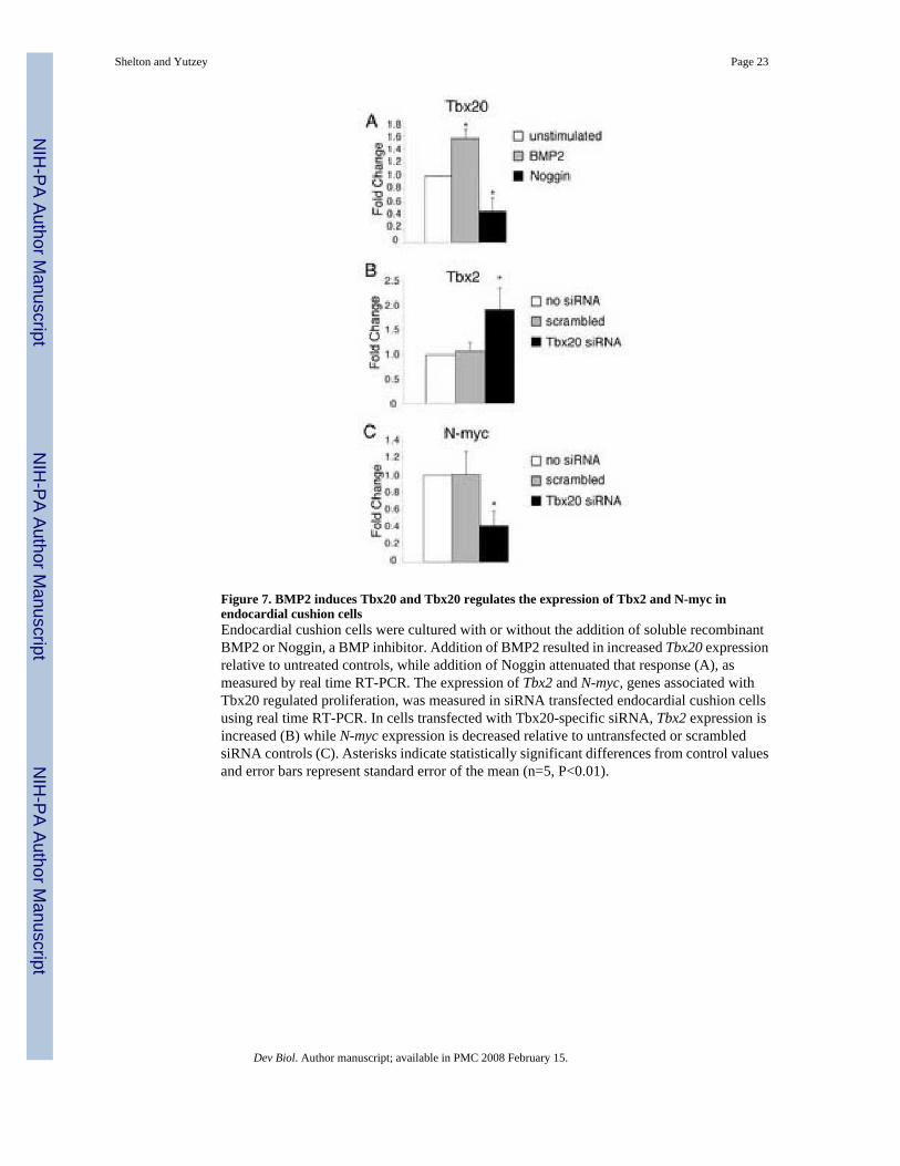
Figure 7. BMP2 induces Tbx20 and Tbx20 regulates the expression of Tbx2 and N-myc inendocardial cushion cellsEndocardial cushion cells were cultured with or without the addition of soluble recombinantBMP2 or Noggin, a BMP inhibitor. Addition of BMP2 resulted in increased Tbx20 expressionrelative to untreated controls, while addition of Noggin attenuated that response (A), asmeasured by real time RT-PCR. The expression of Tbx2 and N-myc, genes associated withTbx20 regulated proliferation, was measured in siRNA transfected endocardial cushion cellsusing real time RT-PCR. In cells transfected with Tbx20-specific siRNA, Tbx2 expression isincreased (B) while N-myc expression is decreased relative to untransfected or scrambledsiRNA controls (C). Asterisks indicate statistically significant differences from control valuesand error bars represent standard error of the mean (n=5, P<0.01).
Shelton and Yutzey Page 23
Dev Biol. Author manuscript; available in PMC 2008 February 15.
NIH
-PA Author Manuscript
NIH
-PA Author Manuscript
NIH
-PA Author Manuscript
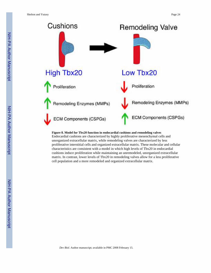
Figure 8. Model for Tbx20 function in endocardial cushions and remodeling valvesEndocardial cushions are characterized by highly proliferative mesenchymal cells andunorganized extracellular matrix, while remodeling valves are characterized by lessproliferative interstitial cells and organized extracellular matrix. These molecular and cellularcharacteristics are consistent with a model in which high levels of Tbx20 in endocardialcushions induce proliferation while maintaining an unremodeled, unorganized extracellularmatrix. In contrast, lower levels of Tbx20 in remodeling valves allow for a less proliferativecell population and a more remodeled and organized extracellular matrix.
Shelton and Yutzey Page 24
Dev Biol. Author manuscript; available in PMC 2008 February 15.
NIH
-PA Author Manuscript
NIH
-PA Author Manuscript
NIH
-PA Author Manuscript