Extracellular matrix turnover and outflow resistance
16
Extracellular Matrix Turnover and Outflow Resistance Kate E. Keller, Aga Mini, John M. Bradley, Mary J. Kelley, and Ted S. Acott Casey Eye Institute, Oregon Health & Science University, Portland, Oregon, USA Abstract Normal homeostatic adjustment of elevated intraocular pressure (IOP) involves remodeling the extracellular matrix (ECM) of the trabecular meshwork (TM). This entails sensing elevated IOP, releasing numerous activated proteinases to degrade existing ECM and concurrent biosynthesis of replacement ECM components. To increase or decrease IOP, the quantity, physical properties and/ or organization of new components should be somewhat different from those replaced in order to modify outflow resistance. ECM degradation and replacement biosynthesis in the outflow pathway must be tightly controlled and focused to retain the complex structural organization of the tissue. Recently identified podosome- or invadopodia-like structures (PILS) may aid in the focal degradation of ECM and organization of replacement components. Keywords Aqueous humor outflow resistance; extracellular matrix turnover; biosynthetic extracellular matrix replacement Introduction Increased resistance to aqueous humor flow through the trabecular meshwork (TM) results in elevated intraocular pressure (IOP), which is a primary risk factor for primary open-angle glaucoma (POAG)(Boland and Quigley, 2007). Normal homeostatic adjustment of outflow resistance appears to be triggered by cellular sensing of changes in stretch or distortion of the cribriform or juxtacanalicular region of the TM (Bradley et al., 2001; Acott and Kelley, 2008). When these signals are received, extracellular matrix (ECM) turnover is adjusted to shift the balance toward higher or lower resistance. This homeostatic adjustment appears to begin with selective proteinase secretion and activation, targeted ECM cleavage and fragment uptake, and biosynthesis of replacement ECM components (Acott and Kelley, 2008). Since mRNA levels for many ECM genes in the TM are relatively high (Vittal et al., 2005), ongoing ECM turnover must be a regular maintenance event, even when the resistance does not need to be modified (Bradley et al., 1998). Various proteinases and their inhibitors are expressed in the TM in a variety of instances. These include matrix metalloproteinases (MMPs), membrane type (MT) MMPs, ADAMs ( A Disintegrin-like And Metalloproteinase), ADAMTSs (ADAM with Thrombo Spondin type 1 © 2008 Elsevier Ltd. All rights reserved. Correspondence: E-mail: [email protected] Casey Eye Institute / OHSU, 3375 SW Terwilliger, Portland, OR 97239-4197, Phone: 503-494-8455, Fax: 503-418-2399. Publisher's Disclaimer: This is a PDF file of an unedited manuscript that has been accepted for publication. As a service to our customers we are providing this early version of the manuscript. The manuscript will undergo copyediting, typesetting, and review of the resulting proof before it is published in its final citable form. Please note that during the production process errors may be discovered which could affect the content, and all legal disclaimers that apply to the journal pertain. NIH Public Access Author Manuscript Exp Eye Res. Author manuscript; available in PMC 2010 April 1. Published in final edited form as: Exp Eye Res. 2009 April ; 88(4): 676–682. doi:10.1016/j.exer.2008.11.023. NIH-PA Author Manuscript NIH-PA Author Manuscript NIH-PA Author Manuscript
Transcript of Extracellular matrix turnover and outflow resistance
Extracellular Matrix Turnover and Outflow Resistance
Kate E. Keller, Aga Mini, John M. Bradley, Mary J. Kelley, and Ted S. AcottCasey Eye Institute, Oregon Health & Science University, Portland, Oregon, USA
AbstractNormal homeostatic adjustment of elevated intraocular pressure (IOP) involves remodeling theextracellular matrix (ECM) of the trabecular meshwork (TM). This entails sensing elevated IOP,releasing numerous activated proteinases to degrade existing ECM and concurrent biosynthesis ofreplacement ECM components. To increase or decrease IOP, the quantity, physical properties and/or organization of new components should be somewhat different from those replaced in order tomodify outflow resistance. ECM degradation and replacement biosynthesis in the outflow pathwaymust be tightly controlled and focused to retain the complex structural organization of the tissue.Recently identified podosome- or invadopodia-like structures (PILS) may aid in the focal degradationof ECM and organization of replacement components.
KeywordsAqueous humor outflow resistance; extracellular matrix turnover; biosynthetic extracellular matrixreplacement
IntroductionIncreased resistance to aqueous humor flow through the trabecular meshwork (TM) results inelevated intraocular pressure (IOP), which is a primary risk factor for primary open-angleglaucoma (POAG)(Boland and Quigley, 2007). Normal homeostatic adjustment of outflowresistance appears to be triggered by cellular sensing of changes in stretch or distortion of thecribriform or juxtacanalicular region of the TM (Bradley et al., 2001; Acott and Kelley,2008). When these signals are received, extracellular matrix (ECM) turnover is adjusted toshift the balance toward higher or lower resistance. This homeostatic adjustment appears tobegin with selective proteinase secretion and activation, targeted ECM cleavage and fragmentuptake, and biosynthesis of replacement ECM components (Acott and Kelley, 2008). SincemRNA levels for many ECM genes in the TM are relatively high (Vittal et al., 2005), ongoingECM turnover must be a regular maintenance event, even when the resistance does not needto be modified (Bradley et al., 1998).
Various proteinases and their inhibitors are expressed in the TM in a variety of instances. Theseinclude matrix metalloproteinases (MMPs), membrane type (MT) MMPs, ADAMs (ADisintegrin-like And Metalloproteinase), ADAMTSs (ADAM with ThromboSpondin type 1
© 2008 Elsevier Ltd. All rights reserved.Correspondence: E-mail: [email protected] Casey Eye Institute / OHSU, 3375 SW Terwilliger, Portland, OR 97239-4197, Phone:503-494-8455, Fax: 503-418-2399.Publisher's Disclaimer: This is a PDF file of an unedited manuscript that has been accepted for publication. As a service to our customerswe are providing this early version of the manuscript. The manuscript will undergo copyediting, typesetting, and review of the resultingproof before it is published in its final citable form. Please note that during the production process errors may be discovered which couldaffect the content, and all legal disclaimers that apply to the journal pertain.
NIH Public AccessAuthor ManuscriptExp Eye Res. Author manuscript; available in PMC 2010 April 1.
Published in final edited form as:Exp Eye Res. 2009 April ; 88(4): 676–682. doi:10.1016/j.exer.2008.11.023.
NIH
-PA Author Manuscript
NIH
-PA Author Manuscript
NIH
-PA Author Manuscript
motifs), plasminogen activators (PA), tissue inhibitors of metalloproteinases (TIMPs), PAinhibitors, and serine proteinase inhibitors (serpins) (Table 1 and Table 2)(Alexander et al.,1991;Borras, 2003;Fuchshofer et al., 2003;Lo et al., 2003;Vittal et al., 2005;Oh et al., 2006).The significance of MMPs in outflow resistance was shown when treatment of anteriorsegments in perfusion culture with MMPs was found to increase outflow, while specificinhibition of MMP activity decreased outflow facility (Bradley et al., 1998). Also, whenpressure was increased in perfusion culture there was a concomitant increase in outflow facility,which stabilized over time. This homeostatic pressure response likely involves MMPs, sinceadjustment of outflow facility was concurrent with increased MMP activity and ECM turnover.The proteolytic degradation of ECM components probably affects structural organization andalters biological interactions, leading to a modified outflow resistance. However, despite manyyears of research, the ECM components that contribute to outflow resistance remain elusive(Acott and Kelley, 2008). Glycosaminoglycans (GAGs) are thought to be a major contributingfactor since treatment with enzymes that specifically degrade one or more type of GAG, orwith agents that modify GAG chain biosynthesis, increase outflow facility (Barany andScotchbrook, 1954;Francois, 1975;Knepper et al., 1984;Sawaguchi et al., 1992;Sawaguchi etal., 1993;Hubbard et al., 1997;Johnson and Bahler, 1999;Keller et al., 2008). However, GAG-specific enzymatic changes in outflow of humans or higher primates remain controversial(Acott and Kelley, 2008).
In addition to their degradation activities, many of these proteinases participate in cell-cell andcell-matrix interactions, process latent proteins to active forms, convert structural ECMproteins to signaling molecules, change tissue architecture, and provide cues for cellproliferation and survival (Page-McCaw et al., 2007). Thus, degradation of structuralcomponents alone may not directly result in IOP adjustment, but that proteolytic alterationscould cause secondary effects that modify outflow resistance. In order to facilitate theseadjustments, replacement ECM is likely to differ from what was degraded. Biosynthesis ofreplacement molecules is therefore a crucial component for normal homeostatic adjustment ofoutflow resistance.
Matrix MetalloproteinasesMMPs are a group of 23 related proteinases that degrade ECM (Page-McCaw et al., 2007). AllMMPs have a similar domain structure and in their simplest form are composed of a prodomainand a catalytic domain (Fig. 1)(Chakraborti et al., 2003; Ra and Parks, 2007). MMPs may alsocontain C-terminal hemopexin-like (PEX) domains, which function in substrate recognition orproteinase localization, that are separated from the N-terminal catalytic domain by a flexiblehinge region. Some MMPs also have a transmembrane or a glycosylphosphatidylinositolanchor, which anchors them to the cell surface. MMPs are synthesized as zymogens, inactiveproenzymes, where the prodomain confers latency to the active enzymes. A free thiol of aconserved cysteine residue in the prodomain electrostatically interacts with a Zn2+ ion that ishistidine-ligated into the active site of the catalytic domain in the sequence HEXXHXXGXXH(Fig. 1). Extracellular posttranslational cleavage, usually by another MMP or by a furinproprotein convertase, disrupts the thiol-Zn2+ interaction and releases the biologically activeproteinase, a process known as the cysteine switch (Chakraborti et al., 2003). Serineproteinases, such as plasmin and thrombin, can activate proMMPs, while interaction with αVintegrin has been implicated for MMP2 (Brooks et al., 1996; Ra and Parks, 2007). However,for most MMPs, the actual physiologic activation mechanisms are uncertain.
Numerous MMPs have been detected in TM cells and tissue (Table 1) and MMP mRNA andprotein levels were upregulated in response to mechanical stretching, elevated pressure andvarious cytokines or growth factors (Alexander et al., 1991;Samples et al., 1993;Alexander etal., 1998;Bradley et al., 1998;Bradley et al., 2000;Gonzalez et al., 2000;WuDunn, 2001;Fleenor
Keller et al. Page 2
Exp Eye Res. Author manuscript; available in PMC 2010 April 1.
NIH
-PA Author Manuscript
NIH
-PA Author Manuscript
NIH
-PA Author Manuscript
et al., 2003;Pang et al., 2003;Vittitow and Borras, 2004;Oh et al., 2006;Kelley et al., 2007).MMPs -2, -14, -15 and -16 are constitutively expressed at relatively high levels in the TM(Alexander et al., 1991;Bradley et al., 2001;Vittal et al., 2005). MMPs -1, -3, and -9 arenormally expressed at low levels but are massively upregulated and activated in response tovarious stimulations. Agents that increase outflow facility in perfusion culture, or lasertrabeculoplasty surgery and medications that lower IOP in POAG patients, also affectexpression and/or activation of certain MMPs (Parshley et al., 1996;Johnson, 1997;Petersonet al., 1999;Shearer and Crosson, 2002;Fleenor et al., 2003;Pang et al., 2003;Oh et al.,2006;Sanka et al., 2007;Bahler et al., 2008). Addition of purified MMPs to human anteriorsegment perfusion cultures increased outflow facility, while inhibiting their endogenousactivity by TIMP2 or synthetic MMP inhibitors reduced outflow facility (Bradley et al.,1998). This argues strongly that controlled ECM turnover is required for maintenance ofoutflow resistance and that MMP regulation seems to be a major component for adjustingoutflow resistance. Immunofluorescence studies showed that MMP2 strongly stained normalhuman TM in the uveal, corneoscleral and juxtacanalicular regions and the inner wall ofSchlemm’s canal (Ronkko et al., 2007). In POAG TM, immunostaining intensity for MMPs-1 and -3 was greatly increased, while by Western blot, MMPs -2 and -9 were reduced comparedto age-matched control eyes (Ronkko et al., 2007;Govindarajan et al., 2008). The increase inMMPs -1 and -3 levels suggests that POAG TM cells continually strive to correct outflowresistance, but instead the persistent action of these MMPs likely causes an overproduction ofreplacement ECM and/or cause cleavage of ECM components that are not usually accessibleto degradation. TGFβ2, a cytokine whose levels are increased in POAG aqueous humor anddecreases outflow facility in perfusion culture, increased expression, but not activation, of pro-MMP2 (Fuchshofer et al., 2003;Gottanka et al., 2004;Fleenor et al., 2006).
Compartmentalization of MMPs has been noted in many cell types and tissues, but especiallyin invadopodia and podosomes of invasive cells (Chen and Wang, 1999). The termcompartmentalization refers to the location of the inactive enzyme in the pericellular matrix,and its site of activation (Ra and Parks, 2007). Due to the potency of these enzymes in degradingECM, MMPs are targeted to specific locations where they become highly concentrated. Thisregulates activation of latent MMPs and targets their catalytic activity to certain substrates inthe pericellular environment. For instance, MMP14 (MT1-MMP) cleaves pro-MMP2 and pro-MMP13 to their active forms on the cell surface, which triggers potent pericellular proteolyticactivity (Itoh and Seiki, 2006). Recent identification of clusters of podosome- or invadopodia-like structures (PILS) in TM cells show that MMPs -2 and -14 are localized to regions of thecell that appear to function in both adhesion and matrix degradation (Fig. 2)(Linder, 2007;Aga et al., 2008). Focusing MMP14 at such structures could increase its local concentrationto allow homodimerization of its PEX domains. This in turn would facilitate MMP2 activationand further augment targeted proteolytic activity at the cell surface. PILS have been found inTM tissue suggesting that these focal regions of regulated and targeted ECM turnover may beimportant in regulating outflow facility in response to elevated IOP (Aga et al., 2008).
Active MMPs degrade many different ECM molecules (Table 1)(Chakraborti et al., 2003),most of which are also expressed in the TM (Acott and Kelley, 2008). Rather than degradingall ECM, it is likely that only certain molecules or parts of molecules are degraded. This focal,targeted degradation is probably facilitated by PILS. For instance, collagen IV α3 is cleavedby MMP9 and collagen XVIII by other MMPs to release tumstatin or endostatin, respectively,but the structural triple-helical portion is not degraded (Page-McCaw et al., 2007). Both thesecollagen types are expressed in the TM (Hann et al., 2001;Ohlmann et al., 2005) and bothstatins bind integrins, which launch a signaling cascade that disrupts the actin cytoskeleton(Bix and Iozzo, 2005). Therefore, in the TM, proteolytically-released fragments could impactoutflow resistance via cytoskeletal adjustments.
Keller et al. Page 3
Exp Eye Res. Author manuscript; available in PMC 2010 April 1.
NIH
-PA Author Manuscript
NIH
-PA Author Manuscript
NIH
-PA Author Manuscript
ADAMs and ADAMTSsAnother class of zinc-dependent metalloproteinases is the adamalysins, which are categorizedinto two subfamilies known as the ADAMs and the ADAMTSs (Table 1). Both these proteinasegroups are involved in ectodomain shedding, activation of cell surface receptors and growthfactors and ECM protein cleavage (Apte, 2004;Huovila et al., 2005;Porter et al., 2005). Themajor difference between the two subfamilies is that ADAMs are mostly transmembraneproteins while ADAMTSs are secreted molecules that bind to the ECM.
There are currently 29 ADAM family members that cleave a wide range of substrates (Huovilaet al., 2005). Similar to MMPs, ADAMs have a multi-domain structure and are synthesized asproenzymes that are activated by a cysteine switch (Fig. 1). A terminal aspartate residue in thesequence HEXXHXXGXXHD distinguishes the adamalysins from the MMPs. Lying C-terminal to the catalytic domain is the disintegrin domain, which contains an integrin-bindingmotif, a cysteine-rich region, an EGF-like domain, a transmembrane region and a C-terminalcytosolic tail. The tail contains multiple motifs which interact with other cytosolic proteins andcan be phosphorylated on Ser/Thr and Tyr residues (Huovila et al., 2005).
There are 19 ADAMTSs and 3 ADAMTS-like enzymes. Like the ADAMs, ADAMTSs havea wide range of substrates including collagen, versican, and aggrecan (Porter et al., 2005). Thedomain structure of the ADAMTSs is similar to that of ADAMs and MMPs, but there are somedifferences that distinguish them (Fig. 1). There is some debate as to whether the prodomainsconfer latency to the ADAMTS enzyme, but the prodomains are removed by furin-cleavagein the secretory pathway or by MMP17 on the cell surface (Apte, 2004). The ancillary regionis comprised of a disintegrin-like domain, a central thrombospondin-like (TS) domain, acysteine-rich domain, a spacer domain, and a varying number (0–14) of C-terminal TSdomains. The ancillary domain can be proteolytically cleaved, which either augments orinhibits activity of the ADAMTS enzyme (Gao et al., 2002).
Although ADAMs and ADAMTS are expressed in other eye tissues (Wride et al., 2006), littleis known about the action of these proteinases in the TM. Our studies have shown that TNFαand/or IL-1α treatment of TM cells stimulate mRNA expression of ADAMs -9 and -10 andADAMTSs -4, -5, -7 and -13 (Chen et al., 2005; Acott and Kelley, 2008). TGFβ2 treatment ofglaucomatous TM cells increased ADAMTS5 mRNA expression (Fleenor et al., 2006). A neo-epitope of versican that is exposed following ADAMTS4 cleavage was found to localize toPILS (Fig. 2)(Aga et al., 2008) and in areas of the TM that experience high segmental flow, asassessed by fluorescent Qdot nanoparticle labeling of anterior segments (Bradley et al.,2008). Immunolocalization of ADAMTS4 showed that its expression was highly increased inthe JCT region in human anterior segments that were subjected to elevated pressure (Keller etal., unpublished observations). Moreover, treatment of human and pig anterior segments inperfusion culture with ADAMTS4 increased outflow facility. Further studies are required toclarify the possible roles of ADAMs and ADAMTSs in outflow resistance.
Tissue Inhibitors of the MMPsIn order to regulate these potent proteinases, MMP activity needs to be tightly controlled. TMcells can downregulate MMP mRNA or protein levels, change the activation state of theseenzymes or release inhibitors that inactivate the enzymes. There are four TIMPs all of whichare expressed in human TM cells (Table 2). Factors known to upregulate MMP expression alsoaffect TIMP mRNA expression and therefore a delicate balance likely exists in vivo betweenMMP activation and inhibition by TIMPs (Samples et al., 1993;Pang et al., 2003;Oh et al.,2006). TIMP2 not only acts as an inhibitor of MMP activity, but it is also involved in activationof pro-MMP2 (Wang et al., 2000). The C-terminal domain of TIMP2 simultaneously binds tothe C-terminus of pro-MMP2 and the N-terminus of activated MMP14 (Fig. 3). This trimeric
Keller et al. Page 4
Exp Eye Res. Author manuscript; available in PMC 2010 April 1.
NIH
-PA Author Manuscript
NIH
-PA Author Manuscript
NIH
-PA Author Manuscript
complex recruits an adjacent MMP14 molecule, which is responsible for the resultant cleavageof pro-MMP2. The relative concentration and binding of TIMP2 influences whether MMP14will participate in cleavage of MMP2 and whether it will cleave other substrates (Kudo et al.,2007). In perfused anterior segment organ culture, increased pressure resulted in more activatedMMP2 (Bradley et al., 2001). In cell culture, mechanical stretch, which is presumablemimicking pressure increases, increased MMPs -2 and 14 while decreasing TIMP2 levels(Bradley et al., 2001). This reduction in TIMP2 appears to bring it to the correct level tofacilitate activation of MMP2 without completely blocking the process. TIMP1 inhibitsADAM10 but not ADAM17, whereas TIMP3 inhibits both enzymes (Rapti et al., 2008).TIMP3 inhibits ADAMTSs -4 and -5 activity (Kashiwagi et al., 2001).
Other Proteinases and inhibitorsMany other proteinases are expressed in the TM and may function directly or indirectly tomodulate outflow resistance (Table 1). Tissue plasminogen activator (tPA), a serine proteinasethat converts plasminogen to plasmin, is expressed in the TM (Park et al., 1987;Shuman et al.,1988;Chen et al., 2005) and by Schlemm’s canal endothelial cells (Stamer et al., 1998). Sinceplasmin can activate proMMPs, the action of tPA may be a secondary effect rather than directECM proteolysis. tPA activity was reduced in response to dexamethasone, a corticosteroid thatcan induce glaucoma and decrease outflow facility in human anterior perfusion culture (Snyderet al., 1993;Clark et al., 1995). Urokinase-type PA (uPA) distribution overlaps with that of tPAand may also be involved in ECM turnover (Tripathi et al., 1990;Chen et al., 2005;Vittal et al.,2005).
Serpins are a class of serine proteinase inhibitors (Table 2) (Gettins, 2002). There are 16members of this family including antithrombin, α1-proteinase inhibitor (PAI-1), andantitrypsin and they function in a wide range of physiological processes. Serpin expression ismodulated by various stimulations in TM cells (Zhao et al., 2004;Chen et al., 2005;Rozsa etal., 2006;Fan et al., 2008). SerpinA3 (α1-chymotrypsin) was upregulated, while serpinB2(PAI-2) was downregulated by corticosteroid treatment of human TM cells (Lo et al.,2003;Rozsa et al., 2006;Fan et al., 2008). SerpinE1 (PAI-1) was upregulated 6.8-fold byTGFβ2 treatment of glaucomatous TM cells and by TGFβ1 in HTM cells (Zhao et al.,2004;Fleenor et al., 2006). Furthermore, TGFβ2-induced expression and secretion of PAI-1reduced MMP2 activity (Fuchshofer et al., 2003). Thus, modulation of serpin expression inresponse to various factors may affect MMP activity. Aqueous humor contains significantamounts of serpinC1 (antithrombin III), serpinA1 (α-1-antitrypsin), α-2-macroglobulin, andtPA (Tripathi et al., 1988;Ando et al., 1993;Rao et al., 2000). Interestingly, α-2-macroglobulinis an endogenous inhibitor of ADAMTSs -4 and-5 (Tortorella et al., 2004). Increasedconcentrations of these inhibitors in aqueous humor may alter MMP activity in active flowareas.
Regulation of proteinases by GAGs and other ECM moleculesMMPs and other proteinases in the TM may bind GAGs, which could limit diffusion of theactive enzyme in tissue (Page-McCaw et al., 2007). GAGs are long sugar chains that are usuallyattached to a core protein. Since GAGs are thought to be a significant source of outflowresistance, GAG binding may directly affect expression and/or activation of MMPs. The typesof GAGs are chondroitin/dermatan sulfate (CS/DS), heparin/heparan sulfate (HS), keratansulfate (KS) and hyaluronan (HA). Chondroitin-4-sulfate (C-4-S) concomitantly binds MMP2and MMP16 to facilitate the conversion of latent pro-MMP2 to its active form (Iida et al.,2007). It is feasible that MMP2 located in PILS could, in part, be activated by a similarmechanism. MMP2 can also bind tightly to HS chains of syndecan-2, which suppresses itsactivation (Munesue et al., 2007). Hyaluronan, applied exogenously to corneal explants in
Keller et al. Page 5
Exp Eye Res. Author manuscript; available in PMC 2010 April 1.
NIH
-PA Author Manuscript
NIH
-PA Author Manuscript
NIH
-PA Author Manuscript
organ culture, resulted in increased expression and activation of MMPs -2 and -9 (Isnard et al.,2001), while treatment of cartilage chondrocytes with low molecular weight HA fragmentsincreased MMP3 mRNA expression (Ohno et al., 2005). Together, these studies suggest thatGAG chains, or GAG chain fragments, may facilitate expression and activation of pro-MMPsand therefore play a role in targeted degradation of pericellular matrix. TM GAG content varieswith age and POAG disease progression (Gong et al., 1992; Knepper et al., 1996; Knepper etal., 1996), so minor changes may profoundly affect proteinase activity and/or inhibition andpotentially disrupt normal ECM turnover during homeostatic adjustment of IOP.
CD44, the major hyaluronan receptor, also plays an important role in MMP regulation. CD44can attenuate the secretion and activation of MMP2 (Takahashi et al., 1999). CD44 is also areceptor for active MMP9 in keratinocytes and this interaction subsequently activates latentTGFβ (Yu and Stamenkovic, 1999). MMP14 can bind directly to CD44 via its PEX domains,which results in shedding of a soluble 70kDa extracellular fragment (sCD44) (Kajita et al.,2001; Mori et al., 2002). sCD44 concentrations were higher in aqueous humor of POAGpatients than in age-matched control samples (Knepper et al., 2002; Nolan et al., 2007). Thus,CD44 not only regulates MMP activity, but is itself a substrate for MMP cleavage.
Other proteinases and inhibitors also bind GAGs and ECM molecules, e.g. ADAM12 binds tosyndecan-4 and ADAM-9 binds α6α1 and αvα5 integrins (Huovila et al., 2005). Integrin-ADAMassociation facilitates cell-cell interactions and sequesters the enzyme in an inactive state atthe cell surface (Bridges and Bowditch, 2005). There is no evidence to suggest that thedisintegrin domains of ADAMTSs also bind integrins (Porter et al., 2005). ADAMTSs bindssulfated GAGs in the ECM or on the cell surface, or to HS and CS chains of syndecan-1 (Apte,2004; Gao et al., 2004). The spacer region of ADAMTS4 binds the C-terminal region offibronectin, which may inhibit its enzymatic activity (Fig. 1)(Hashimoto et al., 2004). MMPinhibitors also bind GAGs. The N-terminal region of TIMP3 interacts with HS and possiblyCS GAG chains (Yu et al., 2000). Serpin activity is also profoundly affected by the presenceof GAGs, such as HS and DS, which accelerate interaction with their target proteinases (Pikeet al., 2005).
Replacement ECMAn integral component of any normal ongoing or homeostatic resistance adjustment involvingECM turnover is the biosynthetic replacement of degraded components (Acott and Kelley,2008). Modifications in outflow resistance will likely include changes in levels or organizationof ECM components. TM cells alter mRNA expression of many ECM genes in response tomechanical stretching, exposure to exogenous soluble factors and synthetic agents or bytreatment with IOP-lowering drugs and laser trabeculoplasty (Lo et al., 2003; Zhao et al.,2004; Vittal et al., 2005; Fleenor et al., 2006; Rozsa et al., 2006; Fan et al., 2008). ECM genemRNA levels also vary between non-glaucomatous and glaucomatous eyes (Diskin et al.,2006; Liton et al., 2006). PILS could potentially coordinate the biosynthetic replacement andorganization of ECM components in degraded areas. Some of the ECM molecules that aresynthesized are different from their predecessors and can have extra exons included, orexcluded, which may contain additional ECM or cell binding sites (Vittal et al., 2005; Zhaoand Russell, 2005; Keller et al., 2007). Continued exposure of TM cells to elevated IOP, orfrom many years of normal homeostatic adjustment, could conceivably cause an excess of cell-cell, cell-protein and/or protein-protein interactions that would contribute to the observedincreased rigidity of the TM in POAG or with age (Gabelt and Kaufman, 2005; Schlunck etal., 2008). In addition, stiffness could be exacerbated by an increase in ECM cross-linking. Forinstance, POAG TM cells have increased protein levels and activity of the enzyme tissuetransglutaminase (Tovar-Vidales et al., 2008). Increased stiffness may hinder detection ofmechanical stretch/distortion signals by cells in the JCT region. Consequently, the normal
Keller et al. Page 6
Exp Eye Res. Author manuscript; available in PMC 2010 April 1.
NIH
-PA Author Manuscript
NIH
-PA Author Manuscript
NIH
-PA Author Manuscript
signals that are required to initiate ECM turnover may not be received and the typicalmechanisms to adjust outflow resistance would not be activated. Alternatively, such ECMcross-linking could change susceptibility of ECM substrates to proteinase cleavage andturnover.
AcknowledgmentsSupport was provided by NIH EY003279, EY008247, EY010572, the Glaucoma Research Foundation (to KEK), andby an unrestricted grant to Casey Eye Institute from Research to Prevent Blindness, New York, NY.
ReferencesAcott TS, Kelley MJ. Extracellular matrix in the trabecular meshwork. Exp Eye Res 2008;86:543–561.
[PubMed: 18313051]Aga M, Bradley JM, et al. Specialized podosome- or invadopodia-like structures (PILS) for focal
trabecular meshwork extracellular matrix turnover. Invest Ophthalmol Vis Sci. 2008In PressAlexander JP, Samples JR, et al. Growth factor and cytokine modulation of trabecular meshwork matrix
metalloproteinase and TIMP expression. Curr Eye Res 1998;17:276–285. [PubMed: 9543636]Alexander JP, Samples JR, et al. Expression of matrix metalloproteinases and inhibitor by human
trabecular meshwork. Invest Ophthalmol Vis Sci 1991;32:172–180. [PubMed: 1846130]Ando H, Twining SS, et al. MMPs and proteinase inhibitors in the human aqueous humor. Invest
Ophthalmol Vis Sci 1993;34:3541–3548. [PubMed: 7505006]Apte SS. A disintegrin-like and metalloprotease (reprolysin type) with thrombospondin type 1 motifs:
the ADAMTS family. Int J Biochem Cell Biol 2004;36:981–985. [PubMed: 15094112]Bahler CK, Howell KG, et al. Prostaglandins increase trabecular meshwork outflow facility in cultured
human anterior segments. Am J Ophthalmol 2008;145:114–119. [PubMed: 17988642]Barany EH, Scotchbrook S. Influence of testicular hyaluronidase on the resistance to flow through the
angle of the anterior chamber. Acta Physiol Scand 1954;30:240–248. [PubMed: 13158098]Bix G, Iozzo RV. Matrix revolutions: "tails" of basement-membrane components with angiostatic
functions. Trends Cell Biol 2005;15:52–60. [PubMed: 15653078]Boland MV, Quigley HA. Risk factors and open-angle glaucoma: classification and application. J
Glaucoma 2007;16:406–418. [PubMed: 17571004]Borras T. Gene expression in the trabecular meshwork and the influence of intraocular pressure. Prog
Retin Eye Res 2003;22:435–463. [PubMed: 12742391]Bradley JM, Anderssohn AM, et al. Mediation of laser trabeculoplasty-induced matrix metalloproteinase
expression by IL-1beta and TNFalpha. Invest Ophthalmol Vis Sci 2000;41:422–430. [PubMed:10670472]
Bradley JM, Keller KE, et al. Quantum dot analyses of segmental flow patterns in the trabecularmeshwork. Invest Ophthalmol Vis Sci 2008;49E-abstract 1635
Bradley JM, Kelley MJ, et al. Effects of mechanical stretching on trabecular matrix metalloproteinases.Invest Ophthalmol Vis Sci 2001;42:1505–1513. [PubMed: 11381054]
Bradley JM, Vranka J, et al. Effect of matrix metalloproteinases activity on outflow in perfused humanorgan culture. Invest Ophthalmol Vis Sci 1998;39:2649–2658. [PubMed: 9856774]
Bridges LC, Bowditch RD. ADAM-Integrin Interactions: potential integrin regulated ectodomainshedding activity. Curr Pharm Des 2005;11:837–847. [PubMed: 15777238]
Brooks PC, Stromblad S, et al. Localization of matrix metalloproteinase MMP-2 to the surface of invasivecells by interaction with integrin alpha v beta 3. Cell 1996;85:683–693. [PubMed: 8646777]
Chakraborti S, Mandal M, et al. Regulation of matrix metalloproteinases: an overview. Mol Cell Biochem2003;253:269–285. [PubMed: 14619979]
Chen WT, Wang JY. Specialized surface protrusions of invasive cells, invadopodia and lamellipodia,have differential MT1-MMP, MMP-2, and TIMP-2 localization. Ann N Y Acad Sci 1999;878:361–371. [PubMed: 10415741]
Keller et al. Page 7
Exp Eye Res. Author manuscript; available in PMC 2010 April 1.
NIH
-PA Author Manuscript
NIH
-PA Author Manuscript
NIH
-PA Author Manuscript
Chen Y, Kelley MJ, et al. DNA microarray analysis of gene expression in trabecular meshwork cells inresponse to TNFα and IL-1α. Invest Ophthalmol Vis Sci 2005;46E-abstract 1349
Clark AF, Wilson K, et al. Dexamethasone-induced ocular hypertension in perfusion-cultured humaneyes. Invest Ophthalmol Vis Sci 1995;36:478–489. [PubMed: 7843916]
Diskin S, Kumar J, et al. Detection of differentially expressed glycogenes in trabecular meshwork of eyeswith primary open-angle glaucoma. Invest Ophthalmol Vis Sci 2006;47:1491–1499. [PubMed:16565384]
Fan BJ, Wang DY. Gene expression profiles of human trabecular meshwork cells induced bytriamcinolone and dexamethasone. Invest Ophthalmol Vis Sci 2008;49:1886–1897. [PubMed:18436822]
Fleenor DL, Pang IH, et al. Involvement of AP-1 in interleukin-1alpha-stimulated MMP-3 expression inhuman trabecular meshwork cells. Invest Ophthalmol Vis Sci 2003;44:3494–3501. [PubMed:12882799]
Fleenor DL, Shepard AR, et al. TGFbeta2-induced changes in human trabecular meshwork: implicationsfor intraocular pressure. Invest Ophthalmol Vis Sci 2006;47:226–234. [PubMed: 16384967]
Francois J. The importance of the mucopolysaccharides in intraocular pressure regulation. InvestOphthalmol 1975;14:173–176. [PubMed: 123231]
Fuchshofer R, Welge-Lussen U, et al. The effect of TGF-beta2 on human trabecular meshworkextracellular proteolytic system. Exp Eye Res 2003;77:757–765. [PubMed: 14609564]
Gabelt BT, Kaufman PL. Changes in aqueous humor dynamics with age and glaucoma. Prog Retin EyeRes 2005;24:612–637. [PubMed: 15919228]
Gao G, Plaas A, et al. ADAMTS4 (aggrecanase-1) activation on the cell surface involves C-terminalcleavage by glycosylphosphatidyl inositol-anchored membrane type 4-matrix metalloproteinase andbinding of the activated proteinase to chondroitin sulfate and heparan sulfate on syndecan-1. J BiolChem 2004;279:10042–10051. [PubMed: 14701864]
Gao G, Westling J, et al. Activation of the proteolytic activity of ADAMTS4 (aggrecanase-1) by C-terminal truncation. J Biol Chem 2002;277:11034–11041. [PubMed: 11796708]
Gettins PG. Serpin structure, mechanism, and function. Chem Rev 2002;102:4751–4804. [PubMed:12475206]
Gong H, Freddo TF, et al. Age-related changes of sulfated proteoglycans in the normal human trabecularmeshwork. Exp Eye Res 1992;55:691–709. [PubMed: 1478279]
Gonzalez P, Epstein DL. Genes upregulated in the human trabecular meshwork in response to elevatedintraocular pressure. Invest Ophthalmol Vis Sci 2000;41:352–361. [PubMed: 10670462]
Gottanka J, Chan D, et al. Effects of TGF-beta2 in perfused human eyes. Invest Ophthalmol Vis Sci2004;45:153–158. [PubMed: 14691167]
Govindarajan B, Salomon RG, et al. Role of oxidatively modified extracellular matrix proteins and matrixmetalloproteinases in glaucomatous trabecular meshwork. invest Ophthalmol Vis Sci 2008;49E-abstract 1645
Hann CR, Springett MJ, et al. Ultrastructural localization of collagen IV, fibronectin, and laminin in thetrabecular meshwork of normal and glaucomatous eyes. Ophthalmic Res 2001;33:314–324.[PubMed: 11721183]
Hashimoto G, Shimoda M, et al. ADAMTS4 (aggrecanase-1) interaction with the C-terminal domain offibronectin inhibits proteolysis of aggrecan. J Biol Chem 2004;279:32483–32491. [PubMed:15161923]
Hubbard WC, Johnson M, et al. Intraocular pressure and outflow facility are unchanged following acuteand chronic intracameral chondroitinase ABC and hyaluronidase in monkeys. Exp Eye Res1997;65:177–190. [PubMed: 9268586]
Huovila AP, Turner AJ, et al. Shedding light on ADAM metalloproteinases. Trends Biochem Sci2005;30:413–422. [PubMed: 15949939]
Iida J, Wilhelmson KL, et al. Cell surface chondroitin sulfate glycosaminoglycan in melanoma: role inthe activation of pro-MMP-2 (progelatinase A). Biochem J 2007;403:553–563. [PubMed: 17217338]
Isnard N, Legeais JM, et al. Effect of hyaluronan on MMP expression and activation. Cell Biol Int2001;25:735–739. [PubMed: 11482897]
Keller et al. Page 8
Exp Eye Res. Author manuscript; available in PMC 2010 April 1.
NIH
-PA Author Manuscript
NIH
-PA Author Manuscript
NIH
-PA Author Manuscript
Itoh Y, Seiki M. MT1-MMP: a potent modifier of pericellular microenvironment. J Cell Physiol2006;206:1–8. [PubMed: 15920734]
Johnson DH. The effect of cytochalasin D on outflow facility and the trabecular meshwork of the humaneye in perfusion organ culture. Invest Ophthalmol Vis Sci 1997;38:2790–2799. [PubMed: 9418732]
Johnson DH, Bahler CH. Heparitinase increases outflow facility in the human eye. Invest OphthalmolVis Sci 1999;40:S504.
Kajita M, Itoh Y, et al. Membrane-type 1 matrix metalloproteinase cleaves CD44 and promotes cellmigration. J Cell Biol 2001;153:893–904. [PubMed: 11381077]
Kashiwagi M, Tortorella M, et al. TIMP-3 is a potent inhibitor of aggrecanase 1 (ADAM-TS4) andaggrecanase 2 (ADAM-TS5). J Biol Chem 2001;276:12501–12504. [PubMed: 11278243]
Keller KE, Bradley JM, et al. Effects of modifiers of glycosaminoglycan biosynthesis on outflow facilityin perfusion culture. Invest Ophthalmol Vis Sci 2008;49:2495–2505. [PubMed: 18515587]
Keller KE, Kelley MJ, et al. Extracellular matrix gene alternative splicing by trabecular meshwork cellsin response to mechanical stretching. Invest Ophthalmol Vis Sci 2007;48:1164–1172. [PubMed:17325160]
Kelley MJ, Rose AY, et al. Synergism of TNF and IL-1 in the Induction of Matrix Metalloproteinase-3in Trabecular Meshwork. Invest Ophthalmol Vis Sci 2007;48:2634–2643. [PubMed: 17525194]
Knepper PA, Farbman AI, et al. Exogenous hyaluronidases and degradation of hyaluronic acid in therabbit eye. Invest Ophthalmol Vis Sci 1984;25:286–293. [PubMed: 6698747]
Knepper PA, Goossens W, et al. Glycosaminoglycans of the human trabecular meshwork in primaryopen-angle glaucoma. Invest Ophthalmol Vis Sci 1996;37:1360–1367. [PubMed: 8641839]
Knepper PA, Goossens W, et al. Glycosaminoglycan stratification of the juxtacanalicular tissue in normaland primary open-angle glaucoma. Invest Ophthalmol Vis Sci 1996;37:2414–2425. [PubMed:8933758]
Knepper PA, Mayanil CS, et al. Aqueous humor in primary open-angle glaucoma contains an increasedlevel of CD44S. Invest Ophthalmol Vis Sci 2002;43:133–139. [PubMed: 11773023]
Kudo T, Takino T, et al. Substrate choice of membrane-type 1 matrix metalloproteinase is dictated bytissue inhibitor of metalloproteinase-2 levels. Cancer Sci 2007;98:563–568. [PubMed: 17425593]
Linder S. The matrix corroded: podosomes and invadopodia in extracellular matrix degradation. TrendsCell Biol 2007;17:107–117. [PubMed: 17275303]
Liton PB, Luna C, et al. Genome-wide expression profile of human trabecular meshwork cultured cells,nonglaucomatous and primary open angle glaucoma tissue. Mol Vis 2006;12:774–790. [PubMed:16862071]
Lo WR, Rowlette LL, et al. Tissue differential microarray analysis of dexamethasone induction revealspotential mechanisms of steroid glaucoma. Invest Ophthalmol Vis Sci 2003;44:473–485. [PubMed:12556371]
Mori H, Tomari T, et al. CD44 directs membrane-type 1 matrix metalloproteinase to lamellipodia byassociating with its hemopexin-like domain. EMBO J 2002;21:3949–3959. [PubMed: 12145196]
Munesue S, Yoshitomi Y, et al. A novel function of syndecan-2, suppression of matrixmetalloproteinase-2 activation, which causes suppression of metastasis. J Biol Chem2007;282:28164–28174. [PubMed: 17623663]
Nolan MJ, Giovingo MC, et al. Aqueous humor sCD44 concentration and visual field loss in primaryopen-angle glaucoma. J Glaucoma 2007;16:419–429. [PubMed: 17700283]
Oh DJ, Martin JL, et al. Effect of latanoprost on the expression of matrix metalloproteinases and theirtissue inhibitors in human trabecular meshwork cells. Invest Ophthalmol Vis Sci 2006;47:3887–3895. [PubMed: 16936101]
Ohlmann AV, Ohlmann A, et al. Localization of collagen XVIII and endostatin in the human eye. CurrEye Res 2005;30:27–34. [PubMed: 15875362]
Ohno S, Ohno-Nakahara M, et al. Induction of MMP-3 by hyaluronan oligosaccharides intemporomandibular joint chondrocytes. J Dent Res 2005;84:1005–1009. [PubMed: 16246931]
Page-McCaw A, Ewald AJ, et al. Matrix metalloproteinases and the regulation of tissue remodelling. NatRev Mol Cell Biol 2007;8:221–233. [PubMed: 17318226]
Keller et al. Page 9
Exp Eye Res. Author manuscript; available in PMC 2010 April 1.
NIH
-PA Author Manuscript
NIH
-PA Author Manuscript
NIH
-PA Author Manuscript
Pang IH, Fleenor DL, et al. Aqueous outflow-enhancing effect of tert-butylhydroquinone: involvementof AP-1 activation and MMP-3 expression. Invest Ophthalmol Vis Sci 2003;44:3502–3510.[PubMed: 12882800]
Pang IH, Hellberg PE, et al. Expression of matrix metalloproteinases and their inhibitors in humantrabecular meshwork cells. Invest Ophthalmol Vis Sci 2003;44:3485–3493. [PubMed: 12882798]
Park JK, Tripathi RC, et al. Tissue plasminogen activator in the trabecular endothelium. InvestOphthalmol Vis Sci 1987;28:1341–1345. [PubMed: 3112034]
Parshley DE, Bradley JM, et al. Laser trabeculoplasty induces stromelysin expression by trabecularjuxtacanalicular cells. Invest Ophthalmol Vis Sci 1996;37:795–804. [PubMed: 8603864]
Peterson JA, Tian B, et al. Latrunculin-A increases outflow facility in the monkey. Invest OphthalmolVis Sci 1999;40:931–941. [PubMed: 10102290]
Pike RN, Buckle AM, et al. Control of the coagulation system by serpins. Getting by with a little helpfrom glycosaminoglycans. FEBS J 2005;272:4842–4851. [PubMed: 16176258]
Porter S, Clark IM, et al. The ADAMTS metalloproteinases. Biochem J 2005;386:15–27. [PubMed:15554875]
Ra HJ, Parks WC. Control of matrix metalloproteinase catalytic activity. Matrix Biol 2007;26:587–596.[PubMed: 17669641]
Rao PV, Allingham RR, et al. Antithrombin III, a serpin family protease inhibitor, is a major heparinbinding protein in porcine aqueous humor. Biochem Biophys Res Commun 2000;272:1–5. [PubMed:10872794]
Rapti M, Atkinson SJ, et al. The isolated N-terminal domains of TIMP-1 and TIMP-3 are insufficient forADAM10 inhibition. Biochem J 2008;411:433–439. [PubMed: 18215140]
Ronkko S, Rekonen P, et al. Matrix metalloproteinases and their inhibitors in the chamber angle of normaleyes and patients with primary open-angle glaucoma and exfoliation glaucoma. Graefes Arch ClinExp Ophthalmol 2007;245:697–704. [PubMed: 17028863]
Rozsa FW, Reed DM, et al. Gene expression profile of human trabecular meshwork cells in response tolong-term dexamethasone exposure. Mol Vis 2006;12:125–141. [PubMed: 16541013]
Samples JR, Alexander JP, et al. Regulation of the levels of human trabecular matrix metalloproteinasesand inhibitor by interleukin-1 and dexamethasone. Invest Ophthalmol Vis Sci 1993;34:3386–3395.[PubMed: 8225873]
Sanka K, Maddala R, et al. Influence of actin cytoskeletal integrity on matrix metalloproteinase-2activation in cultured human trabecular meshwork cells. Invest Ophthalmol Vis Sci 2007;48:2105–2114. [PubMed: 17460268]
Sawaguchi S, Lam TT, et al. Effects of Glycosaminoglycan-Degrading Enzymes on Bovine TrabecularMeshwork in Organ Culture. J Glaucoma 1993;2:80–86.
Sawaguchi S, Yue BY, et al. Effects of intracameral injection of chondroitinase ABC in vivo. ArchOphthalmol 1992;110:110–117. [PubMed: 1731702]
Schlunck G, Han H, et al. Substrate rigidity modulates cell matrix interactions and protein expression inhuman trabecular meshwork cells. Invest Ophthalmol Vis Sci 2008;49:262–269. [PubMed:18172101]
Shearer TW, Crosson CE. Adenosine A1 receptor modulation of MMP-2 secretion by trabecularmeshwork cells. Invest Ophthalmol Vis Sci 2002;43:3016–3020. [PubMed: 12202524]
Shuman MA, Polansky JR, et al. Tissue plasminogen activator in cultured human trabecular meshworkcells. Predominance of enzyme over plasminogen activator inhibitor. Invest Ophthalmol Vis Sci1988;29:401–405. [PubMed: 3125123]
Snyder RW, Stamer WD, et al. Corticosteroid treatment and trabecular meshwork proteases in cell andorgan culture supernatants. Exp Eye Res 1993;57:461–468. [PubMed: 8282032]
Stamer WD, Roberts BC, et al. Isolation, culture, and characterization of endothelial cells from Schlemm'scanal. Invest Ophthalmol Vis Sci 1998;39:1804–1812. [PubMed: 9727403]
Takahashi K, Eto H, et al. Involvement of CD44 in matrix metalloproteinase-2 regulation in humanmelanoma cells. Int J Cancer 1999;80:387–395. [PubMed: 9935179]
Keller et al. Page 10
Exp Eye Res. Author manuscript; available in PMC 2010 April 1.
NIH
-PA Author Manuscript
NIH
-PA Author Manuscript
NIH
-PA Author Manuscript
Tortorella MD, Arner EC, et al. Alpha2-macroglobulin is a novel substrate for ADAMTS-4 andADAMTS-5 and represents an endogenous inhibitor of these enzymes. J Biol Chem2004;279:17554–17561. [PubMed: 14715656]
Tovar-Vidales T, Roque R, et al. Tissue transglutaminase expression and activity in normal andglaucomatous human trabecular meshwork cells and tissues. Invest Ophthalmol Vis Sci2008;49:622–628. [PubMed: 18235007]
Tripathi RC, Park JK, et al. Tissue plasminogen activator in human aqueous humor and its possibletherapeutic significance. Am J Ophthalmol 1988;106:719–722. [PubMed: 3143267]
Tripathi RC, Tripathi BJ, et al. Localization of urokinase-type plasminogen activator in human eyes: animmunocytochemical study. Exp Eye Res 1990;51:545–552. [PubMed: 2123459]
Vittal V, Rose A, et al. Changes in gene expression by trabecular meshwork cells in response tomechanical stretching. Invest Ophthalmol Vis Sci 2005;46:2857–2868. [PubMed: 16043860]
Vittitow J, Borras T. Genes expressed in the human trabecular meshwork during pressure-inducedhomeostatic response. J Cell Physiol 2004;201:126–137. [PubMed: 15281095]
Wang Z, Juttermann R, et al. TIMP-2 is required for efficient activation of proMMP-2 in vivo. J BiolChem 2000;275:26411–26415. [PubMed: 10827175]
Wride MA, Geatrell J, et al. Proteases in eye development and disease. Birth Defects Res C EmbryoToday 2006;78:90–105. [PubMed: 16622853]
WuDunn D. The effect of mechanical strain on matrix metalloproteinase production by bovine trabecularmeshwork cells. Curr Eye Res 2001;22:394–397. [PubMed: 11600941]
Yu Q, Stamenkovic I. Localization of matrix metalloproteinase 9 to the cell surface provides a mechanismfor CD44-mediated tumor invasion. Genes Dev 1999;13:35–48. [PubMed: 9887098]
Yu WH, Yu S, et al. TIMP-3 binds to sulfated glycosaminoglycans of the extracellular matrix. J BiolChem 2000;275:31226–31232. [PubMed: 10900194]
Zhao X, Ramsey KE, et al. Gene and protein expression changes in human trabecular meshwork cellstreated with transforming growth factor-beta. Invest Ophthalmol Vis Sci 2004;45:4023–4034.[PubMed: 15505052]
Zhao X, Russell P. Versican splice variants in human trabecular meshwork and ciliary muscle. Mol Vis2005;11:603–608. [PubMed: 16110303]
Keller et al. Page 11
Exp Eye Res. Author manuscript; available in PMC 2010 April 1.
NIH
-PA Author Manuscript
NIH
-PA Author Manuscript
NIH
-PA Author Manuscript
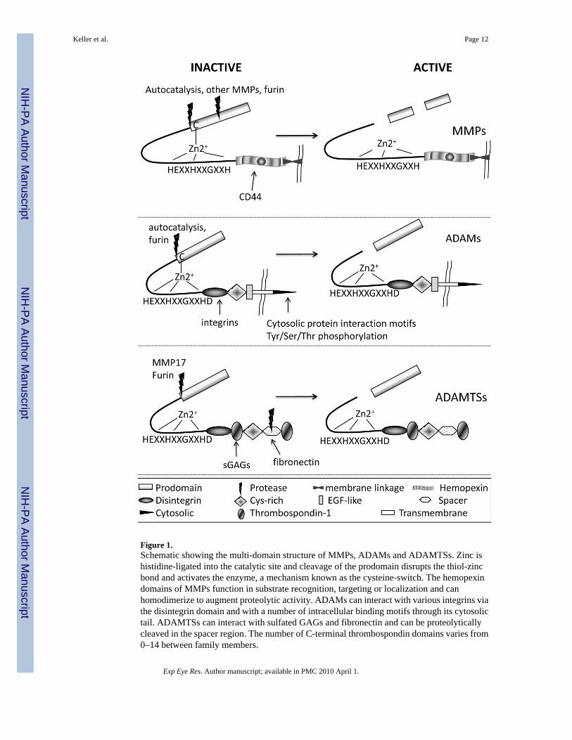
Figure 1.Schematic showing the multi-domain structure of MMPs, ADAMs and ADAMTSs. Zinc ishistidine-ligated into the catalytic site and cleavage of the prodomain disrupts the thiol-zincbond and activates the enzyme, a mechanism known as the cysteine-switch. The hemopexindomains of MMPs function in substrate recognition, targeting or localization and canhomodimerize to augment proteolytic activity. ADAMs can interact with various integrins viathe disintegrin domain and with a number of intracellular binding motifs through its cytosolictail. ADAMTSs can interact with sulfated GAGs and fibronectin and can be proteolyticallycleaved in the spacer region. The number of C-terminal thrombospondin domains varies from0–14 between family members.
Keller et al. Page 12
Exp Eye Res. Author manuscript; available in PMC 2010 April 1.
NIH
-PA Author Manuscript
NIH
-PA Author Manuscript
NIH
-PA Author Manuscript
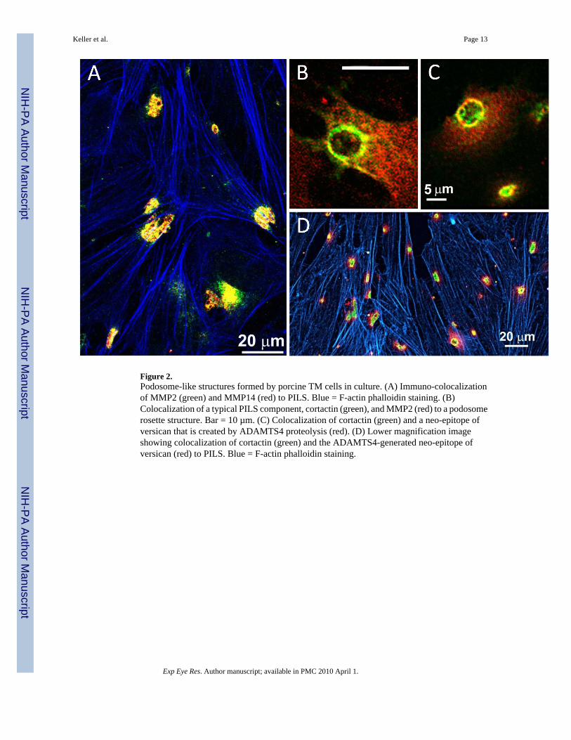
Figure 2.Podosome-like structures formed by porcine TM cells in culture. (A) Immuno-colocalizationof MMP2 (green) and MMP14 (red) to PILS. Blue = F-actin phalloidin staining. (B)Colocalization of a typical PILS component, cortactin (green), and MMP2 (red) to a podosomerosette structure. Bar = 10 µm. (C) Colocalization of cortactin (green) and a neo-epitope ofversican that is created by ADAMTS4 proteolysis (red). (D) Lower magnification imageshowing colocalization of cortactin (green) and the ADAMTS4-generated neo-epitope ofversican (red) to PILS. Blue = F-actin phalloidin staining.
Keller et al. Page 13
Exp Eye Res. Author manuscript; available in PMC 2010 April 1.
NIH
-PA Author Manuscript
NIH
-PA Author Manuscript
NIH
-PA Author Manuscript
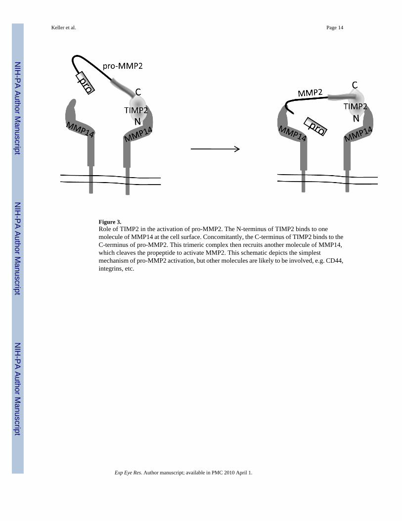
Figure 3.Role of TIMP2 in the activation of pro-MMP2. The N-terminus of TIMP2 binds to onemolecule of MMP14 at the cell surface. Concomitantly, the C-terminus of TIMP2 binds to theC-terminus of pro-MMP2. This trimeric complex then recruits another molecule of MMP14,which cleaves the propeptide to activate MMP2. This schematic depicts the simplestmechanism of pro-MMP2 activation, but other molecules are likely to be involved, e.g. CD44,integrins, etc.
Keller et al. Page 14
Exp Eye Res. Author manuscript; available in PMC 2010 April 1.
NIH
-PA Author Manuscript
NIH
-PA Author Manuscript
NIH
-PA Author Manuscript
NIH
-PA Author Manuscript
NIH
-PA Author Manuscript
NIH
-PA Author Manuscript
Keller et al. Page 15
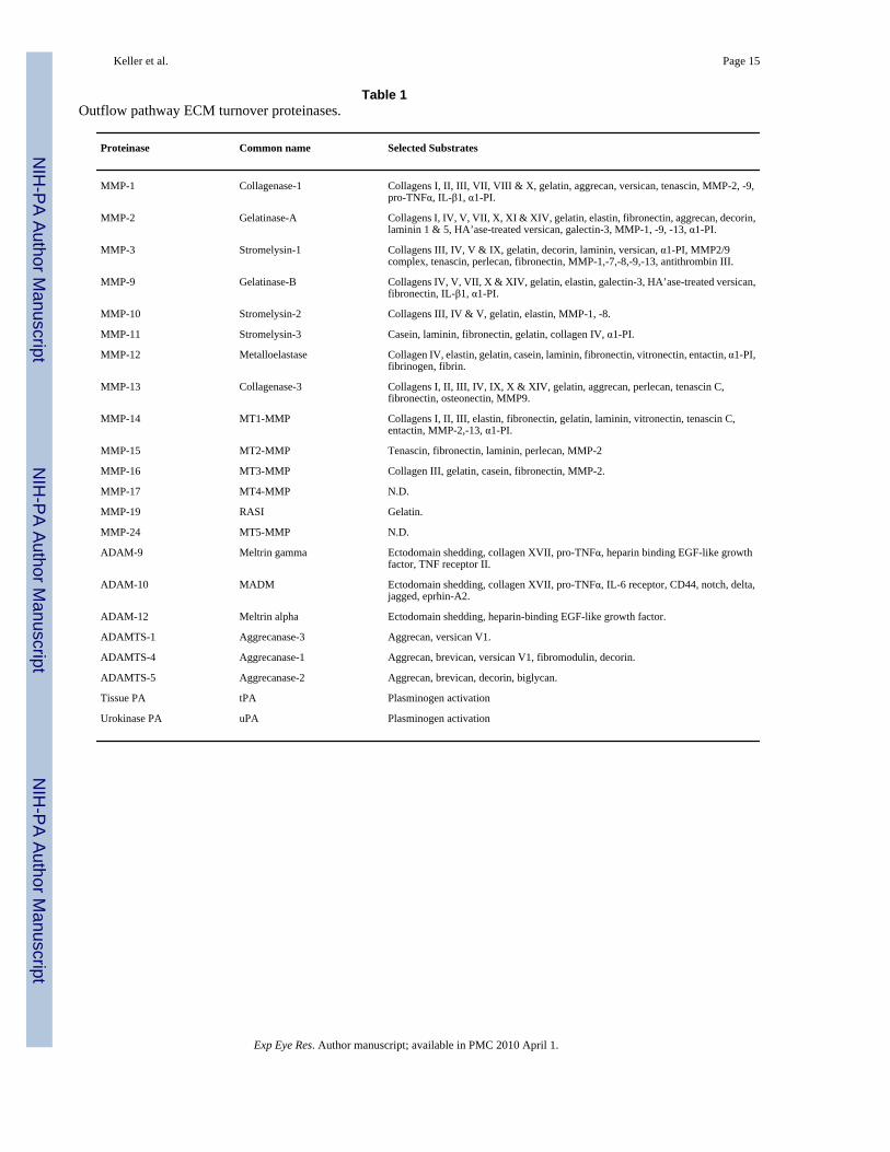
Table 1Outflow pathway ECM turnover proteinases.
Proteinase Common name Selected Substrates
MMP-1 Collagenase-1 Collagens I, II, III, VII, VIII & X, gelatin, aggrecan, versican, tenascin, MMP-2, -9,pro-TNFα, IL-β1, α1-PI.
MMP-2 Gelatinase-A Collagens I, IV, V, VII, X, XI & XIV, gelatin, elastin, fibronectin, aggrecan, decorin,laminin 1 & 5, HA’ase-treated versican, galectin-3, MMP-1, -9, -13, α1-PI.
MMP-3 Stromelysin-1 Collagens III, IV, V & IX, gelatin, decorin, laminin, versican, α1-PI, MMP2/9complex, tenascin, perlecan, fibronectin, MMP-1,-7,-8,-9,-13, antithrombin III.
MMP-9 Gelatinase-B Collagens IV, V, VII, X & XIV, gelatin, elastin, galectin-3, HA’ase-treated versican,fibronectin, IL-β1, α1-PI.
MMP-10 Stromelysin-2 Collagens III, IV & V, gelatin, elastin, MMP-1, -8.
MMP-11 Stromelysin-3 Casein, laminin, fibronectin, gelatin, collagen IV, α1-PI.
MMP-12 Metalloelastase Collagen IV, elastin, gelatin, casein, laminin, fibronectin, vitronectin, entactin, α1-PI,fibrinogen, fibrin.
MMP-13 Collagenase-3 Collagens I, II, III, IV, IX, X & XIV, gelatin, aggrecan, perlecan, tenascin C,fibronectin, osteonectin, MMP9.
MMP-14 MT1-MMP Collagens I, II, III, elastin, fibronectin, gelatin, laminin, vitronectin, tenascin C,entactin, MMP-2,-13, α1-PI.
MMP-15 MT2-MMP Tenascin, fibronectin, laminin, perlecan, MMP-2
MMP-16 MT3-MMP Collagen III, gelatin, casein, fibronectin, MMP-2.
MMP-17 MT4-MMP N.D.
MMP-19 RASI Gelatin.
MMP-24 MT5-MMP N.D.
ADAM-9 Meltrin gamma Ectodomain shedding, collagen XVII, pro-TNFα, heparin binding EGF-like growthfactor, TNF receptor II.
ADAM-10 MADM Ectodomain shedding, collagen XVII, pro-TNFα, IL-6 receptor, CD44, notch, delta,jagged, eprhin-A2.
ADAM-12 Meltrin alpha Ectodomain shedding, heparin-binding EGF-like growth factor.
ADAMTS-1 Aggrecanase-3 Aggrecan, versican V1.
ADAMTS-4 Aggrecanase-1 Aggrecan, brevican, versican V1, fibromodulin, decorin.
ADAMTS-5 Aggrecanase-2 Aggrecan, brevican, decorin, biglycan.
Tissue PA tPA Plasminogen activation
Urokinase PA uPA Plasminogen activation
Exp Eye Res. Author manuscript; available in PMC 2010 April 1.
NIH
-PA Author Manuscript
NIH
-PA Author Manuscript
NIH
-PA Author Manuscript
Keller et al. Page 16
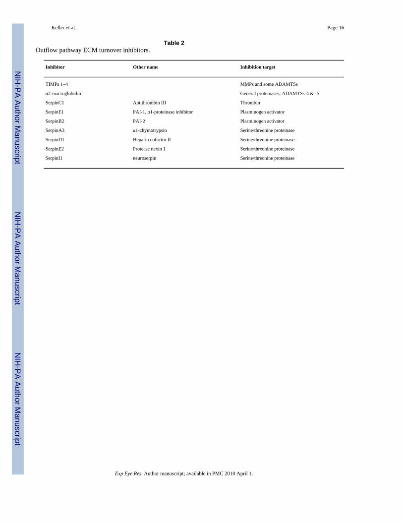
Table 2Outflow pathway ECM turnover inhibitors.
Inhibitor Other name Inhibition target
TIMPs 1–4 MMPs and some ADAMTSs
α2-macroglobulin General proteinases, ADAMTSs-4 & -5
SerpinC1 Antithrombin III Thrombin
SerpinE1 PAI-1, α1-proteinase inhibitor Plasminogen activator
SerpinB2 PAI-2 Plasminogen activator
SerpinA3 α1-chymotrypsin Serine/threonine proteinase
SerpinD1 Heparin cofactor II Serine/threonine proteinase
SerpinE2 Protease nexin 1 Serine/threonine proteinase
SerpinI1 neuroserpin Serine/threonine proteinase
Exp Eye Res. Author manuscript; available in PMC 2010 April 1.