Inhibition of Lymphogenous Metastasis Using Adeno-Associated Virus-Mediated Gene Transfer of a...
10
2005;65:6901-6909. Cancer Res JianMin Lin, Alshad S. Lalani, Thomas C. Harding, et al. Soluble VEGFR-3 Decoy Receptor Adeno-Associated Virus-Mediated Gene Transfer of a Inhibition of Lymphogenous Metastasis Using Updated version http://cancerres.aacrjournals.org/content/65/15/6901 Access the most recent version of this article at: Cited Articles http://cancerres.aacrjournals.org/content/65/15/6901.full.html#ref-list-1 This article cites by 35 articles, 15 of which you can access for free at: Citing articles http://cancerres.aacrjournals.org/content/65/15/6901.full.html#related-urls This article has been cited by 32 HighWire-hosted articles. Access the articles at: E-mail alerts related to this article or journal. Sign up to receive free email-alerts Subscriptions Reprints and . [email protected] Department at To order reprints of this article or to subscribe to the journal, contact the AACR Publications Permissions . [email protected] Department at To request permission to re-use all or part of this article, contact the AACR Publications Research. on December 23, 2013. © 2005 American Association for Cancer cancerres.aacrjournals.org Downloaded from Research. on December 23, 2013. © 2005 American Association for Cancer cancerres.aacrjournals.org Downloaded from
-
Upload
independent -
Category
Documents
-
view
1 -
download
0
Transcript of Inhibition of Lymphogenous Metastasis Using Adeno-Associated Virus-Mediated Gene Transfer of a...

2005;65:6901-6909. Cancer Res JianMin Lin, Alshad S. Lalani, Thomas C. Harding, et al. Soluble VEGFR-3 Decoy ReceptorAdeno-Associated Virus-Mediated Gene Transfer of a Inhibition of Lymphogenous Metastasis Using
Updated version
http://cancerres.aacrjournals.org/content/65/15/6901
Access the most recent version of this article at:
Cited Articles
http://cancerres.aacrjournals.org/content/65/15/6901.full.html#ref-list-1
This article cites by 35 articles, 15 of which you can access for free at:
Citing articles
http://cancerres.aacrjournals.org/content/65/15/6901.full.html#related-urls
This article has been cited by 32 HighWire-hosted articles. Access the articles at:
E-mail alerts related to this article or journal.Sign up to receive free email-alerts
Subscriptions
Reprints and
To order reprints of this article or to subscribe to the journal, contact the AACR Publications
Permissions
To request permission to re-use all or part of this article, contact the AACR Publications
Research. on December 23, 2013. © 2005 American Association for Cancercancerres.aacrjournals.org Downloaded from
Research. on December 23, 2013. © 2005 American Association for Cancercancerres.aacrjournals.org Downloaded from

Inhibition of Lymphogenous Metastasis Using Adeno-Associated
Virus-Mediated Gene Transfer of a Soluble VEGFR-3
Decoy Receptor
JianMin Lin,1Alshad S. Lalani,
1Thomas C. Harding,
1Melissa Gonzalez,
1Wei-Wei Wu,
1
Bo Luan,1Guang Huan Tu,
1Kathryn Koprivnikar,
1Melinda J. VanRoey,
1
Yulong He,2Kari Alitalo,
2and Karin Jooss
1
1Department of Preclinical Oncology and Immunology, Cell Genesys, Inc., South San Francisco, California and 2Molecular/Cancer BiologyLaboratory and Ludwig Institute for Cancer Research, Haartman Institute, Biomedicum Helsinki, University of Helsinki, Helsinki, Finland
Abstract
The presence of metastases in regional lymph nodes is astrong indicator of poor patient survival in many types ofcancer. It has recently been shown that the lymphangiogenicgrowth factor, vascular endothelial growth factor-C (VEGF-C),and its receptor, VEGF receptor-3 (VEGFR3), may play apivotal role in the promotion of metastasis to regional lymphnodes. In this study, human prostate and melanoma tumormodels that preferentially metastasize to the lymph nodesfollowing s.c. tumor cell implantation were established fromlymph node metastases via in vivo selection. Melanomatumor cell sublines established from lymph node metastasisexpress higher amounts of VEGF-C than the parental tumorcells. The inhibition of tumor-derived VEGF-C with a solubleVEGFR3 decoy receptor, sVEGFR3-Fc, expressed via a recom-binant adeno-associated viral vector, potently blocks tumor-associated lymphangiogenesis and tumor metastasis to thelymph nodes, when the treatment was initiated before thetumor implantation. In addition, sVEGFR3-Fc serum levelsrequired for efficient blockade of lymph node metastasesare strictly dependent on the VEGF-C levels generated bythe primary tumor. Recombinant adeno-associated virus–mediated gene transfer of sVEGFR3-Fc may represent afeasible therapeutic strategy for blockade of lymphogenousmetastasis. (Cancer Res 2005; 65(15): 6901-9)
Introduction
Recent studies have shown that a member of the vascularendothelial growth factor (VEGF) family, VEGF-C, stimulateslymphangiogenesis and lymphatic endothelial cell growth andmigration upon binding to its receptor, VEGFR-3 (1). VEGF-C hasalso been shown to promote lymphatic-mediated metastasis viainduction of tumor-associated lymphangiogenesis (2–5) in numeroussolid cancers (6–13). In support of these are preclinical datademonstrating that VEGF-C overexpression in cancer cellssignificantly increases tumor-associated lymphangiogenesis, resultingin enhanced metastasis to regional lymph nodes (2, 5, 11, 14, 15). Inaddition, blockade of VEGF-C/D–mediated signaling has beenshown to suppress tumor lymphangiogenesis and lymph nodemetastases in mice (16–19). We examined the effects of a soluble
VEGF-C inhibitor, called sVEGFR3-Fc, on tumor-induced lympho-genous and lung metastases in several tumor models thatpreferentially metastasize to the lymph nodes following s.c.implantation.A recombinant adeno-associated virus (rAAV) vector was used
for gene delivery of sVEGFR3-Fc. AAV is a parvovirus that shows nopathogenicity in humans and cannot replicate without helperfunctions provided by another virus such as adenovirus orherpesvirus (20). rAAV vectors lead to long-term expression oftherapeutic transgenes in liver and muscle (21).The results of this study show that stable systemic expression of
sVEGFR3-Fc following rAAV-mediated gene transfer potentlyinhibits tumor-associated lymphangiogenesis and the developmentof lymph node metastases in highly metastatic melanoma andrenal cell carcinoma models, and both lymph node and lungmetastases in a prostate tumor model. Furthermore, the serumlevels of sVEGFR3-Fc necessary to efficiently block lymph nodemetastases in these models are strongly dependent on the amountof VEGF-C generated by the primary tumor.
Materials and Methods
Cell lines. Human prostate carcinoma PC-3, renal carcinoma Caki-2, and
melanoma A375 cell lines were purchased from American Type Culture
Collection (ATCC, Manassas, VA). PC-3-mlg2 and A375-mln1 are sublinesestablished by in vivo selection of lymph node metastases from PC-3 or
A375 s.c. tumor–bearing mice. PC-3-mlg2-VEGF-C is a subline established
by transduction of PC-3-mlg2 cells with a lentiviral vector encoding human
VEGF-C. All of the above tumor cell lines were transduced with a lentiviralvector expressing a luciferase reporter gene and maintained in culture
conditions recommended by ATCC.
Recombinant adeno-associated virus vector construction. AAVvector plasmid encoding sVEGFR3-Fc was constructed as previously
described (22). Pseudotyped rAAV serotype-8 vector stocks were produced
in HEK 293 cells using calcium phosphate triple transfection of the rAAV
vector expression plasmid of interest in combination with the AAV-8 serotypehelper plasmid p5e18-VD2/8 (23) and pXX-6 (20). Virions were isolated on
two sequential CsCl gradients and titers determined by dot blot using
radioactive probe specific for the rAAV transgene.
In vivo gene transfer of sVEGFR3. Female NCR nu/nude mice (Taconic,Germantown, NY) were housed under specific pathogen-free conditions and
treated according to the Institute of Laboratory Animal Research Guide for
the Care and Use of Laboratory Animals. Mice were injected with a singledose of rAAV-sVEGFR3-Fc into the quadriceps muscle (50 AL/muscle) or
into the portal vein (dosing volume of 200 AL). Mice were bled by alternate
retro-orbital puncture on scheduled intervals to measure the serum levels of
human sVEGFR3-Fc by ELISA. Because the ratio between physical andinfectious vector particles differ between rAAV vector preparations, efficacy
was correlated with sVEGFR-3-Fc serum levels and not with the injected
physical particle dose.
Requests for reprints: JianMin Lin, Department of Preclinical Oncology andImmunology, Cell Genesys, Inc., 500 Forbes Boulevard, South San Francisco, CA 94080.Phone: 650-266-2911; Fax: 650-266-3300; E-mail: [email protected].
I2005 American Association for Cancer Research.
www.aacrjournals.org 6901 Cancer Res 2005; 65: (15). August 1, 2005
Research Article
Research. on December 23, 2013. © 2005 American Association for Cancercancerres.aacrjournals.org Downloaded from

sVEGFR3 detection by ELISA. sVEGFR3-Fc was quantitated using asandwich ELISA. Briefly, microtiter plates were coated with anti-human IgG
capture antibody (Bethyl Laboratories, Montgomery, TX) and blocked with
PBS-1% bovine serum albumin-0.05% Tween 20 buffer. Recombinant human
VEGFR3-Fc (R&D Systems, Minneapolis, MN) was used for standard curves.Samples and the standards were incubated in the wells for 1 hour, washed
extensively, and then incubated with 50 ng/mL biotinylated goat anti-
human VEGFR3 antiserum (R&D Systems) for 1 hour. The samples were
then incubated with horseradish peroxidase–conjugated streptavidin (BDPharMingen, San Diego, CA) and detected using Sure Blue TMB substrate
(KPL, Gaithersburg, MD) at 450/650 nm optical densities. The detection
sensitivity for sVEGFR3-Fc using this ELISA is f1 to 5 ng/mL.
Xenotransplantation, metastasis detection, and evaluation. In all
efficacy studies, rAAV vectors were administered to mice before tumor
challenge. Following rAAV injection, mice were challenged s.c. with 3 � 106
of either PC-3-mlg2 or A375-mln1 tumor cells. Primary tumor volumes
were determined by measurements using digital calipers and calculated as
length � width2 � 0.5. Mice were euthanized at either 3 or 5 weeks after
tumor cell inoculation and the lymph nodes were collected and analyzed by
bioluminescence imaging. Briefly, the mice were given i.p. with 1.5 mg/g
Luciferin substrate (Xenogen Corp., Alameda, CA). Fifteen minutes later, the
mice were euthanized and six lymph nodes, including the axillary and
inguinal from both sides, were collected and placed in a Petri dish for
bioluminescence imaging analysis. A set of six lymph nodes collected from a
naı̈ve mouse was used to determine the background CCD (complete count
density) counts in every experiment. The metastases burden of each mouse
was calculated based on total bioluminescence (CCD counts) of the lymph
nodes collected from each mouse. In a separate study, 5 � 106 Caki-2 tumor
cells were given 10 days following rAAV vector injection. Lymph nodes were
collected from each animal at 2 weeks after tumor inoculation and the
volume calculated as (p/6) � (length � width)3/2.
For selection of metastatic variants, lymph nodes with bioluminescence
CCD counts above 1 � 105 were collected for establishment of primary
cultures. Briefly, lymph nodes were minced, trypsinized at 37jC for 15minutes, and placed in a fresh culture dish with growth medium. Tumor
cells were selected by repeated trypsinization every 2 days for a total of five
passages. Approximately 3 � 106 cells were implanted in the s.c. tissue of
the dorsal flank of female NCR nu/nude mice for outgrowth and furthermetastatic selection.
Reverse transcription-PCR and ELISA detection of VEGF-C, VEGF-D,and VEGF-A. Frozen cell pellets and pieces of frozen, unthawed tumors
were homogenized in TRIzol reagent (Invitrogen Life Technologies,Carlsbad, CA) to isolate total RNA. Genomic DNA was removed from the
samples using DNA-free (Ambion, Austin, TX). RNA samples were converted
to cDNA and analyzed by quantitative PCR using TaqMan One-Step RT-PCRMaster Mix Reagents kit (Applied Biosystems, Foster City, CA) and Assay-
on-Demand reagents for hVEGF-C, mVEGF-C, hVEGF-A, hVEGF-D,
hGAPDH, and mGAPDH (all from Applied Biosystems). A solid-phase
capture ELISA was also used to quantify human VEGF-C protein fromhomogenized tumor tissues and tissue culture supernatants using a
commercially available kit (R&D Systems).
Quantitative detection of human tumor cell metastases. The
detection of human tumor cells in mouse lymph nodes was based on thequantitative detection of human alu sequences present in mouse lymph
node DNA extracts as previously described (24). Briefly, genomic DNA was
extracted from harvested tissue and human DNA detected by quantitativePCR using primers specific for human alu sequences. A quantitative
measure of amplifiable mouse DNA was obtained through amplification of
the mouse GAPDH genomic DNA sequence with mGAPDH.
Evaluation of lymphangiogenesis by lymphatic mapping. Anesthe-tized Caki-2 tumor-bearing mice were injected with 100 AL of 1% isosulfan
blue (lymphazurin, U.S. Surgical Corp., Norwalk, CT) intratumorally. After
10 minutes, mice were photographed for observation of the dye going
through the draining lymphatic vessels and isosulfan blue–stained draininglymph nodes.
Immunohistochemical analysis. Tissues harvested from animals were
fixed in 4% paraformaldehyde, infiltrated with 30% sucrose, and frozen in
OCT compound. Cryostat sections (5-25 Am) were rehydrated in TBS,permeabilized with 0.1% Triton X-100 (Sigma), and incubated in 10%
normal serum. Primary antibodies including goat polyclonal anti-CD105
(Santa Cruz Biotech, Santa Cruz, CA) and goat polyclonal anti-LYVE-1 (R&D
Systems) were applied overnight at 4jC. A secondary rabbit anti-goat Alexa594 (Molecular Probes, Eugene, OR) antibody was incubated for 30 minutes
at room temperature. Slides were mounted in Vectashield Mounting
Medium with 4V,6-diamidino-2-phenylindole (DAPI; Vector Laboratories,
Burlingame, CA) and analyzed by fluorescence microscopy using a ZeissAxioplan microscope equipped with a SPOT RT Slider digital camera
(Diagnostic Instruments, Inc., Sterling Heights, MI). Images were acquired
from serial sections using Image Pro Plus software (MediaCybernetics,
Silver Springs, MD).
Results
Recombinant adeno-associated virus vectors encoding asVEGFR3-Fc decoy receptor results in sustained systemicexpression of a potent VEGF-C inhibitor following genetransfer in vitro and in vivo . To evaluate the antilymphangiogenicand antimetastatic effects of a human sVEGFR3 decoy receptorusing systemic gene transfer, a recombinant AAV vector thatencodes a secreted form of human VEGFR3 (sVEGFR3-Fc) under thecontrol of a constitutive CAG promoter was constructed (data notshown). The soluble receptor comprises the first three amino-terminal immunoglobulin-like domains of the cellular VEGFR3 andwas fused to the heavy chain of the human IgG1 Fc moiety (17). Thistruncated sVEGFR3-Fc has previously been shown to specificallybind to VEGF-C and VEGF-D with high affinity and, therefore,competes with the binding of the ligands to their cognate cellularreceptor (17). A corresponding rAAV vector expressing no trans-gene, termed rAAV-Null, served as a vector control. rAAV vectorspseudotyped with the capsid from AAV serotype-8 were used inthese studies because it results in significantly higher expressionlevels of the transgene in serum compared with AAV-2 vectors (25).The expression of sVEGFR3-Fc following rAAV-mediated gene
transfer was verified in vitro and in vivo . A single administration of1 � 1011 vg of rAAV-sVEGFR3-Fc into the quadricep muscles orportal vein of immunodeficient mice resulted in sVEGFR3-Fcserum levels of f1 Ag/mL 1 week following gene transfer, andincreased over time, reaching steady-state levels of 8 to 10 Ag/mLatf4 to 5 weeks following vector administration (data not shown).Furthermore, sustained expression of sVEGFR3-Fc was detected inthe sera of mice for >4 months following a single injection of rAAV-sVEGFR3-Fc and animals showed no evidence of adverse healththroughout the entire course of the experiments (data not shown).In addition, the biological activity of rAAV-produced sVEGFR3-Fcwas verified by measuring its ability to block VEGF-C–inducedproliferation of Ba/F3 cells expressing a chimeric VEGFR-3/erythropoietin receptor in vitro (17). rAAV-derived sVEGFR3-Fcinhibited VEGF-C–induced mitogenic activity in a dose-dependentmanner with a IC50 off30 ng/mL when Ba/F3-VEGFR3/EpoR cellswere stimulated with 100 ng/mL of human VEGF-C for 72 hours(data not shown). Thus, long-term sustained systemic expression ofa biologically active form of sVEGFR3-Fc is achieved following asingle administration of rAAV vectors in vivo .Establishment of a lymphogenous metastatic melanoma
tumor model. A metastatic human tumor model, termed A375-mln1, was developed to evaluate the effects of rAAV-sVEGFR3-Fc–mediated gene transfer on tumor-induced lymphangiogenesis andlymphogenous metastasis. To monitor metastasis, tumor cells werefirst transduced with a lentiviral vector encoding the luciferase
Cancer Research
Cancer Res 2005; 65: (15). August 1, 2005 6902 www.aacrjournals.org
Research. on December 23, 2013. © 2005 American Association for Cancercancerres.aacrjournals.org Downloaded from

reporter gene. The luciferase-expressing tumor cells can be readilyvisualized in vitro or ex vivo by bioluminescence imaging. Theluciferase-positive A375 tumors grown s.c. metastasize to the lungsbut have a low incidence for metastasizing to the lymph nodes. TheA375-mln1 tumor variants established from lymph node metasta-ses of A375 tumor-bearing mice showed a >3-fold increase in lymphnode metastases following s.c. tumor cell injection compared withthe parental A375-challenged animals (Fig. 1A).Because VEGF-C is a potent lymphangiogenic factor that
stimulates tumor lymphangiogenesis and lymphogenous metasta-sis (26, 27), the correlation between the metastatic capacity andVEGF-C expression in A375-mln1 tumor model was characterized.To do so, we evaluated whether in vivo–selected subpopulations oftumor cells that preferentially metastasize to lymph nodes expressmore human VEGF-C than the parental cell lines. The expression ofVEGF-C was increased >4-fold at the mRNA level (Fig. 1B), andf2-fold at the protein level (Fig. 1C), in the metastatic A375-mln1
subline compared with the parental A375 cells in vitro (Fig. 1Band C). In addition, the expression of VEGF-A was increased 3-foldin the selected tumor subline (Fig. 1B). Similar to our in vitroobservations, we detected an approximate 2-fold increase in VEGF-Cprotein levels in tumors arising from the metastatic A375-mln1cells compared with tumors grown from the parental A375 cells, asquantitated by ELISA (Fig. 1C). Interestingly, no significant increasein the expression of the lymphangiogenic growth factor VEGF-Dwas observed in the A375-mln1 metastatic tumor subline (Fig. 1B).In summary, a tumor model that preferentially metastasizes to thelymph nodes following s.c. tumor cell challenge was developed, andan increase in the expression of VEGF-C, but not VEGF-D, by theprimary tumor correlated with a higher incidence of lymph nodemetastasis in vivo .rAAV-sVEGFR3-Fc–mediated gene transfer inhibits lymph
node metastasis in an A375 human melanoma model but isless effective in blocking lung metastasis. The antimetastatic
Figure 1. Tumor cell lines established from lymph node metastases express higher levels of VEGF-C and VEGF-A and result in higher incidence of lymph nodemetastasis following subsequent s.c. implantation in nude mice. A, a metastatic melanoma tumor cell variant, termed A375-mln1, was established from metastaticlymph nodes of mice bearing s.c. A375 tumors following one round of selection in vivo . The tumor cells were genetically modified to express the luciferase reporter geneand the frequency of lymph node metastasis following s.c. implantation of A375 parental and A375-mln1 metastatic variant was determined by bioluminescence.Columns, mean frequencies of lymph node metastasis; bars, SE (n z 7, six experiments). B, the metastatic A375-mln-1 cell line expresses increased mRNA levels ofVEGF-A and VEGF-C, but not VEGF-D, compared with the parental A375 cell line. Total RNA was isolated from tissue culture cells and the expression of VEGF-A,VEGF-C, and VEGF-D was determined by quantitative PCR. Columns, average cycle threshold (CT ); bars, SE (n = 6). Fold difference (indicated next to asterisk)in RNA expression between A375 and A375-mln1 was determined using the comparative CT method according to manufacturer’s instructions (Applied Biosystems).All values are normalized to GAPDH. C, total protein was extracted from cultured cells (in vitro ) or primary tumor tissue (in vivo) and assayed for human VEGF-Cexpression by ELISA. Columns, means; bars, SE (n z 4, two experiments). The relative fold difference of VEGF-C expression from in vivo–selected A375-mln1 versusparental tumor cells is indicated by the asterisk.
Control of Lymph Node Metastasis by rAAV-sVEGFR3-Mediated Gene Transfer
www.aacrjournals.org 6903 Cancer Res 2005; 65: (15). August 1, 2005
Research. on December 23, 2013. © 2005 American Association for Cancercancerres.aacrjournals.org Downloaded from

effects of rAAV-sVEGFR3-Fc–mediated gene transfer were firstevaluated in the human A375-mln1 metastatic melanoma model. Inthis model, lymph node and lung metastases appear concurrentlyand can be detected in all animals challenged s.c. with A375-mln1by 3 weeks postimplantation (data not shown). A single injection ofrAAV-sVEGFR3-Fc (at either 3 � 1011 or 1.5 � 1011 vg/animal) orrAAV-Null (3 � 1011 vg/animal) was given into the portal vein ofmice 10 days before A375-mln1 metastatic tumor cell challenge toevaluate dose-dependent efficacy. rAAV-sVEGFR3-Fc treatmenthad no effect on the growth of the primary tumor (Fig. 2A). Lymphnodes and lungs were collected 3 weeks after tumor challenge forevaluation of lymphogenous and lung metastasis (Fig. 2B and C).
Systemic expression of sVEGFR3-Fc following rAAV-mediated genetransfer inhibited metastasis to the lymph nodes in a dose-dependent manner (Fig. 2B). Only two of seven mice (28%, P = 0.01)developed lymph node metastases following treatment with thehigh dose of rAAV-sVEGFR3-Fc, which resulted in stable sVEGFR3-Fc serum levels of f20 Ag/mL. In contrast, all nine mice (100%)developed lymph node metastases after receiving an equivalentdose of the rAAV-Null vector. No antimetastatic efficacy wasobserved in animals that received the lower dose of vector, whichresulted in f2 Ag/mL of serum sVEGFR3-Fc. Interestingly, serumsVEGFR3-Fc levels of 20 Ag/mL that were effective in blockinglymph node metastasis were less efficacious in preventing theformation of lung metastases. In the group treated with the highdose of rAAV-sVEGFR3-Fc, five of seven (71%) mice developed lungmetastases, compared with all animals in the control group (Fig.2C). However, the overall tumor burden in the lungs was slightlylower in the animals receiving the high dose of the therapeuticvector than in the lungs of control animals (P = 0.028). The low-dose treatment of rAAV-sVEGFR3-Fc had no impact on A375-mln1lung metastasis.rAAV-sVEGFR3-Fc–mediated gene transfer inhibits tumor-
induced lymphangiogenesis and metastasis in a human kidneytumor model. rAAV-sVEGFR3-Fc treatment was further evaluatedin a renal cell carcinoma, Caki-2, model. A pilot study showed thatCaki-2 tumor cells rapidly induce lymphangiogenesis and undergolymph node metastasis after s.c. implantation in nude mice.Because the portal vein and i.m. routes of vector administrationresult in comparable sVEGFR3-Fc serum levels, the less invasivei.m. vector administration route was utilized for the remainder ofthe experiments in the study. A single injection of rAAV-sVEGFR3-Fc (at either 3 � 1011 or 1.5 � 1011 vg/animal) was administeredi.m. to mice 10 days before Caki-2 tumor cell challenge to evaluatedose-dependent antimetastatic efficacy. Sustained sVEGFR3-Fcserum levels of f35 and 10 Ag/mL, respectively, were detectedfollowing rAAV-mediated gene transfer (data not shown). At2 weeks after tumor inoculation, lymph nodes were harvestedand analyzed for the presence of tumor metastases. Due to weakluciferase activity in the Caki-2 cell line, the efficacy of the therapyin this model was analyzed by injecting lymphazurin (1% isosulfanblue), a dye that specifically stains lymphatic vessels and lymphnodes (28, 29), into the tumor. This approach has the additionaladvantage of providing an evaluation of lymphangiogenesis aswell as lymph node metastasis blockade. A clear enlargementand increase in number of the draining lymphatic vessels froms.c.-grown tumors was evident when lymphazurin was injectedintratumorally in animals treated with the rAAV-Null vector(Fig. 3A). In contrast, no tumor-associated lymphatic vessels wereapparent in rAAV-sVEGFR3-Fc–treated animals, which correlatedwith a lack of dye-positive lymph nodes (Fig. 3B). rAAV-sVEGFR3-Fc–mediated gene transfer also resulted in an f60% (P < 0.01)reduction of Caki-2–induced lymph node enlargement versus thecontrol treatment (Fig. 3C). Furthermore, PCR analysis done onlymph nodes isolated from these animals revealed the presence ofsignificantly more human genome (alu) sequences in controlanimals than in rAAV-sVEGFR3-Fc–treated animals (Fig. 3D),indicating a reduced number of human tumor cells in the lymphnodes of treated animals. In summary, a reduction in the numberof lymphatic vessels and a 70% reduction of human cells present inthe lymph nodes was observed in Caki-2 tumor-bearing animalsthat continuously expressed 35 Ag/mL of sVEGFR3-Fc in the serumfollowing rAAV-sVEGFR3-Fc gene transfer. sVEGFR3-Fc serum
Figure 2. Dose-dependent effects of rAAV-sVEGFR3-Fc on blockade ofA375-mln1 tumor metastasis to the lymph nodes and lungs. Immunodeficientmice were injected with 1.5 � 1011 or 3 � 1011 vg/animal of rAAV-sVEGFR3-Fcor rAAV-Null control vectors. Ten days following rAAV administration, mice werechallenged s.c. with 3 � 106 A375-mln1 tumor cells that stably express aluciferase reporter gene and the primary tumor growth (A ) was monitored overtime. Lymph nodes (B) and the lungs (C ) were evaluated for metastasis bybioluminescence at 3 weeks after s.c. tumor implantation. Shown are thebioluminescence CCD counts of six lymph nodes (axillary and inguinal nodesfrom both sides; B) and the entire lungs (C) collected from each animal. Lymphnodes and lungs collected from naı̈ve mice had V3 � 104 CCD counts(hatched area ), which represent background values. Bars in the scatter plots,mean F SE CCD counts per group.
Cancer Research
Cancer Res 2005; 65: (15). August 1, 2005 6904 www.aacrjournals.org
Research. on December 23, 2013. © 2005 American Association for Cancercancerres.aacrjournals.org Downloaded from

levels of 10 Ag/mL also resulted in reduced inhibition of lymphnode metastases in tumor-bearing mice (data not shown),indicating the dose dependency of the rAAV-sVEGFR3-Fc therapyon lymph node metastasis.rAAV-sVEGFR3-Fc gene transfer inhibits PC-3 prostate
cancer metastasis to lymph nodes and lungs. The antimeta-static effect of rAAV-sVEGFR3-Fc–mediated gene transfer was alsoevaluated in a prostate cancer model, PC-3-mlg2. The PC-3-mlg2tumor cell line was established from lymph node metastases ofPC-3 tumor-bearing mice. Although it has previously been shownthat parental PC-3 tumor cells grown s.c. spontaneously metas-tasize to the lymph nodes (30) and express higher levels of VEGF-Ccompared with in vivo selected A375-mln1 tumor cells (data notshown), we observed that the growth rate of the primary tumorwas poor and varied greatly among animals (data not shown). Incontrast, we observed 100% tumor take and a consistently highrate of lymph node metastasis using the PC-3-mlg2 tumor sublinederived from the lymph node metastases of PC-3 tumor-bearingmice (data not shown). In the PC-3-mlg2 tumor model, lymphnode metastases could be detected in f80% of the animals5 weeks after s.c. tumor challenge, in contrast to lung metastases,which were only detected in 10% to 20% of the animals at thesame time point. The incidence of lung metastases increases overtime, however, reaching 50% by 7 to 8 weeks after tumorchallenge (data not shown). Three dose levels of rAAV-sVEGFR3-
Fc (3 � 1010, 7 � 1010, or 1.5 � 1011 vg/animal) were administeredi.m. to mice 10 days before PC-3-mlg2 tumor cell challenge toevaluate dose-dependent antimetastatic efficacy. With this treat-ment regimen, the primary tumor growth was not affected(Fig. 4A). Lymph nodes and lungs were collected 5 weeks followingtumor challenge for evaluation of metastasis using biolumines-cence imaging. Only 2 of 11 mice (18%, P = 0.01) developed lymphnode metastases following treatment with the highest dose ofrAAV-sVEGFR3-Fc, which resulted in sVEGFR3-Fc serum levels off20 Ag/mL (Fig. 4B). In contrast, 8 of 11 mice (73%) developedlymph node metastases when treated with an equivalent dose ofrAAV-Null vector (Fig. 4B). The potent inhibitory effects ofsVEGFR3-Fc were lost when the treatment dose of rAAV-sVEGFR3-Fc was decreased, with 7 of 13 (54%, P > 0.05) and 7of 12 (58%, P > 0.05) animals developing lymph node metastaseswhen the sVEGFR3-Fc serum levels were 10 and 0.6 Ag/mL,respectively, demonstrating the dose dependency of the rAAV-sVEGFR3-Fc antimetastatic effect. Two of eleven (18%) animalstreated with the control vector had developed lung metastases atthe time of sacrifice, 5 weeks posttreatement, whereas no lungmetastases were detected in any of the mice treated with rAAV-sVEGFR-3-Fc. Although the frequency of lung metastases was lowat the time of sacrifice, these data suggest that lung metastases inthe PC-3-mlg2 model may spread from lymph node metastasesand may be controlled by sVEGFR3-Fc therapy.
Figure 3. rAAV-sVEGFR3-Fc inhibits lymph nodemetastasis in a s.c. Caki-2 model. Immunodeficientmice were challenged s.c. with Caki-2 tumorcells 10 days following rAAV-sVEGFR3-Fcor rAAV-Null vector administration. Two weeksfollowing tumor challenge, randomly selectedmice were injected intratumorally with 1%isosulfan blue for lymphatic vessel mapping(A and B). A typical observation of blue dyedraining through enlarged lymphatic vessels fromthe tumor to the lymph nodes inrAAV-Null–injected Caki-2 tumor-bearing mice(A), which was absent in therAAV-sVEGFR3-Fc–treated animals (B). t, s.c.tumor injected with 1% isosulfan blue; LN,isosulfan blue-stained lymph nodes; filled arrow,isosulfan blue–stained lymphatic vessel. C, meanF SE volume of axillary and inguinal lymph nodesharvested from control and treated mice. D, meanF SE number of human metastatic tumor cellspresent in the lymph nodes of control and treatedmice as determined by PCR analysis usingspecific human genome (alu ) sequence probes.
Control of Lymph Node Metastasis by rAAV-sVEGFR3-Mediated Gene Transfer
www.aacrjournals.org 6905 Cancer Res 2005; 65: (15). August 1, 2005
Research. on December 23, 2013. © 2005 American Association for Cancercancerres.aacrjournals.org Downloaded from

rAAV-sVEGFR3-Fc gene transfer blocks the development oflymph node metastasis by inhibiting tumor-associatedlymphangiogenesis. To confirm the mechanism by whichrAAV-sVEGFR3-Fc therapy blocks lymphatic-mediated metastasis,primary tumors from PC-3-mlg2 and Caki-2 tumor-bearinganimals pretreated with rAAV-sVEGFR3-Fc or AAV-null vectorswere evaluated for tumor-associated lymphatic vessels byhistologic analysis using the lymphatic-specific cell surfacemarker, LYVE-1 (Fig. 5A and B). Immunohistochemical stainingof tumors from AAV-control–treated animals revealed thepresence of irregular LYVE-1–positive lymphatic vessels thatpermeated into the tumor tissue and often formed hyperplasticclusters containing invasive tumor cells (Fig. 5A and B , top).In contrast, the primary tumor tissues derived from rAAV-sVEGFR3-Fc–treated animals were devoid of LYVE-1–positivelymphatic vessels within the tumor (Fig. 5A and B , bottom).Importantly, non–tumor-associated LYVE-1–positive lymphaticvessels detected in the normal skin tissue adjacent to the tumorperiphery remained unaffected by the rAAV-sVEGFR3-Fc treat-ment (Fig. 5A and C). This suggests that sVEGFR-3 treatmentdoes not impede preexisting or non–tumor-associated lymphaticvessels. Interestingly, tumor-associated blood vessels, as detectedby CD105 immunostaining, was unaffected by rAAV-sVEGFR3-Fctreatment in either PC-3-mlg2 or Caki-2 tumors as the number of
CD105-positive vessels within the primary tissue were equivalentto those observed in control tumors (Fig. 5A-D).rAAV-sVEGFR3-Fc serum levels required for blockade of
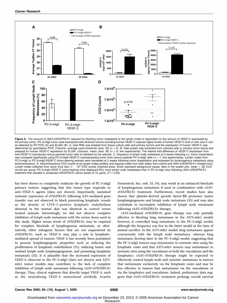
tumor-induced lymph node metastasis are dependent on theamount of VEGF-C expressed by the primary tumor. rAAV-sVEGFR3-Fc–mediated gene delivery aims at sequestering andneutralizing the bioactivity of the VEGF-C ligand by occluding itsinteraction to cellular VEGFR3. To evaluate the mechanism ofaction for rAAV-sVEGFR3-Fc, PC-3-mlg2 tumor lines were trans-duced with a lentiviral vector expressing human VEGF-C. Thetransduced cells, termed PC-3-mlg2-VEGF-C, express higher levelsof VEGF-C in vitro and in vivo than the parental PC-3-mlg2 cells(f9-fold and f12-fold, respectively) as determined by reversetranscription-PCR analysis and ELISA (Fig. 6A and B). Consistentwith the putative role of VEGF-C in lymph node metastasis, PC-3-mlg2-VEGF-C–challenged animals developed lymph node metas-tases in 100% of animals within 3 weeks following tumor cellchallenge, whereas only f50% of PC-3-mlg2–challenged animalsdeveloped lymph node metastases at the same time point (Fig. 6C).The PC-3-mlg2-VEGF-C and PC-3-mlg2 tumor models were used
to determine if the sVEGFR3-Fc serum levels required for efficientblockade of lymph node metastases correlated with the VEGF-Clevels generated by the primary tumors. rAAV-sVEGFR3-Fc at adose of 1.5 � 1011 vg/animal (resulting in sVEGFR3-Fc serum levelsof f15 Ag/mL) was injected i.m. in mice 10 days before tumor cellchallenge. Five weeks later, lymph nodes were collected forevaluation of lymphogenous metastasis using bioluminescenceimaging. As shown in Fig. 6D , only 10% of the animals challengedwith PC-3-mlg2 tumor cells, but 57% of the animals challengedwith the VEGF-C overexpressing PC-3-mlg2 tumor cells, developedlymph node metastases. These data suggest that the sVEGFR3-Fcserum levels required for efficient blockade of lymph nodemetastases correlate with the amount of VEGF-C generated bythe primary tumor.
Discussion
VEGF-C and its signal transduction pathway has been charac-terized in recent years, and a major role in lymphangiogenesis andlymphogenous tumor metastasis has been suggested for thisgrowth factor (2–4). In the studies presented here, we show that ahuman melanoma cell line established from lymph node metas-tases of A375 tumor-bearing animals expresses 4-fold higher VEGF-Clevels compared with the parental A375 cells. No significantdifferences in the expression of the lymphangiogenic factor, VEGF-D,were observed in the metastatic subline compared with theirparental tumor cells in culture. Furthermore, s.c. implantation oftumors derived from metastatic cells expressing increased levels ofVEGF-C resulted in a significantly higher incidence of lymph nodemetastases than from the unselected parental cells, supporting thehypothesis that lymph node metastasis are driven mainly by theexpression of VEGF-C generated by the primary tumor (11, 15).The study presented here also examined whether a rAAV vector
expressing a sVEGFR3-Fc fusion protein that sequesters VEGF-Ccould block the metastatic spread of tumor cells from the primarys.c. tumor site to the lymph nodes in three distinct metastatictumor models. Gene transfer of rAAV-sVEGFR3-Fc effectivelyblocked tumor metastasis to regional lymph nodes in the humanprostate cancer (PC-3-mlg2), melanoma (A375-mln1), and renal cellcarcinoma (Caki-2) metastatic tumor models. These resultsconfirm the recent findings by He et al. (19), demonstrating that
Figure 4. Systemic expression of rAAV-sVEGFR3-Fc inhibits PC-3-mlg2 tumormetastasis to the lymph nodes in immunodeficient mice. Immunodeficientmice were injected i.m. with rAAV-sVEGFR3-Fc at 3 � 1010, 7 � 1010, or1.5 � 1011 vg/animal resulting in sustained systemic sVEGFR3-Fc levels of0.6, 10, and 20 Ag/mL, respectively. Ten days following rAAV administration,mice were challenged s.c. with 3 � 106 PC-3-mlg2 tumor cells that stablyexpress a luciferase reporter gene and the growth of the primary tumor (A) wasmeasured over time. Lymph nodes were evaluated for lymphogenous metastasisby bioluminescence at 5 weeks after tumor implantation. B, bioluminescenceCCD counts of six lymph nodes (axillary and inguinal nodes from both sides)from control and treated mice. Lymph nodes collected from naı̈ve mice hadV1 � 104 CCD counts (hatched area ), which represent background values.Bars in the scatter plots, mean F SE CCD counts per group.
Cancer Research
Cancer Res 2005; 65: (15). August 1, 2005 6906 www.aacrjournals.org
Research. on December 23, 2013. © 2005 American Association for Cancercancerres.aacrjournals.org Downloaded from

suppression of VEGFR3 signaling inhibits tumor metastasis to thelymph nodes and extend these findings by showing that thisantimetastatic effect is highly dose dependent.rAAV-mediated gene delivery resulting in sustained sVEGFR3-Fc
serum levels of 15 Ag/mL or above efficiently blocked lymph-nodemetastasis by up to 70% in all three tumor models. In contrast,sVEGFR3-Fc serum levels of 10 Ag/mL and below had reduced orminimal effect. Moreover, tumors that express more VEGF-Crequired higher doses of vector to block metastatic spread of tumorcells. Earlier onset and higher frequency of lymph node metastasisin vivo was seen in a metastatic PC-3-mlg2 tumor cell line thatwas engineered to overexpresses VEGF-C (PC-3-mlg2-VEGF-C)compared with the parental cell line, and higher levels of serumsVEGFR3-Fc were required to block trafficking of these cells tolymph nodes. Thus, there is a direct correlation between theexpression levels of VEGF-C in primary tumors, the development oflymph node metastases in vivo and the sVEGFR3-Fc serum levelsrequired for therapeutic benefit.
Constitutive systemic expression of sVEGFR3-Fc following AAV-mediated gene transfer resulted in the potent blockade of tumor-associated lymphangiogenesis and did not seem to modulate‘‘classic’’ neoangiogenesis at the primary tumor site. Our studiesshow that rAAV-sVEGFR3-Fc treatment resulted in the completeinhibition of LYVE-1–positive lymphatic vessels, but had no effectin blocking CD105-associated blood vessels within the primarytumor tissue in either the PC3-mlg2 or the Caki-2 metastatictumor models. This suggests that sVEGFR3-Fc specifically targetsinteractions of VEGF-C to its high affinity receptor, VEGFR3,rather than mitigating potential downstream angiogenic signalsemanating from the low-affinity interaction of VEGF-C toVEGFR2. In addition, although elevated levels of VEGF-A wereobserved in both the PC-3-mlg2 and A375-mln1 metastatic linesin vitro , the growth of the primary tumor was not affected byrAAV-sVEGFR3-Fc treatment using the vector doses used in ourtumor studies. Indeed, blockade of VEGF-A signaling pathwaysusing systemic gene delivery of a sVEGFR1/R2-Fc decoy receptor
Figure 5. rAAV-sVEGFR3-Fc inhibits tumor-associated lymphangiogenesis but not angiogenesis in PC-3 prostate and Caki-2 renal carcinoma tumor models. At3 weeks following tumor implantation, PC3-mlg2 (A ) and Caki-2 (B ) primary tumors from control (top ) and rAAV-sVEGFR3-Fc–treated (bottom ) mice were harvestedand analyzed for lymphatic vessels using a murine LYVE-1 antibody (red staining ) and for blood vessels using a murine CD105 antibody (green staining ) byimmunohistochemistry. Tissue sections were counterstained with DAPI (blue ). Dotted line, tumor-stroma boundary where ‘‘T’’ represents the tumor tissue and ‘‘S’’represents the adjacent normal skin. LYVE-1+ lymphatic vessels (white arrows) were observed within the tumor in control-treated specimens and were completelydevoid within the tumor periphery in rAAV-sVEGFR3-Fc–treated tumors. LYVE-1 staining within the normal skin adjacent to the tumor periphery showed no difference inthe density of preexisting lymphatic vessels (yellow arrows ) following rAAV-sVEGFR3-Fc treatment compared with control tumor sections. The density of CD105+
tumor-associated blood vessels (green arrow ) was similar in rAAV-sVEGFR3-Fc–treated tumor tissues compared with the controls. C and D, mean F SE numberof immunostained vessel structures was enumerated from nine serial sections of four representative PC-3-mlg2 tumors (C ) and two representative Caki-2 tumors (D )in each experimental group. PC-3 tumors: P = 0.0001 for LYVE-1, P > 0.5 for CD105; Caki-2 tumors: P = 0.0001 for LYVE-1, P > 0.3 for CD105.
Control of Lymph Node Metastasis by rAAV-sVEGFR3-Mediated Gene Transfer
www.aacrjournals.org 6907 Cancer Res 2005; 65: (15). August 1, 2005
Research. on December 23, 2013. © 2005 American Association for Cancercancerres.aacrjournals.org Downloaded from
has been shown to completely eradicate the growth of PC-3-mlg2primary tumors, suggesting that this tumor type responds toanti–VEGF-A agents (data not shown). Importantly, sustainedsystemic expression of sVEGFR3-Fc following AAV-mediated genetransfer was not observed to block preexisting lymphatic vesselsas the density of LYVE-1–positive lymphatic endotheliumdetected in the normal skin was identical in control versustreated animals. Interestingly, we did not observe completeinhibition of lymph node metastasis with the vector doses used inthis study. Higher serum levels of sVEGFR3-Fc may be requiredfor complete blockade of lymphogenous metastasis, or, alter-natively, other mitogenic factors that are not sequestered bysVEGFR3-Fc, such as VEGF-A, may play a role in lymphatic-mediated spread of tumors. VEGF-A has recently been postulatedto possess lymphangiogenic properties such as inducing theproliferation of lymphatic endothelium (31), inducing tumor andsentinel lymph node lymphangiogenesis, and promoting lymphaticmetastasis (32). It is plausible that the increased expression ofVEGF-A observed in the PC-3-mlg2 (data not shown) and A375-mln1 tumor models may contribute to the lack of completeinhibition of lymph node metastasis following rAAV-sVEGFR3-Fctherapy. Thus, clinical regimens that directly target VEGF-A, suchas the neutralizing VEGF-A monoclonal antibody Avastin
(Genentech, Inc.; refs. 33, 34), may result in an enhanced blockadeof lymphogenous metastasis if used in combination with rAAV-sVEGFR3-Fc treatment. Furthermore, recent studies have alsoshown that platelet-derived growth factor-BB promotes tumorlymphangiogenesis and lymph node metastasis (35) and may alsocontribute to incomplete inhibition of lymph node metastasisfollowing rAAV-sVEGFR3-Fc therapy.rAAV-mediated sVEGFR3-Fc gene therapy was only partially
effective in blocking lung metastases in the A375-mln1 model;however, it controlled lung metastases in the PC-3-mlg2 model,although the frequency was low in the latter model at the time ofanimal sacrifice. In the A375-mln1 model, lung metastases appearconcurrently with the lymph node metastases, whereas lungmetastases develop later in the PC-3-mlg2 model, suggesting thatthe PC-3-mlg2 tumors may metastasize to systemic sites using thelymphatic route and that A375-mln1 tumors may metastasize tosystemic sites using the vasculature or both the vasculature and thelymphatics. rAAV-sVEGFR3-Fc therapy might be expected toeffectively control lymph node and systemic metastases in tumorsthat metastasize exclusively via the lymphatic route and may beless effective in tumors that metastasize via the vasculature orvia the lymphatics and vasculature. Indeed, preliminary data sug-gests that rAAV-sVEGFR3-Fc treatment prolongs overall survival
Figure 6. The amount of rAAV-sVEGFR3-Fc required for blocking tumor metastasis to the lymph nodes is dependent on the amount of VEGF-C expressed bythe primary tumor. PC-3-mlg2 tumor cells transduced with lentiviral vectors encoding human VEGF-C express higher levels of human VEGF-C both in vitro and in vivoas detected by RT-PCR (A ) and ELISA (B). A, total RNA was isolated from tissue culture cells and primary tumors and the expression of human VEGF-C wasdetermined by quantitative PCR. Columns, average cycle threshold; bars, SE (n = 5). B, total protein was extracted from cultured cells or primary tumor tissue andassayed for human VEGF-C expression by ELISA. Columns, mean; bars, SE (n z 6, two experiments). The relative fold difference of VEGF-C expression fromlenti-VEGF-C–transduced versus parental tumor cells is indicated by the asterisk. C, frequency of lymph node metastasis at 3 weeks following s.c. tumor implantationwas increased significantly using PC-3-mlg2-VEGF-C overexpressing tumor lines versus parental PC-3-mlg2 cells (n = 7, five experiments). Lymph nodes fromPC-3-mlg2 or PC-3-mlg2-VEGF-C tumor-bearing animals were harvested at 5 weeks following tumor implantation and evaluated for lymphogenous metastasis usingbioluminescence. D, bioluminescence CCD counts of six lymph nodes (axillary and inguinal nodes from both sides) from control and rAAV-sVEGFR3-Fc–treated mice.Lymph nodes collected from naı̈ve mice had V1 � 104 CCD counts (hatched area ), which represent background values. Bars in the scatter plot, mean F SE CCDcounts per group. PC-3-mlg2-VEGF-C tumor-bearing mice displayed 50% more lymph node metastases than in PC-3-mlg2 mice following rAAV-sVEGFR3-Fctreatment that resulted in sustained sVEGFR3-Fc serum levels of 15 Ag/mL (P < 0.05).
Cancer Research
Cancer Res 2005; 65: (15). August 1, 2005 6908 www.aacrjournals.org
Research. on December 23, 2013. © 2005 American Association for Cancercancerres.aacrjournals.org Downloaded from
of PC-3-mlg2 tumor-bearing animals, as would be expected in thismodel, which apparently metastasizes via the lymphatics only.Potent antimetastatic effects were observed in the tumor
models when rAAV-sVEGFR3-Fc was administered to animalsbefore tumor challenge, suggesting that rAAV-sVEGFR3-Fc genetransfer may inhibit early processes of lymphangiogenesis bypreventing VEGF-C–mediated lymphatic vessel formation at theprimary tumor site. However, survival studies in tumor-bearinganimals treated with rAAV-sVEGFR3-Fc will be required todefinitively determine whether rAAV-sVEGFR3-Fc therapy inhibitsmetastasis and improves overall survival in animals withpreexisting tumors. If rAAV-sVEGFR3-Fc is effective at blockingonly the initial phases of lymphatic vessel development andlymphatic dissemination of tumor cells, its best use might be asprevention therapy in lymph node–negative cancer patientsfollowing surgery. Alternatively, if rAAV-sVEGFR3-Fc gene therapyis effective in both early and later stage tumors (i.e., if it improves
survival in animals with preexisting tumors), it could be a viabletreatment strategy to prevent further metastatic spread inpatients with tumors that are not readily accessible for surgicalresection.In summary, we have shown the antimetastatic effect of rAAV-
sVEGFR3-Fc treatment in three different animal tumor models.rAAV-mediated gene transfer of sVEGFR3-Fc may represent afeasible therapeutic strategy for blockade of lymphogeneous andsystemic metastasis.
Acknowledgments
Received 2/7/2005; revised 4/14/2005; accepted 5/17/2005.The costs of publication of this article were defrayed in part by the payment of page
charges. This article must therefore be hereby marked advertisement in accordancewith 18 U.S.C. Section 1734 solely to indicate this fact.
We thank the members of the animal facility for excellent technical assistance, JeffWaugh and Dr. James P. Quigley for technical support of reverse transcription-PCRand Taqman assays, and Satya Yendluri for providing help in vector production.
References1. Karkkainen MJ, Alitalo K. Lymphatic endothelialregulation, lymphoedema, and lymph node metastasis.Semin Cell Dev Biol 2002;13:9–18.
2. Stacker SA, Baldwin ME, Achen MG. The role of tumorlymphangiogenesis in metastatic spread. FASEB J 2002;16:922–34.
3. Eriksson U, Alitalo K. VEGF receptor 1 stimulatesstem-cell recruitment and new hope for angiogenesistherapies. Nat Med 2002;8:775–7.
4. Karkkainen MJ, Petrova TV. Vascular endothelialgrowth factor receptors in the regulation of angio-genesis and lymphangiogenesis. Oncogene 2000;19:5598–605.
5. Mandriota SJ, Jussila L, Jeltsch M, et al. Vascularendothelial growth factor-C-mediated lymphangiogen-esis promotes tumour metastasis. EMBO J 2001;20:672–82.
6. Liu XE, Sun XD, Wu JM. Expression and significance ofVEGF-C and FLT-4 in gastric cancer. World J Gastro-enterol 2004;10:352–5.
7. Yonemura Y, Endo Y, Fujita H, et al. Role of vascularendothelial growth factor C expression in the develop-ment of lymph node metastasis in gastric cancer. ClinCancer Res 1999;5:1823–9.
8. Tsurusaki T, Kanda S, Sakai H, et al. Vascularendothelial growth factor-C expression in humanprostatic carcinoma and its relationship to lymph nodemetastasis. Br J Cancer 1999;80:309–13.
9. Akagi K, Ikeda Y, Miyazaki M, et al. Vascularendothelial growth factor-C (VEGF-C) expression inhuman colorectal cancer tissues. Br J Cancer 2000;83:887–91.
10. Hashimoto I, Kodama J, Seki N, et al. Vascularendothelial growth factor-C expression and its relation-ship to pelvic lymph node status in invasive cervicalcancer. Br J Cancer 2001;85:93–7.
11. Skobe M, Hawighorst T, Jackson DG, et al. Inductionof tumor lymphangiogenesis by VEGF-C promotesbreast cancer metastasis. Nat Med 2001;7:192–8.
12. Shushanov S, Bronstein M, Adelaide J, et al. VEGFc
and VEGFR3 expression in human thyroid pathologies.Int J Cancer 2000;86:47–52.
13. Schietroma C, Cianfarani F, Lacal PM, et al. Vascularendothelial growth factor-C expression correlates withlymph node localization of human melanoma metasta-ses. Cancer 2003;98:789–97.
14. Stacker SA, Achen MG, Jussila L, Baldwin ME, AlitaloK. Lymphangiogenesis and cancer metastasis. Nat RevCancer 2002;2:573–83.
15. Karpanen T, Egeblad M, Karkkainen MJ, et al.Vascular endothelial growth factor C promotes tumorlymphangiogenesis and intralymphatic tumor growth.Cancer Res 2001;61:1786–90.
16. Karpanen T, Alitalo K. Lymphatic vessels as targets oftumor therapy? J Exp Med 2001;194:F37–42.
17. Makinen T, Jussila L, Veikkola T, et al. Inhibition oflymphangiogenesis with resulting lymphedema in trans-genic mice expressing soluble VEGF receptor-3. NatMed 2001;7:199–205.
18. Stacker SA, Caesar C, Baldwin ME, et al. VEGF-Dpromotes the metastatic spread of tumor cells via thelymphatics. Nat Med 2001;7:186–91.
19. He Y, Kozaki K, Karpanen T, et al. Suppression oftumor lymphangiogenesis and lymph node metastasisby blocking vascular endothelial growth factor receptor3 signaling. J Natl Cancer Inst 2002;94:819–25.
20. Xiao X, Li J, Samulski RJ. Production of high-titerrecombinant adeno-associated virus vectors in theabsence of helper adenovirus. J Virol 1998;72:2224–32.
21. Grimm D, Kay MA. From virus evolution to vectorrevolution: use of naturally occurring serotypes ofadeno-associated virus (AAV) as novel vectors forhuman gene therapy. Curr Gene Ther 2003;3:281–304.
22. He Y, Rajantie I, Pajusola K, et al. VEGFR3 mediatedactivation of lymphatic endothelium is crucial for tumorcell entry and spread via lymphatic vessels. Cancer Res2005;65:4739–46.
23. Xiao W, Berta SC, Lu MM, Moscioni AD, Tazelaar J,Wilson JM. Adeno-associated virus as a vector for liver-directed gene therapy. J Virol 1998;72:10222–6.
24. Zijlstra A, Mellor R, Panzarella G, et al. A quantitativeanalysis of rate-limiting steps in the metastatic cascade
using human-specific real-time polymerase chain reac-tion. Cancer Res 2002;62:7083–92.
25. Gao GP, Alvira MR, Wang L, Calcedo R, Johnston J,Wilson JM. Novel adeno-associated viruses from rhesusmonkeys as vectors for human gene therapy. Proc NatlAcad Sci U S A 2002;99:11854–9.
26. Jeltsch M, Kaipainen A, Joukov V, et al. Hyperplasia oflymphatic vessels in VEGF-C transgenic mice. Science1997;276:1423–5.
27. Veikkola T, Jussila L, Makinen T, et al. Signalling viavascular endothelial growth factor receptor-3 is suffi-cient for lymphangiogenesis in transgenic mice. EMBO J2001;20:1223–31.
28. Echt ML, Finan MA, Hoffman MS, Kline RC, RobertsWS, Fiorica JV. Detection of sentinel lymph nodes withlymphazurin in cervical, uterine, and vulvar malignan-cies. South Med J 1999;92:204–8.
29. Beitsch PD, Clifford E, Whitworth P, Abarca A.Improved lymphatic mapping technique for breastcancer. Breast J 2001;7:219–23.
30. Rubio N, Villacampa MM, El Hilali N, Blanco J.Metastatic burden in nude mice organs measured usingprostate tumor PC-3 cells expressing the luciferase geneas a quantifiable tumor cell marker. Prostate 2000;44:133–43.
31. Nagy JA, Vasile E, Feng D, et al. Vascular permeabilityfactor/vascular endothelial growth factor induces lym-phangiogenesis as well as angiogenesis. J Exp Med 2002;196:1497–506.
32. Hirakawa S, Kodama S, Kunstfeld R, Kajiya K, BrownLF, Detmar M. VEGF-A induces tumor and sentinellymph node lymphangiogenesis and promotes lym-phatic metastasis. J Exp Med 2005;201:1089–99.
33. Fernando NH, Hurwitz HI. Targeted therapy ofcolorectal cancer: clinical experience with bevacizumab.Oncologist 2004;9:11–8.
34. Salgaller ML. Technology evaluation: bevacizumab,Genentech/Roche. Curr Opin Mol Ther 2003;5:657–67.
35. Cao R, Bjorndahl MA, Religa P, et al. PDGF-BBinduces intratumoral lymphangiogenesis and pro-motes lymphatic metastasis. Cancer Cell 2004;6:333–45.
Control of Lymph Node Metastasis by rAAV-sVEGFR3-Mediated Gene Transfer
www.aacrjournals.org 6909 Cancer Res 2005; 65: (15). August 1, 2005
Research. on December 23, 2013. © 2005 American Association for Cancercancerres.aacrjournals.org Downloaded from

![Synthesis, in silico, in vitro, and in vivo investigation of 5-[11C]methoxy-substituted sunitinib, a tyrosine kinase inhibitor of VEGFR-2](https://static.fdokumen.com/doc/165x107/6333b54176a7ca221d0858ae/synthesis-in-silico-invitro-and-invivo-investigation-of-5-11cmethoxy-substituted.jpg)