Induction of Circular Episomes during Rescue and Replication of Adeno-Associated Virus in...
22
Induction of Circular Episomes during Rescue and Replication of Adeno-Associated Virus in Experimental Models of Virus Latency Sergei A. Musatov, Tara A. Scully, Lorita Dudus, and Krishna J. Fisher 1 Department of Pathology and Laboratory Medicine, Gene Therapy Program, Tulane University Medical Center, New Orleans, Louisiana 70112 Received June 8, 2000; returned to author for revision June 23, 2000; accepted July 3, 2000 The synthesis of linear duplex replicative structures (monomers, head-to-head, and tail-to-tail dimers) is an important hallmark of the productive phase of the adeno-associated virus (AAV) life cycle. These structures are generated by a strand-displacement replication mechanism and believed to be a reservoir for single-stranded DNA genomes. During the course of studies with recombinant versions of AAV (rAAV), we discovered the assembly of circular duplex provirus derivatives in latently infected cell lines under conditions permissive for replication (i.e., helper virus dependent). These novel structures were cloned by bacterial trapping revealing a markedly homogeneous structure that included a single copy of the rAAV genome joined head-to-tail about the inverted terminal repeats (ITR). Restriction and sequence analysis of the point of circularization revealed a so-called “TRT” domain, consisting of a single ITR hairpin palindrome flanked by 59 and 39 D sequence elements. The circular conformation was additionally characterized by Southern blotting and confirmed by purification on an ethidium bromide–CsCl gradient where the buoyant density was consistent with circular supercoiled DNA. These findings suggest that AAV replication is accompanied by the assembly of circular duplex structures. © 2000 Academic Press INTRODUCTION Adeno-associated virus (AAV) is a single-stranded eu- karyotic virus classified in the family Parvoviridae. At least 10 different serotypes of AAV have been identified in warm-blooded animals, 3 of which are human derived (AAV-2, -3, and -5) (Hoggan et al., 1966). The linear 1.6– 2.0 3 10 6 kDa viral genome (4675 bp) is packaged within a nonenveloped protein capsid of icosahedral symmetry and is approximately 20 nm in diameter (Janik et al., 1984; Srivastava et al., 1983). AAV possesses several unique features that sets it apart from other parvoviruses (so- called autonomous parvoviruses) including its depen- dence on viral helper function for replication (e.g., ade- novirus and herpes virus) (Atchison et al., 1965; Schle- hofer et al., 1986), wide host range, and the absence of associated pathology. The AAV genome similarly shows distinction from other members of the Parvoviridae fam- ily; most notable is the structure of the palindromic se- quences that flank both the 59 and the 39 ends. In AAV the genome termini are true inverted repeats of 145 nucleo- tides, the first 125 of which are capable of forming a T-shaped duplex structure (Berns and Kelly, 1974; Koczot et al., 1973). The formation of this terminal hairpin-like conformation is required for replication of the viral ge- nome, which occurs via a single-strand displacement mechanism (Berns, 1990; Hauswirth and Birns, 1977; Straus et al., 1976b). The AAV inverted terminal repeats (ITRs) also serve a role in host chromosomal integration (Linden et al., 1996; Yang et al., 1997). Positioned be- tween the ITRs are two transcriptional domains that encode viral polypeptides involved in replication (Rep proteins) and packaging (Cap proteins) of progeny viri- ons (Srivastava et al., 1983). Both loci generate multiple translation products via differential use of promoter ele- ments and alternative splicing of pre-mRNA (Cassinotti et al., 1988; Mendelson et al., 1986; Murnane, 1995). The AAV lifecycle is punctuated by both latent and replicative episodes, the latter of which is conditional upon the host cell’s physiology, particularly as it relates to a secondary viral infection (Atchison et al., 1965; Hog- gan et al., 1966). In its most stable form, wild-type AAV exists as a latent provirus preferentially targeting sites within human chromosome 19. Structural analysis of integrated provirus reveals a mechanism that results in tandem concatemers that are heterogeneous in struc- ture (Cheung et al., 1980; Handa et al., 1977; Laughlin et al., 1986). When a cell harboring an integrated AAV pro- virus is challenged with a secondary infection by mem- bers of the Adenovirus or Herpesvirus families, a series of events unfold that result in a productive infection; this process is marked by rescue and replication the AAV genome. These viruses encode helper functions that, along with the products from cellular genes, are re- cruited by AAV. The tight interrelation between AAV and helper virus has been most thoroughly studied in the setting of an adenovirus infection. Here adenoviral 1 To whom correspondence and reprint requests should be ad- dressed at School of Medicine, Department of Pathology and Labora- tory Medicine SL79, 1430 Tulane Avenue, New Orleans, LA, 70112. Fax: (504) 587-7389. E-mail: [email protected]. Virology 275, 411–432 (2000) doi:10.1006/viro.2000.0504, available online at http://www.idealibrary.com on 0042-6822/00 $35.00 Copyright © 2000 by Academic Press All rights of reproduction in any form reserved. 411
-
Upload
independent -
Category
Documents
-
view
0 -
download
0
Transcript of Induction of Circular Episomes during Rescue and Replication of Adeno-Associated Virus in...
dt(
Virology 275, 411–432 (2000)doi:10.1006/viro.2000.0504, available online at http://www.idealibrary.com on
Induction of Circular Episomes during Rescue and Replication of Adeno-Associated Virusin Experimental Models of Virus Latency
Sergei A. Musatov, Tara A. Scully, Lorita Dudus, and Krishna J. Fisher1
Department of Pathology and Laboratory Medicine, Gene Therapy Program, Tulane University Medical Center, New Orleans, Louisiana 70112
Received June 8, 2000; returned to author for revision June 23, 2000; accepted July 3, 2000
The synthesis of linear duplex replicative structures (monomers, head-to-head, and tail-to-tail dimers) is an important hallmarkof the productive phase of the adeno-associated virus (AAV) life cycle. These structures are generated by a strand-displacementreplication mechanism and believed to be a reservoir for single-stranded DNA genomes. During the course of studies withrecombinant versions of AAV (rAAV), we discovered the assembly of circular duplex provirus derivatives in latently infected celllines under conditions permissive for replication (i.e., helper virus dependent). These novel structures were cloned by bacterialtrapping revealing a markedly homogeneous structure that included a single copy of the rAAV genome joined head-to-tail aboutthe inverted terminal repeats (ITR). Restriction and sequence analysis of the point of circularization revealed a so-called “TRT”domain, consisting of a single ITR hairpin palindrome flanked by 59 and 39 D sequence elements. The circular conformation wasadditionally characterized by Southern blotting and confirmed by purification on an ethidium bromide–CsCl gradient where thebuoyant density was consistent with circular supercoiled DNA. These findings suggest that AAV replication is accompanied by the
assembly of circular duplex structures. © 2000 Academic PressINTRODUCTION
Adeno-associated virus (AAV) is a single-stranded eu-karyotic virus classified in the family Parvoviridae. Atleast 10 different serotypes of AAV have been identifiedin warm-blooded animals, 3 of which are human derived(AAV-2, -3, and -5) (Hoggan et al., 1966). The linear 1.6–2.0 3 106 kDa viral genome (4675 bp) is packaged withina nonenveloped protein capsid of icosahedral symmetryand is approximately 20 nm in diameter (Janik et al., 1984;Srivastava et al., 1983). AAV possesses several uniquefeatures that sets it apart from other parvoviruses (so-called autonomous parvoviruses) including its depen-dence on viral helper function for replication (e.g., ade-novirus and herpes virus) (Atchison et al., 1965; Schle-hofer et al., 1986), wide host range, and the absence ofassociated pathology. The AAV genome similarly showsdistinction from other members of the Parvoviridae fam-ily; most notable is the structure of the palindromic se-quences that flank both the 59 and the 39 ends. In AAV thegenome termini are true inverted repeats of 145 nucleo-tides, the first 125 of which are capable of forming aT-shaped duplex structure (Berns and Kelly, 1974; Koczotet al., 1973). The formation of this terminal hairpin-likeconformation is required for replication of the viral ge-nome, which occurs via a single-strand displacement
1 To whom correspondence and reprint requests should be ad-ressed at School of Medicine, Department of Pathology and Labora-
ory Medicine SL79, 1430 Tulane Avenue, New Orleans, LA, 70112. Fax:
504) 587-7389. E-mail: [email protected].411
mechanism (Berns, 1990; Hauswirth and Birns, 1977;Straus et al., 1976b). The AAV inverted terminal repeats(ITRs) also serve a role in host chromosomal integration(Linden et al., 1996; Yang et al., 1997). Positioned be-tween the ITRs are two transcriptional domains thatencode viral polypeptides involved in replication (Repproteins) and packaging (Cap proteins) of progeny viri-ons (Srivastava et al., 1983). Both loci generate multipletranslation products via differential use of promoter ele-ments and alternative splicing of pre-mRNA (Cassinottiet al., 1988; Mendelson et al., 1986; Murnane, 1995).
The AAV lifecycle is punctuated by both latent andreplicative episodes, the latter of which is conditionalupon the host cell’s physiology, particularly as it relatesto a secondary viral infection (Atchison et al., 1965; Hog-gan et al., 1966). In its most stable form, wild-type AAVexists as a latent provirus preferentially targeting siteswithin human chromosome 19. Structural analysis ofintegrated provirus reveals a mechanism that results intandem concatemers that are heterogeneous in struc-ture (Cheung et al., 1980; Handa et al., 1977; Laughlin etal., 1986). When a cell harboring an integrated AAV pro-virus is challenged with a secondary infection by mem-bers of the Adenovirus or Herpesvirus families, a seriesof events unfold that result in a productive infection; thisprocess is marked by rescue and replication the AAVgenome. These viruses encode helper functions that,along with the products from cellular genes, are re-cruited by AAV. The tight interrelation between AAV andhelper virus has been most thoroughly studied in the
setting of an adenovirus infection. Here adenoviral0042-6822/00 $35.00Copyright © 2000 by Academic PressAll rights of reproduction in any form reserved.
1eiaaagrp(tspdr(
412 MUSATOV ET AL.
genes E1A, E1B, E4, E2A, and VA RNA have been impli-cated to have pivotal roles (Carter et al., 1983; Janik et al.,
981, 1989; Richardson and Westphal, 1981, 1983). Inter-stingly, the adenovirus gene products that are directly
nvolved in adenoviral DNA replication (i.e., E2A, E2B,nd terminal protein) do not appear to contribute similarctivities during replication of the AAV genome (Carter etl., 1992; Myers et al., 1980; Straus et al., 1976a). In aeneral sense, viral helper activities act to enhance or
egulate AAV and cellular gene expression, while cellularroteins direct replication of the rescued AAV genome
Berns et al., 1988). Once AAV has launched its replica-ion cycle, intracellular compartments become filled withtructures that are hallmarks of AAV replication. Thelayers here include linear duplex monomer (RFm) andimer (RFd) structures; however, Southern blot analysis
eveals a heterogeneous “smear” of other productsHong et al., 1992, 1994; Wang et al., 1995).
There are two principal templates for AAV replication,integrated provirus and single-stranded genomes thatare encapsidated in virion particles. Both result in anintracellular milieu of replication duplex linear interme-diates and single-stranded genomes that are virtuallyindistinguishable by Southern blotting (Hauswirth andBirns, 1977; Laughlin et al., 1986; Straus et al., 1976b). Forthe purposes of discussion, we will consider replicationfrom integrated provirus with the understanding that thesame mechanism applies to infectious virus templates.According to one model, an important early step forexcision of integrated provirus is the assembly of termi-nal repeat sequences into a Holliday-like cruciformstructure (Wang et al., 1996; Ward and Berns, 1991).Resolution of the Holliday conformation by crossing overwithin the palindrome releases an episomal derivative ofthe AAV genome containing a covalently cross-linkedterminal domain similar in structure to the T-shapedhairpin of the single-stranded genome. This model alsopredicts that nonviral DNA ends generated at the exci-sion site retain a mirror half of the Holliday cruciform,again organized as a covalently cross-linked hairpin(Ward and Berns, 1991). Although both viral and nonviralDNA ends are cross-linked by a terminal hairpin struc-ture, only the AAV sequence is replicated due to thepresence of a cis-acting motif called the terminal reso-lution site (trs), located between A and D regions, that isrecognized and nicked by AAV Rep 68/78 proteins (Imand Muzyczka, 1990; Wang et al., 1996). Because the trsis juxtaposed downstream to the ITR palindrome, Rep-mediated nicking creates a 39-OH primer from whichcell-mediated leading-strand synthesis can initiate. Thecomplementary strand is displaced during replication,eventually giving rise to a second replication template. Ifnicking fails to occur at the trs site located at the oppos-ing end of the genome, the newly formed 39 end folds onitself to form a hairpin, allowing strand elongation to
continue (Berns, 1990; Hong et al., 1994; Straus et al.,1976b). The result is a second round of replication andthe production of a dimer episomal duplex intermediate.As predicted by the current single-strand displacementreplication mechanism for AAV, dimer structures are in-variably organized as head-to-head or tail-to-tail, butnever head-to-tail (Hong et al., 1994; Ward and Berns,1991). The model also predicts that single-stranded ge-nomes with (1) and (2) polarities are produced withequal efficiency and are available for packaging (Mayoret al., 1969).
Although the current strand-displacement mechanismfor AAV replication correctly predicts the linear RF con-formation, the marked heterogeneity of newly synthe-sized AAV DNAs during a productive infection makes itdifficult to preclude the possible involvement of lessabundant intermediate structures. During the course ofstudies in our laboratory with recombinant versions ofAAV (rAAV), we discovered the assembly of circular du-plex provirus derivatives (called cAAV) in latently infectedcell lines under conditions permissive for replication.Similar to linear replicative forms, the intracellular copynumber of cAAV structures was greatest when a fullcomplement of AAV and adenovirus helper functionswere provided in trans. In this report, we characterize thestructure and assembly of cAAV in several different mod-els of AAV replication. Our data suggest that cAAV inter-mediates are remarkably homogeneous in structure andcontain a single copy of the AAV genome that is circu-larized by condensation of the 59 and 39 ITRs into aso-called “TRT” domain. Functional studies indicate thatcAAV can replicate along the accepted strand-displace-ment pathway following resolution of the TRT domain orby a mechanism that preserves the integrity of the TRTdomain and in turn the circular conformation. The puta-tive assembly of a circular duplex structure is an unusualobservation for the productive phase of the AAV lifecycle, yet it suggests the involvement of a replicationpathway that has yet to be elucidated. The data pre-sented here should be of critical importance for under-standing the biological significance of cAAV structuresfor AAV replication.
RESULTS
Circular rAAV genomes are intermediates of virusreplication in cell culture
The assembly of circular duplex intermediates duringAAV replication was initially detected with the aid of abacterial trapping technique (Duan et al., 1998). Briefly,the procedure is based on a vector (AV.EGFPori) thatcarries a bacterial origin of replication (Ori) and ampicil-lin resistance gene (Amp) in tandem with a reporterminigene (CMV-EGFP). The strategy here was to engi-neer a rAAV vector that housed all the information forreplicating in bacteria under antibiotic selection, contin-
gent upon circularization of a linear duplex intermediate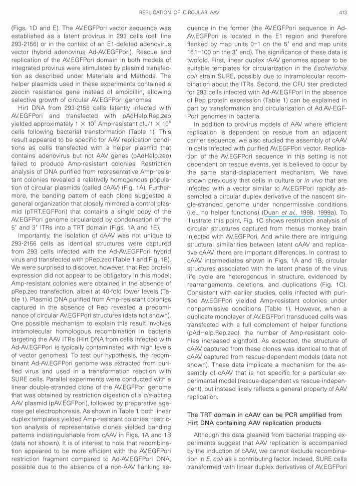
2fvWeApbcnOitAobfSltArdtp(trp
qAf1tscbfopP
rcitdtsisg(icistcslrCfndt(nccsspdr
TH
pbt
413REPLICATION OF CIRCULAR AAV
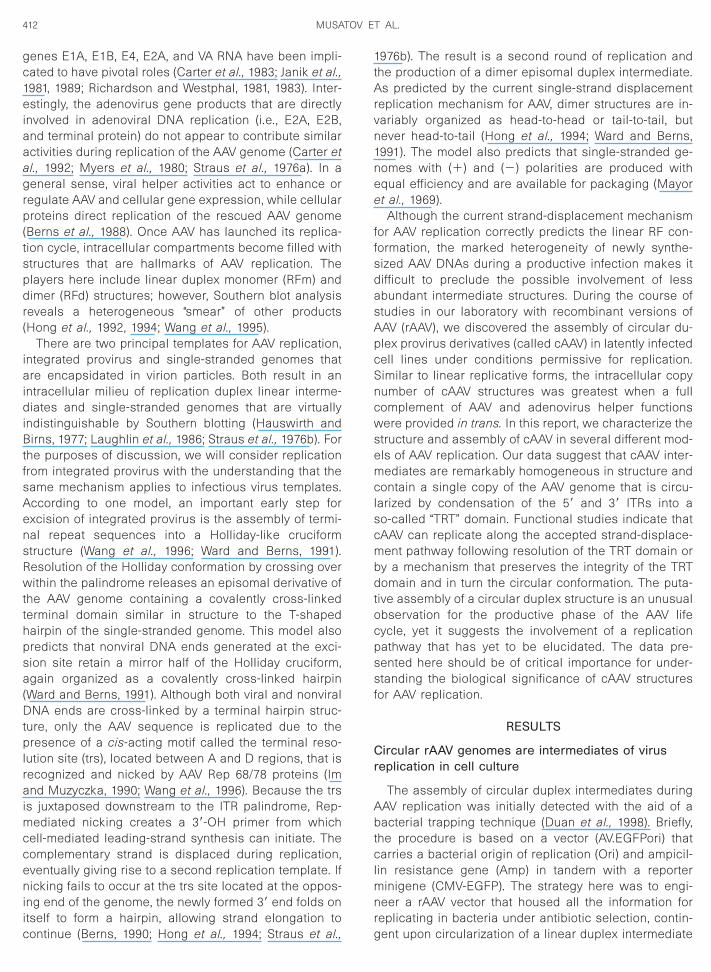
(Figs. 1D and E). The AV.EGFPori vector sequence wasestablished as a latent provirus in 293 cells (cell line293-2156) or in the context of an E1-deleted adenovirusvector (hybrid adenovirus Ad-AV.EGFPori). Rescue andreplication of the AV.EGFPori domain in both models ofintegrated provirus were stimulated by plasmid transfec-tion as described under Materials and Methods. Thehelper plasmids used in these experiments contained azeocin resistance gene instead of ampicillin, allowingselective growth of circular AV.EGFPori genomes.
Hirt DNA from 293-2156 cells latently infected withAV.EGFPori and transfected with pAdHelp.Rep.zeoyielded approximately 1 3 105 Amp-resistant cfu/1 3 106
cells following bacterial transformation (Table 1). Thisresult appeared to be specific for AAV replication condi-tions as cells transfected with a helper plasmid thatcontains adenovirus but not AAV genes (pAdHelp.zeo)failed to produce Amp-resistant colonies. Restrictionanalysis of DNA purified from representative Amp-resis-tant colonies revealed a relatively homogenous popula-tion of circular plasmids (called cAAV) (Fig. 1A). Further-more, the banding pattern of each clone suggested ageneral organization that closely mirrored a control plas-mid (pTRT.EGFPori) that contains a single copy of theAV.EGFPori genome circularized by condensation of the59 and 39 ITRs into a TRT domain (Figs. 1A and 1E).
Importantly, the isolation of cAAV was not unique to93-2156 cells as identical structures were captured
rom 293 cells infected with the Ad-AV.EGFPori hybridirus and transfected with pRep.zeo (Table 1 and Fig. 1B).e were surprised to discover, however, that Rep protein
xpression did not appear to be obligatory in this model;mp-resistant colonies were obtained in the absence ofRep.zeo transfection, albeit at 40-fold lower levels (Ta-le 1). Plasmid DNA purified from Amp-resistant coloniesaptured in the absence of Rep revealed a predomi-ance of circular AV.EGFPori structures (data not shown).ne possible mechanism to explain this result involves
ntramolecular homologous recombination in bacteriaargeting the AAV ITRs (Hirt DNA from cells infected withd-AV.EGFPori is typically contaminated with high levelsf vector genomes). To test our hypothesis, the recom-inant Ad-AV.EGFPori genome was extracted from puri-
ied virus and used in a transformation reaction withURE cells. Parallel experiments were conducted with a
inear double-stranded clone of the AV.EGFPori genomehat was obtained by restriction digestion of a cis-actingAV plasmid (pAV.EGFPori), followed by preparative aga-
ose gel electrophoresis. As shown in Table 1, both linearuplex templates yielded Amp-resistant colonies; restric-
ion analysis of representative clones yielded bandingatterns indistinguishable from cAAV in Figs. 1A and 1B
data not shown). It is of interest to note that recombina-ion appeared to be more efficient with the AV.EGFPoriestriction fragment compared to Ad-AV.EGFPori DNA,
ossible due to the absence of a non-AAV flanking se- tuence in the former (the AV.EGFPori sequence in Ad-V.EGFPori is located in the E1 region and therefore
lanked by map units 0–1 on the 59 end and map units6.1–100 on the 39 end). The significance of these data iswofold. First, linear duplex rAAV genomes appear to beuitable templates for circularization in the Escherichiaoli strain SURE, possibly due to intramolecular recom-ination about the ITRs. Second, the CFU titer predicted
or 293 cells infected with Ad-AV.EGFPori in the absencef Rep protein expression (Table 1) can be explained inart by transformation and circularization of Ad.AV-EGF-ori genomes in bacteria.
In addition to provirus models of AAV where efficienteplication is dependent on rescue from an adjacentarrier sequence, we also studied the assembly of cAAV
n cells infected with purified AV.EGFPori vector. Replica-ion of the AV.EGFPori sequence in this setting is notependent on rescue events, yet is believed to occur by
he same stand-displacement mechanism. We havehown previously that cells in culture or in vivo that are
nfected with a vector similar to AV.EGFPori rapidly as-embled a circular duplex derivative of the nascent sin-le-stranded genome under nonpermissive conditions
i.e., no helper functions) (Duan et al., 1998, 1999a). Tollustrate this point, Fig. 1C shows restriction analysis ofircular structures captured from rhesus monkey brain
njected with AV.EGFPori. And while there are intriguingtructural similarities between latent cAAV and replica-
ive cAAV, there are important differences. In contrast toAAV intermediates shown in Figs. 1A and 1B, circulartructures associated with the latent phase of the virus
ife cycle are heterogenous in structure, evidenced byearrangements, deletions, and duplications (Fig. 1C).onsistent with earlier studies, cells infected with puri-
ied AV.EGFPori yielded Amp-resistant colonies underonpermissive conditions (Table 1). However, when auplicate monolayer of AV.EGFPori transduced cells was
ransfected with a full complement of helper functionspAdHelp.Rep.zeo), the number of Amp-resistant colo-ies increased eightfold. As expected, the structure ofAAV captured from these clones was identical to that ofAAV captured from rescue-dependent models (data nothown). These data implicate a mechanism for the as-embly of cAAV that is not specific for a particular ex-erimental model (rescue-dependent vs rescue-indepen-ent), but instead likely reflects a general property of AAV
eplication.
he TRT domain in cAAV can be PCR amplified fromirt DNA containing AAV replication products
Although the data gleaned from bacterial trapping ex-eriments suggest that AAV replication is accompaniedy the induction of cAAV, we cannot exclude recombina-
ion in E. coli as a contributing factor. Indeed, SURE cells
ransformed with linear duplex derivatives of AV.EGFPori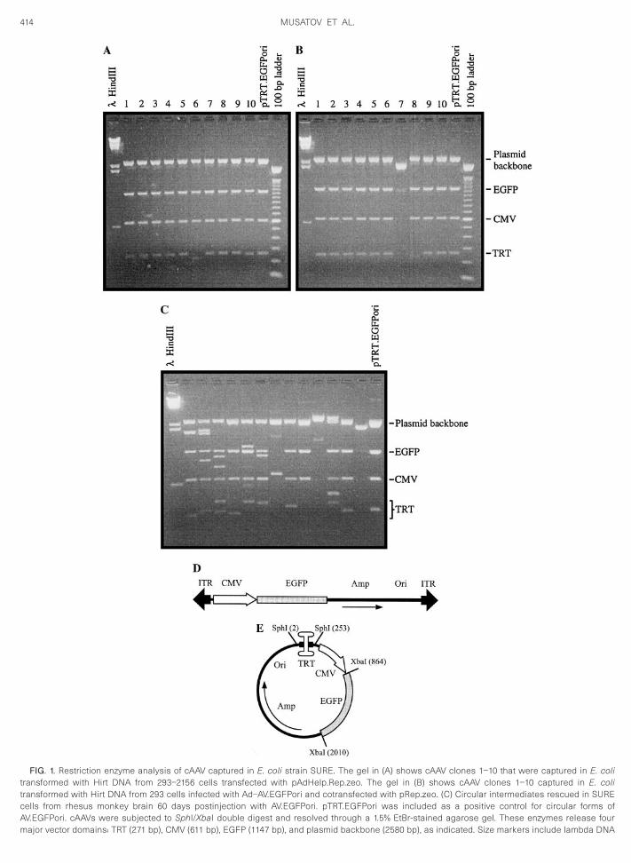
414 MUSATOV ET AL.
FIG. 1. Restriction enzyme analysis of cAAV captured in E. coli strain SURE. The gel in (A) shows cAAV clones 1–10 that were captured in E. colitransformed with Hirt DNA from 293–2156 cells transfected with pAdHelp.Rep.zeo. The gel in (B) shows cAAV clones 1–10 captured in E. colitransformed with Hirt DNA from 293 cells infected with Ad–AV.EGFPori and cotransfected with pRep.zeo. (C) Circular intermediates rescued in SUREcells from rhesus monkey brain 60 days postinjection with AV.EGFPori. pTRT.EGFPori was included as a positive control for circular forms ofAV.EGFPori. cAAVs were subjected to SphI/XbaI double digest and resolved through a 1.5% EtBr-stained agarose gel. These enzymes release four
major vector domains: TRT (271 bp), CMV (611 bp), EGFP (1147 bp), and plasmid backbone (2580 bp), as indicated. Size markers include lambda DNA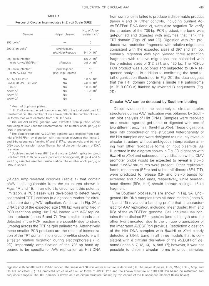
ftPqtt(2
C
seotttciiBpbfwehf
g1iRtotodsnp
A
c
D
415REPLICATION OF CIRCULAR AAV
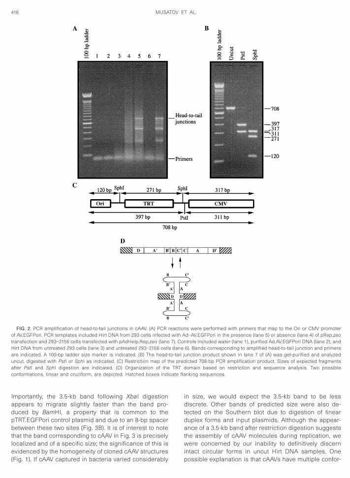
yielded Amp-resistant colonies (Table 1) that containcAAV indistinguishable from the structures shown inFigs. 1A and 1B. In an effort to circumvent this potentiallimitation, a PCR assay was developed to detect newlyassembled TRT junctions (a diagnostic marker for circu-larization) during AAV replication. As shown in Fig. 2A, aDNA band of the expected size (708 bp) was amplified inPCR reactions using Hirt DNA loaded with AAV replica-tion products (lanes 5 and 7). Two smaller bands alsodetected in the PCR reaction are possibly due to strandjumping across the TRT hairpin palindrome. Alternatively,these smaller PCR products are the result of isomeriza-tion of the TRT domain into a cruciform-like structure witha faster relative migration during electrophoresis (Fig.2D). Importantly, amplification of the 708-bp band ap-peared to be specific for AAV replication as Hirt DNA
digested with HindIII and a 100-bp ladder. The linear AV.EGFPori vectoOri are indicated. (E) The predicted structure of circular forms of AV.E
TABLE 1
Rescue of Circular Intermediates in E. coli Strain SURE
Sample Helper plasmidNo. of Amp-
resistant cfua
293 cellsb — 0
293-2156 cellsb pAdHelp.zeo 0pAdHelp.Rep.zeo 9.1 3 104
293 cells infected — 6.0 3 102
with Ad–AV.EGFPorib pRep.zeo 2.5 3 104
293 cells infectedb pAdHelp.zeo 12with AV.EGFPori pAdHelp.Rep.zeo 97
d–AV.EGFPoric NA 1.8 3 102
Linear ds AV.EGFPorid NA 4.0 3 103
RFm-Ae NA 1.0 3 103
cAAV-Ae NA 1.1 3 106
cAAV-Be NA 0AAV-Ce NA 0
a Mean of duplicate plates.b Hirt DNA was extracted from cells and 5% of the total yield used for
transformation. The number of cfu shown reflects the number of circu-lar forms that were captured from 1 3 106 cells.
c The Ad–AV.EGFPori genome was extracted from purified virionsand 50 ng of DNA used for transformation. The number of cfu per mg of
NA is presented.d The double-stranded AV.EGFPori genome was excised from plas-
mid pAV.EGFPori by digestion with restriction enzymes that leave 0-and 8-bp sequences flanking 59 and 39 ITRs, respectively, and 50 ng ofDNA used for transformation The number of cfu per microgram of DNAis shown.
e Double-stranded linear (RFm) and circular (cAAV) replication prod-ucts from 293–2156 cells were purified to homogeneity (Figs. 4 and 5)and 2 ng samples used for transformation. The number of cfu per mg ofDNA is shown.
sequence analysis. The TRT domain is drawn as a cruciform structure flanke
from control cells failed to produce a discernable product(lanes 4 and 6). Other controls, including purified Ad-AV.EGFPori DNA (lane 2), were also negative. To verifythe structure of the 708-bp PCR product, the band wasgel-purified and digested with enzymes that flank theTRT domain (Figs. 2B and 2C). Digestion with PstI pro-duced two restriction fragments with relative migrationsconsistent with the expected sizes of 397 and 311 bp.Similarly, digestion with SphI yielded three restrictionragments with relative migrations that coincided withhe predicted sizes of 317, 271, and 120 bp. The 708-bpCR product was subcloned and subjected to DNA se-uence analysis. In addition to confirming the head-to-
ail organization illustrated in Fig. 2C, the data suggesthat the TRT domain contains a single ITR palindromeA9-B9-B-C9-C-A) flanked by inverted D sequences (Fig.D).
ircular AAV can be detected by Southern blotting
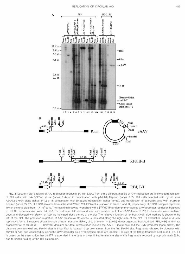
Direct evidence for the assembly of circular duplextructures during AAV replication was obtained by South-rn blot analysis of Hirt DNAs. Samples were resolvedn a neutral agarose gel uncut or digested with one of
wo different enzymes, BamHI or XbaI. These digestionsake into consideration the structural heterogeneity ofhe Hirt samples and were designed to uniquely reveal aircular structure without ambiguous interpretation aris-
ng from other replicative forms or input plasmids. Asllustrated in the diagram shown in Fig. 3B, digestion withamHI or XbaI and subsequent hybridization with a CMVromoter probe would be expected to reveal a 3.5-kband if cAAV structures were present. Other replicative
orms, monomers (RFm) and tail-to-tail dimers (RFd, T-T),ere predicted to release 0.9- and 0.8-kb bands forxtended and closed ends, respectively, while head-to-ead dimers (RFd, H-H) should liberate a single 1.5-kb
ragment.The Southern blot results are shown in Fig. 3A. Undi-
ested Hirt DNA samples from all three models (lanes 5,1, and 15) revealed a banding profile that is character-
stic for AAV replication, including linear duplex RFm andFd of the AV.EGFPori genome. Cell line 293-2156 con-
ains three distinct RFm species (one full length and thether two truncated) due to the unique organization of
he integrated AV.EGFPori provirus. Restriction digestionf the Hirt DNA samples with BamHI or XbaI clearlyetected a 3.5-kb band in all three models that is con-istent with a circular derivative of the AV.EGFPori ge-ome (lanes 6, 7, 12, 13, 16, and 17); however, it was notossible to discern circular forms in uncut samples.
ture is depicted (D). The major domains, ITRs, CMV, EGFP, Amp, andi and the known structure of pTRT.EGFPori based on restriction and
r strucGFPor
d by two copies of the D sequence element (black boxes).
pbtle(
Hauac cate fla
416 MUSATOV ET AL.
Importantly, the 3.5-kb band following XbaI digestionappears to migrate slightly faster than the band pro-duced by BamHI, a property that is common to the
TRT.EGFPori control plasmid and due to an 8-bp spaceretween these two sites (Fig. 3B). It is of interest to note
hat the band corresponding to cAAV in Fig. 3 is preciselyocalized and of a specific size; the significance of this isvidenced by the homogeneity of cloned cAAV structures
FIG. 2. PCR amplification of head-to-tail junctions in cAAV. (A) PCR rof AV.EGFPori. PCR templates included Hirt DNA from 293 cells infectedtransfection and 293–2156 cells transfected with pAdHelp.Rep.zeo (lane
irt DNA from untreated 293 cells (lane 3) and untreated 293–2156 cellre indicated. A 100-bp ladder size marker is indicated. (B) The head-ncut, digested with PstI or SphI as indicated. (C) Restriction map of tfter PstI and SphI digestion are indicated. (D) Organization of theonformations, linear and cruciform, are depicted. Hatched boxes indi
Fig. 1). If cAAV captured in bacteria varied considerably
in size, we would expect the 3.5-kb band to be lessdiscrete. Other bands of predicted size were also de-tected on the Southern blot due to digestion of linearduplex forms and input plasmids. Although the appear-ance of a 3.5-kb band after restriction digestion suggeststhe assembly of cAAV molecules during replication, wewere concerned by our inability to definitively discernintact circular forms in uncut Hirt DNA samples. One
s were performed with primers that map to the Ori or CMV promoterd–AV.EGFPori in the presence (lane 5) or absence (lane 4) of pRep.zeotrols included water (lane 1), purified Ad.AV.EGFPori DNA (lane 2), and
6). Bands corresponding to amplified head-to-tail junction and primersunction product shown in lane 7 of (A) was gel-purified and analyzedicted 708-bp PCR amplification product. Sizes of expected fragmentsomain based on restriction and sequence analysis. Two possiblenking sequences.
eactionwith A7). Con
s (laneto-tail jhe pred
TRT d
possible explanation is that cAAVs have multiple confor-
417REPLICATION OF CIRCULAR AAV
FIG. 3. Southern blot analysis of AAV replication products. (A) Hirt DNAs from three different models of AAV replication are shown; cotransfectionof 293 cells with pAV.EGFPori alone (lanes 2–4) or in combination with pAdHelp.Rep.zeo (lanes 5–7), 293 cells infected with hybrid virusAd–AV.EGFPori alone (lanes 8–10) or in combination with pRep.zeo transfection (lanes 11–13), and transfection of 293–2156 cells with pAdHelp.Rep.zeo (lanes 15–17). Hirt DNA isolated from untreated 293 or 293–2156 cells is shown in lanes 1 and 14, respectively. Hirt DNA samples represent10% of the total yield from 1 3 106 cells. The resulting blot was hybridized with a [32P]dCTP random-primer-labeled CMV promoter restriction fragment.pTRT.EGFPori was spiked with Hirt DNA from untreated 293 cells and used as a positive control for cAAV (lanes 18–20). Hirt samples were analyzeduncut and digested with BamHI or XbaI as indicated along the top of the blot. The relative migration of lambda HindIII size markers is shown to theleft of the blot. The predicted migration of AAV replicative structures is indicated along the right side of the blot. (B) Restriction maps of duplexreplicative forms. Structures shown include a linear monomer (RFm), circular monomer (cAAV), dimer organized head-to-head (RFd, H-H), and dimerorganized tail-to-tail (RFd, T-T). Relevant domains for data interpretation include the AAV ITR (solid box) and the CMV promoter (open arrow). Thedistance between XbaI and BamHI sites is 8 bp. XhoI is located 10 bp downstream from the first BamHI site. Fragments released by digestion withBamHI or XbaI and visualized by using the CMV promoter as a hybridization probe are labeled. The size of the 0.9-kb fragment in RFm and RFd, T-Tis based on the assumption that the ITR is extended. In the case of cross-linked termini the size of this fragment is reduced by approximately 62 bp
due to hairpin folding of the ITR palindrome.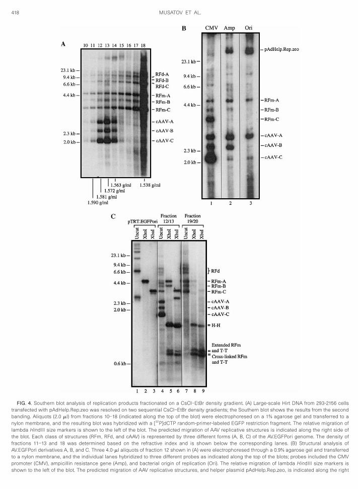
n
418 MUSATOV ET AL.
FIG. 4. Southern blot analysis of replication products fractionated on a CsCl–EtBr density gradient. (A) Large-scale Hirt DNA from 293-2156 cellstransfected with pAdHelp.Rep.zeo was resolved on two sequential CsCl–EtBr density gradients; the Southern blot shows the results from the secondbanding. Aliquots (2.0 ml) from fractions 10–18 (indicated along the top of the blot) were electrophoresed on a 1% agarose gel and transferred to a
ylon membrane, and the resulting blot was hybridized with a [32P]dCTP random-primer-labeled EGFP restriction fragment. The relative migration oflambda HindIII size markers is shown to the left of the blot. The predicted migration of AAV replicative structures is indicated along the right side ofthe blot. Each class of structures (RFm, RFd, and cAAV) is represented by three different forms (A, B, C) of the AV.EGFPori genome. The density offractions 11–13 and 18 was determined based on the refractive index and is shown below the corresponding lanes. (B) Structural analysis ofAV.EGFPori derivatives A, B, and C. Three 4.0-ml aliquots of fraction 12 shown in (A) were electrophoresed through a 0.9% agarose gel and transferredto a nylon membrane, and the individual lanes hybridized to three different probes as indicated along the top of the blots; probes included the CMVpromoter (CMV), ampicillin resistance gene (Amp), and bacterial origin of replication (Ori). The relative migration of lambda HindIII size markers is
shown to the left of the blot. The predicted migration of AAV replicative structures, and helper plasmid pAdHelp.Rep.zeo, is indicated along the right
c
io
419REPLICATION OF CIRCULAR AAV
mations (e.g., supercoiled and relaxed) and therefore arenot concentrated at any one location following agarosegel electrophoresis. Detection would be further compro-mised by the complexity and smearing of DNA structuresthat is characteristic of uncut Hirt samples from AAVreplication.
Purification and characterization of cAAV
To further substantiate the induction of duplex circularmolecules during AAV replication, Hirt DNA from 293-2156 cells transfected with pAdHelp.Rep.zeo was re-solved by two sequential rounds of CsCl–EtBr densitygradient ultracentrifugation; the Southern blot in Fig. 4Ashows the banding profile obtained from the secondgradient. The blot was probed with a [32P]dCTP random-primer-labeled EGFP restriction fragment revealing acomplex, yet discernable, banding pattern. Critical tounderstanding the Southern blot banding pattern is therecognition that the 293-2156 cell line used in theseexperiments contains three provirus derivatives of theoriginal AV.EGFPori genome; RFm A is full length, whileforms B and C contain structural deletions. Each of thethree provirus structures are efficiently rescued and rep-licated and appear to be the consequence of the AAVintegration mechanism; AAV integration has been shownpreviously to be associated with complex rearrange-ments, including concatemerization and deletions (Duanet al., 1997; McLaughlin et al., 1988). Consistent with theaccepted strand-displacement replication mechanismfor AAV, RFm give rise to RFd due to ITR priming at thedistal end of newly synthesized RFm structures. Andwhile RFm and RFd species are represented throughoutthe gradient, the peak fractions (18–21) are found at aCsCl–EtBr density consistent with linear DNA. Yet themost significant observation is the presence of threeadditional bands in the region of the density gradient thatcorrespond to supercoiled circular DNA (density 1.57–1.62 g/ml) (Radloff et al., 1967). In similar fashion to RFdforms, cAAV Forms A, B, and C can be traced to linearRFm Forms A, B, and C.
Experiments were performed to provide preliminarydata as to the structure of provirus Forms B and C; FormA is assumed to be full length based on its relativemigration on a neutral agarose gel. To test our hypothe-sis that Forms B and C are provirus deletion mutants ofthe AV.EGFPori genome, a sample of fraction 12 from thedensity gradient (Fig. 4A) was resolved on a neutralagarose gel in triplicate and transferred to a nylon mem-brane, and individual lanes were hybridized with a radio-
side of the blot. (C) Restriction analysis of DNA from high- and low-deseparately (12 and 13 or 19 and 20), dialyzed, and subjected to restrictincluded in the analysis as a positive control for circular monomer form
f the blot. The predicted migration of AAV replicative structures is indicatedlabeled restriction fragment mapping to the CMV pro-moter, ampicillin resistance gene, or ColE1 origin ofreplication. As shown in Fig. 4B, there is good correspon-dence of the hybridization signal between RFm and cAAV(RFd are not well discerned in the blot) and between theprovirus derivatives themselves. For instance, Form Aproduces a strong hybridization signal with all threeprobes supporting our monomer-length assignment.Forms B and C, however, appear to contain substantialdeletions in Ori (Form B) or both Amp and Ori (Form C)based on reduced hybridization signals. Importantly, thispattern is seen with both RFm and cAAV, supporting acommon origin. Input helper plasmid (pAdHelp.Rep.zeo)was also detected in lanes 2 and 3 since it contains Oriand a plasmid sequence that partially overlaps the Ampprobe, yet lacks the CMV promoter and therefore is notdetected in lane 1. The apparent deletion of Amp and Oridomains from cAAV-B and cAAV-C provides a logicalexplanation for their null phenotype during bacterial trap-ping; virtually all circular forms cloned in bacteria areconsistent with a structure that contains a full-lengthgenome (Fig. 1).
Gradient fractions representing circular (12/13) andlinear (19/20) DNAs were independently pooled, dialyzedto remove CsCl salt, and analyzed by Southern blotting(Fig. 4C). Plasmid pTRT.EGFPori was included in theexperiment to serve as a positive control for circularforms of the AV.EGFPori vector. Uncut samples of fraction12/13 (lane 4) reveal cAAV, RFm, and RFd as before, whilean uncut sample of fraction 19/20 (lane 7) is clearlylacking bands that correspond to cAAV. The multipleconformations seen in an uncut sample of pTRT.EGFPori(lane 1) were gathered into a single band of 4.6 (lane 2)or 3.4 kb (lane 3) following digestion with XhoI (singlecutter) or XbaI (double cutter), respectively. Restrictiondigestion of fraction 12/13 with the same enzymes pro-duced a band of a size equivalent to that seen withrestricted pTRT.EGFPori (compare lanes 2 and 5 for XhoI
ut, lanes 3 and 6 for XbaI cut), accompanied by the lossof all three circular forms that were evident in the uncutsample (compare lanes 4, 5, and 6). The 4.6 and 3.4-kbbands seen in lanes 5 and 6 are likely due to lineariza-tion of cAAV-A. Other notable hybridization signals pro-duced by restriction digestion of fraction 12/13 includesbands of 4.2 (lane 5) and 3.0 kb (lane 6). These bandslikely correspond to linearized cAAV-B; this is best illus-trated by comparing the relative migration of the 4.2-kbband shown in lane 5 to RFm-B shown in lane 7. The fateof cAAV-C after digestion with XhoI or XbaI is less clear
actions. Two fractions from each portion of the gradient were pooledstion with a single (XhoI) or a double (XbaI) cutter. pTRT.EGFPori wasrelative migration of lambda HindIII size markers is shown to the left
nsity fron dige
s. The
along the right side of the blot.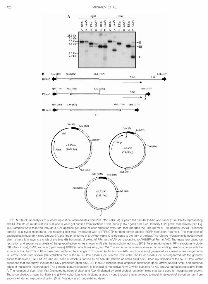
420 MUSATOV ET AL.
FIG. 5. Structural analysis of purified replication intermediates from 293–2156 cells. (A) Supercoiled circular (cAAV) and linear (RFm) DNAs representingAV.EGFPori structural derivatives A, B, and C were gel purified from fractions 12/13 (density 1.577 g/ml) and 19/20 (density 1.526 g/ml), respectively (see Fig.4C). Samples were resolved through a 1.2% agarose gel uncut or after digestion with SphI that liberates the ITRs (RFm) or TRT domain (cAAV). Followingtransfer to a nylon membrane, the resulting blot was hybridized with a [32P]dCTP random-primer-labeled EGFP restriction fragment. The migration ofsupercoiled circular (I), nicked circular (II), and linear (III) forms of cAAV derivative C is indicated to the right of the blot. The relative migration of lambda HindIIIsize markers is shown to the left of the blot. (B) Schematic drawing of RFm and cAAV corresponding to AV.EGFPori Forms A–C. The maps are based onrestriction and sequence analysis of the gel-purified genomes shown in (A) after being subcloned into pSP72. Relevant domains in RFm structures includeITR (black arrow), CMV promoter (open arrow), EGFP (shaded box), Amp, and Ori. The same domains are shown in corresponding cAAV structures with theexception that the ITRs in RFm have been replaced by a single TRT domain (solid box) in cAAV. Junction sites (J) generated as a result of rearrangementsin Forms B and C are shown. (C) Restriction map of the AV.EGFPori provirus locus in 293–2156 cells. The 23-kb provirus locus is organized into five genomesubunits (labeled C, DIR, A1, A2, and A3), each of which is flanked by an AAV ITR (shown as small solid box). Other key domains of the AV.EGFPori vectorsequence that are shown include the CMV promoter (open box), EGFP cDNA (shaded box), ampicillin resistance gene (arrow labeled Amp), and bacterialorigin of replication (hatched box). The genome subunit labeled C is identical to replicative Form C while subunits A1, A2, and A3 represent replicative FormA. The location of ScaI, XhoI, PstI (indicated by open circles), and XbaI (indicated by solid circles) restriction sites that were used for mapping are shown.The large shaded arrows that flank the DIR–A1 subunit junction indicate a large inverted repeat that is believed to result in deletion of the ori domain from
subunit A1 during rescue/replication (S. A. Musatov et al., unpublished data).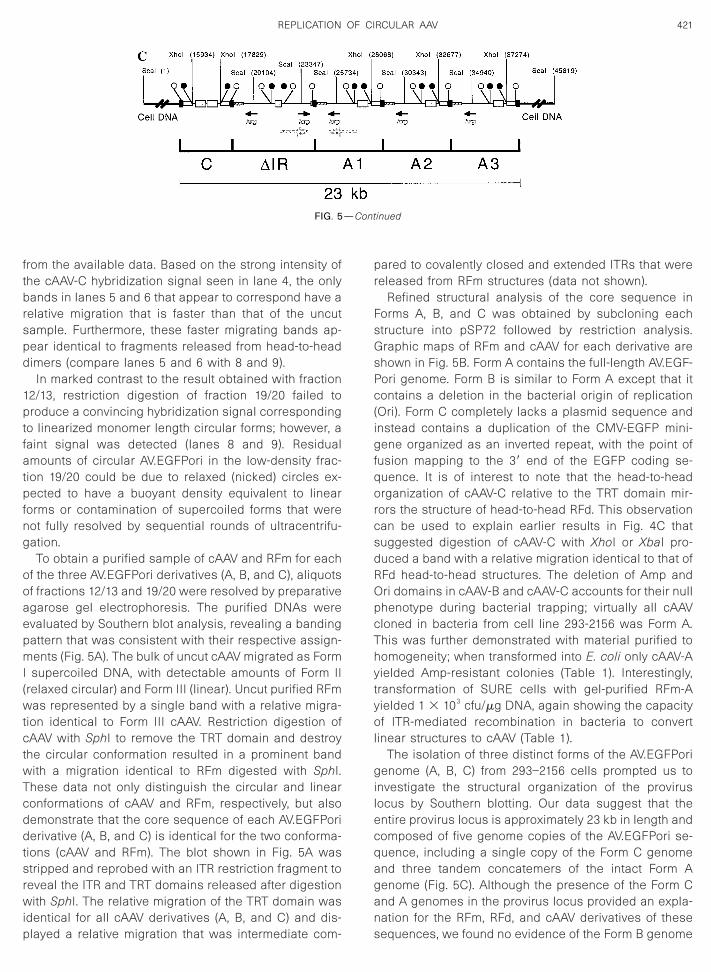
Tcddtsrwip
—Cont
421REPLICATION OF CIRCULAR AAV
from the available data. Based on the strong intensity ofthe cAAV-C hybridization signal seen in lane 4, the onlybands in lanes 5 and 6 that appear to correspond have arelative migration that is faster than that of the uncutsample. Furthermore, these faster migrating bands ap-pear identical to fragments released from head-to-headdimers (compare lanes 5 and 6 with 8 and 9).
In marked contrast to the result obtained with fraction12/13, restriction digestion of fraction 19/20 failed toproduce a convincing hybridization signal correspondingto linearized monomer length circular forms; however, afaint signal was detected (lanes 8 and 9). Residualamounts of circular AV.EGFPori in the low-density frac-tion 19/20 could be due to relaxed (nicked) circles ex-pected to have a buoyant density equivalent to linearforms or contamination of supercoiled forms that werenot fully resolved by sequential rounds of ultracentrifu-gation.
To obtain a purified sample of cAAV and RFm for eachof the three AV.EGFPori derivatives (A, B, and C), aliquotsof fractions 12/13 and 19/20 were resolved by preparativeagarose gel electrophoresis. The purified DNAs wereevaluated by Southern blot analysis, revealing a bandingpattern that was consistent with their respective assign-ments (Fig. 5A). The bulk of uncut cAAV migrated as FormI supercoiled DNA, with detectable amounts of Form II(relaxed circular) and Form III (linear). Uncut purified RFmwas represented by a single band with a relative migra-tion identical to Form III cAAV. Restriction digestion ofcAAV with SphI to remove the TRT domain and destroythe circular conformation resulted in a prominent bandwith a migration identical to RFm digested with SphI.
hese data not only distinguish the circular and linearonformations of cAAV and RFm, respectively, but alsoemonstrate that the core sequence of each AV.EGFPorierivative (A, B, and C) is identical for the two conforma-
ions (cAAV and RFm). The blot shown in Fig. 5A wastripped and reprobed with an ITR restriction fragment toeveal the ITR and TRT domains released after digestionith SphI. The relative migration of the TRT domain was
dentical for all cAAV derivatives (A, B, and C) and dis-
FIG. 5
layed a relative migration that was intermediate com-
pared to covalently closed and extended ITRs that werereleased from RFm structures (data not shown).
Refined structural analysis of the core sequence inForms A, B, and C was obtained by subcloning eachstructure into pSP72 followed by restriction analysis.Graphic maps of RFm and cAAV for each derivative areshown in Fig. 5B. Form A contains the full-length AV.EGF-Pori genome. Form B is similar to Form A except that itcontains a deletion in the bacterial origin of replication(Ori). Form C completely lacks a plasmid sequence andinstead contains a duplication of the CMV-EGFP mini-gene organized as an inverted repeat, with the point offusion mapping to the 39 end of the EGFP coding se-quence. It is of interest to note that the head-to-headorganization of cAAV-C relative to the TRT domain mir-rors the structure of head-to-head RFd. This observationcan be used to explain earlier results in Fig. 4C thatsuggested digestion of cAAV-C with XhoI or XbaI pro-duced a band with a relative migration identical to that ofRFd head-to-head structures. The deletion of Amp andOri domains in cAAV-B and cAAV-C accounts for their nullphenotype during bacterial trapping; virtually all cAAVcloned in bacteria from cell line 293-2156 was Form A.This was further demonstrated with material purified tohomogeneity; when transformed into E. coli only cAAV-Ayielded Amp-resistant colonies (Table 1). Interestingly,transformation of SURE cells with gel-purified RFm-Ayielded 1 3 103 cfu/mg DNA, again showing the capacityof ITR-mediated recombination in bacteria to convertlinear structures to cAAV (Table 1).
The isolation of three distinct forms of the AV.EGFPorigenome (A, B, C) from 293–2156 cells prompted us toinvestigate the structural organization of the proviruslocus by Southern blotting. Our data suggest that theentire provirus locus is approximately 23 kb in length andcomposed of five genome copies of the AV.EGFPori se-quence, including a single copy of the Form C genomeand three tandem concatemers of the intact Form Agenome (Fig. 5C). Although the presence of the Form Cand A genomes in the provirus locus provided an expla-nation for the RFm, RFd, and cAAV derivatives of these
inued
sequences, we found no evidence of the Form B genome
422 MUSATOV ET AL.
(Fig. 5B). However, positioned between the Form C andA1 sequences, we identified a rearranged genome (la-beled DIR) that contained an inverted duplication of theAV.EGFPori genome; sequence analysis of the clonedDIR genome indicated that the junction of the inversionoriginates in the EGFP open reading frame and extendsthrough the ampicillin resistance gene (Fig. 5C). To testthe hypothesis that the DIR subunit was involved in theassembly of the Form B genome during rescue, theregion of the provirus locus spanning the DIR and A1genomes was cloned and transfected into 293 cellsalong with helper functions. Interestingly, Southern blotanalysis of Hirt DNA revealed that the predominant rep-lication products were consistent with linear and circularderivatives of AV.EGFPori Form A and B (data not shown).Therefore, while Form B is not physically associated withthe AV.EGFPori provirus locus in 293-2156 cells, it isassembled during rescue/replication of the A1 subunitpresumably due to the large inverted repeat that flanksthe DIR-A1 junction (see large shaded arrows in Fig. 5C).The dynamics of the AV.EGFPori provirus locus in 293-2156 cells, including the assembly of Form B, will bepublished elsewhere (S. A. Musatov et al., unpublisheddata).
Assembly of cAAV during wild-type AAV replication
It is important to emphasize at this juncture that theexperimental design of our studies relied on plasmidtransfection to stimulate a permissive intracellular milieufor AAV replication. Furthermore, we elected to use plas-mids that contain the minimal adenovirus (E1, E2a, E4,and VA RNA) and AAV (Rep) helper functions for replica-tion. We recognize that this approach may inadvertentlyalter the normal processivity of AAV replication, possiblyestablishing an environment for the assembly of unusualstructures (e.g., cAAV). We therefore set out to determinewhether the induction of cAAV was indicative of experi-mental models that more closely mirror “wild-type” AAVreplication. To exclude the possibility that the absence ofcap genes artificially enhanced the induction of cAAVdue to a block in single-stranded DNA packaging, werepeated our experiments with another plasmid(pAdHelp.Rep.Cap.zeo) that encodes a full complementof helper functions necessary to support the AAV pro-ductive phase, including virion assembly. Hirt DNA fromthese experiments contained cAAV according to South-ern blotting and bacterial trapping with no deviation fromresults obtained in the absence of cap genes (data notshown). We also tested the formation of cAAV duringwild-type AAV replication using psub201 (Samulski et al.,1987) as a template. In these experiments adenovirushelper functions were provided either by plasmid trans-fection or by adenovirus infection. Hirt DNA was ex-tracted from cells 48 h posttransfection and analyzed by
Southern blotting; DNAs were electrophoresed uncut,digested with HindIII, or double-digested with HindIII andBamHI to distinguish linear RFm, RFd, and cAAV (Fig. 6A).As shown in Fig. 6B, both RFm and RFd structures werereadily detected in samples of uncut Hirt DNA regardlessof whether helper functions were provided by plasmidtransfection (lane 1) or virus infection (lane 4); however,the assembly of RFd structures appeared to be moreefficient with adenovirus infection (compare RFd band inlanes 1 and 4). Digestion of Hirt DNA samples withHindIII (lanes 2 and 5) resulted in the appearance of 1.9-and 3.6-kb bands, consistent with restricted RFm/RFd(tail-to-tail) and RFd (head-to-head), respectively. A bandwith a size equivalent to that of the wild-type AAV unitlength genome (4.6 kb) was also evident in HindIII cutDNA, corresponding to either uncut RFm or cAAV. Thatthis 4.6-kb band represented cAAV and not incompletedigestion of RFm was confirmed by double digestion withHindIII and BamHI. Under these conditions, cAAV isconverted to a unique fragment of 3.7 kb (Fig. 6A) whichwas clearly evident in the two Hirt DNA samples (Fig. 6B,lanes 3 and 6). It is of interest to note that the relativecopy number of cAAV in the presence of adenovirusinfection is nearly equivalent to that of linear RFm basedon band intensities (compare the 4.6-kb band in lane 4with the 3.7-kb band in lane 6). Finally, we also detectedthe assembly of cAAV in a 293 cell line latently infectedwith a rAAV vector that expresses human blood coagu-lation factor IX (AV.hFIXori), suggesting that the effect canbe achieved with rAAV vectors other than AV.EGFPori(data not shown). Collectively, our findings strongly sug-gest that the formation of cAAV is an intrinsic property ofAAV replication regardless of the replication model (res-cue-dependent or rescue-independent), virus structure(wild-type or recombinant), or the method by whichhelper functions are delivered (viral infection or plasmidtransfection).
cAAV intermediates replicate as circular molecules
The assembly of circular duplex forms during AAVreplication has important implications for the currentstrand-displacement model. At issue here is the potentialfor circular structures to serve as replication templates.To test this hypothesis we took advantage of the meth-ylation pattern that is conferred on plasmid DNA whenpropagated in dam1 bacteria; plasmids grown in thisgenetic background are sensitive to DpnI, a restrictionenzyme that selectively cleaves methylated GATC sitesbut is inactive toward the same sequence when hemi-methylated or unmethylated (as would be expected ofDNA that is replicated in mammalian cells).
293 cells were cotransfected with cis-acting plasmidpAV.EGFPori and helper plasmid pAdHelp.Rep.zeo, con-ditions that result in efficient rescue and replication ofthe AV.EGFPori sequence. As a control, cells were trans-
fected with pAV.EGFPori in the absence of helper plas-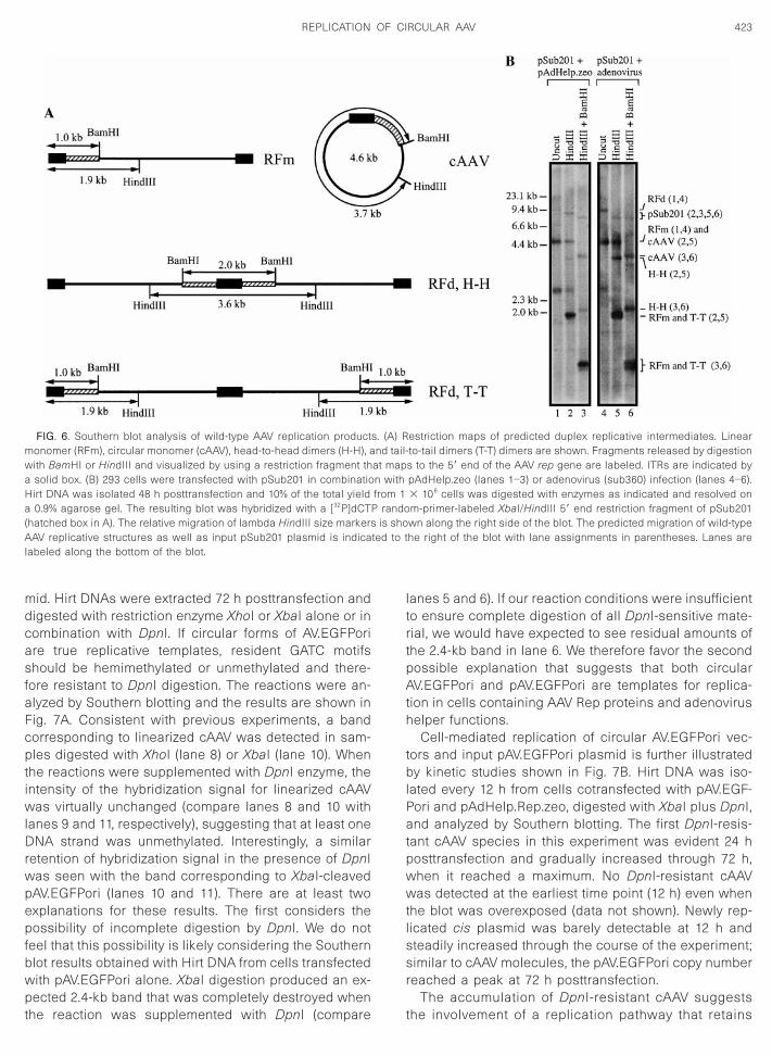
asfaFcp
wpep
ssr
423REPLICATION OF CIRCULAR AAV
mid. Hirt DNAs were extracted 72 h posttransfection anddigested with restriction enzyme XhoI or XbaI alone or incombination with DpnI. If circular forms of AV.EGFPori
re true replicative templates, resident GATC motifshould be hemimethylated or unmethylated and there-
ore resistant to DpnI digestion. The reactions were an-lyzed by Southern blotting and the results are shown inig. 7A. Consistent with previous experiments, a bandorresponding to linearized cAAV was detected in sam-les digested with XhoI (lane 8) or XbaI (lane 10). When
the reactions were supplemented with DpnI enzyme, theintensity of the hybridization signal for linearized cAAVwas virtually unchanged (compare lanes 8 and 10 withlanes 9 and 11, respectively), suggesting that at least oneDNA strand was unmethylated. Interestingly, a similarretention of hybridization signal in the presence of DpnI
as seen with the band corresponding to XbaI-cleavedAV.EGFPori (lanes 10 and 11). There are at least twoxplanations for these results. The first considers theossibility of incomplete digestion by DpnI. We do not
feel that this possibility is likely considering the Southernblot results obtained with Hirt DNA from cells transfectedwith pAV.EGFPori alone. XbaI digestion produced an ex-pected 2.4-kb band that was completely destroyed when
FIG. 6. Southern blot analysis of wild-type AAV replication productmonomer (RFm), circular monomer (cAAV), head-to-head dimers (H-H), awith BamHI or HindIII and visualized by using a restriction fragment tha solid box. (B) 293 cells were transfected with pSub201 in combinatioHirt DNA was isolated 48 h posttransfection and 10% of the total yielda 0.9% agarose gel. The resulting blot was hybridized with a [32P]dCTP(hatched box in A). The relative migration of lambda HindIII size markersAAV replicative structures as well as input pSub201 plasmid is indicalabeled along the bottom of the blot.
the reaction was supplemented with DpnI (compare t
lanes 5 and 6). If our reaction conditions were insufficientto ensure complete digestion of all DpnI-sensitive mate-rial, we would have expected to see residual amounts ofthe 2.4-kb band in lane 6. We therefore favor the secondpossible explanation that suggests that both circularAV.EGFPori and pAV.EGFPori are templates for replica-tion in cells containing AAV Rep proteins and adenovirushelper functions.
Cell-mediated replication of circular AV.EGFPori vec-tors and input pAV.EGFPori plasmid is further illustratedby kinetic studies shown in Fig. 7B. Hirt DNA was iso-lated every 12 h from cells cotransfected with pAV.EGF-Pori and pAdHelp.Rep.zeo, digested with XbaI plus DpnI,and analyzed by Southern blotting. The first DpnI-resis-tant cAAV species in this experiment was evident 24 hposttransfection and gradually increased through 72 h,when it reached a maximum. No DpnI-resistant cAAVwas detected at the earliest time point (12 h) even whenthe blot was overexposed (data not shown). Newly rep-licated cis plasmid was barely detectable at 12 h and
teadily increased through the course of the experiment;imilar to cAAV molecules, the pAV.EGFPori copy numbereached a peak at 72 h posttransfection.
The accumulation of DpnI-resistant cAAV suggests
estriction maps of predicted duplex replicative intermediates. Linearto-tail dimers (T-T) dimers are shown. Fragments released by digestions to the 59 end of the AAV rep gene are labeled. ITRs are indicated bypAdHelp.zeo (lanes 1–3) or adenovirus (sub360) infection (lanes 4–6).3 106 cells was digested with enzymes as indicated and resolved onm-primer-labeled XbaI/HindIII 59 end restriction fragment of pSub201wn along the right side of the blot. The predicted migration of wild-typehe right of the blot with lane assignments in parentheses. Lanes are
s. (A) Rnd tail-
at mapn withfrom 1
randois sho
ted to t
he involvement of a replication pathway that retains
is inc
424 MUSATOV ET AL.
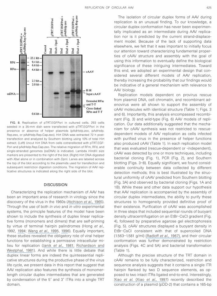
the integrity of the TRT domain. Furthermore, it ap-pears that this reaction is markedly less efficient com-pared to replication of linear RFm (Fig. 7B). In attempt-ing to understand this observation, it is important torecognize that replication of cAAV as a circular mole-cule is likely compromised by a competing reactionthat involves resolution of the TRT domain, therebyconverting cAAV to replication-competent linear RFm.According to this line of reasoning, TRT-mediated res-cue depletes the intracellular pool of cAAV while con-comitantly fueling the replication of linear RFm. Toevaluate the relative activity of these two opposingpathways, 293 cells were transfected with cAAVcloned in bacteria (i.e., pTRT.EGFPori) in the presenceor absence of viral helper plasmid. Southern blot anal-ysis of Hirt DNA from cells cotransfected with pTR-T.EGFPori and pAdHelp.Rep.Cap.zeo revealed a band-ing pattern that is characteristic of AAV infections,including the synthesis of RFm, RFd, and single-stranded genomes (ssDNA) (Fig. 8). The production of
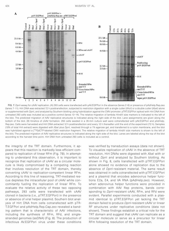
FIG. 7. DpnI-assay for cAAV replication. (A) 293 cells were transfected(lanes 7–11). Hirt DNA was extracted 72 h posttransfection, subjected toor supplemented with DpnI, and analyzed by Southern blotting using hybuntreated 293 cells was included as a positive control (lanes 12–14). Tthe blot. The predicted migration of AAV replicative structures is indicbottom of the blot. (B) Kinetics of cAAV formation. 293 cells seeded iRep.zeo. Cells were harvested and Hirt DNA extracted 12 h posttransfe(10% of total Hirt extract) were digested with XbaI plus DpnI, resolved twas hybridized against a [32P]dCTP-labeled CMV restriction fragment.the blot. The predicted migration of AAV replicative structures is indicataccording to the harvest time point. Hirt DNA from untreated 293 cells
infectious AV.EGFPori virus under these conditions
was verified by transduction assays (data not shown).To visualize replication of cAAV in the absence of TRTresolution, Hirt DNAs were digested with XbaI with orwithout DpnI and analyzed by Southern blotting. Asshown in Fig. 8, cells transfected with pTRT.EGFPorialone showed no evidence of replication due to theabsence of DpnI-resistant material. The same resultwas obtained in cells cotransfected with pTRT.EGFPoriand a plasmid that encodes adenovirus helper func-tions E2a, E4, and VA RNA (pAdHelp.zeo). However,when adenovirus helper functions were provided incombination with AAV Rep proteins, bands corre-sponding to DpnI-resistant cAAV, RFm, and RFd wereevident. Parallel experiments conducted with a plas-mid identical to pTRT.EGFPori yet lacking the TRTdomain failed to produce DpnI-resistant cAAV or linearRF structures under replicative conditions (data notshown). These findings establish a critical role for theTRT domain and suggest that cAAV can replicate as acircular molecule or serve as a precursor for linear
AV.EGFPori in the absence (lanes 2–6) or presence of pAdHelp.Rep.zeotion digestion with a single cutter (XhoI) or a double cutter (XbaI) aloneon against the CMV promoter. pTRT.EGFPori spiked with Hirt DNA fromtive migration of lambda HindIII size markers is indicated to the left ofong the right side of the blot. Lane assignments are given along themm culture well were cotransfected with pAV.EGFPori and pAdHelp.d every 12 h thereafter until the end of the experiment (72 h). Samplesa 1% agarose gel, and transferred to a nylon membrane, and the blot
lative migration of lambda HindIII size markers is shown to the left ofg the right side of the blot. Lanes are labeled along the top of the blot
luded as a control.
with prestricridizati
he relaated aln a 35-ction anhroughThe reed alon
RFm following resolution of the TRT domain.
425REPLICATION OF CIRCULAR AAV
DISCUSSION
Characterizing the replication mechanism of AAV hasbeen an important area of research in virology since thediscovery of the virus in the 1960s (Atchison et al., 1965).Through the use of both in vivo and in vitro experimentalsystems, the principle features of the model have beenshown to include the synthesis of duplex linear replica-tive forms (monomers and dimers) that are self-primingby virtue of terminal hairpin palindromes (Hong et al.,1992, 1994; Wang et al., 1995, 1996). Equally important,these studies revealed the obligatory role of viral helperfunctions for establishing a permissive intracellular mi-lieu for replication (Janik et al., 1981; Richardson andWestphal, 1981). And while there is little debate thatduplex linear forms are indeed the quintessential repli-cative structures during the productive phase of the viruslife cycle, we present data in this report that suggest thatAAV replication also features the synthesis of monomer-length circular duplex intermediates that are generatedby condensation of the 59 and 39 ITRs into a single TRT
FIG. 8. Replication of pTRT.EGFPori in cultured cells. 293 cellsseeded in a 35-mm dish were transfected with pTRT.EGFPori in thepresence or absence of helper plasmids (pAdHelp.zeo, pAdHelp.Rep.zeo, or pAdHelp.Rep.Cap.zeo). Hirt DNA was extracted 72 h post-transfection and analyzed by Southern blotting using 10% of total Hirtextract. (Left) Uncut Hirt DNA from cells cotransfected with pTRT.EGF-Pori and pAdHelp.Rep.Cap.zeo. The relative migration of RFm, RFd, andsingle-stranded genomes (ssDNA) is indicated. Lambda HindIII sizemarkers are presented to the right of the blot. (Right) Hirt DNA digestedwith XbaI alone or in combination with DpnI. Lanes are labeled acrossthe top of the blot according to the plasmids used for transfection andsubsequent restriction digestion conditions. The migration of AAV rep-licative structures is indicated along the right side of the blot.
domain.
The isolation of circular duplex forms of AAV duringreplication is an unusual finding. To our knowledge, acircular duplex conformation has never been experimen-tally implicated as an intermediate during AAV replica-tion nor is it predicted by the current strand-displace-ment model. Because of the lack of supporting dataelsewhere, we felt that it was important to initially focusour attention toward characterizing fundamental proper-ties of cAAV structure and assembly with the goal ofusing this information to eventually define the biologicalsignificance of these intriguing intermediates. Towardthis end, we adopted an experimental design that con-sidered several different models of AAV replication,thereby increasing the probability that our findings wouldbe indicative of a general mechanism with relevance toAAV biology.
Replication models dependent on provirus rescuefrom plasmid DNA, cell chromatin, and recombinant ad-enovirus were all shown to support the assembly ofcAAV molecules with identical structure (Table 1; Figs. 3and 6). Importantly, this analysis encompassed recombi-nant (Fig. 3) and wild-type (Fig. 6) AAV models of repli-cation. Our data additionally suggested that the mecha-nism for cAAV synthesis was not restricted to rescue-dependent models of AAV replication as cells infectedwith purified virus in the presence of helper functionsalso produced cAAV (Table 1). In each replication modelthat was evaluated (rescue-dependent or -independent),cAAV was detected by one or more techniques, includingbacterial cloning (Fig. 1), PCR (Fig. 2), and Southernblotting (Figs. 3–8). Equally significant, we found consid-erable continuity between data collected by differentdetection methods; this is best illustrated by the struc-tural uniformity of cAAV predicted from Southern blotting(Fig. 3A) and observed by bacterial cloning (Figs. 1A and1B). While these and other data support our hypothesisthat AAV replication is accompanied by the assembly ofcircular duplex intermediates, the ability to purify thesestructures to homogeneity provided definitive proof oftheir existence. Purification of cAAV was accomplishedin three steps that included sequential rounds of buoyantdensity ultracentrifugation on an EtBr–CsCl gradient (Fig.4), followed by preparative agarose gel electrophoresis(Fig. 5). cAAV structures displayed a buoyant density inEtBr–CsCl consistent with that of supercoiled DNA(1.563–1.581 g/ml) (Radloff et al., 1967), and their circularconformation was further demonstrated by restrictionanalysis (Figs. 4C and 5A) and bacterial transformation(Table 1).
Although the precise structure of the TRT domain incAAV remains to be fully characterized, restriction andsequence analysis suggests that it contains a single ITRhairpin flanked by two D sequence elements, as op-posed to two intact ITRs ligated end-to-end. Interestingly,Xiao et al. (Xiao et al., 1997) recently described the
construction of a plasmid (pDD-2) that contains a 165-bp
sdc
dcWordci(wcricpdtptsccInAri
tagdstdcPadtpntahpulrttisTt
426 MUSATOV ET AL.
ITR sequence that is nearly indistinguishable from theTRT domain described in the present report. When trans-fected into cells along with adenovirus and AAV helperfunctions, the engineered pDD-2 plasmid was shown toefficiently template the synthesis of linear duplex inter-mediates (RFm and RFd) and progeny virus. Xiao et al.(Xiao et al., 1997) proposed a mechanism that involvesthe isomerization of the duplex 165-bp domain in pDD-2into a Holliday-like conformation. The cruciform structureis resolved by cellular factors giving rise to linear repli-cation intermediates with cross-linked ITR hairpins ateither end; in the presence of Rep proteins the trs site iscleaved, allowing strand-displacement replication to ini-tiate. Because of the remarkable similarity between the165-bp domain in pDD-2 and the TRT in cAAV, the mech-anism proposed by Xiao et al. may be applicable to cAAVintermediates described here. Indeed, our data suggestthat cAAV structures spawn linear duplex monomers anddimers, and progeny virus under replicative conditions(Fig. 8).
Extrapolating the model proposed by Xiao et al. (Xiaoet al., 1997) to cAAV, resolution of the TRT domain ini-tiates a reaction mechanism that favors products (i.e.,linear duplex replicative forms) while concurrently de-pleting the pool of circular precursors. The progressiveloss of circular structures over time is predicted becausethere is no compensatory loop to replace those cAAVmolecules that are resolved to linear intermediates. Weconducted experiments to test this hypothesis and weresurprised to discover that the intracellular copy numberof cAAV followed a kinetic profile that actually increasedthrough time (Fig. 7B). Furthermore, these structuresappeared to represent newly replicated material evi-denced by resistance to a methylation-sensitive restric-tion enzyme DpnI. These data suggest the involvement ofan alternative replication pathway that may include anovel priming mechanism that does not destroy the in-tegrity of the TRT or the overall circular conformation.Interestingly, earlier studies with plasmids containingAAV ITRs have reported a similar activity, without neces-sarily making reference to the AAV replication pathway.Using a battery of experimental techniques, includingdirect visualization of replication intermediates with elec-tron microscopy, Yalkinoglu et al. (Yalkinoglu et al., 1991)
howed that a plasmid with a single AAV ITR (pA2Y1)isplayed bidirectional replication in cells treated withhemical carcinogens (Yalkinoglu et al., 1991). These
studies were conducted in the absence of AAV or ade-novirus helper functions, revealing a prominent role forcellular factors. Although this finding could be inter-preted as a carcinogen-inducible DNA amplification re-sponse with little relevance to AAV biology, it is importantto emphasize that the ability of genotoxic agents tosupport limited AAV replication is well documented(Yalkinoglu et al., 1988). Even more remarkable are stud-
ies with cis-acting AAV plasmids that contain a cloned tuplex version of the viral genome with deletions inritical ITR domains (Hong et al., 1992; Wang et al., 1996).hen transfected into cells along with a full complement
f helper functions, these ITR-mutated genomes failed toescue from the plasmid backbone or synthesize linearuplex replicative forms. However, in most if not allases, the entire cis-acting plasmid replicated, produc-
ng DpnI-resistant plasmid DNA that was either linearform III) or relaxed circular (form II). One explanation that
as offered considered the possibility that the AAV ITRontains an internal origin of replication that does notequire hairpin priming as predicted for linear duplexntermediates (Wang et al., 1995, 1996). Although repli-ation of cis-acting AAV plasmids in these studies ap-eared to be favored in settings where rescue of the AAVomain was impeded due to a deletion in one or both of
he ITRs, we observed replication of a cis-acting AAVlasmid (pAV.EGFPori) in our studies despite having in-
act ITRs at both ends of the genome (Fig. 7). These datauggest that replication of cis-acting AAV plasmids in-luding cAAV can follow one of two pathways, possiblyontingent upon the timing of rescue. In the absence of
TR or TRT resolution, replication initiates from an inter-al origin and proceeds through a circular conformation.lternatively, should rescue antecede the initiation of
eplication, a linear duplex intermediate is liberated lead-ng to conventional strand-displacement synthesis.
In addition to replication mechanisms, we are awarehat other cellular pathways could be important for thessembly of cAAV. Of particular interest here is homolo-ous recombination, a pathway that caught our attentionuring the course of bacterial trapping experiments. Ashown in Table 1, we were surprised and intrigued to find
hat circular structures identical to those synthesizeduring AAV replication in mammalian cells could beloned in bacteria transformed with linear duplex AV.EGF-ori templates. We interpreted this observation to reflect
recombination activity that involved the ITR palin-romes. Indeed, recombination pathways in bacteria that
arget inverted repeat sequences have been exploitedreviously for the development of in vivo cloning tech-iques (Chartier et al., 1996; Oliner et al., 1993). Restric-
ion and sequence analysis of the TRT in cAAV revealedstructure that is composed of a single copy of ITR
airpin flanked by inverted D elements (Fig. 2D). Oneossible explanation for this organization is intramolec-lar homologous recombination involving the ITRs of
inear RFm. This event would correctly account for theeduction in hairpin copy number by a factor of 1. Fur-hermore, because the D domain does not participate inhe formation of the hairpin in linear monomer duplexntermediates, homologous recombination should pre-erve the integrity of the D sequence as is seen in theRT domain. The alternative model for AAV circulariza-
ion in bacteria, end-to-end ligation, would be predicted
o yield a TRT domain composed of two complete ITRs.
mcpuot
427REPLICATION OF CIRCULAR AAV
We have never encountered this conformation in ourstudies. Furthermore, we have never detected dimer cir-cular structures, excluding intermolecular recombinationevents. Our discovery of ITR-mediated recombination inbacteria has important implications for data interpreta-tion here and previously (Duan et al., 1998, 1999a). Firstand foremost, the AAV circularization mechanisms inbacteria may be conserved in mammalian cells, therebysupporting the role of a linear duplex precursor andhomologous recombination. At present the biochemistryand genetics of homologous recombination in E. coli are
uch better understood compared to analogous pro-esses in mammalian cells. This opens a new area ofossible investigation of ITR-mediated recombinationsing bacterial systems, in vivo or in vitro. Recent studiesf recircularization of linear plasmid multimers via in-
ramolecular homologous recombination in E. coli re-vealed this process to be recA-independent despite thecentral role of this gene product during homologousrecombination. While other genes, recB, recC, and sbcB,were found to have a clear influence on the efficiency oftransformation with plasmid multimers, in the absence ofthese gene products recombination could nonethelessproceed efficiently by an alternative, as yet unknown,pathway (Barth et al., 1996). Aside from the possibleadvantage of using bacteria to study the mechanism ofITR-mediated recombination, we also learned from ourstudies that bacterial trapping as a method for cloningcircular AAV structures (latent or replicative) carries aninherent level of background (due to putative homolo-gous recombination of linear duplex intermediates) thatmust be taken into consideration during data interpreta-tion. Indeed, this potential limitation prompted us to usePCR, Southern blotting, and CsCl–EtBr density gradientsto validate the synthesis and structure of cAAV in ourstudies. Importantly, our conclusions regarding the as-sembly of cAAV during AAV replication do not depend ondata generated by bacterial trapping, allowing us to usethis technique in a supportive role without compromisingthe accuracy of our data interpretation.
An important question that remains to be elucidated isthe role of cAAV intermediates during AAV replication.Clearly, cAAV structures can contribute to the pool oflinear RFm and RFd following resolution of the TRT do-main (Fig. 8) (Xiao et al., 1997). This observation com-bined with data that suggest that cAAV intermediatesalso have the capacity to self-replicate (Figs. 7 and 8),points to a role that is similar to that of linear RFm. Forthis reason, we propose the abbreviation “RFCm” to de-note a replicative form that has a circular and monomerconfiguration. And while we believe that our data areconsistent with this assignment, it is difficult to ignorethe relatively low copy number compared to linear RFstructures (Fig. 3A). At first glance, this finding wouldseem to diminish the importance of cAAV for supporting
replication. However, it is important to recognize that theabove line of reasoning does not take into account thefact that while RFCm contributes to the pool of RFmfollowing resolution of the TRT, the reverse reaction (RFmto RFCm) is at best fueled by homologous recombinationas described above. Therefore, we speculate that theeffective concentration of RFCm at any given time ispotentially greater than that visualized by Southern blot-ting.
An alternative explanation for the low copy number ofRFCm considers the prospect that AAV replication islikely served by mechanisms that do not participate di-rectly in the task of bulking the cell with massive quan-tities of replication templates. For instance, RFCm couldbe involved in (or the product of) a surveillance pathwaythat is critical for maintaining the integrity of the AAVgenome, particularly the ITRs. Interestingly, there is atleast one event during AAV replication that appears to bedependent (in part) on the assembly of a circular confor-mation of the viral genome. In studies with cloned AAVgenomes containing mutations in the ITR domains, Sam-ulski et al. (Samulski et al., 1983) discovered an activityduring replication that repaired the damaged ITR. This“activity” does not implicate a particular protein, butrather a pathway that recognizes the damaged end anduses the intact ITR as a template for repair. The pathwaywas proposed to be a type of “gene correction,” and it isnot unique to AAV. Indeed, adenovirus and herpesvirusboth have been shown to repair damaged cis-actingterminal repeat sequences that are important for repli-cation (Graham et al., 1989; Lechner and Kelly, 1977;Roizman, 1979). Interestingly, a circular conformation hasbeen implicated in both instances. In fact, adenovirus isknown to replicate via strand-displacement of linear tem-plates, yet assembles a rare population of circular du-plex intermediates during the process. This pattern isremarkably similar to our findings with AAV, thereby pro-viding an important precedence.
The gene correction mechanism proposed by Samul-ski et al. (Samulski et al., 1983) begins with a templatethat models integrated AAV but not infectious single-stranded genomes, the reasons for which are twofold.First, the integration mechanism of AAV results in theassembly of a provirus structure that is both complex andprone to inflicting damage to the ITRs (Yang et al., 1997).Second, we propose that ITR-mediated gene correctionis a prerequisite of virion packaging, thereby ensuringthat infectious single-stranded genomes are intact. Start-ing with a plasmid model of integrated AAV provirus thatcontains an intact ITR at one end and a damaged end atthe other, the process begins by resolution of the intactITR following its isomerization into a Holliday-like cruci-form (resolution of the cruciform is possibly mediated bycellular proteins). The product of cruciform resolution isa linear duplex with covalently closed hairpins at bothends. One of the large AAV Rep proteins (68/78) binds to
a specific binding site in the A domain and institutes a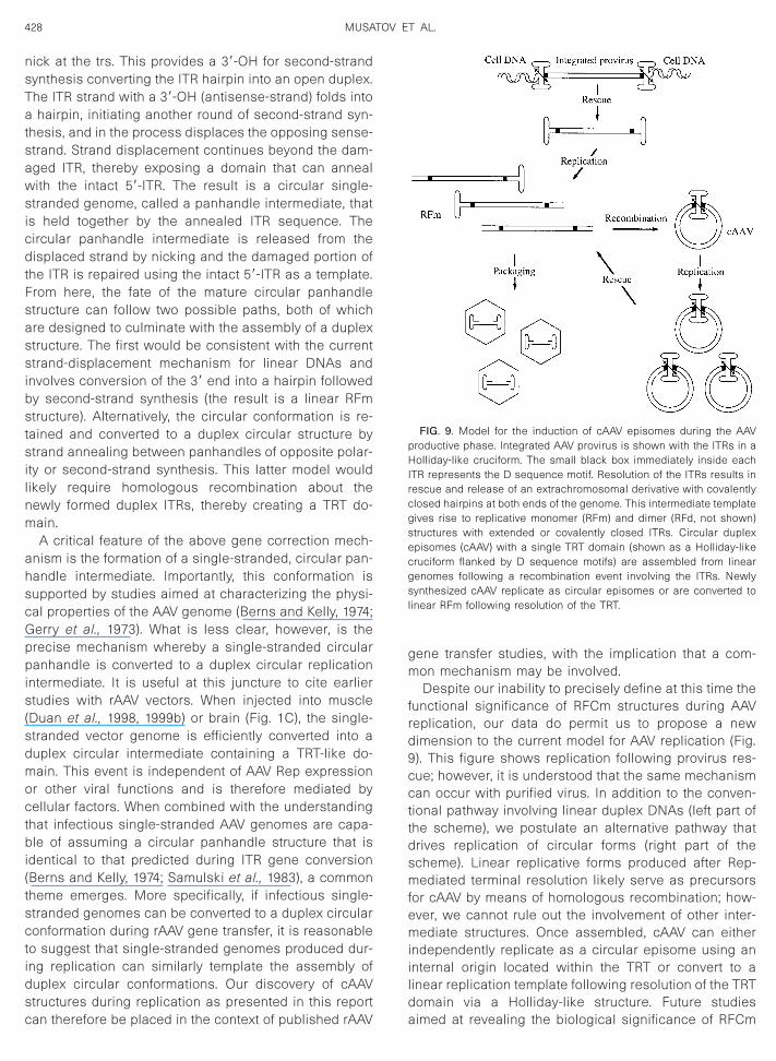
sdmoctbi(tsctidsc
gm
frd9ccttdsmfemiild
428 MUSATOV ET AL.
nick at the trs. This provides a 39-OH for second-strandsynthesis converting the ITR hairpin into an open duplex.The ITR strand with a 39-OH (antisense-strand) folds intoa hairpin, initiating another round of second-strand syn-thesis, and in the process displaces the opposing sense-strand. Strand displacement continues beyond the dam-aged ITR, thereby exposing a domain that can annealwith the intact 59-ITR. The result is a circular single-stranded genome, called a panhandle intermediate, thatis held together by the annealed ITR sequence. Thecircular panhandle intermediate is released from thedisplaced strand by nicking and the damaged portion ofthe ITR is repaired using the intact 59-ITR as a template.From here, the fate of the mature circular panhandlestructure can follow two possible paths, both of whichare designed to culminate with the assembly of a duplexstructure. The first would be consistent with the currentstrand-displacement mechanism for linear DNAs andinvolves conversion of the 39 end into a hairpin followedby second-strand synthesis (the result is a linear RFmstructure). Alternatively, the circular conformation is re-tained and converted to a duplex circular structure bystrand annealing between panhandles of opposite polar-ity or second-strand synthesis. This latter model wouldlikely require homologous recombination about thenewly formed duplex ITRs, thereby creating a TRT do-main.
A critical feature of the above gene correction mech-anism is the formation of a single-stranded, circular pan-handle intermediate. Importantly, this conformation issupported by studies aimed at characterizing the physi-cal properties of the AAV genome (Berns and Kelly, 1974;Gerry et al., 1973). What is less clear, however, is theprecise mechanism whereby a single-stranded circularpanhandle is converted to a duplex circular replicationintermediate. It is useful at this juncture to cite earlierstudies with rAAV vectors. When injected into muscle(Duan et al., 1998, 1999b) or brain (Fig. 1C), the single-
tranded vector genome is efficiently converted into auplex circular intermediate containing a TRT-like do-ain. This event is independent of AAV Rep expression
r other viral functions and is therefore mediated byellular factors. When combined with the understanding
hat infectious single-stranded AAV genomes are capa-le of assuming a circular panhandle structure that is
dentical to that predicted during ITR gene conversionBerns and Kelly, 1974; Samulski et al., 1983), a commonheme emerges. More specifically, if infectious single-tranded genomes can be converted to a duplex circularonformation during rAAV gene transfer, it is reasonable
o suggest that single-stranded genomes produced dur-ng replication can similarly template the assembly ofuplex circular conformations. Our discovery of cAAVtructures during replication as presented in this report
an therefore be placed in the context of published rAAV aene transfer studies, with the implication that a com-on mechanism may be involved.Despite our inability to precisely define at this time the
unctional significance of RFCm structures during AAVeplication, our data do permit us to propose a newimension to the current model for AAV replication (Fig.). This figure shows replication following provirus res-ue; however, it is understood that the same mechanisman occur with purified virus. In addition to the conven-
ional pathway involving linear duplex DNAs (left part ofhe scheme), we postulate an alternative pathway thatrives replication of circular forms (right part of thecheme). Linear replicative forms produced after Rep-ediated terminal resolution likely serve as precursors
or cAAV by means of homologous recombination; how-ver, we cannot rule out the involvement of other inter-ediate structures. Once assembled, cAAV can either
ndependently replicate as a circular episome using annternal origin located within the TRT or convert to ainear replication template following resolution of the TRTomain via a Holliday-like structure. Future studies
FIG. 9. Model for the induction of cAAV episomes during the AAVproductive phase. Integrated AAV provirus is shown with the ITRs in aHolliday-like cruciform. The small black box immediately inside eachITR represents the D sequence motif. Resolution of the ITRs results inrescue and release of an extrachromosomal derivative with covalentlyclosed hairpins at both ends of the genome. This intermediate templategives rise to replicative monomer (RFm) and dimer (RFd, not shown)structures with extended or covalently closed ITRs. Circular duplexepisomes (cAAV) with a single TRT domain (shown as a Holliday-likecruciform flanked by D sequence motifs) are assembled from lineargenomes following a recombination event involving the ITRs. Newlysynthesized cAAV replicate as circular episomes or are converted tolinear RFm following resolution of the TRT.
imed at revealing the biological significance of RFCm
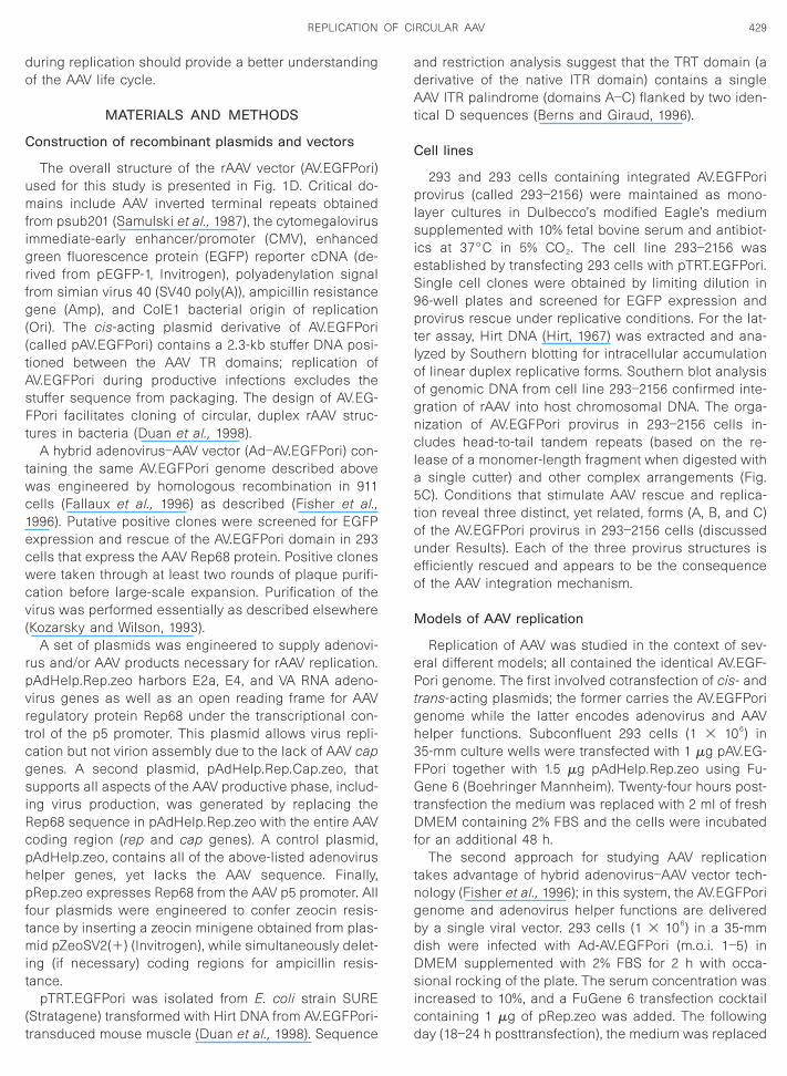
gsiRcphpftmit
(t
adAt
C
plsi
tgh
429REPLICATION OF CIRCULAR AAV
during replication should provide a better understandingof the AAV life cycle.
MATERIALS AND METHODS
Construction of recombinant plasmids and vectors
The overall structure of the rAAV vector (AV.EGFPori)used for this study is presented in Fig. 1D. Critical do-mains include AAV inverted terminal repeats obtainedfrom psub201 (Samulski et al., 1987), the cytomegalovirusimmediate-early enhancer/promoter (CMV), enhancedgreen fluorescence protein (EGFP) reporter cDNA (de-rived from pEGFP-1, Invitrogen), polyadenylation signalfrom simian virus 40 (SV40 poly(A)), ampicillin resistancegene (Amp), and ColE1 bacterial origin of replication(Ori). The cis-acting plasmid derivative of AV.EGFPori(called pAV.EGFPori) contains a 2.3-kb stuffer DNA posi-tioned between the AAV TR domains; replication ofAV.EGFPori during productive infections excludes thestuffer sequence from packaging. The design of AV.EG-FPori facilitates cloning of circular, duplex rAAV struc-tures in bacteria (Duan et al., 1998).
A hybrid adenovirus–AAV vector (Ad–AV.EGFPori) con-taining the same AV.EGFPori genome described abovewas engineered by homologous recombination in 911cells (Fallaux et al., 1996) as described (Fisher et al.,1996). Putative positive clones were screened for EGFPexpression and rescue of the AV.EGFPori domain in 293cells that express the AAV Rep68 protein. Positive cloneswere taken through at least two rounds of plaque purifi-cation before large-scale expansion. Purification of thevirus was performed essentially as described elsewhere(Kozarsky and Wilson, 1993).
A set of plasmids was engineered to supply adenovi-rus and/or AAV products necessary for rAAV replication.pAdHelp.Rep.zeo harbors E2a, E4, and VA RNA adeno-virus genes as well as an open reading frame for AAVregulatory protein Rep68 under the transcriptional con-trol of the p5 promoter. This plasmid allows virus repli-cation but not virion assembly due to the lack of AAV cap
enes. A second plasmid, pAdHelp.Rep.Cap.zeo, thatupports all aspects of the AAV productive phase, includ-
ng virus production, was generated by replacing theep68 sequence in pAdHelp.Rep.zeo with the entire AAVoding region (rep and cap genes). A control plasmid,AdHelp.zeo, contains all of the above-listed adenoviruselper genes, yet lacks the AAV sequence. Finally,Rep.zeo expresses Rep68 from the AAV p5 promoter. All
our plasmids were engineered to confer zeocin resis-ance by inserting a zeocin minigene obtained from plas-
id pZeoSV2(1) (Invitrogen), while simultaneously delet-ng (if necessary) coding regions for ampicillin resis-ance.
pTRT.EGFPori was isolated from E. coli strain SUREStratagene) transformed with Hirt DNA from AV.EGFPori-
ransduced mouse muscle (Duan et al., 1998). Sequencend restriction analysis suggest that the TRT domain (aerivative of the native ITR domain) contains a singleAV ITR palindrome (domains A–C) flanked by two iden-
ical D sequences (Berns and Giraud, 1996).
ell lines
293 and 293 cells containing integrated AV.EGFPorirovirus (called 293–2156) were maintained as mono-
ayer cultures in Dulbecco’s modified Eagle’s mediumupplemented with 10% fetal bovine serum and antibiot-
cs at 37°C in 5% CO2. The cell line 293–2156 wasestablished by transfecting 293 cells with pTRT.EGFPori.Single cell clones were obtained by limiting dilution in96-well plates and screened for EGFP expression andprovirus rescue under replicative conditions. For the lat-ter assay, Hirt DNA (Hirt, 1967) was extracted and ana-lyzed by Southern blotting for intracellular accumulationof linear duplex replicative forms. Southern blot analysisof genomic DNA from cell line 293–2156 confirmed inte-gration of rAAV into host chromosomal DNA. The orga-nization of AV.EGFPori provirus in 293–2156 cells in-cludes head-to-tail tandem repeats (based on the re-lease of a monomer-length fragment when digested witha single cutter) and other complex arrangements (Fig.5C). Conditions that stimulate AAV rescue and replica-tion reveal three distinct, yet related, forms (A, B, and C)of the AV.EGFPori provirus in 293–2156 cells (discussedunder Results). Each of the three provirus structures isefficiently rescued and appears to be the consequenceof the AAV integration mechanism.
Models of AAV replication
Replication of AAV was studied in the context of sev-eral different models; all contained the identical AV.EGF-Pori genome. The first involved cotransfection of cis- andrans-acting plasmids; the former carries the AV.EGFPorienome while the latter encodes adenovirus and AAVelper functions. Subconfluent 293 cells (1 3 106) in
35-mm culture wells were transfected with 1 mg pAV.EG-FPori together with 1.5 mg pAdHelp.Rep.zeo using Fu-Gene 6 (Boehringer Mannheim). Twenty-four hours post-transfection the medium was replaced with 2 ml of freshDMEM containing 2% FBS and the cells were incubatedfor an additional 48 h.
The second approach for studying AAV replicationtakes advantage of hybrid adenovirus–AAV vector tech-nology (Fisher et al., 1996); in this system, the AV.EGFPorigenome and adenovirus helper functions are deliveredby a single viral vector. 293 cells (1 3 106) in a 35-mmdish were infected with Ad-AV.EGFPori (m.o.i. 1–5) inDMEM supplemented with 2% FBS for 2 h with occa-sional rocking of the plate. The serum concentration wasincreased to 10%, and a FuGene 6 transfection cocktailcontaining 1 mg of pRep.zeo was added. The following
day (18–24 h posttransfection), the medium was replaced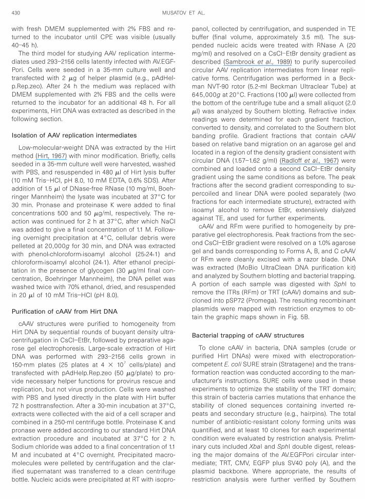
pDref
I
msw
r3c
t
cgfpfia
pogowaArcpt
B
pcfuetspnqciimp
430 MUSATOV ET AL.
with fresh DMEM supplemented with 2% FBS and re-turned to the incubator until CPE was visible (usually40–45 h).
The third model for studying AAV replication interme-diates used 293–2156 cells latently infected with AV.EGF-Pori. Cells were seeded in a 35-mm culture well andtransfected with 2 mg of helper plasmid (e.g., pAdHel-
.Rep.zeo). After 24 h the medium was replaced withMEM supplemented with 2% FBS and the cells were
eturned to the incubator for an additional 48 h. For allxperiments, Hirt DNA was extracted as described in the
ollowing section.
solation of AAV replication intermediates
Low-molecular-weight DNA was extracted by the Hirtethod (Hirt, 1967) with minor modification. Briefly, cells
eeded in a 35-mm culture well were harvested, washedith PBS, and resuspended in 480 ml of Hirt lysis buffer
(10 mM Tris–HCl, pH 8.0, 10 mM EDTA, 0.6% SDS). Afteraddition of 1.5 ml of DNase-free RNase (10 mg/ml, Boeh-inger Mannheim) the lysate was incubated at 37°C for0 min. Pronase and proteinase K were added to finaloncentrations 500 and 50 mg/ml, respectively. The re-
action was continued for 2 h at 37°C, after which NaClwas added to give a final concentration of 1.1 M. Follow-ing overnight precipitation at 4°C, cellular debris werepelleted at 20,000g for 30 min, and DNA was extractedwith phenol:chloroform:isoamyl alcohol (25:24:1) andchloroform:isoamyl alcohol (24:1). After ethanol precipi-tation in the presence of glycogen (30 mg/ml final con-centration, Boehringer Mannheim), the DNA pellet waswashed twice with 70% ethanol, dried, and resuspendedin 20 ml of 10 mM Tris–HCl (pH 8.0).
Purification of cAAV from Hirt DNA
cAAV structures were purified to homogeneity fromHirt DNA by sequential rounds of buoyant density ultra-centrifugation in CsCl–EtBr, followed by preparative aga-rose gel electrophoresis. Large-scale extraction of HirtDNA was performed with 293–2156 cells grown in150-mm plates (25 plates at 4 3 107 cells/plate) andransfected with pAdHelp.Rep.zeo (50 mg/plate) to pro-
vide necessary helper functions for provirus rescue andreplication, but not virus production. Cells were washedwith PBS and lysed directly in the plate with Hirt buffer72 h posttransfection. After a 30-min incubation at 37°C,extracts were collected with the aid of a cell scraper andcombined in a 250-ml centrifuge bottle. Proteinase K andpronase were added according to our standard Hirt DNAextraction procedure and incubated at 37°C for 2 h.Sodium chloride was added to a final concentration of 1.1M and incubated at 4°C overnight. Precipitated macro-molecules were pelleted by centrifugation and the clar-ified supernatant was transferred to a clean centrifuge
bottle. Nucleic acids were precipitated at RT with isopro- rpanol, collected by centrifugation, and suspended in TEbuffer (final volume, approximately 3.5 ml). The sus-pended nucleic acids were treated with RNase A (20mg/ml) and resolved on a CsCl–EtBr density gradient asdescribed (Sambrook et al., 1989) to purify supercoiledcircular AAV replication intermediates from linear repli-cative forms. Centrifugation was performed in a Beck-man NVT-90 rotor (5.2-ml Beckman Ultraclear Tube) at645,000g at 20°C. Fractions (100 ml) were collected fromthe bottom of the centrifuge tube and a small aliquot (2.0ml) was analyzed by Southern blotting. Refractive indexreadings were determined for each gradient fraction,converted to density, and correlated to the Southern blotbanding profile. Gradient fractions that contain cAAVbased on relative band migration on an agarose gel andlocated in a region of the density gradient consistent withcircular DNA (1.57–1.62 g/ml) (Radloff et al., 1967) were
ombined and loaded onto a second CsCl–EtBr densityradient using the same conditions as before. The peak
ractions after the second gradient corresponding to su-ercoiled and linear DNA were pooled separately (two
ractions for each intermediate structure), extracted withsoamyl alcohol to remove EtBr, extensively dialyzedgainst TE, and used for further experiments.
cAAV and RFm were purified to homogeneity by pre-arative gel electrophoresis. Peak fractions from the sec-nd CsCl–EtBr gradient were resolved on a 1.0% agaroseel and bands corresponding to Forms A, B, and C cAAVr RFm were cleanly excised with a razor blade. DNAas extracted (MoBio UltraClean DNA purification kit)nd analyzed by Southern blotting and bacterial trapping.
portion of each sample was digested with SphI toemove the ITRs (RFm) or TRT (cAAV) domains and sub-loned into pSP72 (Promega). The resulting recombinantlasmids were mapped with restriction enzymes to ob-
ain the graphic maps shown in Fig. 5B.
acterial trapping of cAAV structures
To clone cAAV in bacteria, DNA samples (crude orurified Hirt DNAs) were mixed with electroporation-ompetent E. coli SURE strain (Stratagene) and the trans-
ormation reaction was conducted according to the man-facturer’s instructions. SURE cells were used in thesexperiments to optimize the stability of the TRT domain;
his strain of bacteria carries mutations that enhance thetability of cloned sequences containing inverted re-eats and secondary structure (e.g., hairpins). The totalumber of antibiotic-resistant colony forming units wasuantified, and at least 10 clones for each experimentalondition were evaluated by restriction analysis. Prelim-
nary cuts included XbaI and SphI double digest, releas-ng the major domains of the AV.EGFPori circular inter-
ediate; TRT, CMV, EGFP plus SV40 poly (A), and thelasmid backbone. Where appropriate, the results of
estriction analysis were further verified by Southern
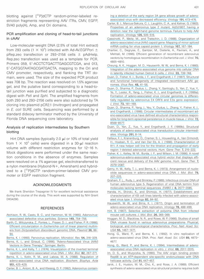
BB
B
C
C
C
C
D
D
D
D
F
F
G
G
H
H
H
H
H
H
I
J
431REPLICATION OF CIRCULAR AAV
blotting against [32P]dCTP random-primer-labeled re-striction fragments representing AAV ITRs, CMV, EGFP,SV40 poly(A), Amp, and Ori domains.
PCR amplification and cloning of head-to-tail junctionsin cAAV
Low-molecular-weight DNA (2.5% of total Hirt extract)from 293 cells (1 3 106) infected with Ad-AV.EGFPori 6pRep.zeo transfection or 293–2156 cells 6 pAdHelp.Rep.zeo transfection was used as a template for PCR.Primers 059, 59-ACCTCTGACTTGAGCGTCGA, and 013,59-CATGGTAATAGCGATGACTA, mapping to the Ori andCMV promoter, respectively, and flanking the TRT do-main, were used. The size of the expected PCR productwas 708 bp. The reaction was resolved on 1.5% agarosegel, and the putative band corresponding to a head-to-tail junction was purified and subjected to a diagnosticdigest with PstI and SphI. The amplified fragments fromboth 293 and 293–2156 cells were also subcloned by TAcloning into plasmid pCR2.1 (Invitrogen) and propagatedin E. coli SURE cells. Sequencing was performed by astandard dideoxy terminator method by the University ofFlorida DNA sequencing core laboratory.
Analysis of replication intermediates by Southernblotting
Hirt DNA samples (typically 2.0 ml or 10% of total yieldfrom 1 3 106 cells) were digested in a 30-ml reactionvolume with different restriction enzymes for 12–16 h.Mock digestions were performed under the same reac-tion conditions in the absence of enzymes. Sampleswere resolved on a 1% agarose gel, electrotransferred toa nylon membrane (Hybond-N1, Amersham) and hybrid-ized to a [32P]dCTP random-primer-labeled CMV pro-moter or EGFP restriction fragment.
ACKNOWLEDGMENTS
We thank Sheridon Trepagnier IV for excellent technical assistanceduring the course of the study. This work was supported by NIH GrantDK54290.
REFERENCES
Atchison, R. W., Casto, B. C., and Hammon, W. M. (1965). Adenovirus-associated defective virus particles. Science 149, 754–756.
Barth, C., Wilczynska, Z., Pontes, L., Fraser, D. J., and Fisher, P. R. (1996).Efficient circularization in Escherichia coli of linear plasmid multim-ers from Dictyostellium discoideum genomic DNA. Plasmid 36, 86–94.
erns, K. I. (1990). Parvovirus replication. Microbiol. Rev. 54, 316–329.erns, K. I., and Giraud, C. (1996). “Adeno-Associated Virus (AAV)
Vectors in Gene Therapy.” Springer, Berlin.erns, K. I., and Kelly, T. J. (1974). Visualization of the inverted terminal
repetition in adeno-associated virus DNA. J. Mol. Biol. 82, 267–271.Berns, K. I., Kotin, R. M., and Labow, M. A. (1988). Regulation of
adeno-associated virus DNA replication. Biochem. Biophys. Acta951, 425–429.
Carter, B. J., Antoni, B. A., and Klessig, D. F. (1992). Adenovirus contain-
ing a deletion of the early region 2A gene allows growth of adeno-associated virus with decreased efficiency. Virology 191, 473–476.
arter, B. J., Marcus-Sekura, C. J., Laughlin, C. A., and Ketner, G. (1983).Properties of an adenovirus type 2 mutant, Ad2d1807, having adeletion near the right-hand genome terminus: Failure to help AAVreplication. Virology 126, 505–516.
assinotti, P., Weitz, M., and Tratschin, J. D. (1988). Organization ofadeno-associated virus (AAV) capsid gene: Mapping of minor splicedmRNA coding for virus capsid protein 1. Virology 167, 167–184.
hartier, C., Degryse, E., Gantzer, M., Dieterle, A., Pavirani, A., andMehtali, M. (1996). Efficient generation of recombinant adenovirusvectors by homologous recombination in Escherichia coli. J. Virol. 70,4805–4810.
heung, A. K., Hoggan, M. D., Hauswirth, W. W., and Berns, K. I. (1980).Integration of the adeno-associated virus genome into cellular DNAin latently infected human Detroit 6 cells. J. Virol. 33, 739–748.
uan, D., Fisher, K. J., Burda, J. F., and Engelhardt, J. F. (1997). Structuraland functional heterogeneity of integrated recombinant AAV ge-nomes. Virus Res. 48, 41–56.
uan, D., Sharma, P., Dudus, L., Zhang, Y., Sanlioglu, S., Yan, Z., Yue, Y.,Ye, Y., Lester, R., Yang, J., Fisher, K. J., and Engelhardt, J. F. (1999a).Formation of adeno-associated virus circular genomes is differen-tially regulated by adenovirus E4 ORF6 and E2a gene expression.J. Virol. 73, 161–169.
uan, D., Sharma, P., Yang, J., Yeu, Y., Dudus, L., Zhang, Y., Fisher, K. J.,and Engelhardt, J. (1998). Circular intermediates of recombinant ad-eno-associated virus have defined structural characteristics respon-sible for long-term episomal persistence in muscle tissue. J. Virol. 72,8568–8577.
uan, D., Yan, Z., Yue, Y., and Engelhardt, J. F. (1999b). Structuralanalysis of adeno-associated virus transduction circular intermedi-ates. Virology 261, 8–14.
allaux, F. J., Kranenburg, O., Cramer, S. J., Houweling, A., Van Ormondt,H., Hoeben, R. C., and Van Der Eb, A. J. (1996). Characterization of911: A new helper cell line for the titration and propagation of earlyregion 1-deleted adenoviral vectors. Hum. Gene Ther. 7, 215–222.
isher, K. J., Kelley, W. M., Burda, J. F., and Wilson, J. M. (1996). A noveladenovirus-adeno-associated virus hybrid vector that displays effi-cient rescue and delivery of the AAV genome. Hum. Gene Ther. 7,2079–2087.
erry, H. W., Kelly, T. J., and Berns, K. I. (1973). Arrangement of nucle-otide sequences in adeno-associated virus DNA. J. Mol. Biol. 79,207–225.
raham, F. L., Rudy, J., and Brinkley, P. (1989). Infectious circular DNA ofhuman adenovirus type 5: Regeneration of viral DNA termini frommolecules lacking terminal sequences. EMBO J. 8, 2077–2085.
anda, H., Shiroki, K., and Shimojo, H. (1977). Establishment andcharacterization of KB cell lines latently infected with adeno-associ-ated virus type 1. Virology 82, 84–92.
auswirth, W. W., and Birns, K. I. (1977). Origin and termination ofadeno-associated virus DNA replication. Virology 78, 488–499.
irt, B. (1967). Selective extraction of polyoma DNA from infectedmouse cell cultures. J. Mol. Biol. 26, 365–369.
oggan, M. D., Blacklow, N. R., and Rowe, W. P. (1966). Studies of smallDNA viruses found in various adenovirus preparations: Physical,biological, and immunological characteristics. Proc. Natl. Acad. Sci.USA 55, 1467–1471.
ong, G., Ward, P., and Berns, K. I. (1992). In vitro replication ofadeno-associated virus DNA. Proc. Natl. Acad. Sci. USA 89, 4673–4677.
ong, G., Ward, P., and Berns, K. I. (1994). Intermediates of adeno-associated virus DNA replication in vitro. J. Virol. 68, 2011–2015.
m, D. S., and Muzyczka, N. (1990). The AAV origin binding proteinRep68 is an ATP-dependent site-specific endonuclease with DNAhelicase activity. Cell 61, 447–457.
anik, J. E., Huston, M. M., Cho, K., and Rose, J. A. (1989). Efficient
synthesis of adeno-associated virus structural proteins requires both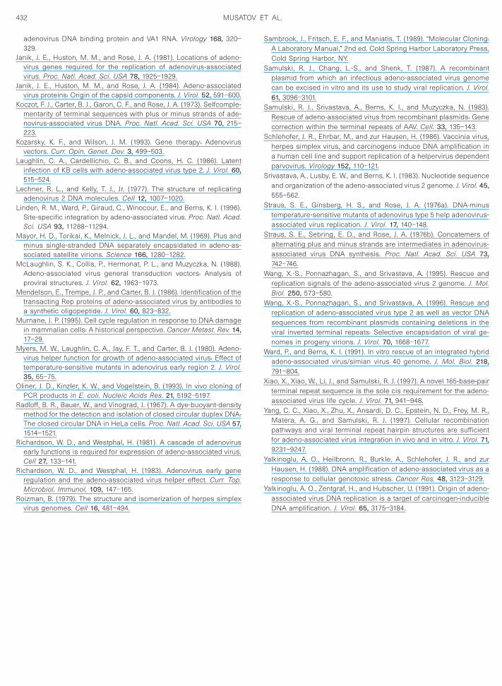
K
K
L
L
M
M
M
M
M
O
R
R
R
R
S
S
S
S
S
S
S
W
W
W
X
Y
Y
Y
432 MUSATOV ET AL.
adenovirus DNA binding protein and VA1 RNA. Virology 168, 320–329.
Janik, J. E., Huston, M. M., and Rose, J. A. (1981). Locations of adeno-virus genes required for the replication of adenovirus-associatedvirus. Proc. Natl. Acad. Sci. USA 78, 1925–1929.
Janik, J. E., Huston, M. M., and Rose, J. A. (1984). Adeno-associatedvirus proteins: Origin of the capsid components. J. Virol. 52, 591–600.
oczot, F. J., Carter, B. J., Garon, C. F., and Rose, J. A. (1973). Selfcomple-mentarity of terminal sequences with plus or minus strands of ade-novirus-associated virus DNA. Proc. Natl. Acad. Sci. USA 70, 215–223.
ozarsky, K. F., and Wilson, J. M. (1993). Gene therapy: Adenovirusvectors. Curr. Opin. Genet. Dev. 3, 499–503.
Laughlin, C. A., Cardellichio, C. B., and Coons, H. C. (1986). Latentinfection of KB cells with adeno-associated virus type 2. J. Virol. 60,515–524.
echner, R. L., and Kelly, T. J., Jr. (1977). The structure of replicatingadenovirus 2 DNA molecules. Cell 12, 1007–1020.
inden, R. M., Ward, P., Giraud, C., Winocour, E., and Berns, K. I. (1996).Site-specific integration by adeno-associated virus. Proc. Natl. Acad.Sci. USA 93, 11288–11294.ayor, H. D., Torikai, K., Melnick, J. L., and Mandel, M. (1969). Plus andminus single-stranded DNA separately encapsidated in adeno-as-sociated satellite virions. Science 166, 1280–1282.cLaughlin, S. K., Collis, P., Hermonat, P. L., and Muzyczka, N. (1988).Adeno-associated virus general transduction vectors: Analysis ofproviral structures. J. Virol. 62, 1963–1973.endelson, E., Trempe, J. P., and Carter, B. J. (1986). Identification of thetransacting Rep proteins of adeno-associated virus by antibodies toa synthetic oligopeptide. J. Virol. 60, 823–832.urnane, J. P. (1995). Cell cycle regulation in response to DNA damagein mammalian cells: A historical perspective. Cancer Metast. Rev. 14,17–29.yers, M. W., Laughlin, C. A., Jay, F. T., and Carter, B. J. (1980). Adeno-virus helper function for growth of adeno-associated virus: Effect oftemperature-sensitive mutants in adenovirus early region 2. J. Virol.35, 65–75.
liner, J. D., Kinzler, K. W., and Vogelstein, B. (1993). In vivo cloning ofPCR products in E. coli. Nucleic Acids Res. 21, 5192–5197.
adloff, B. R., Bauer, W., and Vinograd, J. (1967). A dye-buoyant-densitymethod for the detection and isolation of closed circular duplex DNA:The closed circular DNA in HeLa cells. Proc. Natl. Acad. Sci. USA 57,1514–1521.
ichardson, W. D., and Westphal, H. (1981). A cascade of adenovirusearly functions is required for expression of adeno-associated virus.Cell 27, 133–141.
ichardson, W. D., and Westphal, H. (1983). Adenovirus early generegulation and the adeno-associated virus helper effect. Curr. Top.Microbiol. Immunol. 109, 147–165.
oizman, B. (1979). The structure and isomerization of herpes simplex
virus genomes. Cell 16, 481–494.ambrook, J., Fritsch, E. F., and Maniatis, T. (1989). “Molecular Cloning:A Laboratory Manual,” 2nd ed. Cold Spring Harbor Laboratory Press,Cold Spring Harbor, NY.
amulski, R. J., Chang, L.-S., and Shenk, T. (1987). A recombinantplasmid from which an infectious adeno-associated virus genomecan be excised in vitro and its use to study viral replication. J. Virol.61, 3096–3101.
amulski, R. J., Srivastava, A., Berns, K. I., and Muzyczka, N. (1983).Rescue of adeno-associated virus from recombinant plasmids: Genecorrection within the terminal repeats of AAV. Cell. 33, 135–143.
chlehofer, J. R., Ehrbar, M., and zur Hausen, H. (1986). Vaccinia virus,herpes simplex virus, and carcinogens induce DNA amplification ina human cell line and support replication of a helpervirus dependentparvovirus. Virology 152, 110–121.
rivastava, A., Lusby, E. W., and Berns, K. I. (1983). Nucleotide sequenceand organization of the adeno-associated virus 2 genome. J. Virol. 45,555–562.
traus, S. E., Ginsberg, H. S., and Rose, J. A. (1976a). DNA-minustemperature-sensitive mutants of adenovirus type 5 help adenovirus-associated virus replication. J. Virol. 17, 140–148.
traus, S. E., Sebring, E. D., and Rose, J. A. (1976b). Concatemers ofalternating plus and minus strands are intermediates in adenovirus-associated virus DNA synthesis. Proc. Natl. Acad. Sci. USA 73,742–746.
ang, X.-S., Ponnazhagan, S., and Srivastava, A. (1995). Rescue andreplication signals of the adeno-associated virus 2 genome. J. Mol.Biol. 250, 573–580.
ang, X.-S., Ponnazhagan, S., and Srivastava, A. (1996). Rescue andreplication of adeno-associated virus type 2 as well as vector DNAsequences from recombinant plasmids containing deletions in theviral inverted terminal repeats: Selective encapsidation of viral ge-nomes in progeny virions. J. Virol. 70, 1668–1677.
ard, P., and Berns, K. I. (1991). In vitro rescue of an integrated hybridadeno-associated virus/simian virus 40 genome. J. Mol. Biol. 218,791–804.
iao, X., Xiao, W., Li, J., and Samulski, R. J. (1997). A novel 165-base-pairterminal repeat sequence is the sole cis requirement for the adeno-associated virus life cycle. J. Virol. 71, 941–948.
ang, C. C., Xiao, X., Zhu, X., Ansardi, D. C., Epstein, N. D., Frey, M. R.,Matera, A. G., and Samulski, R. J. (1997). Cellular recombinationpathways and viral terminal repeat hairpin structures are sufficientfor adeno-associated virus integration in vivo and in vitro. J. Virol. 71,9231–9247.
alkinoglu, A. O., Heilbronn, R., Burkle, A., Schlehofer, J. R., and zurHausen, H. (1988). DNA amplification of adeno-associated virus as aresponse to cellular genotoxic stress. Cancer Res. 48, 3123–3129.
alkinoglu, A. O., Zentgraf, H., and Hubscher, U. (1991). Origin of adeno-associated virus DNA replication is a target of carcinogen-inducible
DNA amplification. J. Virol. 65, 3175–3184.