Anterograde delivery of brain-derived neurotrophic factor to striatum via nigral transduction of...
12
Anterograde delivery of brain-derived neurotrophic factor to striatum via nigral transduction of recombinant adeno- associated virus increases neuronal death but promotes neurogenic response following stroke Elin Gustafsson, 1 Gunnar Andsberg, 1 Vladimer Darsalia, 1 Paul Mohapel, 1 Ronald J. Mandel, 2 Deniz Kirik, 3 Olle Lindvall 1 and Zaal Kokaia 1 1 Section of Restorative Neurology, Wallenberg Neuroscience Center, University of Lund, BMC A-11 SE-221 84 Lund, Sweden 2 Department of Neuroscience, The Powell Gene Therapy Center, McKnight Brain Institute, University of Florida College of Medicine, PO Box 100244, Gainesville, FL 32610-0244, USA 3 Section of Neurobiology, Wallenberg Neuroscience Center, University of Lund, BMC A-11 SE-221 84 Lund, Sweden Keywords: anterograde transport, BDNF, BrdU, focal cerebral ischaemia, GFP, neurogenesis, neuroprotection, substantia nigra Abstract To explore the role of brain-derived neurotrophic factor for survival and generation of striatal neurons after stroke, recombinant adeno- associated viral vectors carrying brain-derived neurotrophic factor or green fluorescent protein genes were injected into right rat substantia nigra 4–5 weeks prior to 30 min ipsilateral of middle cerebral artery occlusion. The brain-derived neurotrophic factor– recombinant adeno-associated viral transduction markedly increased the production of brain-derived neurotrophic factor protein by nigral cells. Brain-derived neurotrophic factor was transported anterogradely to the striatum and released in biologically active form, as revealed by the hypertrophic response of striatal neuropeptide Y-positive interneurons. Animals transduced with brain-derived neurotrophic factor-recombinant adeno-associated virus also exhibited abnormalities in body posture and movements, including tilted body to the right, choreiform movements of left forelimb and head, and spontaneous, so-called ‘barrel’ rotation along their long axis. The continuous delivery of brain-derived neurotrophic factor had no effect on the survival of striatal projection neurons after stroke, but exaggerated the loss of cholinergic, and parvalbumin- and neuropeptide Y-positive, g-aminobutyric acid-ergic interneurons. The high brain-derived neurotrophic factor levels in the animals subjected to stroke also gave rise to an increased number of striatal cells expressing doublecortin, a marker for migrating neuroblasts, and cells double-labelled with the mitotic marker, 5-bromo-2 0 -deoxyur- idine-5 0 monophosphate, and early neuronal (Hu) or striatal neuronal (Meis2) markers. Our findings indicate that long-term anterograde delivery of high levels of brain-derived neurotrophic factor increases the vulnerability of striatal interneurons to stroke-induced damage. Concomitantly, brain-derived neurotrophic factor potentiates the stroke-induced neurogenic response, at least at early stages. Introduction Brain-derived neurotrophic factor (BDNF) is a member of a trophic factor family called neurotrophins, and plays a major role in survival and differentiation during embryonic development of the nervous sys- tem (for review, see Davies, 1994). In the adult brain, ischaemic insults lead to marked increases of BDNF levels in cortical and hippocampal neurons (Lindvall et al., 1992; Takeda et al., 1993; Kokaia et al., 1995; Arai et al., 1996; Kokaia et al., 1996; Kokaia et al., 1998b), and it has been proposed that these changes of endogeneous BDNF levels might be a neuroprotective response (Lindvall et al., 1994). In agreement with this hypothesis, mice heterozygous for BDNF gene deletion exhibit larger cerebral infarcts as compared with wild-type animals after stroke (Endres et al., 2000), and blockade of endogeneous levels of BDNF by intraventricular infusion of TrkB-Fc fusion protein reduces the survival of several types of forebrain neurons following global forebrain ischaemia (Larsson et al., 1999). To study the effect of increased BDNF levels on neuronal survival after stroke, several routes of delivery have been used. BDNF has been reported to protect against neuronal death following global forebrain ischaemia when delivered by intraventricular infusion (Beck et al., 1994) or by transplantation of genetically modified primary fibroblasts into the hippocampus (Ferrer et al., 1998). In addition, recombinant BDNF reduces the damage after hypoxia-ischaemia both in cell culture (Cheng & Mattson, 1994) and when infused intra- cerebroventricularly in the neonatal brain (Han & Holtzman, 2000; Han et al., 2000). Intraventricular (Scha ¨bitz et al., 1997), intravenous (Scha ¨bitz et al., 2000) and intraparenchymal (Yamashita et al., 1997; Yanamoto et al., 2000) infusion of BDNF also protect cortical neurons against stroke-induced damage. We have demonstrated that continuous BDNF delivery mediated by direct gene transfer to the striatum with recombinant adeno-associated viral (rAAV) vectors partially protects striatal neurons against stroke-induced death (Andsberg et al., 2002). European Journal of Neuroscience, Vol. 17, pp. 2667–2678, 2003 ß Federation of European Neuroscience Societies doi:10.1046/j.1460-9568.2003.02713.x Correspondence: Dr Z. Kokaia, as above. E-mail: [email protected] Received 29 November 2002, revised 11 March 2003, accepted 2 April 2003
-
Upload
independent -
Category
Documents
-
view
1 -
download
0
Transcript of Anterograde delivery of brain-derived neurotrophic factor to striatum via nigral transduction of...

Anterograde delivery of brain-derived neurotrophic factor tostriatum via nigral transduction of recombinant adeno-associated virus increases neuronal death but promotesneurogenic response following stroke
Elin Gustafsson,1 Gunnar Andsberg,1 Vladimer Darsalia,1 Paul Mohapel,1 Ronald J. Mandel,2 Deniz Kirik,3
Olle Lindvall1 and Zaal Kokaia1
1Section of Restorative Neurology, Wallenberg Neuroscience Center, University of Lund, BMC A-11 SE-221 84 Lund, Sweden2Department of Neuroscience, The Powell Gene Therapy Center, McKnight Brain Institute, University of Florida College of Medicine,PO Box 100244, Gainesville, FL 32610-0244, USA3Section of Neurobiology, Wallenberg Neuroscience Center, University of Lund, BMC A-11 SE-221 84 Lund, Sweden
Keywords: anterograde transport, BDNF, BrdU, focal cerebral ischaemia, GFP, neurogenesis, neuroprotection, substantia nigra
Abstract
To explore the role of brain-derived neurotrophic factor for survival and generation of striatal neurons after stroke, recombinant adeno-associated viral vectors carrying brain-derived neurotrophic factor or green fluorescent protein genes were injected into right ratsubstantia nigra 4–5 weeks prior to 30 min ipsilateral of middle cerebral artery occlusion. The brain-derived neurotrophic factor–recombinant adeno-associated viral transduction markedly increased the production of brain-derived neurotrophic factor protein bynigral cells. Brain-derived neurotrophic factor was transported anterogradely to the striatum and released in biologically active form, asrevealed by the hypertrophic response of striatal neuropeptide Y-positive interneurons. Animals transduced with brain-derivedneurotrophic factor-recombinant adeno-associated virus also exhibited abnormalities in body posture and movements, includingtilted body to the right, choreiform movements of left forelimb and head, and spontaneous, so-called ‘barrel’ rotation along their longaxis. The continuous delivery of brain-derived neurotrophic factor had no effect on the survival of striatal projection neurons after stroke,but exaggerated the loss of cholinergic, and parvalbumin- and neuropeptide Y-positive, g-aminobutyric acid-ergic interneurons. Thehigh brain-derived neurotrophic factor levels in the animals subjected to stroke also gave rise to an increased number of striatal cellsexpressing doublecortin, a marker for migrating neuroblasts, and cells double-labelled with the mitotic marker, 5-bromo-20-deoxyur-idine-50monophosphate, and early neuronal (Hu) or striatal neuronal (Meis2) markers. Our findings indicate that long-term anterogradedelivery of high levels of brain-derived neurotrophic factor increases the vulnerability of striatal interneurons to stroke-induced damage.Concomitantly, brain-derived neurotrophic factor potentiates the stroke-induced neurogenic response, at least at early stages.
Introduction
Brain-derived neurotrophic factor (BDNF) is a member of a trophic
factor family called neurotrophins, and plays a major role in survival
and differentiation during embryonic development of the nervous sys-
tem (for review, see Davies, 1994). In the adult brain, ischaemic insults
lead to marked increases of BDNF levels in cortical and hippocampal
neurons (Lindvall et al., 1992; Takeda et al., 1993; Kokaia et al., 1995;
Arai et al., 1996; Kokaia et al., 1996; Kokaia et al., 1998b), and it has
been proposed that these changes of endogeneous BDNF levels might
be a neuroprotective response (Lindvall et al., 1994). In agreement
with this hypothesis, mice heterozygous for BDNF gene deletion
exhibit larger cerebral infarcts as compared with wild-type animals
after stroke (Endres et al., 2000), and blockade of endogeneous levels
of BDNF by intraventricular infusion of TrkB-Fc fusion protein
reduces the survival of several types of forebrain neurons following
global forebrain ischaemia (Larsson et al., 1999).
To study the effect of increased BDNF levels on neuronal survival
after stroke, several routes of delivery have been used. BDNF has
been reported to protect against neuronal death following global
forebrain ischaemia when delivered by intraventricular infusion (Beck
et al., 1994) or by transplantation of genetically modified primary
fibroblasts into the hippocampus (Ferrer et al., 1998). In addition,
recombinant BDNF reduces the damage after hypoxia-ischaemia both
in cell culture (Cheng & Mattson, 1994) and when infused intra-
cerebroventricularly in the neonatal brain (Han & Holtzman, 2000;
Han et al., 2000). Intraventricular (Schabitz et al., 1997), intravenous
(Schabitz et al., 2000) and intraparenchymal (Yamashita et al.,
1997; Yanamoto et al., 2000) infusion of BDNF also protect cortical
neurons against stroke-induced damage. We have demonstrated
that continuous BDNF delivery mediated by direct gene transfer
to the striatum with recombinant adeno-associated viral (rAAV)
vectors partially protects striatal neurons against stroke-induced death
(Andsberg et al., 2002).
European Journal of Neuroscience, Vol. 17, pp. 2667–2678, 2003 � Federation of European Neuroscience Societies
doi:10.1046/j.1460-9568.2003.02713.x
Correspondence: Dr Z. Kokaia, as above.
E-mail: [email protected]
Received 29 November 2002, revised 11 March 2003, accepted 2 April 2003

Recent experimental studies have indicated that BDNF can also
influence neurogenesis in the adult brain. Intraventricular infusion of
BDNF (Zigova et al., 1998; Pencea et al., 2001) or overexpression of
the BDNF gene in the ventricular zone (Benraiss et al., 2001) increases
the number of new neurons generated from the subventricular zone
(SVZ) in several forebrain structures. Stroke leads to increased
production of new striatal neurons from precursor cells in the SVZ
(Arvidsson et al., 2002; Parent et al., 2002). Because the number of
new striatal neurons showing long-term survival was small, we
proposed that delivery of BDNF might be one approach to promote
this neurogenic response (Arvidsson et al., 2002).
The objectives of the present study were twofold. First, to test
whether striatal BDNF levels can be increased by intranigral transduc-
tion of rAAV carrying the BDNF gene. Endogeneous BDNF is
transported efficiently in nigrostriatal axons in the anterograde direc-
tion (von Bartheld et al., 1996; Altar et al., 1997; Smith et al., 1997).
This route of delivery should therefore increase the BDNF content in
the striatum without altering its structural integrity. Second, to explore
whether continuous high intrastriatal BDNF levels, obtained through
anterograde transport from substantia nigra, can influence neuronal
survival and neurogenesis after stroke.
Materials and methods
Vector production
The rAAV2 vectors used in this study (BDNF–rAAV and GFP–rAAV)
were produced at the University of Florida Powell Gene Therapy
Center, USA. Virus production followed a two plasmid adenovirus-
free method, as reported in detail previously (Hauswirth et al., 2000).
Briefly, the helper/packaging plasmid pDG (Grimm et al., 1998) that
supplies all the necessary helper functions as well as rep and cap in
trans was cotransfected by calcium phosphate precipitation with vector
plasmid, plasmid cytomegalovirus promoter with chicken R-actin
intervening sequence-BDNF (pCBA–BDNF), in near confluent 293
cells. After lysing the cells, rAAV was prepared by Iodixanol cen-
trifugation and hand-packed heparin column purification, as described
previously (Zolotukhin et al., 1999).
Animals and experimental groups
All animal related procedures were conducted in accordance with local
ethical guidelines and approved animal care protocols. Thirty-six male
Wistar rats weighing approximately 210 g at the beginning of the
experiment (Taconic M & B A/S, Ry, Denmark) were housed under
12 h light : 12 h dark cycle with ad libitum access to food and water. All
animals were anaesthetized by inhalation of 1% halothane and injected
unilaterally in the substantia nigra with either BDNF–rAAV (n¼ 18) or
GFP–rAAV (n¼ 18). At 4 weeks after transduction, four animals were
tested for the effect of catecholamine inhibition, and another four rats
were implanted with recording electrodes for monitoring cortical and
striatal electroencephalogram (EEG) (n¼ 2 for BDNF–rAAV- and
n¼ 2 for GFP–rAAV-transduced animals for both analyses). All
animals were tested for spontaneous rotation. Four rats transduced
with BDNF–rAAV were used for immunocytochemical assessment of
BDNF levels prior to stroke, and another six rats (three transduced with
BDNF–rAAV and three with GFP–rAAV) for determination of BDNF
tissue levels with ELISA.
At 5 weeks after viral transduction, 15 rats were subjected to stroke
using middle cerebral artery occlusion (MCAO: n¼ 7 for BDNF–rAAV
and n¼ 8 for GFP–rAAV), and six rats transduced with GFP–rAAV
were sham-operated. From the day after MCAO or sham treatment, and
for 1 week thereafter, all animals received daily injections [50 mg/kg,
intraperitoneally (i.p.)] of 5-bromo-20-deoxyuridine-50-monophosphate
(BrdU). At 1 week after MCAO, all animals were tested for sponta-
neous rotation, and at 2 weeks were evaluated immunocytochemically.
Viral injections
The BDNF–rAAV or GFP–rAAV vectors were injected under halo-
thane anaesthezia in the right substantia nigra (2 mL, 0.5 mL/min) using
a 10-mL Hamilton microsyringe fitted with a glass micropipette (outer
diameter 60–80mm). The injection coordinates were: 5.3 mm caudal
and 2.0 mm lateral from bregma, and 7.0 mm ventral from brain
surface, with tooth bar at �2.3 mm (Paxinos & Watson, 1997).
Middle cerebral artery occlusion
Stroke was induced by MCAO, as described previously (Andsberg
et al., 1998; Kokaia et al., 1998a; Andsberg et al., 2002). The rats were
first anaesthetized by inhalation of 3.5% halothane in N2O : O2
(70 : 30), and then intubated and ventilated artificially with inhalation
of 1–1.5% halothane during the rest of operation. The tail artery was
catheterized for blood sampling and blood pressure recording, and
0.1 mL of heparin (300 IU/mL) was given. Mean arterial blood pres-
sure, PO2, PCO2, pH and blood glucose concentration were monitored
(Table 1), and body temperature was maintained at 37� 0.5 8C with a
heating blanket. The middle cerebral artery (MCA) was occluded by an
intraluminal filament technique (Koizumi et al., 1986; Zhao et al.,
1994). In brief, the external carotid and common carotid arteries were
ligated, and the internal carotid artery was temporarily closed by a
microvascular clip. A small incision was then made in the common
carotid artery, and a filament, which had a distal cylinder of silicon
rubber, was inserted into the internal carotid artery through the
common carotid artery to close the origin of MCA. Animals were
then extubated and allowed to wake up to resume spontaneous breath-
ing. The filament was withdrawn after 30 min. Sham-operated animals
were treated identically, except that MCA was not occluded. The
efficiency of MCAO was determined in GFP–rAAV-transduced ani-
mals at 1 h of reperfusion on the basis of neurological assessment
(Bederson et al., 1986; Andsberg et al., 1998). Only animals with
Table 1. Physiological parameters in rats subjected to intranigral GFP–rAAV-
or BDNF–rAAV transduction and middle cerebral artery occlusion or sham
surgery
Physiological parameters
Animal group
GFP–rAAV–sham
GFP–rAAV–MCAO
BDNF–rAAV–MCAO
Glucose concentration (mM) 3.6� 0.9 4.0� 0.8 7.3� 1.8�y
pH 7.40� 0.02 7.41� 0.02 7.44� 0.03�
pCO2 (mmHg) 37.4� 1.6 36.0� 1.9 34.4� 3.6pO2 (mmHg) 107.7� 16.8 102.9� 11.8 103.0� 10.5
Body temperature (8C)Start of occlusion 37.0� 0.4 37.2� 0.3 37.2� 0.2Start of reperfusion 37.0� 0.5 37.4� 0.6 37.2� 0.5After 1 h of reperfusion 36.3� 0.6 37.4� 0.5� 38.3� 0.6�y
After 24 h of reperfusion 38.1� 0.2 38.2� 0.2 38.8� 0.7Mean arterial bloodpressure (mmHg)
108� 12 111� 13 99� 10
Values are means � SD. �Significantly different from GFP–rAAV–sham.ySignificantly different from GFP–rAAV–MCAO, P< 0.05, ANOVA followedby Bonferroni–Dunn post-hoc test. All physiological parameters weremeasured just before the induction of ischaemia, and temperature, in addition,at the start of reperfusion, and after 1 and 24 h of reperfusion. The BDNF-treated rats were not fasted before surgery due to their unstable physiologicalcondition. However, their glucose value was still within the predefined rangeand below the level which is known to exaggerate ischaemic damage (Li et al.,1994; Li et al., 1995).
2668 E. Gustafsson et al.
� 2003 Federation of European Neuroscience Societies, European Journal of Neuroscience, 17, 2667–2678

pronounced gait disturbances and circling or walking to the left were
included in the study. The behavioural abnormalities in BDNF–rAAV-
transduced animals did not allow for this neurological assessment.
Behavioural analysis
Assessment of rotational behaviour was performed at 4 and 6 weeks
after viral transduction (1 week before and 1 week after MCAO) in
automated rotometer bowls (Ungerstedt & Arbuthnott, 1970). Spon-
taneous rotation was monitored for 30 min, and asymmetry scores
expressed as mean net full 908 turns, with ipsilateral rotations assigned
a positive value.
Electrode implantation and EEG analyses
The rats were anaesthetized and bipolar stainless-steel recording
electrodes were implanted bilaterally into the striatum and cortex
using a Kopf stereotaxic frame. Coordinates were, with tooth bar at
�3.3 mm: 0.8 mm posterior to bregma; 2.6 mm lateral to midline; and
on the cortical surface (cortex); 0.8 mm posterior to bregma; 5.0 mm
lateral to midline; and 5.0 mm ventral from dura (striatum). On the next
day, EEG recording and behavioural observation were performed for
several hours on freely moving animals.
Catecholamine depletion
Rats were injected with the reversible inhibitor of dopamine and
noradrenaline synthesis, a-methyl-para-tyrosine (AMPT; 250 mg/kg;
i.p., Sigma). The motor behaviour of the animals was observed at 2, 4
and 8 h after drug administration.
Immunocytochemistry
The procedures used for immunocytochemical staining are described
in detail elsewhere (Larsson et al., 2002). Briefly, the animals were
deeply anaesthetized with pentobarbital and transcardially perfused
with 50 mL of saline followed by ice-cold 4% paraformaldehyde in
0.1 M phosphate buffer (PB). The brains were removed, postfixed with
the same fixative overnight, and then transferred to 20% sucrose
solution. Forty-micrometre-thick coronal sections were then cut in
10 series using a freezing microtome and stored in cryoprotectant
solution at �20 8C. Immunocytochemistry was performed on free-
floating sections in potassium phosphate-buffered saline (KPBS). The
following primary antibodies were used: neuronal-specific antigen
(NeuN, 1 : 100, mouse monoclonal, Chemicon, Temecula, CA,
USA), choline acetyltransferase (ChAT, 1 : 500; rabbit polyclonal,
Chemicon), GFP (1 : 1000; rabbit polyclonal, Clontech, Palo Alto,
CA, USA), neuropeptide Y (NPY, 1 : 1500; rabbit polyclonal, Sigma),
parvalbumin (1 : 2000; mouse monoclonal, Sigma), BDNF (1 : 1000;
rabbit polyclonal, a gift from Dr Q. Yan), BrdU (1 : 100; rat mono-
clonal, Harlan Sera-Laboratory, Loughborough, UK), Hu (1 : 500;
mouse monoclonal, Chemicon), doublecortin (DCX, 1 : 250; rabbit
polyclonal, a gift from Dr J. G. Gleeson and Dr C. A. Walsh) and Meis2
(1 : 1000; rabbit polyclonal, a gift from Dr A. M. Buchberg).
For single staining, sections were rinsed and endogenous peroxidase
activity was blocked by 3% hydrogen peroxide and 10% methanol.
After rinses, sections were incubated with the primary antibody in 2%
appropriate serum in 0.25% Triton X-100 overnight at 4 8C. On the
next day, sections were rinsed and incubated with the appropriate
biotinylated secondary antibody: horse antimouse (1 : 200 for NeuN
and parvalbumin, Vector Laboratories, Burlingame, CA, USA) and
biotinylated goat antirabbit (1 : 200 for ChAT, NPY, BDNF and GFP,
Vector Laboratories) in KPBS containing 0.25% Triton X-100 for 1 h.
Sections were then rinsed, incubated in avidin–biotin complex (Elite
ABC Kit, Vector Laboratories) for 1 h and developed by reaction with
diaminobenzidine.
For double-label immunocytochemistry, sections were first treated
with 1 M hydrochloric acid at 65 8C for 30 min, and then rinsed and
incubated for 36 h with BrdU antibody together with NeuN, Hu, DCX
or Meis2 antibodies and appropriate sera. Sections were subsequently
rinsed and incubated for 2 h with Cy3-conjugated donkey antirat
(1 : 400 for BrdU, Jackson ImmunoResearch, West Grove, PA,
USA) and biotinylated horse-antimouse (NeuN and Hu) or goat-
antirabbit (DCX and Meis2) secondary antibodies (1 : 200, Vector
Laboratories). After rinsing, sections were incubated for 2 h with
Alexa 488-conjugated streptavidin (1 : 250, Molecular Probes,
Eugene, OR, USA), mounted on glass slides and cover-slipped. When
staining for Hu, the streptavidin step was preceded by tyramide
amplification procedure (TSA biotin system, NEN, Boston, MA,
USA).
BDNF ELISA
Measurement of BDNF protein levels was performed in tissue samples
from substantia nigra and striatum (n¼ 3 for BDNF–rAAV- and n¼ 3
for GFP–rAAV-transduced animals) using BDNF Emax ImmunoAssay
System (Promega, Madison, WI, USA) according to manufacturer’s
recommendations. Briefly, each well of 96-well polystyrene ELISA
plates (Nunc MaxiSorpTM) was coated with monoclonal anti-BDNF
antibody (1 : 1000). On the next day after rinses with washing buffer,
wells were incubated with Block & Sample (BS) buffer for 1 h. Tissue
extract (duplicate) and BDNF standards (triplicate) were loaded to
wells and incubated for 2 h. After several washes, each well was loaded
with BS buffer containing chicken polyclonal anti-BDNF antibody
(1 : 500) and incubated for 2 h. Horseradish peroxidase-conjugated
secondary antibody (1 : 2000) in BS buffer was added and incubated
for 1 h followed by several washes and incubation with TMB solution
containing peroxidase substrate for 10 min. The reaction was stopped
with 1 M phosphoric acid, and the absorbance at 450 nm was recorded
on a plate reader (Anthos Labtec Instr., Salzburg, Austria). Standard
curve of BDNF was determined for each ELISA plate and the values
obtained from the absorbance reader were expressed as pg/mg tissue.
Cell counting
Numbers of NeuN-, NPY-, ChAT- and parvalbumin-positive neurons
were quantified using stereological procedures (West et al., 1991;
West, 1999) in every 20th (for NeuN and NPY) or 10th (for ChAT and
parvalbumin) section throughout the striatum (Larsson et al., 2001;
Andsberg et al., 2002). The image of the section was displayed on the
screen of a computer using an Olympus BH-2 microscope and a CCD-
IRIS colour video camera, both controlled by CAST-GRIDTM software
(Olympus, Denmark). The border of the striatum ipsilateral to MCAO
was first marked at low magnification (� 1.25 objective). Sampling
and cell counting were then performed using� 100 (for NeuN) or� 40
objectives (for ChAT, NPY and parvalbumin).
The number of DCX-, BrdU–Hu- and BrdU–Meis2-immunoposi-
tive cells was quantified in the striatum (including SVZ) using a grid in
an epifluorescence microscope. The number of neurons counted in six
sections was pooled. Double-labelling was validated using a confocal
laser scanning microscope (Leica) in one randomly chosen section
from every other animal. A cell was considered as double-labelled
when staining with both antibodies was colocalized in a minimum of
three consecutive sections in a sequential z-series with 1-mm interval.
Cell volume measurement
To investigate if biologically active BDNF was released in the striatum
in sufficient amounts to exert trophic effects on BDNF-responsive
NPY-positive interneurons (Croll et al., 1994; Andsberg et al., 2002),
mean cell volume was estimated using the rotator method (Weber et al.,
� 2003 Federation of European Neuroscience Societies, European Journal of Neuroscience, 17, 2667–2678
BDNF – neuroprotection and neurogenesis after stroke 2669

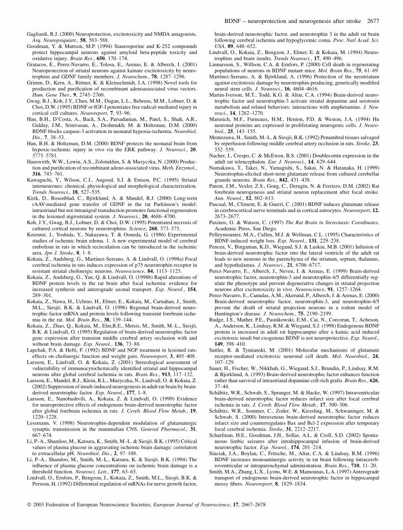
Fig. 1. (A) Photomicrograph showing brain-derived neurotrophic factor (BDNF)-immunoreactivity in a coronal section from a rat subjected to BDNF–recombinantadeno-associated viral (rAAV) transduction unilaterally in the substantia nigra. Note the high BDNF immunoreactivity distributed equally throughout the wholestriatum. (B) Levels of BDNF immunoreactivity in the striatum ipsi- and contralateral to GFP–rAAV (n¼ 2) and BDNF–rAAV (n¼ 4) transduction, as assessed usingoptical density measurements. (C–F) BDNF immunoreactivity in the substantia nigra contra- (C, E) and ipsilateral (D, F) to BDNF–rAAV transduction. (E, F)Enlargements of the areas marked by boxes in C and D, respectively. (G and H) Levels of BDNF protein as assessed by ELISA in (G) striatum and (H) substantia nigraof animals transduced with GFP–rAAV (n¼ 3) or BDNF–rAAV (n¼ 3). All values are mean� SEM. Scale bar, 500mm (C and D); 100mm (E and F).
� 2003 Federation of European Neuroscience Societies, European Journal of Neuroscience, 17, 2667–2678
2670 E. Gustafsson et al.

1997), available in the C.A.S.T.-GRID software (Olympus). Between
50 and 75 NPY-positive cells per striatum were analysed in every 20th
section through the striatum.
Quantification of BDNF immunoreactivity
Relative levels of BDNF immunoreactivity were measured in the
striatum ipsilateral to rAAV transduction as mean optical densities
by computerized image analysis using Image 1.52 software (Wayne
Rasband, NIMH) in three sections from each animal. Background
optical density, determined in a structure lacking BDNF immunor-
eactivity (corpus callosum), was subtracted from each measurement.
Statistical analysis
All comparisons were performed using one-way analysis of variance
(ANOVA), followed by Bonferroni–Dunn post-hoc test. Significance
was set at P< 0.05.
Results
Characterization of transgene expression and behaviouralconsequences
To study the efficacy of transduction in the substantia nigra and the
distribution of anterogradely transported BDNF in the ipsilateral
striatum, we used GFP and BDNF immunocytochemistry. It has been
observed that, within 1–2 weeks after intraparenchymal transduction
of rAAV-carrying genes, elevated levels of the corresponding proteins
are detected immunocytochemically with further increases over sub-
sequent weeks (Mandel & Kirik, unpublished observation). Significant
staining was detected in the substantia nigra and striatum of all
transduced animals (Fig. 1A, D and F), with a similar pattern and
intensity for both transgenes (data not shown). In the substantia nigra,
neurons of both the pars reticulata and compacta were intensely
stained, and the neuropil was moderately immunoreactive. Some
neurons in and around the injection tract were also GFP- and
BDNF-immunoreactive. The striatum ipsilateral to the BDNF–rAAV
nigral transduction was extensively stained with the BDNF antibody
(Fig. 1A). The strong immunoreactivity in the neuropil was equally
distributed in all directions with no detectable cellular staining. In
animals transduced with GFP–rAAV, BDNF-immunoreactivity was
not observed either in substantia nigra or striatum.
The semiquantitative analysis revealed about ninefold increase of
BDNF immunoreactivity in the ipsilateral striatum of BDNF–rAAV-
transduced rats, as compared with the striatum in GFP–rAAV-trans-
duced animals (Fig. 1B). Similarly, ELISA showed more than a 20-fold
increase of BDNF protein levels in the striatum ipsilateral to BDNF–
rAAV transduction (Fig. 1G). There were no pronounced changes of
BDNF protein levels in the contralateral striatum. The BDNF levels
were 190-fold higher in BDNF–rAAV- as compared with GFP–rAAV-
transduced nigra (Fig. 1H). There was also a 14-fold increase of BDNF
levels in the contralateral substantia nigra, possibly due to transport of
BDNF from transduced cells.
In order to determine whether the striatal BDNF which had been
transported anterogradely following the nigral rAAV transduction
was biologically active, the volume of NPY-positive neurons was
measured in BDNF- and GFP-transduced animals subjected to MCAO,
and in sham-treated rats injected with GFP–rAAV. The volume of
NPY-positive neurons was 2.2- and 1.8-fold larger in the BDNF–
rAAV-transduced animals subjected to MCAO as compared with GFP–
rAAV-transduced animals with or without MCAO, respectively
(Fig. 2).
Fig. 2. (A–C) Photomicrographs of NPY-immunoreactive striatal interneurons in animals transduced with (A, B) GFP–recombinant adeno-associated virus (rAAV)or (C) brain-derived neurotrophic factor (BDNF)–rAAV and subjected to (A) sham treatment or (B and C) middle cerebral artery occlusion (MCAO). (C) Note thehypertrophy of NPY-positive neurons in animals transduced with BDNF–rAAV. (D) Volume of NPY-positive striatal interneurons, as measured using the rotatormethod, in animals transduced with GFP–rAAV (n¼ 14) or BDNF–rAAV (n¼ 6) and subjected to sham treatment (n¼ 6) or MCAO (n¼ 14). All values aremean�SEM. �P< 0.05, one-way ANOVA followed by Bonferroni–Dunn post-hoc test. Scale bar, 100mm (C).
� 2003 Federation of European Neuroscience Societies, European Journal of Neuroscience, 17, 2667–2678
BDNF – neuroprotection and neurogenesis after stroke 2671

The body weight of rats injected with BDNF–rAAV was lower than
that of GFP–rAAV animals at 3 weeks after transduction, but was
relatively stable thereafter (data not shown). Animals transduced with
BDNF–rAAV also exhibited abnormalities in body posture and move-
ments. The head and frontal part of the body were constantly tilted to
the right (transduced) side with periodic choreiform movements of the
left forelimb and head. Most BDNF–rAAV-transduced rats exhibited
spontaneous, so-called ‘barrel’ rotation along their long axis. The rats
also had problems with balance when rearing, and showed repeated
head nodding movements. They were hyperactive and rotated sponta-
neously towards the transduced side, which was not observed in GFP–
rAAV animals (Fig. 3A). The MCAO did not alter this spontaneous
rotation (Fig. 3B). Despite the behavioural abnormalities, the BDNF–
rAAV-transduced rats were able to drink, eat and groom themselves.
We hypothesized that seizure development had contributed to the
observed behavioural abnormalities, and therefore EEG was recorded
from the frontal cortex and striatum in four animals at 4 weeks after
viral transduction. However, no seizure activity was detected and the
rats did not loose consciousness, were responsive to touch and hand-
ling, and exhibited exploratory behaviour in the open-field. Moreover,
the clonic movements of the forelimb were observed only contral-
aterally to the side of BDNF–rAAV transduction.
We also investigated whether the motor abnormalities could be due
to effects of BDNF on the function of nigrostriatal neurons (e.g. striatal
dopamine release). At 2 h after injection of the dopamine and nora-
drenaline synthesis inhibitor, AMPT, movements were slower in both
GFP–rAAV- and BDNF–rAAV-transduced rats, and between 4 and 8 h,
the hypokinetic effect of the drug became even more pronounced.
However, the tilted posture and barrel rotation were still observed in
the BDNF–rAAV-transduced animals.
Effect of BDNF gene transfer on survival of striatal neuronsafter MCAO
The stroke-induced neuronal damage in the striatum was first assessed
by combining immunocytochemical staining with the neuron-specific
marker NeuN and stereological procedures (Andsberg et al., 1998;
Larsson et al., 1999; Larsson et al., 2001; Andsberg et al., 2002).
Because more than 90% of striatal neurons are medium-sized spiny
projection neurons (Kawaguchi et al., 1995), the loss of NeuN-positive
cells after MCAO primarily reflects the death of these cells. The
MCAO caused 64% and 71% reduction of the number of NeuN-
positive cells in animals transduced with GFP–rAAV and BDNF–
rAAV, respectively (Fig. 4A). The lesion was mainly localized to the
dorsolateral striatum (Fig. 4C and D) (Memezawa et al., 1992;
Andsberg et al., 1998). There was no significant difference in the
survival of NeuN-positive cells between GFP–rAAV- and BDNF–
rAAV-transduced rats.
In addition to medium-sized spiny projection neurons, the striatum
contains three major classes of interneurons which can be distin-
Fig. 4. (A) Total number of NeuN-stained cells in the striatum quantified using stereological procedures in animals transduced with GFP–recombinant adeno-associated virus (rAAV, n¼ 13) or brain-derived neurotrophic factor (BDNF)–rAAV (n¼ 6) and subjected to sham treatment (n¼ 6) or 30 min of middle cerebralartery occlusion (MCAO, n¼ 13). (B–D) NeuN-stained coronal sections from the central part of the striatum in representative animals from the different groups. TheMCAO groups showed similar, uniform loss of NeuN-stained cells in the ipsilateral dorsolateral striatum (�). Values are means�SEM. #P< 0.05, one-way ANOVA
followed by Bonferroni–Dunn post-hoc test.
Fig. 3. Spontaneous rotation as assessed at 4 weeks after intranigral injection ofGFP–recombinant adeno-associated viral (rAAV, n¼ 8) and brain-derivedneurotrophic factor (BDNF)–rAAV (n¼ 7) vectors [A, before middle cerebralartery occlusion (MCAO)] and at 1 week after MCAO (B). ‘–’ means rotation tothe left and ‘ þ’ means rotation to the right. Note the pronounced rotationalasymmetry towards the injected (right) side in BDNF–rAAV-transduced ani-mals, which is maintained after MCAO.
� 2003 Federation of European Neuroscience Societies, European Journal of Neuroscience, 17, 2667–2678
2672 E. Gustafsson et al.

guished on basis of their immunoreactivity to ChAT, parvalbumin or
NPY (Kawaguchi et al., 1995). We quantified the ischaemic damage to
these classes of interneurons. The MCAO caused a significant (27%)
loss of parvalbumin-expressing interneurons in GFP–rAAV-trans-
duced animals (Fig. 5A). This loss was further exaggerated (to
65%) in BDNF–rAAV-transduced rats. In agreement with previous
reports (Chesselet et al., 1990; Uemura et al., 1990; Andsberg et al.,
1998; Kokaia et al., 1998a; Andsberg et al., 2002), MCAO caused no
significant reduction of the number of NPY- or ChAT-positive striatal
interneurons in GFP–rAAV-transduced animals. However, in the
BDNF-rAAV transduced animals subjected to MCAO, there was a
40% and 50% loss of NPY- and ChAT-positive interneurons, respec-
tively (Fig. 5B and C).
Effect of BDNF gene transfer on stroke-induced striatalneurogenesis
We also assessed whether the elevated BDNF levels could influence
the initial phase of stroke-induced striatal neurogenesis (Arvidsson
et al., 2002). First, the number of DCX-positive cells was counted in
the striatum ipsilateral to MCAO (Fig. 6). DCX is a marker of
migrating neuroblasts, and in the adult brain is expressed mostly in
the SVZ and rostral migratory stream with single cells detectable in the
striatum (Nacher et al., 2001). Stroke triggers the migration of DCX-
positive cells from the SVZ to the damaged striatum (Arvidsson et al.,
2002; Parent et al., 2002). We observed here that at 2 weeks after the
stroke, the number of DCX-positive cells in the GFP–rAAV-transduced
animals had increased to more than 231% of that in sham-treated
animals. Transduction with BDNF–rAAV led to a further increase
in the number of DCX-positive striatal cells (to 522% and 233% of that
in GFP–rAAV–sham and GFP–rAAV–MCAO groups, respectively,
Fig. 6).
To confirm the neuronal phenotype of the new striatal cells, all
animals were injected daily with BrdU for 1 week following MCAO,
and were killed 1 week thereafter. BrdU is a thymidine analogue,
which is incorporated into DNA during cell division. We double-
immunostained sections with antibodies against BrdU and Hu, an early
neuronal marker, which starts to be expressed in neurons soon after
differentiation and remains in mature cells (Marusich et al., 1994). The
BrdU–Hu-double-labelled neurons (Fig. 7A–F) were mainly distrib-
uted close to the lateral ventricle, the corpus callosum and the anterior
commisure. Many cells were also detected in the border zone between
the damaged and the spared medial parts of the striatum. Quantifica-
tion revealed increased numbers of BrdU–Hu-double-labelled striatal
cells in the GFP–rAAV-transduced animals subjected to MCAO as
compared with sham-treated rats (Fig. 7G). In the BDNF–rAAV-
transduced animals, the number of BrdU–Hu-double-labelled striatal
cells was further increased to 253% and 726% as compared with GFP–
rAAV-transduced animals with and without MCAO, respectively.
In order to characterize the early phenotype of the new striatal
neurons, we quantified the number of BrdU–Meis2-positive cells.
Meis2 is a transcription factor which is expressed at high levels by
striatal precursors during embryonic development, and also in the adult
striatum (Toresson et al., 2000). Similar to BrdU–Hu-positive cells,
BDNF–rAAV transduction significantly increased the number of
BrdU–Meis2-double-labelled striatal neurons after stroke (Fig. 7H).
Discussion
The present study shows that injection of rAAV carrying the BDNF
gene into the rat substantia nigra leads to increased production of
BDNF by nigral neurons, and anterograde transport and release of
high amounts of this neurotrophic factor in the ipsilateral striatum.
Continuous supply of BDNF via this route does not protect striatal
projection neurons from stroke-induced damage. On the contrary,
anterogradely delivered BDNF exaggerates the loss of parvalbumin-
positive interneurons and causes the death of resistant NPY- and ChAT-
positive striatal interneurons following stroke. Concomitantly,
Fig. 5. Number of (A) parvalbumin-, (B) neuropeptide Y (NPY)- and (C)choline acetyltransferase (ChAT)- immunoreactive neurons in the striatumipsilateral to sham surgery (n¼ 6) or 30 min of middle cerebral artery occlusion(MCAO, n¼ 13) in animals subjected to intranigral GFP–recombinant adeno-associated viral (rAAV, n¼ 13) or BDNF–rAAV (n¼ 6) transduction. Valuesare mean� SEM. �P< 0.05, one-way ANOVA, followed by Bonferroni–Dunnpost-hoc test.
� 2003 Federation of European Neuroscience Societies, European Journal of Neuroscience, 17, 2667–2678
BDNF – neuroprotection and neurogenesis after stroke 2673

FIG. 6.
FIG. 7.
� 2003 Federation of European Neuroscience Societies, European Journal of Neuroscience, 17, 2667–2678
2674 E. Gustafsson et al.

increased levels of intrastriatal BDNF promote the initial phase of
stroke-induced neurogenesis in the striatum.
Under normal conditions, approximately 14% of striatal BDNF has
been transported anterogradely from the substantia nigra (Altar et al.,
1997). Our immunocytochemical and ELISA data clearly document
that after transduction of nigral cells with BDNF–rAAV, large amounts
of BDNF are similarly transported in nigrostriatal axons to the
striatum. By using this natural route of BDNF delivery, homogeneous
distribution of high levels of this neurotrophic factor is obtained
throughout the striatum. The observed hypertrophy of NPY-positive
neurons indicates that sufficient levels of BDNF are released in
biologically active form. Moreover, the BDNF–rAAV-transduced ani-
mals stopped gaining weight, similar to that which has been reported
previously with intracerebroventricular (Lapchak & Hefti, 1992; Sauer
et al., 1993; Pelleymounter et al., 1995; Siuciak et al., 1996), suprani-
gral (Altar et al., 1992; Martin-Iverson et al., 1994) and striatal (Altar
et al., 1992) delivery of BDNF. These animals also exhibited abnormal
motor behaviour, including choreiform movements, spontaneous cir-
cling and barrel rotation. Intracerebral infusion of BDNF can lead to
behavioural seizures (Scharfman et al., 2002). However, the EEG
recordings and administration of a catecholamine synthesis inhibitor
provided no evidence that seizures or increased dopamine release,
respectively, were responsible for the motor abnormalities. We
hypothesize that the continuous delivery of BDNF leads to increased
striatal glutamate levels, which induce the behavioural changes. In
agreement, intrastriatal injection of excitatory amino acids gives rise to
increased locomotor activity, choreiform movements, contralateral
turning behaviour and ipsilateral barrel rotation (Toth & Lajtha,
1989; Vecsei & Beal, 1991).
Several studies have demonstrated that delivery of exogeneous
BDNF counteracts MCAO-induced damage, predominantly in the
cerebral cortex (Schabitz et al., 1997; Yamashita et al., 1997; Schabitz
et al., 2000; Yanamoto et al., 2000; Zhang & Pardridge, 2001). BDNF
also protects striatal neurons from excitotoxic lesions (Martınez-
Serrano & Bjorklund, 1996; Bemelmans et al., 1999; Perez-Navarro
et al., 1999; Perez-Navarro et al., 2000; Gratacos et al., 2001). In
accordance, we have previously observed that direct intrastriatal
injection of BDNF–rAAV vector at 4–5 weeks prior to the ischaemic
insult used here leads to a small increase in the number of surviving
projection neurons in the dorsolateral striatum, with more pronounced
sparing of parvalbumin-containing interneurons (Andsberg et al.,
2002). Interestingly, the survival of cholinergic interneurons following
global forebrain ischaemia is dependent on endogenous BDNF (Lars-
son et al., 1999). Scavenging of endogenous BDNF by intraventricular
injection of TrkB-Fc fusion protein causes approximately 50% reduc-
tion in the number of ChAT-positive interneurons in the dorsolateral
striatum following a 30-min insult.
In contrast to these observations, we found here that anterograde
delivery of BDNF to the striatum increased the ischaemic damage to
several neuronal populations. BDNF aggravated the stroke-induced
loss of parvalbumin-positive cells and also caused degeneration of
NPY- and ChAT-positive interneurons, which was not seen in GFP–
rAAV-injected animals subjected to MCAO. There was no significant
effect of BDNF on the survival of striatal projection neurons. The
paradoxically increased vulnerability of striatal interneurons, induced
by the continuous anterograde delivery of BDNF, probably illustrates
that the level and duration and route of delivery determine the effect of
this neurotrophin on neuronal survival. In accordance, the immuno-
cytochemical analysis revealed that the BDNF levels were much
higher and equally distributed throughout the whole striatum following
intranigral as compared with the direct intrastriatal injection of
BDNF–rAAV, which gave rise to patchy appearance of BDNF immu-
noreactivity close to the injection site (Andsberg et al., 2002). Simi-
larly, glial cell line-derived neurotrophic factor (GDNF)–rAAV
transduction supports much higher levels of GDNF protein as com-
pared with striatal transduction using the same vector (Kirik et al.,
2000). We propose that low and moderate levels of BDNF are
neuroprotective. However, when BDNF is delivered continuously in
high amounts, exaggeration of excitotoxic mechanisms overrides the
neuroprotective effect of BDNF and increases the vulnerability of
striatal cells to ischaemic damage. Stroke leads to depolarization of
corticostriatal neurons and glutamate release, giving rise to excitotoxic
damage to striatal neurons (Gagliardi, 2000; Sattler & Tymianski,
2001). It is tempting to speculate that the increased vulnerability of
striatal neurons to ischaemic injury, mediated by the high, homoge-
nously distributed levels of BDNF, is due to the ability of this
neurotrophic factor to enhance excitatory glutamatergic synaptic
transmission (for review, see Lessmann, 1998). BDNF also increases
glutamate release in cortical neurons (Akaneya et al., 1997; Car-
mignoto et al., 1997; Takei et al., 1997; Numakawa et al., 1999) and
astrocytes (Pascual et al., 2001).
Our finding that BDNF can enhance neuronal vulnerability is
consistent with reports demonstrating that BDNF increases oxidative
stress-dependent cell death (Gwag et al., 1995) and potentiates the
necrosis induced by oxygen–glucose deprivation or N-methyl-D-aspar-
tate (NMDA) (Koh et al., 1995) in cultured cortical neurons, and also
that intrahippocampal infusion of BDNF exacerbates the loss of CA3
pyramidal neurons induced by kainic acid (Rudge et al., 1998).
Interestingly, the high-affinity neurotrophin receptor tyrosine-kinase
inhibitor K252a enhances the survival of hippocampal neurons after
exposure to high levels of free radicals (Goodman & Mattson, 1994).
Moreover, it was recently reported that the lack of another neurotro-
phin, NT-3, in conditional knockout mice leads to reduced infarct
volume after MCAO, whereas addition of NT-3 exaggerates the death
of cultured cortical neurons after oxygen–glucose deprivation (Bates
et al., 2002).
We have previously shown that 2 h of MCAO leads to neurogenesis
in the SVZ, and that the new neurons migrate to the stroke-damaged
part of the striatum, where they express morphological markers of
striatal projection neurons (Arvidsson et al., 2002). The present data
Fig. 6. (A–D) Photomicrographs showing DCX-immunoreactivity in coronal sections through the dorsomedial striatum (A) contra- and (B) ipsilateral to the insult at2 weeks after middle cerebral artery occlusion (MCAO) in animals injected with brain-derived neurotrophic factor (BDNF)–recombinant adeno-associated virus(rAAV). Note the large number of DCX-positive cells in the ipsilateral striatum (B; enlarged in C and D); distributed in a density gradient from the SVZ, bordereddorsally by the corpus callosum and medially by the lateral ventricle (LV). (E) Number of DCX-positive cells in the striatum ipsilateral to sham surgery (n¼ 6) or30 min of MCAO (n¼ 13) in animals injected with GFP–rAAV (n¼ 13) or BDNF–rAAV (n¼ 6) in the substantia nigra. Values are mean�SEM. �P< 0.05, one-wayANOVA, followed by Bonferroni–Dunn post-hoc test. Scale bar, 200mm (A and B); 30 mm (C and D).
Fig. 7. (A–C) Consecutive 1-mm confocal images in z-dimension showing (A) Hu or (B) 5-bromo-20-deoxyuridine-50-monophosphate (BrdU) immunoreactivityseparately or (C) as merged images. (D–F) Confocal 3D reconstruction of neurons from the striatum ipsilateral to middle cerebral artery occlusion (MCAO) showing(D) Hu and (E) BrdU immunoreactivity separately or (F) as a merged image. Reconstructed orthogonal images are presented as viewed in the x–z (top) and y–z (right)planes. Arrows indicate double-labelled cells. (G, H) Number of cells double-labelled with BrdU and (G) Hu or (H) Meis2 in the striatum ipsilateral to sham surgery(n¼ 6) or 30 min of MCAO (n¼ 13) in animals injected with GFP–recombinant adeno-associated virus (rAAV, n¼ 13) or brain-derived neurotrophic factor (BDNF)–rAAV (n¼ 6). All values are mean�SEM. �P< 0.05, one-way ANOVA, followed by Bonferroni–Dunn post-hoc test. Scale bar, 30mm (A–C); 20 mm (D–F).
� 2003 Federation of European Neuroscience Societies, European Journal of Neuroscience, 17, 2667–2678
BDNF – neuroprotection and neurogenesis after stroke 2675

indicate that this neurogenic response is promoted by long-term BDNF
delivery. In the BDNF-exposed ischaemic striatum, there was a marked
increase in the number of DCX-immunoreactive migrating neuro-
blasts, and cells double-labelled with BrdU and early neuronal (Hu)
and striatal neuronal (Meis2) markers. The BDNF-induced promotion
of striatal neurogenesis following stroke is in agreement with previous
observations that BDNF increases the number of neurons in embryonic
striatal cultures (Ahmed et al., 1995), and that new neurons are
recruited to the striatum after intraventricular injection of adeno-virus
carrying the BDNF gene (Benraiss et al., 2001) or intraventricular
administration of BDNF (Pencea et al., 2001). Thus, the increase in the
number of new striatal neurons observed here could be explained either
by this direct effect of BDNF on striatal neurogenesis or by BDNF
modulating the stroke-induced generation of new neurons. The mode
of action of BDNF during striatal neurogenesis remains to be explored.
It has been shown that the majority of new neurons in the dentate gyrus
and the striatum die within the first weeks after ischaemic and epileptic
insults (Ekdahl et al., 2001; Arvidsson et al., 2002). At least in the
dentate gyrus, this seems to occur through a caspase-mediated apop-
totic mechanism (Ekdahl et al., 2001). One possibility could be that
BDNF counteracts the degeneration of the newly formed neurons. In
support of this hypothesis, BDNF knockout mice show increased
apoptotic death of precursor cells in the dentate gyrus and the SVZ
(Linnarsson et al., 2000). Furthermore, BDNF counteracts caspase-3
activation-dependent apoptotic cell death in the ischaemia–hypoxia
model (Han et al., 2000).
In conclusion, we have found that transduction of nigrostriatal
neurons with a BDNF–rAAV vector provides an efficient route for
long-term delivery of this neurotrophic factor to the striatum. However,
high striatal levels of BDNF protein as generated here produce motor
abnormalities. In contrast to previously described neuroprotective
effects of BDNF, the anterogradely delivered, high BDNF levels
aggravated the ischaemic damage in the striatum, probably by enhan-
cing glutamate-evoked excitotoxicity. In the same animals, BDNF
promoted the initial phase of stroke-induced striatal neurogenesis. It is
conceivable that anterograde delivery of BDNF via nigral afferents
will be a useful tool to explore further the role of BDNF for the balance
between neuronal death and neurogenesis in the striatum after brain
insults. Our data show that the actions of BDNF on neuronal survival
and neurogenesis are complex, and underscore the notion that many
aspects of BDNF function have to be taken into account when
considering potential therapeutic applications of this neurotrophin.
Acknowledgements
We thank Monica Lundahl for excellent technical assistance. This work wassupported by the Swedish Research Council, Swedish Gene Therapy Program,Kock, Crafoord, Elsa and Thorsten Segerfalk Foundations, the Swedish StrokeFoundations, the Swedish Association of Neurologically Disabled, and theSwedish Society for Medical Research. Vector production was supported byNIH RO1 NS36302.
Abbreviations
AMPT, a-methyl-para-tyrosine; ANOVA, analysis of variance; BDNF, brain-derived neurotrophic factor; BrdU, 5-bromo-20-deoxyuridine-50-monopho-sphate; BS, Block & Sample; ChAT, choline acetyltransferase; DCX,doublecortin; EEG, electroencephalogram; GDNF, glial cell line-derived neuro-trophic factor; GFP, green fluorescent protein; i.p., intraperitoneally; KPBS,potassium phosphate-buffered saline; MCA, middle cerebral artery; MCAO,middle cerebral artery occlusion; NeuN, neuronal-specific antigen; NMDA, N-methyl-D-aspartate; NPY, neuropeptide Y; PB, phosphate buffer; pCBA, plasmidcytomegalovirus promoter with chicken R-actin intervening, sequence; rAAV,recombinant adeno-associated virus; SVZ, subventricular zone.
References
Ahmed, S., Reynolds, B.A. & Weiss, S. (1995) BDNF enhances the differ-entiation but not the survival of CNS stem cell-derived neuronal precursors. J.Neurosci., 15, 5765–5778.
Akaneya, Y., Tsumoto, T., Kinoshita, S. & Hatanaka, H. (1997) Brain-derivedneurotrophic factor enhances long-term potentiation in rat visual cortex. J.Neurosci., 17, 6707–6716.
Altar, C.A., Boylan, C.B., Jackson, C., Hershenson, S., Miller, J., Wiegand, S.J.,Lindsay, R.M. & Hyman, C. (1992) Brain-derived neurotrophic factoraugments rotational behavior and nigrostriatal dopamine turnover in vivo.Proc. Natl. Acad. Sci. USA, 89, 11,347–11,351.
Altar, C.A., Cai, N., Bliven, T., Juhasz, M., Conner, J.M., Acheson, A.L.,Lindsay, R.M. & Wiegand, S.J. (1997) Anterograde transport of brain-derived neurotrophic factor and its role in the brain. Nature, 389, 856–860.
Andsberg, G., Kokaia, Z., Bjorklund, A., Lindvall, O. & Martınez-Serrano, A.(1998) Amelioration of ischaemia-induced neuronal death in the rat striatumby NGF-secreting neural stem cells. Eur. J. Neurosci., 10, 2026–2036.
Andsberg, G., Kokaia, Z., Klein, R.L., Muzyczka, N., Lindvall, O. & Mandel,R.J. (2002) Neuropathological and behavioral consequences of adeno-asso-ciated viral vector-mediated continuous intrastriatal neurotrophin delivery ina focal ischemia model in rats. Neurobiol. Dis., 9, 187–204.
Arai, S., Kinouchi, H., Akabane, A., Owada, Y., Kamii, H., Kawase, M. &Yoshimoto, T. (1996) Induction of brain-derived neurotrophic factor (BDNF)and the receptor trk B mRNA following middle cerebral artery occlusion inrat. Neurosci. Lett., 211, 57–60.
Arvidsson, A., Collin, T., Kirik, D., Kokaia, Z. & Lindvall, O. (2002) Neuronalreplacement from endogenous precursors in the adult brain after stroke.Nature Med., 8, 963–970.
von Bartheld, C.S., Byers, M.R., Williams, R. & Bothwell, M. (1996) Ante-rograde transport of neurotrophins and axodendritic transfer in the devel-oping visual system. Nature, 379, 830–833.
Bates, B., Hirt, L., Thomas, S.S., Akbarian, S., Le, D., Amin-Hanjani, S.,Whalen, M., Jaenisch, R. & Moskowitz, M.A. (2002) Neurotrophin-3promotes cell death induced in cerebral ischemia, oxygen-glucose depriva-tion, and oxidative stress: possible involvement of oxygen free radicals.Neurobiol. Dis., 9, 24–37.
Beck, T., Lindholm, D., Castren, E. & Wree, A. (1994) Brain-derived neuro-trophic factor protects against ischemic cell damage in rat hippocampus. J.Cereb. Blood Flow Metab., 14, 689–692.
Bederson, J.B., Pitts, L.H., Tsuji, M., Nishimura, M.C., Davis, R.L. & Bart-kowski, H. (1986) Rat middle cerebral artery occlusion: evaluation of themodel and development of a neurologic examination. Stroke, 17, 472–476.
Bemelmans, A.P., Horellou, P., Pradier, L., Brunet, I., Colin, P. & Mallet, J.(1999) Brain-derived neurotrophic factor-mediated protection of striatalneurons in an excitotoxic rat model of Huntington’s disease, as demonstratedby adenoviral gene transfer. Hum. Gene Ther., 10, 2987–2997.
Benraiss, A., Chmielnicki, E., Lerner, K., Roh, D. & Goldman, S.A. (2001)Adenoviral brain-derived neurotrophic factor induces both neostriatal andolfactory neuronal recruitment from endogenous progenitor cells in the adultforebrain. J. Neurosci., 21, 6718–6731.
Carmignoto, G., Pizzorusso, T., Tia, S. & Vicini, S. (1997) Brain-derivedneurotrophic factor and nerve growth factor potentiate excitatory synaptictransmission in the rat visual cortex. J. Physiol. (Lond.), 498, 153–164.
Cheng, B. & Mattson, M.P. (1994) NT-3 and BDNF protect CNS neuronsagainst metabolic/excitotoxic insults. Brain Res., 640, 56–67.
Chesselet, M.F., Gonzales, C., Lin, C.S., Polsky, K. & Jin, B.K. (1990) Ischemicdamage in the striatum of adult gerbils: relative sparing of somatostatinergicand cholinergic interneurons contrasts with loss of efferent neurons. Exp.Neurol., 110, 209–218.
Croll, S.D., Wiegand, S.J., Anderson, K.D., Lindsay, R.M. & Nawa, H. (1994)Regulation of neuropeptides in adult rat forebrain by the neurotrophinsBDNF and NGF. Eur. J. Neurosci., 6, 1343–1353.
Davies, A.M. (1994) The role of neurotrophins in the developing nervoussystem. J. Neurobiol., 25, 1334–1348.
Ekdahl, C.T., Mohapel, P., Elmer, E. & Lindvall, O. (2001) Caspase inhibitorsincrease short-term survival of progenitor-cell progeny in the adult rat dentategyrus following status epilepticus. Eur. J. Neurosci., 14, 937–945.
Endres, M., Fan, G., Hirt, L., Fujii, M., Matsushita, K., Liu, X., Jaenisch, R. &Moskowitz, M.A. (2000) Ischemic brain damage in mice after selectivelymodifying BDNF or NT4 gene expression. J. Cereb. Blood Flow Metab., 20,139–144.
Ferrer, I., Ballabriga, J., Marti, E., Perez, E., Alberch, J. & Arenas, E. (1998)BDNF up-regulates TrkB protein and prevents the death of CA1 neuronsfollowing transient forebrain ischemia. Brain Pathol., 8, 253–261.
� 2003 Federation of European Neuroscience Societies, European Journal of Neuroscience, 17, 2667–2678
2676 E. Gustafsson et al.
Gagliardi, R.J. (2000) Neuroprotection, excitotoxicity and NMDA antagonists.Arq. Neuropsiquiatr., 58, 583–588.
Goodman, Y. & Mattson, M.P. (1994) Staurosporine and K-252 compoundsprotect hippocampal neurons against amyloid beta-peptide toxicity andoxidative injury. Brain Res., 650, 170–174.
Gratacos, E., Perez-Navarro, E., Tolosa, E., Arenas, E. & Alberch, J. (2001)Neuroprotection of striatal neurons against kainate excitotoxicity by neuro-trophins and GDNF family members. J. Neurochem., 78, 1287–1296.
Grimm, D., Kern, A., Rittner, K. & Kleinschmidt, J.A. (1998) Novel tools forproduction and purification of recombinant adenoassociated virus vectors.Hum. Gene Ther., 9, 2745–2760.
Gwag, B.J., Koh, J.Y., Chen, M.M., Dugan, L.L., Behrens, M.M., Lobner, D. &Choi, D.W. (1995) BDNF or IGF-I potentiates free radical-mediated injury incortical cell cultures. Neuroreport, 7, 93–96.
Han, B.H., D’Costa, A., Back, S.A., Parsadanian, M., Patel, S., Shah, A.R.,Gidday, J.M., Srinivasan, A., Deshmukh, M. & Holtzman, D.M. (2000)BDNF blocks caspase-3 activation in neonatal hypoxia-ischemia. Neurobiol.Dis., 7, 38–53.
Han, B.H. & Holtzman, D.M. (2000) BDNF protects the neonatal brain fromhypoxic-ischemic injury in vivo via the ERK pathway. J. Neurosci., 20,5775–5781.
Hauswirth, W.W., Lewin, A.S., Zolotukhin, S. & Muzyczka, N. (2000) Produc-tion and purification of recombinant adeno-associated virus. Meth. Enzymol.,316, 743–761.
Kawaguchi, Y., Wilson, C.J., Augood, S.J. & Emson, P.C. (1995) Striatalinterneurones: chemical, physiological and morphological characterization.Trends Neurosci., 18, 527–535.
Kirik, D., Rosenblad, C., Bjorklund, A. & Mandel, R.J. (2000) Long-termrAAV-mediated gene transfer of GDNF in the rat Parkinson’s model:intrastriatal but not intranigral transduction promotes functional regenerationin the lesioned nigrostriatal system. J. Neurosci., 20, 4686–4700.
Koh, J.Y., Gwag, B.J., Lobner, D. & Choi, D.W. (1995) Potentiated necrosis ofcultured cortical neurons by neurotrophins. Science, 268, 573–575.
Koizumi, J., Yoshida, Y., Nakazawa, T. & Ooneda, G. (1986) Experimentalstudies of ischemic brain edema. 1. A new experimental model of cerebralembolism in rats in which recirculation can be introduced in the ischemicarea. Jpn J. Stroke, 8, 1–8.
Kokaia, Z., Andsberg, G., Martınez-Serrano, A. & Lindvall, O. (1998a) Focalcerebral ischemia in rats induces expression of p75 neurotrophin receptor inresistant striatal cholinergic neurons. Neuroscience, 84, 1113–1125.
Kokaia, Z., Andsberg, G., Yan, Q. & Lindvall, O. (1998b) Rapid alterations ofBDNF protein levels in the rat brain after focal ischemia: evidence forincreased synthesis and anterograde axonal transport. Exp. Neurol., 154,289–301.
Kokaia, Z., Nawa, H., Uchino, H., Elmer, E., Kokaia, M., Carnahan, J., Smith,M.L., Siesjo, B.K. & Lindvall, O. (1996) Regional brain-derived neuro-trophic factor mRNA and protein levels following transient forebrain ische-mia in the rat. Mol. Brain Res., 38, 139–144.
Kokaia, Z., Zhao, Q., Kokaia, M., Elm,R.E., Metsis, M., Smith, M.-L., Siesjo,B.K. & Lindvall, O. (1995) Regulation of brain-derived neurotrophic factorgene expression after transient middle cerebral artery occlusion with andwithout brain damage. Exp. Neurol., 136, 73–88.
Lapchak, P.A. & Hefti, F. (1992) BDNF and NGF treatment in lesioned rats:effects on cholinergic function and weight gain. Neuroreport, 3, 405–408.
Larsson, E., Lindvall, O. & Kokaia, Z. (2001) Stereological assessment ofvulnerability of immunocytochemically identified striatal and hippocampalneurons after global cerebral ischemia in rats. Brain Res., 913, 117–132.
Larsson, E., Mandel, R.J., Klein, R.L., Muzyczka, N., Lindvall, O. & Kokaia, Z.(2002) Suppression of insult-induced neurogenesis in adult rat brain by brain-derived neurotrophic factor. Exp. Neurol., 177, 1–8.
Larsson, E., Nanobashvili, A., Kokaia, Z. & Lindvall, O. (1999) Evidencefor neuroprotective effects of endogenous brain-derived neurotrophic factorafter global forebrain ischemia in rats. J. Cereb. Blood Flow Metab., 19,1220–1228.
Lessmann, V. (1998) Neurotrophin-dependent modulation of glutamatergicsynaptic transmission in the mammalian CNS. General Pharmacol., 31,667–674.
Li, P.-A., Shamloo, M., Katsura, K., Smith, M.-L. & Siesjo, B.K. (1995) Criticalvalues of plasma glucose in aggravating ischemic brain damage: correlationto extracellular pH. Neurobiol. Dis., 2, 97–108.
Li, P.-A., Shamloo, M., Smith, M.-L., Katsura, K. & Siesjo, B.K. (1994) Theinfluence of plasma glucose concentrations on ischemic brain damage is athreshold function. Neurosci. Lett., 177, 63–65.
Lindvall, O., Ernfors, P., Bengzon, J., Kokaia, Z., Smith, M.L., Siesjo, B.K. &Persson, H. (1992) Differential regulation of mRNAs for nerve growth factor,
brain-derived neurotrophic factor, and neurotrophin 3 in the adult rat brainfollowing cerebral ischemia and hypoglycemic coma. Proc. Natl Acad. Sci.USA, 89, 648–652.
Lindvall, O., Kokaia, Z., Bengzon, J., Elmer, E. & Kokaia, M. (1994) Neuro-trophins and brain insults. Trends Neurosci., 17, 490–496.
Linnarsson, S., Willson, C.A. & Ernfors, P. (2000) Cell death in regeneratingpopulations of neurons in BDNF mutant mice. Mol. Brain Res., 75, 61–69.
Martınez-Serrano, A. & Bjorklund, A. (1996) Protection of the neostriatumagainst excitotoxic damage by neurotrophin-producing, genetically modifiedneural stem cells. J. Neurosci., 16, 4604–4616.
Martin-Iverson, M.T., Todd, K.G. & Altar, C.A. (1994) Brain-derived neuro-trophic factor and neurotrophin-3 activate striatal dopamine and serotoninmetabolism and related behaviors: interactions with amphetamine. J. Neu-rosci., 14, 1262–1270.
Marusich, M.F., Furneaux, H.M., Henion, P.D. & Weston, J.A. (1994) Huneuronal proteins are expressed in proliferating neurogenic cells. J. Neuro-biol., 25, 143–155.
Memezawa, H., Smith, M.-L. & Siesjo, B.K. (1992) Penumbral tissues salvagedby reperfusion following middle cerebral artery occlusion in rats. Stroke, 23,552–559.
Nacher, J., Crespo, C. & McEwen, B.S. (2001) Doublecortin expression in theadult rat telencephalon. Eur. J. Neurosci., 14, 629–644.
Numakawa, T., Takei, N., Yamagishi, S., Sakai, N. & Hatanaka, H. (1999)Neurotrophin-elicited short-term glutamate release from cultured cerebellargranule neurons. Brain Res., 842, 431–438.
Parent, J.M., Vexler, Z.S., Gong, C., Derugin, N. & Ferriero, D.M. (2002) Ratforebrain neurogenesis and striatal neuron replacement after focal stroke.Ann. Neurol., 52, 802–813.
Pascual, M., Climent, E. & Guerri, C. (2001) BDNF induces glutamate releasein cerebrocortical nerve terminals and in cortical astrocytes. Neuroreport, 12,2673–2677.
Paxinos, G. & Watson, C. (1997) The Rat Brain in Stereotaxic Coordinates.Academic Press, San Diego.
Pelleymounter, M.A., Cullen, M.J. & Wellman, C.L. (1995) Characteristics ofBDNF-induced weight loss. Exp. Neurol., 131, 229–238.
Pencea, V., Bingaman, K.D., Wiegand, S.J. & Luskin, M.B. (2001) Infusion ofbrain-derived neurotrophic factor into the lateral ventricle of the adult ratleads to new neurons in the parenchyma of the striatum, septum, thalamus,and hypothalamus. J. Neurosci., 21, 6706–6717.
Perez-Navarro, E., Alberch, J., Neveu, I. & Arenas, E. (1999) Brain-derivedneurotrophic factor, neurotrophin-3 and neurotrophin-4/5 differentially reg-ulate the phenotype and prevent degenerative changes in striatal projectionneurons after excitotoxicity in vivo. Neuroscience, 91, 1257–1264.
Perez-Navarro, E., Canudas, A.M., Akerund, P., Alberch, J. & Arenas, E. (2000)Brain-derived neurotrophic factor, neurotrophin-3, and neurotrophin-4/5prevent the death of striatal projection neurons in a rodent model ofHuntington’s disease. J. Neurochem., 75, 2190–2199.
Rudge, J.S., Mather, P.E., Pasnikowski, E.M., Cai, N., Corcoran, T., Acheson,A., Anderson, K., Lindsay, R.M. & Wiegand, S.J. (1998) Endogenous BDNFprotein is increased in adult rat hippocampus after a kainic acid inducedexcitotoxic insult but exogenous BDNF is not neuroprotective. Exp. Neurol.,149, 398–410.
Sattler, R. & Tymianski, M. (2001) Molecular mechanisms of glutamatereceptor-mediated excitotoxic neuronal cell death. Mol. Neurobiol., 24,107–129.
Sauer, H., Fischer, W., Nikkhah, G., Wiegand, S.J., Brundin, P., Lindsay, R.M.& Bjorklund, A. (1993) Brain-derived neurotrophic factor enhances functionrather than survival of intrastriatal dopamine cell-rich grafts. Brain Res., 626,37–44.
Schabitz, W.R., Schwab, S., Spranger, M. & Hacke, W. (1997) Intraventricularbrain-derived neurotrophic factor reduces infarct size after focal cerebralischemia in rats. J. Cereb. Blood Flow Metab., 17, 500–506.
Schabitz, W.R., Sommer, C., Zoder, W., Kiessling, M., Schwaninger, M. &Schwab, S. (2000) Intravenous brain-derived neurotrophic factor reducesinfarct size and counterregulates Bax and Bcl-2 expression after temporaryfocal cerebral ischemia. Stroke, 31, 2212–2217.
Scharfman, H.E., Goodman, J.H., Sollas, A.L. & Croll, S.D. (2002) Sponta-neous limbic seizures after intrahippocampal infusion of brain-derivedneurotrophic factor. Exp. Neurol., 174, 201–214.
Siuciak, J.A., Boylan, C., Fritsche, M., Altar, C.A. & Lindsay, R.M. (1996)BDNF increases monoaminergic activity in rat brain following intracereb-roventricular or intraparenchymal administration. Brain Res., 710, 11–20.
Smith, M.A., Zhang, L.X., Lyons, W.E. & Mamounas, L.A. (1997) Anterogradetransport of endogenous brain-derived neurotrophic factor in hippocampalmossy fibers. Neuroreport, 8, 1829–1834.
� 2003 Federation of European Neuroscience Societies, European Journal of Neuroscience, 17, 2667–2678
BDNF – neuroprotection and neurogenesis after stroke 2677

Takeda, A., Onodera, H., Sugimoto, A., Kogure, K., Obinata, M. & Shibahara,S. (1993) Coordinated expression of messenger RNAs for nerve growthfactor, brain-derived neurotrophic factor and neurotrophin-3 in the rathippocampus following transient forebrain ischemia. Neuroscience, 55,23–31.
Takei, N., Sasaoka, K., Inoue, K., Takahashi, M., Endo, Y. & Hatanaka, H.(1997) Brain-derived neurotrophic factor increases the stimulation-evoked release of glutamate and the levels of exocytosis-associatedproteins in cultured cortical neurons from embryonic rats. J. Neurochem.,68, 370–375.
Toresson, H., Parmar, M. & Campbell, K. (2000) Expression of Meis and Pbxgenes and their protein products in the developing telencephalon: implica-tions for regional differentiation. Mech. Dev., 94, 183–187.
Toth, E. & Lajtha, A. (1989) Motor effects of intracaudate injection ofexcitatory amino acids. Pharmacol. Biochem. Behav., 33, 175–179.
Uemura, Y., Kowall, N.W. & Beal, M.F. (1990) Selective sparing of NADPH-diaphorase-somatostatin-neuropeptide Y neurons in ischemic gerbil striatum.Ann. Neurol., 27, 620–625.
Ungerstedt, U. & Arbuthnott, G.W. (1970) Quantitative recording of rotationalbehavior in rats after 6-hydroxy-dopamine lesions of the nigrostriatal dopa-mine system. Brain Res., 24, 485–493.
Vecsei, L. & Beal, M.F. (1991) Comparative behavioral and neurochemicalstudies with striatal kainic acid- or quinolinic acid-lesioned rats. Pharmacol.Biochem. Behav., 39, 473–478.
Weber, U.J., Bock, T., Buschard, K. & Pakkenberg, B. (1997) Total number andsize distribution of motor neurons in the spinal cord of normal and EMC-virus infected mice – a stereological study. J. Anat., 191, 347–353.
West, M.J. (1999) Stereological methods for estimating the total number ofneurons and synapses: issues of precision and bias. Trends Neurosci., 22,51–61.
West, M.J., Slomianka, L. & Gundersen, H.J. (1991) Unbiased stereologicalestimation of the total number of neurons in the subdivisions of the rathippocampus using the optical fractionator. Anat. Rec., 231, 482–497.
Yamashita, K., Wiessner, C., Lindholm, D., Thoenen, H. & Hossmann, K.A.(1997) Post-occlusion treatment with BDNF reduces infarct size in a modelof permanent occlusion of the middle cerebral artery in rat. Metab. BrainDis., 12, 271–280.
Yanamoto, H., Nagata, I., Sakata, M., Zhang, Z., Tohnai, N., Sakai, H. &Kikuchi, H. (2000) Infarct tolerance induced by intra-cerebral infusion ofrecombinant brain-derived neurotrophic factor. Brain Res., 859, 240–248.
Zhang, Y. & Pardridge, W.M. (2001) Neuroprotection in transient focal brainischemia after delayed intravenous administration of brain-derived neuro-trophic factor conjugated to a blood–brain barrier drug targeting system.Stroke, 32, 1378–1384.
Zhao, Q., Smith, M.-L. & Siesjo, B.K. (1994) The w-conopeptide SNX-111, anN-type calcium channel blocker, dramatically ameliorates brain damage dueto transient focal ischaemia. Acta Physiol. Scand., 150, 459–461.
Zigova, T., Pencea, V., Wiegand, S.J. & Luskin, M.B. (1998) Intraventricularadministration of BDNF increases the number of newly generated neurons inthe adult olfactory bulb. Mol. Cell. Neurosci., 11, 234–245.
Zolotukhin, S., Byrne, B.J., Mason, E., Zolotukhin, I., Potter, M., Chesnut, K.,Summerford, C., Samulski, R.J. & Muzyczka, N. (1999) Recombinantadeno-associated virus purification using novel methods improves infectioustiter and yield. Gene Ther., 6, 973–985.
� 2003 Federation of European Neuroscience Societies, European Journal of Neuroscience, 17, 2667–2678
2678 E. Gustafsson et al.