Thrombospondin 3 is a developmentally regulated heparin binding protein

B R A I N R E S E A R C H 1 3 8 3 ( 2 0 1 1 ) 9 0 – 9 8
ava i l ab l e a t www.sc i enced i r ec t . com
www.e l sev i e r . com/ loca te /b ra i n res
Research Report
EphB4 is developmentally and differentially regulated in bloodvessels throughout the forebrain neurogenic niche in themouse brain: Implications for vascular remodeling
Dannia Colín-Castelána, Bryan V. Phillips-Farfánb, Gabriel Gutiérrez-Ospinaa,Alma Lilia Fuentes-Fariasc, Armida Báez-Saldañaa, Patricia Padilla-Cortésd,Esperanza Meléndez-Herrerac,⁎aDepartamento de Biología Celular y Fisiología and Grupo de Investigación de Células Troncales, IMPULSA 02,Instituto de Investigaciones Biomédicas Universidad Nacional Autónoma de México, 04510, MexicobLaboratorio de Neuromorfometría, Instituto Nacional de Pediatría, Insurgentes Sur 3700, Letra C, Col. Insurgentes Cuicuilco,Del. Coyoacán, 04530, MexicocLaboratorio de Ecología Sensorial, Facultad de Biología, Universidad Michoacana de San Nicolás de Hidalgo,Ciudad Universitaria, Morelia Michoacán, 58230, MexicodUnidad de Cromatografía, Instituto de Investigaciones Biomédicas, Universidad Nacional Autónoma de México, 04510, Mexico
A R T I C L E I N F O
⁎ Corresponding author. Fax: +52 443 3167412E-mail address: [email protected] (Abbreviations: E, embryonic day; FNN, fore
0006-8993/$ – see front matter © 2011 Elsevidoi:10.1016/j.brainres.2011.01.110
A B S T R A C T
Article history:Accepted 31 January 2011
Neurogenesis is a process influenced by environmental cues that create highly specificfunctional niches. Recently, the role of blood vessels in the maintenance and functioning ofneurogenic niches during development and in adult life has been hallmarked. In addition totheir trophic support for the highly demanding neurogenic process, blood vessels regulateneuroblast differentiation and migration and define functional domains. Sinceneurogenesis along the forebrain neurogenic niche (FNN) is a multistage process, inwhich neuroblast proliferation, differentiation and migration are spatially restricted tospecific locations; we evaluated the structural features of vascular beds that support theseprocesses during critical time points in their development. Additionally, we studied themolecular identity of the endothelial components of vascular beds using the expression ofthe venous marker EphB4. Our results show that blood vessels along the FNN: 1) are presentvery early in development; 2) define the borders of the FNN since early developmentalstages; 3) experience constant remodeling until achieving their mature structure; 4) showvenous features during perinatal developmental times; and 5) down-regulate their EphB4expression as development proceeds. Collectively, our results describe the formation of theintricate vascular network that may support neurogenesis along the FNN and show thatblood vessels along this neurogenic niche are dynamic entities that experience significantstructural and molecular remodeling throughout development.
© 2011 Elsevier B.V. All rights reserved.
Keywords:Blood vesselVeinNeurogenesisVascular remodelingDevelopment
1.E. Meléndez-Herrera).brain neurogenic niche; PD, postnatal day
er B.V. All rights reserved.
91B R A I N R E S E A R C H 1 3 8 3 ( 2 0 1 1 ) 9 0 – 9 8
1. Introduction
Neurogenesis comprises several stages; including prolifera-tion of stem cells, amplifying-cells and neuroblasts, neuro-blast migration and neuroblast terminal differentiation. Theforebrain neurogenic niche (FNN), formed by the subventri-cular zone (SVZ), rostral migratory stream (RMS) and olfactorybulb (OB), constitutes an anatomical entity where neurogen-esis remains active postnatally (Doetsch and Alvarez-Buylla,1996; Lois and Alvarez-Buylla, 1993). Neuroblasts arising fromprecursors at each of these sites migrate towards theglomerular and granular layers of the OB (Luskin, 1993, 1998).Substantial experimental evidence supports the concept thatthe fate of new olfactory interneurons in the postnatal brain isspecified by the site where they are born (Mendoza-Torre-blanca et al., 2008; Merkle et al., 2007), a circumstance thatappears to depend on the embryological origin of the stem cellsubsets that colonized distinctive FNN domains earlier duringembryonic development (Young et al., 2007). However, thefinal phenotype of new olfactory interneurons in the postnatalbrain seems not to be irreversibly defined by their fatecommitment at birth sites, since both transit-amplifyingneural precursors and neuroblasts are capable of phenotypicplasticity (Hack et al., 2005; Seidenfaden et al., 2006). Instead,the specification of neuronal fate appears to be a progressiveprocess that occurs as neuronal precursors migrate and losetheir ability to proliferate along the FNN (Hack et al., 2005;Luskin, 1998).
In addition to regional identity factors, extrinsic micro-environmental elements may still influence fate specificationof new olfactory interneurons (Ninkovic and Gotz, 2007).Indeed, it is now recognized that specialized microenviron-ments are partly responsible for maintaining postnatalneurogenesis (Ninkovic and Gotz, 2007). Many elements(i.e., cell types and their secreted products) may “condition”such microenviroments. Among them, blood vessels areparticularly important because they are likely to providestem cells and transit-amplifying cells with short and longrange signals to help maintain their self-renewal anddifferentiation properties (Riquelme et al., 2008; Shen et al.,2004, 2008; Tavazoie et al., 2008). Blood vessels may alsoserve as a scaffold through which neuroblasts find theirway while migrating towards the OB (Bovetti et al., 2007;Whitman et al., 2009).
A full characterization of the vascular bed that supportsneuroblast proliferation, commitment and migration alongthe FNN will thus improve our understanding of the require-ments for sustaining postnatal neurogenesis. At this time, theprecise structural and molecular identities of the vascularelements that support neurogenesis at different stages are notwell defined. Thus, in this work we evaluated the presenceand developmental pattern of the generic endothelial cellmarker PECAM-1 to establish the distribution and develop-mental remodeling of the vascular bed along the FNN, duringselected prenatal and postnatal ages. In addition, we charac-terized the expression pattern of the ephrin receptor EphB4, aknown venous marker.
Our results showed intense vascular remodeling takingplace along the FNN during prenatal and early postnataldevelopment. Interestingly, starting at the earliest develop-
mental stages, blood vessels bordered the FNN. In addition,FNN blood vessels first show a venous identity and asdevelopment proceeds, the venous marker EphB4 is downregulated and/or restricted to specific domains along the FNN.Together, these results support the existence of highlyspecialized vascular niches or compartments along the FNN.
2. Results
2.1. Blood vessels in the developing FNN re-orient duringearly embryonic stages
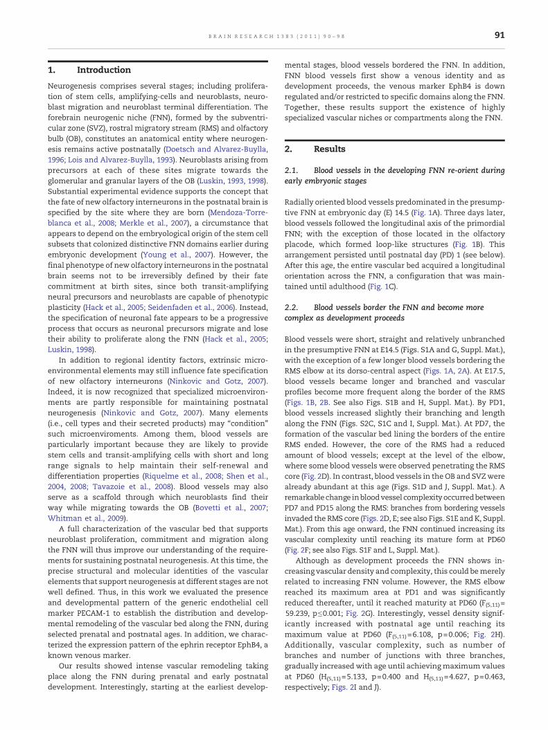
Radially oriented blood vessels predominated in the presump-tive FNN at embryonic day (E) 14.5 (Fig. 1A). Three days later,blood vessels followed the longitudinal axis of the primordialFNN; with the exception of those located in the olfactoryplacode, which formed loop-like structures (Fig. 1B). Thisarrangement persisted until postnatal day (PD) 1 (see below).After this age, the entire vascular bed acquired a longitudinalorientation across the FNN, a configuration that was main-tained until adulthood (Fig. 1C).
2.2. Blood vessels border the FNN and become morecomplex as development proceeds
Blood vessels were short, straight and relatively unbranchedin the presumptive FNN at E14.5 (Figs. S1A and G, Suppl. Mat.),with the exception of a few longer blood vessels bordering theRMS elbow at its dorso-central aspect (Figs. 1A, 2A). At E17.5,blood vessels became longer and branched and vascularprofiles become more frequent along the border of the RMS(Figs. 1B, 2B. See also Figs. S1B and H, Suppl. Mat.). By PD1,blood vessels increased slightly their branching and lengthalong the FNN (Figs. S2C, S1C and I, Suppl. Mat.). At PD7, theformation of the vascular bed lining the borders of the entireRMS ended. However, the core of the RMS had a reducedamount of blood vessels; except at the level of the elbow,where some blood vessels were observed penetrating the RMScore (Fig. 2D). In contrast, blood vessels in the OB and SVZwerealready abundant at this age (Figs. S1D and J, Suppl. Mat.). Aremarkablechange inbloodvessel complexityoccurredbetweenPD7 and PD15 along the RMS: branches from bordering vesselsinvaded the RMS core (Figs. 2D, E; see also Figs. S1E and K, Suppl.Mat.). From this age onward, the FNN continued increasing itsvascular complexity until reaching its mature form at PD60(Fig. 2F; see also Figs. S1F and L, Suppl. Mat.).
Although as development proceeds the FNN shows in-creasing vascular density and complexity, this could bemerelyrelated to increasing FNN volume. However, the RMS elbowreached its maximum area at PD1 and was significantlyreduced thereafter, until it reached maturity at PD60 (F(5,11)=59.239, p≤0.001; Fig. 2G). Interestingly, vessel density signif-icantly increased with postnatal age until reaching itsmaximum value at PD60 (F(5,11)=6.108, p=0.006; Fig. 2H).Additionally, vascular complexity, such as number ofbranches and number of junctions with three branches,gradually increased with age until achievingmaximum valuesat PD60 (H(5,11)=5.133, p=0.400 and H(5,11)=4.627, p=0.463,respectively; Figs. 2I and J).
92 B R A I N R E S E A R C H 1 3 8 3 ( 2 0 1 1 ) 9 0 – 9 8
93B R A I N R E S E A R C H 1 3 8 3 ( 2 0 1 1 ) 9 0 – 9 8
2.3. EphB4 is developmentally regulated andheterogeneously expressed along the FNN asdevelopment proceeds
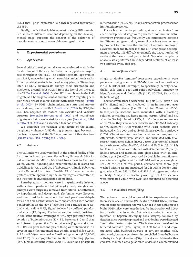
PECAM-1 positive blood vessels were immunoreactive forEphB4 along the FNN at E14.5, E17.5 and PD1 (Figs. 3A–I).Surprisingly, EphB4 labeling was not associated with FNNblood vessels at PD7 (Figs. 3J–L). At PD15, EphB4 immunoreac-tivity was again present in blood vessels along the RMS, butnot in those located in the OB. A few blood vessels runningthrough the SVZ also displayed EphB4 immunoreactivity(Figs. 3M–O); this was also observed at PD60. Finally, bloodvessels along the RMS and within the OB were not EphB4positive at PD60 (Figs. 3P–R).
3. Discussion
Neurogenesis is influenced by the environmental signals thatneuronal precursors encounter at different locations. Thesesignals are likely supplied by distinct cellular elements, amongwhich endothelial cells are known to play an important role.In this regard, endothelial cells are critical components ofbrain homeostasis (Rizzo and Leaver, 2010). They interact withglial and neuronal elements throughout the brain to definehighly specific functional domains (De Seranno et al., 2004).Recently, it has been shown that they play a crucial role for themaintenance and function of neurogenic niches duringdevelopment and in adult life (Palmer et al., 2000; Shenet al., 2004; Tavazoie et al., 2008).
As brain vascular beds are basically composed by veins andarteries, it is likely that the notable physiological andmolecular differences between both vessel populations couldsustain different neurogenic events, such as neuronal differ-entiation (Mukouyama et al., 1997) or neuroblast migration(Anstrom et al., 2005).
The expression pattern of the generic endothelial cellmarker PECAM-1 and the venousmarker EphB4was studied atselected developmental ages, to characterize the dynamicremodeling of the specialized endothelium that supportsneurogenesis along the FNN.
3.1. Intense vascular remodeling occurs throughout theFNN during prenatal and perinatal developmental ages
Blood vessels were present in the presumptive FNN as early asE14.5. During this developmental age they were scarce, poorlybranched and predominantly radially oriented. From E17.5until PD1, blood vessels along the FNN underwent gradual
Fig. 1 – Blood vessels change their orientation as embryonic devsagittal sections of the FNN and their corresponding photomicrogat E14.5 (A) and E17.5 (B) and in-vivo blood vessel filling with dextrradial orientation of most blood vessels at E14.5 (A). In contrast, athe direction of neuroblast migration except for some blood vessuntil adulthood (C). Empty arrows on E14.5 and E17.5 photomicrolocation of RMS elbow is shown by boxes. LGE, lateral ganglionicrostral migratory stream; SVZ, subventricular zone. Scale bar 50
remodeling and became more abundant, longer and moreramified. They also changed their orientation to a longitudinalcourse. From PD1 until PD60 blood vessels continued increas-ing in complexity and length, until reaching maximum levelsat PD60. At the same time, the area of the FNN decreased withage. These results could be related to the intense angiogenicactivity that begins at E14.5 and wanes by PD60 (Figs. S2A andB, Suppl. Mat).
3.2. Blood vessels border the FNN from earlydevelopmental stages
Blood vessels began bordering the dorsal aspect of the FNN asearly as E14.5. Then, they appeared more profusely in theventral aspect of the FNN by E17.5. By around PD7 bloodvessels fully bordered the FNN. These observations may beimportant, since the mechanisms that keep migrating neuro-blasts inside the RMS borders are poorly understood (Belvin-drah et al., 2007). In this context, blood vessels could establisha “physical barrier”: by themselves during embryonic andearly postnatal ages or together with astrocytes starting atPD7, when astrocytes begin their appearance on the borders ofthe FNN (De Seranno et al., 2004; Peretto et al., 2005).Interestingly, we have previously shown that FNN bloodvessels might function as a local source of semaphorins,molecules known to participate as chemorepellents or che-moattractants (Melendez-Herrera et al., 2008).
3.3. EphB4-positive blood vessels support earlyneurogenesis throughout the FNN
During perinatal developmental stages (E14.5, E17.5 and PD1),FNN blood vessels show a venous identity, as suggested byEphB4 expression. As development proceeds (PD7), EphB4 isdown-regulated along the FNN. Interestingly, during earlystages in the formation of the FNN, a time during whichneuroblasts migrate in clusters directly associated to bloodvessels without a glial scaffold (E14.5-PD1), blood vessels showEphB4 expression. Together, these observations are in agree-ment with previous data showing that germinal cells areassociated to venous vessels while migrating (Anstrom et al.,2005). At later stages this might not be the case, because fromPD7 until adulthood, EphB4 is down-regulated in blood vesselsinside the FNN.
Alternatively, EphB4 expression in perinatal blood vesselscould be an indicator of capillary sprouting and not specifi-cally a venous marker, as has been recently suggested (Tayloret al., 2007). However, it is precisely at the developmentaltimes when major remodeling is occurring (between PD1 and
elopment proceeds. Diagrams representing blood vessels inraphs showing immunofluorescence against PECAM-1 (green)an (green) and DAPI counterstaining (blue) at PD60. Notice thet E17.5 (B) blood vessels were tangentially oriented followingel loops in the OP (white arrows). This orientation persistedgraphs show long vessels delimiting FNN borders. Theeminence; OP, olfactory placode; OV olfactory ventricle; RMS,μm.

Fig. 2 – Blood vessels border the FNN since early developmental stages and experience constant remodeling during prenataland postnatal ages. Blood vessels stained by immunofluorescence against PECAM-1 (A-E) or by in-vivo blood vessel filling withdextran (F), in sagittal sections of the RMS elbow at different developmental ages. Blood vessels at E14.5 were scarce and did notshow a lot of branching (A). The vascular tree increased in complexity with increasing age along the FNN (B-F). However, bloodvessels were present in the borders of the presumptive FNN (dashed lines) since early development and at all ages studied.Scale bar 50 μm. All graphs show means±S.E.M. The area of the RMS elbow decreased with increasing developmental stage(G; all pair-wise comparisons were significantly different, p≤0.05 or 0.01, except E14.5 vs. E17.5) and the total length of vesselsper observation area at every age is shown as vessel density (H; PD60 was significantly different from all other ages, p≤0.05,except for PD7). Number of branches and number of vessels with three branches is also shown (no significant differenceswere found).
94 B R A I N R E S E A R C H 1 3 8 3 ( 2 0 1 1 ) 9 0 – 9 8
95B R A I N R E S E A R C H 1 3 8 3 ( 2 0 1 1 ) 9 0 – 9 8
PD60) that EphB4 expression is down-regulated throughoutthe FNN.
Finally, the fact that EphB4 expression along FNN vascularbed shifts to different locations depending on the develop-mental stage, supports the concept of the existence ofvascular compartments across this neurogenic niche.
4. Experimental procedures
4.1. Age selection
Several critical developmental ages were selected to study theestablishment of the vascular niche that supports neurogen-esis throughout the FNN. The earliest prenatal age studiedwas E14.5, an age during which neuroblast migration is radialfrom the lateral ventricle to the olfactory placode. Three dayslater, at E17.5, neuroblasts change their orientation andmigrate as a continuous stream from the lateral ventricles tothe OB (Tucker et al., 2006). During PD1, neuroblasts in the RMSmigrate as a homogenous mass and until PD7 migrating cellsalong the FNN are in direct contact with blood vessels (Perettoet al., 2005). By PD15, chain migration starts and matureastrocytes appear in the RMS (Peretto et al., 2005). By PD60, theFNN has acquired its mature non-vascular and vascularstructure (Melendez-Herrera et al., 2008) and neuroblastsmigrate as chains ensheated by astrocytes (Lois et al., 1996;Peretto et al., 2005) and associated to blood vessels.
We described the vascular architecture of the lateralganglionic eminence (LGE) during prenatal ages, because ithas been shown that the SVZ is a remnant of this structure(Tucker et al., 2006; Young et al., 2007).
4.2. Animals
The CD1 mice we used were bred in the animal facility of theInstituto de Investigaciones Biomédicas, Universidad Nacio-nal Autónoma de México. Mice had free access to food andwater. Animal handling and experimentation followed theGuidelines for Care and Use of Laboratory Animals publishedby the National Institutes of Health. All of the experimentalprotocols were approved by the animal rights' committee atthe Instituto de Investigaciones Biomédicas.
Timed-pregnant mothers were intraperitoneally injectedwith sodium pentobarbital (45 mg/kg body weight) andembryos were surgically removed from uterus, anesthetizedby hypothermia and decapitated. The brains were dissectedand then fixed by immersion in buffered paraformaldehydefor 24 h at 4 °C. Postnatal mice were anesthetized with sodiumpentobarbital on the day of sacrifice and perfused transcar-dially with saline (0.9%, Sigma) followed by buffered parafor-maldehyde (4%; Sigma). The brains were removed, post-fixedin the same fixative overnight at 4 °C, cryo-protected with asolution of buffered sucrose (30%; J.T. Baker) at 4 °C until theysank, frozen in pre-chilled 2-methylbutane (Sigma) and storedat −80 °C. Sagittal sections (30 μm thick) were obtained with acryostat and either mounted onto gelatin-coated slides (E14.5,E17.5 and PD1) or preserved as free floating sections (PD7, PD15and PD60) in a cryoprotective solution containing glycerol(25%; Sigma), ethylene glycol (25%; J.T. Baker) and phosphate
buffered saline (PBS, 50%). Finally, sections were processed forimmunofluorescence.
For each experimental procedure, at least two brains fromeach developmental stage were processed. For immunohisto-chemistry protocols we frequently use consecutive sectionsfor different antigens and try to employ at least two sectionsby protocol to minimize the number of animals employed.However, since the thickness of the FNN changes as develop-ment proceeds, it is difficult to quantify the exact number ofsections that were used per animal. Vascular complexityanalysis was performed in independent sections of at leasttwo animals by studied age.
4.3. Immunofluorescence
Single or double immunofluorescence experiments wereperformed using a rat anti PECAM-1 monoclonal antibody(1:150; MEC13.3, BD Pharmigen) to specifically identify endo-thelial cells and a goat anti-EphB4 polyclonal antibody toidentify venous endothelial cells (1:150; SC-7285, Santa CruzBiotechnology).
Sections were rinsed twice with PBS plus 0.3% Triton X-100(PBTx; Sigma) and then incubated in an immuno-retrieversolution with citrate (Bio SB Inc.) for 30 min at 65 °C.Subsequently, sections were incubated with a blockingsolution containing 5% horse normal serum (Gibco) and 5%albumin (Roche) diluted in PBTx, for 30 min at room temper-ature. Then, they were incubated with anti-PECAM-1 antibodyat 4 °C overnight. After washing with PBTx, sections wereincubated with a goat anti-rat biotinilated secondary antibody(1:750; Chemicon) for two hours at room temperature.Afterwards, sections were washed and incubated with fluo-rescein-conjugated avidin (1:200; Vector Laboratories) dilutedin bicarbonate buffer (NaHCO3 0.1 M and NaCl 0.1 M pH 8.3)for 90 min. Sections were stained with 4′,6-diamino-2-pheny-lindole (DAPI) and mounted onto glass slides with an anti-fading agent (DAKO) or processed by double immunofluores-cence incubating them with anti-EphB4 antibody overnight at4 °C. At the end of this period, sections were thoroughlywashed with PBTx and incubated for 2 h with a donkey anti-goat Alexa Fluor 555 (1:750; A-21432, Invitrogen) secondaryantibody. Finally, after washing overnight at 4 °C, sectionswere incubated 2 min with DAPI and mounted as describedabove.
4.4. In-vivo blood-vessel filling
We performed in-vivo blood-vessel filling experiments usingfluorescein labeled dextran (1%; dextran, 2,000,000 MW, Invitro-gen) in order to visualize the vascular bed in the adult mousebrain. PD60 mice were anesthetized by intra-peritoneal injec-tion of sodiumpentobarbital. Animals received an intra-cardiacinjection of heparin (0.5 mg/kg body weight), followed bydextran. Mice were decapitated and their brains were dissected2 min after dextran injection. The brains were post-fixed inbuffered formalin (10%; Sigma) at 4 °C for 48 h and cryo-protected with buffered sucrose at 30% for another 48 h.Afterwards, brains were frozen in pre-chilled 2-methylbutanewith dry ice. Sagittal sections (50 μmthick)were obtainedwith acryostat, mounted onto gelatinized slides and counterstained
97B R A I N R E S E A R C H 1 3 8 3 ( 2 0 1 1 ) 9 0 – 9 8
with DAPI for 2 min. After rinsing with PBS, sections were coverslipped with an anti-fading agent (Dako).
4.5. Image acquisition and analysis
All the sections were observed using an Olympus BX51WImicroscope equipped with a Disk Scanning Unit (DSU;Olympus) and an automatic slide (Micro Bright Field Biosci-ence). All images were taken with the Stereo Investigatorsoftware (Micro Bright Field Bioscience) and processed withImageJ (NIH).
For vascular complexity analysis the ImageJ Analyze-Skeleton plugin was employed (http://pacific.mpi-cbg.de/AnalyzeSkeleton). Briefly, original photomicrographs wereused as templates to draw blood vessels conserving theirthickness features. This drawn image was made binary andthen skeletonized. The analysis provided results of total vessellength, number of branches, number of triple points, etc. Inevery image, FNN total area was measured and graphedagainst developmental age (Fig. S1G, Suppl. Mat.). For vesseldensity the total length of vessels per observation area wasanalyzed (Leunig et al., 1992). All statistical comparisons weremade with SigmaStat software. Area and vessel densitycomparisons were made with one-way ANOVAs and theHolm-Sidak post-hoc test was used for pair-wise multiplecomparison procedures. On the other hand, the number ofbranches and triple points were compared with Kruskal–Wallis tests and Dunn's test was used for pair-wise multiplecomparison procedures.
Supplementary materials related to this article can befound online at doi:10.1016/j.brainres.2011.01.110.
Acknowledgments
This work was supported by grants from Dirección General deAsuntos del Personal Académico (PAPIIT no. IN210105) andfrom IMPULSA 02, Universidad Nacional Autónoma deMéxico.Additional funding was provided by grants from ConsejoNacional de Ciencia y Tecnología (CONACYT no. P45872-M).Esperanza Meléndez-Herrera was a postdoctoral fellow thatreceived support from DGAPA-UNAM.
R E F E R E N C E S
Anstrom, J.A., Thore, C.R., Moody, D.M., Challa, V.R., Block, S.M.,Brown, W.R., 2005. Germinal matrix cells associate with veinsand a glial scaffold in the human fetal brain. Brain Res. Dev.Brain Res. 160, 96–100.
Fig. 3 – The venous marker EphB4 is developmentally and differe(red) expression pattern on PECAM-1-positive blood vessels (greeN, Q) and LGE/SVZ (C, F, I, L, O, R) at E14.5 (A–C), E17.5 (D–F), PD1 (Gblood vessels were immunoreactive for EphB4 at E14.5 (A–C), E17with blood vessels at PD7 (J–L). At PD15 there was some EphB4 imand SVZ (O), but not in the OB (M). At PD60 blood vessels along theto the SVZ (R). Scale bar 50 μm.
Belvindrah, R., Hankel, S., Walker, J., Patton, B.L., Muller, U., 2007.Beta1 integrins control the formation of cell chains in the adultrostral migratory stream. J. Neurosci. 27, 2704–2717.
Bovetti, S., Hsieh, Y.C., Bovolin, P., Perroteau, I., Kazunori, T.,Puche, A.C., 2007. Blood vessels form a scaffold for neuroblastmigration in the adult olfactory bulb. J. Neurosci. 27,5976–5980.
De Seranno, S., Estrella, C., Loyens, A., Cornea, A., Ojeda, S.R.,Beauvillain, J.C., Prevot, V., 2004. Vascular endothelial cellspromote acute plasticity in ependymoglial cells of theneuroendocrine brain. J. Neurosci. 24, 10353–10363.
Doetsch, F., Alvarez-Buylla, A., 1996. Network of tangentialpathways for neuronal migration in adult mammalian brain.Proc. Natl Acad. Sci. USA 93, 14895–14900.
Hack, M.A., Saghatelyan, A., de Chevigny, A., Pfeifer, A.,Ashery-Padan, R., Lledo, P.M., Gotz, M., 2005. Neuronal fatedeterminants of adult olfactory bulb neurogenesis.Nat. Neurosci. 8, 865–872.
Leunig, M., Yuan, F., Menger, M.D., Boucher, Y., Goetz, A.E.,Messmer, K., Jain, R.K., 1992. Angiogenesis, microvasculararchitecture, microhemodynamics, and interstitial fluidpressure during early growth of human adenocarcinomaLS174T in SCID mice. Cancer Res. 52, 6553–6560.
Lois, C., Alvarez-Buylla, A., 1993. Proliferating subventricular zonecells in the adult mammalian forebrain can differentiate intoneurons and glia. Proc. Natl Acad. Sci. USA 90,2074–2077.
Lois, C., Garcia-Verdugo, J.M., Alvarez-Buylla, A., 1996. Chainmigration of neuronal precursors. Science 271, 978–981.
Luskin, M.B., 1993. Restricted proliferation and migration ofpostnatally generated neurons derived from the forebrainsubventricular zone. Neuron 11, 173–189.
Luskin, M.B., 1998. Neuroblasts of the postnatal mammalianforebrain: their phenotype and fate. J. Neurobiol. 36,221–233.
Melendez-Herrera, E., Colin-Castelan, D., Varela-Echavarria, A.,Gutierrez-Ospina, G., 2008. Semaphorin-3A and its receptorneuropilin-1 are predominantly expressed in endothelial cellsalong the rostral migratory stream of young and adult mice.Cell Tissue Res. 333, 175–184.
Mendoza-Torreblanca, J.G., Martinez-Martinez, E.,Tapia-Rodriguez, M., Ramirez-Hernandez, R., Gutierrez-Ospina, G., 2008. The rostral migratory stream is a neurogenicniche that predominantly engenders periglomerular cells:in vivoevidence in the adult rat brain. Neurosci. Res. 60, 289–299.
Merkle, F.T., Mirzadeh, Z., Alvarez-Buylla, A., 2007. Mosaicorganization of neural stem cells in the adult brain. Science317, 381–384.
Mukouyama, Y., Kuroyanagi, H., Shirasawa, T., Tomoda, T., Saffen,D., Oishi, M., Watanabe, T., 1997. Induction of protein tyrosinephosphatase epsilon transcripts during NGF-induced neuronaldifferentiation of PC12D cells and during the development ofthe cerebellum. Brain Res. Mol. Brain Res. 50, 230–236.
Ninkovic, J., Gotz, M., 2007. Signaling in adult neurogenesis: fromstem cell niche to neuronal networks. Curr. Opin. Neurobiol.17, 338–344.
Palmer, T.D., Willhoite, A.R., Gage, F.H., 2000. Vascular niche for
ntially regulated in blood vessels throughout the FNN. EphB4n) in sagittal sections of the OB (A, D, G, J, M, P), RMS (B, E, H, K,–I), PD7 (J–L), PD15 (M–O) and PD60 (P–R). All PECAM-1 positive.5 (D–F) and PD1 (G–I). Notice that EphB4 was not associatedmunoreactivity associated with blood vessels in the RMS (N)RMS (Q) and OB (P) were negative for this marker, in contrast
98 B R A I N R E S E A R C H 1 3 8 3 ( 2 0 1 1 ) 9 0 – 9 8
adult hippocampal neurogenesis. J. Comp. Neurol. 425,479–494.
Peretto, P., Giachino, C., Aimar, P., Fasolo, A., Bonfanti, L., 2005.Chain formation and glial tube assembly in the shift fromneonatal to adult subventricular zone of the rodent forebrain.J. Comp. Neurol. 487, 407–427.
Riquelme, P.A., Drapeau, E., Doetsch, F., 2008. Brainmicro-ecologies: neural stem cell niches in the adultmammalian brain. Philos. Trans. R. Soc. Lond. B Biol. Sci. 363,123–137.
Rizzo, M.T., Leaver, H.A., 2010. Brain endothelial cell death: modes,signaling pathways, and relevance to neural development,homeostasis, and disease. Mol. Neurobiol. 42, 52–63.
Seidenfaden, R., Desoeuvre, A., Bosio, A., Virard, I., Cremer, H.,2006. Glial conversion of SVZ-derived committed neuronalprecursors after ectopic grafting into the adult brain. Mol. Cell.Neurosci. 32, 187–198.
Shen, Q., Goderie, S.K., Jin, L., Karanth, N., Sun, Y., Abramova, N.,Vincent, P., Pumiglia, K., Temple, S., 2004. Endothelial cellsstimulate self-renewal and expand neurogenesis of neuralstem cells. Science 304, 1338–1340.
Shen, Q., Wang, Y., Kokovay, E., Lin, G., Chuang, S.M., Goderie, S.K.,Roysam, B., Temple, S., 2008. Adult SVZ stem cells lie in avascular niche: a quantitative analysis of niche cell–cellinteractions. Cell Stem Cell 3, 289–300.
Tavazoie, M., Van der Veken, L., Silva-Vargas, V., Louissaint, M.,Colonna, L., Zaidi, B., Garcia-Verdugo, J.M., Doetsch, F., 2008.A specialized vascular niche for adult neural stem cells. CellStem Cell 3, 279–288.
Taylor, A.C., Murfee, W.L., Peirce, S.M., 2007. Expression alongadult ratmicrovascular networks: EphB4 ismore than a venousspecific marker. Microcirculation 14, 253–267.
Tucker, E.S., Polleux, F., LaMantia, A.S., 2006. Position and timespecify the migration of a pioneering population of olfactorybulb interneurons. Dev. Biol. 297, 387–401.
Whitman, M.C., Fan, W., Rela, L., Rodriguez-Gil, D.J., Greer, C.A.,2009. Blood vessels form a migratory scaffold in the rostralmigratory stream. J. Comp. Neurol. 516, 94–104.
Young, K.M., Fogarty, M., Kessaris, N., Richardson, W.D., 2007.Subventricular zone stem cells are heterogeneous with respectto their embryonic origins and neurogenic fates in the adultolfactory bulb. J. Neurosci. 27, 8286–8296.
Copyright © 2022 FDOKUMEN