The potential role of metalloproteinases in neurogenesis in the gerbil hippocampus following global...
15
The Potential Role of Metalloproteinases in Neurogenesis in the Gerbil Hippocampus Following Global Forebrain Ischemia Luiza Wo ´ jcik-Stanaszek 1 , Joanna Sypecka 1 , Patrycja Szymczak 1 , Malgorzata Ziemka-Nalecz 1 , Michel Khrestchatisky 2 , Santiago Rivera 2 , Teresa Zalewska 1 * 1 NeuroRepair Department, Mossakowski Medical Research Centre, Polish Academy of Sciences, Warsaw, Poland, 2 Neurobiologie des Interactions Cellulaires et Neurophysiopathologie (NICN), UMR 6184, CNRS, Aix-Marseille University, Marseille, France Abstract Background: Matrix metalloproteinases (MMPs) have recently been considered to be involved in the neurogenic response of adult neural stem/progenitor cells. However, there is a lack of information showing direct association between the activation of MMPs and the development of neuronal progenitor cells involving proliferation and/or further differentiation in vulnerable (Cornus Ammoni-CA1) and resistant (dentate gyrus-DG) to ischemic injury areas of the brain hippocampus. Principal Findings: We showed that dynamics of MMPs activation in the dentate gyrus correlated closely with the rate of proliferation and differentiation of progenitor cells into mature neurons. In contrast, in the damaged CA1 pyramidal cells layer, despite the fact that some proliferating cells exhibited antigen specific characteristic of newborn neuronal cells, these did not attain maturity. This coincides with the low, near control-level, activity of MMPs. The above results are supported by our in vitro study showing that MMP inhibitors interfered with both the proliferation and differentiation of the human neural stem cell line derived from umbilical cord blood (HUCB-NSCs) toward the neuronal lineage. Conclusion: Taken together, the spatial and temporal profiles of MMPs activity suggest that these proteinases could be an important component in neurogenesis-associated processes in post-ischemic brain hippocampus. Citation: Wo ´ jcik-Stanaszek L, Sypecka J, Szymczak P, Ziemka-Nalecz M, Khrestchatisky M, et al. (2011) The Potential Role of Metalloproteinases in Neurogenesis in the Gerbil Hippocampus Following Global Forebrain Ischemia. PLoS ONE 6(7): e22465. doi:10.1371/journal.pone.0022465 Editor: Mike O. Karl, Center for Regenerative Therapies Dresden, Germany Received August 12, 2010; Accepted June 28, 2011; Published July 25, 2011 Copyright: ß 2011 Wo ´ jcik-Stanaszek et al. This is an open-access article distributed under the terms of the Creative Commons Attribution License, which permits unrestricted use, distribution, and reproduction in any medium, provided the original author and source are credited. Funding: This work was supported by MSHE grants: 1266/P01/2006/31 and 0154/B/P01/2010/38, and by the Letten Foundation grant to MK and SR. The funders had no role in study design, data collection and analysis, decision to publish, or preparation of the manuscript. Competing Interests: The authors have declared that no competing interests exist. * E-mail: [email protected] Introduction Many recent studies have noted that ischemia resembles other brain injuries in producing enhanced neurogenesis in neuropro- liferative regions of the adult rodent brain, including the subventricular zone (SVZ) of the lateral ventricles and the subgranular zone (SGZ) of the dentate gyrus (DG) of the hippocampus [1–4]. Ectopic neurogenesis has also been observed in degenerated hippocampal CA1 in animal models of global ischemia [5,6]. The discovery of neurogenic responses subsequent to ischemic injury has led to the hypothesis that the expansion of the pool of endogenous progenitors could augment the regenerative capacity of the damaged areas. Therefore, the identification of mechanisms that promote the proliferation of progenitors, migration toward injured brain areas and differentiation into the phenotype of lost neuronal cells has become particularly relevant to the development of stem cell-based therapies. It is hypothesized that following ischemic insult, neurogenesis proceeds as it does during embryonic development, involving the concerted action of cell surface and extracellular matrix molecules, thereby providing an environment which may be instructive or permissive to neurogenesis associated processes [7]. In this context, enzymes that modify the extracellular matrix and modulate both axonal guidance and cell adhesion molecules are particularly interesting [8]. The matrix metalloproteinases (MMPs) are one such group of proteinases known to play important roles in the ECM remodeling required for developmental processes. MMPs belong to a family of secreted or membrane-bound endopeptidases, with 25 distinct mammalian gene products [9]. MMPs participate in numerous physiological and pathological processes through the processing of a variety of pericellular substrates including extracellular matrix proteins, cell surface receptors, cell adhesion molecules and growth factors [10,11]. Whereas early up-regulation of MMPs, in particular gelatinases MMP-2 and MMP-9, has been mostly investigated in the context of their detrimental roles in brain ischemic injury [12,13], their involvement in the neurogenic response of adult neural stem/ progenitor cells in the ischemic brain has only been considered recently. MMPs are expressed abundantly in neural stem cells isolated from the human central nervous system (CNS) [14] and according to Mannello et al. [15] they have regulatory roles during the proliferation and differentiation of neural precursor cells in the embryonic mouse brain. Furthermore, Morris et al. [16] reported PLoS ONE | www.plosone.org 1 July 2011 | Volume 6 | Issue 7 | e22465
-
Upload
independent -
Category
Documents
-
view
1 -
download
0
Transcript of The potential role of metalloproteinases in neurogenesis in the gerbil hippocampus following global...

The Potential Role of Metalloproteinases inNeurogenesis in the Gerbil Hippocampus FollowingGlobal Forebrain IschemiaLuiza Wojcik-Stanaszek1, Joanna Sypecka1, Patrycja Szymczak1, Malgorzata Ziemka-Nalecz1, Michel
Khrestchatisky2, Santiago Rivera2, Teresa Zalewska1*
1 NeuroRepair Department, Mossakowski Medical Research Centre, Polish Academy of Sciences, Warsaw, Poland, 2 Neurobiologie des Interactions Cellulaires et
Neurophysiopathologie (NICN), UMR 6184, CNRS, Aix-Marseille University, Marseille, France
Abstract
Background: Matrix metalloproteinases (MMPs) have recently been considered to be involved in the neurogenic responseof adult neural stem/progenitor cells. However, there is a lack of information showing direct association between theactivation of MMPs and the development of neuronal progenitor cells involving proliferation and/or further differentiationin vulnerable (Cornus Ammoni-CA1) and resistant (dentate gyrus-DG) to ischemic injury areas of the brain hippocampus.
Principal Findings: We showed that dynamics of MMPs activation in the dentate gyrus correlated closely with the rate ofproliferation and differentiation of progenitor cells into mature neurons. In contrast, in the damaged CA1 pyramidal cellslayer, despite the fact that some proliferating cells exhibited antigen specific characteristic of newborn neuronal cells, thesedid not attain maturity. This coincides with the low, near control-level, activity of MMPs. The above results are supported byour in vitro study showing that MMP inhibitors interfered with both the proliferation and differentiation of the humanneural stem cell line derived from umbilical cord blood (HUCB-NSCs) toward the neuronal lineage.
Conclusion: Taken together, the spatial and temporal profiles of MMPs activity suggest that these proteinases could be animportant component in neurogenesis-associated processes in post-ischemic brain hippocampus.
Citation: Wojcik-Stanaszek L, Sypecka J, Szymczak P, Ziemka-Nalecz M, Khrestchatisky M, et al. (2011) The Potential Role of Metalloproteinases in Neurogenesis inthe Gerbil Hippocampus Following Global Forebrain Ischemia. PLoS ONE 6(7): e22465. doi:10.1371/journal.pone.0022465
Editor: Mike O. Karl, Center for Regenerative Therapies Dresden, Germany
Received August 12, 2010; Accepted June 28, 2011; Published July 25, 2011
Copyright: � 2011 Wojcik-Stanaszek et al. This is an open-access article distributed under the terms of the Creative Commons Attribution License, which permitsunrestricted use, distribution, and reproduction in any medium, provided the original author and source are credited.
Funding: This work was supported by MSHE grants: 1266/P01/2006/31 and 0154/B/P01/2010/38, and by the Letten Foundation grant to MK and SR. The fundershad no role in study design, data collection and analysis, decision to publish, or preparation of the manuscript.
Competing Interests: The authors have declared that no competing interests exist.
* E-mail: [email protected]
IntroductionMany recent studies have noted that ischemia resembles other
brain injuries in producing enhanced neurogenesis in neuropro-
liferative regions of the adult rodent brain, including the
subventricular zone (SVZ) of the lateral ventricles and the
subgranular zone (SGZ) of the dentate gyrus (DG) of the
hippocampus [1–4]. Ectopic neurogenesis has also been observed
in degenerated hippocampal CA1 in animal models of global
ischemia [5,6].
The discovery of neurogenic responses subsequent to ischemic
injury has led to the hypothesis that the expansion of the pool of
endogenous progenitors could augment the regenerative capacity
of the damaged areas. Therefore, the identification of mechanisms
that promote the proliferation of progenitors, migration toward
injured brain areas and differentiation into the phenotype of lost
neuronal cells has become particularly relevant to the development
of stem cell-based therapies.
It is hypothesized that following ischemic insult, neurogenesis
proceeds as it does during embryonic development, involving the
concerted action of cell surface and extracellular matrix molecules,
thereby providing an environment which may be instructive or
permissive to neurogenesis associated processes [7]. In this context,
enzymes that modify the extracellular matrix and modulate both
axonal guidance and cell adhesion molecules are particularly
interesting [8].
The matrix metalloproteinases (MMPs) are one such group of
proteinases known to play important roles in the ECM remodeling
required for developmental processes.
MMPs belong to a family of secreted or membrane-bound
endopeptidases, with 25 distinct mammalian gene products [9].
MMPs participate in numerous physiological and pathological
processes through the processing of a variety of pericellular
substrates including extracellular matrix proteins, cell surface
receptors, cell adhesion molecules and growth factors [10,11].
Whereas early up-regulation of MMPs, in particular gelatinases
MMP-2 and MMP-9, has been mostly investigated in the context of
their detrimental roles in brain ischemic injury [12,13], their
involvement in the neurogenic response of adult neural stem/
progenitor cells in the ischemic brain has only been considered
recently. MMPs are expressed abundantly in neural stem cells
isolated from the human central nervous system (CNS) [14] and
according to Mannello et al. [15] they have regulatory roles during
the proliferation and differentiation of neural precursor cells in the
embryonic mouse brain. Furthermore, Morris et al. [16] reported
PLoS ONE | www.plosone.org 1 July 2011 | Volume 6 | Issue 7 | e22465

that mRNA expression of both MMP-9 and/or MMP-2 in neural
progenitor cells of the SVZ increased several-fold after ischemic
insult in adult rats. The report published by Lu et al. [17] showed
that up-regulation of MMP-9 and MMP-2 in the SGZ of the
dentate gyrus was compatible with the peak of post-ischemic
neurogenesis in adult primate brains. It has been further proposed
that MMP-9 facilitates neuroblast migration after ischemic stroke
[18–20]. Altogether, these data strongly suggest the participation of
MMPs in ischemic injury repair, favoring the migration of precursor
stem cells from neurogenic into injured sites to replenish lost cells.
Despite ever-growing information concerning the involvement
of MMPs in neurogenesis-associated processes in vitro and ex vivo in
experimental stroke models, the proof of relevance in vivo after
transient forebrain ischemia is still missing. Our previous study
indicates that MMPs might indeed contribute to global ischemia-
stimulated neurogenesis [21]. In the current work we further
extend our investigation and evaluate whether the activation of
MMPs in the brain hippocampus parallels the rate of neuronal
progenitor cell proliferation and/or further differentiation after
forebrain ischemia. In an effort to further elucidate the
involvement of MMPs in neurogenesis-associated processes, we
have also tested the effect of MMPs inhibitors on the development
of a neural stem cell line derived from human umbilical cord blood
(HUCB-NSCs). Our results show that dynamic evolution of
MMPs activity matches the progression of proliferation and
differentiation of stem/progenitor cells into mature neurons,
highlighting the potential role of these extracellular proteinases
in ischemia-induced neurogenesis.
Materials and Methods
The following primary antibodies (source and final dilution)
were used for tissue staining: rat polyclonal anti-BrdU (AbD
Serotec, Raleigh, NC, 1:200), mouse monoclonal anti-neuronal
nuclear antigen (NeuN; Chemicon, Temecula, CA, 1:500), mouse
monoclonal anti-neurofilament 200 (NF-200, Sigma, Saint Louis,
MO, 1:500), and rabbit polyclonal anti-GFAP (DakoCytomation,
Glostrup, Denmark, 1:1000). Anti-rat FITC conjugated (Bethyl
Lab, Montgomery, TX, 1:1000), anti-mouse Alexa 546 (Invitro-
gen, Carlsbad, CA, 1:500), and anti-rabbit Alexa 546 (Invitrogen,
Carlsbad, CA, 1:500) respectively, served as the secondary
antibodies. For the immunocytochemistry of HUCB-NSCs, the
primary antibodies utilized were: mouse monoclonal anti-Ki67
(Novocastra Lab Ltd., Newcastle,UK, 1:100), mouse monoclonal
anti-TuJ1 (Covance, Emeryville, CA, 1:500), mouse monoclonal
anti-MAP2 (Sigma, St Louis, MO, 1:500), mouse monoclonal anti-
galactocerebroside (GalC) (Chemicon, Temecula, CA, 1:200) and
anti S100beta (Swant, Bellinzona, Switzerland, 1:1000). Second-
ary antibodies were conjugated to anti-mouse or anti-rabbit Alexa
Fluor 546 (Invitrogen Molecular Probes, Eugene, OR, 1:1000).
As mentioned in the Results section, some experiments were
conducted with the following pharmacological agents added to the
assay buffer or culture medium: broad-spectrum inhibitors of MMP
– 1,10-O-phenanthroline (Merck, Whitehouse Station, NY,1 mM),
and GM6001 (a peptidyl hydroxamate, Sigma-Aldrich Co, 25 mM),
a non-selective MMPs inhibitor - doxycycline (Sigma-Aldrich,
St Louis, MO, 60 mM ), competitive inhibitor of MMP-2 and
MMP-9 SB-3CT [3-(4-phenoxyphenylsulfonyl)-propylthiirane, Sig-
ma, 10 mM), an inhibitor of serine proteinases - Pefabloc SC [4-(2-
Aminoethyl)-benzenesulfonyl fluoride, hydrochloride] (Roche Appl
Sci, Mannheim, Germany, 5 mM) and the furin inhibitor Dec-
RVKR-CMK (Calbiochem, San Diego, CA, 50 mM).
GM6001 was prepared as 1 mM stock solution and SB-3CT as
50 mM stock solution, each in DMSO. All the other agents were
dissolved in PBS. For each agent, a corresponding diluting solution
was used in these experiments as control.
In vivo experimental designIschemic model. All experimental treatments were approved
by the Local Commission of Ethics for Experiments on Animals.
Male Mongolian gerbils, weighing 50–70 g were used in the
experiments. The animals were allowed free access to food and
water. Forebrain ischemia was performed as described previously
[22] by 5-minute bilateral ligation of the common carotid arteries
under halothane/N2O anesthesia in strictly controlled normo-
thermic conditions. Sham-operated gerbils served as controls. In
all experiments six animals per time point and treatment were
used.
BrdU labeling. 5-bromo-2-deoxyuridine (BrdU; Sigma-
Aldrich) dissolved in physiological saline was administered
intraperitoneally (50 mg/kg per injection, in sterile 0.9% NaCl
plus 0.007 N NaOH). Two-injection paradigms were used. In
some experiments the animals received a single dose of BrdU and
were sacrificed 24 h after the injection. This procedure was used to
determine the number of cells that incorporated BrdU during a
24 h period at a specific time point after ischemia (6–28 days). In
other experiments the animals received BrdU injections twice daily
(12 h apart) for 3 consecutive days starting 6 days after the onset of
ischemia. Animals in this group were sacrificed 14 and 28 days
after the insult. This allowed us to determine the phenotype of the
newborn cells. Sham-operated gerbils served as controls.
Tissue preparation and immunohistochemistry. At the
scheduled time points, anesthetized animals were perfused
transcardially first with phosphate buffered saline (PBS) followed
by a fixative solution (4% paraformaldehyde, PFA, in 0.1 M
phosphate buffer, pH 7.4). The brains were removed, and post-
fixed for 3 h at 4uC in the same fixative solution. Following post
fixation, brains were cryoprotected overnight in 20% sucrose
solution (in 0.1 M PBS), frozen on dry ice and stored at 270uC.
Double-labeled immunofluorescence was performed on free
floating 25 mm coronal cryostat sections (seven to nine per
animal) comprising the hippocampal formation of 6 animals per
group.
For BrdU immunostaining, DNA was first denaturated in 2 N
hydrochloric acid at 37uC for 60 min. Then tissue sections were
incubated in 0.1 M sodium tetraborate (pH 8.5) for 15 min, blocked
with 10% normal goat serum in PBS containing 0.25% Triton X-
100 for 60 min, and incubated with anti-BrdU overnight at 4uC.
Following the washing procedure, the primary antibodies were
revealed by appropriate secondary anti-rat IgG2a FITC conjugated
antibodies for 60 min at room temperature and in the dark.
The differentiation of BrdU-positive cells was monitored with
mouse NF-200 and mouse NeuN as neuronal markers and GFAP
as an astrocytic marker. After BrdU staining, the brain-tissue
sections were incubated with primary antibodies overnight at 4uC.
After being rinsed in PBS, the sections were exposed to secondary
antibodies 1 h at room temperature. Negative controls were
processed in the same manner on adjacent sections but with the
primary antibodies omitted. Double labeling to determine the
expression of phenotypic markers by BrdU expressing cells was
verified using a confocal laser scanning microscope (LSM 510,
Carl Zeiss, Jena, Germany). A helium-neon laser (543 nm) was
utilized in the excitation of Alexa Fluor 546, while an argon laser
(488) was applied in the excitation of FITC.
The number of BrdU-positive cells in the DG and CA1 area
was assessed in an average of 7–9 coronal hippocampal sections
per animal. To avoid double counting we did not analyze adjacent
sections. Every section was evaluated using a computerized
The Role of Metalloproteinases in Neurogenesis
PLoS ONE | www.plosone.org 2 July 2011 | Volume 6 | Issue 7 | e22465

Figure 1. Time- course of cell proliferation in the adult gerbil hippocampus after global ischemia. Animals were subjected to 5 min ofglobal forebrain ischemia followed by 6, 9, 11, 14, 21 or 28 days of reperfusion. BrdU was administered 24 h prior to sacrifice and brains wereprocessed for BrdU immunohistochemistry. Graphs show the number of BrdU-labeled nuclei in the neurogenic SGZ of the DG (A) and in CA1 (B) in asham operated control animal (C) and at different times after ischemia. The number of proliferating, BrdU-positive cells, increases markedly at 9–11days of reperfusion in SGZ of the DG and at 6 days of reperfusion in CA1. Values represent the means 6 SEM of six animals per time point. One-wayANOVA and Bonferroni test: **p,0.01 and ***p,0.001 indicate a statistically significant difference vs control value.doi:10.1371/journal.pone.0022465.g001
The Role of Metalloproteinases in Neurogenesis
PLoS ONE | www.plosone.org 3 July 2011 | Volume 6 | Issue 7 | e22465

microscope system, and positive cells were displayed on a
computer screen. All of the counting was performed under the
fluorescence microscope and using a 206 objective.
In situ zymography. In order to localize activity of MMP-2
and MMP-9 within the brain hippocampus in control and ischemic
animals and in HUCB-NSCs we conducted in situ zymography
according to previously described methods [12,23]. Thawed frozen,
non-fixed coronal brain sections (25 mm thick) or HUCB-NSCs
cultured on glass cover slips were incubated for 3 h at 37uC in a
humid dark chamber in reaction buffer containing 50 mg/ml of
FITC-labeled DQ-gelatin (Invitrogen Molecular Probes, Eugene,
OR) that is quenched intramolecularly. Gelatin-FITC cleavage by
tissue metalloproteinases (gelatinases) releases peptides whose
fluorescence is representative of proteolytic activity. The sections
were rinsed in PBS and fixed in cold 4% PFA for 20 min then
mounted in fluorescent mounting medium (Dako) and observed
using fluorescence microscopy. To confirm that the proteolytic
activity is attributable to MMPs, some sections in each experiment
were incubated in the above conditions with a broad spectrum
inhibitor of metalloproteinases, 1 mM 1,10-O-phenanthroline.
Fluorescence was visualized using an Axiovert 25 fluorescence
microscope (Carl Zeiss, Jena, Germany) and confocal microscope.
Images were captured on the Videotronic CCD-4230 camera, and
processed by Axiovision image analysis system. All images subjected
to direct comparisons were captured at the same exposure and
digital gain settings to eliminate confounds of differential background
intensity or false-positive fluorescent signals across sections.
In the next series of experiments double fluorescent labeling
was performed in order to identify the cell types expressing
gelatinolytic activity. After zymography, the sections were rinsed in
PBS at pH 7.4, (3610 min), preincubated in a blocking solution
(10% goat serum+0.5% Triton X-100), and then incubated
overnight at 4uC with proteins specific for either astrocytes
(GFAP) or neurons (NF-200 and NeuN).
Culture and treatments of HUCB-NSCsHUCB-NSCs [24] were cultured as a mixed population of
committed adherent progenitors and free-floating undifferentiated
Figure 2. Newly-divided cells in the DG of adult gerbil hippocampus after global ischemia. Animals were subjected to 5 min of globalforebrain ischemia followed by reperfusion. BrdU was administered 24 h prior to sacrifice. Brain sections were double-labeled with anti BrdU antibody(green) and anti-NF-200 (red) (A, B, C, G) or anti-GFAP (red) (D, E, F, H). Confocal photomicrographs show immunohistochemical reaction in control DG(A, D), 9 days after ischemia (B, E), and 28 days after ischemia (C, F). G, H represent magnification (z-stacks) of the picture C and F. Photomicrographsare representative of observations made from six animals per time point. Scale bar 10 mm. Abbreviations: s.g.z – subgranular zone, g.z. –granularzone.doi:10.1371/journal.pone.0022465.g002
The Role of Metalloproteinases in Neurogenesis
PLoS ONE | www.plosone.org 4 July 2011 | Volume 6 | Issue 7 | e22465

cells in F12/DMEM+2% FBS+ITS medium (Gibco) in stabilized
conditions of 37uC and 5% CO2 in a fully humidified atmosphere.
The pooled fractions of adherent and floating HUCB-NSCs were
seeded at a density of 104cells/cm2 onto fibronectin-coated glass
plates (10 mg/ml in PBS). Prior to seeding fibronectin remained on
culture dishes overnight at 4uC without air drying and the excess
of substrate was then removed and plates rinsed with warm PBS.
Following cell adhesion, the standard medium was replaced with
the serum-free equivalent, either with or without MMPs inhibitor
– SB-3CT, GM6001 or doxycycline, - at limiting concentrations.
HUCB-NSCs cultures were left for 8 days in vitro (DIV) to grow
and differentiate under the given conditions.
Culture growth. The same number of cells was plated on
fibronectin-coated coverslips to estimate growth rate. Our
observations found fibronectin to be a more effective ECM
component in mediating cell growth and differentiation of HUCB-
NSCs, as compared with laminin and collagen [25]. At the
designated times of culture (4, or 8 DIV) in the presence or
absence of MMP inhibitors, the standard medium was discarded
and cells incubated (3 h at 37uC) with a medium containing
Alamar Blue (Promega Corp.,Madison,WI). Fluorescence was
read using a MultiScan Ascent FL (LabSystems Oy,Helsinki,
Finland) spectrofluorimeter (by excitation wavelength 545 nm/
emission 590 nm), and its level (proportional to the number of
viable cells present on the multiwell plates) was converted to the
number of surviving cells.
Immunocytochemistry. The cell cultures were fixed for
20 min with 4% PFA diluted in PBS. A blocking solution,
containing 10% normal goat serum in PBS, was applied for 1 h at
25uC. The capacity of HUCB-NSCs to generate neurons and glia
was examined through application of specific antibodies against
neuronal (Tuj1, MAP2), oligodendroglial (GalC) and astrocytic
(S100beta) antigens. Cell proliferation was evaluated using anti-
Ki67 determining cells in the mitotic cycle. Immunoreaction with
Figure 3. Neural identity of newly divided cells in the CA1 area after global ischemia. Animals were subjected to 5 min of global forebrainischemia followed by reperfusion. BrdU was administered 24 h prior to sacrifice. Brain sections from a control animal (A, D) and from an animal 6 days(B, E ) and 28 days (C, F) after ischemia were stained for BrdU immunoreactivity (green) and neuron-specific NF-200 (A, B, C, G) or the astrocyte-specific GFAP (D, E, F, H) markers (red). C, H represent magnification (z-stacks) of the picture C and E. Six days after ischemia the higher than in controlnumber of BrdU+ cells are seen within the damaged pyramidal cell. Intensive BrdU labeling was observed in strata oriens and radiatum. Dependingon the CA1 area BrdU-positive cells expressed distinct antigens – NF-200 exclusively in pyramidal cell layer and GFAP in strata oriens and radiatum aswell as in pyramidal cell layer. Photomicrographs are representative of observations made from six animals per time point. Scale bar 10 mm.Abbreviations: p.l. – pyramidal layer; s.o – stratum oriens; s.r.- stratum radiatum.doi:10.1371/journal.pone.0022465.g003
The Role of Metalloproteinases in Neurogenesis
PLoS ONE | www.plosone.org 5 July 2011 | Volume 6 | Issue 7 | e22465

the primary antibodies was carried out overnight at 4uC. Cells
were rinsed with PBS and then incubated for 1 h at RT with an
appropriate secondary antibody conjugated to Alexa Fluor-546
(1:1000, Molecular Probes). Controls for specificity of
immunostaining were processed with either the primary or the
secondary antibody excluded. Cell nuclei were visualized using
30 min incubation at RT with 5 mM Hoechst 33258 (Sigma). The
labeled cells were examined under fluorescence and confocal
microscope. Images were captured and processed as described
above.
Analyses of HUCB-NSCs differentiation. Neurons,
astrocytes and oligodendrocytes were counted manually. To
quantify the percentages of cells expressing a specific marker in
a given experiment, the number of immunoreactive cells in the
Figure 4. Neurogenesis in the adult gerbil hippocampus after global ischemia. Animals were subjected to 5 min of global forebrainischemia followed by reperfusion. BrdU was administered twice daily 6–9 days after ischemia. Brain sections from a control animal (A, D, G), and froman animal 14 days (B, E, H) or 28 days (C, F, I, K, L) after ischemia were stained for BrdU immunoreactivity (green), for specific neuronal markers (NF-200and NeuN -red) and astrocytic marker (GFAP-red) in the DG (A, B, C) and the CA1 (D–I). J, K, L represent magnification (z –stacks) of the picture C, F, I,respectively. Note co-localization of BrdU with mature NeuN-positive neurons in SGZ, SG and hilus at 14 days after ischemia (B) and increased numberof BrdU/NeuN positive cells in SG at 28 days after ischemia (C, J). Photomicrographs are representative of observations made from six animals pertime point. Scale bar 10 mm. Abbreviations: s.g.z - subgranular zone, g.z.- granular zone, h - hilus, p.l.- pyramidal layer.doi:10.1371/journal.pone.0022465.g004
The Role of Metalloproteinases in Neurogenesis
PLoS ONE | www.plosone.org 6 July 2011 | Volume 6 | Issue 7 | e22465
The Role of Metalloproteinases in Neurogenesis
PLoS ONE | www.plosone.org 7 July 2011 | Volume 6 | Issue 7 | e22465

whole population was determined, relative to the total number of
Hoechst-positive non-apoptotic nuclei. In a typical experiment,
5000 cells per marker were counted and the results expressed as a
percentage of total Hoechst-stained nuclei.
Statistical analysisAll values are given as mean +/2 SEM. Differences between
means were also determined using one-way analysis of variance
(ANOVA) followed by posthoc Bonferroni test. Statistical
significance was deemed to be present if p,0.05.
Results
Time course of cell proliferation in the gerbilhippocampus after global ischemia
The time course of cell proliferation in the adult gerbil
hippocampus was studied at specific time points after ischemic
injury (6, 9, 11, 14, 21 or 28 days). For this purpose, animals received
a single dose of BrdU 24 h prior to sacrifice. The number of newly
generated cells was determined in the DG and CA1 structures by
monitoring the incorporation and subsequent immunohistochemical
detection of BrdU. A number of cells incorporating BrdU were
found in the hippocampus of the normal, adult brain. After ischemia
BrdU+ cell density increased markedly, though the labeling pattern
revealed structure-dependent differences. In the DG the elevated
presence of BrdU-positive cells was seen predominantly within the
neurogenic SGZ, with the highest density (as compare to the sham
control) observed between 9 and 11 days of reperfusion (Figs. 1A,
2B,E) [F(2, 75) = 16.04, p,.001]. Thereafter, cell proliferation
appeared to return to the control level. Double labeling shows that
some cells proliferating within 24 h in the SGZ express neuronal
marker - NF-200 (Fig. 2A, B, C, G). GFAP-expressing cells were
found occasionally among the BrdU-labeled population in the SGZ.
Significant ischemia-induced elevation of the presence of BrdU+ cell
nuclei within 24 h, as compared with the sham, was also observed
throughout the CA1 region, with the greatest number of dividing
cells present after 6 days of reperfusion [F(1, 51) = 30.48, p,.001].
The prolongation of recovery up to 28 days led to a reduction in the
presence of immunoreactive cells, but numbers remained higher
than in the sham animals [F(1, 49) = 7.90, p = .008] (Fig. 1B). As
Fig. 3 demonstrates, the intensity of BrdU labeling is different
depending on the topographical area. Only a few BrdU- positive
cells were detected within the destroyed pyramidal cell layer, while
the neighbouring structures, the stratum oriens and stratum radiatum,
showed numerous cell nuclei with BrdU being incorporated.
Importantly, these BrdU+cells expressed distinct antigens – NF-
200 only in the layer vulnerable to ischemia, and GFAP in the entire
CA1- in pyramidal layer as well as in strata oriens and radiatum.
Phenotypic characterization of proliferating cells afterhypoxia/ischemia
To further characterize the fate of cells incorporating BrdU,
sections from sham and ischemic gerbils were double-stained for
BrdU and known neuronal antigens - NF-200 (detected in immature
as well as in mature neurons) and NeuN (for mature neuronal cells)
and GFAP for astrocytes. For the purpose of these studies, animals
received multiple BrdU injections on days 6–9 after ischemia, and
were sacrificed 14 and 28 days of reperfusion. Representative images
are shown in Fig. 4. Striking proliferation of the precursor population
was observed during the investigated period of recovery following
ischemia in the DG with the most pronounced expansion of BrdU-
positive cells at 4 weeks (Fig. 4C, J). Numerous BrdU-positive nuclei
in this area were closely associated with mature neurons expressing
NeuN, though some showed positive immunoreaction with NF-200
(see Fig. 2), a marker of neurofilaments.
BrdU/NeuN labeled cells were also observed occasionally in the
hilus. At the time of recovery, particularly at 28 days, numerous
BrdU/NeuN-positive, newly-generated cells, integrated the granu-
lar cell layer, suggesting the occurrence of short-distance migration
(Fig. 4C). Some (albeit significant fewer) double-stained cells of the
BrdU/NF-200 and BrdU/NeuN types also appeared in the SGZ of
control animals showing the basal level of cell differentiation.
During the investigated time of reperfusion the numbers of
BrdU-positive cells in the damaged CA1 pyramidal cell layer
outnumbered those in the control. Some of them, sparsely
distributed within this structure, showed double labeling with
BrdU and NF200, which was better manifested at 28 days of
recovery (Fig. 4D, E, F). We did not observe co-localization with
NeuN, the marker specific of developed neurons. An increasing
number of BrdU-positive cells were observed in the structures
neighboring the pyramidal CA1 layer. However, these expressed
the GFAP antigen and showed morphology characteristic of
astrocytes (Fig. 4G,H,I).
The effect of ischemia/reperfusion on the activity ofMMPs in the hippocampus
In order to test whether changes in net MMP activity
accompanied enhanced proliferation and differentiation of stem/
progenitor cells in response to ischemia, we used in situ zymography
in ex vivo brain slices, followed by double staining with anti-NF200,
NeuN, and GFAP antibodies. We found that in the investigated
hippocampal subfields, transient forebrain ischemia affects the
activity of MMPs differentially. As depicted in Fig. 5 (A, B, C, D) the
gelatinase-associated proteolytic activity in the DG increased
progressively across the time of reperfusion. The increment in
activity coincides spatially and temporally with the development of
stem/progenitor cells. In the neurogenic SGZ the activity of MMPs
was seen in NF200-positive neurons as well as in neurons expressing
NeuN (Fig. 6). A strong fluorescence signal was observed in Hoechst
counterstained neuronal nuclei. However, it was also seen in the
cytoplasmic compartment and outside cell bodies. Gelatinolytic
activity was also found in the hilus in cells expressing NeuN.
In contrast, in the ischemia-damaged pyramidal cell layer the
activity of gelatinases dropped below the control levels during
reperfusion (Fig. 5 E, F, G, H). The fluorescence signal in CA1 was
detected in neurons expressing NF-200 antigen (Fig. 7). At
comparable time points incipient activation of MMPs either
associated with or surrounding astrocytes immunoreactive against
the GFAP antibody, was found in adjacent brain areas – the stratum
oriens and stratum radiatum. In situ zymography in the presence of
1 mM 1,10-O-phenanthroline, a broad spectrum MMP inhibitor,
very significantly inhibited the gelatinolytic activity (not shown).
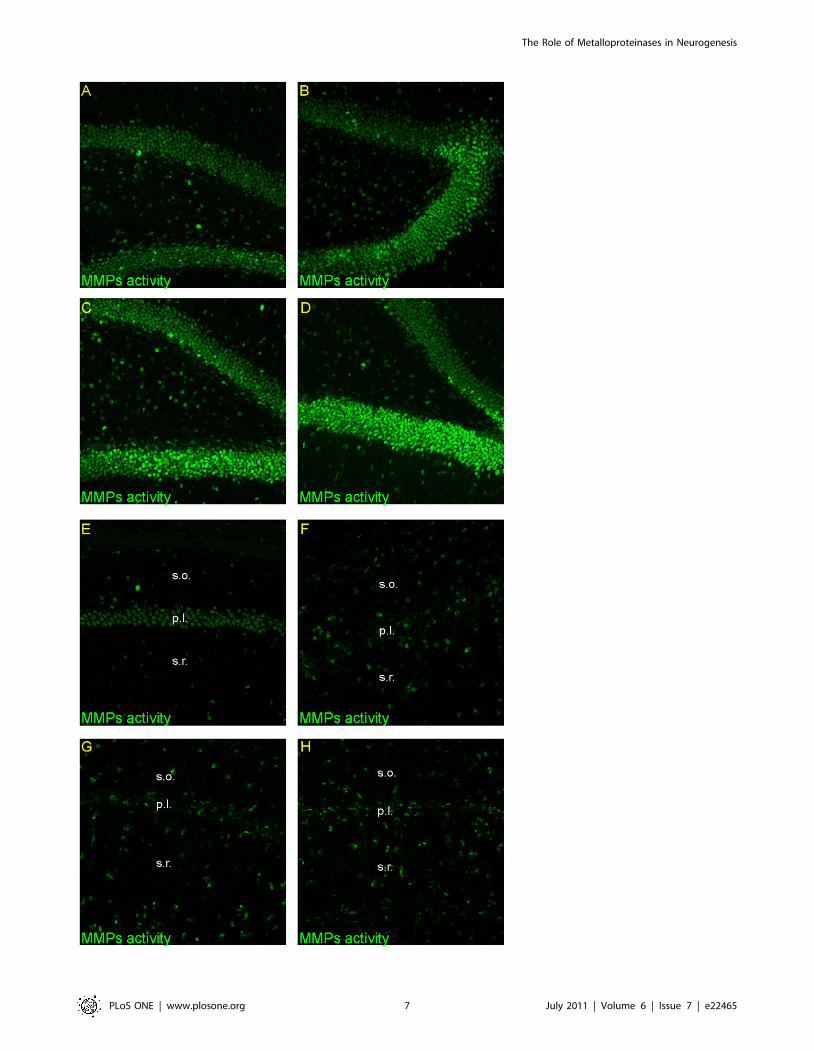
Figure 5. Activity of metalloproteinases in the adult gerbil hippocampus after global ischemia. Animals were subjected to 5 min ofglobal forebrain ischemia followed by reperfusion. Confocal photomicrographs showing in situ zymography in DG and CA1 from a control animal (A,E) and from ischemic animals sacrificed at 7 (B, F), 14 (C, G) and 28 days (D, H) after ischemia. Note the increase of fluorescence signal in the DG and instrata oriens and stratum radiatum with simultaneous decrease in pyramidal cell layer of the CA1 area across the time of reperfusion. All imagessubjected to direct comparisons were captured at the same exposure and digital gain settings. Photomicrographs are representative of observationsmade from six animals per time point. Abbreviations: s.o – stratum oriens; s.r. – stratum radiatum.doi:10.1371/journal.pone.0022465.g005
The Role of Metalloproteinases in Neurogenesis
PLoS ONE | www.plosone.org 8 July 2011 | Volume 6 | Issue 7 | e22465

The effect of MMP inhibitors: SB-3CT, GM6001 anddoxycycline on the gelatinolytic activity, growth,proliferation and differentiation of cultured HUCB-NSCs
Equivalent numbers of HUCB-NSCs were plated onto
fibronectin-coated coverslips and grown in serum-free medium
with/or without added pharmacological agents, SB-3CT, GM
6001 or doxycycline. Fig. 8A illustrates the average time-course of
growth rate during 8 days in culture. Quantitation of cells in
control conditions (without inhibitors) shows continuous increase
in the number of viable cells vs initially plated (c.10-fold). The
addition of MMPs inhibitor – SB-3CT (10 mM), GM6001 (25 mM)
or doxycycline (60 mM) - to the culture medium significantly
reduced the viability of HUCB-NSCs at 4 and 8 days in culture.
Figure 6. Distribution of metalloproteinases in the DG after global ischemia. Animals were subjected to 5 min of global forebrain ischemiafollowed by reperfusion. Confocal photomicrographs following co-staining of in situ zymography (green) with neuronal markers: NF200 (D, E, F, K)(red) and NeuN (A, B, C, J) (red) or the astrocyte marker GFAP (G, H, I, L) (red) in control ( A, D, G) and at 7 (B, E, H) or 28 days (C, F, I, J, K, L) afterischemia. J, K, L represent magnification (z-stacks)of the picture C,F,I, respectively. Note that MMPs activity was principally associated with neurons.Photomicrographs are representative of observations made from six animals per time point. Scale bar 10 mm Abbreviations: g.z – granular zone; s.g.z-subgranular zone.doi:10.1371/journal.pone.0022465.g006
The Role of Metalloproteinases in Neurogenesis
PLoS ONE | www.plosone.org 9 July 2011 | Volume 6 | Issue 7 | e22465

At this time, the number of living cells was reduced by about 30%
in the presence of SB-3CT and GM 6001 and 60% by
doxycycline, as compared with untreated cells [F(3, 52) = 10.42,
p,.001]. Despite a significantly lower proportion of live to total
cells in the treated cultures as compared with the control, living
cells show a stable growth rate.
In order to test the influence of the MMP inhibitors on cell
proliferation, we used a marker of dividing cells, Ki67. Quantitation
of immunopositive cells indicates that HUCB-NSCs proliferate
consistently on fibronectin-coated plates. The addition of SB-3CT,
GM6001 or doxycycline resulted in a markedly reduced proportion
of Ki67-positive cells (in relation to control) - to the average range
Figure 7. Distribution of metalloproteinases in the CA1 after global ischemia. Confocal photomicrographs following co-staining of in situzymography (green) with neuronal markers: NeuN (A, B, C, J) (red) NF200 (D, E, F, K) (red) or the astrocyte marker GFAP (red) (G, H, I, L) in control (A, D,G) and at 7 (B, E, H) or 28 days (C, F, I, J, K, L) after ischemia. J, K, L represent magnification (z-stacks) of the picture C, F, I respectively. Note theassociation of MMPs activity with reactive astrocytes GFAP positive in the stratum oriens and stratum radiatum, probably because of the glialreactivity. Photomicrographs are representative of observations made from six animals per time point. Scale bar 10 mm. Abbreviations: p.l.- pyramidallayer; s.o – stratum oriens; s.r. – stratum radiatum.doi:10.1371/journal.pone.0022465.g007
The Role of Metalloproteinases in Neurogenesis
PLoS ONE | www.plosone.org 10 July 2011 | Volume 6 | Issue 7 | e22465

Figure 8. Effect of SB-3CT, GM6001 and doxycycline on the growth and proliferation of HUCB-NSCs. Equivalent numbers of HUCB-NSCswere plated on fibronectin-coated coverslips and grown 8 days in serum-free medium with or without SB-3CT (10 mM), GM6001 (25 mM) ordoxycycline (60 mM). A) Graph shows an average number of surviving cells after 4, and 8 days in culture. The addition of SB-3CT, GM6001 ordoxycycline to the incubation medium decreased the number of surviving cells. The results (mean values +/2 SEM) represent five independentexperiments. One-way ANOVA and Bonferroni test: **p,.01; ***p,.001, between treatments (inhibitors vs control value). B) Graph shows the rate ofHUCB-NSCs proliferation expressed as a % of control of Ki67- immunopositive cells. The present of SB-3CT, GM6001 or doxycycline in the culture
The Role of Metalloproteinases in Neurogenesis
PLoS ONE | www.plosone.org 11 July 2011 | Volume 6 | Issue 7 | e22465

20%–30% at 4 and 40–50% at 8 days in culture [F(3, 56) = 22.16,
p,.001] (Figs. 8B and 9).
We next tested whether these inhibitors might affect the
differentiation of HUCB-NSCs into neurons and glia. In control
cultures at 8 DIV, approximately 32% of the entire cell population
was immunopositive for Tuj1 (immature neurons) while 17% were
stained for MAP-2 (mature neurons). Analysis of cells immuno-
labelled for S100beta and GalC showed that astroglia and
oligodendroglia accounted for 5% and 4% of cells, respectively
(Fig. 10). We also tried to determine which cell phenotypes express
endogenous MMPs. High-resolution confocal analysis indicated
that MMP activity was localized in all tested cell types: in
immature and differentiated neurons (Tuj1- and MAP2+,
astrocytes (S100beta+) and oligodendrocytes (GalC+) (Fig. 11).
This observation suggests the involvement of metalloproteinases in
HUCB-NSCs differentiation. The presence of investigated phar-
macological agents altered the profile of differentiation - they
inhibited the generation of neurons and promoted the differen-
tiation into oligodendrocytes and astrocytes (Fig. 10). As shown in
the graph, among the remaining cells, the percentage of immature
neurons (Tuj1-positive) was reduced to 15% in the presence SB-
3CT (see also Fig. 9) and to 5% and 7.5% in the cultures treated
with GM6001 and doxycycline, respectively [control vs inhibitors
F(3, 26) = 21.92, p,.001]. The addition of MMP inhibitors
decreased as well the proportion of mature neurons (MAP-2+),
with the most pronounced effect of SB-3CT, which almost
completely blocked their generation [control vs inhibitors F(3,
18) = 11,73. In the presence of doxycyline and GM6001 p = .003,
in the presence of SB-3CT p,.001].
Concomitantly, the percentage of cells expressing the astroglial
and oligodendroglial markers (S100beta and GalC) was higher,
and increased by about 6- and 8-fold, respectively, in the presence
of GM6001 and doxycycline, as compared with the untreated
culture. The number of ofr GalC- or S100beta-positive cells was
affected similarly in the presence of both these inhibitors [F(2,
14) = 65,41, p,.001 and F(2, 16) = 32,35, p,.001, respectively].
The less effect on the promotion of glial cells differentiation was
noted in the case of SB-3CT – the cells expressing GalC increased
only 1.5-fold and S100beta about 2.5-fold, versus control.
In contrast, the serine proteinase inhibitor Pefabloc (5 mM) and
the furin inhibitor Dec-RVKR-CMK (50 mM) did not alter the
profile of differentiation, as compared with control cultures (results
not shown).
Discussion
In agreement with recent studies, our results demonstrate that
adult neural progenitors proliferate in situ in response to forebrain
ischemia. Furthermore, current data also demonstrate that the
stimulation of neural stem cell development in the dentate area of
the hippocampus after forebrain ischemia is accompanied by a
substantial increase in MMPs activity. The timing and magnitude
of the elevation of net proteolytic activity after ischemia correlate
well with the acceleration of stem/progenitor cells proliferation
and further differentiation in the DG, which strongly supports the
contribution of MMPs in post-ischemic neurogenesis. Our
observations provide a new insight into the role of MMPs in
contrast with the detrimental roles traditionally attributed to these
enzymes in cerebral ischemia [13,26,27], and are in line with the
particularly interesting data reported recently by Barkho et al [19],
supporting a role for endogenous MMPs in stroke-induced
neurogenesis.
The analysis of post-ischemic rate of cell birth and differenti-
ation reveals different neurogenic potentials of the hippocampal
subfields. In the known neurogenic zone of the DG, cell
proliferation increased markedly, compared to control animals.
Some of the proliferating BrdU-labeled cells expressed NF-200 as
early as 24 h after the injection of BrdU, indicating that some
dividing progenitor cells commit to neuronal differentiation.
Interestingly, we and others have previously shown MMP activity
reduced markedly the number of proliferating cells. The results (mean values +/2 SEM) represent five independent experiments. One-way ANOVAand Bonferroni test: ***p,.001, between treatments (inhibitors vs control value).doi:10.1371/journal.pone.0022465.g008
Figure 9. Effect of SB-3CT (10 mM) on the Ki67 and Tuj1 positive cells in HUCB-NSs culture. Equivalent numbers of HUCB-NSCs wereplated on fibronectin-coated coverslips and grown 8 days in serum-free medium with or without SB-3CT (10 mM). Cells were stained for Ki67 (red) andTuj1 (green). The cell nuclei counterstained with Hoechst (blue). Note the decreased number of immunolabeled cells in the presence of SB-3CT. Scalebar 20 mm.doi:10.1371/journal.pone.0022465.g009
The Role of Metalloproteinases in Neurogenesis
PLoS ONE | www.plosone.org 12 July 2011 | Volume 6 | Issue 7 | e22465
in the immature neurons [28,29]. At later time points (14 and 28
days) after ischemic insult numerous progenitor cells relocate into
the granular cell layer and become mature granule neurons in line
with previous studies showing that NeuN is expressed after SGZ
neuronal precursors migrate into the granular cell layer (1,2). The
double BrdU-GFAP positive cells found occasionally are probably
neural stem cells and may suggest GFAP labeling of non
proliferating astroglia present in neurogenic zones [30]. This
finding supports previous reports where the proliferation of
astroglia in the DG after global ischemia is unremarkable [31,32].
In contrast, in the damaged CA1 pyramidal layer we noticed a
small number of BrdU+ cells, only slightly greater than in the
control. Some of these cells exhibited the NF-200 antigen which
appeared early in neuronal development, but did not express
mature neuronal antigen-NeuN, suggesting they undergo pro-
grammed cell death before attaining maturity. Moreover, we
found no evidence of SGZ neural stem cells migration into the
CA1 to replace neurons lost after ischemia in line with previously
reported data showing that proliferating cells in the SGZ travel
only to the adjacent GCL [33]. From the above, it follows that in
the present experimental conditions, the expected endogenous
regenerative capacity fails as a source of meaningful compensation
for lost neuronal circuits and that the CA1 area merely displays
gliogenesis. These data are in disagreement with previous reports
indicating that newly-formed neurons originated in the brain
neurogenic zones (SVZ or SGZ), have the capacity to migrate into
the injured CA1 area to integrate the existing brain circuitry [5,6].
The reasons for such a discrepancy may be due to differences in
experimental protocols.
It is worth noting that the vast majority of proliferating
BrdU+cells were present in the stratum oriens and stratum radiatum of
the CA1, rather than in the pyramidal cell layer. These cells
exhibited characteristic features of astroglial phenotype: flat spread
or stellate cells with filamentous GFAP staining.
A clear picture of the factors responsible for neurogenesis in the
SGZ is still elusive. Several findings clearly support that ischemia-
induced neuronal progenitor development is certainly controlled
by the cooperative action of many intrinsic and extrinsic factors,
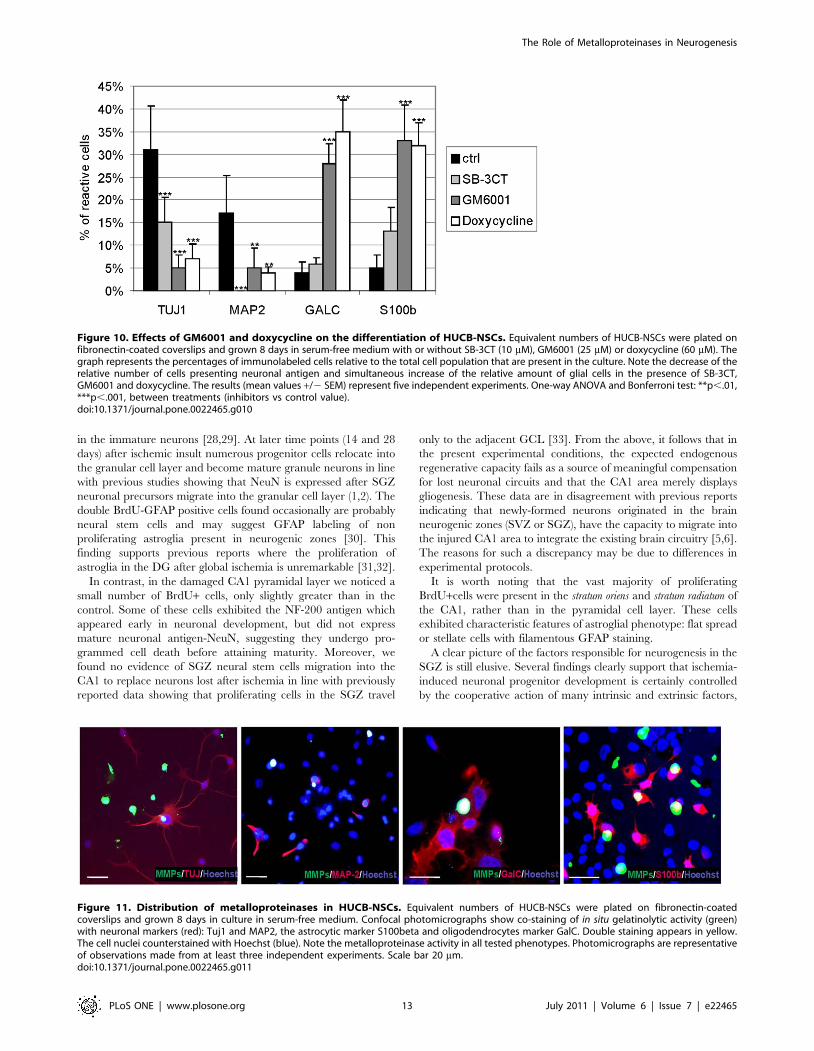
Figure 10. Effects of GM6001 and doxycycline on the differentiation of HUCB-NSCs. Equivalent numbers of HUCB-NSCs were plated onfibronectin-coated coverslips and grown 8 days in serum-free medium with or without SB-3CT (10 mM), GM6001 (25 mM) or doxycycline (60 mM). Thegraph represents the percentages of immunolabeled cells relative to the total cell population that are present in the culture. Note the decrease of therelative number of cells presenting neuronal antigen and simultaneous increase of the relative amount of glial cells in the presence of SB-3CT,GM6001 and doxycycline. The results (mean values +/2 SEM) represent five independent experiments. One-way ANOVA and Bonferroni test: **p,.01,***p,.001, between treatments (inhibitors vs control value).doi:10.1371/journal.pone.0022465.g010
Figure 11. Distribution of metalloproteinases in HUCB-NSCs. Equivalent numbers of HUCB-NSCs were plated on fibronectin-coatedcoverslips and grown 8 days in culture in serum-free medium. Confocal photomicrographs show co-staining of in situ gelatinolytic activity (green)with neuronal markers (red): Tuj1 and MAP2, the astrocytic marker S100beta and oligodendrocytes marker GalC. Double staining appears in yellow.The cell nuclei counterstained with Hoechst (blue). Note the metalloproteinase activity in all tested phenotypes. Photomicrographs are representativeof observations made from at least three independent experiments. Scale bar 20 mm.doi:10.1371/journal.pone.0022465.g011
The Role of Metalloproteinases in Neurogenesis
PLoS ONE | www.plosone.org 13 July 2011 | Volume 6 | Issue 7 | e22465

which may contribute to the difference in SGZ/CA1 neurogenic
potential. One of the most interesting findings obtained in the
current work is that ischemia elicits contrasting effects on the
spatial pattern of net MMP activity that matches the progression of
proliferation in the DG across time and correlates well with the
process of differentiation of stem/progenitor cells into mature
neurons. Such a spatio-temporal relationship between activation of
MMPs and neurogenesis may suggest a casual link between these
processes. This finds strong support in our cell culture experiments
showing that the SB-3CT mediated inhibition of endogenous
MMP activity, (particularly MMP-2 and MMP-9), of HUCB-
NSCs significantly reduced both their proliferation and their
differentiation toward the neuronal lineage. However, we cannot
exclude that the reduced number of immature and mature
neurons (Tuj1- and MAP-2 positive, respectively) might likely be
related to a lower level of generation thereof, and/or their
decreased survival. Simultaneously, the number of oligodendro-
cytes and astrocytes augmented compared to the control, probably
due to increased proliferation. These data support our in vivo
results relative to the involvement of MMPs in the development of
progenitors. They also provide an intriguing model in which
MMPs could modulate the nuclear regulated functions of HUCB-
NSCs. Further support to stress the importance of MMPs in
neurogenesis as compared with other proteinases stems from the
failure of serine proteinase and furin inhibitors (Pefabloc and Dec-
RVKR-CMK) to modulate this process. Consistent with this
notion, there remain emerging in vitro and in vivo data pointing to
regulatory roles of MMPs in neuroblast migration across tissue
matrices [18,34] as well as during the proliferation and
differentiation of neural precursor cells after global and focal
ischemia in rodents [17,19]. On the other hand, partial
preservation of HUCB-NSCs proliferation in the presence of
metalloproteinase inhibitor suggests that the process is not entirely
dependent on MMP activity.
It is not possible at present to define precisely which of their
pleiotropic functions of MMPs are directly linked to post-ischemic
neurogenesis. One likely scenario would involve proteolytic
modulation of guidance molecules and/or the remodeling of the
ECM [35]. The latter may uncover cryptic sites or liberate soluble
fragments [36,37] that promote migration. It is therefore plausible
that the relocation of cells from the SGZ to the GCL in the DG
observed in our study may be facilitated by MMP-mediated
breakdown of ECM barriers impeding cell movement. Comple-
mentary to their role in cell migration, MMPs could also promote
neurite extension of newly integrated progenitor cells as suggested
by recent findings involving MMP-2 and MMP-3 in dendro-axonal
growth of cortical immature neurons [38,39].
MMP-mediated conversion of several trophic factors to their
biologically active forms may also produce signals supporting
neurogenesis [40].
One of the interesting results from this study is the presence of
MMPs activity in neuronal nuclei in the DG area, consistent with
recent findings showing gelatinolytic activity in the neuronal nuclei
of ischemic brain as well as in the nuclei of cultured astrocytes [41–
43]. Nuclear metalloproteinase activity may influence the mecha-
nisms by which neural stem/progenitor cells adjust their gene
expression program but no information is available at present.
Clearly, further investigation will be necessary to confirm this
hypothesis. Nevertheless, regardless of the mechanism of MMPs
action in the DG, the spatial and temporal relationship between the
activation of MMPs and accelerated BrdU incorporation argues
strongly for their involvement in ischemia-induced neurogenesis.
Why neurogenesis fails in the post-ischemic CA1 pyramidal cell
layer is currently unclear. This may obey to different events that
challenge tissue restoration including substantial ECM deposition
and glial scar formation [44,45], along with high levels of at least
one inhibitor of metalloproteinases (TIMP-1) found in reactive
astrocytes after global ischemia [12]. The latter may contribute to
the low MMP activity observed in this area and favor altogether the
formation of a glial scar that hampers the remodeling of the
damaged tissue required for its regeneration. The high level of
invading microglial cells usually found in the early stages of the
ischemic lesion may also contribute to disturb the homeostasis of the
extracellular environment necessary for progenitors to proliferate
and differentiate. In this regard, it has been proposed that early
microglial expression of MMP-9 in the CA1 of rats after global
ischemia, contributes to early neurodegeneration in this area [12].
Unlike in the pyramidal cell layer significant activation of
gelatinase-associated activity was observed in the stratum oriens and
stratum radiatum at some distance from the injured site. As we have
reported previously, ischemia-induced changes are located in the
dendritic region of CA1 pyramidal neurons of the stratum radiatum
and then spread out to the stratum oriens [46]. In these regions the
stress response of postinjured tissue seems to be associated with the
appearance of reactive astrocytes and increased MMP activity that
may facilitate delayed astrocyte-mediated tissue remodeling and
repair at the periphery of the lesion.
ConclusionsThe spatio-temporal relationship between neurogenic-associat-
ed processes and gelatinase activity observed after forebrain
ischemia in the dentate gyrus of the adult rodent brain may indicate
that the metalloproteinases are among the discussed mechanism(s)
which govern the development of stem/progenitor cells. The
importance of MMPs in neurogenesis finds strong support in our
in vitro study showing inhibition of proliferation and differentia-
tion in the presence of metalloproteinases inhibitors.
Author Contributions
Conceived and designed the experiments: LW-S PS TZ. Performed the
experiments: LW-S JS PS MZ-N TZ. Analyzed the data: LW-S JS PS MK
SR TZ. Wrote the paper: MK SR TZ.
References
1. Liu J, Solway K, Messing RO, Sharp FR (1998) Increased neurogenesis in the
dentate gyrus after transient global ischemia in gerbils. J Neurosci 18:
7768–7778.
2. Jin K, Minami M, Lan JQ, Mao XO, Batteur S, et al. (2001) Neurogenesis in the
dentate subgranular zone and rostral subventricular zone after focal cerebral
ischemia in the rat. Proc Natl Acad Sci USA 98: 4710–4715.
3. Parent JM, Vexler ZS, Gong C, Derugin N, Ferriero DM (2002) Rat forebrain
neurogenesis and striatal neuronal replacement after focal stroke. Ann Neurol
52: 802–813.
4. Burns TC, Varfaillie CM, Low WC (2009) Stem cells for ischemic brain injury:
A critical review. J Comp Neurol 515: 125–144.
5. Nakatomi H, Kuriu T, Okabe S, Yamamoto S, Hatano O, et al. (2002)
Regeneration of hippocampal pyramidal neurons after ischemic brain injury by
recruitment of endogenous neural progenitors. Cell 110: 429–441.
6. Bendel O, Bueters T, Euler M, Ogren SO, Sandin J, et al. (2005) Reappearance
of hippocampal CA1 neurons after ischemia is associated with recovery of
learning and memory. J Cereb Blood Flow Metab 25: 1586–1595.
7. Bovetti S, Bovolin P, Perroteau I, Puche AC (2007) Subventricular zone-derived
neuroblast migration to the olfactory bulb is modulated by matrix remodeling.
Eur J Neurosci 25: 2021–2033.
8. Ethel IM, Ethel DW (2007) Matrix metalloproteinases in brain development and
remodeling: Synaptic functions and targets. J Neurosci Res 85: 2813–2823.
The Role of Metalloproteinases in Neurogenesis
PLoS ONE | www.plosone.org 14 July 2011 | Volume 6 | Issue 7 | e22465

9. Rivera S, Khrestchatisky M, Kaczmarek L, Rosenberg GA, Jaworski DM (2010)
Metzincin Proteases and their Inhibitors, Foes or Friends in Nervous System
Physiology? J Neurosci 30: 15337–15357.
10. Dzwonek J, Rylski M, Kaczmarek L (2004) Matrix metalloproteinases and their
endogenous inhibitors in neuronal physiology of the adult brain. FEBS Lett 567:
129–135.
11. Yong VW (2005) Metalloproteinases mediators of pathology and regeneration in
the CNS. Nat Rev Neurosci 6: 931–944.
12. Rivera S, Ogier C, Jourquin J, Timsit S, Szklarczyk AW, et al. (2002) Gelatinase
B and TIMP-1 are regulated in a cell- and time- dependent manner in
association with neuronal death and glial reactivity after global forebrain
ischemia. Eur J Neurosci 15: 19–32.
13. Gasche Y, Soccal PM, Kanemitsu M, Copin J-C (2006) Matrix metalloprotei-
nases and diseases of the central nervous system with a special emphasis on
ischemic brain. Front Biosci 11: 1289–1301.
14. Frolichsthal-Schoeller P, Vescovi AL, Krekoski CA, Murphy G, Edwards DR,
et al. (1999) Expression and modulation of matrix metalloproteinase-2 and tissue
inhibitors of metalloproteinases in human embryonic CNS stem cells.
NeuroReport 10: 45–351.
15. Mannello F, Tonti GAM, Bagnara GP, Papa S (2006) Role and function of
matrix metalloproteinases in the differentiation and biological characterization
of mesenchymal stem cell. Stem Cells 24: 475–481.
16. Morris DC, Zhang ZG, Zhang R, LeTourmeau Y, Gregg SR, et al. (2006)
Stroke increases expression of matrix metalloproteinases and p21–activated
protein kinase in neural progenitor cells. Acad Emerg Med 13: S194.
17. Lu L, Tonchev AB, Kaplamadzhiev DB, Boneva NB, Mori Y, et al. (2008)
Expression of Matrix Metalloproteinases in the Neurogenic Niche of the Adult
Monkey Hippocampus after Ischemia. Hippocampus 18: 1074–1084.
18. Lee SR, Kim H-J, Rogowska J, Zhao BQ, Bhide P, et al. (2006) Involvement of
Matrix Metalloproteinase in Neuroblast Cell Migration from the Subventricular
Zone after Stroke. J Neurosci 26: 3491–3495.
19. Barkho BZ, Munoz AE, Li X, Li L, Cunningham LA, et al. (2008) Endogenous
Matrix Metalloproteinase MMP-3 and MMP-9 Promote Differentiation and
Migration of Adult Neural Progenitor Cells in Response to Chemokines. Stem
Cells 26: 3139–3149.
20. Kang SS, Kook JH, Hwang S, Park SH, Nam SC, et al. (2008) Inhibition of
matrix metalloproteinase-9 attenuated neural progenitor cell migration after
photothrombotic ischemia. Brain Res 1228: 20–6.
21. Wojcik L, Sawicka A, Rivera S, Zalewska T (2009) Neurogenesis in gerbil
hippocampus following brain ischemia: focus on the involvement of metallo-
proteinases. Acta Neurobiol Exp 69: 52–61.
22. Domanska-Janik K, Bong P, Bronisz-Kowalczyk A, Zajac H, Zabłocka B (1999)
AP1 transcriptional factor activation and its relation to apoptosis of hippocampal
CA1 pyramidal neurons after transient ischemia in gerbils. J Neurosci Res 57:
840–846.
23. Ogier C, Bernard A, Chollet AM, Le Diguardher T, Hanessian S, et al. (2006)
Matrix metalloproteinase -2 (MMP-2) regulates astrocyte motility in connection
with the actin cytoskeleton and integrins. Glia 54: 272–284.
24. Buzanska L, Jurga M, Stachowiak EK, Stachowiak MK, Domanska-Janik K
(2006) Neural stem-like cell line derived froma nonhematopoietic population of
human umbilical cord blood. Stem Cells Dev 15: 391–406.
25. Szymczak P, Sypecka J, Zalewska T (2009) Relationship between extracellular
matrix components and MMPs activity during development of neural stem cells
from umbilical cord blood (HUCB-NSC). (Abstract) Acta Neurobiol Exp 69:
293.
26. Asahi M, Asahi K, Jung JC, del Zoppo GJ, Fini ME, et al. (2000) Role for matrix
metalloproteinase 9 after focal cerebral ischemia: effects of gene knockout and
enzyme inhibition with BB-94. J Cereb Blood Flow Metab 20: 1681–1689.
27. Zalewska T, Ziemka-Nalecz M, Sarnowska A, Domanska-Janik K (2002)
Involvement of MMPs in delayed neuronal death after global ischemia. ActaNeurobiol Exp 62: 53–61.
28. Lee CZ, Xu B, Hashimoto T, Yang GY, Young WL (2004) Doxycycline
suppresses cerebral matrix metalloproteinase-9 and angiogenesis induced byfocal hyperstimulation of vascular endothelial growth factor in a mouse model.
Stroke 35: 1715–1719.29. Jablonska A, Wojcik L, Zalewska T, Domanska-Janik K, Lukomska B
Neurogenesis in the adult rat brain after human umbilical cord blood stem
cells transplantation in an experimental model of cerebral ischemic injury;alterations in the expression of doublecortin and metalloproteinases.(Abstract)
7th International Stem Cell School in Regenerative Medicine. 70 p.30. Wurmser AE, Palmer TD, Gage FH (2004) Cellular interactions in the stem cell
niche. Science 304: 1253–1254.31. Kato H, Takahashi A, Itoyama Y (2003) Cell cycle protein expression in
proliferating microglia and astrocytes following transient global cerebral
ischemia in the rat. Brain Res Bull 60: 215–221.32. Tonchev AB, Yamashima T, Zhao L, Okano HJ, Okano H (2003) Proliferation
of neural and neuronal progenitors after global brain ischemia in young adultmacaque monkeys. Mol Cell Neurosci 23: 292–301.
33. Jin K, Sun Y, Xie L, Peel A, Mao XO, et al. (2003) Directed migration of
neuronal precursors into the ischemic cerebral cortex and striatum. Mol CellNeurosci 24: 171–189.
34. Tsukatani T, Fillmore HL, Hamilton HR, Holbrook EH, Costanzo RM (2003)Matrix metalloproteinase expression in the olfactory epithelium. Neuro Report
14: 1135–1140.35. Nagase H, Woessner JF, Jr. (1999) Matrix metalloproteinases. J Biol Chem 274:
21491–21494.
36. Gianelli G, Falk-Marzillier J, Schiraldi O, Stetler-Stevenson WG, Quaranta V(1997) Induction of cell migration by matrix metalloprotease-2 cleavage of
laminin-5. Science 277: 225–228.37. Xu J, Rodriguez D, Petitclerc E, Kim JJ, Hangai M, et al. (2001) Proteolytic
exposure of a cryptic site within collagen type IV is required for angiogenesis and
tumor growth in vivo. J Cell Biol 154: 1069–1080.38. Gonthier B, Koncina E, Satkauskas S, Perraut M, Roussel G, et al. (2009) A
PKC-dependent recruitment of MMP-2 controls semaphorin-3A growth-promoting effect in cortical dendrites. PLoS One 4: e5099.
39. Ould-yahoui A, Tremblay E, Sbai O, Ferhat L, Bernard A, et al. (2009) A newrole for TIMP-1 in modulating neurite outgrowth and morphology of cortical
neurons. PLoS One 4: e8289.
40. Bruno MA, Cuello AC (2006) Activity-dependent release of precursor nervegrowth factor, conversion to mature nerve growth factor, and its degradation by
protease cascade. Proc Natl Acad Sci USA 103: 6735–6740.41. Amantea D, Corasaniti MT, Mercuri NB, Bernardi G, Bagetta G (2007) Brain
regional and cellular localization of gelatinase activity in rat that have undergone
transient middle cerebral artery occlusion. Neuroscience 152: 8–17.42. Yang Y, Candelario-Jalil E, Thompson JF, Cuadrado E, Estrada EY, et al.
(2010) Increased intranuclear metalloproteinase activity in neurons interfereswith oxidative DNA repair in focal cerebral ischemia. J Neurochem 112:
134–149.43. Sbai O, Ould-Yahoui A, Ferhalt L, Gueye Y, Bernard A, et al. (2010)
Differential vesicular distribution and trafficking of MMP-2, MMP-9, and their
inhibitors in astrocytes. Glia 58: 344–366.44. Fawcett JW, Asher RA (1999) The glial scar and central nervous system repair.
Brain Res Bull 49: 377–391.45. Nedergaard M, Dirmagl U (2005) Role of glial cells in cerebral ischemia. Glia
50: 281–286.
46. Ziemka-Nalecz M, Zalewska T, Zajac H, Domanska-Janik K (2003) Decrease ofPKC precedes other cellular signs of calpain activation after transient cerebral
ischemia. Neurochem Int 42: 205–214.
The Role of Metalloproteinases in Neurogenesis
PLoS ONE | www.plosone.org 15 July 2011 | Volume 6 | Issue 7 | e22465