Clean public areas, facilities and equipment D1.HHK.CL3.02 Trainee Manual
Upload
independentCategory
view
0download
0
BioMed CentralNeural Development
ss
Open AcceResearch articleCyclin D1 fine-tunes the neurogenic output of embryonic retinal progenitor cellsGaurav Das1,2, Yoon Choi3,4, Piotr Sicinski3,4 and Edward M Levine*1,2Address: 1Department of Ophthalmology and Visual Sciences, John A Moran Eye Center, University of Utah, Salt Lake City, UT 84132, USA, 2Department of Neurobiology and Anatomy, University of Utah, Salt Lake City, UT 84132, USA, 3Department of Pathology, Harvard Medical School, Boston, MA 02115, USA and 4Department of Cancer Biology, Dana-Farber Cancer Institute, Boston, MA 02115, USA
Email: Gaurav Das - [email protected]; Yoon Choi - [email protected]; Piotr Sicinski - [email protected]; Edward M Levine* - [email protected]
* Corresponding author
AbstractBackground: Maintaining the correct balance of proliferation versus differentiation in retinalprogenitor cells (RPCs) is essential for proper development of the retina. The cell cycle regulatorcyclin D1 is expressed in RPCs, and mice with a targeted null allele at the cyclin D1 locus (Ccnd1-/
-) have microphthalmia and hypocellular retinas, the latter phenotype attributed to reduced RPCproliferation and increased photoreceptor cell death during the postnatal period. How cyclin D1influences RPC behavior, especially during the embryonic period, is unclear.
Results: In this study, we show that embryonic RPCs lacking cyclin D1 progress through the cellcycle at a slower rate and exit the cell cycle at a faster rate. Consistent with enhanced cell cycleexit, the relative proportions of cell types born in the embryonic period, such as retinal ganglioncells and photoreceptor cells, are increased. Unexpectedly, cyclin D1 deficiency decreases theproportions of other early born retinal neurons, namely horizontal cells and specific amacrine celltypes. We also found that the laminar positioning of horizontal cells and other cell types is alteredin the absence of cyclin D1. Genetically replacing cyclin D1 with cyclin D2 is not efficient atcorrecting the phenotypes due to the cyclin D1 deficiency, which suggests the D-cyclins are notfully redundant. Replacement with cyclin E or inactivation of cyclin-dependent kinase inhibitorp27Kip1 restores the balance of RPCs and retinal cell types to more normal distributions, whichsuggests that regulation of the retinoblastoma pathway is an important function for cyclin D1 duringembryonic retinal development.
Conclusion: Our findings show that cyclin D1 has important roles in RPC cell cycle regulation andretinal histogenesis. The reduction in the RPC population due to a longer cell cycle time and to anenhanced rate of cell cycle exit are likely to be the primary factors driving retinal hypocellularityand altered output of precursor populations in the embryonic Ccnd1-/- retina.
BackgroundThe vertebrate retina is composed of seven major cellclasses that arise from a common source, the retinal pro-
genitor cell (RPC) population. Although RPCs at anygiven stage are largely multipotential, they are constrainedsuch that each cell class is generated in a temporal, albeit
Published: 5 May 2009
Neural Development 2009, 4:15 doi:10.1186/1749-8104-4-15
Received: 25 September 2008Accepted: 5 May 2009
This article is available from: http://www.neuraldevelopment.com/content/4/1/15
© 2009 Das et al.; licensee BioMed Central Ltd. This is an open access article distributed under the terms of the Creative Commons Attribution License (http://creativecommons.org/licenses/by/2.0), which permits unrestricted use, distribution, and reproduction in any medium, provided the original work is properly cited.
Page 1 of 23(page number not for citation purposes)

Neural Development 2009, 4:15 http://www.neuraldevelopment.com/content/4/1/15
overlapping order. Production of retinal ganglion cells(RGCs), horizontal cells, and cone photoreceptors is initi-ated at the earliest stage of retinal neurogenesis, followedby amacrine cells and rod photoreceptors, which is thenfollowed by bipolar cells and Müller glia. The relative pro-portion of cells in each class differs widely. For example,cones account for approximately 3% and rods approxi-mately 97% of the photoreceptors in the mouse retina,and rod photoreceptors are the most abundant cell classaccounting for approximately 70% of all retinal cells [1].In general, the early-born cell classes constitute a muchsmaller percentage of the retina than do the late-born cellclasses [1,3]. While cell death contributes to the final celldistribution of the adult retina [4,6], the initial allocationof precursor cells (that is, RPCs that exit the cell cycle) toeach class is a predominant factor in setting their relativeproportions.
In addition to generating the different cell classes, RPCsneed to proliferate in order to produce enough cells topopulate the retina. In the rat retina, RPC proliferationdrives an approximately 400-fold expansion of total cellnumber in a 17-day period between embryonic day (E)14and postnatal day (P)8 [7]. This interval also correspondsto when the bulk of the RPC population exits the cell cycleto generate precursors [2]. Thus, RPCs are exposed to com-peting forces that either influence them to stay in the cellcycle in order to produce enough cells or to exit the cellcycle at the appropriate time in order to generate the cor-rect proportion of cells corresponding to each cell class.
It is generally accepted that multiple cell-extrinsic and -intrinsic factors play important roles in establishing thecorrect balance between RPC proliferation and precursorgeneration during development [8-10]. While it is impor-tant to understand how these different factors are inte-grated into networks, an understanding of the molecularmechanisms used to exit the cell cycle during the transi-tion from RPC to precursor is also needed.
D-type cyclins promote progression from G1 to S phase individing cells by activating cyclin-dependent kinases 4 or6 (CDK4/6) and by sequestering cyclin-dependent kinaseinhibitors such as cyclin-dependent kinase inhibitor 1B(CDKN1B, henceforth referred to as P27KIP1) [11]. Thenet result is enhanced CDK2 activity, inactivation of retin-oblastoma proteins, and activation of DNA replication. D-cyclins are also downstream of various signaling pathwaysand, thus, are well positioned to co-ordinate cell cycleprogression with the extracellular environment [11,12].Mice have three D-cyclin genes: cyclin D1 (Ccnd1), cyclinD2 (Ccnd2) and cyclin D3 (Ccnd3). The expression andrequirement of the D-cyclins during development is tissuespecific [13]. Surprisingly, mouse embryos lacking allthree D-cyclins develop until E16.5, when they die due to
heart abnormalities combined with severe anemia [14].Although developmental defects are apparent in thesemice prior to E16.5, proliferation of many tissues, includ-ing the retina, still occurs, indicating that the D-cyclins arenot absolutely required for cell cycle progression.
Ccnd1 is the predominant D-cyclin in the developing ret-ina and is highly expressed in RPCs but absent from exitedprecursors and differentiated cells [15,18] (this study).Zebrafish embryos treated with a Ccnd1 morpholinoexhibit small eye [19] and mice lacking Ccnd1 have smalleyes and hypocellular retinas due to reduced RPC prolifer-ation and postnatal retinal cell death [17,20,21]. How-ever, the impact of Ccnd1 on embryonic retinaldevelopment has not been directly assessed.
In this study, we characterized the embryonic retinal phe-notype in Ccnd1-/- mice. We found that the cell cycle rateof the Ccnd1-/- RPC population is slower than normal andthis population undergoes a faster rate of depletion due toan increased rate of cell cycle exit. Consistent with this,RGCs and photoreceptors are overrepresented. Surpris-ingly, other early-born embryonic cell classes in the retina,namely horizontal and amacrine cells, are underrepre-sented in the absence of Ccnd1. Analysis of retinas fromnewborn mice in which Ccnd1 is replaced by Ccnd2 revealthat the proportions of at least some cell types remainaltered, suggesting a unique requirement for Ccnd1 inRPCs. We also analyzed the retinas of newborn mice inwhich Ccnd1 is replaced by human Cyclin E (hCcne) or inCcnd1-/-, p27Kip1-/- double mutants and found that theproportions of cell types approach a more normal distri-bution. These findings led us to propose that Ccnd1 con-trols the timing of cell cycle exit in embryonic RPCs and,by doing so, contributes to the appropriate allocation ofprecursor cells to each cell class. We also propose thatCcnd1 contributes to the correct proliferative expansion ofthe retina by influencing the time it takes for RPCs to tran-sit through the cell cycle and by maintaining a sufficientnumber of RPCs during the period of neurogenesis.
Materials and methodsAnimalsCcnd1-/- mice were purchased from Jackson Laboratories(Bar Harbor, ME, USA). Drs Matthew Fero and James Rob-erts (Fred Hutchinson Cancer Center, Seattle, WA) kindlyprovided the p27Kip1-/- mice. The mouse strains contain-ing the Ccnd2 cDNA targeted to the Ccnd1 locus (Ccnd1D2/
D2) and human Ccne cDNA targeted to the Ccnd1 locus(Ccnd1hE/hE) were maintained in the Sicinski laboratory.The noon of the day a vaginal plug was observed was des-ignated E0.5. Genotyping was done as previouslydescribed [17,22-24]. All animal use and care was con-ducted in accordance with protocols approved by the Uni-versity of Utah Institutional Animal Care and Use
Page 2 of 23(page number not for citation purposes)

Neural Development 2009, 4:15 http://www.neuraldevelopment.com/content/4/1/15
Committee and set forth in the Association for Research inVision and Ophthalmology (ARVO) Statement for theUse of Animals. Efforts were made to minimize discom-fort to animals and, when possible, the number of ani-mals needed per analysis was kept to a minimum.
ImmunohistochemistryTissue preparation and immunohistochemistry were doneas previously described [25]. Radial cryosections throughthe retina were cut at a thickness of 10 m. Primary anti-bodies are listed in Table 1. Antigen unmasking (0.18 mMcitric acid, 77 M sodium citrate, pH 6.0, 15 minutes, 90–95°C) was performed prior to incubation with the prolif-erating cell nuclear antigen (PCNA) antibody. Hydrochlo-ric acid treatment (2N HCl, 30 minutes, roomtemperature) was performed prior to incubation with thebromodeoxyuridine (BrdU) antibody.
Image analysisSections were analyzed by epi-fluorescence using a NikonE-600 microscope and images captured in gray scale modewith a Spot-RT slider CCD camera (Diagnostic Instru-ments, Sterling Heights, MI, USA). Confocal images werescanned using an Olympus Fluoview 1000 microscope.Color (RGB) images were assembled from individualmonochrome channels using Photoshop CS (Adobe Sys-tems Inc., San Jose, CA, USA). The levels function wasused to adjust the digital images to be consistent with vis-ual observations.
Marker quantification and statistical analysisThe relative proportions, lineal densities, or areal densi-ties of marker-positive (+) or -negative (-) cells were quan-tified at E12, E14.5 and P0. For each genotype, aminimum of three animals from at least two litters wassampled. For each animal, three different non-adjacentcentral-retina sections were used for cell counting.
At E12, epi-fluorescence images of whole retinal sectionswere captured. Cell populations were quantified over thetotal area of the sections (marker+ cells/mm2 retina). Theexception was for PCNA+ cells, which were quantified as apercentage of the total cell population ((PCNA+ cells/DAPI+ cells) × 100). PCNA+ population was sampled fromthe dorsal retina, where neurogenesis initiates. At E14.5,marker+ cells were calculated as a percentage of total cells(marker+ cells/DAPI+ cells) from 400×-magnified confocalimages (1,600 × 1,600 resolution), captured at compara-ble dorsal-medial regions. At P0, marker+ cells were quan-tified as a percentage of total cells from confocal images ofmedial-central retina, within 200 m of the optic nervehead. Neurofilament medium (NEFM)+ horizontal andSRY-box containing gene 2 (SOX2)+ amacrine cells werequantified as a ratio of the unit length of apical surface ofthe retina (marker+ cells/mm retina) because of their
sparse, linear distribution. The entire peripheral-central-peripheral extent of individual sections was used for thesemeasurements. All cell counts, area, and length measure-ments were done using Adobe Photoshop CS and ImageJ(NIH). Students' t-test was performed using Kaleidagraphstatistical and graphing software (Synergy Software, Read-ing, PA, USA) to determine statistical significance in themarker+ cell population between mutant and control sam-ples. In all graphs, numbers inside bars indicate thenumber of samples analyzed. Error bars indicate standarddeviation.
Window-labeling using thymidine analogs to measure cell cycle timesRetinas with lens attached were cultured for 2.5 hours andsequentially exposed to two thymidine analogs fordefined intervals. At P0, 5-iodo-2'-deoxy-uridine (IdU)was added to the culture medium for the first 2 hours andreplaced with 5-bromo-2'-deoxy-uridine (BrdU) for thefinal 30 minutes. At E14.5, BrdU was added to the culturemedium for the first 2 hours and replaced with 5-ethynyl-2'-deoxy-uridine (EdU) for the final 30 minutes. As previ-ously described [26-28], a combination of mouse anti-BrdU (clone B44; BD Biosciences, San Jose, CA, USA) andrat anti-BrdU (clone BU1/75; Serotec, Raleigh, NC, USA)were used to detect the analogs at P0. For the E14.5 sam-ples, the mouse anti-BrdU antibody was used to detectBrdU (EdU is also detected), and EdU was specificallydetected using the Click-it Reaction (Molecular Probes,Carlsbad, CA, USA) [29]. PCNA was used at both ages toidentify RPCs in all phases of the cell cycle [15]. Thelength of the cell cycle (Tc) in hours was calculated by theformulae:
or
and the length of the S-phase (Ts) in hours was calculatedby the formulae:
or
At E14.5 cell counts were done from a single central fieldon each section (at least three sections per animal), gener-ally on the same side. At P0, cell counts were done on sixfields spanning an entire section (at least of two sectionsper animal). Dorsal-ventral orientation was lost upon dis-
( ) [ ]T h PCNA cells/IdU only cells at Pc = × + +2 0
( ) [ ]T h PCNA cells/BrdU only cells at E14.5c = × + +2
( ) [ ]T h BrdU cells/IdU only cells at P0s = × + +2
( ) [ ]T h EdU cells/BrdU only cells at E14.5s = × + +2
Page 3 of 23(page number not for citation purposes)
Neural Development 2009, 4:15 http://www.neuraldevelopment.com/content/4/1/15
secting eyes out. A more detailed analysis of this assay willappear in a forthcoming manuscript (GD and EML).
Cell cycle exit assay and RGC birthdatingPregnant mice were injected once with a dose of BrdU (10mg/ml stock in 0.1 M Tris (pH 7): 100 g/gm of bodyweight injected) at E13.5 or E18.5 and sacrificed 24 hourslater at E14.5 and P0.5, respectively. Sections were co-labeled with antibodies against BrdU and PCNA andimaged by confocal microscopy. The cell cycle exit indexwas calculated as the percentage of BrdU+ cells that werePCNA- ((BrdU+, PCNA- cells/Total BrdU+ cells) × 100).
To measure the production of RGCs from RPCs, sectionsfrom the same animals used for the cell cycle exit indexwere co-labeled with antibodies against BrdU and POUdomain, class 4, transcription factor 2 (POU4F2; formerlyBRN3B). The index for RGC production was calculated asthe percentage of BrdU+ cells that were POU4F2+ ((BrdU+,POU4F2+ cells/Total BrdU+ cells) × 100). Cell counts weredone from a single dorsal-central field per section (at leasttwo sections per animal) retina at E14.5. For P0 samples,counts were done from two peripheral fields at oppositeends per section (at least two sections per animal).
ResultsCCND1 expression pattern during the early stages of retinal developmentIn the mouse retina, CCND1 protein is expressed as earlyas E11 [17]. However, a systematic analysis of its expres-sion pattern during early retinal development has notbeen done. Therefore, we examined CCND1 expressionfrom E9.5 to E14.5, the period of optic cup formation andonset of retinal neurogenesis (Figure 1). CCND1 proteinis expressed as early as E9.5 in several tissues that give riseto the eye, including the optic vesicle and surface ecto-derm, as well as in the adjacent diencephalic neuroepithe-lium (Figure 1A). At E11, CCND1 expression is strongestin the central region of the neural retina and in the lensvesicle (Figure 1B), and the high level of CCND1 expres-sion in the neural retina spreads outward by E12 andreaches the peripheral retina by approximately E14.5 (Fig-ure 1C, D). This dynamic pattern is reminiscent of thewave of neurogenesis. To examine this relationship fur-ther, these sections were also labeled with the Tuj1 anti-body (Figure 1E–H), which detects the acetylated form ofclass III beta-Tubulin (acTUBB3) and reveals the initialformation of the differentiated cell layer (DCL) [30,31]. Adirect comparison of the CCND1 (dashed lines) andacTUBB3 expression patterns (Figure 1I–L) indicates thatthe high level of CCND1 expression in the neuroblast
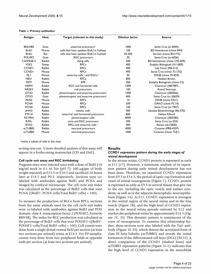
Table 1: Primary antibodies
Antigen Host Target (relevant to this study) Dilution factor Source
BHLHB5 Goat amacrine precursors1 1000 Santa Cruz (sc-6045)BrdU Mouse cells that have uptaken BrdU in S-phase 100 BD biosciences (clone B44)BrdU Rat cells that have uptaken BrdU in S-phase 50-200 Serotec (clone BU1/75)
POU4F2 Goat RGC precursors 50 Santa Cruz (sc-6026)CASPASE-3 Rabbit dying cells 500 BD biosciences (clone C92-605)
VSX2 Sheep RPCs 400 Exalpha Biologicals (X1180P)CCND1 Rabbit RPCs 400 Lab Vision (RB-212)CCND1 Mouse RPCs 400 Santa Cruz (clone 72-13G)
ISL1 Mouse amacrine cells 1 and RGCs 1 50 DSHB (clone 39.4D5)HES1 Rabbit RPCs 800 Nadean BrownMITF Mouse RPE 500 Exalpha Biologicals (clone C5)NEFM Rabbit RGC and horizontal cells 1000 Chemicon (AB1987)NR2E3 Rabbit rod precursors 100 Anand SwaroopOTX2 Rabbit photoreceptor and amacrine precursors1 1000 Chemicon (AB9566)OTX2 Goat photoreceptor and amacrine precursors1 400 Santa Cruz (sc-30659)PAX6 Mouse RPCs 10 DSHB (clone P3U1)PCNA Mouse RPCs 500 DAKO (clone PC10)PCNA Rabbit RPCs 100 Santa Cruz (sc-7907)pHH3 Rabbit mitotic cells 500 Upstate Biotechnology (06-570)PTF1A Rabbit amacrine1 and horizontal precursors 800 Helena EdlundRCVRN Rabbit photoreceptor cells 4000 Chemicon (AB5585)RXR Rabbit cone and RGC precursors 200 Santa Cruz (sc-555)SOX2 Rabbit RPCs and amacrine cells 1 400 Abcam (ab15830)
acTUBB3 Rabbit neuronal precursors 4000 Covance (PRB-435P)acTUBB3 Mouse neuronal precursors 1000 Covance (clone TUJ1)
1marks a subset of cells in this class
Page 4 of 23(page number not for citation purposes)

Neural Development 2009, 4:15 http://www.neuraldevelopment.com/content/4/1/15
layer (NBL) precedes the wave of acTUBB3 expression, buttheir relative timing and similar patterns suggest they arelinked.
Patterning and apoptosis are unaltered by Ccnd1 inactivation at embryonic agesSince Ccnd1 is expressed during optic cup formation, thehypocellularity of the Ccnd1-/- retina could be due toaltered regional patterning. However, analysis of severalmarkers of optic vesicle and cup patterning did not revealdifferences in the establishment or size of the neural reti-nal domain (Additional file 1). Likewise, we did notobserve obvious differences in apoptosis at any of theembryonic ages analyzed as revealed by activated caspase3 (CASP3) immunoreactivity (Additional file 2) or byTUNEL assay (data not shown).
Cell cycle time is longer in the Ccnd1-/- RPC populationHaving ruled out major changes in retinal domain forma-tion and cell death, we measured other parameters thatcould cause the hypocellularity observed in the Ccnd1-/-
retina. At birth, Ccnd1-/- retinas show a three-fold decrease
in total cells and a concomitant three-fold decrease in cellsthat incorporate tritiated thymidine [21]. While thesefindings suggest reduced RPC proliferation prior to P0, wedirectly analyzed proliferative activity during the embry-onic period, first by detection of phosphorylated histoneH3 (pHH3), a marker of RPCs in M-phase (Additional file3) [15]. Fewer pHH3+ cells are evident by E14.5, confirm-ing that RPC proliferation is reduced in the embryonicCcnd1-/- retina.
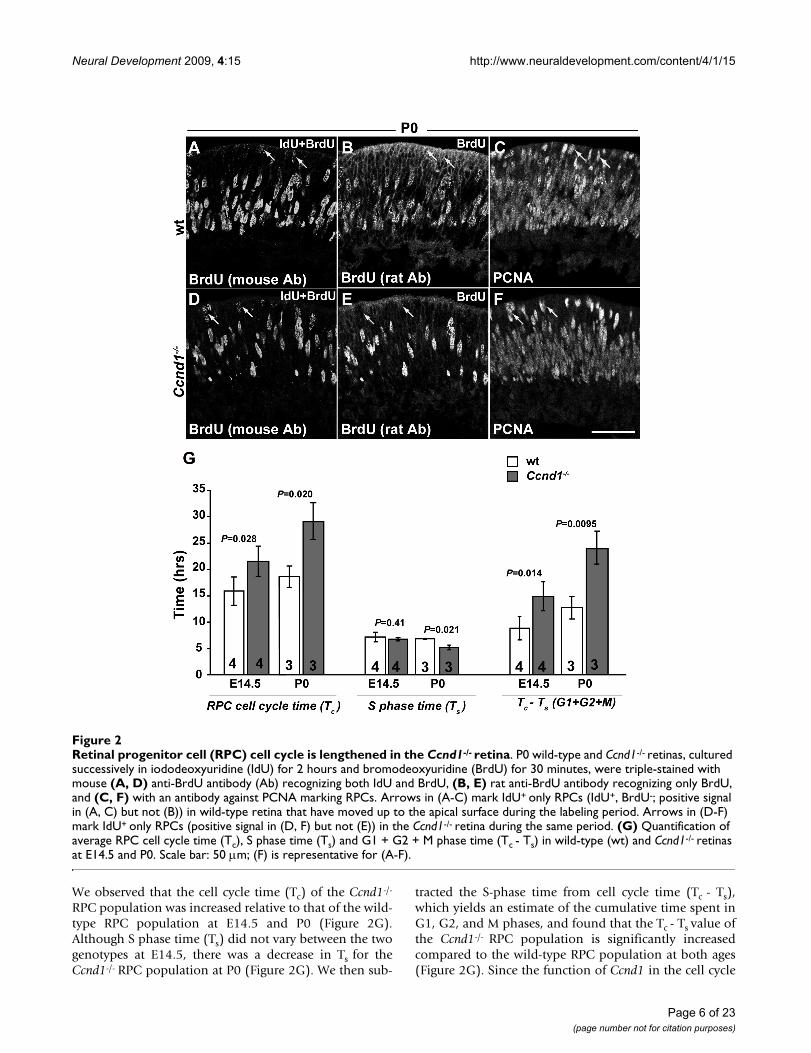
To get an estimate of the cell cycle time and related meas-ures, we adapted a window-labeling paradigm that uti-lizes two thymidine analogs that can be differentiallydetected [27] (manuscript in preparation). Frozen sec-tions from P0 retinas were triple-labeled with a mouseanti-BrdU antibody identifying both IdU and BrdU (Fig-ure 2A, D), a rat anti-BrdU antibody identifying only BrdU(Figure 2B, E) and an anti-PCNA antibody for labeling thecomplete RPC cohort (Figure 2C, F) [15]. BrdU and EdUon E14.5 sections were detected as described (see Materi-als and methods).
Expression patterns of CCND1 and acTUBB3 during early retinal developmentFigure 1Expression patterns of CCND1 and acTUBB3 during early retinal development. Wild-type retinas were double-labeled with antibodies against (A-D) CCND1 and (E-H) acTUBB3. (I-L) Merged images. Dashed lines indicate the peripheral extent of strong CCND1+ cells in retinas from E11 (B, F, J), E12 (C, G, K), and E14.5 (D, H, L) embryos. Asterisks in (A, E, I) indicate that this region of the neuroepithelium is folded over in the section. Abbreviations: D, diencephalon; DCL, differenti-ated cell layer; L, lens; NBL, neuroblast layer; NR, neural retina; OV, optic vesicle; PNR, presumptive neural retina; SE, surface ectoderm. Scale bars: 100 m; (K) is representative for (A-C, E-G, I-K); (L) is representative for (D, H, L).
Page 5 of 23(page number not for citation purposes)
Neural Development 2009, 4:15 http://www.neuraldevelopment.com/content/4/1/15
We observed that the cell cycle time (Tc) of the Ccnd1-/-
RPC population was increased relative to that of the wild-type RPC population at E14.5 and P0 (Figure 2G).Although S phase time (Ts) did not vary between the twogenotypes at E14.5, there was a decrease in Ts for theCcnd1-/- RPC population at P0 (Figure 2G). We then sub-
tracted the S-phase time from cell cycle time (Tc - Ts),which yields an estimate of the cumulative time spent inG1, G2, and M phases, and found that the Tc - Ts value ofthe Ccnd1-/- RPC population is significantly increasedcompared to the wild-type RPC population at both ages(Figure 2G). Since the function of Ccnd1 in the cell cycle
Retinal progenitor cell (RPC) cell cycle is lengthened in the Ccnd1-/- retinaFigure 2Retinal progenitor cell (RPC) cell cycle is lengthened in the Ccnd1-/- retina. P0 wild-type and Ccnd1-/- retinas, cultured successively in iododeoxyuridine (IdU) for 2 hours and bromodeoxyuridine (BrdU) for 30 minutes, were triple-stained with mouse (A, D) anti-BrdU antibody (Ab) recognizing both IdU and BrdU, (B, E) rat anti-BrdU antibody recognizing only BrdU, and (C, F) with an antibody against PCNA marking RPCs. Arrows in (A-C) mark IdU+ only RPCs (IdU+, BrdU-; positive signal in (A, C) but not (B)) in wild-type retina that have moved up to the apical surface during the labeling period. Arrows in (D-F) mark IdU+ only RPCs (positive signal in (D, F) but not (E)) in the Ccnd1-/- retina during the same period. (G) Quantification of average RPC cell cycle time (Tc), S phase time (Ts) and G1 + G2 + M phase time (Tc - Ts) in wild-type (wt) and Ccnd1-/- retinas at E14.5 and P0. Scale bar: 50 m; (F) is representative for (A-F).
Page 6 of 23(page number not for citation purposes)
Neural Development 2009, 4:15 http://www.neuraldevelopment.com/content/4/1/15
is thought to be specific to the G1 phase, this suggestedthat the increase in Tc was due to a longer G1 phase,although we cannot exclude potential changes in G2 or Mphases. In sum, these findings demonstrate that Ccnd1 isrequired to ensure an appropriate rate of passage throughthe cell cycle and that the slower rate of proliferation inthe absence of Ccnd1 is likely to contribute to the hypocel-lularity of the Ccnd1-/-retina.
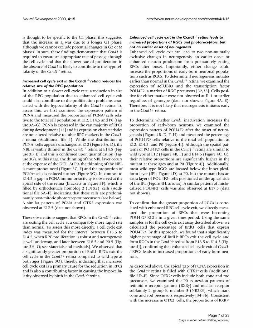
Increased cell cycle exit in the Ccnd1-/- retina reduces the relative size of the RPC populationIn addition to a slower cell cycle rate, a reduction in sizeof the RPC population due to enhanced cell cycle exitcould also contribute to the proliferation problems asso-ciated with the hypocellularity of the Ccnd1-/- retina. Toassess this, we first examined the expression pattern ofPCNA and measured the proportion of PCNA+ cells rela-tive to the total cell population at E12, E14.5 and P0 (Fig-ure 3A–G). PCNA is expressed in the vast majority of RPCsduring development [15] and its expression characteristicsare not altered relative to other RPC markers in the Ccnd1-
/- retina (Additional file 4). While the distribution ofPCNA+ cells appears unchanged at E12 (Figure 3A, D), theNBL is visibly thinner in the Ccnd1-/- retina at E14.5 (Fig-ure 3B, E) and this is confirmed upon quantification (Fig-ure 3G). At this stage, the thinning of the NBL layer occursat the expense of the DCL. At P0, the thinning of the NBLis more pronounced (Figure 3C, F) and the proportion ofPCNA+ cells is reduced further (Figure 3G). In contrast toE14.5, a gap in PCNA immunoreactivity is observed at theapical side of the retina (brackets in Figure 3F), which isfilled by orthodenticle homolog 2 (OTX2)+ cells (Addi-tional file 5A–F), indicating that these cells are predomi-nantly post-mitotic photoreceptor precursors (see below).A similar pattern of PCNA and OTX2 expression wasobserved at E17.5 (data not shown).
These observations suggest that RPCs in the Ccnd1-/- retinaare exiting the cell cycle at a comparably more rapid ratethan normal. To assess this more directly, a cell cycle exitindex was measured for the interval between E13.5 toE14.5, when RPC proliferation is robust and neurogenesisis well underway, and later between E18.5 and P0.5 (Fig-ure 3H–O; see Materials and methods). We observed thata significantly greater proportion of BrdU+ RPCs exit thecell cycle in the Ccnd1-/- retina compared to wild type atboth ages (Figure 3O), thereby indicating that increasedcell cycle exit is a primary cause for the reduction in RPCsand is also a contributing factor in causing the hypocellu-larity observed by birth in the Ccnd1-/- retina.
Enhanced cell cycle exit in the Ccnd1-/- retina leads to increased proportions of RGCs and photoreceptors, but not an earlier onset of neurogenesisEnhanced cell cycle exit can lead to two non-mutuallyexclusive changes in neurogenesis: an earlier onset orenhanced neuron production from prematurely exitingRPCs after onset. Importantly, either change couldincrease the proportions of early born neuronal popula-tions such as RGCs. To determine if neurogenesis initiatesearlier than normal in the Ccnd1-/- retina, we examined theexpression of acTUBB3 and the transcription factorPOU4F2, a marker of RGC precursors [32,33]. Cells posi-tive for either marker were not observed at E11 or earlierregardless of genotype (data not shown; Figure 4A, E).Therefore, it is not likely that neurogenesis initiates earlyin the Ccnd1-/- retina.
To determine whether Ccnd1 inactivation increases theproportion of early-born neurons, we examined theexpression pattern of POU4F2 after the onset of neuro-genesis (Figure 4B–D, F–H) and measured the percentageof POU4F2+ cells relative to the total cell population atE12, E14.5, and P0 (Figure 4I). Although the spatial pat-terns of POU4F2+ cells in the Ccnd1-/- retina are similar towild type at E12 (Figure 4B, F) and E14.5 (Figure 4C, G),their relative proportions are significantly higher in themutant at these ages and at P0 (Figure 4J). Additionally,most wild-type RGCs are located below the inner plexi-form layer (IPL; Figure 4D) at P0, but the mutant has anextra layer of POU4F2+ cells positioned on the apical sideof the IPL (Figure 4H, arrows). A similar pattern of mislo-calized POU4F2+ cells was also observed at E17.5 (datanot shown).
To confirm that the greater proportion of RGCs is corre-lated with enhanced RPC cell cycle exit, we directly meas-ured the proportion of RPCs that were becomingPOU4F2+ RGCs in a given time period. Using the samesamples as for the cell cycle exit assay described above, wecalculated the percentage of BrdU+ cells that expressPOU4F2+. By this approach, we found that a significantlyhigher percentage of BrdU+ RPCs exit the cell cycle andform RGCs in the Ccnd1-/- retina from E13.5 to E14.5 (Fig-ure 4J), confirming that enhanced cell cycle exit of Ccnd1-
/- RPCs leads to increased proportions of early born neu-rons.
As described above, the apical 'gap' of PCNA expression inthe Ccnd1-/- retina is filled with OTX2+ cells (Additionalfile 5D–F). Since OTX2+ cells include both cone and rodprecursors, we examined the P0 expression patterns ofretinoid × receptor gamma (RXR) and nuclear receptorsubfamily 2, group E, member 3 (NR2E3), which markcone and rod precursors respectively [34-36]. Consistentwith the increase in OTX2+ cells, the proportions of RXR+
Page 7 of 23(page number not for citation purposes)
Neural Development 2009, 4:15 http://www.neuraldevelopment.com/content/4/1/15
cells (Figure 5A, D, G) and NR2E3+ cells (Figure 5B, E, H)are increased in the Ccnd1-/- retina. The increased propor-tion of photoreceptors is confirmed by the expression ofrecoverin (RCVRN; Figure 5C, F, I), a calcium-bindingprotein expressed in photoreceptors at perinatal ages [37].Further, an apparent increase in cells expressing blue cone
opsin (OPN1SW) at P0 and rhodopsin at P4 (data notshown) support our conclusion that cones and rods con-tribute to the relative increase in photoreceptor produc-tion.
Gradual depletion of retinal progenitor cell (RPC) population in the Ccnd1-/- retina is caused by enhanced cell cycle exitFigure 3Gradual depletion of retinal progenitor cell (RPC) population in the Ccnd1-/- retina is caused by enhanced cell cycle exit. (A-F) Wild-type (wt) and Ccnd1-/- retinas were labeled with an antibody against PCNA from E12 to P0. Dashed lines in (A, B, D, E) demarcate the differentiated cell layer (DCL) from the neuroblast layer (NBL). Brackets in (F) show the 'apical gap' in the P0 mutant retina. (G) Quantification of PCNA+ cells from E12, E14.5 and P0 retinas. (H) Schematic represen-tation of cell cycle exit assay. (I-N) Wild-type and Ccnd1-/- retina samples, collected at 24 h following a single bromodeoxyuri-dine (BrdU) injection at E13.5, were co-labeled with antibodies against PCNA and BrdU to measure rate of cell cycle exit, as outlined in (H). Arrowheads in (I-N) indicate cells that had exited the cell cycle in the last 24 h. (O) Quantification of exited cells (BrdU+, PCNA-) as a percentage of BrdU+ cells at E14 and P0.5. Abbreviations: DCL, differentiated cell layer; L, lens; NBL; neuroblast layer NR; neural retina. Scale bars: 100 m; (E) is representative for (B, E); (F) for (A, C, D, F); (N) for (I-N).
Page 8 of 23(page number not for citation purposes)

Neural Development 2009, 4:15 http://www.neuraldevelopment.com/content/4/1/15
The proportions of horizontal and amacrine cells are reduced in the Ccnd1-/- retina, despite increased cell cycle exitSince the decrease in the relative proportion of the RPCpopulation correlates with increased neurogenesis in theembryonic Ccnd1-/- retina, it stands to reason that the pro-portions of other early-born cell types, such as horizontaland amacrine cells, would also be increased. We found,however, that these cell types are in fact underrepresentedat E17.5 (data not shown) and P0 (Figure 6). Horizontalcells, which express NEFM, are positioned in a single linetowards the outer part of the NBL (Figure 6A, arrows). In
the Ccnd1-/- retina, these cells are spaced further apart, anddisplaced toward the IPL (Figure 6C, arrows). This reduc-tion in horizontal cells is clearly indicated in retinal wholemounts (Figure 6B, D), by quantification of their linealdensity on retinal sections (Figure 6E), and with othermarkers of horizontal cells (aquaporin 4, prox1, and cal-bindin; data not shown). We also observed a distinctreduction in a subpopulation of amacrine cells marked bySOX2 and islet1 (ISL1) co-expression (Figure 6F–L) [38].Whereas SOX2+, ISL1+ cells appear as an orderly bi-layeron both sides of the IPL (Figure 6F–H), the cells posi-tioned below the IPL are mostly absent in the Ccnd1-/- ret-
Retinal ganglion cells (RGCs) are overproduced in the Ccnd1-/- retinaFigure 4Retinal ganglion cells (RGCs) are overproduced in the Ccnd1-/- retina. (A-H) Wild-type (wt) and Ccnd1-/- retinas were stained with an antibody against POU4F2, which marks a majority of RGCs, at E11, E12, E14.5 and P0. Arrows in (H) mark the extra layer of RGCs in the Ccnd1-/- retina at P0. (I) Quantification of relative proportions of POU4F2+ RGCs. (J) Quantification of relative rate of POU4F2+ RGC production between E13.5 to E14.5. Abbreviations: DCL, differentiated cell layer; L, lens; NBL, neuroblast layer; NR, neural retina. Scale bars: 100 m; (G) is representative for (C, G); (H) for (A, B, D, E, F, H).
Page 9 of 23(page number not for citation purposes)

Neural Development 2009, 4:15 http://www.neuraldevelopment.com/content/4/1/15
ina and the remaining cells are spaced further apart(Figure 6I–K).
Ccnd1 deficiency has different effects on distinct precursor populationsAlthough our analysis of apoptosis (Additional file 2 anddata not shown) suggests that cell death is not contribut-ing to the embryonic phenotype, we cannot entirely ruleit out as a factor in causing the reduction in horizontalcells and SOX2+, ISL1+ amacrine cells as these cell typesare normally in low abundance. Furthermore, as it is hardto discern NEFM+ horizontal cells during early neurogen-esis and SOX2 expression is indicative of advanced stagesof maturation, the reductions in these markers could also
be due to delayed differentiation. This is unlikely, how-ever, as cells expressing these markers continue to appearreduced at later ages (data not shown). Another possibil-ity is that the Ccnd1 deficiency is causing an underproduc-tion in the post-mitotic precursors from which theseparticular cell types arise. To address this, we examinedretinas at earlier stages of development using markersexpressed in newly generated precursors of horizontal,amacrine, and photoreceptor cells (Figure 7). Pancreasspecific transcription factor, 1a (Ptf1a) encodes a basichelix-loop-helix transcription factor expressed in horizon-tal cell precursors and a subset of amacrine cell precursors[39-41]. Basic helix-loop-helix family, member e22(Bhlhe22, henceforth referred to as Bhlhb5) is another
Proportion of photoreceptor cells is increased in the Ccnd1-/- retinaFigure 5Proportion of photoreceptor cells is increased in the Ccnd1-/- retina. Expression pattern of cone precursor marker (A, D) RXR, (B, E) rod precursor marker NR2E3, and (C, F) general photoreceptor marker RCVRN at P0. (G-I) Quantifi-cation of relative proportions of RXR+, NR2E3+ RCVRN+ cells, respectively, at P0. Abbreviations: NR, neural retina. Scale bars: 100 m (D) is representative for (A, D); (E) for (B, E); (F) for (C, F).
Page 10 of 23(page number not for citation purposes)

Neural Development 2009, 4:15 http://www.neuraldevelopment.com/content/4/1/15
basic helix-loop-helix factor expressed in embryonic pre-cursors that give rise to GABAergic and displaced amacrinecells [42]. Otx2 is predominantly expressed in photore-ceptor precursors, although a subset of RGC and amacrineprecursors transiently express Otx2 at the start of their dif-ferentiation [43-45].
Whereas a few cells in the E12 wild-type dorsal retinaexpress PTF1A, significantly fewer PTF1A+ cells aredetected in the Ccnd1-/- retina (Figure 7A, D, G). In con-trast, BHLHB5+ cells, which are more abundant at this age,do not differ in their relative proportions between the
wild-type and Ccnd1-/- retina (Figure 7B, E, G). OTX2expression is evident in the retinal pigmented epithelium,peripheral retina, and isolated cells in the NBL (Figure 7C,F) and quantification of OTX2+ cells in the NBL reveals adecrease in their proportion in the Ccnd1-/- retina (Figure7G). This decrease is also reflected in RXR immunoreac-tivity (data not shown), which suggests a drop in coneprecursor production at this age. At E14.5, the generaltrends for each marker are similar to that found at E12(Figure 7H–N), but it appears that the proportion ofOTX2+ cells is catching up in the mutant (Figure 7N). AtP0, the proportion of PTF1A+ cells remains reduced in the
Reduced densities of horizontal and amacrine cells in the Ccnd1-/- retinaFigure 6Reduced densities of horizontal and amacrine cells in the Ccnd1-/- retina. (A, C) Expression pattern of NEFM at P0 is shown. Arrows point to representative horizontal cells. Bright staining in the differentiated cell layer (DCL) is due to NEFM expression in retinal ganglion cells (RGCs). (B, D) Retinal whole mounts stained with NEFM antibody reveal differences in horizontal cell density across retina. Tissues were imaged from basal surface to reduce interference from NEFM immunoreac-tivity in RGCs. Insets show boxed regions. (E) Quantification of NEFM+ horizontal cells at P0. (F-K) Expression patterns of SOX2 (F, I) and ISL1 (G, J) at P0 (merged images in (H, K)) are shown. (L) Quantification of SOX2+, ISL1B+ amacrine cells at P0. Abbreviations: DCL, differentiated cell layer; NBL; neuroblast layer. Scale bars: 100 m; (C) is representative for (A, C); (D) for (B, D).
Page 11 of 23(page number not for citation purposes)

Neural Development 2009, 4:15 http://www.neuraldevelopment.com/content/4/1/15
Ccnd1-/- retina (Figure 7O, R, U) and the relative propor-tion of Bhlhb5+ cells does not differ between the wild-typeand Ccnd1-/- retina (Figure 7U), although their distribu-tion is altered (Figure 7P, U). The relative proportion ofOTX2+ cells in the Ccnd1-/- retina is greater than in wild-type at P0 (Figure 7Q, T, U) and the larger proportions of
RCVRN+, NR2E3+, and RXR+ cells (Figure 5) collectivelysupport the idea that, by P0, rod and cone precursor pro-duction is enhanced in the absence of Ccnd1.
To gain insight into the potential relationships betweenthese precursor populations, we directly compared the
Ccnd1-deficiency causes alterations in the proportions of precursor cell populationsFigure 7Ccnd1-deficiency causes alterations in the proportions of precursor cell populations. Expression patterns of (A, D, H, K, O, R) PTF1A, (B, C, I, L, P, S) BHLHB5 and (C, F, J, M, Q, T) OTX2 at E12 (A-F), E14.5 (H-M), and P0 (O-T) are shown. (G, N, U) Quantification of marker+ cells at E12 (G), E14.5 (N), and P0 (U). Abbreviations: DCL, differentiated cell layer; NBL, neuroblast layer; PR, peripheral retina; RPE, retinal pigmented epithelium. Scale bar: 100 m; (F) is representative for (A-F); (M) for (H-M); (T) for (O-T).
Page 12 of 23(page number not for citation purposes)

Neural Development 2009, 4:15 http://www.neuraldevelopment.com/content/4/1/15
expression patterns of PTF1A, BHLHB5, and OTX2 at E12,E14.5 and P0 (Figure 8). Regardless of age, PTF1A is notexpressed in the same cells as BHLHB5 or OTX2, whichsuggests that PTF1A+ precursors are distinct fromBHLHB5+ precursors (Figure 8A, D, G, J, M, P) and OTX2+
precursors (Figure 8B, E, H, K, N, Q). In contrast, OTX2and BHLHB5 are co-expressed in a subset of cells fromboth populations (Figure 8C, F, I, L, O, R, arrowheads). AtE12, cells co-expressing OTX2 and BHLHB5 persist in theCcnd1-/- retina even though OTX2+ cells are fewer (Figure8C, F). At E14.5 and P0, the majority of cells co-expressingBHLHB5 and OTX2 are found in the NBL and not in theapical layer of OTX2+ cells (Figure 8I, O), which areinstead marked by RXR or NR2E3 (data not shown), andthese relationships are maintained in the Ccnd1-/- retina(Figure 8L, R; data not shown). These observations suggestthat the combinatorial expression of OTX2 and BHLHB5marks multiple precursor populations. In sum, althoughwe cannot definitively rule out apoptosis or altered differ-entiation as contributing factors, these data strongly sug-gest that Ccnd1 inactivation alters the production ofspecific cell populations from the earliest times after onsetof neurogenesis by altering the relative output of precur-sor cells from RPCs.
Ccnd2 cannot completely rescue the Ccnd1-/- retinal phenotypeGenetic replacement of Ccnd1 by Ccnd2 in Ccnd1D2/D2
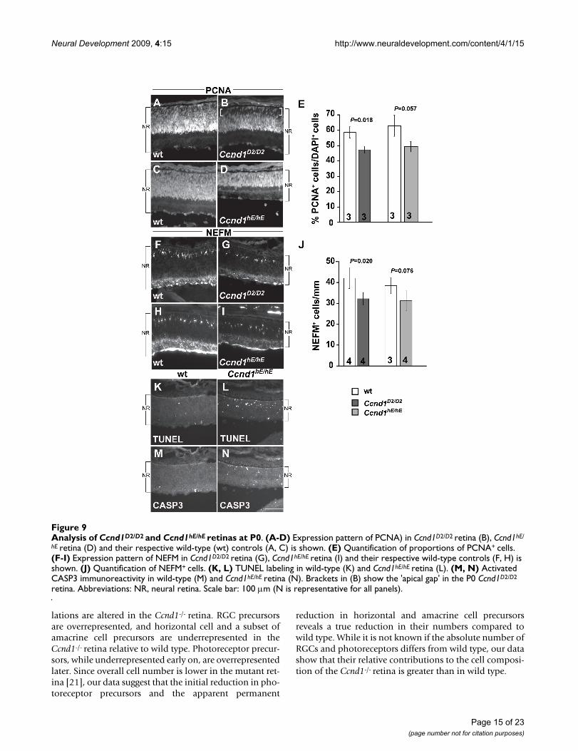
knock-in mice restores the histological appearance of theadult retina and electroretinographic response of photore-ceptors [22]. In this model, the Ccnd2 cDNA sequence isinserted into the Ccnd1 locus and regulated by the Ccnd1promoter and enhancer elements. We examined the P0retina in these mice to determine if Ccnd2 is sufficient tocorrect the developmental changes due to Ccnd1 defi-ciency (Figure 9). PCNA immunolabeling reveals that,similar to the Ccnd1-/- retina, the RPC layer is thinner inthe Ccnd1D2/D2 retina compared to its wild-type control,with a 'gap' at the apical surface (Figure 9A, B; brackets inB) that is filled with OTX2+ cells (Additional file 5J–L).Scoring of PCNA+ cells reveals that their proportion is sig-nificantly reduced (Figure 9E). Unlike the Ccnd1-/- retina,POU4F2+ cells are not mis-positioned on the apical side ofthe IPL layer (Additional file 6E). Quantification ofNEFM+ horizontal cells revealed a significant decrease intheir numbers (Figure 9F, G, J) although not to the samemagnitude as in the Ccnd1-/- retina (Figure 6E). In agree-ment with this trend, there appear to be fewer PTF1A+,SOX2+, and BHLHB5+ cells in the Ccnd1D2/D2 retina com-pared to its wild-type control (Additional file 6B–D, F–H).These findings indicate that Ccnd2 is not sufficient tocompletely compensate for Ccnd1 in retinal cell produc-tion.
Genetic manipulation of downstream cell cycle regulators minimizes the impact of the Ccnd1 deficiency on embryonic retinal developmentGenetic and biochemical evidence suggests that the rate-limiting function of Ccnd1 in promoting cell cycle pro-gression is to stimulate Ccne activity [23]. Based on thismodel, the altered cell production in the Ccnd1-/- RPCpopulation could be due to limited Ccne activity. Toaddress this, we analyzed the newborn retina in a mousestrain in which the human Ccne cDNA is inserted into theCcnd1 locus. In this strain, referred to as Ccnd1hE/hE,human Ccne is expressed in place of Ccnd1. Similar to theCcnd1D2/D2 mouse, the adult retina in this model appearshistologically normal and electrophysiological propertiesare better than in the Ccnd1-/- retina [23].
Our initial analysis revealed that the Ccnd1hE/hE retina isthinner than its wild-type counterpart, which may be dueto an increase in apoptosis, especially in the NBL (Figure9K–N). In contrast to the Ccnd1-/- and Ccnd1D2/D2 retinas,PCNA staining shows that the RPC layer extends all theway to the apical edge, similar to the wild-type control(Figure 9C, D). Whereas the proportions of PCNA+ andNEFM+ cell populations are not significantly differentfrom wild type, they exhibit downward trends (Figure 9E,H, I, J). The relative proportions and positions of cellsexpressing POU4F2, PTF1A, SOX2, and BHLHB5 appearto be similar between the Ccnd1hE/hE and its wild-type con-trol retina (Additional file 6I–P). These findings suggestthat Ccne is more efficient than Ccnd2 in replacing Ccnd1to control the balance of retinal cell types produced.
The sequestration of P27KIP1 by CCND1 protein is onemechanism by which CCND1 is predicted to enhanceCCNE activity and promote cell cycle progression [11].Consistent with this, genetic inactivation of p27Kip1 alle-viates many of the phenotypes seen in the Ccnd1-/- mouse[46,47]. Furthermore, ectopic proliferation occurs in thep27Kip1-/- retina and its overexpression inhibits RPC pro-liferation [48,50]. To test whether the removal of p27Kip1restores the balance of cell types in the absence of Ccnd1,we analyzed Ccnd1-/-, p27Kip1-/- double mutant retinas atP0 (Additional file 7). We found that the expression pat-tern of PCNA in the double mutant retina is more similarto the control retina (Ccnd1+/-) than to the Ccnd1-/- retina,which is indicated by the absence of an 'apical gap' instaining (Additional file 7A, G, S). The cellular distribu-tions of POU4F2+ RGCs, NEFM+ horizontal cells as well asother cell populations expressing PTF1A, SOX2, andBHLHB5 in the double mutant also appear more similarto the control patterns than those in the Ccnd1-/- retina(Additional file 7B–F, H–L, T–X). For comparative pur-poses, the expression patterns for these markers in thep27Kip1-/- retina are shown in Additional file 7M–R. Thesum of our observations from the Ccnd1hE/hE and Ccnd1-/-,
Page 13 of 23(page number not for citation purposes)

Neural Development 2009, 4:15 http://www.neuraldevelopment.com/content/4/1/15
p27Kip1-/- mice suggest that Ccnd1's influence on precur-sor cell output is dependent on its role in regulating Ccneand p27Kip1.
DiscussionWe report here that Ccnd1 has important functions in reg-ulating embryonic retinal histogenesis. In addition tohypocellularity due to changes in proliferation, the rela-tive proportions of multiple post-mitotic precursor popu-
Relationship between PTF1A+, BHLHB5+, and OTX2+ precursorsFigure 8Relationship between PTF1A+, BHLHB5+, and OTX2+ precursors. Retinal sections at (A-F) E12, (G-L) E14.5, and (M-R) P0 were double-labeled with combinations of antibodies against PTF1A, BHLHB5 and OTX2. Arrowheads in (C, F, I, L, O, R) show examples of cell co-expressing OTX2 and BHLHB5. Arrows in (B, C, E, F, H, I, K, L) point to the retinal pigmented epithelium (RPE). Abbreviations: DCL, differentiated cell layer; NBL, neuroblast layer; RPE, retinal pigmented epithelium; wt, wild type. Scale bar: 100 m; (R) is representative for all panels.
Page 14 of 23(page number not for citation purposes)
Neural Development 2009, 4:15 http://www.neuraldevelopment.com/content/4/1/15
lations are altered in the Ccnd1-/- retina. RGC precursorsare overrepresented, and horizontal cell and a subset ofamacrine cell precursors are underrepresented in theCcnd1-/- retina relative to wild type. Photoreceptor precur-sors, while underrepresented early on, are overrepresentedlater. Since overall cell number is lower in the mutant ret-ina [21], our data suggest that the initial reduction in pho-toreceptor precursors and the apparent permanent
reduction in horizontal and amacrine cell precursorsreveals a true reduction in their numbers compared towild type. While it is not known if the absolute number ofRGCs and photoreceptors differs from wild type, our datashow that their relative contributions to the cell composi-tion of the Ccnd1-/- retina is greater than in wild type.
Analysis of Ccnd1D2/D2 and Ccnd1hE/hE retinas at P0Figure 9Analysis of Ccnd1D2/D2 and Ccnd1hE/hE retinas at P0. (A-D) Expression pattern of PCNA) in Ccnd1D2/D2 retina (B), Ccnd1hE/
hE retina (D) and their respective wild-type (wt) controls (A, C) is shown. (E) Quantification of proportions of PCNA+ cells. (F-I) Expression pattern of NEFM in Ccnd1D2/D2 retina (G), Ccnd1hE/hE retina (I) and their respective wild-type controls (F, H) is shown. (J) Quantification of NEFM+ cells. (K, L) TUNEL labeling in wild-type (K) and Ccnd1hE/hE retina (L). (M, N) Activated CASP3 immunoreactivity in wild-type (M) and Ccnd1hE/hE retina (N). Brackets in (B) show the 'apical gap' in the P0 Ccnd1D2/D2
retina. Abbreviations: NR, neural retina. Scale bar: 100 m (N is representative for all panels).
Page 15 of 23(page number not for citation purposes)

Neural Development 2009, 4:15 http://www.neuraldevelopment.com/content/4/1/15
The changes outlined above are likely to be the result ofCcnd1's roles in the cell cycle. Since cell cycle transit timeis longer and the relative rate of cell cycle exit is enhancedin the absence of Ccnd1, it is possible that these changesin proliferation are linked. While longer cell cycle timesare predictive of increased cell cycle exit in the brain[51,52], this does not appear to be the case in thezebrafish retina [53,54]. So whether the lengthening ofthe cell cycle directly causes enhanced cell cycle exit in theCcnd1-/- retina is not clear. Regardless, we propose thatCcnd1 is required for establishing the proper balance ofcell types produced during embryonic retinal develop-ment by mediating cell cycle exit, or in other words, therate of precursor output from RPCs.
Ccnd1 regulates the timing of cell cycle exit in a limited manner after the onset of neurogenesisInterestingly, not all Ccnd1-/- RPCs exit prematurely, andthe extent of RPC depletion, while significant, is notsevere. A reasonable percentage of Ccnd1-/- RPCs remain inthe cell cycle even as neurogenesis progresses. This is notdue to restricted expression as Ccnd1 is widely expressedin RPCs and throughout most of the cell cycle (this study)[15,18]. Rather, D-cyclins are not absolutely required forproliferation in embryonic RPCs since proliferation stilloccurs in mice deficient in Ccnd1, Ccnd2, and Ccnd3 [14].We also found no evidence that Ccnd1-/- RPCs exit the cellcycle before the normal onset of neurogenesis eventhough Ccnd1 is abundantly expressed as early as E9.5.This is not because RPCs are inherently unable to initiateneurogenesis early since precocious neurons are producedin the paired box gene 6 (Pax6-/-) and hairy and enhancerof split 1 (Hes1-/-) retinas [55,57]. Furthermore, thespreading wave of neurogenesis is not altered in theCcnd1-/- retina, even though wild-type RPCs increase theirlevel of CCND1 expression just ahead of the neurogenicwave. These observations led us to propose a model stat-ing that once neurogenesis begins, a limited number ofRPCs become Ccnd1-dependent and their timing of cellcycle exit is determined by their level of CCND1 expres-sion or activity (Figure 10A). We also predict that Ccnd1-dependent RPCs are generated continuously during reti-nal development and have limited proliferative potential.Otherwise, Ccnd1 deficiency should have caused a morediscontinuous or severe decline in the RPC population. Itappears then that downregulation of Ccnd1 is an impor-tant step in the transition of RPCs to post-mitotic precur-sors. Consistent with this, forced expression of CCND1 inphotoreceptor precursors causes unscheduled prolifera-tion, differentiation defects, and apoptosis [58].
Mechanism of Ccnd1-mediated cell cycle exitD-cyclins regulate the retinoblastoma pathway by bindingto and activating CDK4/6 and by sequestering CDK2inhibitors such as P27KIP1 [11]. Both mechanisms ulti-
mately lead to inactivation of retinoblastoma proteins byCDK2/4/6-mediated phosphorylation, allowing cells toprogress from G1 to S phase and undergo DNA replica-tion. Although the importance of the retinoblastomapathway in continuously cycling cells is not clear, it is crit-ical in many cell lineages for differentiation [59-61]. Inthe mouse retina, genetic deletion of the retinoblastomaproteins (Rb1, Rbl1/p107, or Rbl2/p130) uncouples cellcycle exit and differentiation, resulting in ectopic prolifer-ating cells that express markers of multiple precursor celltypes in the retina [62,65]. Further evidence of this decou-pling is seen in the postnatal p107 single copy mutant ret-ina where mature horizontal cells proliferate extensively,all the while retaining their differentiated characteristics[66]. This suggests that retinoblastoma pathway activityregulates cell cycle exit of RPCs and controls the post-mitotic state for some period of time after cell cycle exit.Additional evidence to this effect comes from geneticstudies of molecular regulators in this pathway: inactiva-tion of cyclin-dependent kinase inhibitors such asp27Kip1, p57Kip2, and p19Ink4d cause ectopic prolifera-tion [9,48,50,67]. Forced expression of Ccnd1, the large T-antigen from simian virus 40 or the human papillomavi-rus type 16 (HPV-16) E7 protein (negative regulators ofretinoblastoma proteins) in post-mitotic photoreceptorprecursors causes inappropriate cell cycle re-entry andsubsequent cell death or tumorigenesis depending on thenature of the transgene construct [58,68-72]. A similarphenomenon is also observed for other retinal cell types[73,75]. Furthermore, Rb1 phosphorylation is greatlydiminished in the Ccnd1-/- retina, probably due to reduc-tions in CDK2 and CDK4 activities [23,46,47,76]. Sincethe retinoblastoma proteins are expressed in dynamic andtemporal patterns in mouse RPCs [60,64,77], their expres-sion levels in individual RPCs may determine the timingof Ccnd1-dependence (Figure 10A). However, othermechanisms such as extracellular signaling are also likelyto contribute to tempering retinoblastoma protein activityin continuously cycling RPCs [12].
Our results indicate that Ccnd2 may not influence RPCcell cycle exit in the same manner as Ccnd1. Although theretina in the Ccnd1D2/D2 mouse is not as severely affectedas in the Ccnd1-/- mouse, cell production is not restored tonormal proportions. Limited Ccnd2 expression is not thelikely reason for this [22]. Rather, molecular analyses indi-cate that CCND2 activity is not identical to CCND1[22,46,47,78]. The newborn Ccnd1hE/hE retina also has amore normal cellular composition than the Ccnd1-/- retinaand may surpass the extent of rescue in the Ccnd1D2/D2 ret-ina. Interpretation of the phenotype is complicated byenhanced cell death, which is not observed in the new-born Ccnd1-/- or Ccnd1D2/D2 retinas. This is probably dueto high hCcne expression as endogenous Ccne is normallyexpressed at low levels [18] (unpublished observations).
Page 16 of 23(page number not for citation purposes)

Neural Development 2009, 4:15 http://www.neuraldevelopment.com/content/4/1/15
Models of Ccnd1 function in the retinaFigure 10Models of Ccnd1 function in the retina. (A) General model of Ccnd1-dependence in retinal progenitor cells (RPCs). Most, if not all RPCs express CCND1. At least one division before cell cycle exit, RPCs become dependent on CCND1 to remain in the cell cycle (RPCs in gray box). Those that retain sufficiently high Ccnd1 levels or activity continue to divide whereas those that drop below a threshold will produce at least one post-mitotic precursor cell (P). It is not known if the other daughters of each division are Ccnd1-dependent, nor is the mode of division known for Ccnd1-dependent RPCs. It is presumed that at least some of the RPCs that persist contribute to the RPC population at later stages. (B) Model of how cell production is altered in the absence of Ccnd1 during early retinal development. In the wild-type retina, a proportion of CCND1-dependent RPCs will produce precursors that differentiate into cone, horizontal, or amacrine cells (O/P). In the Ccnd1-/- retina, Ccnd1-dependent RPCs exit at least one division sooner, resulting in a gradual reduction in the size of the RPC population and an enhancement in the relative production of retinal ganglion cell (RGC) precursors at the expense of other precursor types. This could be due to an instructive role for Ccnd1 in cell fate specification or to a consequence of RPC competence and/or altered environment at the time of exit. The inability of RPCs to replenish the PTF1A precursor population (as it does for the OTX2 precursor pop-ulation) suggests that most RPCs lose their competence to make PTF1A precursors (R*). Similar mechanisms may influence the output of other precursor populations.
Page 17 of 23(page number not for citation purposes)

Neural Development 2009, 4:15 http://www.neuraldevelopment.com/content/4/1/15
While hCcne may rescue premature cell cycle exit due tothe Ccnd1 deficiency, it could also activate apoptosis bycausing an incompatible activation of proliferation anddifferentiation pathways in precursor cells. Nevertheless,instead of functionally replacing Ccnd1, ectopicallyexpressed hCCNE bypasses the retinoblastoma proteins[23], by partnering with CDK2 to induce S-phase entrywithout sufficient RB1 phosphorylation [79]. A similarbypass mechanism appears to be operating in the Ccnd1-/
-, p27Kip1-/- retina [46,47] and the more normal distribu-tion of cell types in the newborn Ccnd1-/-, p27Kip1-/- retinaat P0 supports the idea that p27Kip1 is downstream ofCcnd1 in regulating the production of precursor popula-tions. The sum of these findings agrees with the modelthat Ccnd1-mediated regulation of the retinoblastomapathway is an important mechanism for controlling thetiming of cell cycle exit in embryonic RPCs.
Ccnd1 influences the production of precursor cells allocated to multiple cell typesIn multipotential progenitor cell lineages, enhanced ratesof cell cycle exit tend to cause reductions in late-born celltypes that may or may not be accompanied by increases inthe production of early-born cell types [55,57,80-83].Interestingly, the changes in cell production that occur inthe embryonic Ccnd1-/- retina diverge from this generalrule. RGC production is enhanced whereas unexpectedly,production of other early-born cell types, namely hori-zontal cells, SOX2+, ISL1+ amacrine cells, and cones (ini-tially), is reduced, and these types of alterations areindicative of changes in cell fate specification (Figure10B). Since Ccnd1 is expressed in RPCs and not in post-mitotic precursors, how might Ccnd1 inactivation pro-duce these changes?
One possibility is that Ccnd1 has an instructive role in ret-inal cell fate determination, similar to Ccne in the thoracicNB6-4 neuroblast lineage in Drosophila [84]. Ccnd1 mayprevent a subset of early neurogenic RPCs from becomingRGCs by directing them toward horizontal, amacrine, orcone cell fates. Indeed, production of PTF1A+ precursors isreduced in the Ccnd1-/-retina, and Ptf1a inactivationresults in a cell fate switch from horizontal and amacrinecells to RGCs [39-41]. Although OTx2+ (and RXR+) pre-cursors are also underrepresented in the Ccnd1-/- retina atE12 and E14.5, it is unclear how Ccnd1 could instructphotoreceptor fate since inactivation of OTx2 in photore-ceptor precursors causes conspicuous amacrine cell over-production and apoptosis by P0 [45], two changes notobserved in the Ccnd1-/- retina. Regardless, if Ccnd1 isinstructive for cell fate, we predict that the mechanisminvolved could operate independently of its role in timingRPC cell cycle exit since altering precursor cell fates doesnot necessarily involve changes in proliferation.
Another possibility is that Ccnd1 deficiency could producecell fate changes that are linked to the altered timing ofcell cycle exit (Figure 10B). In this scenario, an early neu-rogenic, Ccnd1-dependent RPC is competent to becomean RGC, but is prevented from doing so because itexpresses CCND1 and stays in the cell cycle. As CCND1levels drop below a threshold in a subsequent cell cycle,the RPC exits and differentiates into the other early-borncell types (that is, horizontal, amacrine, cone; O/P precur-sor in Figure 10B) because of changes in its competenceand/or in its surrounding environmental milieu. In theabsence of CCND1, the Ccnd1-dependent RPC exits atleast one cell cycle sooner and differentiates into an RGCat the expense of other early-born cell types (Figure 10B).Attractive features of this model are that it incorporatescurrent ideas on retinal development: that RPCs aremultipotential; that temporal shifts in RPC competenceoccur as development progresses; and that the concertedactions of cell-extrinsic and -intrinsic pathways mediatecell fates [85]. Importantly, it doesn't invoke a functionfor Ccnd1 beyond controlling the timing of cell cycle exit.
An unresolved issue, however, is that while this modelaccounts for enhanced RGC production early and pho-toreceptor production late, it fails to explain the persistentunderproduction of other early-born cell types in themutant. If RPCs are multipotential and premature cellcycle exit is a continuous and ongoing process in theCcnd1-/- retina, then the RPCs that exit subsequentlyshould compensate for the earlier exited RPCs and pro-duce the precursors that are initially underproduced.While this is observed for the OTX2+, RXR+ precursors(cones), production of PTF1A+ precursors (horizontalcells and some amacrine cells) fails to 'catch up'. One pos-sibility is that most RPCs lose their competence to pro-duce PTF1A+ precursors (R* in Figure 10B). In the Ccnd1mutant, the PTF1A-incompent RPCs are unable to com-pensate for the early underproduction of PTF1A+ precur-sors; thereby resulting in a permanent deficit in theseprecursors and the cell types they give rise to.
The BHLHB5+ cell population is unique in that its propor-tion does not vary between the wild type in the Ccnd1-/-
retina, at least up to P0. Given the idea that subsets ofRPCs may utilize different proteins to control cell cycleexit [18], BHLHB5+ precursors may not require Ccnd1 toregulate the number of RPCs needed for their production.The fact that the proportion of BHLHB5+ precursorsremains consistent may also be an indication that produc-tion of this cell population is dependent on non-cellautonomous feedback signaling [86-88].
As mentioned at the start of this section, a more rapid rateof RPC depletion due to enhanced neurogenesis shouldcause a reduction or absence in the last generated cell
Page 18 of 23(page number not for citation purposes)

Neural Development 2009, 4:15 http://www.neuraldevelopment.com/content/4/1/15
types. Interestingly, rods, bipolar cells, and Müller glia arepresent in the postnatal Ccnd1-/- retina as are PCNA+ cells[21] (unpublished observations), which indicates thatRPCs persist until the last stages of normal histogenesis.This could occur if our model of Ccnd1-dependence inembryonic RPCs also holds for postnatal RPCs. If true,then the rate of RPC decline may not be steep enough todeplete the population prior to production of the last-born cell types, although again, we would expect a drop intheir numbers. Our observation of an increased propor-tion of rod precursors at P0 suggests that they are beingproduced at the expense of bipolar cells and Müller glia,similar to what may be happening for RGC precursors andthe other early-generated precursor populations. Assess-ing this is difficult, however, because of the extensive celldeath in the postnatal Ccnd1-/- retina, when bipolar cellsand Müller glia are being produced [21,89]. Alternatively,RPCs in the postnatal period may not require Ccnd1 tocontrol timing of cell cycle exit, and one possible explana-tion is that Ccnd3 takes over, a scenario analogous to D-cyclin utilization in cerebellar granule precursors, whichdepend on Ccnd1 early and Ccnd2 late, to produce the cor-rect number of granule cells [82,90]. Ccnd3 is normallyexpressed in Müller glia and possibly in RPCs at the endof histogenesis (that is, P5 and older). Importantly,CCND3 expression is upregulated by P0 in the Ccnd1-/-
retina (unpublished observation) [47], which suggests apossible compensatory mechanism for maintaining post-natal RPCs.
Does Ccnd1 regulate laminar positioning of retinal cells?Retinal cells occupy distinct locations in the retina andcells of the same cell type generally occupy the same lam-inar position. Unexpectedly, we found that the locationsof cells belonging to several different classes are altered inthe Ccnd1-/- retina. For example, RGCs are distributed onboth sides of the IPL and horizontal cells are positionedcloser than normal to the IPL. Why this occurs is not clear,but Ccnd1 can influence cell migration via the ROCKpathway [91,92]. Important to note, however, is that hor-izontal cells briefly reside in this position during theirnormal course of differentiation [93,94]. Whether Ccnd1has a direct role in regulating precursor cell positioning/migration or if these changes are due to indirect effects ofaltered differentiation or because of compromised cell-cell interactions due to the changes in the proportions ofretinal cell types awaits further analysis.
ConclusionThis study elucidates the roles of Ccnd1 in embryonic ret-inal development. We show that Ccnd1 is expressed glo-bally in RPCs and contributes to two aspects ofproliferation control – the rate of cell cycle progressionand the timing of cell cycle exit. Ccnd1 is also required toensure that precursor populations are produced in their
appropriate proportions. We propose that Ccnd1 does thisthrough its control of cell cycle exit and that the perma-nent reduction in the PTF1A+ precursor population in theCcnd1-/- retina is the result of a temporal shift in RPC com-petence. More studies are needed to address whetherCcnd1 also has a direct role in regulating precursor fatesand, if so, whether p27Kip1 or other cell cycle regulatorsthat are downstream of Ccnd1 are involved. This workprovides further evidence for the model that cell cycle reg-ulators contribute to the neurogenic output of multipo-tential progenitor populations.
AbbreviationsBrdU: 5-bromo-2'-deoxy-uridine; DCL: differentiated celllayer; E: embryonic day; EdU: 5-ethynyl-2'-deoxy-uridine;IdU: 5-iodo-2'-deoxy-uridine; IPL: inner plexiform layer;NBL: neuroblast layer; P: postnatal day; RGC: retinal gan-glion cell; RPC: retinal progenitor cell.
Competing interestsThe authors declare that they have no competing interests.
Authors' contributionsEML and GD conceived the study and contributed to theexperimental design, interpretation of data, and prepara-tion of the figures and manuscript. GD contributed to theexperimental design, collected and analyzed the data, andprepared the figures and manuscript. YC bred and geno-typed the knock-in mouse lines, and prepared the eyes foranalysis. EML and PS oversaw the work done in theirrespective laboratories. All authors read and approved themanuscript.
Additional material
Additional file 1Expression domains of neural retina and retinal pigmented epithelium markers are not altered in the Ccnd1-/- eye prior to the onset of neu-rogenesis. Wild-type and Ccnd1-/- retinas at E9.5 and E11 were stained with antibodies against (A, B, I, J) PAX6, (C, D, K, L) SOX2, (E, F, M, N) VSX2 and (K, L, O, P) MITF. The asterisk in (D) indicates that this region of the neuroepithelium is folded over in the section. Abbrevia-tions: NR, neural retina; OV, optic vesicle; PNR, presumptive neural ret-ina; PRPE, presumptive retinal pigmented epithelium; RPE, retinal pigmented epithelium. Scale bar: 100 m; (P) is representative for (A-P).Click here for file[http://www.biomedcentral.com/content/supplementary/1749-8104-4-15-S1.tiff]
Page 19 of 23(page number not for citation purposes)

Neural Development 2009, 4:15 http://www.neuraldevelopment.com/content/4/1/15
AcknowledgementsWe thank Dr Sabine Fuhrmann for critical reading of the manuscript and members of the Levine and Fuhrmann laboratories for their insights and assistance. We also thank Drs Nadean Brown, Helena Edlund, Alejandro Sanchez-Alvarado, and Anand Swaroop for reagents and advice. The ISL1 and PAX6 monoclonal antibodies, developed by Drs TM Jessell and A Kawakami, respectively, were obtained from the Developmental Studies Hybridoma Bank developed under the auspices of the NICHD and main-tained by The University of Iowa, Department of Biological Sciences, Iowa City, IA, USA. This work was supported by R01 grants EY013760 (EML), CA108950 (PS), and CA083688 (PS), by NEI vision core grant EY0014800, and unrestricted funding by Research to Prevent Blindness to the Moran Eye Center. EML is a Research to Prevent Blindness Sybil Harrington Scholar and PS is a Fellow of the Leukemia and Lymphoma Society.
References1. Jeon CJ, Strettoi E, Masland RH: The major cell populations of
the mouse retina. J Neurosci 1998, 18:8936-8946.2. Rapaport DH, Wong LL, Wood ED, Yasumura D, LaVail MM: Timing
and topography of cell genesis in the rat retina. J Comp Neurol2004, 474:304-324.
3. Finlay BL: The developing and evolving retina: using time toorganize form. Brain Res 2008, 1192:5-16.
4. Farah MH, Easter SS Jr: Cell birth and death in the mouse retinalganglion cell layer. J Comp Neurol 2005, 489:120-134.
5. Strom RC, Williams RW: Cell production and cell death in thegeneration of variation in neuron number. J Neurosci 1998,18:9948-9953.
Additional file 2Cell death is not altered in the Ccnd1-/- retina during embryonic development. Sections from (A-D) wild-type (wt) and (E-H) Ccnd1-/-
retinas were stained with an antibody against activated-CASPASE 3, a marker of dying cells. No differences were observed in the pattern or number of immunoreactive cells at E12, E14.5, or E17.5. At P0, Ccnd1-
/- retinas showed a slight increase in the number of activated CASP3+ cells. Bright dots in (D) are non-specific background staining. Scale bars: 100 m; (G) is representative for (A, C, E, G); (H) for (B, D, F, H).Click here for file[http://www.biomedcentral.com/content/supplementary/1749-8104-4-15-S2.jpeg]
Additional file 3Phosphorylated histone H3 immunoreactivity. Expression patterns of pHH3 at (A, D) E12, (B, E) E14.5, and (C, F) P0 in wild-type (wt) (A-C) and Ccnd1-/- retinas (D-F) are shown. Scale bars: 100 m; (D) is rep-resentative for (A, D); (F) for (B, C, E, F).Click here for file[http://www.biomedcentral.com/content/supplementary/1749-8104-4-15-S3.jpeg]
Additional file 4Co-expression patterns of retinal progenitor cell markers are main-tained in the Ccnd1-/- retina. (A-D) Expression patterns of VSX2 and HES1 at E12 (A, B) and P0 (C, D) in wild-type (wt) retinas are shown. (E-H) Expression patterns of VSX2 and HES1 at E12 (E, F) and P0 (G, H) in Ccnd1-/- retinas are shown. (I-N) Co-expression patterns of PCNA and VSX2 at P0 in wild-type (I-K) and Ccnd1-/- retinas (L-N) are shown. (O-T) Co-expression patterns of PCNA and HES1 at P0 in wild-type (O-Q) and Ccnd1-/- retinas (R-T) are shown. Note that in all cases the co-expression relationships are maintained, indicating that the altered expression patterns in the Ccnd1-/- retina are due to the decrease in retinal progenitor cell numbers and not to direct regulation of the marker pro-teins. Abbreviations: NR, neural retina. Scale bars: 100 m; (H) is rep-resentative for (A-H); (T) for (I-T).Click here for file[http://www.biomedcentral.com/content/supplementary/1749-8104-4-15-S4.jpeg]
Additional file 5Co-expression patterns of PCNA and OTX2 in Ccnd1-/-, Ccnd1D2/D2, and Ccnd1hE/hE retinas at P0. (A-R) Ccnd1-/- (D-F), Ccnd1D2/D2 (J-L) and Ccnd1hE/hE (P-R) retinas and their respective wild type controls ((A-C), (G-I), and (M-O), respectively) at P0 were double-labeled with antibodies against PCNA and OTX2 Merged images show that OTX2-expressing cells completely fill the PCNA- 'gap' in the Ccnd1-/- and Ccnd1D2/D2 retinas (F, L). Abbreviations: DCL, differentiated cell layer; NBL, neuroblast layer; PRL, photoreceptor cell layer; RPE, retinal pig-mented epithelium. Scale bar: 100 m; (R) is representative for all pan-els.Click here for file[http://www.biomedcentral.com/content/supplementary/1749-8104-4-15-S5.jpeg]
Additional file 6Expression patterns of POU4F2, PTF1A, SOX2, and BHLHB5 in Ccnd1D2/D2 and Ccnd1hE/hE retinas at P0. POU4F2 expression in (A, I) wild-type (wt) controls, (E) Ccnd1D2/D2 and (M) Ccnd1hE/hE retina at P0, is shown. PTF1A expression in (B, J) wild-type controls, (F) Ccnd1D2/D2 and (N) Ccnd1hE/hE retina at P0 is shown. SOX2 expression in (C, K) wild-type controls, (G) Ccnd1D2/D2 and (O) Ccnd1hE/hE retina at P0 is shown. BHLHB5 expression in (D, L) wild-type controls, (H) Ccnd1D2/D2 and (P) Ccnd1hE/hE retina at P0 is also shown. Abbrevia-tions: NR, neural retina. Scale bar: 100 m; (P) is representative for all panels.Click here for file[http://www.biomedcentral.com/content/supplementary/1749-8104-4-15-S6.tiff]
Additional file 7Expression patterns of PCNA, POU4F2, NEFM, PTF1A, SOX2, and BHLHB5 in wild type (Ccnd1+/-), Ccnd1-/- single null, p27Kip1-/-
single null, and Ccnd1-/-, p27Kip1-/-double null retinas at P0. (A, G, M, S) PCNA expression in retinal progenitor cells, showing absence of a significant apical gap in the Ccnd1-/-, p27Kip1-/- double null (S) com-pared to Ccnd1-/- (G). The distributions of (B, H, N, T) POU4F2+ reti-nal ganglion cells, (C, I, O, U) NEFM+ horizontal cells in the outer neuroblast layer, (D, J, P, V) PTF1A+, (E, K, Q, W) SOX2+ and (F, L, R, X) BHLHB5+ precursors in the double null (bottom row) are more sim-ilar to wild type (top row) than to the Ccnd1-/-(second row). Expression patterns for each of the markers in the p27Kip1-/- retina (third row) are shown for comparison. Scale bar: 100 m; (X) is representative for all panels.Click here for file[http://www.biomedcentral.com/content/supplementary/1749-8104-4-15-S7.tiff]
Page 20 of 23(page number not for citation purposes)

Neural Development 2009, 4:15 http://www.neuraldevelopment.com/content/4/1/15
6. Finlay BL, Pallas SL: Control of cell number in the developingmammalian visual system. Prog Neurobiol 1989, 32:207-234.
7. Alexiades MR, Cepko C: Quantitative analysis of proliferationand cell cycle length during development of the rat retina.Dev Dyn 1996, 205:293-307.
8. Dyer MA, Cepko CL: Regulating proliferation during retinaldevelopment. Nat Rev Neurosci 2001, 2:333-342.
9. Levine EM, Green ES: Cell-intrinsic regulators of proliferationin vertebrate retinal progenitors. Sem Cell Dev Biol 2004,15:63-74.
10. Martins RA, Pearson RA: Control of cell proliferation by neuro-transmitters in the developing vertebrate retina. Brain Res2008, 1192:37-60.
11. Kozar K, Sicinski P: Cell cycle progression without cyclin D-CDK4 and cyclin D-CDK6 complexes. Cell Cycle 2005,4:388-391.
12. Musgrove EA: Cyclins: roles in mitogenic signaling and onco-genic transformation. Growth Factors 2006, 24:13-19.
13. Ciemerych MA, Kenney AM, Sicinska E, Kalaszczynska I, Bronson RT,Rowitch DH, Gardner H, Sicinski P: Development of miceexpressing a single D-type cyclin. Genes Dev 2002,16:3277-3289.
14. Kozar K, Ciemerych MA, Rebel VI, Shigematsu H, Zagozdzon A, Sicin-ska E, Geng Y, Yu Q, Bhattacharya S, Bronson RT, Akashi K, SicinskiP: Mouse development and cell proliferation in the absenceof D-cyclins. Cell 2004, 118:477-491.
15. Barton KM, Levine EM: Expression patterns and cell cycle pro-files of PCNA, MCM6, Cyclin D1, Cyclin A2, Cyclin B1 andphosphorylated histone H3 in the developing mouse retina.Dev Dynamics 2008, 237:672-682.
16. Blackshaw S, Harpavat S, Trimarchi J, Cai L, Huang H, Kuo WP,Weber G, Lee K, Fraioli RE, Cho SH, Yung R, Asch E, Ohno-MachadoL, Wong WH, Cepko CL: Genomic analysis of mouse retinaldevelopment. PLoS Biol 2004, 2:E247.
17. Sicinski P, Donaher JL, Parker SB, Li T, Fazeli A, Gardner H, HaslamSZ, Bronson RT, Elledge SJ, Weinberg RA: Cyclin D1 provides alink between development and oncogenesis in the retina andbreast. Cell 1995, 82:621-630.
18. Trimarchi JM, Stadler MB, Cepko CL: Individual retinal progeni-tor cells display extensive heterogeneity of gene expression.PLoS ONE 2008, 3:e1588.
19. Duffy KT, McAleer MF, Davidson WR, Kari L, Kari C, Liu CG, FarberSA, Cheng KC, Mest JR, Wickstrom E, Dicker AP, Rodeck U: Coor-dinate control of cell cycle regulatory genes in zebrafishdevelopment tested by cyclin D1 knockdown with mor-pholino phosphorodiamidates and hydroxyprolyl-phosphonopeptide nucleic acids. Nucleic Acids Res 2005, 33:4914-4921.
20. Fantl V, Stamp G, Andrews A, Rosewell I, Dickson C: Mice lackingcyclin D1 are small and show defects in eye and mammarygland development. Genes Dev 1995, 9:2364-2372.
21. Ma C, Papermaster D, Cepko CL: A unique pattern of photore-ceptor degeneration in cyclin D1 mutant mice. Proc Natl AcadSci USA 1998, 95:9938-9943.
22. Carthon BC, Neumann CA, Das M, Pawlyk B, Li T, Geng Y, Sicinski P:Genetic replacement of cyclin D1 function in mouse devel-opment by cyclin D2. Mol Cell Biol 2005, 25:1081-1088.
23. Geng Y, Whoriskey W, Park MY, Bronson RT, Medema RH, Li T,Weinberg RA, Sicinski P: Rescue of cyclin D1 deficiency byknockin cyclin E. Cell 1999, 97:767-777.
24. Fero ML, Rivkin M, Tasch M, Porter P, Carow CE, Firpo E, Polyak K,Tsai LH, Broudy V, Perlmutter RM, Kaushansky K, Roberts JM: A syn-drome of multiorgan hyperplasia with features of gigantism,tumorigenesis, and female sterility in p27(Kip1)-deficientmice. Cell 1996, 85:733-744.
25. Clark AM, Yun S, Veien ES, Wu YY, Chow RL, Dorsky RI, Levine EM:Negative regulation of Vsx1 by its paralog Chx10/Vsx2 isconserved in the vertebrate retina. Brain Res 2008,1192:99-113.
26. Iulianella A, Sharma M, Durnin M, Vanden Heuvel GB, Trainor PA:Cux2 (Cutl2) integrates neural progenitor developmentwith cell-cycle progression during spinal cord neurogenesis.Development 2008, 135:729-741.
27. Burns KA, Kuan CY: Low doses of bromo- and iododeoxyurid-ine produce near-saturation labeling of adult proliferativepopulations in the dentate gyrus. Eur J Neurosci 2005,21:803-807.
28. Quinn JC, Molinek M, Martynoga BS, Zaki PA, Faedo A, Bulfone A,Hevner RF, West JD, Price DJ: Pax6 controls cerebral corticalcell number by regulating exit from the cell cycle and speci-fies cortical cell identity by a cell autonomous mechanism.Dev Biol 2007, 302:50-65.
29. Buck SB, Bradford J, Gee KR, Agnew BJ, Clarke ST, Salic A: Detec-tion of S-phase cell cycle progression using 5-ethynyl-2'-deoxyuridine incorporation with click chemistry, an alterna-tive to using 5-bromo-2'-deoxyuridine antibodies. BioTech-niques 2008, 44:927-929.
30. Sharma RK, Netland PA: Early born lineage of retinal neuronsexpress class III beta-tubulin isotype. Brain Res 2007,1176:11-17.
31. Sigulinsky CL, Green ES, Clark AM, Levine EM: Vsx2/Chx10ensures the correct timing and magnitude of Hedgehog sig-naling in the mouse retina. Dev Biol 2008, 317:560-575.
32. Gan L, Wang SW, Huang Z, Klein WH: POU domain factor Brn-3b is essential for retinal ganglion cell differentiation and sur-vival but not for initial cell fate specification. Dev Biol 1999,210:469-480.
33. Qiu F, Jiang H, Xiang M: A comprehensive negative regulatoryprogram controlled by Brn3b to ensure ganglion cell specifi-cation from multipotential retinal precursors. J Neurosci 2008,28:3392-3403.
34. Bumsted O'Brien KM, Cheng H, Jiang Y, Schulte D, Swaroop A, Hen-drickson AE: Expression of photoreceptor-specific nuclearreceptor NR2E3 in rod photoreceptors of fetal human ret-ina. Invest Ohthalmol Vis Sci 2004, 45:2807-2812.
35. Chen J, Rattner A, Nathans J: The rod photoreceptor-specificnuclear receptor Nr2e3 represses transcription of multiplecone-specific genes. J Neurosci 2005, 25:118-129.
36. Mori M, Ghyselinck NB, Chambon P, Mark M: Systematic immu-nolocalization of retinoid receptors in developing and adultmouse eyes. Invest Ophthalmol Vis Sci 2001, 42:1312-1318.
37. Sharma RK, O'Leary TE, Fields CM, Johnson DA: Development ofthe outer retina in the mouse. Brain Res 2003, 145:93-105.
38. Taranova OV, Magness ST, Fagan BM, Wu Y, Surzenko N, Hutton SR,Pevny LH: SOX2 is a dose-dependent regulator of retinal neu-ral progenitor competence. Genes Dev 2006, 20:1187-1202.
39. Dullin JP, Locker M, Robach M, Henningfeld KA, Parain K, Afelik S,Pieler T, Perron M: Ptf1a triggers GABAergic neuronal cellfates in the retina. BMC Dev Biol 2007, 7:110.
40. Fujitani Y, Fujitani S, Luo H, Qiu F, Burlison J, Long Q, Kawaguchi Y,Edlund H, MacDonald RJ, Furukawa T, Fujikado T, Magnuson MA,Xiang M, Wright CV: Ptf1a determines horizontal and ama-crine cell fates during mouse retinal development. Develop-ment 2006, 133:4439-4450.
41. Nakhai H, Sel S, Favor J, Mendoza-Torres L, Paulsen F, Duncker GI,Schmid RM: Ptf1a is essential for the differentiation ofGABAergic and glycinergic amacrine cells and horizontalcells in the mouse retina. Development 2007, 134:1151-1160.
42. Feng L, Xie X, Joshi PS, Yang Z, Shibasaki K, Chow RL, Gan L:Requirement for Bhlhb5 in the specification of amacrine andcone bipolar subtypes in mouse retina. Development 2006,133:4815-4825.
43. Baas D, Bumsted KM, Martinez JA, Vaccarino FM, Wikler KC, Barn-stable CJ: The subcellular localization of Otx2 is cell-type spe-cific and developmentally regulated in the mouse retina.Brain Res Mol Brain Res 2000, 78:26-37.
44. Bovolenta P, Mallamaci A, Briata P, Corte G, Boncinelli E: Implica-tion of OTX2 in pigment epithelium determination and neu-ral retina differentiation. J Neurosci 1997, 17:4243-4252.
45. Nishida A, Furukawa A, Koike C, Tano Y, Aizawa S, Matsuo I, Furu-kawa T: Otx2 homeobox gene controls retinal photoreceptorcell fate and pineal gland development. Nat Neurosci 2003,6:1255-1263.
46. Geng Y, Yu Q, Sicinska E, Das M, Bronson RT, Sicinski P: Deletionof the p27Kip1 gene restores normal development in cyclinD1-deficient mice. Proc Natl Acad Sci USA 2001, 98:194-199.
47. Tong W, Pollard JW: Genetic evidence for the interactions ofcyclin D1 and p27(Kip1) in mice. Mol Cell Biol 2001,21:1319-1328.
48. Levine EM, Close J, Fero M, Ostrovsky A, Reh TA: p27(Kip1) regu-lates cell cycle withdrawal of late multipotent progenitorcells in the mammalian retina. Dev Biol 2000, 219:299-314.
Page 21 of 23(page number not for citation purposes)

Neural Development 2009, 4:15 http://www.neuraldevelopment.com/content/4/1/15
49. Dyer MA, Cepko CL: p27Kip1 and p57Kip2 regulate prolifera-tion in distinct retinal progenitor cell populations. J Neurosci2001, 21:4259-4271.
50. Cunningham JJ, Levine EM, Zindy F, Goloubeva O, Roussel MF,Smeyne RJ: The cyclin-dependent kinase inhibitors p19(Ink4d)and p27(Kip1) are coexpressed in select retinal cells and actcooperatively to control cell cycle exit. Mol Cell Neurosci 2002,19:359-374.
51. Calegari F, Haubensak W, Haffner C, Huttner WB: Selectivelengthening of the cell cycle in the neurogenic subpopulationof neural progenitor cells during mouse brain development.J Neurosci 2005, 25:6533-6538.
52. Calegari F, Huttner WB: An inhibition of cyclin-dependentkinases that lengthens, but does not arrest, neuroepithelialcell cycle induces premature neurogenesis. J Cell Sci 2003,116:4947-4955.
53. Baye LM, Link BA: Interkinetic nuclear migration and the selec-tion of neurogenic cell divisions during vertebrate retinogen-esis. J Neurosci 2007, 27:10143-10152.
54. Baye LM, Link BA: The disarrayed mutation results in cell cycleand neurogenesis defects during retinal development inzebrafish. BMC Dev Biol 2007, 7:28.
55. Lee HY, Wroblewski E, Philips GT, Stair CN, Conley K, Reedy M,Mastick GS, Brown NL: Multiple requirements for Hes 1 duringearly eye formation. Dev Biol 2005, 284:464-478.
56. Philips GT, Stair CN, Young Lee H, Wroblewski E, Berberoglu MA,Brown NL, Mastick GS: Precocious retinal neurons: Pax6 con-trols timing of differentiation and determination of cell type.Dev Biol 2005, 279:308-321.
57. Tomita K, Ishibashi M, Nakahara K, Ang SL, Nakanishi S, Guillemot F,Kageyama R: Mammalian hairy and Enhancer of split homolog1 regulates differentiation of retinal neurons and is essentialfor eye morphogenesis. Neuron 1996, 16:723-734.
58. Skapek SX, Lin SC, Jablonski MM, McKeller RN, Tan M, Hu N, Lee EY:Persistent expression of cyclin D1 disrupts normal photore-ceptor differentiation and retina development. Oncogene2001, 20:6742-6751.
59. Skapek SX, Pan YR, Lee EY: Regulation of cell lineage specifica-tion by the retinoblastoma tumor suppressor. Oncogene 2006,25:5268-5276.
60. Zhang J, Gray J, Wu L, Leone G, Rowan S, Cepko CL, Zhu X, CraftCM, Dyer MA: Rb regulates proliferation and rod photorecep-tor development in the mouse retina. Nat Genet 2004,36:351-360.
61. Chen D, Opavsky R, Pacal M, Tanimoto N, Wenzel P, Seeliger MW,Leone G, Bremner R: Rb-mediated neuronal differentiationthrough cell-cycle-independent regulation of E2f3a. PLoS Biol2007, 5:e179.
62. Dyer MA, Bremner R: The search for the retinoblastoma cell oforigin. Nat Rev Cancer 2005, 5:91-101.
63. Bremner R, Chen D, Pacal M, Livne-Bar I, Agochiya M: The RB pro-tein family in retinal development and retinoblastoma: newinsights from new mouse models. Dev Neurosci 2004,26:417-434.
64. Donovan SL, Schweers B, Martins R, Johnson D, Dyer MA: Compen-sation by tumor suppressor genes during retinal develop-ment in mice and humans. BMC Biol 2006, 4:14.
65. Chen D, Livne-bar I, Vanderluit JL, Slack RS, Agochiya M, Bremner R:Cell-specific effects of RB or RB/p107 loss on retinal develop-ment implicate an intrinsically death-resistant cell-of-originin retinoblastoma. Cancer Cell 2004, 5:539-551.
66. Ajioka I, Martins RA, Bayazitov IT, Donovan S, Johnson DA, Frase S,Cicero SA, Boyd K, Zakharenko SS, Dyer MA: Differentiated hor-izontal interneurons clonally expand to form metastaticretinoblastoma in mice. Cell 2007, 131:378-390.
67. Dyer MA, Cepko CL: p57(Kip2) regulates progenitor cell pro-liferation and amacrine interneuron development in themouse retina. Development 2000, 127:3593-3605.
68. al-Ubaidi MR, Font RL, Quiambao AB, Keener MJ, Liou GI, OverbeekPA, Baehr W: Bilateral retinal and brain tumors in transgenicmice expressing simian virus 40 large T antigen under con-trol of the human interphotoreceptor retinoid-binding pro-tein promoter. J Cell Biol 1992, 119:1681-1687.
69. al-Ubaidi MR, Hollyfield JG, Overbeek PA, Baehr W: Photoreceptordegeneration induced by the expression of simian virus 40
large tumor antigen in the retina of transgenic mice. Proc NatlAcad Sci USA 1992, 89:1194-1198.
70. Albert DM, Griep AE, Lambert PF, Howes KA, Windle JJ, Lasudry JG:Transgenic models of retinoblastoma: what they tell usabout its cause and treatment. Trans Am Ophthalmol Soc 1994,92:385-400. discussion 400-381.
71. Howes KA, Lasudry JG, Albert DM, Windle JJ: Photoreceptor celltumors in transgenic mice. Invest Ophthalmol Vis Sci 1994,35(2):342-351.
72. Howes KA, Ransom N, Papermaster DS, Lasudry JG, Albert DM,Windle JJ: Apoptosis or retinoblastoma: alternative fates ofphotoreceptors expressing the HPV-16 E7 gene in the pres-ence or absence of p53. Genes Dev 1994, 8:1300-1310.
73. Hammang JP, Baetge EE, Behringer RR, Brinster RL, Palmiter RD,Messing A: Immortalized retinal neurons derived from SV40T-antigen-induced tumors in transgenic mice. Neuron 1990,4:775-782.
74. Hammang JP, Behringer RR, Baetge EE, Palmiter RD, Brinster RL,Messing A: Oncogene expression in retinal horizontal cells oftransgenic mice results in a cascade of neurodegeneration.Neuron 1993, 10:1197-1209.
75. Windle JJ, Albert DM, O'Brien JM, Marcus DM, Disteche CM, Ber-nards R, Mellon PL: Retinoblastoma in transgenic mice. Nature1990, 343:665-669.
76. Landis MW, Pawlyk BS, Li T, Sicinski P, Hinds PW: Cyclin D1-dependent kinase activity in murine development and mam-mary tumorigenesis. Cancer Cell 2006, 9:13-22.
77. Spencer C, Pajovic S, Devlin H, Dinh QD, Corson TW, Gallie BL: Dis-tinct patterns of expression of the RB gene family in mouseand human retina. Gene Expr Patterns 2005, 5:687-694.
78. Baker GL, Landis MW, Hinds PW: Multiple functions of D-typecyclins can antagonize pRb-mediated suppression of prolifer-ation. Cell Cycle 2005, 4:330-338.
79. Woo RA, Poon RY: Cyclin-dependent kinases and S phase con-trol in mammalian cells. Cell Cycle 2003, 2:316-324.
80. de Nooij JC, Hariharan IK: Uncoupling cell fate determinationfrom patterned cell division in the Drosophila eye. Science1995, 270:983-985.
81. de Nooij JC, Letendre MA, Hariharan IK: A cyclin-dependentkinase inhibitor, Dacapo, is necessary for timely exit fromthe cell cycle during Drosophila embryogenesis. Cell 1996,87:1237-1247.
82. Huard JM, Forster CC, Carter ML, Sicinski P, Ross ME: Cerebellarhistogenesis is disturbed in mice lacking cyclin D2. Develop-ment 1999, 126:1927-1935.
83. Ohnuma S, Hopper S, Wang KC, Philpott A, Harris WA: Co-ordi-nating retinal histogenesis: early cell cycle exit enhancesearly cell fate determination in the Xenopus retina. Develop-ment 2002, 129:2435-2446.
84. Berger C, Pallavi SK, Prasad M, Shashidhara LS, Technau GM: A crit-ical role for cyclin E in cell fate determination in the centralnervous system of Drosophila melanogaster. Nat Cell Biol 2005,7:56-62.
85. Livesey FJ, Cepko CL: Vertebrate neural cell-fate determina-tion: lessons from the retina. Nat Rev 2001, 2:109-118.
86. Belliveau MJ, Cepko CL: Extrinsic and intrinsic factors controlthe genesis of amacrine and cone cells in the rat retina. Devel-opment 1999, 126:555-566.
87. Reh TA, Tully T: Regulation of tyrosine hydroxylase-containingamacrine cell number in larval frog retina. Dev Biol 1986,114:463-469.
88. Ma L, Cantrup R, Varrault A, Colak D, Klenin N, Gotz M, McFarlaneS, Journot L, Schuurmans C: Zac1 functions through TGFbetaIIto negatively regulate cell number in the developing retina.Neural Dev 2007, 2:11.
89. Young RW: Cell death during differentiation of the retina inthe mouse. J Comp Neurol 1984, 229:362-373.
90. Pogoriler J, Millen K, Utset M, Du W: Loss of cyclin D1 impairscerebellar development and suppresses medulloblastomaformation. Development 2006, 133:3929-3937.
91. Li Z, Wang C, Jiao X, Lu Y, Fu M, Quong AA, Dye C, Yang J, Dai M,Ju X, Zhang X, Li A, Burbelo P, Stanley ER, Pestell RG: Cyclin D1regulates cellular migration through the inhibition of throm-bospondin 1 and ROCK signaling. Mol Cell Biol 2006,26:4240-4256.
Page 22 of 23(page number not for citation purposes)

Neural Development 2009, 4:15 http://www.neuraldevelopment.com/content/4/1/15
Publish with BioMed Central and every scientist can read your work free of charge
"BioMed Central will be the most significant development for disseminating the results of biomedical research in our lifetime."
Sir Paul Nurse, Cancer Research UK
Your research papers will be:
available free of charge to the entire biomedical community
peer reviewed and published immediately upon acceptance
cited in PubMed and archived on PubMed Central
yours — you keep the copyright
Submit your manuscript here:http://www.biomedcentral.com/info/publishing_adv.asp
BioMedcentral
92. Li Z, Wang C, Prendergast GC, Pestell RG: Cyclin D1 functions incell migration. Cell Cycle 2006, 5:2440-2442.
93. Poche RA, Kwan KM, Raven MA, Furuta Y, Reese BE, Behringer RR:Lim1 is essential for the correct laminar positioning of reti-nal horizontal cells. J Neurosci 2007, 27:14099-14107.
94. Edqvist PH, Hallbook F: Newborn horizontal cells migrate bi-directionally across the neuroepithelium during retinaldevelopment. Development 2004, 131:1343-1351.
Page 23 of 23(page number not for citation purposes)
Copyright © 2022 FDOKUMEN