Predictors of withdrawal: Possible precursors of avoidant personality disorder
Upload
sitmrewariCategory
view
3download
0
Do Hematopoietic Cells Exposed to a Neurogenic EnvironmentMimic Properties of Endogenous Neural Precursors?
P. Walczak1,2,*, N. Chen1,2, J.E. Hudson1,2, A.E. Willing1,2,3,4,5, S.N. Garbuzova-Davis1,2,S. Song1,6, P.R. Sanberg1,2,4,5,7, J. Sanchez-Ramos1,4,6, P.C. Bickford1,2,4, and T.Zigova1,2,3,41 Center of Excellence for Aging and Brain Repair, University of South Florida College of Medicine,Tampa, Florida
2 Department of Neurosurgery, University of South Florida College of Medicine, Tampa, Florida
3 Department of Anatomy, University of South Florida College of Medicine, Tampa, Florida
4 Department of Pharmacology and Therapeutics, University of South Florida College of Medicine,Tampa, Florida
5 Department of Pathology, University of South Florida College of Medicine, Tampa, Florida
6 Department of Neurology, University of South Florida College of Medicine, James A. Haley VAHospital, Tampa, Florida
7 Department of Psychiatry, University of South Florida College of Medicine, Tampa, Florida
AbstractHematopoietic progenitors are cells, which under challenging experimental conditions can developunusual phenotypic properties, rather distant from their original mesodermal origin. As previouslyreported, cells derived from human umbilical cord blood (HUCB) or human bone marrow (BM)under certain in vivo or in vitro conditions can manifest neural features that resemble features ofneural-derived cells, immunocytochemically and in some instances also morphologically. Thepresent study explored how hematopoietic-derived cells would respond to neurogenic signals fromthe subventricular zone (SVZ) of adult and aged (6 and 16 months old) rats. The mononuclear fractionof HUCB cells was transplanted into the SVZ of immunosuppressed (single cyclosporin or three-drug treatment) animals. The triple-suppression paradigm allowed us to protect transplanted humancells within the brain and to explore further their phenotypic and migratory properties. One weekafter implantation, many surviving HUCB cells were located within the SVZ and the vertical limbof the rostral migratory stream (RMS). The migration of HUCB cells was restricted exclusively tothe pathway leading to the olfactory bulb. In younger animals, grafted cells navigated almost halfwaythrough the vertical limb, whereas, in the older animals, the migration was less pronounced. Theoverall cell survival was greater in younger animals than in older ones. Immunocytochemistry forsurface CD antigen expression showed that many HUCB cells, either cultured or within the brainparenchyma, retained their hematopoietic identity. A few cells, identified by using human-specificantibodies (anti-human nuclei, or mitochondria) expressed nestin and doublecortin, markers ofendogenous neural progenitors. Therefore, it is believed that the environment of the neurogenic SVZ,even in aged animals, was able to support survival, “neuralization,” and migratory features of HUCB-derived cells.
*Correspondence to: Piotr Walczak, MD, PhD, Center of Excellence for Aging and Brain Repair, Department of Neurosurgery, Universityof South Florida College of Medicine, 12901 Bruce B. Downs Blvd., Tampa, FL 33612. E-mail: [email protected].
NIH Public AccessAuthor ManuscriptJ Neurosci Res. Author manuscript; available in PMC 2006 December 14.
Published in final edited form as:J Neurosci Res. 2004 April 15; 76(2): 244–254.
NIH
-PA Author Manuscript
NIH
-PA Author Manuscript
NIH
-PA Author Manuscript
Keywordsaging; hematopoietic progenitors; immunosuppression; migration; neural antigens; transplantation
Advances in stem cell research have opened promising avenues for treatment of multipledisorders. Cord blood cell transplantation was already been applied in hematology (Gluckmanet al., 1989) and cancer therapy (Wagner et al., 1995). Research on cell-based therapies fordiseases of the nervous system has focused on stem/progenitor cells for replenishment ofmissing cellular components or metabolic products (Sanchez-Pernaute et al., 2001). Embryonicstem cells, currently considered one of the most promising sources of potentially therapeuticcells, are plagued with numerous ethical and technical concerns. Therefore, scientists arelooking for alternative cell sources as effective as embryonic stem cells but more readilyavailable and less controversial.
Currently there is an explosion of knowledge on the pluripotency and plasticity of severalnonembryonic cell types, including bone marrow (Azizi et al., 1998; Sanchez-Ramos et al.,2000), skin (Toma et al., 2001), umbilical cord blood (Ha et al., 2001; Buzanska et al., 2002;Zigova et al., 2002), fetal liver (Hao et al., 2003), umbilical cord Wharton’s jelly (Mitchell etal., 2003), and peripheral blood (Zhao et al., 2003). The results have not been fully repeatableand frequently raise doubts about whether transdifferentiation actually occurs (Morshead etal., 2002; Wagers et al., 2002; Wells, 2002). Some researchers claim that the explanation fora broader differentiation repertoire of postembryonic progenitor cells is a result of cell fusion(Ying et al., 2002; Terada et al., 2002). On the other hand, there is evidence suggesting theexistence of circulating (Labat et al., 2000) or scattered (Young et al., 2001) primitive cells inorgans. These cells do not seem to be committed to any phenotype but acquire a phenotypeaccording to the environment in which they reside. Under normal circumstances, thedifferentiation repertoire of tissue-specific stem cells may be limited by anatomical boundaries,but experimental manipulations (placing the progenitors in ectopic locations) may expand theirdifferentiation potential over conventional anatomical limits.
Human umbilical cord progenitors are an example of potentially pluripotent cells. Thesenoncontroversial, immunologically immature cells are easily obtained, carry a low risk ofblood-borne pathogen transmission, and are rich in progenitor cells. Under variousexperimental conditions, they may acquire the phenotype of hepatocytes (Newsome et al.,2003), myofibroblasts (Kadner et al., 2002), osteoblasts (Rosada et al., 2003), and all neurallines (Sanchez-Ramos et al., 2001; Bicknese et al., 2002; Buzanska et al., 2002; Zigova et al.,2002). Human umbilical cord blood (HUCB) transplantation has been successfully used(without causing tumors) as a therapeutic method for bone marrow (BM) repopulation (Lu etal., 1996). As the extensive collection of histocompatibility-typed HUCB cells in blood banksgrows, the matching of cord blood for specific patients may become as simple as the matchingrequired for a blood transfusion.
The rodent brain is one of the most extensively used tools to study therapeutic approaches forneurodegenerative diseases. Among all parts of the brain, only the hippocampus and thesubventricular zone (SVZ) have the unique ability to generate neurons throughout life, evenin adulthood and senescence (Cameron and McKay, 1999). We have chosen SVZ of aged ratbrain as a model with which to study pluripotent properties of hematopoietic progenitors,because it still contains developmental signals for generation, migration, and differentiation ofneural cells. In many ways, some neurodegenerative disorders develop a microenvironmentsimilar to that of the aged brain. Therefore, this neurogenic environment is ideal for testing thecapacity of progenitor cells for neural differentiation. In addition, the RMS, a natural extensionof the SVZ, serves as a pathway for endogenous neuroblasts to reach their final destination in
Walczak et al. Page 2
J Neurosci Res. Author manuscript; available in PMC 2006 December 14.
NIH
-PA Author Manuscript
NIH
-PA Author Manuscript
NIH
-PA Author Manuscript
the olfactory bulb and possibly provides instructive signals for HUCB cells, which may mimicthe migratory behavior of their endogenous SVZ-derived counterparts.
In the current study, we used the mononuclear fraction of HUCB cells to characterizephenotypic properties and migratory behavior within adult and aged brain. Our study providesinsights into the possible “neuralization” of hematopoietic progenitors and addressesdifficulties of xenografting.
MATERIALS AND METHODSHUCB Cells
Cryopreserved samples of the mononuclear fraction of HUCB cells from anonymous donorswere obtained from Cambrex Poietics, Inc. According to company specification, cord bloodwas collected by puncturing the umbilical cord stub, and the mononuclear fraction was isolatedon a Ficoll/Hypaque density gradient within 24 hr after collection. A mixture of dimethylsulfoxide and RPMI media was used for cryopreservation. Upon arrival, the samples werethawed into 10 ml of Dulbecco’s minimal essential medium (DMEM; Gibco, Grand Island,NY) with 10% fetal bovine serum and 0.1% Gentamicin (Sigma, St. Louis, MO), spun down(400 g/7 min), and resuspended in 1 ml of the same media to determine the viability of the cellsin each sample. The viability of cells was determined using either trypan blue staining or thefluorescent fluorescein diacetate/propidium iodide method (Zigova et al., 1996). Both methodsshowed initial viability ranging from 70% to 95%, which usually dropped no more than 5%after grafting.
Green Fluorescent Protein BM CellsAdult transgenic green fluorescent protein (GFP) mice (C57BL/6-Tg-N) from JacksonLaboratories were used as donors for BM and SVZ cells. According to the procedure previouslydescribed (Song and Sanchez-Ramos, 2002), marrow was flushed with phosphate-buffered saltsolution (PBS; pH 7.2) supplemented with 0.5% bovine serum albumin, from femurs and tibiaeof anesthetized animals. Cells were disaggregated by frequent trituration and then filteredthrough 30-μm nylon mesh to remove lipid and tissue fragments, washed by centrifugation,and resuspended in 800 μl of PBS buffer for each sample containing 108 cells. A small aliquotof suspension was dropped on a slide and checked under the fluorescent light for green-fluorescing cells. The viability of the sample was determined in a manner similar to that usedfor cord blood (see above).
Preparation of Cell CultureThe HUCB cells were plated in DMEM, supplemented with fetal bovine serum and gentamicinon poly-L-lysine-coated eight-well dishes (Nunc, Napperville, IL), according to procedurespreviously described (Zigova et al., 2002), at a seeding density of 100,000 cells/cm2, and placedinto a water-jacketed incubator (95% O2, 5% CO2) for 7 days, with one exchange of media onday 4. The cells were washed with 0.1 M PBS for 5 min, fixed with cold 4% paraformaldehydein 0.1 M PBS for 10 min, and washed thrice in cold PBS. Soon after fixation, the slides wereused for immunocytochemical detection of cell-type-specific antigen expression. In someinstances, we used flasks to prelabel the HUCB cells with cholera toxin subunit b (CTb)conjugated to fluorescein isothiocyanate (FITC; Molecular Probes, Eugene, OR), modifyingthe staining procedure described by Willing et al. (2002). Briefly, 1 day prior to transplantation,lyophilized CTb was mixed with culture media to a final concentration of 2 μg/ml, and cellswere incubated in the presence of CTb for 12–24 hr, then harvested, washed three times, andsuspended in Hank’s balanced salt solution (Gibco) at a concentration of 50,000 cells/μl. Cellswith viability no less than 70% were used for transplantation. A similar procedure forpreparation of cultures was employed with GFP-BM cells from mouse donors.
Walczak et al. Page 3
J Neurosci Res. Author manuscript; available in PMC 2006 December 14.
NIH
-PA Author Manuscript
NIH
-PA Author Manuscript
NIH
-PA Author Manuscript

AnimalsTransplantation experiments were performed on adult 6 (n = 25)- and 16 (n = 25)-month-oldFisher 344 male rats (NIA, Bethesda, MD) weighing 350 – 450 g, housed two per cage. Animalswere maintained on a 12-hr light/12-hr dark cycle, with a continuous supply of food and water.All investigations described in this report were performed in accordance with the NationalInstitute of Health guidelines for the care and use of laboratory animals and were approved bythe University of South Florida Institutional Animal Care and Use Committee.
TransplantationThe immunosuppression treatment was started 2 days prior to transplantation either with dailyinjections of cyclosporin A (10 mg/kg, i.p., n = 40) or with a three-drug therapy (n = 10):cyclosporin A (10 mg/kg, i.p.), azathioprine (5 mg/kg, i.p.), and methylprednisolone (1.5 mg/kg, i.m.; Pedersen et al., 1997). All animals were immunosuppressed for the entire survivalperiod.
Cells (either HUCB or murine GFP-BM), were resuspended in HBSS and diluted into aconcentration of 50,000 cells/μl. Animals received a unilateral injection of cells (total 100,000cells in 2 μl) into the anterior part of the SVZ [level skull (AP) = 1.6, (ML) = 1.5, (DV)= – 4.2,as measured from bregma and dura (Paxinos and Watson, 1986)]. By using a 10-μl Flexifillmicrosyringe (170 μm OD; World Precision Instruments), cells were introduced slowly (at arate of 0.5 μl/min) into the brain. The needle was left in place for 5 min and then withdrawnslowly. The incision was closed with surgical glue (Vetbond; Vet Products Labs). Aftersurgery, animals were placed individually into cages and allowed to survive for 1 week.
Tissue ProcessingOne week after transplantation, animals were anesthetized with pentobarbital (150 mg/kg) andtranscardially perfused with 200 ml of 0.1 M PBS, followed by 250 ml of 4% paraformaldehydein the same buffer. The brains were postfixed for 24 hr in 4% paraformaldehyde andcryopreserved in 30% sucrose. Twenty-micrometer-thick cryostat sagittal sections (from bothhemispheres) were thaw mounted on charged slides and completely dried before storing at –20°C. When ready to use, sections were adjusted to room temperature. Every sixth section perbrain was stained with cresyl violet, dehydrated, and sealed with Permount.
ImmunohistochemistryAfter confirmation of graft localization with cresyl violet staining, every other section fromthe region containing graft was processed for human-specific antigens (human nuclei ormitochondria; 1:50; Chemicon, Temecula, CA). Cell-type-specific antibodies [nestin; 1:500(BD, San Jose, CA); doublecortin; 1:2,000 (Chemicon); TuJ1; 1:2,000 (Covance); GFAP;1:500 (Dako, Carpenteria, CA); CD4, CD8, CD45; 1:50 (Chemicon); CD133; 1:50 (MiltenyiBiotech); OX-42; 1:50 (Serotec, Bicester, United Kingdom)] were employed to identifyexpression of hematopoietic and neural antigens and response of the host to grafted cells.Antibody against caspase-3 (1:1,000; R&D Systems, Minneapolis, MN) was used to detectcells undergoing apoptosis. Sections were incubated with primary antibodies diluted in 0.1 MPBS containing 10% normal goat serum overnight at 4°C and on the next day treated with theappropriate secondary antibodies. Negative controls were prepared using identical procedures,with omission of the primary antibody. For nuclear counterstain, we used a mountingVectashield medium with DAPI (Vector, Burlingame, CA). Immunostained brain sectionswere analyzed using the Olympus BX60 and IX71 epifluorescence microscopes with OlympusDP-70 digital acquisition system. Images were obtained using × 20 and × 40 objectives andprocessed with Photoshop software.
Walczak et al. Page 4
J Neurosci Res. Author manuscript; available in PMC 2006 December 14.
NIH
-PA Author Manuscript
NIH
-PA Author Manuscript
NIH
-PA Author Manuscript
Quantitative Estimation of Surviving HUCB Cells Within the BrainTo determine the number of surviving cells within the SVZ, one week after transplantation,the counts were collected only from triply immunosuppressed animals (n = 10). The data werecollected from three younger (6 months) and three older (16 months) animals. This selectionwas based on the number of human cells found per section (minimum 50 cells). The remainingfour animals were excluded from the quantitative analysis. Only those cells were counted thatcolocalized nuclear DAPI and cytoplasmic mitochondrial marker. Because we only used countsfrom every sixth brain section, the total count was an extrapolation for the remaining sectionswith graft. An estimate of the number of surviving cells in these sections was obtained usingAbercrombie’s (1946) formula. While this semiquantitative method is biased, it is sufficientfor the purpose of our study, which is the optimization of this short-term xenografting model.To determine the dispersion of grafted cells from the needle track and injection area, we countedthe number of sections containing HUCB cells. Migratory behavior of HUCB cells within theRMS was estimated by measuring the distance from the center of the injection site within theSVZ.
RESULTSMorphology and Immunophenotype of Cultured HUCB Cells
Mononuclear HUCB cells were plated in cell culture dishes to provide an in vitro comparisonwith cells grafted into the brain parenchyma. Twenty-four hours after plating, the cells werequite uniform in their size and shape. Over time in vitro, a wide range of distinct morphologiesbecame evident, including round cells, spindle-shaped or fibroblast-like cells, and theoccasional neuron-like polar cells. In these short-term monolayer cultures, single cells orsmaller clumps were observed, whereas formation of clusters was quite rare. Independently oftheir morphological appearance, the majority of HUCB cells retained their hematopoieticphenotype and expressed the pan-leukocyte marker CD45 (Fig. 1A, parts A, B). The remainingcell population (~10%) did not reveal CD45 antigen, indicating that these cells were eitherhematopoietic progenitor cells (possibly expressing CD34 antigen) or even more immature,perhaps pluripotent cells (Chen et al., 2003).
The cultures were immunolabeled for several neural antigens, including nestin, glial fibrillaryacidic protein (GFAP), and βIII-tubulin (TuJ1). Only a subpopulation (~10%) of the small,round cells expressed nestin (Fig. 1A, part C). Among the morphologically diversified cellswith larger bodies and prominent processes, 35% were GFAP immunoreactive (Fig. 1A, partD). At this time point, only a few (>0.5%) round and occasionally polarized cells with neuron-like morphology expressed the early neuronal marker βIII-tubulin (Fig. 1A, parts E, F).
Prelabeling of HUCB Cells With the Fluorescent TagTo identify HUCB cells within the brain parenchyma, the cultures were incubated with CTbconjugated to the fluorescent FITC probe prior to surgery. After 12 hr of incubation with CTb,only approximately 40% of mononuclear HUCB cells were labeled (Fig. 1B, parts A, B). Evenincreasing the incubation time up to 3 days failed to improve labeling efficiency, suggestingthat unlabeled cells presumably do not have the VAMP receptor responsible for internalizationof the dye (McIntosh and Schnitzer, 1999). In one set of experiments, CTb-prelabeled HUCBcells were cultured for an extended period (14 –21 days) to determine whether the dye affectsnormal cellular development. In those cells, which were originally labeled, the intensity ofsignal was very strong in the perikarya and diminished in newly growing processes (not shown).On the other hand, prelabeled cells transplanted into the SVZ were usually surrounded by blurrybrain tissue, indicating possible dye release. This, however, suggests that some of thetransplanted labeled cells could have been damaged; this could lead to dye release and possibletransfer to endogenous cells (Fig. 1B, part C; Fig. 2A, part D).
Walczak et al. Page 5
J Neurosci Res. Author manuscript; available in PMC 2006 December 14.
NIH
-PA Author Manuscript
NIH
-PA Author Manuscript
NIH
-PA Author Manuscript

Identification of Grafted Cells Within the BrainOn cresyl violet-stained sections, darkly labeled, round HUCB cells were easily discerned fromsurrounding endogenous SVZ cells (Fig. 2A, part A). In triply immunosuppressed animals,grafted cells were randomly dispersed within the SVZ and adjacent areas, without a restraininglayer of glia or macrophage infiltration. Immunocytochemistry against human antigens was aspecific and reliable method for tracking grafted cells. The two human-specific markers(human nuclei, Fig. 2A, part B; mitochondria, Fig. 2A, part C) detected the HUCB cells withover 95% efficiency. The mitochondria had a prominent granular pattern, indirectly suggestingthe metabolic activity of the HUCB mitochondrial pool.
Immunological/Inflammatory ResponseOne week after grafting of HUCB cells to the SVZ of cyclosporin A-immunosuppressed Fisher344 rats (6 or 16 months old), we noticed signs of graft rejection. Cresyl violet staining showeddensely packed, small cells infiltrating the area of the graft (Fig. 2B, part A). To identify theorigin of these cells, we performed immunostaining for human-specific markers, but fewlabeled cells were found. To determine the phenotype of infiltrating cells, a series ofimmunohistochemical reactions against the host cells, including lymphocytes, astroglia, andmicroglia/macrophages, was performed. T lymphocytes expressing CD4 or CD8 antigensrepresented an insignificant fraction of the infiltrate; most of the cells were expressing OX-42,a marker of macrophages and microglia (Fig. 2B, part B). The round, unbranched morphologyof OX-42 cells, known to be the main effectors of acute inflammatory/immunological responsewithin the brain, is characteristic of activated microglia of the host. At the same time, anunusually strong intensity of immunolabeling for fibrillary acidic protein (GFAP) was detectedin the proximity of the graft (data not shown). Therefore, to improve the graft survival, a three-drug treatment (see Materials and Methods) was substituted for the cyclosporin-onlyimmunosuppression paradigm. This resulted in significantly reduced microglia/macrophagesinfiltration in both age groups (Fig. 2B, part C), and numerous easily identifiable HUCB cellssurvived within the SVZ (Fig. 2B, part F) and RMS (Fig. 3A, part F). After strongerimmunosuppression, we found approximately 7% of surviving cells in 6-month-old animalsand 3% cells in 16-month-old animals. Immunocytochemistry for caspase-3 antigen identifiedonly single grafted cells positive for this apoptotic marker (Fig. 2B, part D).
Immunogenicity of Mouse BM CellsGFP bone marrow cells from transgenic mouse were transplanted into the SVZ of adult (6 and16 months old) cyclosporin A-immunosuppressed rats. The number of activated microglialcells was much lower than in HUCB grafted cells. In addition, GFP bone marrow-derived cellssurvived significantly better (~10%), dispersed within the brain (Fig. 2B, part C), and attainedcomplex morphology (Hudson et al., this issue).
Distribution/Migration of Grafted CellsThe injection of a cell suspension into the brain parenchyma, using a commercially availableFlexifill microsyringe, employs high pressure on the target region and is accompanied bypassive dispersion of injected cells. This feature has to be taken into consideration whenanalyzing active migration of cells. To define the extent of passive dispersion, a transplantexperiment was performed in which animals were perfused almost immediately (3 hr) aftertransplantation (Fig. 3A, part A). Immunohistochemical and histological analysis revealed thatneedle passage caused a visible track; on fixed sagittal sections, it appeared as an approximately70-μm-thick gap in tissue. Within the target area, presumably the SVZ (place of cell deposit),there was a round cavity (~150 μm in diameter). This graft site was usually filled with cells;single cells frequently infiltrated the surrounding parenchyma. The largest distance of passivedispersion was ~200 μm from the graft center. One week after grafting, the needle track was
Walczak et al. Page 6
J Neurosci Res. Author manuscript; available in PMC 2006 December 14.
NIH
-PA Author Manuscript
NIH
-PA Author Manuscript
NIH
-PA Author Manuscript
filled with moderately packed multiform cells (Fig. 2A, part A and B part A). Immunolabelingdefined them either as host micro- and astroglia or as grafted cells. In both age groups, humanimmunopositive cells were found distant from those cells previously defined as passivelymigrating cells. Interestingly, a fraction of grafted cells was migrating exclusively along theRMS either toward the olfactory bulb or backward on the dorsal surface of the lateral ventricle.Whereas the tendency to migrate caudally was not very marked and could be misread as passivedistribution of cells, the migration in RMS toward olfactory bulb was prominent and supportedby cues operating within this pathway. In older animals, grafted cells were found usually inthe initial part of RMS (~400 μm from the center of the graft deposit); in younger animals,these cells reached the elbow of the pathway (~1,000 μm from the center of the graft deposit),which is the transition from vertical to horizontal limb (Fig. 3A, part F). In addition to thedistance, the number of migrating cells also differed between experimental age groups. In 16-month-old rats, very few cells entered the RMS (Fig. 3A, part D), whereas, in 6-month-oldrats, the number of human cells entering the pathway was significantly greater (Fig. 3A, partF). The transvascular distribution of human cells was also evident. Grafted cells wereoccasionally detected in proximity to or within cortical vessels (Fig. 3A, part E).
Phenotype of Transplanted HUCB CellsConsistently with the in vitro experiment, 1 week after grafting, approximately 90% oftransplanted cells expressed the panleukocyte marker CD45 (Fig. 3B, part D), whereas fewcells (> 1%) were positive for CD133 (Fig. 3B, part C), a marker common for hematopoieticand neural progenitors (Uchida et al., 2000). Surprisingly, a low number of the grafted cellsexpressed lymphocytic markers (Fig. 3B, part E), the host immune system response eliminatedthem. Although the astroglial marker GFAP and the early neuronal marker TuJ1 were observedin culture experiments, these markers were not expressed in grafted cells 1 week aftertransplantation. In some cases, colocalization of human-specific labeling with markers ofmigrating SVZ neuroblasts such as nestin (Fig. 3B, part A) and doublecortin (Fig. 3B, part B)was observed in the SVZ or RMS but never in cortical parts or the corpus callosum.
DISCUSSIONIn this study, we have begun to define properties of HUCB-derived cells focusing on theirdifferentiation potential and migration within the neurogenic (SVZ) parenchyma of aged ratbrain. We have compared the phenotype of transplanted HUCB-derived cells (eitherhematopoietic or neural) with the antigenic profile of cultured HUCB cells in the same timeframe. We also have found that, with aggressive immunosuppression treatment, asubpopulation of mononuclear HUCB cells survived and migrated within the RMS, similarlyto endogenous neuroblasts. Under both in vivo and in vitro conditions, the majority of HUCBcells retained their hematopoietic identity, whereas a few expressed markers of neural lineages.The survival and migratory abilities of these cells within the ectopic location were better inyounger animals than in older ones. These findings also suggest that neurogenic signalsproduced by SVZ are weaker with increasing age of the animal.
Why Use the Mononuclear Fraction and Not a Selected Population of HUCB Cells?Numerous reports have shown potential trophic benefits (Chen et al., 2001; Lu et al., 2002;Garbuzova-Davis et al., 2003), or even replacement of missing enzymes (Sanberg et al.,2003), after systemic administration of the mononuclear cord blood cells. Several groups areexploring the differentiation potential of HUCB-derived progenitors. However, the lack ofappropriate markers makes it very difficult to dissect a specific population of pluripotent cells.We can partially rely on hematopoietic CD markers for immature cells, such as CD34, CD133,or CD117, but these also do not guarantee the recognition of pluripotent cells within the HUCBcell population. The full mononuclear fraction is a mixture of cells including the minute
Walczak et al. Page 7
J Neurosci Res. Author manuscript; available in PMC 2006 December 14.
NIH
-PA Author Manuscript
NIH
-PA Author Manuscript
NIH
-PA Author Manuscript
subpopulation of presumably “real progenitor cells”; therefore, elimination or selection of cellsbased on available surface antigens may not give a satisfactory answer. Thus initialcharacterization of the full mononuclear fraction suggests four possible explanations for thecurrent and previous results: 1) naïve cells differentiate depending on environmental cues, 2)hematopoietic cells transdifferentiate into neural cells, 3) HUCB cells fuse with neural cells toproduce a cellular chimera, 4) mature cells dedifferentiate to an earlier stage of cellulardevelopment.
Pretransplantation Labeling of CellsHowever, fluorescent labeling of cells prior to grafting is a very useful method for furtheranalysis; cholera toxin does not appear to be a sufficient labeling method for HUCB-derivedcells. Very low labeling efficiency and evidence of dye transfer to host cells suggest thenecessity for utilization of different methods of tagging, such as lenti-GFP transfections(Woods et al., 2001) or iron nanoparticles (Frank et al., 2003). An optimal solution for celldetection would be utilization of hematopoietic progenitors from transgenic GFP mouse. Atthe same time, this approach would help to minimize the immunosuppressive treatment.
Phenotypic Features of Mononuclear HUCB Cells In Vitro and In VivoThe mononuclear HUCB fraction consists mostly of committed lymphocytic and monocyticblood lines (~90%) characterized by the common panleukocyte marker CD45. Only a smallportion of HUCB cells (1–4%) consists of hematopoietic progenitors identifiable by surfacemarkers such as CD34, CD133, CD90, and CD117. By using existing markers, we canrecognize ~95% of the mononuclear population; the remaining ~5% of cells were notcharacterized by the markers we used, so further, definitive markers have to be employed.
A growing body of evidence indicates that, in every organism, there is a pool of circulating(Labat et al., 2000) and/or tissue residual (Prockop, 1997; Young et al., 2001) pluripotentprogenitor cells able to differentiate in vitro into multiple phenotypes. These cells migrate towherever there is damage, become organ-specific progenitor cells, and repopulate missingphenotypes. Blood is a natural vehicle for these progenitors, especially in the early stages ofdevelopment, and is an optimal source of expandable progenitors (Kawano et al., 2003) thatcould be utilized for therapeutic purposes. There are numerous reports showing evidence fordifferentiation of cord-blood-derived progenitors into neural lineages in vitro (Sanchez-Ramoset al., 2001; Bicknese et al., 2002) and in vivo (Zigova et al., 2002). In hematology, it isrelatively easy to determine functional differentiation by testing bone marrow repopulationafter lethal irradiation (Hogan et al., 2002). In experiments related to nervous tissue, the taskof functional integration is more complicated.
Most studies testing potential neural differentiation are based on the presence of antigensdefined as “specific” for neural lineages. However, there are caveats with this approach; theproteins that are considered characteristic for neural cells (and are responsible for sophisticatedfunctions of neurons or glia) could be expressed by cells committed to quite distant nonneurallineages through the experimental manipulations and not necessarily support the completeneural performance of the cell. In our study, we noticed that a surprisingly high number of cellsexpressed the astroglial protein GFAP (35%), whereas, at the same time, 90% of cells expressedthe hematopoietic marker CD45. This duality in the phenotype was observed in many cell types(Davidoff et al., 2002) under normal and experimental conditions. The other two neuralmarkers, nestin and early neuronal βIII-tubulin, are present in our short-term cultures only invery low numbers. Other investigators reported the presence of these antigens or mRNAs incultured HUCB cells under similar conditions or after exposure to proliferative/differentiatingfactors (Sanchez-Ramos et al., 2001; Bicknese et al., 2002; Buzanska et al., 2002). However,none of these studies addressed the issues of stability of expression after removal of stimulating
Walczak et al. Page 8
J Neurosci Res. Author manuscript; available in PMC 2006 December 14.
NIH
-PA Author Manuscript
NIH
-PA Author Manuscript
NIH
-PA Author Manuscript

factors, or coexistence of hematopoietic and neural proteins in HUCB cells. The present studyshows that, even though we see the expression of neural markers in culture, we do not see themin the transplants in a similar time frame. The occasional presence of nestin-positive (Fig. 3B,part A) and CD133-positive (Fig. 3B, part C) HUCB cells is a very promising finding. Basedon a recent report by Ha et al. (2003), human cord blood monocytes coexpress these twomarkers, thus supporting the idea that cord blood cells can adopt a neuronal fate. This, however,has to be addressed in long-term studies, which allow these cells to develop appropriatephenotypes and more complex morphologies. Further experiments in our laboratory will focuson identification of the pluripotent HUCB population and the coexistence of two phenotypes(hematopoietic and neural) in one cell (Chen et al., 2003).
Do HUCB Cells Survive in the Rat Brain?We have demonstrated that the xenotransplantation model is a reliable method for testingbiological features of human cells; however, optimization of experimental conditions has tobe carefully performed. A critical issue in our current experiment was immunosuppression.We realized that graft rejection was never a problem in studies utilizing neonatal hosts (Zigovaet al., 2000, 2002). However, with adult and senescent rats, we observed a remarkableimmunological response. Cyclosporin treatment appeared to be insufficient to protect theintraparenchymal graft. This may have happened for several reasons. One reason is the highlipid solubility of the cyclosporin, which may be trapped in the cerebral endothelial cells ofthe blood– brain barrier. Thus the effectiveness of cyclosporin relies on T-lymphocytesuppression prior to intraparenchymal penetration. In our case, we did not see lymphocyticinfiltrate (probably because of cyclosporin treatment or the short survival period) but theimmunological/inflammatory rejection was associated with activated microglial cells.Utilization of the three-drug pretreatment (Pedersen et al., 1997) substantially decreasedmicroglial activity and allowed us to detect human cells within the brain parenchyma. However,in comparison with the BM cells from the transgenic GFP mouse, survival was poor. Thisfinding is in agreement with observed deviations from the rule of hyperacute rejection ofxenograft (Platt, 1996), suggesting that there are concordant and disconcordant xenografts. Inthat case, animals may produce natural xenoreactive antibodies against some species (e.g.,human) and reject such grafts in hyperacute mechanism (disconcordant system). These animalsdo not produce xenoreactive antibodies against different species (e.g., mouse), thus rejectingtransplant in an allograft-like mechanism (concordant system).
As a result of pretransplantation manipulations, up to 95% of grafted cells undergo cell death(Sortwell et al., 2000; Ahn et al., 2003). To minimize the effect of apoptosis, we cultured cellsprior to transplantation for 2 days. Immunostaining against caspase-3 1 week after graftingshowed very low number of human cells undergoing apoptosis (Fig. 2B, part D). Relativelylow cell survival (3–7%) and marginal apoptosis in transplanted cells may reflect an alreadystabilized situation, whereas the majority of apoptotically dying cells would probably bedetected shortly (1–2 days) after grafting.
Active Migration or Just Passive Traversing Through the RMS?One of the most interesting findings of this study is the migration of HUCB cells within thewell-defined pathway utilized by endogenous cells. It has been previously shown that otherneural-derived cells can use signals within this pathway and obey their instructions (Zigova etal., 1996, 2000). In the current study, HUCB cells that are known to move within the bloodstream, their natural environment, were also able to understand cues governing migration inthis neurogenic region. It is possible that, besides their inherent migratory abilities, these cellsmay have more features in common with neuroblasts in this region. Therefore, additionalinformation has to be acquired to define/confirm whether these cells have CXCR receptors andpossibly other determinants that might be responsible for migration. The fact that migration in
Walczak et al. Page 9
J Neurosci Res. Author manuscript; available in PMC 2006 December 14.
NIH
-PA Author Manuscript
NIH
-PA Author Manuscript
NIH
-PA Author Manuscript

older animals is slightly slower also suggests that these cells respond to cues in the pathway,and it is not just a passive diffusion of cells through the RMS. Long-term studies will answerthe questions of whether HUCB cells can reach the horizontal limb of the RMS and whetherthey recognize signals within the olfactory bulb directing them to switch from tangentialmigration to the radial mode (Kishi, 1987).
Where Do We Go From Here?This area of research is very young and will need a lot of nurturing before there are conclusiveresults. Our laboratory is focused on properties of HUCB-derived cells with respect to findingthe most suitable population of cells that could be used as a source of multipotent progenitorsfor various therapeutic applications. Ongoing experiments will answer the question of whetherthe duality of HUCB cell phenotype is a natural feature or a feature acquired in the process ofexposure to serum-containing culture conditions. We also have to examine long-term survivaland different routes of cell administration, followed by behavioral testing, to confirm that thesecells are good candidates for cell-based strategies designed to slow the aging process and treatage-related neurodegenerative disorders of the human brain. Future experiments should revealwhether immunorejection is manageable and HUCB cells can survive for a long time withinthe rodent brain. If necessary, we will perform xenotransplantation experiments with neonatalGFP mouse blood-derived cells or allografts with neonatal rat blood-derived cells transfectedwith GFP.
Acknowledgements
This work is dedicated to the memory of Dr. Tanja Zigova, my irreplaceable mentor, teacher and friend; and in thehonor of Dr. David Cahill, Professor and Chair of the Department of Neurosurgery, in gratitude for his kindness,support, and encouragement. We are grateful to Marci McCall for editorial assistance. Human umbilical cord bloodcells were obtained from Cambrex, Poietics, Inc. P.R.S. is a cofounder and A.E.W., J.S.R., S.G.D., P.C.B., and T.Z.are consultants of Saneron CCELL Therapeutics, Inc. (Tampa, FL). This study was supported by NIH/NIA grant R01AG20927-01 to T.Z.
ReferencesAbercrombie M. Estimation of nuclear population from microtome sections. Anatomical Record
1946;94:239 –247.Ahn YH, Emgard M, Brundin P. Ultrastructural characterization of dissociated embryonic ventral
mesencephalic tissue treated with neuroprotectants. Cell Transplant 2003;12:235–241. [PubMed:12797378]
Azizi SA, Stokes D, Augelli BJ, DiGirolamo C, Prockop DJ. Engraftment and migration of human bonemarrow stromal cells implanted in the brains of albino rats—similarities to astrocyte grafts. Proc NatlAcad Sci USA 1998;95:3908 –3913. [PubMed: 9520466]
Bicknese AR, Goodwin HS, Quinn CO, Henderson VC, Chien SN, Wall DA. Human umbilical cordblood cells can be induced to express markers for neurons and glia. Cell Transplant 2002;11:261–264.[PubMed: 12075991]
Buzanska L, Machaj EK, Zablocka B, Pojda Z, Domanska-Janik K. Human cord blood-derived cellsattain neuronal and glial features in vitro. J Cell Sci 2002;115:2131–2138. [PubMed: 11973354]
Cameron HA, McKay RD. Restoring production of hippocampal neurons in old age. Nat Neurosci1999;2:894 –7. [PubMed: 10491610]
Chen J, Sanberg PR, Li Y, Wang L, Lu M, Willing AE, Sanchez-Ramos J, Chopp M. Intravenousadministration of human umbilical cord blood reduces behavioral deficits after stroke in rats. Stroke2001;32:2682–2688. [PubMed: 11692034]
Chen N, Song S, Hudson JE, Willing AE, Garbuzova-Davis S, Sanchez-Ramos J, Sanberg PR, ZigovaT. Neural and hematopoietic antigens in cultured human umbilical cord blood cells. ExperimentalNeurology 2003;181:89.
Walczak et al. Page 10
J Neurosci Res. Author manuscript; available in PMC 2006 December 14.
NIH
-PA Author Manuscript
NIH
-PA Author Manuscript
NIH
-PA Author Manuscript

Davidoff MS, Middendorff R, Kofuncu E, Muller D, Jezek K, Holstein AF. Leydig cells of the humantestis possess astrocyte and oligodendrocyte marker molecules. Acta Histochem 2002;104:39 – 49.[PubMed: 11993850]
Frank JA, Miller BR, Arbab AS, Zywicke HA, Jordan EK, Lewis BK, Bryant LH Jr, Bulte JW. Clinicallyapplicable labeling of mammalian and stem cells by combining superparamagnetic iron oxides andtransfection agents. Radiology 2003;228:480 – 487. [PubMed: 12819345]
Garbuzova-Davis S, Willing AE, Zigova T, Saporta S, Justen EB, Lane JC, Hudson JE, Chen N, DavisCD, Sanberg PR. Intravenous administration of human umbilical cord blood cells in a mouse modelof amyotrophic lateral sclerosis: distribution, migration, and differentiation. J Hematother Stem CellRes 2003;12:255–270. [PubMed: 12857367]
Gluckman E, Broxmeyer HA, Auerbach AD, Friedman HS, Douglas GW, Devergie A, Esperou H,Thierry D, Socie G, Lehn P, et al. Hematopoietic reconstitution in a patient with Fanconi’s anemiaby means of umbilical-cord blood from an HLA-identical sibling. N Engl J Med 1989;321:1174 –1178. [PubMed: 2571931]
Ha Y, Choi JU, Yoon DH, Yeon DS, Lee JJ, Kim HO, Cho YE. Neural phenotype expression of culturedhuman cord blood cells in vitro. Neuroreport 2001;12:3523–3527. [PubMed: 11733704]
Ha Y, Lee JE, Kim KN, Cho YE, Yoon DH. Intermediate filament nestin expressions in human cordblood monocytes (HCMNCs). Acta Neurochir 2003;145:483– 487.
Hao HN, Zhao J, Thomas RL, Parker GC, Lyman WD. Fetal human hematopoietic stem cells candifferentiate sequentially into neural stem cells and then astrocytes in vitro. J Hematother Stem CellRes 2003;12:23–32. [PubMed: 12662433]
Hogan CJ, Shpall EJ, Keller G. Differential long-term and multilineage engraftment potential fromsubfractions of human CD34+ cord blood cells transplanted into NOD/SCID mice. Proc Natl AcadSci USA 2002;99:413– 418. [PubMed: 11782553]
Kadner A, Hoerstrup SP, Tracy J, Breymann C, Maurus CF, Melnitchouk S, Kadner G, Zund G, TurinaM. Human umbilical cord cells: a new cell source for cardiovascular tissue engineering. Ann ThoracSurg 2002;74:S1422–S1428. [PubMed: 12400830]
Kawano Y, Kobune M, Yamaguchi M, Nakamura K, Ito Y, Sasaki K, Takahashi S, Nakamura T, ChibaH, Sato T, Matsunaga T, Azuma H, Ikebuchi K, Ikeda H, Kato J, Niitsu Y, Hamada H. Ex vivoexpansion of human umbilical cord hematopoietic progenitor cells using a coculture system withhuman telomerase catalytic subunit (hTERT)-transfected human stromal cells. Blood 2003;101:532–540. [PubMed: 12393449]
Kishi K. Golgi studies on the development of granule cells of the rat olfactory bulb with reference tomigration in the subependymal layer. J Comp Neurol 1987;258:112–124. [PubMed: 3571532]
Labat ML, Milhaud G, Pouchelet M, Boireau P. On the track of a human circulating mesenchymal stemcell of neural crest origin. Biomed Pharmacother 2000;54:146 –162. [PubMed: 10840592]
Lu D, Sanberg PR, Mahmood A, Li Y, Wang L, Sanchez-Ramos J, Chopp M. Intravenous administrationof human umbilical cord blood reduces neurological deficit in the rat after traumatic brain injury.Cell Transplant 2002;11:275–281. [PubMed: 12075993]
Lu L, Shen RN, Broxmeyer HE. Stem cells from bone marrow, umbilical cord blood and peripheral bloodfor clinical application: current status and future application. Crit Rev Oncol Hematol 1996;22:61–78. [PubMed: 8679101]
McIntosh DP, Schnitzer JE. Caveolae require intact VAMP for targeted transport in vascular endothelium.Am J Physiol 1999;277:H2222–H2232. [PubMed: 10600840]
Mitchell KE, Weiss ML, Mitchell BM, Martin P, Davis D, Morales L, Helwig B, Beerenstrauch M, Abou-Easa K, Hildreth T, Troyer D, Medicetty S. Matrix cells from Wharton’s jelly form neurons and glia.Stem Cells 2003;21:50 – 60. [PubMed: 12529551]
Morshead CM, Benveniste P, Iscove NN, van der Kooy D. Hematopoietic competence is a rare propertyof neural stem cells that may depend on genetic and epigenetic alterations. Nat Med 2002;8:268 –273. [PubMed: 11875498]
Newsome PN, Johannessen I, Boyle S, Dalakas E, McAulay KA, Samuel K, Rae F, Forrester L, TurnerML, Hayes PC, Harrison DJ, Bickmore WA, Plevris JN. Human cord blood-derived cells candifferentiate into hepatocytes in the mouse liver with no evidence of cellular fusion. Gastroenterology2003;124:1891–1900. [PubMed: 12806622]
Walczak et al. Page 11
J Neurosci Res. Author manuscript; available in PMC 2006 December 14.
NIH
-PA Author Manuscript
NIH
-PA Author Manuscript
NIH
-PA Author Manuscript

Pedersen EB, Zimmer J, Finsen B. Triple immunosuppression protects murine intracerebral, hippocampalxenografts in adult rat hosts: effects on cellular infiltration, major histocompatibility complex antigeninduction and blood– brain barrier leakage. Neuroscience 1997;78(3):685–701. [PubMed: 9153651]
Platt JL. The immunological barriers to xenotransplantation. Crit Rev Immunol 1996;16:331–358.[PubMed: 8954254]
Prockop DJ. Marrow stromal cells as stem cells for nonhematopoietic tissues. Science 1997;276:71–74.[PubMed: 9082988]
Rosada C, Justesen J, Melsvik D, Ebbesen P, Kassem M. The human umbilical cord blood: a potentialsource for osteoblast progenitor cells. Calcif Tissue Int 2003;72:135–142. [PubMed: 12457262]
Sanberg PR, Willing AE, Austin LA, Mallery J, Floyd R, Koksen SL, Davis C, Garbuzova-Davis S.Cerebral intraventricular transplantation of human umbilical cord blood cells as a potential treatmentof Sanfilippo syndrome type. Experimental Neurology 2003;181:104 –105.
Sanchez-Pernaute R, Harvey-White J, Cunningham J, Bankiewicz KS. Functional effect of adeno-associated virus mediated gene transfer of aromatic L-amino acid decarboxylase into the striatum of6-OHDA-lesioned rats. Mol Ther 2001;4:324 –330. [PubMed: 11592835]
Sanchez-Ramos J, Song S, Cardozo-Pelaez F, Hazzi C, Stedeford T, Willing A, Freeman TB, Saporta S,Janssen W, Patel N, Cooper DR, Sanberg PR. Adult bone marrow stromal cells differentiate intoneural cells in vitro. Exp Neurol 2000;164:247–256. [PubMed: 10915564]
Sanchez-Ramos JR, Song S, Kamath SG, Zigova T, Willing A, Cardozo-Pelaez F, Stedeford T, ChoppM, Sanberg PR. Expression of neural markers in human umbilical cord blood. Exp Neurol2001;171:109 –115. [PubMed: 11520125]
Song S, Sanchez-Ramos J. Preparation of neural progenitors from bone marrow and umbilical cord blood.Methods Mol Biol 2002;198:79 – 88. [PubMed: 11951643]
Sortwell CE, Pitzer MR, Collier TJ. Time course of apoptotic cell death within mesencephalic cellsuspension grafts: implications for improving grafted dopamine neuron survival. Exp Neurol2000;165:268 –277. [PubMed: 10993687]
Terada N, Hamazaki T, Oka M, Hoki M, Mastalerz DM, Nakano Y, Meyer EM, Morel L, Petersen BE,Scott EW. Bone marrow cells adopt the phenotype of other cells by spontaneous cell fusion. Nature2002;416:542–545. [PubMed: 11932747]
Toma JG, Akhavan M, Fernandes KJ, Barnabe-Heider F, Sadikot A, Kaplan DR, Miller FD. Isolation ofmultipotent adult stem cells from the dermis of mammalian skin. Nat Cell Biol 2001;3:778 –784.[PubMed: 11533656]
Uchida N, Buck DW, He D, Reitsma MJ, Masek M, Phan TV, Tsukamoto AS, Gage FH, Weissman IL.Direct isolation of human central nervous system stem cells. Proc Natl Acad Sci USA 2000;97:14720–14725. [PubMed: 11121071]
Wagers AJ, Sherwood RI, Christensen, Weissman IL. Little evidence for developmental plasticity ofadult hematopoietic stem cells. Science 2002;297:2256 –2259. [PubMed: 12215650]
Wagner JE, Kernan NA, Steinbuch M, Broxmeyer HE, Gluckman E. Allogeneic sibling umbilical-cord-blood transplantation in children with malignant and non-malignant disease. Lancet 1995;346:214 –219. [PubMed: 7616801]
Wells WA. Is transdifferentiation in trouble? J Cell Biol 2002;157:15– 8. [PubMed: 11916984]Willing AE, Garbuzova-Davis S, Sanberg PR, Saporta S. Routes of stem cell administration in the adult
rodent. Methods Mol Biol 2002;198:357–374. [PubMed: 11951638]Woods NB, Mikkola H, Nilsson E, Olsson K, Trono D, Karlsson S. Lentiviral-mediated gene transfer
into haematopoietic stem cells. J Intern Med 2001;249:339 –343. [PubMed: 11298854]Ying QL, Nichols J, Evans EP, Smith AG. Changing potency by spontaneous fusion. Nature
2002;416:545–548. [PubMed: 11932748]Young HE, Steele TA, Bray RA, Hudson J, Floyd JA, Hawkins K, Thomas K, Austin T, Edwards C,
Cuzzourt J, Duenzl M, Lucas PA, Black AC Jr. Human reserve pluripotent mesenchymal stem cellsare present in the connective tissues of skeletal muscle and dermis derived from fetal, adult, andgeriatric donors. Anat Rec 2001;264:51– 62. [PubMed: 11505371]
Zhao Y, Glesne D, Huberman E. A human peripheral blood monocyte-derived subset acts as pluripotentstem cells. Proc Natl Acad Sci USA 2003;100:2426 –2431. [PubMed: 12606720]
Walczak et al. Page 12
J Neurosci Res. Author manuscript; available in PMC 2006 December 14.
NIH
-PA Author Manuscript
NIH
-PA Author Manuscript
NIH
-PA Author Manuscript
Zigova T, Betarbet R, Soteres BJ, Brock S, Bakay RA, Luskin MB. A comparison of the patterns ofmigration and the destinations of homo-topically transplanted neonatal subventricular zone cells andheterotopically transplanted telencephalic ventricular zone cells. Dev Biol 1996;173:459 –474.[PubMed: 8606005]
Zigova T, Pencea V, Sanberg PR, Luskin MB. The properties of hNT cells following transplantation intothe subventricular zone of the neonatal forebrain. Exp Neurol 2000;163:31–38. [PubMed: 10785441]
Zigova T, Song S, Willing AE, Hudson JE, Newman MB, Saporta S, Sanchez-Ramos J, Sanberg PR.Human umbilical cord blood cells express neural antigens after transplantation into the developingrat brain. Cell Transplant 2002;11:265–274. [PubMed: 12075992]
Walczak et al. Page 13
J Neurosci Res. Author manuscript; available in PMC 2006 December 14.
NIH
-PA Author Manuscript
NIH
-PA Author Manuscript
NIH
-PA Author Manuscript
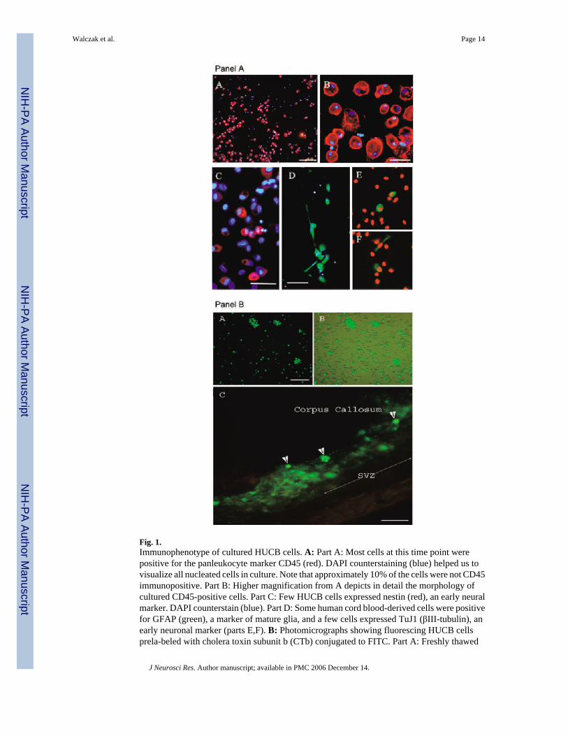
Fig. 1.Immunophenotype of cultured HUCB cells. A: Part A: Most cells at this time point werepositive for the panleukocyte marker CD45 (red). DAPI counterstaining (blue) helped us tovisualize all nucleated cells in culture. Note that approximately 10% of the cells were not CD45immunopositive. Part B: Higher magnification from A depicts in detail the morphology ofcultured CD45-positive cells. Part C: Few HUCB cells expressed nestin (red), an early neuralmarker. DAPI counterstain (blue). Part D: Some human cord blood-derived cells were positivefor GFAP (green), a marker of mature glia, and a few cells expressed TuJ1 (βIII-tubulin), anearly neuronal marker (parts E,F). B: Photomicrographs showing fluorescing HUCB cellsprela-beled with cholera toxin subunit b (CTb) conjugated to FITC. Part A: Freshly thawed
Walczak et al. Page 14
J Neurosci Res. Author manuscript; available in PMC 2006 December 14.
NIH
-PA Author Manuscript
NIH
-PA Author Manuscript
NIH
-PA Author Manuscript

HUCB cells were cultured for 24 hr and then incubated with cholera toxin (green) for 12 hrprior to transplantation. Part B: The same culture visualized through a combined fluorescentand brightfield microscope showing that not every cell was labeled with this dye. Part C: CTb-labeled green fluorescing HUCB cells within the subventricular zone of a 16-month-old Fisher344 rat. One week after grafting, we found numerous single cells or clumps of cells intenselylabeled with the dye (arrowheads). In some instances, the blurriness surrounding the graft inthe brain parenchyma may indicate possible dye leakage. Scale bars = 200 μm in A(A); 30μm in A(B); 100 μm in A(C,D); B(A,B); 50 μm in A(C), B(C).
Walczak et al. Page 15
J Neurosci Res. Author manuscript; available in PMC 2006 December 14.
NIH
-PA Author Manuscript
NIH
-PA Author Manuscript
NIH
-PA Author Manuscript

Fig. 2.Identification of HUCB cells within the subventricular zone of adult rat brain 1 week aftertransplantation. These animals were subjected to triple immunosuppression. A: Part A: Brightfield photomicrograph of cresyl violet-stained sagittal brain section of a 6-month-old ratshowing morphology and distribution of grafted HUCB cells. Easily identifiable darkly stainedgrafted cells (see also inset) are evenly distributed throughout the SVZ. Part B: Human nuclei-specific immunostaining (red) identifies HUCB-derived cells injected into the SVZ. Theinset shows the pattern of nuclear labeling at higher magnification. Part C: To detecttransplanted cells, we also used human-specific antibody against mitochondria (red). BlueDAPI counterstaining reveals the main anatomical structures and allows quantitative analysis
Walczak et al. Page 16
J Neurosci Res. Author manuscript; available in PMC 2006 December 14.
NIH
-PA Author Manuscript
NIH
-PA Author Manuscript
NIH
-PA Author Manuscript
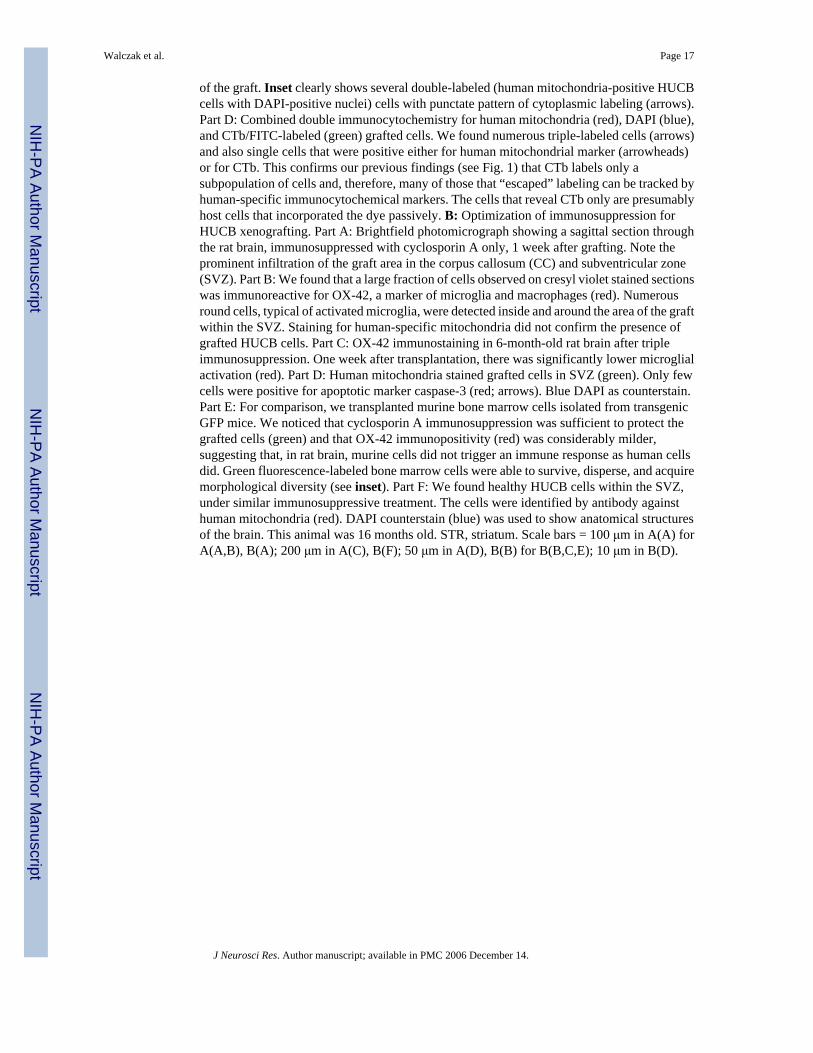
of the graft. Inset clearly shows several double-labeled (human mitochondria-positive HUCBcells with DAPI-positive nuclei) cells with punctate pattern of cytoplasmic labeling (arrows).Part D: Combined double immunocytochemistry for human mitochondria (red), DAPI (blue),and CTb/FITC-labeled (green) grafted cells. We found numerous triple-labeled cells (arrows)and also single cells that were positive either for human mitochondrial marker (arrowheads)or for CTb. This confirms our previous findings (see Fig. 1) that CTb labels only asubpopulation of cells and, therefore, many of those that “escaped” labeling can be tracked byhuman-specific immunocytochemical markers. The cells that reveal CTb only are presumablyhost cells that incorporated the dye passively. B: Optimization of immunosuppression forHUCB xenografting. Part A: Brightfield photomicrograph showing a sagittal section throughthe rat brain, immunosuppressed with cyclosporin A only, 1 week after grafting. Note theprominent infiltration of the graft area in the corpus callosum (CC) and subventricular zone(SVZ). Part B: We found that a large fraction of cells observed on cresyl violet stained sectionswas immunoreactive for OX-42, a marker of microglia and macrophages (red). Numerousround cells, typical of activated microglia, were detected inside and around the area of the graftwithin the SVZ. Staining for human-specific mitochondria did not confirm the presence ofgrafted HUCB cells. Part C: OX-42 immunostaining in 6-month-old rat brain after tripleimmunosuppression. One week after transplantation, there was significantly lower microglialactivation (red). Part D: Human mitochondria stained grafted cells in SVZ (green). Only fewcells were positive for apoptotic marker caspase-3 (red; arrows). Blue DAPI as counterstain.Part E: For comparison, we transplanted murine bone marrow cells isolated from transgenicGFP mice. We noticed that cyclosporin A immunosuppression was sufficient to protect thegrafted cells (green) and that OX-42 immunopositivity (red) was considerably milder,suggesting that, in rat brain, murine cells did not trigger an immune response as human cellsdid. Green fluorescence-labeled bone marrow cells were able to survive, disperse, and acquiremorphological diversity (see inset). Part F: We found healthy HUCB cells within the SVZ,under similar immunosuppressive treatment. The cells were identified by antibody againsthuman mitochondria (red). DAPI counterstain (blue) was used to show anatomical structuresof the brain. This animal was 16 months old. STR, striatum. Scale bars = 100 μm in A(A) forA(A,B), B(A); 200 μm in A(C), B(F); 50 μm in A(D), B(B) for B(B,C,E); 10 μm in B(D).
Walczak et al. Page 17
J Neurosci Res. Author manuscript; available in PMC 2006 December 14.
NIH
-PA Author Manuscript
NIH
-PA Author Manuscript
NIH
-PA Author Manuscript

Fig. 3.Distribution and migration of grafted HUCB cells. A: Part A: Brightfield photomicrograph ofthe sagittal section through the hemisphere of a 6-month-old rat (3 hr after grafting) with needletrack penetrating corpus callosum and targeting SVZ. Part B: Higher magnification of the samebrain area showing a deposit of HUCB cells within the SVZ. Part C: Grafted cells, prelabeledwith CTb (green), migrated along the rostral migratory stream (RMS) delineated by dashedlines. Part D: Additionally, we used an antibody against human nuclei (green) to identify cellsin the RMS. The migratory pathway is highlighted by immunostaining against doublecortin(red). This marker identifies endogenous migrating neuronal progenitors within the RMS. PartE: Grafted cells, identified with human specific antibody against mitochondria (green), were
Walczak et al. Page 18
J Neurosci Res. Author manuscript; available in PMC 2006 December 14.
NIH
-PA Author Manuscript
NIH
-PA Author Manuscript
NIH
-PA Author Manuscript

frequently found inside or in close proximity to cortical vessels surrounding the needle track.Part F: The same human specific antibody conjugated to rhodamine (red) was used to detectcells within the RMS. The slide was counterstained with nuclear DAPI (blue). B: Theimmunophenotype of HUCB cells within the rat brain one week after grafting. Parts A,B:Neural antigens. Part A: Occasionally, HUCB-derived cells within the migratory pathway co-expressed nestin, an early marker of neural progenitors. Asterisks indicate several endogenousneural progenitors (red) within the pathway; arrow points to CTb-labeled (green), nestin-positive (yellow) HUCB cell. Part B: Some HUCB cells were identified within the SVZ by adual immunostaining for human nuclei (green) and neuronal marker doublecortin (red). Arrowindicates a cell expressing both markers. LV, lateral ventricle. Parts C–E: Hematopoieticantigens. Part C: Few HUCB cells within the SVZ were positive for human-specific CD133antigen (red). This marker is common for neural and hematopoietic progenitors. Part D:Expression of CD45, a panleukocyte marker, was detected in some HUCB-derived cells (red).Part E: Human-specific CD4 positivity was found in only a few cells (red), indicating thepossibility that the subpopulation of mature lymphocytes, major inducers of rejection, mighthave been eliminated from the graft and that further strong immunoshielding may not berequired. Scale bar = 500 μm in A(A); 50 μm in A(B), in A(D) for A(D,E), and in B(B) for B(B–E); 150 μm in A(C,F); 25 μm in B(A).
Walczak et al. Page 19
J Neurosci Res. Author manuscript; available in PMC 2006 December 14.
NIH
-PA Author Manuscript
NIH
-PA Author Manuscript
NIH
-PA Author Manuscript
Copyright © 2022 FDOKUMEN