Cyclin D1 fine-tunes the neurogenic output of embryonic retinal progenitor cells
Upload
independentCategory
view
1download
0
Neuron
Article
Polycomb Limits the Neurogenic Competenceof Neural Precursor Cells to PromoteAstrogenic Fate TransitionYusuke Hirabayashi,1 Nao Suzki,1 Masafumi Tsuboi,1 Takaho A. Endo,2 Tetsuro Toyoda,2 Jun Shinga,3 Haruhiko Koseki,3
Miguel Vidal,3,4 and Yukiko Gotoh1,*1Institute of Molecular and Cellular Biosciences, University of Tokyo, Tokyo, Japan2RIKEN Genomic Sciences Center3RIKEN Center for Allergy and Immunology
Kanagawa, Japan4Centro de Investigaciones Biologicas, Consejo Superior de Investigaciones Cientıficas, Madrid, Spain
*Correspondence: [email protected] 10.1016/j.neuron.2009.08.021
SUMMARY
During neocortical development, neural precursorcells (NPCs, or neural stem cells) produce neuronsfirst and astrocytes later. Although the timing of thefate switch from neurogenic to astrogenic is criticalfor determining the number of neurons, the mecha-nisms are not fully understood. Here, we show thatthe polycomb group complex (PcG) restricts neuro-genic competence of NPCs and promotes the transi-tion of NPC fate from neurogenic to astrogenic. Inac-tivation of PcG by knockout of the Ring1B or Ezh2gene or Eed knockdown prolonged the neurogenicphase of NPCs and delayed the onset of the astro-genic phase. Moreover, PcG was found to repressthe promoter of the proneural gene neurogenin1 ina developmental-stage-dependent manner. Theseresults demonstrate a role of PcG: the temporal regu-lation of NPC fate.
INTRODUCTION
The mechanism regulating the timing of stem cell fate switching
is crucial for understanding tissue development. Neurons and
astrocytes in the neocortex are derived from common multipo-
tent NPCs which sequentially pass through phases of expansion,
neurogenesis and astrogenesis (Hirabayashi and Gotoh, 2005;
Miller and Gauthier, 2007). The production of neurons before
astrocytes allows the initial establishment of a neuronal network
and subsequent integration of astrocytes into the network. The
timing of the neurogenic-to-astrogenic fate switch is critical for
brain development, in particular because the length of the neuro-
genic phase is a key parameter in determining the number of
neurons in each brain region.
NPC fate is regulated by both extracellular signals and cell-
intrinsic programs (Desai and McConnell, 2000). The extracellular
signals, including ciliary neurotrophic factor (CNTF), leukemia
inhibitory factor (LIF), and cardiotrophin-1 (CT-1), activate the
600 Neuron 63, 600–613, September 10, 2009 ª2009 Elsevier Inc.
JAK-STAT pathway, which plays a pivotal role in the promotion
of astrocytic differentiation (Barnabe-Heider et al., 2005; Bonni
et al., 1997; He et al., 2005; Johe et al., 1996; Rajan and McKay,
1998). The bone morphogenetic protein (BMP) and Notch path-
ways also interact with the JAK-STAT pathway to promote
astrocytic differentiation (Gaiano and Fishell, 2002; Kamakura
et al., 2004; Nakashima et al., 1999). However, some ligands
that activate the JAK-STAT pathway are expressed during the
neurogenic phase when astrocytic genes are silent (Molne
et al., 2000). At least three mechanisms have been proposed to
account for the suppression of astrocytic genes during the
neurogenic phase. First, STAT responsive elements at the astro-
cytic gene loci are inaccessible for STAT proteins during the
neurogenic phase due to DNA or histone H3K9 methylation
(Fan et al., 2005; Song and Ghosh, 2004; Takizawa et al., 2001).
Second, astrocytic genes are inactivated by erbB4-NCoR
signaling during the neurogenic phase (Sardi et al., 2006). And
third, STAT proteins are inactivated by the proneural basic
helix-loop-helix (bHLH) transcription factors present during the
neurogenic phase.
Genetic and molecular evidence has established the essential
roles of proneural bHLH proteins in suppressing astrocytic fate
(Guillemot, 2007), such that deletion of bHLH genes causes
precocious astrocyte differentiation and loss of the neuronal
fate (Nieto et al., 2001; Tomita et al., 2000). Two such bHLH tran-
scription factors, Neurogenin (Ngn) 1 and Ngn2, are expressed
solely during the neurogenic, not in the astrocytic, phase of
neocortical development (Sun et al., 2001, and references
therein). Ngn1 expression in vitro and in vivo can suppress astro-
cytic differentiation of NPCs in part because Ngn1 sequesters
the p300/CBP-Smad1 complex from STAT3, which leads to
STAT3 inactivation/dephosphorylation (Cai et al., 2000; Guil-
lemot, 2007; Sun et al., 2001). Since STAT3 activation is neces-
sary for astrogenesis to occur (He et al., 2005), the expression of
bHLH proteins can suppress astrogenesis even when astrocytic
genes are released from erbB4-NCoR signaling and demethy-
lated. Turning off the bHLH proteins is therefore a critical event
for the onset of astrocyte differentiation.
The regulation of proneural bHLH genes, primarily Ngn1 and
Ngn2 in the neocortex, is thus key for understanding phase

Neuron
Neurogenic-to-Gliogenic Fate Switch by Polycomb
transition in NPCs (Guillemot, 2007). Wnt signaling has been
shown to induce the expression of ngn1 and ngn2 and promote
neocortical neuronal differentiation (Hirabayashi et al., 2004;
Israsena et al., 2004). However, Wnt ligands continue to be
expressed during the astrogenic phase (Shimogori et al., 2004),
suggesting that their effect on Ngn1 and Ngn2 is somehow
blocked.
The neurogenic-to-astrogenic fate switch can be observed
even within clones of single NPCs in culture, suggesting that
cell-intrinsic programs also play essential roles (Naka et al.,
2008; Qian et al., 2000). The intrinsic mechanisms may change
the responsiveness of NPCs to fate-regulating extracellular sig-
nals and thus control the timing of the fate switch. For instance,
intrinsic mechanisms may suppress Wnt induction of Ngn
expression during the gliogenic phase and derepress STAT3
so that NPCs can respond to astrocyte-promoting cytokines.
It has become increasingly clear in recent years that epige-
netic events control responsiveness of each cell to extrinsic
signals and govern cell fate decisions. Polycomb group (PcG)
proteins have emerged as central players in these epigenetic
programming events. PcG proteins are transcription repressors
that function by modulating chromatin structure (Schwartz and
Pirrotta, 2007). They reside in two main complexes referred to
as polycomb repressive complexes 1 and 2 (PRC1 and PRC2).
PRC2 contains Eed, Suz12, and the methyltransferase Ezh1
or Ezh2 that catalyzes histone H3 lysine 27 trimethylation
(H3K27me3) (Cao and Zhang, 2004; Shen et al., 2008). This
histone modification then provides a platform to recruit PRC1,
which contains the ubiquitin ligase Ring1 (Ring1A or Ring1B),
an essential component for PcG-mediated repression (de Na-
poles et al., 2004). Although PcG proteins have been demon-
strated to play critical roles for the maintenance of ES cells as
well as adult stem cell populations such as hematopoietic and
neural stem cells (Spivakov and Fisher, 2007; Valk-Lingbeek
et al., 2004), their roles in multipotent progenitors during tissue
development have largely remained unclear.
In this study, we show that the PcG proteins epigenetically
suppress the ngn1 locus during the astrogenic phase and thus
trigger the neurogenic-to-astrogenic fate switching of NPCs
and regulate the duration of the neurogenic phase in the devel-
oping neocortex.
RESULTS
Temporal Restriction of Neurogenic Competencein the Neocortical NPCsPreviously, we and others showed that the canonical Wnt
pathway induces expression of ngn1 and ngn2 and functions
instructively to promote neuronal differentiation of neocortical
NPCs (Guillemot, 2007; Hirabayashi and Gotoh, 2005; Hirabaya-
shi et al., 2004; Israsena et al., 2004; Zhou et al., 2006). It was
puzzling, however, that Wnt ligands continue to be expressed
during the astrogenic phase (Shimogori et al., 2004). We exam-
ined whether the response of NPCs to Wnt signaling is depen-
dent on the developmental stage using in vitro culture methods
(Qian et al., 2000). Neuroepithelial cells isolated from E11.5
neocortex were cultured for 3, 6, and 9 days in vitro (DIV) as
a suspension in the presence of fibroblast growth factor (FGF)
2 and epidermal growth factor (EGF) and subsequently plated
onto poly-D-lysine-coated dishes. Cells were infected with either
control retrovirus or retrovirus encoding a stabilized b-catenin
(S33Yb-catenin), which constitutively activates the canonical
Wnt pathway (Korinek et al., 1997). Under these conditions,
more than 99% of infected (GFP-positive) cells are nestin-posi-
tive undifferentiated NPCs, and 3 and 9 DIV cultures correspond
to the neurogenic and astrogenic phases, respectively (Hira-
bayashi and Gotoh, 2005; Qian et al., 2000). Three days after
infection of 3 DIV cultures with S33Yb-catenin virus, the propor-
tion of cells positive for the neuronal marker b3-tubulin (TuJ1)
markedly increased (Figures 1A and 1B). In contrast, expression
of S33Yb-catenin had less of an effect on neuronal differentiation
in 9 DIV cultures (Figures 1A and 1B). Expression of S33Yb-cat-
enin also had less of an effect on neuronal differentiation of 3 DIV
culture isolated from E17.5 neocortex compared to that of 3 DIV
culture isolated from E11.5 neocortex (Figure 1B). Therefore, the
effects of S33Yb-catenin on neuronal differentiation appear to
decrease with age. As a control, enforced expression of ngn1
in NPCs of any age did induce neuronal differentiation (Figures
1A and 1B). Consistent with this observation, the effect of
S33Yb-catenin on increasing the amounts of ngn1 and ngn2
mRNAs were also reduced in 9 DIV NPCs compared to 3 DIV
NPCs (Figures 1C and S3A). These results suggest that NPCs
lose their capacity to differentiate into neurons in response to
the canonical Wnt pathway during late stages of development.
In addition, they imply that a step between b-catenin activation
and ngn1 expression is blocked in the late/astrogenic stage of
development.
We considered the possibility that this blockade was due to
the absence of components of the b-catenin/TCF transcriptional
complex at late stages. However, we found that S33Yb-catenin
stimulates expression of SuperTOP-FLASH, a luciferase reporter
gene construct expressed under the control of TCF-responsive
elements (Kaykas et al., 2004), in both E11.5 + 3 DIV and 12 DIV
cultures (Figure 1D). Moreover, treatment of cultures for 2 hr
with the GSK3 inhibitor SB216763, which activates the canonical
Wnt pathway (Cohen and Goedert, 2004), induced Axin2, an
immediate-early gene in the canonical Wnt pathway (Aulehla
et al., 2003), to a similar extent in both E11.5 + 3 DIV and 9 DIV
cultures (Figure S1). Therefore, b-catenin appears to be able to
form an active transcriptional complex with TCF even in late-
stage cells.
Temporal Change of the Chromatin State at NeurogeninPromotersWe tested whether the epigenetic chromatin state of ngn gene
loci changes during development. We first examined the DNA
methylation status of the ngn1 promoter by the bisulfite direct
sequence method (Clark et al., 1994) but found that the ngn1
promoter was barely methylated in either E11.5 + 3 DIV or 9 DIV
cultures (Figure S2). We next investigated posttranslational
modification of histone H3 bound to the ngn promoters using
chromatin immunoprecipitation (ChIP). Histone H3 acetylation
at Lys9 and Lys14 (H3K9K14ac) or trimethylation at Lys9 and
Lys27 (H3K9me3 and H3K27me3) in general correlates with
open or closed chromatin state, respectively (Berger, 2002;
Cao and Zhang, 2004). H3K9K14ac was enriched at the proximal
Neuron 63, 600–613, September 10, 2009 ª2009 Elsevier Inc. 601

Neuron
Neurogenic-to-Gliogenic Fate Switch by Polycomb
(around TATA box) region of the ngn1 and ngn2 promoters
compared to the intergenic region in E11.5 + 3 DIV cultures,
but its levels were reduced gradually in the late-stage cultures
Control
Infection
Ngn1
S33Yβ-catenin
+ 3 DIV + 3 DIV+ 6 DIV
E11.5 E17.5
+ 9 DIV
TuJ1
+ c
ells
/GF
P+
cells
[%
]
0
20
40
60
80
100
A
B
C
D
0
5
10
15
20
25
Lu
cife
rase
Act
ivit
y (A
rbit
rary
Un
it)
SuperTOP SuperFOP0
50
100
150
Lu
cife
rase
Act
ivit
y (A
rbit
rary
Un
it)
SuperTOP SuperFOP
Control
S33Yβ-catenin
3 DIV 12 DIV
Ng
n1
mR
NA
/Gap
dh
mR
NA
(Arb
itra
ry U
nit
)
9 DIV3 DIV
0
0.5
1
1.5
2
2.5
3
3.5
4
4.5
5
S33Yβ-cateninCon S33Y
β-cateninConInfection
TuJ1/GFP
E11
.5 +
3 D
IVE
11.5
+ 9
DIV
Control S33Yβ-catenin Ngn1 Figure 1. NPCs Lose Their Capacity to
Differentiate into Neurons in Response to
the Canonical Wnt Pathway at Late Stages
of Development
NPCs isolated from E11.5 were cultured for 3, 6, or
9 DIV, and E17.5 cells were cultured for 3 DIV. Next
the cells were infected with a retrovirus encoding
GFP alone (control), GFP together with S33Yb-cat-
enin, or Ngn1 and incubated for an additional
3 days with FGF2.
(A) Anti-GFP (green) and TuJ1 (red) immunofluo-
rescence are shown for typical fields in control,
S33Yb-catenin, or Ngn1-expressing cells.
(B) The percentage of TuJ1+ cells among GFP+
cells was determined. Data are means + SEM of
values from four samples.
(C) E11.5 NPCs cultured for the indicated periods
were infected with a retrovirus encoding GFP
alone (control) or GFP together with S33Yb-cate-
nin and incubated for 3 days with FGF2 and subse-
quently for 3 hr without FGF2. The amount of ngn1
mRNA was determined at the end of these treat-
ments by quantitative PCR. Data are means +
SEM of values from three samples.
(D) E11.5 NPCs cultured for the indicated periods
were transfected with pMX-S33Yb-catenin (open
bars) or empty vector (filled bars), together with
Super TOP-FLASH or Super FOP-FLASH (a
negative control reporter plasmid with a mutated
TCF binding site) for 20 hr. Data are means +
SEM of values from three samples. Scale bar,
20 mm.
(Figures 2A–2C and S3B). Furthermore,
the levels of H3K27me3 at the ngn1 and
ngn2 promoters increased gradually in
late-stage cultures (Figures 3A and S3C),
whereas the levels of H3K9me3 and
H3K9me2 at these promoters remained
low in both early and late cultures (data
not shown). Developmental increases in
the levels of H3K27me3 at the ngn1 and
ngn2 promoters were also found in NPCs
isolated from E14 and E17 neocortices
(Figures S3D and S5A). These results
suggest that the chromatin state of the
ngn promoters gradually becomes closed
over time during development, and ngn
genes are thus transcribed less in the
late stages. Consistent with this notion,
the levels of RNA polymerase II associ-
ated with the ngn promoters were
reduced in the late cultures (Figures 2D,
S3E, and S5B).
We then asked whether stage-depen-
dent changes in histone deacetylation
affected ngn expression by treating NPCs
with the histone deacetylases (HDAC) inhibitor valproic acid
(VPA) (Gottlicher et al., 2001). VPA treatment for 16 hr increased
the levels of H3K9K14ac at the ngn1 promoter (Figure 2E)
602 Neuron 63, 600–613, September 10, 2009 ª2009 Elsevier Inc.

Neuron
Neurogenic-to-Gliogenic Fate Switch by Polycomb
and significantly increased ngn1 expression (Figures 2F and
S4A) in E11.5 + 9 DIV culture. In contrast, VPA treatment caused
only a small increase in ngn1 expression in E11.5 + 0 or 3 DIV
cultures (Figures 2F and S4A). This suggests that histone deace-
tylation suppresses the expression of ngn1 in late-stage
cultures.
Imm
un
op
reci
pit
ated
DN
A/in
pu
t[%
]
0
0.5
0.4
0.3
0.2
0.1
3 9 15 DIV DIV DIV
Control IgGH3K9K14ac
Ngn1
A
A B
B C
C
9 DIV
0
0.05
0.1
0.15
0.2
3 6 9
Po
l II/i
np
ut
[%]
DIV
Ngn1 TATAD
3 9 15 3 9 15
TATA intergenic
0
8
6
4
2H3K
9K14
ac/H
3(A
rbit
rary
Un
it)
Ngn1 Ngn1 3’
+ VPA- VPA
E
(TATA)
Ngn1 mRNA (+VPA)Ngn1 mRNA (–VPA)
0
2
4
6
8
10
12
0 3 6 9
DIV
Fo
ld in
crea
se
F
Figure 2. Decreases in the Level of H3K9K14 Acetylation at the ngn1
Promoter Result in the Repression of ngn1 mRNA Expression
Chromatin complex was immunoprecipitated from E11.5 neocortical NPCs
cultured for 3, 9, or 15 DIV with anti-H3K9K14ac (A–C) or anti-RNA polymerase
II (D). The immunoprecipitates were subjected to PCR amplification of �1299
to �976 (TCF site [A], �45 to +39 [TATA box, B and D], and +5170 to +5274
[intergenic, C] base pairs from the transcription start site of ngn1 gene. (E)
The level of acetylation at the ngn1 promoter in VPA-treated (filled bars) or
untreated cells (open bars) was determined by ChIP. (F) The fold change of
ngn1 mRNA amounts caused by VPA treatment at indicated culture periods
was measured (see Figure S4A for absolute values). Data are means + SEM
of values from three samples.
PRC1 Suppresses ngn1 Expression in the Late Stageof Neocortical DevelopmentWe then examined the role of H3K27me3 in ngn expression. Tri-
methylation of H3K27 is catalyzed by PRC2, which includes Eed,
Suz12, and the histone methyltransferase Ezh2 (or Ezh1) (Cao
and Zhang, 2004; Shen et al., 2008). This modification is recog-
nized by PRC1, which induces a closed chromatin state
(Schwartz and Pirrotta, 2007). We inactivated PRC1 by condi-
tional deletion of an essential component, the ubiquitin ligase
Ring1B (de Napoles et al., 2004; Voncken et al., 2003). Neocor-
tical NPCs were isolated from E12.5 mice containing both
Ring1B alleles flanked by the loxP sequences (Ring1Bf/f) and
a transgene expressing tamoxifen-dependent Cre recombinase
under the control of the endogenous ROSA26 promoter
(ROSA26::ERT2-Cre) (Cales et al., 2007; Endoh et al., 2008).
Treatment of NPCs isolated from the Ring1Bf/f;ERT2-Cre mice
with 4-hydroxy-tamoxifen (4-OHT) effectively reduced Ring1B
expression (Figure 3B). Following treatment, the levels of ngn1
mRNA in the 9 DIV cultures markedly increased (�8-fold),
whereas those in the 3 DIV cultures exhibited a much smaller
increase (�1.6-fold) (Figures 3C and S4B). This indicates that
the Ring1B gene contributes to the suppression of ngn1 expres-
sion in the late stages of neocortical development. Inhibition of
HDAC further enhanced ngn1 expression induced by the loss
of Ring1B gene to levels comparable with those in the early stage
(3 DIV) cultures (Figures 3D and S4B). This suggests that HDACs
and PRC1 function cooperatively in the suppression of ngn1 at
late stages of neocortical development. Consistent with this
notion, expression of S33Yb-catenin enhanced the levels of
ngn1 mRNA even in late stage (9 DIV) cultures when Ring1B
was deleted (Figure 3E). On the other hand, the levels of ngn2
mRNA were only moderately enhanced or even reduced by the
loss of Ring1B gene and by inhibition of HDACs, with or with-
out activation of Wnt signaling (Figures S3F, S3G, S3H, and
Table S1). Therefore, additional mechanisms might also con-
tribute to the suppression of ngn2 expression in the late stage
of neocortical development.
Next, we examined ngn1 expression in vivo during neocortical
development. Immunohistochemistry revealed that Ngn1 was
highly expressed at E14.5 in the ventricular zone (VZ) of
neocortex, but its expression gradually declined until E17.5,
when only a small number of cells at the neocortical VZ were
found to express Ngn1 protein (Figures 3H and S6A). However,
when the Ring1B gene was deleted in Ring1Bf/f;ERT2-Cre
mice by injecting tamoxifen intraperitoneally into pregnant
mice at 12.5 days postcoitum (dpc) (Figure 3G), the number of
Ngn1-expressing cells in the VZ increased at E17.0 or E17.5 to
levels comparable with those in the late neurogenic phase
(e.g., E16.5, see Figure S6A) (Figures 3H–3J). Ring1B deletion
did not increase the number of Ngn1-expressing cells at early
stages of the neurogenic phase (e.g., E14.5, Figure S6B).
Increase in Ngn1 protein by Ring1B deletion in the neocortex
at E17.5 was confirmed by western blot analysis (Figures 3L
and 3M). Quantitative RT-PCR analysis also indicated that the
amounts of ngn1 mRNA, but not those of ngn2 mRNA, were
significantly greater in the lysates of Ring1B-deficient cortices
compared to those of control ones prepared at E18.5 (Fig-
ures 3K and S3I). These results demonstrate an in vivo role for
Neuron 63, 600–613, September 10, 2009 ª2009 Elsevier Inc. 603

Neuron
Neurogenic-to-Gliogenic Fate Switch by Polycomb
ANgn1 TATA
B C
D E
H3K
27m
e3/in
pu
t [%
]
0
2
4
6
8
3 6DIV
9
— +
Ring1B
X ERT2-CreRing1Bfl/fl
4-OHT
GAPDH
Ngn1
WB
Ring1B
3 DIV
1
Ng
n1
mR
NA
/Gap
dh
mR
NA
(Arb
itra
ry U
nit
)
Ng
n1
mR
NA
/Gap
dh
mR
NA
(Arb
itra
ry U
nit
)
Ng
n1
mR
NA
/Gap
dh
mR
NA
(Arb
itra
ry U
nit
)
Ng
n1
mR
NA
/Gap
dh
mR
NA
(Arb
itra
ry U
nit
)
0
5
10
ControlKO
+ +——Con KOKOCon
VPARing1B
9 DIV
in vivo
12 DIV
0
5
109 DIV
1
ControlKO
Ring1B
Ng
n1
mR
NA
/Gap
dh
mR
NA
(Arb
itra
ry U
nit
)N
gn
1 p
rote
in/G
apd
h p
rote
in(A
rbit
rary
Un
it)
Con ConInfection
J K
ML
Control Ring1BKO
ConRing1B
KO
VZ VZ
0
5
10
15
20
No
. of
Ng
n1+
cel
ls/a
rea
(Arb
itra
ry u
nit
)
** **Control Ring1B KO
F G
H I
Rin
g1B
/TO
PR
ON
gn
1/T
OP
RO
(-) FGF2
(-) FGF2
(-) FGF2
0
2
4
6
8
10
0
20
40
60
80
100
S33Yβ-catenin
S33Yβ-catenin
KOConRing1B KOCon
Control Ring1BKO
Control Ring1BKO
0
0.2
0.4
0.6
0.8
1
WB
Ngn1+
cell No.( ) in vivo Ngn1mRNA( )
in vivoin vivo
Ngn1protein( )
**
GAPDH
0
0.5
1
1.5
2
2.5
3
3.5
Figure 3. Ring1B/PRC1 Suppresses ngn1
Expression at Late Stages of Neocortical
Development
(A) Abundance of H3K27 trimethylation at the ngn1
promoter (TATA box) was assessed by ChIP and
quantitative PCR from E11.5 neocortical NPCs
cultured for 3, 6, or 9 DIV.
(B and C) NPCs isolated from E12.5 Ring1Bf/f;
ERT2-Cre mice were cultured for 0 or 6 DIV and
cultured for an additional 3 DIV with 4-OHT (KO)
or EtOH (Con). After 3 hr of FGF2 deprivation, the
amount of ngn1 mRNA was determined at the
end of these treatments by quantitative PCR
(see Figure S4B for absolute values). Data are
means + SEM of values from three samples. Cell
lysates at 3 DIV were subjected to western blotting
(WB) using the antibodies indicated (B).
(D and E) The cells were further cultured in the
absence of 4-OHT, with or without VPA for 1 day
and, subsequently, without FGF2 for 2 hr (D).
NPCs isolated from E12.5 Ring1Bf/f;ERT2-Cre
mice were cultured for 6 DIV, infected with a retro-
virus encoding GFP (Con) or GFP together with
S33Y b-catenin and cultured for an additional 3
DIV with 4-OHT (KO) or EtOH (Con) (E). The amount
of ngn1 mRNA was then determined. Data are
means + SEM of values from three samples.
(F–M) Tamoxifen was introduced into control
(Ring1Bf/f) mice (F and H) or Ring1Bf/f;ERT2-Cre
mice (G and I) at E12.5. At E17.0, embryos were
fixed and subjected to immunohistochemistry
using the antibodies against Ring1B (F and G) or
Ngn1 (H and I). Nuclei were counterstained with
Topro-3. Note that the number of Ngn1+ cells in
the neocortical ventricular zone (VZ) increased by
Ring1B deletion. Scale bars, (G) 20 mm, (I) 10 mm.
(J) Quantification of Ngn1+ cells at E17.5. Data
are the mean + SEM of values for ten (control) or
nine (KO) areas of corresponding sections from
each embryo analyzed. In (K)–(M), neocortical
lysates were prepared at E18.5 (K) or E17.5 (L, M),
and the amounts of ngn1 mRNA (K) or protein
(L and M) were determined. (K) Data are the
mean + SEM of values for four control cortices
and 12 KO cortices analyzed. (M) Data are the
mean + SD of values for five control cortices and
three KO cortices analyzed by western blotting
(L). *p < 0.05; **p < 0.005.
Ring1B in the suppression of Ngn1 at the end of the neurogenic
phase.
PRC1 Restricts Neurogenic Competence at Late Stagesof Neocortical DevelopmentThe next key question is whether Ring1B is necessary for the
suppression of the neurogenic fate at late stages of develop-
ment. In the absence of neurogenic signals, deletion of Ring1B
from NPCs isolated from Ring1Bf/f;ERT2-Cre mice by 4-OHT
treatment had little effect on neuronal differentiation at either
E11.5 + 3 DIV or 9 DIV (Figures 4A and 4B), suggesting that the
loss of Ring1B on its own does not result in neurogenesis. By
contrast, loss of Ring1B gene significantly enhanced neuronal
differentiation induced by S33Yb-catenin expression at 9 DIV,
but it had less effect at 3 DIV (Figure 4A). Similarly, deletion of
604 Neuron 63, 600–613, September 10, 2009 ª2009 Elsevier Inc.
Ring1B by treatment with 4-OHT enhanced the S33Yb-cate-
nin-induced neuronal differentiation of NPCs from E17.5
Ring1Bf/f;ERT2-Cre mice cultured for 3 DIV (data not shown).
The loss of Ring1B also significantly enhanced neuronal differen-
tiation induced by growth factor deprivation at E11.5 + 9 DIV
(Figure 4B). These results suggest that PRC1 contributes to the
suppression of neurogenic ‘‘competence’’ during late stages of
NPC culture.
The deletion of Ring1B from E11.5 + 9 DIV NPC cultures iso-
lated from Ring1Bf/f;ERT2-Cre mice by 4-OHT treatment had
little effect on the rate of cell proliferation detected by BrdU
incorporation (Figure 4C) or cell death detected by cleaved cas-
pase-3 (less than 3% of infected cells). Furthermore, Ring1B
deletion markedly increased the proportion of neuron-only and
neuron-containing clones at the expense of nonneuronal clones

Neuron
Neurogenic-to-Gliogenic Fate Switch by Polycomb
Figure 4. Ring1B/PRC1 Is Necessary for the
Termination of Neurogenesis in the Neocortex
(A and B) E12.5 Ring1Bf/f;ERT2-Cre NPCs were cultured
for 0 or 6 DIV and then treated with 4-OHT (KO) or EtOH
(Con) for an additional 3 DIV. Cells were then plated and
infected with a retrovirus encoding GFP (Control) or GFP
together with S33Yb-catenin in the presence of FGF2 (A)
or incubated with or without FGF2 (B). After 2 days, the
percentage of TuJ1+ cells among GFP+ cells was deter-
mined. Data are means + SEM of values from three
samples.
(C) Cells at 9 DIV were incubated with BrdU for 2 hr at the
end of the treatments in (A). The percentage of BrdU+ cells
among GFP+ cells was determined. Data are means +
SEM of values from three samples.
(D) A clonal analysis of (A). E12.5 Ring1Bf/f;ERT2-Cre
NPCs were cultured for 9 DIV and then treated with 4-
OHT (KO) or EtOH (Con) for an additional 3 DIV. Cells
were infected with a retrovirus encoding GFP (Control)
or GFP together with S33Yb-catenin at a low titer. After
incubation in the presence of FGF2 for 2 days, cells in
each clone were stained with TuJ1 antibody, and the
clones were classified as containing either only TuJ1+
cells (neuron-only clone), both TuJ1+ and TuJ1– cells
(neuron-containing clone), or only TuJ1– cells (nonneuro-
nal clone).
(E–L) Ring1Bf/f;NestinCreERT2 mice or control (Ring1Bf/f)
mice were administrated with tamoxifen at E13.5 (E–G) or
E16.0 (H–L) and BrdU at E19.0 (E–G). The pups were fixed
at P6.5 (E–G) or P4.5 (H–L) and subjected to immunohis-
tochemistry using the antibodies indicated. Coronal
sections of primary sensory area in the neocortex are
shown. In (G), neuronal differentiation was determined
by examining cells positive for both BrdU and the upper
layer neuronal marker Cux1 (Nieto et al., 2004) located
in the upper layers. In (L), the numbers of Brn2-positive
cells located in the upper layers were determined. Data
are the mean + SD of values for three control and four
KO mice (E–G) and the mean + SEM of values for 16 cor-
responding areas each from two control mice or five
knockout mice (L). MZ, marginal zone ; II/III, layer II/III ;
IV, layer IV. Scale bar, 30 mm (F), 20 mm (K).
Neuron 63, 600–613, September 10, 2009 ª2009 Elsevier Inc. 605
Neuron
Neurogenic-to-Gliogenic Fate Switch by Polycomb
in a clonal assay in the presence of S33Yb-catenin (Figure 4D).
These results indicate that PRC1/Ring1B instructs neuronal
fate rather than promotes selective proliferation or survival of
neuronal progenitors in the late stages of NPC culture.
We then tested whether PRC1/Ring1B indeed suppresses
neurogenesis in vivo at late stages of neocortical development.
To examine late stage phenotypes, we conditionally deleted
the Ring1B gene in the central nervous system using mice
harboring Ring1Bf/f and an ERT2-Cre transgene under the con-
trol of the nestin enhancer (Ring1Bf/f;NestinERT2-Cre mouse).
The major wave of neurogenesis is normally terminated perina-
tally. Birthdating studies have revealed that neocortical neurons
can be labeled by BrdU at E18.5 but not at E19.0 when examined
20 days after BrdU injection (Levers et al., 2001). We confirmed
that BrdU-labeled cells were hardly found in the upper layer of
WB
GAPDH
GFAP
GFA
P+
cells
/GF
P+
cells
[%
]
Co
ntr
ol
E18
.0
Rin
g1B
KO
Sox2GFAP GFAP Sox2 TOPRO
VZ
0
10
20
30
40
50
60
No
. of
So
x2+
cells
/are
a(A
rbit
rary
un
it)
Control Ring1BKO
Con Ring1BKO
Con Ring1BKO
E18
.0
E17
.5P2.
5C
on
tro
lR
ing
1B K
O
Co
ntr
ol
Rin
g1B
KO
Control Ring1B KO
Control
Ring1B KO
Control Ring1B KOControl Ring1B KO
Pax6
Nestin
S100 TOPRO
C
D
H
I
J
L M N
J'
K'
K
E G
A B
F
No
. of
S10
0+ cel
ls/a
rea
(Arb
itra
ry u
nit
)
in vivo (P2.5)
(Arb
itra
ry u
nit
)
0
5
10
15
20
*O P
(-) FGF2
0
2
4
6
8
10
12
14
Sox2
NestinGFAP GFAPNestinTOPRO
Control Ring1BKO
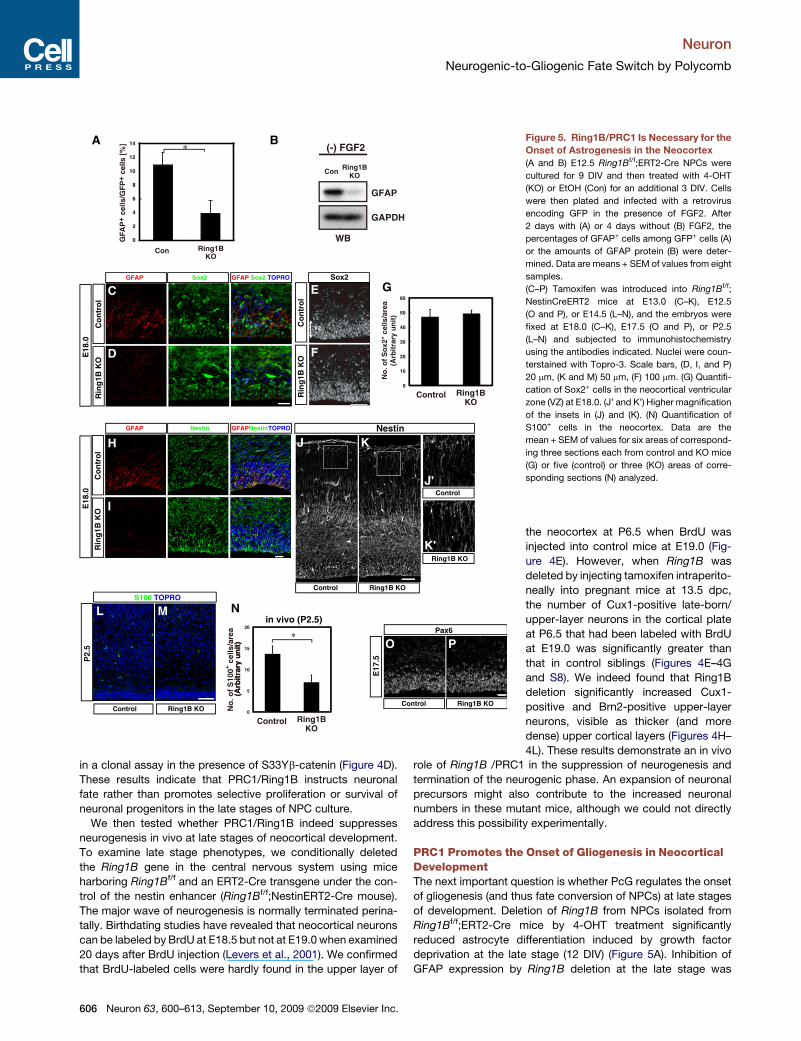
*Figure 5. Ring1B/PRC1 Is Necessary for the
Onset of Astrogenesis in the Neocortex
(A and B) E12.5 Ring1Bf/f;ERT2-Cre NPCs were
cultured for 9 DIV and then treated with 4-OHT
(KO) or EtOH (Con) for an additional 3 DIV. Cells
were then plated and infected with a retrovirus
encoding GFP in the presence of FGF2. After
2 days with (A) or 4 days without (B) FGF2, the
percentages of GFAP+ cells among GFP+ cells (A)
or the amounts of GFAP protein (B) were deter-
mined. Data are means + SEM of values from eight
samples.
(C–P) Tamoxifen was introduced into Ring1Bf/f;
NestinCreERT2 mice at E13.0 (C–K), E12.5
(O and P), or E14.5 (L–N), and the embryos were
fixed at E18.0 (C–K), E17.5 (O and P), or P2.5
(L–N) and subjected to immunohistochemistry
using the antibodies indicated. Nuclei were coun-
terstained with Topro-3. Scale bars, (D, I, and P)
20 mm, (K and M) 50 mm, (F) 100 mm. (G) Quantifi-
cation of Sox2+ cells in the neocortical ventricular
zone (VZ) at E18.0. (J’ and K’) Higher magnification
of the insets in (J) and (K). (N) Quantification of
S100+ cells in the neocortex. Data are the
mean + SEM of values for six areas of correspond-
ing three sections each from control and KO mice
(G) or five (control) or three (KO) areas of corre-
sponding sections (N) analyzed.
the neocortex at P6.5 when BrdU was
injected into control mice at E19.0 (Fig-
ure 4E). However, when Ring1B was
deleted by injecting tamoxifen intraperito-
neally into pregnant mice at 13.5 dpc,
the number of Cux1-positive late-born/
upper-layer neurons in the cortical plate
at P6.5 that had been labeled with BrdU
at E19.0 was significantly greater than
that in control siblings (Figures 4E–4G
and S8). We indeed found that Ring1B
deletion significantly increased Cux1-
positive and Brn2-positive upper-layer
neurons, visible as thicker (and more
dense) upper cortical layers (Figures 4H–
4L). These results demonstrate an in vivo
role of Ring1B /PRC1 in the suppression of neurogenesis and
termination of the neurogenic phase. An expansion of neuronal
precursors might also contribute to the increased neuronal
numbers in these mutant mice, although we could not directly
address this possibility experimentally.
PRC1 Promotes the Onset of Gliogenesis in NeocorticalDevelopmentThe next important question is whether PcG regulates the onset
of gliogenesis (and thus fate conversion of NPCs) at late stages
of development. Deletion of Ring1B from NPCs isolated from
Ring1Bf/f;ERT2-Cre mice by 4-OHT treatment significantly
reduced astrocyte differentiation induced by growth factor
deprivation at the late stage (12 DIV) (Figure 5A). Inhibition of
GFAP expression by Ring1B deletion at the late stage was
606 Neuron 63, 600–613, September 10, 2009 ª2009 Elsevier Inc.

Neuron
Neurogenic-to-Gliogenic Fate Switch by Polycomb
confirmed by western blot analysis (Figure 5B). Therefore,
Ring1B/PRC1 appears to be necessary for the induction of
astrocyte differentiation in the late stage in vitro.
We further examined astrogenesis in vivo. Whereas GFAP-
positive cells could already be detected in the lateral VZ of
control neocortex at E18.0, GFAP expression was barely detect-
able in the corresponding region of Ring1B-deficient mice
(Figures 5C, 5D, 5H, and 5I). Similarly, the number of cells posi-
tive for another astrocytic marker S100b was also markedly
reduced in the neocortex of Ring1B-deficient mice at P2.5
(Figures 5L–5N). Radial fibers of NPCs and the overall brain
architecture appeared normal in these mice as visualized by
immunohistochemistry using antibodies against Nestin (Figures
5H, 5I, and 5J–5K0). These results indicate that the PcG proteins
are essential for promoting the onset of the astrocytic differenti-
ation of NPCs during neocortical development.
Importantly, the numbers of cells positive for the NPC markers
Sox2 or Pax6 were not reduced at E17.5 (Figures 5C–5G, 5O,
and 5P), in fact, the number of Sox2-positive cells increased
slightly at P3 following Ring1B deletion (Figure S6C). These
results demonstrate that PRC1 inactivation does not lead to
precocious differentiation and exhaustion of NPCs during late
stages of neuronal development, supporting the notion that
PRC1 contributes to the temporal switch in NPC differentiation
potential.
In ES cells, Ring1B deletion resulted in derepression of devel-
opmental genes of various lineages (Figure 8A and (Endoh et al.,
2008; van der Stoop et al., 2008)). In contrast, Ring1B deletion in
the late stage (E18.5 + 4 DIV) of neocortical NPCs selectively
derepressed genes associated with the neuronal lineage (Fig-
ure 8A and Table S1). This suggests a restricted/selective func-
tion for PRC1 in the late stage of neocortical development.
PRC2 Promotes Neurogenic-to-Gliogenic FateSwitching in Neocortical NPCsAlthough PRC2 and PRC1 have been proposed to act together
on chromatin silencing, it has also been suggested that these
two complexes have distinct functions. Therefore, we examined
whether PRC2 shares functions with PRC1 in the regulation of
neocortical NPC fate. We first knocked down Eed, an essential
component of PRC2 (Schwartz and Pirrotta, 2007) by intro-
ducing short hairpin RNA (shRNA) against Eed into NPCs by
retroviral infection (see Experimental Procedures). Eed knock-
down dramatically enhanced neuronal differentiation induced
by the GSK3 inhibitor SB216763 in E11.5 + 9 DIV but not 3 DIV
cultures, indicating that reduction of Eed overcame the suppres-
sion of neurogenesis in late-stage cultures (Figure 6A). When Eed
was knocked down, SB216763 treatment also caused a reduc-
tion in the percentage of cells expressing the astrocyte marker
GFAP among GFP-positive cells in E11.5 + 9 DIV culture (Fig-
ure 6B), suggesting that Eed confers gliogenic competence in
late-stage cultures.
Two methyltransferases, Ezh1 and Ezh2, have been identified
as PRC2 components that catalyze H3K27me3 (Cao and Zhang,
2004; Shen et al., 2008). We generated a mouse line in which the
SET domain of Ezh2 was flanked by loxP sequences, and
crossed it with ERT2-Cre mice (Figures 7A and 7B). Treatment
of neocortical NPCs isolated from the Ezh2Dsetf/f;ERT2-Cre
mice at E12.5 with 4-OHT almost completely abolished expres-
sion of intact Ezh2 mRNA (data not shown). The levels of
H3K27me3 were also reduced by 4-OHT treatment in these cells
(Figure 7C), suggesting that Ezh2 plays a major role in catalyzing
H3K27me3 in neocortical NPCs. Ezh2 deletion significantly
increased the levels of ngn1 mRNA, but not those of ngn2
mRNA, in the absence of FGF2 (Figures 7D, S7A, and S7B).
Furthermore, Ezh2 gene deletion markedly enhanced neuronal
differentiation induced either by FGF2 deprivation (Figure 7E)
or by expression of S33Yb-catenin (Figures 7F and 7G) at 9
DIV, whereas it suppressed astrocyte differentiation induced
by S33Yb-catenin (Figure 7H). This supports the notion that
PRC2 restricts neurogenic competence in NPCs and triggers
neurogenic-to-astrogenic fate switching. We further examined
the role of Ezh2 in vivo by conditional deletion of the Ezh2
gene in the central nervous system. When Ezh2 was deleted in
the Ezh2Dsetf/f;ERT2-Cre mice by introducing tamoxifen at
E12.5, there were very little H3K27me3 signals in the ventricular
zone cells of the neocortex at E17.5 (Figures 7I and 7J), again
indicating that Ezh2 is responsible for this histone modification
in these cells. Importantly, in these Ezh2-deficient mice, the
reduction of Ngn1-positive cells in the late neurogenic phase
such as E17.5 was significantly suppressed (Figures 7K–7M).
Consistent with this finding, when Ezh2 was deleted in the
central nervous system in the Ezh2Dsetf/f;NestinERT2-Cre
mice by introducing tamoxifen at E13.5 and E14.5, the number
of S100b-positive cells was reduced in the neocortex at P1.5
(Figures 7P–7R). Taken together, these results support the
notion that PRC2, as well as PRC1, contributes to suppression
of Ngn1 and the transition of NPC fate from neurogenic to astro-
genic during late stages of neocortical development.
DISCUSSION
One of the fundamental questions in understanding tissue devel-
opment is how multipotent progenitors/tissue stem cells give
+– +– +– +–SB216763
shRNA Con Eed Con Eed Con Eed Con Eed
TuJ1
+ ce
lls/G
FP
+ ce
lls [
%]
3 DIV 9 DIV 9 DIV
GFA
P+
cells
/GF
P+
cells
[%
]
+– +–Con Eed Con Eed
SB216763shRNA
B
0
5
10
15
20
0
10
20
30
40A
****
Figure 6. Eed/PRC2 Is Necessary for Suppression of Neurogenesis
and Promotion of Astrogenesis
NPCs from E11.5 neocortex were cultured for 3 or 9 DIV and infected with
a retrovirus encoding GFP with control shRNA (Control) or GFP together
with shRNA for Eed. Cells were then treated with or without the GSK3 inhibitor
SB216763 and cultured for 2 days without FGF2. The percentage of TuJ1+
cells (A) or GFAP+ cells (B) among GFP+ cells was determined. Data are
means + SEM of values from three samples.
Neuron 63, 600–613, September 10, 2009 ª2009 Elsevier Inc. 607

Neuron
Neurogenic-to-Gliogenic Fate Switch by Polycomb
608 N
euron 63, 600–613, September 10, 2009 ª2009 Elsevier Inc.
Neuron
Neurogenic-to-Gliogenic Fate Switch by Polycomb
rise to various cell types in a defined order to achieve appropriate
tissue organization. Neural stem cells (or NPCs) attract much
attention since these cells give rise to neuronal and glial cell
types in a temporally defined sequence in a developmental-
stage-dependent manner with a striking precision. In this study,
we have shown that a PcG-mediated epigenetic mechanism
plays a pivotal role in driving the transition from the neurogenic
phase to the astrogenic phase in NPCs during the mouse
neocortical development. Deletion of Ring1B or Ezh2, as well
as knockdown of Eed, prolonged the neurogenic phase and
delayed the astrogenic phase in cultures of neocortical NPCs.
Importantly, deletion of Ring1B did not reduce the numbers of
Sox2- and Pax6-positive NPCs in the ventricular zone of the
neocortex. Moreover, neuronal differentiation of cultured
neocortical NPCs from Ring1B- or Ezh2-deficient embryos was
still regulated normally and required differentiation conditions,
such as expression of stabilized b-catenin or growth factor
deprivation. These results indicate that PcG proteins regulate
the differentiation capacity of NPCs without affecting the differ-
entiation process per se.
During neocortical development, the neurogenic phase nor-
mally persists for a limited time period (about 11 cell cycles on
average in the mouse neocortex [Takahashi et al., 1999]), and
this restricted period may be a major parameter in determining
the final number of neurons produced during development.
Our results demonstrate that PcG proteins contribute to the
termination of the neurogenic phase, which normally takes place
between E18.5 and E19.0 in the neocortex (Levers et al., 2001).
Indeed, birthdating analysis showed that cells labeled by BrdU at
E19.0 still contributed to upper-layer neurons at P6.5 in Ring1B-
or Ezh2-deficient mice but not in control mice. Interestingly, the
excess neurons produced at around the end of neurogenic
phase appear to be eliminated (probably by cell death [Verney
et al., 2000]) later during postnatal development in both wild-
type and Ring1B-deficient mice (data not shown), suggesting
that these late-born excess neurons fail to integrate into the
appropriate neuronal networks and therefore cannot be sup-
ported by activity/target-dependent survival signals. In other
words, the correct timing of the end of neurogenesis might
help avoid production of excess (unnecessary, undesirable)
neurons.
The roles of PcG in ES cells strikingly differ from those in
NPCs. Components of the PcG are known to localize and
repress a variety of target genes and play an essential role in
the maintenance of pluripotency of ES cells by suppressing
differentiation into multiple lineages (Boyer et al., 2006; Endoh
et al., 2008; Lee et al., 2006). A previous report has shown that
different arrays of genes are labeled with H3K27me3 in ES cells
and ES-derived neuronal progenitors (Mohn et al., 2008), sug-
gesting that PcG targets are different between these cell types.
Indeed, we found that Ring1B deletion in late-stage neocortical
NPCs preferentially increases the expression of genes associ-
ated with neuronal differentiation/development over those asso-
ciated with other lineages based on microarray analyses,
whereas developmental genes in multiple lineages are dere-
pressed by Ring1B deletion in ES cells.
The fate restriction of ES cells during differentiation is accom-
panied by diminished occupancy of H3K27me3 at specific
‘‘bivalent’’ gene promoters involved in the corresponding differ-
entiation process (Mikkelsen et al., 2007), in contrast to the
increased H3K27me3 at ngn loci during fate restriction of
NPCs. Moreover, deletion of Ring1B or Suz12 in ES cells results
in the loss of neurogenic capacity (Pasini et al., 2007; van der
Stoop et al., 2008), whereas deletion of Ring1B in the late
NPCs extended neurogenic capacity. These observations
further support the difference of PcG functions between these
cell types. Thus, this study has unveiled an in vivo role of PcG,
namely, temporal (stage-dependent) fate conversion of multipo-
tent progenitors during development.
We found that PcG is responsible for ngn1 suppression in
late-stage NPCs. Since misexpression of ngn1 extends neuro-
genesis in late-stage NPCs, it is clear that suppression of ngn1
is a prerequisite for the neuronal-to-glial transition of NPC fate.
Therefore, the suppression of ngn1 by PcG may partly account
for the PcG restriction of neurogenic potential and transition to
gliogenesis in the neocortex. However, it is unclear whether
PcG also regulates other genes with similar functions. Ngn2
might be such a target, given that the level of H3K27me3
increases at the ngn2 locus in the late stage of neocortical
NPCs. However, Ring1B deletion by itself did not cause much
increase in ngn2 expression, suggesting that additional mecha-
nisms might account for suppression of ngn2 at late stages of
neocortical development.
Besides ngn1, we did not find any other proneural genes that
were greatly upregulated by Ring1B deletion in neocortical
NPCs. For instance, there was no elevation in neurogenic genes
expressed in the neocortex such as Pax6, Math1, and Mash1
(Britz et al., 2006; Guillemot, 2007; Schuurmans et al., 2004).
Figure 7. Ezh2/PRC2 Is Necessary for Suppression of Neurogenesis and Promotion of Astrogenesis
(A and B) Conditional disruption of the Ezh2 locus. (A) Schematic representation of wild-type Ezh2 allele, the targeting vector, and the resulting floxed and ex
alleles. See the Supplemental Data for the full details (A). Southern blot analysis with EcoRI digestion (upper panel) and PCR analysis using genomic DNA (lower
panel) from ES cells and tails, respectively (B).
(C–H) E12.5 Ezh2Dsetf/f;ERT2-Cre NPCs cultured for 12 DIV were treated as Figure 4 . Cell lysates were subjected to western blotting with antibodies indicated
(C). After 3 hr of FGF2 deprivation, the amount of ngn1 mRNA was determined (D). Cells were infected with a retrovirus encoding GFP and cultured without FGF2
for 2.5 days (E). Cells were infected with a retrovirus encoding GFP (control) or GFP together with S33Yb-catenin in the presence of FGF2 (F–H). After 2.5 days, the
percentages of TuJ1+ cells (E–G) or those of GFAP+ cells (H) among GFP+ cells were determined. (G) Anti-GFP (green) and TuJ1 (red) immunofluorescence are
shown for typical fields in control or S33Yb-catenin expressing control or Ezh2 KO cells. Data are means + SEM of values from three samples.
(I–R) Tamoxifen was introduced into Ezh2Dsetf/f;ERT2-Cre mouse at E12.5 (I–M) or Ezh2Dsetf/f;NestinCreERT2 mouse at E13.5 and E14.5 (N–R). At E17.5 (I–L) or
P1.5 (P–R), embryos were fixed and subjected to immunohistochemistry with antibodies indicated. (M) Quantification of Ngn1+ cells at E17.5. (R) Quantification of
S100+ cells at P1.5. Data are the mean + SEM of values for 24 corresponding areas from three control mice or 32 corresponding areas from four KO mice (M) or 11
corresponding areas from three control mice or seven corresponding areas from two KO mice (R). Scale bars, (J, L, and O) 10 mm, (G and Q) 20 mm.
Neuron 63, 600–613, September 10, 2009 ª2009 Elsevier Inc. 609

Neuron
Neurogenic-to-Gliogenic Fate Switch by Polycomb
Among the basic helix-loop-helix or homeodomain-containing
transcription factors expressed in brain, Dlx2 was significantly
derepressed by Ring1B deletion (Table S1). Although Dlx2 can
contribute to neurogenesis in the ventral telencephalon in
some contexts (Brill et al., 2008; Lim et al., 2009), we do not think
that this gene is responsible for the PcG suppression of neuro-
genesis in the neocortex, since Dlx2 is associated with differen-
tiation of GABAergic interneurons rather than the glutamatergic
neurons (Panganiban and Rubenstein, 2002) we observed in
the Ring1B-deficient mice. Nonetheless, a recent report has
shown that the chromatin remodeling factor Mll1 suppresses
the accumulation of H3K27me3 at the dlx2 locus and thus
confers neurogenic potential in the adult neural stem cells (Lim
et al., 2009). This implies a very interesting possibility that PcG
participates in a common mechanism that suppresses neuro-
genic potential in both dorsal and ventral telencephalon in the
late stages of development, and a small NPC population that
escapes from this mechanism by Mll1 is selected to become
A
B CN N
AA
Wnt Wnt Wnt Wnt
A
Wnt
Neurogenic Competence
N N N N
Wnt Wnt Wnt Wnt
A
Wnt
Neurogenic Competence
WT
Ring1B KO
ESC NPCp value GO9.6E-10 ion transport1.6E-08 synaptic transmission2.6E-08 neuron differentiation1.4E-07 development5.8E-07 gamma-aminobutyric acid signaling pathway6.2E-07 neurotransmitter secretion1.2E-06 potassium ion transport1.4E-06 pattern specification1.4E-06 G-protein coupled receptor protein signaling pathway1.5E-06 regulation of transcription2.9E-06 signal transduction2.9E-06 cell adhesion1.3E-05 nervous system development2.3E-05 brain development2.6E-05 positive regulation of microtubule polymerization6.2E-05 neuron migration6.9E-05 positive regulation of transcription
p value GO1.1E-13 cell adhesion3.0E-13 pattern specification4.4E-13 transmembrane receptor protein tyrosine kinase signaling pathway3.8E-11 organ morphogenesis2.5E-10 immune response2.4E-08 inflammatory response1.5E-07 phosphate transport1.6E-07 skeletal development8.5E-07 axon guidance1.3E-06 eye development2.2E-06 neuron differentiation3.4E-06 proteolysis6.4E-06 positive regulation of angiogenesis1.0E-05 limb morphogenesis2.2E-05 cell-cell signaling2.2E-05 embryonic limb morphogenesis2.3E-05 striated muscle contraction2.4E-05 antigen processing and presentation of peptide antigen via MHC class I2.6E-05 positive regulation of transcription from RNA polymerase II promoter2.9E-05 neuron development4.4E-05 defense response7.2E-05 patterning of blood vessels7.8E-05 G-protein coupled receptor protein signaling pathway
PcG
Me3
K27
PcG
Me3
K27
PcG
ON
or
Proneural genes
(Ngn1 etc)
Neurogenic
Competence
Astrogenic
Competence
Early stage NPCs
(WT)
Late stage NPCs
(PcG mutant)
Neurogenic
Competence
Astrogenic
Competence
OFF
PcG
Proneural genes
(Ngn1 etc)
Late stage NPCs
(WT)
Me3
K27
Me3
K27
PcG
Figure 8. Ring1B/PRC1 Deletion in NPCs Derepresses Genes Associated with the Neuronal Lineage(A) Gene ontology (GO) analysis of Ring1B/PRC1 target genes in ES cells and neocortical NPCs. NPCs isolated from the E18.5 Ring1Bf/f;ERT2-Cre mice were
cultured for 1 DIV and cultured with 4-OHT (KO) or EtOH (Con) as a negative control for an additional 3 DIV. Then cells were plated and cultured 1 day followed by
withdrawal of FGF2. After 2 hr, cells were harvested and subjected to microarray analysis. The significance (p value) of the enrichment is based on a hypergeo-
metric distribution. All the significantly (p value < 10�5) enriched terms classified in GO category Biological Process were indicated.
(B and C) Proposed model of the role of PcG proteins in the temporal fate regulation of NPCs. N, neuron; A, astrocyte.
610 Neuron 63, 600–613, September 10, 2009 ª2009 Elsevier Inc.
Neuron
Neurogenic-to-Gliogenic Fate Switch by Polycomb
adult neural stem cells that continue to produce neurons for life-
time.
Although knockdown of Bmi1, a component of PRC1, resulted
in NPC loss and brain size reduction in a previous study (Fasano
et al., 2007), we did not observe these phenotypes in the Ring1B-
deficient mouse, implicating that Bmi1 and Ring1B may form
distinct complexes that exert different functions. These functions
of Bmi1 may not be related to PRC2, since we found that brain
size reduction was not seen in mice deficient for Ezh2 in the
central nervous system under the conditions corresponding to
Figure 7, although H3K27me3 modification was barely seen in
NPCs from these mice. Functional differences between Bmi1
and PRC2 have also been suggested in the hematopoietic
system and tumors (Lessard et al., 1999; Pietersen et al.,
2008). For example, Bmi1 deletion reduced the numbers of
myeloid and preB cells, whereas Eed deletion increased these
cell types (Lessard et al., 1999).
The levels of H3K27me3 gradually increase over time at the
ngn1 promoter, and it is plausible that, at a certain threshold,
their chromatin state becomes inactivated by PRC1, resulting
in the suppression of ngn expression and the transition of NPC
fate. We propose that the developmental-stage-dependent
accumulation of H3K27me3 at specific gene loci functions as
a timer to drive cell fate switching. Exactly how this accumulation
occurs is not clear at present but might involve either a global
increase in PcG activity or local recruitment of PcG to the ngn1
locus in late stages of neocortical NPCs. In either case, further
analysis of this accumulation may shed light on the mechanism
that underlies the developmental regulation of differentiation
potential.
EXPERIMENTAL PROCEDURES
Generation of Mutant Mice and Animal Treatment
For conditional disruption of the Ezh2 locus, see Supplemental Data. For condi-
tional disruption of the Ring1B or Ezh2 gene, the Ring1Bf/f mouse (Cales et al.,
2007; Endoh et al., 2008) or Ezh2Dsetf/f mouse was crossed with either
a ROSA26::
ERT2-Cre mouse (ERT2-Cre driven by the endogenous Rosa26 promoter) or
a NestinERT2-Cre mouse (ERT2-Cre driven by the nestin enhancer) (Endoh
et al., 2008; Imayoshi et al., 2006).
Tamoxifen (Sigma) was dissolved in corn oil (Nacalai) at a concentration of
10 mg/ml. Pregnant mice were injected intraperitoneally with 150 ml of tamox-
ifen solution for inducing ERT2-Cre activity.
All mice were maintained according to the protocol approved by the Animal
Care and Use Committee of the University of Tokyo.
Expression Constructs and Antibodies
For expression constructs and the antibodies used in this study, see Supple-
mental Data.
Immunohistochemistry, Quantification, and Statistics
Immunohistochemistry was performed as previously described (Britz et al.,
2006; Hirabayashi et al., 2004). Cells positive for the markers were counted
in at least three 200 mm (or 90 mm in Figure 4L) wide bins spanning the primary
sensory area and data are shown as the mean + SEM of values. Data for ChIP
and mRNA quantification are the mean + SEM for three samples. Data for lucif-
erase assay are the mean + SD of values from three samples. All data are
representative of results obtained from at least three independent experi-
ments. Statistical significance is determined by Student’s t test.
Primary NPC Culture, Immunostaining, and Pharmacological
Treatment
Primary NPCs were prepared from the dorsal cerebral cortex of ICR mouse
embryos at E11.5 (E1 was defined as 12 hr after detection of the vaginal
plug). Dissected cortices were transferred to artificial cerebrospinal fluid
(aCSF: 124 mM NaCl, 5 mM KCl, 0.1 mM CaCl2, 26 mM NaHCO3, 1.3 mM
MgCl2, 10 mM glucose) containing 0.05% trypsin (Sigma) and incubated for
10 min on ice to remove overlying epidermal ectoderm. The cortices were
then transferred to aCSF containing 0.1% trypsin, DNase I (0.1 mg/ml) (Roche),
and hyaluronidase (0.67 mg/ml) (Sigma) and incubated at 37�C for 10 min.
After the addition of an equal volume of aCSF containing trypsin inhibitor
(0.7 mg/ml) (Sigma), the neuroepithelium was transferred to DMEM-F12 (1:1)
and mechanically dissociated into single cells. The dissociated cells were
cultured in DMEM-F12 (1:1) supplemented with B27 (Invitrogen), FGF2
(20 ng/ml) (R & D), and EGF (20 ng/ml) (Upstate Biotechnology). For retroviral
infection, cells were mixed with recombinant viruses for 24 hr, washed with
phosphate-buffered saline (PBS), and then incubated in the absence or pres-
ence of FGF2. For immunostaining, cells were fixed with 4% paraformalde-
hyde in PBS, permeabilized with 0.5% Triton X-100 for 30 min, incubated
with primary antibodies for overnight and then with secondary antibodies for
30 min, and mounted in Mowiol (Calbiochem).
Valproic acid (VPA, Sigma) was dissolved in H2O at a concentration of 1 M
and added to culture medium at a final concentration of 1 mM. SB216763 (Toc-
ris) was dissolved in DMSO at a concentration of 10 mM and added to culture
medium at a final concentration of 12 mM. 4-hydroxytamoxifen (4-OHT, Sigma)
was dissolved in EtOH at a concentration of 1 mM and added to culture
medium at a final concentration of 125 nM.
Clonal Analysis
Clonal analysis was performed as previously described (Hirabayashi et al.,
2004). NPCs (7.9 3 102 cells/mm2) were plated on dishes coated with poly-
D-lysine and infected with retroviruses encoding either GFP alone (control)
or both GFP and S33Y b-catenin at a low titer (0.21 infected cells/mm2).
RT-PCR
Total RNA was obtained from infected NPCs using TRIzol (Invitrogen) or
RNAiso (Takara) following the instructions of the manufacturer. Reverse tran-
scription (RT) was performed with 5 mg of total RNA, oligo d(T)12-18 (Invitrogen)
primers, and ReverTra Ace (TOYOBO). The resulting cDNA was subjected to
real-time PCR in a Roche LightCycler with SYBR Premix Ex Taq (Takara).
The abundance of target mRNAs was normalized relative to that of GAPDH
mRNA. The primers were listed in Supplemental Data.
Chromatin Immunoprecipitation Assay
Primary NPCs were cultured in suspension for 0, 3, 6, 9, 12, or 15 days, after
which neurospheres were collected and dissociated. The cells were sus-
pended in lysis solution (1% SDS, 10 mM EDTA, 50 mM Tris-HCl [pH 8.1])
and sonicated to shear genomic chromatin into DNA fragments of �0.5 to
10 kb. The lysate was incubated for 2 hr with Dynabeads Protein A (Invitrogen),
after which the beads were removed and the lysate was incubated overnight at
4�C with antibodies. After the addition of protein A beads, the mixture was
incubated with rotation for 1 hr. The beads were then isolated and washed
consecutively with a low-salt solution (0.1% SDS, 1% Triton X-100, 2 mM
EDTA, 20 mM Tris-HCl [pH 8.1], 150 mM NaCl), a high-salt solution (0.1%
SDS, 1% Triton X-100, 2 mM EDTA, 20 mM Tris-HCl [pH 8.1], 500 mM
NaCl), a LiCl solution (0.25 M LiCl, 1% NP-40, 1% sodium deoxycholate,
1 mM EDTA, 10 mM Tris-HCl [pH 8.1]), and twice with a Tris-EDTA solution
(10 mM Tris-HCl [pH 8.0], 1 mM EDTA). Immune complexes were then eluted
from the beads with a solution containing 10 mM dithiothreitol, 1% SDS, and
0.1 M NaHCO3, after which NaCl was added to a final concentration of 0.2 M
and the eluate was incubated at 65�C overnight to induce the dissociation of
proteins from DNA. The proteins were eliminated by digestion with proteinase
K at 45�C for 1 hr, and the DNA was purified with a QIAquick spin column
(QIAGEN). The eluted DNA was subjected to real-time PCR in a Roche Light-
Cycler with SYBR-green Realtime PCR Master Mix. The abundance of target
genome DNA was normalized relative to that of input. The sense and antisense
primers, respectively, are listed in Supplemental Data.
Neuron 63, 600–613, September 10, 2009 ª2009 Elsevier Inc. 611
Neuron
Neurogenic-to-Gliogenic Fate Switch by Polycomb
RNA Interference
The pSIREN-siLuc (control shRNA) and pSIREN-siEed (Eed shRNA) retroviral
constructs were generated in accordance with the manufacturers’ instructions
(BD Biosciences and Clontech). The target sequence for the Eed shRNA was
50-GCTTTACGGTTATGGAATATC-30.
The reduction of Eed mRNA by introduction of Eed shRNA was�50% in the
experiments of Figure 6 as judged by quantitative RT-PCR. This reduction was
considerably high, since the infection efficiency of shRNA-encoding retrovi-
ruses in Figure 4 was �70%, as judged by GFP fluorescence.
Gene Ontology Analysis
We performed Gene Ontology analysis using Mouse430_2.na25 annotation
data provided by Affymetrix. Observed signals were processed with quantile
normalization and probes were collected whose log2 ratios of intensities to
control were more than 0.5.
All GO terms in the annotation file were statistically evaluated using hyper-
geometric mean. Significantly (p value < 10�5) enriched terms classified in
GO category Biological Process were indicated in Figure 8A.
SUPPLEMENTAL DATA
Supplemental Data include Supplemental Experimental Procedures, eight
figures, and one table and can be found with this article online at http://
www.cell.com/neuron/supplemental/S0896-6273(09)00633-3.
ACKNOWLEDGMENTS
Nestin-CreERT2 mice were kindly provided by Dr. Ryoichiro Kageyama. We
thank Drs J.A. Cooper, M.E. Greenberg, and M. Lamphier for critical reading
and Dr M. Higuchi for technical assistance. This work was supported by
Grant-in-Aid for Scientific Research (A) and on Priority Areas-Molecular Brain
Science from the MEXT of Japan (20022010).
Accepted: August 20, 2009
Published: September 9, 2009
REFERENCES
Aulehla, A., Wehrle, C., Brand-Saberi, B., Kemler, R., Gossler, A., Kanzler, B.,
and Herrmann, B.G. (2003). Wnt3a plays a major role in the segmentation clock
controlling somitogenesis. Dev. Cell 4, 395–406.
Barnabe-Heider, F., Wasylnka, J.A., Fernandes, K.J., Porsche, C., Sendtner,
M., Kaplan, D.R., and Miller, F.D. (2005). Evidence that embryonic neurons
regulate the onset of cortical gliogenesis via cardiotrophin-1. Neuron 48,
253–265.
Berger, S.L. (2002). Histone modifications in transcriptional regulation. Curr.
Opin. Genet. Dev. 12, 142–148.
Bonni, A., Sun, Y., Nadal-Vicens, M., Bhatt, A., Frank, D.A., Rozovsky, I., Stahl,
N., Yancopoulos, G.D., and Greenberg, M.E. (1997). Regulation of gliogenesis
in the central nervous system by the JAK-STAT signaling pathway. Science
278, 477–483.
Boyer, L.A., Plath, K., Zeitlinger, J., Brambrink, T., Medeiros, L.A., Lee, T.I.,
Levine, S.S., Wernig, M., Tajonar, A., Ray, M.K., et al. (2006). Polycomb
complexes repress developmental regulators in murine embryonic stem cells.
Nature 441, 349–353.
Brill, M.S., Snapyan, M., Wohlfrom, H., Ninkovic, J., Jawerka, M., Mastick,
G.S., Ashery-Padan, R., Saghatelyan, A., Berninger, B., and Gotz, M. (2008).
A dlx2- and pax6-dependent transcriptional code for periglomerular neuron
specification in the adult olfactory bulb. J. Neurosci. 28, 6439–6452.
Britz, O., Mattar, P., Nguyen, L., Langevin, L.M., Zimmer, C., Alam, S., Guil-
lemot, F., and Schuurmans, C. (2006). A role for proneural genes in the matu-
ration of cortical progenitor cells. Cereb. Cortex 16 (Suppl 1), i138–i151.
Cai, L., Morrow, E.M., and Cepko, C.L. (2000). Misexpression of basic helix-
loop-helix genes in the murine cerebral cortex affects cell fate choices and
neuronal survival. Development 127, 3021–3030.
612 Neuron 63, 600–613, September 10, 2009 ª2009 Elsevier Inc.
Cales, C., Roman-Trufero, M., Pavon, L., Serrano, I., Melgar, T., Endoh, M.,
Perez, C., Koseki, H., and Vidal, M. (2007). Inactivation of the Polycomb group
protein Ring1B unveils an antiproliferative role in hematopoietic cell expansion
and cooperation with tumorigenesis associated to Ink4a deletion. Mol. Cell
Biol. 28, 1018–1028.
Cao, R., and Zhang, Y. (2004). The functions of E(Z)/EZH2-mediated methyla-
tion of lysine 27 in histone H3. Curr. Opin. Genet. Dev. 14, 155–164.
Clark, S.J., Harrison, J., Paul, C.L., and Frommer, M. (1994). High sensitivity
mapping of methylated cytosines. Nucleic Acids Res. 22, 2990–2997.
Cohen, P., and Goedert, M. (2004). GSK3 inhibitors: development and thera-
peutic potential. Nat. Rev. Drug Discov. 3, 479–487.
de Napoles, M., Mermoud, J.E., Wakao, R., Tang, Y.A., Endoh, M., Appanah,
R., Nesterova, T.B., Silva, J., Otte, A.P., Vidal, M., et al. (2004). Polycomb
group proteins Ring1A/B link ubiquitylation of histone H2A to heritable gene
silencing and X inactivation. Dev. Cell 7, 663–676.
Desai, A.R., and McConnell, S.K. (2000). Progressive restriction in fate poten-
tial by neural progenitors during cerebral cortical development. Development
127, 2863–2872.
Endoh, M., Endo, T.A., Endoh, T., Fujimura, Y., Ohara, O., Toyoda, T., Otte,
A.P., Okano, M., Brockdorff, N., Vidal, M., and Koseki, H. (2008). Polycomb
group proteins Ring1A/B are functionally linked to the core transcriptional
regulatory circuitry to maintain ES cell identity. Development 135, 1513–1524.
Fan, G., Martinowich, K., Chin, M.H., He, F., Fouse, S.D., Hutnick, L., Hattori,
D., Ge, W., Shen, Y., Wu, H., et al. (2005). DNA methylation controls the timing
of astrogliogenesis through regulation of JAK-STAT signaling. Development
132, 3345–3356.
Fasano, C.A., Dimos, J.T., Ivanova, N.B., Lowry, N., Lemischka, I.R., and
Temple, S. (2007). shRNA knockdown of Bmi-1 reveals a critical role for
p21-Rb pathway in NSC self-renewal during development. Cell Stem Cell 1,
87–99.
Gaiano, N., and Fishell, G. (2002). The role of notch in promoting glial and
neural stem cell fates. Annu. Rev. Neurosci. 25, 471–490.
Gottlicher, M., Minucci, S., Zhu, P., Kramer, O.H., Schimpf, A., Giavara, S.,
Sleeman, J.P., Lo Coco, F., Nervi, C., Pelicci, P.G., and Heinzel, T. (2001). Val-
proic acid defines a novel class of HDAC inhibitors inducing differentiation of
transformed cells. EMBO J. 20, 6969–6978.
Guillemot, F. (2007). Spatial and temporal specification of neural fates by tran-
scription factor codes. Development 134, 3771–3780.
He, F., Ge, W., Martinowich, K., Becker-Catania, S., Coskun, V., Zhu, W., Wu,
H., Castro, D., Guillemot, F., Fan, G., et al. (2005). A positive autoregulatory
loop of Jak-STAT signaling controls the onset of astrogliogenesis. Nat. Neuro-
sci. 8, 616–625.
Hirabayashi, Y., and Gotoh, Y. (2005). Stage-dependent fate determination of
neural precursor cells in mouse forebrain. Neurosci. Res. 51, 331–336.
Hirabayashi, Y., Itoh, Y., Tabata, H., Nakajima, K., Akiyama, T., Masuyama, N.,
and Gotoh, Y. (2004). The Wnt/beta-catenin pathway directs neuronal differen-
tiation of cortical neural precursor cells. Development 131, 2791–2801.
Imayoshi, I., Ohtsuka, T., Metzger, D., Chambon, P., and Kageyama, R. (2006).
Temporal regulation of Cre recombinase activity in neural stem cells. Genesis
44, 233–238.
Israsena, N., Hu, M., Fu, W., Kan, L., and Kessler, J.A. (2004). The presence of
FGF2 signaling determines whether beta-catenin exerts effects on prolifera-
tion or neuronal differentiation of neural stem cells. Dev. Biol. 268, 220–231.
Johe, K.K., Hazel, T.G., Muller, T., Dugich-Djordjevic, M.M., and McKay, R.D.
(1996). Single factors direct the differentiation of stem cells from the fetal and
adult central nervous system. Genes Dev. 10, 3129–3140.
Kamakura, S., Oishi, K., Yoshimatsu, T., Nakafuku, M., Masuyama, N., and
Gotoh, Y. (2004). Hes binding to STAT3 mediates crosstalk between Notch
and JAK-STAT signalling. Nat. Cell Biol. 6, 547–554.
Kaykas, A., Yang-Snyder, J., Heroux, M., Shah, K.V., Bouvier, M., and Moon,
R.T. (2004). Mutant Frizzled 4 associated with vitreoretinopathy traps wild-type
Frizzled in the endoplasmic reticulum by oligomerization. Nat. Cell Biol. 6, 52–58.

Neuron
Neurogenic-to-Gliogenic Fate Switch by Polycomb
Korinek, V., Barker, N., Morin, P.J., van Wichen, D., de Weger, R., Kinzler,
K.W., Vogelstein, B., and Clevers, H. (1997). Constitutive transcriptional acti-
vation by a beta-catenin-Tcf complex in APC�/� colon carcinoma. Science
275, 1784–1787.
Lee, T.I., Jenner, R.G., Boyer, L.A., Guenther, M.G., Levine, S.S., Kumar, R.M.,
Chevalier, B., Johnstone, S.E., Cole, M.F., Isono, K., et al. (2006). Control of
developmental regulators by Polycomb in human embryonic stem cells. Cell
125, 301–313.
Lessard, J., Schumacher, A., Thorsteinsdottir, U., van Lohuizen, M., Magnu-
son, T., and Sauvageau, G. (1999). Functional antagonism of the Polycomb-
Group genes eed and Bmi1 in hemopoietic cell proliferation. Genes Dev. 13,
2691–2703.
Levers, T.E., Edgar, J.M., and Price, D.J. (2001). The fates of cells generated at
the end of neurogenesis in developing mouse cortex. J. Neurobiol. 48,
265–277.
Lim, D.A., Huang, Y.C., Swigut, T., Mirick, A.L., Garcia-Verdugo, J.M.,
Wysocka, J., Ernst, P., and Alvarez-Buylla, A. (2009). Chromatin remodelling
factor Mll1 is essential for neurogenesis from postnatal neural stem cells.
Nature 458, 529–533.
Mikkelsen, T.S., Ku, M., Jaffe, D.B., Issac, B., Lieberman, E., Giannoukos, G.,
Alvarez, P., Brockman, W., Kim, T.K., Koche, R.P., et al. (2007). Genome-wide
maps of chromatin state in pluripotent and lineage-committed cells. Nature
448, 553–560.
Miller, F.D., and Gauthier, A.S. (2007). Timing is everything: making neurons
versus glia in the developing cortex. Neuron 54, 357–369.
Mohn, F., Weber, M., Rebhan, M., Roloff, T.C., Richter, J., Stadler, M.B., Bibel,
M., and Schubeler, D. (2008). Lineage-specific polycomb targets and de novo
DNA methylation define restriction and potential of neuronal progenitors. Mol.
Cell 30, 755–766.
Molne, M., Studer, L., Tabar, V., Ting, Y.T., Eiden, M.V., and McKay, R.D.
(2000). Early cortical precursors do not undergo LIF-mediated astrocytic
differentiation. J. Neurosci. Res. 59, 301–311.
Naka, H., Nakamura, S., Shimazaki, T., and Okano, H. (2008). Requirement for
COUP-TFI and II in the temporal specification of neural stem cells in CNS
development. Nat. Neurosci. 11, 1014–1023.
Nakashima, K., Yanagisawa, M., Arakawa, H., Kimura, N., Hisatsune, T.,
Kawabata, M., Miyazono, K., and Taga, T. (1999). Synergistic signaling in fetal
brain by STAT3-Smad1 complex bridged by p300. Science 284, 479–482.
Nieto, M., Schuurmans, C., Britz, O., and Guillemot, F. (2001). Neural bHLH
genes control the neuronal versus glial fate decision in cortical progenitors.
Neuron 29, 401–413.
Nieto, M., Monuki, E.S., Tang, H., Imitola, J., Haubst, N., Khoury, S.J., Cun-
ningham, J., Gotz, M., and Walsh, C.A. (2004). Expression of Cux-1 and
Cux-2 in the subventricular zone and upper layers II-IV of the cerebral cortex.
J. Comp. Neurol. 479, 168–180.
Panganiban, G., and Rubenstein, J.L. (2002). Developmental functions of the
Distal-less/Dlx homeobox genes. Development 129, 4371–4386.
Pasini, D., Bracken, A.P., Hansen, J.B., Capillo, M., and Helin, K. (2007). The
polycomb group protein Suz12 is required for embryonic stem cell differentia-
tion. Mol. Cell. Biol. 27, 3769–3779.
Pietersen, A.M., Horlings, H.M., Hauptmann, M., Langerod, A., Ajouaou, A.,
Cornelissen-Steijger, P., Wessels, L.F., Jonkers, J., van de Vijver, M.J., and
van Lohuizen, M. (2008). EZH2 and BMI1 inversely correlate with prognosis
and TP53 mutation in breast cancer. Breast Cancer Res. 10, R109.
Qian, X., Shen, Q., Goderie, S.K., He, W., Capela, A., Davis, A.A., and Temple, S.
(2000). Timing of CNS cell generation: a programmed sequence of neuron and
glial cell production from isolated murine cortical stem cells. Neuron 28, 69–80.
Rajan, P., and McKay, R.D. (1998). Multiple routes to astrocytic differentiation
in the CNS. J. Neurosci. 18, 3620–3629.
Sardi, S.P., Murtie, J., Koirala, S., Patten, B.A., and Corfas, G. (2006). Prese-
nilin-dependent ErbB4 nuclear signaling regulates the timing of astrogenesis
in the developing brain. Cell 127, 185–197.
Schuurmans, C., Armant, O., Nieto, M., Stenman, J.M., Britz, O., Klenin, N.,
Brown, C., Langevin, L.M., Seibt, J., Tang, H., et al. (2004). Sequential phases
of cortical specification involve Neurogenin-dependent and -independent
pathways. EMBO J. 23, 2892–2902.
Schwartz, Y.B., and Pirrotta, V. (2007). Polycomb silencing mechanisms and
the management of genomic programmes. Nat. Rev. Genet. 8, 9–22.
Shen, X., Liu, Y., Hsu, Y.J., Fujiwara, Y., Kim, J., Mao, X., Yuan, G.C., and
Orkin, S.H. (2008). EZH1 mediates methylation on histone H3 lysine 27 and
complements EZH2 in maintaining stem cell identity and executing pluripo-
tency. Mol. Cell 32, 491–502.
Shimogori, T., VanSant, J., Paik, E., and Grove, E.A. (2004). Members of the
Wnt, Fz, and Frp gene families expressed in postnatal mouse cerebral cortex.
J. Comp. Neurol. 473, 496–510.
Song, M.R., and Ghosh, A. (2004). FGF2-induced chromatin remodeling regu-
lates CNTF-mediated gene expression and astrocyte differentiation. Nat. Neu-
rosci. 7, 229–235.
Spivakov, M., and Fisher, A.G. (2007). Epigenetic signatures of stem-cell iden-
tity. Nat. Rev. Genet. 8, 263–271.
Sun, Y., Nadal-Vicens, M., Misono, S., Lin, M.Z., Zubiaga, A., Hua, X., Fan, G.,
and Greenberg, M.E. (2001). Neurogenin promotes neurogenesis and inhibits
glial differentiation by independent mechanisms. Cell 104, 365–376.
Takahashi, T., Goto, T., Miyama, S., Nowakowski, R.S., and Caviness, V.S., Jr.
(1999). Sequence of neuron origin and neocortical laminar fate: relation to cell
cycle of origin in the developing murine cerebral wall. J. Neurosci. 19, 10357–
10371.
Takizawa, T., Nakashima, K., Namihira, M., Ochiai, W., Uemura, A., Yanagi-
sawa, M., Fujita, N., Nakao, M., and Taga, T. (2001). DNA methylation is a crit-
ical cell-intrinsic determinant of astrocyte differentiation in the fetal brain. Dev.
Cell 1, 749–758.
Tomita, K., Moriyoshi, K., Nakanishi, S., Guillemot, F., and Kageyama, R.
(2000). Mammalian achaete-scute and atonal homologs regulate neuronal
versus glial fate determination in the central nervous system. EMBO J. 19,
5460–5472.
Valk-Lingbeek, M.E., Bruggeman, S.W., and van Lohuizen, M. (2004). Stem
cells and cancer; the polycomb connection. Cell 118, 409–418.
van der Stoop, P., Boutsma, E.A., Hulsman, D., Noback, S., Heimerikx, M.,
Kerkhoven, R.M., Voncken, J.W., Wessels, L.F., and van Lohuizen, M.
(2008). Ubiquitin E3 ligase Ring1b/Rnf2 of polycomb repressive complex 1
contributes to stable maintenance of mouse embryonic stem cells. PLoS
ONE 3, e2235.
Verney, C., Takahashi, T., Bhide, P.G., Nowakowski, R.S., and Caviness, V.S.,
Jr. (2000). Independent controls for neocortical neuron production and histo-
genetic cell death. Dev. Neurosci. 22, 125–138.
Voncken, J.W., Roelen, B.A., Roefs, M., de Vries, S., Verhoeven, E., Marino, S.,
Deschamps, J., and van Lohuizen, M. (2003). Rnf2 (Ring1b) deficiency causes
gastrulation arrest and cell cycle inhibition. Proc. Natl. Acad. Sci. USA 100,
2468–2473.
Zhou, C.J., Borello, U., Rubenstein, J.L., and Pleasure, S.J. (2006). Neuronal
production and precursor proliferation defects in the neocortex of mice with
loss of function in the canonical Wnt signaling pathway. Neuroscience 142,
1119–1131.
Neuron 63, 600–613, September 10, 2009 ª2009 Elsevier Inc. 613
Copyright © 2022 FDOKUMEN