Polycomb group proteins function in the female gametophyte to determine seed development in plants
Upload
independentCategory
view
0download
0
Tumorigenesis and Neoplastic Progression
Deregulated Expression of the Polycomb-GroupProtein SUZ12 Target Genes Characterizes MantleCell Lymphoma
Daniel Martín-Perez,* Esther Sanchez,†
Lorena Maestre,‡ Javier Suela,§
Pierfrancesco Vargiu,* Lorena Di Lisio,*Nerea Martínez,* Javier Alves,¶ Miguel A. Piris,*and Margarita Sanchez-Beato*From the Lymphoma Group,* Molecular Pathology Programme,
the Monoclonal Antibodies Unit,‡ Biotechnology Programme, and
the Cytogenetics Unit,§ Human Genetics Programme, Spanish
National Cancer Research Centre (CNIO), Madrid; the
Department of Pathology,† Hospital Nuestra Senora del Prado,
Talavera de la Reina, Toledo; and the Pathology Department,¶
Hospital La Paz, Madrid, Spain
Polycomb proteins are known to be of great impor-tance in human cancer pathogenesis. SUZ12 is a com-ponent of the Polycomb PRC2 complex that, alongwith EZH2, is involved in embryonic stem cell differ-entiation. EZH2 plays an essential role in many can-cer types, but an equivalent involvement of SUZ12 hasnot been as thoroughly demonstrated. Here we showthat SUZ12 is anomalously expressed in human pri-mary tumors, especially in mantle cell lymphoma(MCL), pulmonary carcinomas and melanoma, and isassociated with gene locus amplification in somecases. Using MCL as a model, functional and genomicstudies demonstrate that SUZ12 loss compromises cellviability, increases apoptosis , and targets genes in-volved in central oncogenic pathways associatedwith MCL pathogenesis. Our results support the hy-pothesis that the abnormal expression of SUZ12accounts for some of the unexplained features ofMCL, such as abnormal DNA repair and increasedresistance to apoptosis. (Am J Pathol 2010, 177:930–942;DOI: 10.2353/ajpath.2010.090769)
The Polycomb group of proteins (PcG) are transcriptionalrepressors essential for regulation of embryogenesis, tissuedevelopment, stem cell self-renewal, and preservation ofcell identity (reviewed in1). PcG proteins modify histone tailsto repress gene expression. Two major PcG complexeshave been described in humans: the polycomb repressive
complex 1 (PRC1), which contains BMI1, MEL18, RING1,RNF2, HPC1, and others, and the polycomb repressivecomplex 2 (PRC2), which typically contains EZH2, SUZ12and various isoforms of EED.2 PRC2 has histone methyl-transferase (HMTase) activity that allows the complex totrimethylate chromatin specifically at lysine 27 of histone H3.PRC1 recognizes this mark and recruits the machinery nec-essary to remodel chromatin structure.3–6
There is mounting evidence of the pathogenic role ofPcG in human cancer.7–10 This is the case for murineBmi1, which collaborates with c-Myc in transforming lym-phoid cells.11,12 Human BMI1 has been found to be dereg-ulated in mantle cell lymphoma (MCL) and in Hodgkin’s anddiffuse large B-cell lymphomas.10,13–16 EZH2 is involved inprogression in prostate cancer and in neoplastic transfor-mation of breast epithelial cells.17,18 This member of thePRC2 complex has HMTase activity and is therefore essen-tial for gene transcription regulation. SUZ12, another impor-tant member of this complex, in conjunction with EED andRBAP48, is up-regulated in colon and breast tumors,19 butits specific function in human cancer is unknown. SUZ12 isa zinc finger protein that has been found at the breakpointsof a recurrent chromosomal translocation in endometrialstromal sarcoma.20 SUZ12 is essential in mouse devel-opment and is required for the proliferation of culturedcells.21 Within the PRC2 complex, SUZ12 is required forthe HMTase activity of the complex.21,22
Supported by grants from the Ministerio de Educacion y Ciencia, Spain(SAF2004-06952-C02-02, SAF2005-00221, and SAF2007-65957-C02-02)and the Ministerio de Sanidad y Consumo, Spain (G03/179, RD06/0020/0083 and RTICC RD06/0020/0107). D.M.-P. is supported by a fellowshipfrom the Ministerio de Educacion y Ciencia (AP2005-3972); L.D.L. issupported by a fellowship from Ministerio de Sanidad y Consumo (FI08/00038); N.M. is supported by a contract from Ministerio de Sanidad yConsumo (CP06/00002).
Accepted for publication April 13, 2010.
Supplemental material for this article can be found on http://ajp.amjpathol.org.
Address reprint requests to Dr. Margarita Sanchez-Beato, Ph.D.,Lymphoma Group, Molecular Pathology Programme, Centro Nacionalde Investigaciones Oncologicas (CNIO), C/ Melchor Fernandez Alma-gro 3, E-28029 Madrid, Spain. E-mail: [email protected].
The American Journal of Pathology, Vol. 177, No. 2, August 2010
Copyright © American Society for Investigative Pathology
DOI: 10.2353/ajpath.2010.090769
930
MCL is a lymphoid malignancy with an aggressiveclinical behavior, whose study has critically improved ourunderstanding of the pathogenic role of multiple onco-genes and survival pathways.23–25 It accounts for around5% to 8% of non-Hodgkin’s lymphomas, and is associ-ated with a chromosomal translocation t(11;14)(q13;q32)that puts the CCND1 gene under the control of the immu-noglobulin heavy chain locus regulatory elements.23
However, this characteristic molecular event does notexplain fully the clinical and biological features of thetumor and is not sufficient for tumoral transformation, ashas been demonstrated in experimental models.26 Sev-eral studies suggest that other molecular events play apathogenic role in MCL pathogenesis, such as ATM lossor nuclear factor �B pathway activation.24,27 Neverthe-less, there are still various MCL oncogenic features thatare not explained by the alterations so far identified.
In this study we have investigated the expression pat-tern of SUZ12 and EZH2 in a large cohort of humannormal tissues and tumors in search of patterns associ-ated with transformation events. We demonstrate thatSUZ12 is anomalously expressed in several human pri-mary tumors, and that it is especially relevant in specifictumors such as MCL, melanoma and pulmonary carcino-mas, where it is associated with gene amplification insome cases. The use of an integrated approach combin-ing genome-wide location assays, functional studies, andgene expression profiling, leads us to conclude thatSUZ12 may be involved in MCL pathogenesis.
Materials and Methods
Production of SUZ12 Monoclonal Antibody
A cDNA encoding the full-length human SUZ12 proteinwas obtained from the laboratory of Dr Yi Zhang (pGEX-KG-SUZ12). The human SUZ12 gene was amplified bypolymerase chain reaction (PCR) and introduced into thepDEST-TH1 expression vector (Invitrogen, Carlsbad, CA)by means of Gateway technology. The MBP-SUZ12 fu-sion protein was then expressed in Escherichia coli strainBL21 (DE3) with 0.4 mmol/L IPTG overnight at 30°C. Thebacteria were lysed with BugBuster reagent (Novagen,Madison, WI). The soluble fraction was purified with amy-lase resin (New England Biolabs, Ipswich, MA), and thejoined protein was eluted with 10 mmol/L maltose. Theprotein-containing fractions were concentrated by Vivaspinultrafiltration (Sartorius Stedim Biotech, Aubagne, France)and used as an immunogen.
Three BALB/c mice were injected intraperitoneally(three times at 14-day intervals) with 100 �g 6 � MBP-SUZ12 fusion protein and Freund’s adjuvant. A 150-�gbooster of the recombinant SUZ12 protein was injectedintraperitoneally, and fused three days later, as de-scribed previously.28,29 Hybridoma supernatants werescreened by enzyme-linked immunosorbent assay. Themouse mAb raised against SUZ12 (220A/A3) was clonedby the limiting dilution technique. Animal experimentswere performed under the experimental protocol ap-
proved by the Institutional Committee for Care and Use ofAnimals, CEUCA no. 001/02.
To confirm that 220A/A3 mAb recognized the humanSUZ12 protein, immunohistochemistry on frozen cytospinpreparations of V5-tagged human SUZ12 expressed inHEK-293T cells was performed. Labeling with the an-ti-V5 mAb confirmed the efficiency of transfection. Acytospin preparation of V5-tagged human SOX4 pro-tein was used as a negative control (SupplementalFigure 1 at http://ajp.amjpathol.org).
Tissue Microarrays and ImmunostainingTechniques
Immunohistochemical expression of SUZ12 and EZH2were assessed using tissue microarray (TMA) technol-ogy for 150 normal and 569 tumoral samples. To thisend, we used a tissue array device (Beecher Instru-ments, Sun Prairie, WI), as previously described.13,30
An additional TMA including 76 MCL cases was alsoused.31 Immunohistochemical staining was performed onthese TMA sections using the following antibodies: McAbSUZ12 (220A/A3) and EZH2 polyclonal antibody (Zymed,San Francisco, CA).32,33 Proliferation indices in MCLcases were evaluated by means of Ki-67 expressionusing MIB1 monoclonal antibody by DAKO (DAKO,Glostrup, Denmark). M.S.-B. and E.S. evaluated the stainingof TMA sections for SUZ12, EZH2, and Ki-67 proteins usinguniform criteria. Discrepancies in the scoring of cases wereresolved after joint examination on a multiheaded micro-scope. To ensure the reproducibility of this method, weused straightforward, clear-cut criteria, and cases werescored as positive (1) or negative (0) for SUZ12 and EZH2antibodies. The threshold was 5% of positive cells for bothantibodies. For Ki-67, the values were scored as nega-tive (0) for fewer than 5% of proliferating cells, positive(1) for 5% to 25%, and strongly positive (2) if more than25% of cells were positive for Ki-67.13 Whole-tissuesections from reactive lymph node, thymus, spleen,and tonsillectomy specimens were used for the exam-ination of benign lymphocyte subpopulations.
FISH
Fluorescence in situ hybridization (FISH) was used todetect SUZ12 copy number changes, as previously re-ported.34 To study SUZ12 amplification we used the bac-terial artificial chromosome clones RP11-290N17 andRP11-640N20 from the BACPAC resources center (Chil-dren’s Hospital Oakland Research Institute, Oakland,CA), which spans the entire 17q11.2 genomic region,and a commercial centromeric probe for chromosome 17(Vysis Inc., Downers Grove, IL), which was used as acontrol for the ploidy level of chromosome 17.
FISH evaluation was performed by J.S. with no previ-ous knowledge of other genetic, clinical, or immunohis-tochemical results. Fluorescence signals were scored ineach sample by counting the number of single-copygene and centromeric signals in an average of 130 (60–210) well-defined nuclei. SUZ12 amplification was recog-
SUZ12 Deregulation in MCL 931AJP August 2010, Vol. 177, No. 2
nized if the SUZ12/chromosome 17 ratio was greater than2 in at least 20% of tumor cells. A sample was consideredto feature a SUZ12 gene gain if the SUZ12/chromosome17 ratio was greater than 1.5 but less than 2.35,36
Cell Lines
Human cell lines derived from MCL patients Jeko-1 andZ138 were kindly provided by Dr. Martínez-Climent andcultured in RPMI 1640 medium supplemented with 1%L-glutamine, 10% fetal bovine serum (Invitrogen), 0.5%penicillin/streptomycin, and 0.1% Fungizone. Cells weremaintained at 37°C in a humidified 5% CO2 incubator.
The HEK293T cell line was obtained from the AmericanType Culture Collection and was cultured in Dulbecco’smodified Eagle’s medium supplemented with 1% L-glu-tamine, 10% fetal bovine serum (Invitrogen), 0.5% peni-cillin/streptomycin and 0.1% Fungizone. Cells were main-tained at 37°C in a humidified 5% CO2 incubator.
Virus Production and Cell Line Infection
Viruses were produced by transient transfection in theHEK293T cell line. Plasmids were produced in the TOP10E. coli strain (Invitrogen) and grown in low-salt Luria Bertanimedium. Plasmids were isolated using Qiagen EndoFreePlasmid Maxi Kit (Qiagen, Venlo, The Netherlands).
For co-transfection, plasmids pCMVdeltaR8.91 (de-rived from pCMVR8.937), pMD.G and the lentiviral vectorpA179.Helix38 containing the shRNAi sequences wereused. Plasmids were co-transfected using FuGene 6(Roche, Basel, Switzerland) following the manufacturer’srecommendations. Target cells (5 � 105) were trans-duced by spinoculation using the viral supernatant.
Vectors and shRNA Design
Different shRNAs were designed using the SIDE program(http://side.bioinfo.cipf.es, last accessed December 4,2008). The shRNAs were designed within the ORF se-quence of SUZ12 to avoid off-target effects associatedwith imperfect matching in the 3� UTR of the target gene.shRNAs were cloned as previously described.39 shRNAsequences used for control and SUZ12 knockdown wereas follows: Scramble: (5�-GAGGAACCAAACCATAACA-3�); shSUZ12.783: (5�-GGATGTAAGTTGTCCAATA-3�);shSUZ12.2076: (5�-GCTGACAATCAAATGAATCAT-3�).
Cell Competition Assays
GFP expression was analyzed by FACS 72 hours afterinfection of the cell lines with lentivirus carrying either theempty vector, or a scrambled control or either of the twoshRNAs against SUZ12. This was done every 2 to 4 days.The evolution of GFP expression was compared with thethird day using the following formula:
% initial ratio �Ratio GFP�/GFPday n
�
Ratio GFP�/GFPday 3� � 100
Cell Growth and Apoptosis
Cell growth was assessed by counting cells in aNeubauer chamber and using trypan blue dye to excludedead cells. For cell cycle analysis, 106 cells were washedwith PBS and fixed with chilled 70% ethanol added dropby drop and incubated in the cold for at least 1 hour. Thecells were then washed again with PBS and resuspendedin 500 �l of PBS. RNase A was added at a final concen-tration of 200 ng/�l and incubated for 30 minutes. Cellswere stained with 10 �l propidium iodide (1 mg/ml) be-fore acquisition in the cytometer.
Cell death was quantitated by annexin V–APC (BDPharMingen, Franklin Lakes, NJ) staining according to themanufacturer’s protocol. Briefly, cells were washed in PBS,resuspended in 500 �l of binding buffer (BD PharMingen)containing 0.5 �g/ml annexin V-APC and 25 �g/mlpropidium iodide, and then analyzed by flow cytom-etry. Cell cycle and apoptosis assays were analyzedwith a FACSCalibur flow cytometer (BD PharMingen).
Histone Extraction
Histones were isolated by acidic extraction in 0.25 mol/LHCl and precipitation with acetone. Briefly, 106 cells wereharvested and incubated overnight in 200 �l of 0.25mol/L HCl at 4°C with shaking. For histone precipitation,8 volumes of acetone were added to the supernatant,and histones were pelleted by centrifugation and washedwith acetone. Histones were air-dried and resuspendedin 0.25 mol/L HCl for subsequent immunoblot analysis.
Western Blot Analysis
Total protein extracts were prepared using radioimmuno-precipitation assay lysis buffer supplemented with proteaseinhibitors. Antibody detection was performed using fluores-cent-labeled secondary antibodies (Alexa 680 and Alexa800, Rockland, Gilbertsville, PA) and an Odyssey infraredsystem scanner (LI-COR Biosciences, Lincoln, NE).
Antibodies for immunoblot analysis included mouseanti-SUZ12 mAb (clone 220A/A3), rabbit anti-histone H3trimethylated at lysine 27 (07-499, Upstate Biotechnol-ogy, Lake Placid, NY), mouse anti-H3 mAb (clone 6.6.2,Upstate Biotechnology), mouse anti-PARP (P248, Sigma-Aldrich Inc., St. Louis, MO), and mouse anti-�-tubulin(clone DM1A, Sigma-Aldrich). Band intensities werequantified using ImageJ 1.34S software (National Insti-tutes of Health, Bethesda, MD).
ChIP-on-Chip
Chromatin Immunoprecipitation
Chromatin immunoprecipitation (ChIP) was assayedusing the ChIP assay kit (Upstate Biotechnology, Billerica,MA) following the manufacturer’s recommendations. Theantibodies used here were specific for SUZ12 (220A/A3and Upstate, 07-379), histone H3 trimethylated at lysine 27,and IgG (Upstate Biotechnology).
932 Martín-Perez et alAJP August 2010, Vol. 177, No. 2
For ChIP-on-chip experiments, three biological repli-cates were amplified using the GenomePlex whole ge-nome amplification kit (Sigma-Aldrich) following the pro-tocol provided by the manufacturer. The same quantity ofeach replicate was mixed in a single tube. Input materialand samples were labeled with Cy3 and Cy5, respec-tively, and hybridized onto the human promoter ChIP-on-chip microarray set (Agilent Technologies Inc., SantaClara, CA).
DNA Microarray Analysis ChIP-on-Chip
The human promoter ChIP-on-chip microarray set cov-ers �5.5 kb upstream to �2.5 kb downstream of thetranscriptional start sites with a total of 487,008 probes.Agilent’s ChIP analytics program (v. 1.3.1) was used forthe analyses.
A whole-chip error model was used to calculate con-fidence values from the enrichment ratio and the signalintensity of each probe (probe P value) and of each set ofthree neighboring probes (probe set P value). Probe setswith significant probe set P values (P � 0.001) and sig-nificant individual probe P values (P � 0.01) were judgedto be bound. Bound regions were assigned to genes ifthey were within 1 kb of the transcription start site regis-tered in at least one of five genomic databases.
PCR of Immunoprecipitated Material
Original sequences for primer design were extracted from theMarch 2006 human reference sequence (NCBI Build 36.1).Immunoprecipitated DNA was subjected to semiquantita-tive PCR using the following primers: ATM: sense strand5�-GTTGTGCAAAGGGGTCAACT-3�, antisense strand 5�-TTGGCGGAACTGAAAGAAG-3�; BCOR: sense strand 5�-GCAAAAGACAGGCGAGCAAG-3�, antisense strand 5�-ACCCCCAGAAAGACCAGGAA-3�; BIRC2: sense strand5�-CCCAGGTGCATTTTTGGAAG-3�, antisense strand 5�-TGCCTGCCAGTCAGTCACAG-3�; CBX2: sense strand5�-TTCTCCCCGCTGTAACCTGA-3�, antisense strand 5�-GCCCGAGATCCAGAACAATG-3�; E2F5: sense strand 5�-TGGATTGCAGTGGCAGGA-3�, antisense strand 5�-GG-CGTGGTAGTGCACACTTG-3�; GADD45G: sense strand5�-GTGCCAGCGTGTATGGTCAA-3�, antisense strand 5�-CGAGTAAGGGCTGCAAAACG-3�; H2AFZ: sense strand5�-AGGGCCTGGGAGTTTTCTTG-3�, antisense strand 5�-CTGTGTACAGCGCAGCCATC-3�; HDAC2: sense strand5�-CTGGAGAAGGAGGCCGTTTC-3�, antisense strand 5�-GCAGACCTGAGGGGGAGAAC-3�; JMJD2D: sense strand5�-AAATATGTACGGGGCAACCA-3�, antisense strand 5�-TGACATCTCCCCTCCCACTA-3�; VAV3: sense strand 5�-GCTCAGCGCACCTAGACGTT-3�, antisense strand 5�-GGCTCAGGTGTTCGACCTTG-3�.
Functional Gene Classification with IngenuityPathways Analysis
We identified functions/pathways classification terms en-riched for SUZ12-bound genes using Ingenuity Pathways
analysis (Redwood City, CA). The probability associatedwith a biological process is a measure of its statisticalsignificance with respect to the functions/pathways/listseligible molecules for the dataset and a reference set ofmolecules that defines the molecules that could pos-sibly have been functions/pathways/lists eligible. Theprobability is that associated with a right-tailed Fisher’sexact test.
Gene Expression Profile
For gene expression profiling, total RNA was extractedfrom cell lines and MCL frozen tumoral samples using theQiagen RNeasy kit (Qiagen). 500 ng of RNA were labeledwith cyanine 5-conjugated dUTP (Cy5) and hybridizedonto the Agilent 44K whole genome microarray chip (Agi-lent Technologies) against a universal human referenceRNA (Stratagene, La Jolla, CA) previously labeled withcyanine 3-conjugated dUTP (Cy3). Slides were scannedin an Agilent G2565AA microarray scanner system anddata were extracted with feature extraction software (Agi-lent Technologies).39
Statistical Analysis
To validate ChIP-on-chip results functionally in MCL tumoralsamples, Pearson correlations between identified SUZ12target genes and SUZ12 expression were calculated usingthe T-Rex program included in the Gene Expression PatternAnalysis Suite (http://www.gepas.org/).40
Gene Set Enrichment Analysis
The gene set enrichment analysis (GSEA) tool (http://www.broad.mit.edu/gsea, last accessed April 7, 2008)41,42
was used to explore functional gene sets, allowing the in-terpretation of complete gene expression data in relation toSUZ12 expression. The gene sets co-regulated with SUZ12expression were identified using Pearson correlation, with aminimum of 10 and a maximum of 500 genes in a gene setbeing required to qualify them for further analysis. The se-lected gene sets corresponded to Biocarta pathways(http://www.biocarta.com, last accessed April 7, 2008), ex-cluding those that were not relevant to either lymphoid cellbiology or cancer. Gene sets with an false discovery rate ofless than 0.25 were considered significant.
Results
SUZ12 Protein Expression Is Restricted toProliferating Cells in Normal Human Tissues
To screen for potential abnormalities in the expression ofSUZ12, we compared expression patterns in tumoralsamples with those in normal tissues. To this end, we firstgenerated a monoclonal antibody (mAb) against SUZ12protein (clone 220A/A3; described in Materials and Meth-ods). Demonstration of 220A/A3 mAb specificity againstSUZ12 in cytospin preparations can be found in the
SUZ12 Deregulation in MCL 933AJP August 2010, Vol. 177, No. 2
supplementary information (Supplemental Figure 1 athttp://ajp.amjpathol.org). These results were confirmed byWestern blot using cell lysate of SUZ12 and SOX4-trans-fected cells (data not shown). The specificity of the mAbgenerated is also demonstrated in the shRNA experi-ments described below. The antibody was found to besuitable for immunohistochemistry, immunofluorescence(data not shown), Western blot detection, and chromatinimmunoprecipitation.
With this mAb, we characterized the expression ofSUZ12 in a large cohort of non-tumoral human tissuesusing a TMA containing a panel of around 50 differentnormal tissues.13 As SUZ12 binds to EZH2 in PRC2 com-plex, we also characterized EZH2 protein expression inthese same tissues. Surprisingly, we found that whereasEZH2 protein is widely detected in a large proportion oftissues, with few exceptions, SUZ12 expression is re-stricted to proliferating cells in reactive lymphoid tissue,germinal cells in the testis, and the epithelium of variousorgans (Figure 1,A–D, and Supplemental Table 1 andSupplemental Figure 2 at http://ajp.amjpathol.org).
SUZ12 Is Overexpressed in a Subset of HumanTumors
We also studied SUZ12 and EZH2 expression in TMAscontaining representative paraffin sections from 569cases of multiple (up to 69) human tumor types.13 EZH2and SUZ12 expression patterns were also different intumoral samples and we found no significant associationbetween these two proteins. While EZH2 protein wasalmost ubiquitously expressed in most tumors (489 out of521, 94%, were positive) although with different intensi-ties, SUZ12 was more restricted to lymphoid, lung, vas-cular, germinal ,and skin tumors (250 out of 497, 50.3%,had detectable expression). Therefore, those casespositive for SUZ12 are usually EZH2-positive, but notvice versa. We also observed a small fraction of tumors
of different types (16/497, 3.2%) in which we coulddetect SUZ12 but not EZH2. Results are summarized inSupplemental Table 2 and Supplemental Figure 2 athttp://ajp.amjpathol.org.
Essentially, SUZ12 expression was more frequent inlymphomas (91%) (notably in MCL); germinal cell-de-rived tumors (70%) (seminomas, teratocarcinomas, andembryonal carcinomas); skin tumors (88%) (melanomasand skin carcinomas); vascular tumors (83%); and pulmo-nary neuroendocrine small-cell carcinomas (70%) (Supple-mental Table 2, A and B, and Supplemental Figure 2 athttp://ajp.amjpathol.org). Therefore, SUZ12 was expressedin tumors characterized by a high growth fraction.
SUZ12 Overexpression Is Associated with GeneLocus Amplification
SUZ12 gene is located at the 17q11.2 locus, which isfrequently found to be translocated in endometrial stro-mal tumors.20 Other cytogenetic alterations have beendescribed at this locus in various types of tumor, such asadditions, deletions and translocations,43–47 meaningthat these cytogenetic alterations could be related totumorigenesis.
To determine whether SUZ12 is amplified in primaryhuman tumors, we used FISH analysis in TMA paraffinsections. 17q11.2 amplification or gain was analyzed inthe same TMAs as were used for IHQ analysis. Fivecases showed gene amplification as determined by thestandard criterion35,36 (SUZ12/centromeric 17 ratio �2.0)and two cases showed increased gene copy number(ratio � 1.5) distributed in several tumoral types such asmesothelioma (amplification in 1/10), melanoma (amplifi-cation in 1/10), skin basal cell carcinoma (gain in 1/10),thyroid follicular carcinoma (amplification in 1/8), leiomy-osarcoma (gain in 1/6), MCL (amplification in 1/10), andovary serous cystoadenocarcinoma (amplification in 1/6).The results are summarized in Supplemental Table 2, Aand B, at http://ajp.amjpathol.org.
Although not exclusive to MCL, the findings of theanomalous expression of SUZ12 in most MCL cases(9/10) compared with its absence in non-tumoral mantlezone cells, and the detection of SUZ12 locus amplifica-tion associated with strong SUZ12 expression promptedus to extend the study to a larger cohort of cases forfurther validation.
Additionally, comparing SUZ12 expression in MCLwith that in other lymphoma types, it was detected inthose lymphomas with a high growth fraction and that arederived from germinal center B cells (diffuse large B-celland Burkitt lymphomas), or was restricted to proliferatingcells in chronic lymphocytic leukemia, follicular lym-phoma, and splenic marginal zone lymphoma cases (Fig-ure 2A). However, in MCL, a tumor characterized bylower or intermediate proliferation, we observed a highlevel of expression of SUZ12 in most (9/10) cases, incontrast with the absence of SUZ12 from the mantle zonecells in reactive lymphoid tissue, which is the normalcounterpart of this tumor type (Figure 2B and Supple-mental Figure 2 at http://ajp.amjpathol.org).
Figure 1. Expression of SUZ12 in non-tumoral human tissues: SUZ12 pro-tein is preferentially detected in proliferating cells in various non-tumoralhuman tissues such as proliferating cells in tonsil germinal center (M, mantlezone; GC, germinal center) (A); proliferating cells in tonsil epithelium (B);germinal cells of the testis (C); and the hair follicle (D). Objective, �100,immersion).
934 Martín-Perez et alAJP August 2010, Vol. 177, No. 2
SUZ12 in Mantle Cell Lymphomas
Therefore, we checked SUZ12 expression and gene lo-cus alterations in an additional group of 76 MCL cases(Supplemental Table 2C at http://ajp.amjpathol.org). Intotal, 46 out of 81 (56%) MCL cases (10 in the generalmultitumor TMA and 71 new evaluable cases in MCL-specific TMA) showed SUZ12 protein expression.
Sixty-seven new MCL samples gave valuable results inthe FISH study. In total, three MCL samples (the previousone and two cases in the new TMA) showed gene am-plification (Figure 2C) and four cases had a high genecopy number (Supplemental Table 2, B and C at http://ajp.amjpathol.org). The three cases with 17q11.2 locusamplification showed SUZ12 expression in more than80% of tumoral cells and, remarkably, two of these caseswere diagnosed as aggressive (blastoid) MCL cases.These data indicate that around 9% of MCL cases hadcytogenetic alterations at the SUZ12 locus. Additionally,polysomy of chromosome 17 was found in 12 MCLsamples.
SUZ12 Silencing by shRNA CompromisesCellular Viability
To further assess the significance of SUZ12 overexpres-sion in MCL we performed an RNAi analysis using alentiviral (HIV)-based RNA interference vector in two
MCL-derived cell lines: Jeko-1 and Z138. The vectorcontains EGFP as a selection marker.
Two sequences (shSUZ12.783 and shSUZ12.2076)correctly induced SUZ12 silencing with a residual ex-pression of less than 20% (Figure 3A). Specificity of theshRNAs was also assessed by measuring trimethylationlevels at lysine 27 of histone H3 (H3K27me3), a hallmarkof PRC2 activity. Levels of H3K27me3 were significantlylower 72 hours after infection with the lentivirus carryingshRNAs against SUZ12 compared with controls (Figure3B). To determine whether SUZ12 inhibition had anyeffect on cell survival or cell growth we designed a strat-egy based on competitive proliferation similar to thatpreviously described (see Materials and Methods).48
Thus, we infected Jeko-1 and Z138 cells and a slight butconstant decrease in GFP� cell number was observed,specifically in those cells transduced with lentivirus car-rying any of the shRNAs against SUZ12 (Figure 3C).
Additionally, to assess the effect of SUZ12 silencing oncell growth directly, we sorted the GFP� fraction in eachcase and measured cell number by trypan blue exclusioncounting. Results showed that cells expressing shRNAsagainst SUZ12 grew less than cells either expressing ascramble control or infected with the empty vector (Fig-ure 3D). Cell cycle analysis by FACS in these GFP�-sorted cells only showed subtle differences: either aslight decrease in G2/M or an increase in SubG1 phase incells deficient in SUZ12 (data not shown). Levels of ap-
Figure 2. SUZ12 in B-cell lymphomas. A: SUZ12 and Ki-67 expression in different types of B-cell lymphoma, showing the correlation between the strength ofSUZ12 expression and proliferation index. B: However, in samples of MCL, a tumor with a relatively low proliferation index, some cases showed increased SUZ12expression, in contrast to the absence of its expression from normal mantle zone cells (Figure 1A), which are the benign counterpart of MCL. C: SUZ12 geneamplification detected by FISH in an SUZ12-positive MCL case. Several copies of SUZ12 (in red) are detected, compared with only two centromeric copies forchromosome 17 (green). (Objective, �100, immersion).
SUZ12 Deregulation in MCL 935AJP August 2010, Vol. 177, No. 2
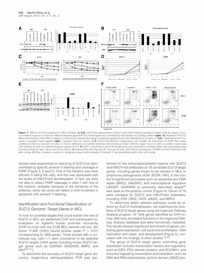
optosis were augmented on silencing of SUZ12 as dem-onstrated by specific annexin V staining and cleavage ofPARP (Figure 3, E and F). One of the hairpins was moreefficient in killing the cells, and this was associated withthe levels of H3K27me3 demethylation. In fact, we werenot able to detect PARP cleavage in Jeko-1 with one ofthe hairpins, probably because of the sensitivity of theantibody, since we could still detect a mild increment inapoptosis with annexin V staining.
Identification and Functional Classification ofSUZ12 Genomic Target Genes in MCL
To look for potential targets that could explain the role ofSUZ12 in MCL we performed ChIP and subsequent hy-bridization on Agilent’s human promoter microarray(ChIP-on-chip) with the Z138 MCL-derived cell line. Wefound 17,605 (3.6%) bound probes (peak P � 0.01)corresponding to 1806 genes. Those probes with a nor-malized log2 ratio �1 were considered to be potentialSUZ12 targets (1424 genes including known SUZ12 tar-get genes such as CDKN2A, GADD45G, BMP2, andWNT49–51).
To determine the accuracy of SUZ12 target gene dis-covery, single-locus semiquantitative PCR was per-
formed on the immunoprecipitated material with SUZ12and H3K27me3 antibodies on 18 candidate SUZ12 targetgenes, including genes known to be relevant in MCL orlymphoma pathogenesis (ATM, BCOR, VAV), in the con-trol of significant processes such as apoptosis and DNArepair (BIRC2, GADD45), and transcriptional regulation(JMJD2). GADD45G (a previously described target)49
was used as the positive control (Figure 4). Eleven of 18were validated for SUZ12 and H3K27me3 antibodies,including ATM, CBX2, VAV3, JMJD2, and BIRC2.
To determine which relevant pathways could be al-tered by SUZ12 overexpression, we examined the func-tions of SUZ12 target genes using the Ingenuity PathwayAnalysis program. Of 1424 genes identified by ChIP-on-chip, 658 have annotated functions in the Ingenuity Path-way Analysis database and were functionally classified.The results showed significant enrichment of genes con-trolling gene expression, cell cycle and proliferation, DNAreplication and repair, and development (Figure 5), con-sistent with the findings of other studies.49,50
The group of SUZ12 target genes controlling geneexpression includes transcription factors and regulators,such as E2F5, POU domain proteins, and SUV39H1, andenzymes regulating transcription and translation, such asDNA and RNA polymerases, jumonji domain (JMJD) pro-
Figure 3. Effects of SUZ12 depletion in MCL cell lines. A: Left, anti-SUZ12 immunoblot of Jeko-1 and Z138 cell lines transduced either with the empty vector,a scrambled sequence or with two different hairpins against SUZ12. Band signals were normalized with tubulin as a loading control (right). B: Depletion of SUZ12affects the function of the PRC2 complex as assessed by immunoblot using an antibody against histone H3 trimethylation at lysine 27 (left). Total histone H3 wasused to normalize band signals (right). Cropped blots are shown. C–F: SUZ12 knockdown compromises cell viability: C: Expression of EGFP that markstransduced cells was tracked over time to observe differences in viability between cells transduced either with the empty vector or with a scramble control andcells transduced with two different hairpins against SUZ12. D: GFP�-sorted Jeko1 and Z138 proliferation was assessed by counting viable cells using trypan blueexclusion along the time. Mean and SD are shown. E: Immunoblot showing specific cleavage of poly (ADP-ribose) polymerase (cPARP) in SUZ12-deficient cellsafter sorting. F: Flow cytometric analysis of apoptosis using double staining of Annexin V and propidium iodide after cell sorting.
936 Martín-Perez et alAJP August 2010, Vol. 177, No. 2

teins, and several eukaryotic translation initiation factors.Development regulators have also been found amongSUZ12 targets in MCL, some of which have been previouslypublished as SUZ12 targets in embryonic cells, such asSOX and FOX family genes, POU domain transcription fac-tors, and BMP2. Another relevant finding is the detection ofSUZ12 in the promoter region of several miRs genes, two ofwhich have been identified as SUZ12 targets (hsa-mir-124aand hsa-mir-18349,52) (identified genes listed in Supple-mental Table 3 at http://ajp.amjpathol.org).
However, the most noteworthy finding was that therewere significant genes among the top SUZ12 targets thatare known to be involved in MCL pathogenesis, some ofwhich were not previously known to be SUZ12 targets.These included those regulating cell cycle (CDKN2A andother INK4 family genes, cyclins, CDKs, CHEK1, MAD2L1,and BUB3); DNA damage and repair genes (ATM,GADD45, several DNA polymerases and topoisomer-ases, MLH1, XRCC family genes, and ERCC familygenes); apoptosis regulators (BCL2 and BCL2 regulatorproteins, BID, several BIRC family members, and others);and we also found members of nuclear factor �B pathway(BCL10, NFKB2, and IKBKG) to be regulated by SUZ12.
SUZ12 Target Gene Expression in SilencedSUZ12 MCL-Derived Cell Lines and MCLTumoral Samples
To validate functionally and elucidate the relevance ofthese ChIP-on-chip findings, we looked at the changes inexpression of the targets associated with SUZ12 expres-sion in both Z138 cell line after SUZ12 silencing and intumoral samples from MCL cases using whole genomeexpression microarrays.
SUZ12 Targets in SUZ12-Silenced Z138 CellLine
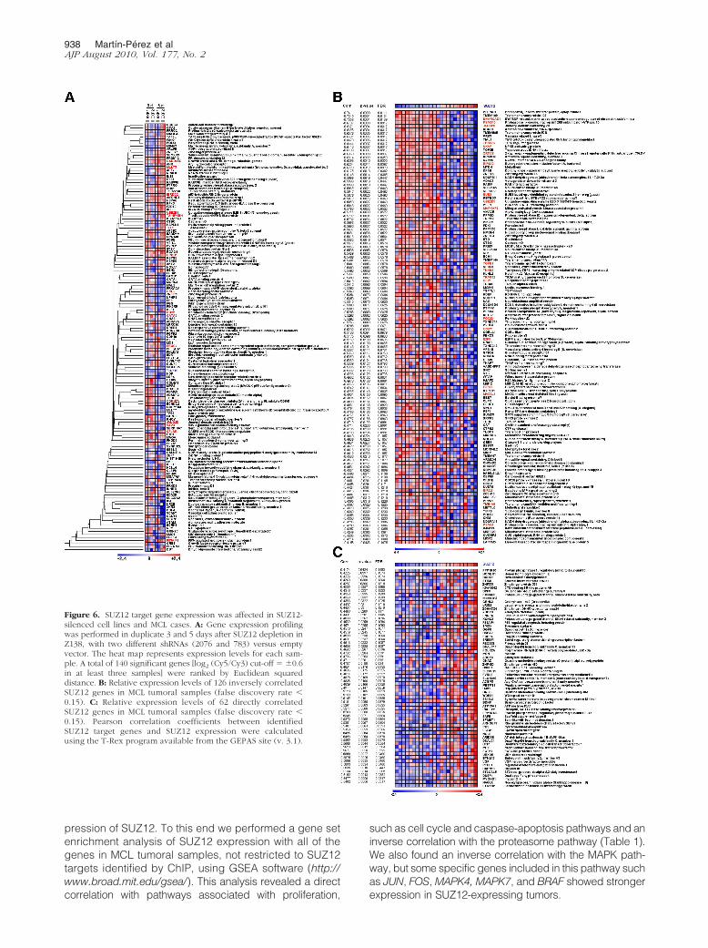
We first analyzed the expression of SUZ12 targets inSUZ12-silenced cells, comparing the expression profileof SUZ12-depleted cells with those infected with the con-trol vector. After SUZ12 silencing in the Z138 cell line,some SUZ12 targets were actually unrepressed. 140transcripts showed an up-regulation or down-regulationof at least 0.6 (log2 scale) and were considered to besignificantly deregulated after SUZ12 silencing. Theseincluded, among others, CDKN2A, GADD45G, genes in-volved in development, such as BMP2, several GATAbinding proteins or differentiation factors like MLLT3 andCBX2 (Figure 6A).
SUZ12 Targets in MCL Samples
We also examined, in MCL tumoral samples, the relationbetween SUZ12 expression and that of genes identifiedby ChIP-on-chip. This analysis revealed that 188 of 642known genes suitable for the analysis (30%) were signif-icantly correlated with SUZ12 expression (Pearson R ��0.4, false discovery rate �0.15). Many of the SUZ12targets were actually down-regulated in SUZ12-positiveMCL samples (126 inversely correlated with SUZ12expression versus 62 with a direct correlation) (Figure6, B and C).
Pathways Co-Regulated with SUZ12 in MCL
Finally, we wanted to determine which characteristics oftumors were associated with changes in SUZ12 levels,identifying functional pathways co-regulated with the ex-
Figure 4. Validation of SUZ12 target genes by semiquantitative ChIP. Single-locus semiquantitative PCR on ChIP samples was performed on severalSUZ12 candidate target genes, with SUZ12 and H3K27me3 antibodies. MouseIgG was used as a negative control.
Figure 5. Functional classification of SUZ12-targeted genes. Functions ofSUZ12 target genes were analyzed using the Ingenuity Pathway Analysisprogram. Of 1424 genes identified by ChIP-on-chip, 658 have annotatedfunctions in the Ingenuity Pathway Analysis database. The number of genesidentified as belonging to each category is included. The probability is thatassociated with a right-tailed Fisher’s exact test.
SUZ12 Deregulation in MCL 937AJP August 2010, Vol. 177, No. 2
pression of SUZ12. To this end we performed a gene setenrichment analysis of SUZ12 expression with all of thegenes in MCL tumoral samples, not restricted to SUZ12targets identified by ChIP, using GSEA software (http://www.broad.mit.edu/gsea/). This analysis revealed a directcorrelation with pathways associated with proliferation,
such as cell cycle and caspase-apoptosis pathways and aninverse correlation with the proteasome pathway (Table 1).We also found an inverse correlation with the MAPK path-way, but some specific genes included in this pathway suchas JUN, FOS, MAPK4, MAPK7, and BRAF showed strongerexpression in SUZ12-expressing tumors.
Figure 6. SUZ12 target gene expression was affected in SUZ12-silenced cell lines and MCL cases. A: Gene expression profilingwas performed in duplicate 3 and 5 days after SUZ12 depletion inZ138, with two different shRNAs (2076 and 783) versus emptyvector. The heat map represents expression levels for each sam-ple. A total of 140 significant genes [log2 (Cy5/Cy3) cut-off � �0.6in at least three samples] were ranked by Euclidean squareddistance. B: Relative expression levels of 126 inversely correlatedSUZ12 genes in MCL tumoral samples (false discovery rate �0.15). C: Relative expression levels of 62 directly correlatedSUZ12 genes in MCL tumoral samples (false discovery rate �0.15). Pearson correlation coefficients between identifiedSUZ12 target genes and SUZ12 expression were calculatedusing the T-Rex program available from the GEPAS site (v. 3.1).
938 Martín-Perez et alAJP August 2010, Vol. 177, No. 2

Discussion
SUZ12 is a core component of the Polycomb PRC2-HMTase complex that has been shown to be involved instem cell maintenance and development. Although somestudies have demonstrated overexpression of SUZ12 incolon and breast tumors,19,53 its real relevance in humancancer is yet to be established.
In this study, we first explored EZH2 and SUZ12 proteinexpression in non-tumoral samples. While EZH2 was widelydetected in almost every tissue analyzed, SUZ12 was re-stricted mainly to those tissue compartments with prolifer-ating cells, such as germinal centers in reactive lymphoidtissue, thymic cortex, epithelial basal cells and germinalcells in the testis. All these tissues are characterized by theirregenerative capacity, suggesting a role for SUZ12 in tissuehomeostasis and in cell cycle and proliferation.
Analysis of tumoral human samples revealed thatEZH2 and SUZ12 are not always expressed simulta-neously. Actually, those cases positive for SUZ12 areusually EZH2-positive, but not vice versa. We also ob-served a small fraction of cases in which we could detectSUZ12 but not EZH2. In these cases we cannot rule outthe possibility that SUZ12 might have additional EZH2-independent functions.
There was a high level of expression of SUZ12 in asubset of tumoral samples including germinal cell-de-rived tumors, melanomas, skin basal cell carcinomas,lung neuroendocrine small-cell carcinoma, pituitary and
parathyroid adenomas, and lymphomas, most remark-ably in MCL, where the high expression of SUZ12 con-trasts with its absence in the non-tumoral mantle zonecells in reactive lymph node. Therefore, our results ex-tend previous observations of the strong expression ofSUZ12 in human tumors.19,53
SUZ12 locus (17q11.2) has been found amplified, as-sociated with protein overexpression in a small subset oftumors. This finding is especially relevant in MCL, whereit seems to be more frequent in blastoid MCL, the ag-gressive variant of this type of lymphoma, since two offour blastoid-MCL cases showed this amplification.Therefore, our finding of SUZ12 amplification in MCL ormelanoma among others types of tumors, along with thepresence of SUZ12 translocations in endometrial sarco-mas of the cervix,20,54 supports the hypothesis thatSUZ12 has an oncogenic function and contributes totumor formation and maintenance. The findings de-scribed here for SUZ12, and the previous results forEZH2,7,17,18 suggest that alteration of the PRC2 complexis a frequent event in human carcinogenesis.
Given the anomalous expression of SUZ12 in MCLtumoral cells compared with the lack of expression intheir normal counterparts and the amplification associ-ated with high levels of expression, we decided to per-form functional analysis in MCL cell lines as a model todepict SUZ12 role in tumorigenesis. To this end, wesilenced SUZ12 expression by RNAi in MCL-derived cell
Table 1. Pathways Co-Regulated with SUZ12 Expression in MCL
Name SizeEnrichment
scoreNormalized
enrichment score PFalse discovery
rate
Gene sets positively correlatedwith SUZ12 expression
Mitochondria pathway 21 0.5952 2.4673 0.0000 0.0053Cell cycle pathway 21 0.5622 2.3809 0.0000 0.0075ARAP pathway 19 0.5528 2.1412 0.0020 0.0283Caspase pathway 21 0.5233 2.1399 0.0040 0.0213CA2� CAM pathway 12 0.6362 1.9820 0.0043 0.0513D4GDI pathway 11 0.6060 1.8114 0.0169 0.1118
Gene sets negatively correlatedwith SUZ12 expression
MAPK pathway 83 �0.4378 �3.4409 0.0000 0.0000Proteasome pathway 19 �0.7375 �2.8678 0.0000 0.0000GH pathway 26 �0.4999 �2.2277 0.0000 0.0395IL1R pathway 29 �0.4244 �2.0417 0.0080 0.0779CHREBP pathway 15 �0.5951 �2.0912 0.0038 0.0832CK1 pathway 12 �0.6534 �2.0431 0.0056 0.0916ARF pathway 16 �0.5428 �1.9669 0.0056 0.1074RARRXR pathway 14 �0.5157 �1.7385 0.0177 0.1367RAB pathway 10 �0.5956 �1.7550 0.0220 0.1392ERK pathway 30 �0.3620 �1.7652 0.0203 0.1402AKAP-centrosome pathway 10 �0.6027 �1.7413 0.0198 0.1413TPO pathway 22 �0.4373 �1.8955 0.0096 0.1444PPARA pathway 51 �0.2753 �1.7689 0.0249 0.1465Cytokine pathway 15 �0.5077 �1.7904 0.0183 0.1473PDGF pathway 26 �0.3973 �1.7772 0.0149 0.1479VEGF pathway 26 �0.3992 �1.7929 0.0135 0.1557TOLL pathway 32 �0.3657 �1.8513 0.0134 0.1642GATA3 pathway 13 �0.5419 �1.7935 0.0080 0.1678TOB1 pathway 16 �0.4963 �1.7958 0.0189 0.1797P38MAPK pathway 37 �0.3407 �1.8065 0.0132 0.1876
GSEA reporting on statistically significant functionally relevant pathways (P � 0.05, false discovery rate � 0.25) and on positive and negativePearson correlation coefficients with a high level of SUZ12 expression. P, nominal probability.
SUZ12 Deregulation in MCL 939AJP August 2010, Vol. 177, No. 2
lines, and evaluated its effect on levels of H3K27me3, cellproliferation, apoptosis, and cell survival. A clear de-crease in H3K27me3 was detected after SUZ12 silencing,demonstrating the interference with PRC2 activity due to thelower levels of SUZ12 in accordance with previous re-ports.21 SUZ12 knockdown resulted in an increased apo-ptosis, as demonstrated by annexin V and PARP cleavageanalysis. When we studied the SUZ12 silencing effect overtime, we observed that loss of SUZ12 compromised cellviability, as demonstrated by cell counting and competitionassays. These results suggest that SUZ12 expression con-tributes to cell survival in MCL cell lines, avoiding apoptosisand increasing cell proliferation.
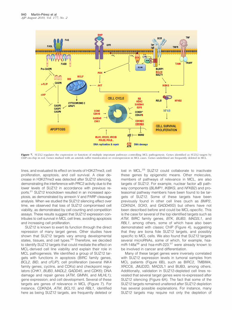
SUZ12 is known to exert its function through the directrepression of many target genes. Other studies haveshown that SUZ12 targets vary among developmentalstates, tissues, and cell types.55 Therefore, we decidedto identify SUZ12 targets that could mediate the effect onMCL-derived cell line viability and explain their role inMCL pathogenesis. We identified a group of SUZ12 tar-gets with functions in apoptosis (BIRC family genes,BCL2, BID, and cFLIP); cell proliferation (several INK4family genes, cyclins, and CDKs) and checkpoint regu-lators (CHK1, BUB3, MADL2, GADD45, and CDK6); DNAdamage and repair genes (ATM, GMNN, and MLHL1);gene expression; and cell development. Several of thesetargets are genes of relevance in MCL (Figure 7). Forinstance, CDKN2A, ATM, BCL10, and RBL1, identifiedhere as being SUZ12 targets, are frequently deleted or
lost in MCL.23 SUZ12 could collaborate to inactivatethese genes by epigenetic means. Other molecules,members of pathways of relevance in MCL, are alsotargets of SUZ12. For example, nuclear factor �B path-way components (BLIMP1, IKBKG, and NFKB2) and pro-teasomal pathway members have been found to be tar-gets of SUZ12. Some of these targets have beenpreviously found in other cell lines (such as BMP2,CDKN2A, SOX3, and GADD45G) but others have notbeen described before and could be MCL-specific. Thisis the case for several of the top identified targets such asATM, BIRC family genes, BTK, BUB3, MAD2L1, andRBL1, among others, some of which have also beendemonstrated with classic ChIP (Figure 4), suggestingthat they are bona fide SUZ12 targets, and possiblyspecific to MCL cells. We also found that SUZ12 targetsseveral microRNAs, some of which, for example, hsa-miR-148a56 and hsa-miR-223,57 were already known tobe involved in cancer and differentiation.
Many of these target genes were inversely correlatedwith SUZ12 expression levels in tumoral samples fromMCL patients (Figure 6B), such as BIRC2, TMBIM4,XRCC6, JMJD2D, MAD2L1 and BUB3, among others.Additionally, validation in SUZ12-depleted cell lines re-vealed that several target genes were re-expressed afterSUZ12 silencing (Figure 6A). The fact that some of theSUZ12 targets remained unaltered after SUZ12 depletionhas several possible explanations. For instance, manySUZ12 targets may require not only the depletion of
Figure 7. SUZ12 regulates the expression or function of multiple important pathways controlling MCL pathogenesis. Genes identified as SUZ12 targets byChIP-on-chip in red. Genes marked with an asterisk suffer translocation or overexpression in MCL cases. Genes underlined are frequently deleted in MCL.
940 Martín-Perez et alAJP August 2010, Vol. 177, No. 2
SUZ12 but also additional events like DNA demethylationor the presence of an activator to be expressed again.1,50
All these findings indicate that SUZ12 could collaboratein deregulating the expression of many important pathwayscontrolling MCL pathogenesis (Figure 7). We propose thatthe abnormal expression of SUZ12 may account for some ofthe still unexplained features of MCL, including abnormalDNA repair and increased resistance to apoptosis.
Interestingly, recent publications have described the ca-pacity of several drugs to block the HMTase activity ofPRC2 complexes.58,59 In fact, LBH589 has proved to beeffective in acute myelogenous leukemia cells. Patients suf-fering from other tumors, like MCL and pulmonary neuroen-docrine small-cell carcinoma, in which PRC2 alterations aredetected, might also benefit from this therapy.
Acknowledgments
We thank Dr. Yi Zhang and Dr. Ru Cao for providing uswith the SUZ12 cDNA for antibody production. We ex-press our gratitude to all members of the MonoclonalAntibodies and Histology and ImmunohistochemistryUnits (CNIO) for their excellent technical contribution andassistance. Special thanks are extended to Esteban Ball-estar, Filipe Jacinto, and Amaia Lujambio from the formerCancer Epigenetics Group (CNIO) for their help with theChIP experiments; to Jorge Luis Martinez from the ProteinTechnology Unit (CNIO) for SUZ12 protein production; toOrlando Dominguez from the Genomics Unit (CNIO) forChIP-on-chip hybridization and help in analysis; and to allof the personnel of the Lymphoma Group (CNIO) for theirhelp and discussions.
References
1. Sparmann A, van Lohuizen M: Polycomb silencers control cell fate,development and cancer. Nat Rev Cancer 2006, 6:846–856
2. Cao R, Wang L, Wang H, Xia L, Erdjument-Bromage H, Tempst P,Jones RS, Zhang Y: Role of histone H3 lysine 27 methylation inPolycomb-group silencing. Science 2002, 298:1039–1043
3. Czermin B, Melfi R, McCabe D, Seitz V, Imhof A, Pirrotta V: Drosophilaenhancer of Zeste/ESC complexes have a histone H3 methyltrans-ferase activity that marks chromosomal Polycomb sites. Cell 2002,111:185–196
4. Shao Z, Raible F, Mollaaghababa R, Guyon JR, Wu CT, Bender W,Kingston RE: Stabilization of chromatin structure by PRC1, a Poly-comb complex. Cell 1999, 98:37–46
5. Muller J, Hart CM, Francis NJ, Vargas ML, Sengupta A, Wild B, MillerEL, O’Connor MB, Kingston RE, Simon JA: Histone methyltransferaseactivity of a Drosophila Polycomb group repressor complex. Cell2002, 111:197–208
6. Kuzmichev A, Nishioka K, Erdjument-Bromage H, Tempst P, ReinbergD: Histone methyltransferase activity associated with a human multipro-tein complex containing the Enhancer of Zeste protein. Genes Dev 2002,16:2893–2905
7. Bracken AP, Pasini D, Capra M, Prosperini E, Colli E, Helin K: EZH2is downstream of the pRB-E2F pathway, essential for proliferation andamplified in cancer. EMBO J 2003, 22:5323–5335
8. Vonlanthen S, Heighway J, Altermatt HJ, Gugger M, Kappeler A,Borner MM, van Lohuizen M, Betticher DC: The bmi-1 oncoprotein isdifferentially expressed in non-small cell lung cancer and correlateswith INK4A-ARF locus expression. Br J Cancer 2001, 84:1372–1376
9. van Kemenade FJ, Raaphorst FM, Blokzijl T, Fieret E, Hamer KM,Satijn DP, Otte AP, Meijer CJ: Coexpression of BMI-1 and EZH2
polycomb-group proteins is associated with cycling cells and degreeof malignancy in B-cell non-Hodgkin lymphoma. Blood 2001,97:3896–3901
10. Bea S, Tort F, Pinyol M, Puig X, Hernandez L, Hernandez S, FernandezPL, van Lohuizen M, Colomer D, Campo E: BMI-1 gene amplification andoverexpression in hematological malignancies occur mainly in mantlecell lymphomas. Cancer Res 2001, 61:2409–2412
11. Haupt Y, Alexander WS, Barri G, Klinken SP, Adams JM: Novel zincfinger gene implicated as myc collaborator by retrovirally acceler-ated lymphomagenesis in E mu-myc transgenic mice. Cell 1991,65:753–763
12. van Lohuizen M, Verbeek S, Scheijen B, Wientjens E, van der GuldenH, Berns A: Identification of cooperating oncogenes in E mu-myctransgenic mice by provirus tagging. Cell 1991, 65:737–752
13. Sanchez-Beato M, Sanchez E, Gonzalez-Carrero J, Morente M, DiezA, Sanchez-Verde L, Martin MC, Cigudosa JC, Vidal M, Piris MA:Variability in the expression of polycomb proteins in different normaland tumoral tissues. A pilot study using tissue microarrays. ModPathol 2006, 19:684–694
14. Dukers DF, van Galen JC, Giroth C, Jansen P, Sewalt RG, Otte AP,Kluin-Nelemans HC, Meijer CJ, Raaphorst FM: Unique polycombgene expression pattern in Hodgkin’s lymphoma and Hodgkin’s lym-phoma-derived cell lines. Am J Pathol 2004, 164:873–881
15. Raaphorst FM, Vermeer M, Fieret E, Blokzijl T, Dukers D, Sewalt RG,Otte AP, Willemze R, Meijer CJ: Site-specific expression of polycomb-group genes encoding the HPC-HPH/PRC1 complex in clinicallydefined primary nodal and cutaneous large B-cell lymphomas. Am JPathol 2004, 164:533–542
16. Sanchez-Beato M, Sanchez E, Garcia JF, Perez-Rosado A, MontoyaMC, Fraga M, Artiga MJ, Navarrete M, Abraira V, Morente M, EstellerM, Koseki H, Vidal M, Piris MA: Abnormal PcG protein expression inHodgkin’s lymphoma. Relation with E2F6 and NFkappaB transcrip-tion factors. J Pathol 2004, 204:528–537
17. Kleer CG, Cao Q, Varambally S, Shen R, Ota I, Tomlins SA, Ghosh D,Sewalt RG, Otte AP, Hayes DF, Sabel MS, Livant D, Weiss SJ, RubinMA, Chinnaiyan AM: EZH2 is a marker of aggressive breast cancerand promotes neoplastic transformation of breast epithelial cells.Proc Natl Acad Sci USA 2003, 100:11606–11611
18. Varambally S, Dhanasekaran SM, Zhou M, Barrette TR, Kumar-SinhaC, Sanda MG, Ghosh D, Pienta KJ, Sewalt RG, Otte AP, Rubin MA,Chinnaiyan AM: The polycomb group protein EZH2 is involved inprogression of prostate cancer. Nature 2002, 419:624–629
19. Kirmizis A, Bartley SM, Farnham PJ: Identification of the polycombgroup protein SU(Z)12 as a potential molecular target for humancancer therapy. Mol Cancer Ther 2003, 2:113–121
20. Koontz JI, Soreng AL, Nucci M, Kuo FC, Pauwels P, van Den BergheH, Cin PD, Fletcher JA, Sklar J: Frequent fusion of the JAZF1 andJJAZ1 genes in endometrial stromal tumors. Proc Natl Acad Sci USA2001, 98:6348–6353
21. Pasini D, Bracken AP, Jensen MR, Lazzerini Denchi E, Helin K: Suz12is essential for mouse development and for EZH2 histone methyl-transferase activity. EMBO J 2004, 23:4061–4071
22. Cao R, Zhang Y: SUZ12 is required for both the histone methyltrans-ferase activity and the silencing function of the EED-EZH2 complex.Mol Cell 2004, 15:57–67
23. Jares P, Colomer D, Campo E: Genetic and molecular pathogenesisof mantle cell lymphoma: perspectives for new targeted therapeutics.Nat Rev Cancer 2007, 7:750–762
24. Martinez N, Camacho FI, Algara P, Rodriguez A, Dopazo A, Ruiz-Ballesteros E, Martin P, Martinez-Climent JA, Garcia-Conde J,Menarguez J, Solano F, Mollejo M, Piris MA: The molecular signatureof mantle cell lymphoma reveals multiple signals favoring cell sur-vival. Cancer Res 2003, 63:8226–8232
25. Rosenwald A, Wright G, Wiestner A, Chan WC, Connors JM, CampoE, Gascoyne RD, Grogan TM, Muller-Hermelink HK, Smeland EB,Chiorazzi M, Giltnane JM, Hurt EM, Zhao H, Averett L, Henrickson S,Yang L, Powell J, Wilson WH, Jaffe ES, Simon R, Klausner RD,Montserrat E, Bosch F, Greiner TC, Weisenburger DD, Sanger WG,Dave BJ, Lynch JC, Vose J, Armitage JO, Fisher RI, Miller TP, LeBlancM, Ott G, Kvaloy S, Holte H, Delabie J, Staudt LM: The proliferation geneexpression signature is a quantitative integrator of oncogenic events thatpredicts survival in mantle cell lymphoma. Cancer Cell 2003, 3:185–197
26. Bodrug SE, Warner BJ, Bath ML, Lindeman GJ, Harris AW, AdamsJM: Cyclin D1 transgene impedes lymphocyte maturation and col-
SUZ12 Deregulation in MCL 941AJP August 2010, Vol. 177, No. 2
laborates in lymphomagenesis with the myc gene. EMBO J 1994,13:2124–2130
27. Stilgenbauer S, Winkler D, Ott G, Schaffner C, Leupolt E, Bentz M,Moller P, Muller-Hermelink HK, James MR, Lichter P, Dohner H:Molecular characterization of 11q deletions points to a pathogenicrole of the ATM gene in mantle cell lymphoma. Blood 1999,94:3262–3264
28. Garcia JF, Roncador G, Garcia JF, Sanz AI, Maestre L, Lucas E,Montes-Moreno S, Fernandez Victoria R, Martinez-Torrecuadrara JL,Marafioti T, Mason DY, Piris MA: PRDM1/BLIMP-1 expression in mul-tiple B and T-cell lymphoma. Haematologica 2006, 91:467–474
29. Montes-Moreno S, Roncador G, Maestre L, Martinez N, Sanchez-Verde L, Camacho FI, Cannata J, Martinez-Torrecuadrada JL, ShenY, Chan WC, Piris MA: Gcet1 (centerin), a highly restricted marker fora subset of germinal center-derived lymphomas. Blood 2008,111:351–358
30. Kallioniemi OP, Wagner U, Kononen J, Sauter G: Tissue microarraytechnology for high-throughput molecular profiling of cancer. HumMol Genet 2001, 10:657–662
31. Tracey L, Aggarwal M, Garcia-Cosio M, Villuendas R, Algara P,Sanchez-Beato M, Sanchez-Aguilera A, Garcia JF, Rodriguez A,Camacho FI, Martinez N, Ruiz-Ballesteros E, Mollejo M, Piris MA:Somatic hypermutation signature in B-cell low-grade lymphomas.Haematologica 2008, 93:1186–1194
32. Caretti G, Di Padova M, Micales B, Lyons GE, Sartorelli V: ThePolycomb Ezh2 methyltransferase regulates muscle gene expressionand skeletal muscle differentiation. Genes Dev 2004, 18:2627–2638
33. Raman JD, Mongan NP, Tickoo SK, Boorjian SA, Scherr DS, GudasLJ: Increased expression of the polycomb group gene. EZH2, intransitional cell carcinoma of the bladder. Clin Cancer Res 2005,11:8570–8576
34. Palacios J, Honrado E, Osorio A, Cazorla A, Sarrio D, Barroso A,Rodriguez S, Cigudosa JC, Diez O, Alonso C, Lerma E, Dopazo J,Rivas C, Benitez J: Phenotypic characterization of BRCA1 andBRCA2 tumors based in a tissue microarray study with 37 immuno-histochemical markers. Breast Cancer Res Treat 2005, 90:5–14
35. Singh B, Stoffel A, Gogineni S, Poluri A, Pfister DG, Shaha AR, PathakA, Bosl G, Cordon-Cardo C, Shah JP, Rao PH: Amplification of the3q26.3 locus is associated with progression to invasive cancer and isa negative prognostic factor in head and neck squamous cell carci-nomas. Am J Pathol 2002, 161:365–371
36. Sholl LM, John Iafrate A, Chou YP, Wu MT, Goan YG, Su L, Huang YT,Christiani DC, Chirieac LR: Validation of chromogenic in situ hybrid-ization for detection of EGFR copy number amplification in nonsmallcell lung carcinoma. Mod Pathol 2007, 20:1028–1035
37. Lois C, Hong EJ, Pease S, Brown EJ, Baltimore D: Germline trans-mission and tissue-specific expression of transgenes delivered bylentiviral vectors. Science 2002, 295:868–872
38. Ruiz-Vela A, Aggarwal M, de la Cueva P et al. Lentiviral (HIV)-basedRNA interference screen in human B-cell receptor regulatory net-works reveals MCL1-induced oncogenic pathways. Blood 2008, 111:1665–1676
39. Ruiz-Vela A, Aggarwal M, de la Cueva P, Treda C, Herreros B, Martin-Perez D, Dominguez O, Piris MA: Lentiviral (HIV)-based RNA interfer-ence screen in human B-cell receptor regulatory networks revealsMCL1-induced oncogenic pathways. Blood 2008, 111:1665–1676
40. Montaner D, Tarraga J, Huerta-Cepas J, Burguet J, Vaquerizas JM,Conde L, Minguez P, Vera J, Mukherjee S, Valls J, Pujana MA, AllozaE, Herrero J, Al-Shahrour F, Dopazo J: Next station in microarray dataanalysis: gEPAS. Nucleic Acids Res 2006, 34:W486–491
41. Subramanian A, Tamayo P, Mootha VK, Mukherjee S, Ebert BL,Gillette MA, Paulovich A, Pomeroy SL, Golub TR, Lander ES, MesirovJP: Gene set enrichment analysis: a knowledge-based approach forinterpreting genome-wide expression profiles. Proc Natl Acad SciUSA 2005, 102:15545–15550
42. Mootha VK, Lindgren CM, Eriksson KF, Subramanian A, Sihag S,Lehar J, Puigserver P, Carlsson E, Ridderstrale M, Laurila E, HoustisN, Daly MJ, Patterson N, Mesirov JP, Golub TR, Tamayo P,Spiegelman B, Lander ES, Hirschhorn JN, Altshuler D, Groop LC:PGC-1alpha-responsive genes involved in oxidative phosphorylationare coordinately downregulated in human diabetes. Nat Genet 2003,34:267–273
43. Onciu M, Schlette E, Medeiros LJ, Abruzzo LV, Keating M, Lai R:Cytogenetic findings in mantle cell lymphoma cases with a high level
of peripheral blood involvement have a distinct pattern of abnormal-ities. Am J Clin Pathol 2001, 116:886–892
44. Espinet B, Sole F, Woessner S, Bosch F, Florensa L, Campo E, CostaD, Lloveras E, Vila RM, Besses C, Montserrat E, Sans-Sabrafen J:Translocation (11;14)(q13;q32) and preferential involvement of chro-mosomes 1, 2, 9, 13, and 17 in mantle cell lymphoma. Cancer GenetCytogenet 1999, 111:92–98
45. Starczynowski DT, Vercauteren S, Telenius A, Sung S, Tohyama K,Brooks-Wilson A, Spinelli JJ, Eaves CJ, Eaves AC, Horsman DE, LamWL, Karsan A: High-resolution whole genome tiling path array CGHanalysis of CD34� cells from patients with low-risk myelodysplasticsyndromes reveals cryptic copy number alterations and predictsoverall and leukemia-free survival. Blood 2008, 112:3412–3424
46. Douglas EJ, Fiegler H, Rowan A, Halford S, Bicknell DC, Bodmer W,Tomlinson IP, Carter NP: Array comparative genomic hybridizationanalysis of colorectal cancer cell lines and primary carcinomas.Cancer Res 2004, 64:4817–4825
47. Kraggerud SM, Skotheim RI, Szymanska J, Eknaes M, Fossa SD,Stenwig AE, Peltomaki P, Lothe RA: Genome profiles of familial/bilateral and sporadic testicular germ cell tumors. Genes Chromo-somes Cancer 2002, 34:168–174
48. Ivanova N, Dobrin R, Lu R, Kotenko I, Levorse J, DeCoste C, SchaferX, Lun Y, Lemischka IR: Dissecting self-renewal in stem cells withRNA interference. Nature 2006, 442:533–538
49. Lee TI, Jenner RG, Boyer LA, Guenther MG, Levine SS, Kumar RM,Chevalier B, Johnstone SE, Cole MF, Isono K, Koseki H, Fuchikami T,Abe K, Murray HL, Zucker JP, Yuan B, Bell GW, Herbolsheimer E,Hannett NM, Sun K, Odom DT, Otte AP, Volkert TL, Bartel DP, MeltonDA, Gifford DK, Jaenisch R, Young RA: Control of developmentalregulators by Polycomb in human embryonic stem cells. Cell 2006,125:301–313
50. Bracken AP, Dietrich N, Pasini D, Hansen KH, Helin K: Genome-widemapping of Polycomb target genes unravels their roles in cell fatetransitions. Genes Dev 2006, 20:1123–1136
51. Kotake Y, Cao R, Viatour P, Sage J, Zhang Y, Xiong Y: pRB familyproteins are required for H3K27 trimethylation and Polycomb repres-sion complexes binding to and silencing p16INK4alpha tumor sup-pressor gene. Genes Dev 2007, 21:49–54
52. Marson A, Levine SS, Cole MF, Frampton GM, Brambrink T,Johnstone S, Guenther MG, Johnston WK, Wernig M, Newman J,Calabrese JM, Dennis LM, Volkert TL, Gupta S, Love J, Hannett N,Sharp PA, Bartel DP, Jaenisch R, Young RA: Connecting microRNAgenes to the core transcriptional regulatory circuitry of embryonicstem cells. Cell 2008, 134:521–533
53. Kirmizis A, Bartley SM, Kuzmichev A, Margueron R, Reinberg D,Green R, Farnham PJ: Silencing of human polycomb target genes isassociated with methylation of histone H3 Lys 27. Genes Dev 2004,18:1592–1605
54. Oliva E, de Leval L, Soslow RA, Herens C: High frequency of JAZF1-JJAZ1 gene fusion in endometrial stromal tumors with smooth muscledifferentiation by interphase FISH detection. Am J Surg Pathol 2007,31:1277–1284
55. Squazzo SL, O’Geen H, Komashko VM, Krig SR, Jin VX, Jang SW,Margueron R, Reinberg D, Green R, Farnham PJ: Suz12 binds tosilenced regions of the genome in a cell-type-specific manner. Ge-nome Res 2006, 16:890–900
56. Lujambio A, Calin GA, Villanueva A, Ropero S, Sanchez-Cespedes M,Blanco D, Montuenga LM, Rossi S, Nicoloso MS, Faller WJ, GallagherWM, Eccles SA, Croce CM, Esteller M: A microRNA DNA methylationsignature for human cancer metastasis. Proc Natl Acad Sci USA2008, 105:13556–13561
57. Stamatopoulos B, Meuleman N, Haibe-Kains B, Saussoy P, Van denNeste E, Michaux L, Heimann P, Martiat P, Bron D, Lagneaux L:MicroRNA-29c and microRNA-223 downregulation has in vivo signif-icance in chronic lymphocytic leukemia and improves disease riskstratification. Blood 2009, 113:5237–5245
58. Fiskus W, Pranpat M, Balasis M, Herger B, Rao R, Chinnaiyan A,Atadja P, Bhalla K: Histone deacetylase inhibitors deplete enhancerof zeste 2 and associated polycomb repressive complex 2 proteins inhuman acute leukemia cells. Mol Cancer Ther 2006, 5:3096–3104
59. Tan J, Yang X, Zhuang L, Jiang X, Chen W, Lee PL, Karuturi RK, TanPB, Liu ET, Yu Q: Pharmacologic disruption of Polycomb-repressivecomplex 2-mediated gene repression selectively induces apoptosisin cancer cells. Genes Dev 2007, 21:1050–1063
942 Martín-Perez et alAJP August 2010, Vol. 177, No. 2
Copyright © 2022 FDOKUMEN