Inactivation of the Polycomb Group Protein Ring1B Unveils an Antiproliferative Role in Hematopoietic...
11
MOLECULAR AND CELLULAR BIOLOGY, Feb. 2008, p. 1018–1028 Vol. 28, No. 3 0270-7306/08/$08.000 doi:10.1128/MCB.01136-07 Copyright © 2008, American Society for Microbiology. All Rights Reserved. Inactivation of the Polycomb Group Protein Ring1B Unveils an Antiproliferative Role in Hematopoietic Cell Expansion and Cooperation with Tumorigenesis Associated with Ink4a Deletion † Carmela Cale ´s, 2 Mo ´nica Roma ´n-Trufero, 1 Leticia Pavo ´n, 2 Iva ´n Serrano, 2 Teresa Melgar, 1 Mitsuhiro Endoh, 3 Claudia Pe ´rez, 4 Haruhiko Koseki, 3 and Miguel Vidal 1,3 * Centro de Investigaciones Biolo ´gicas, Consejo Superior de Investigaciones Cientı ´ficas, Ramiro de Maeztu 9, 28004 Madrid, Spain 1 ; Instituto de Investigaciones Biome ´dicas, Universidad Auto ´noma de Madrid, Consejo Superior de Investigaciones Cientı ´ficas, Arturo Duperier 4, 28029 Madrid, Spain 2 ; Riken Research Center for Allergy and Immunology, 1-7-22 Suehiro, Tsurumi-ku, Yokohama 230-0045, Japan 3 ; and Anatomy and Histopathology, Facultad de Veterinaria, Universidad de Leo ´n, Campus Vegazana, 24071 Leo ´n, Spain 4 Received 26 June 2007/Returned for modification 2 August 2007/Accepted 12 November 2007 Polycomb group (PcG) proteins act as positive regulators of cell proliferation. Ring1B is a PcG gene essential for embryonic development, but its contribution to cell turnover in regenerating tissues in not known. Here, we have generated a conditional mouse mutant line to study the Ring1B role in adult hematopoiesis. Mutant mice developed a hypocellular bone marrow that paradoxically contained an enlarged, hyperproliferating compart- ment of immature cells, with an intact differentiation potential. These alterations were associated with differential upregulation of cyclin D2, which occurred in all mutant bone marrow cells, and of p16 Ink4a , observed only in the differentiated compartment. Concurrent inactivation of Ink4a rescued the defective proliferation of maturing cells but did not affect the hyperproliferative activity of progenitors and resulted in a shortening of the onset of lymphomas induced by Ink4a inactivation. These data show that Ring1B restricts the progenitors’ proliferation and promotes the proliferation of their maturing progeny by selectively altering the expression pattern of cell cycle regulators along hematopoietic differentiation. The novel antiproliferative role of Ring1B’s downregulation of a cell cycle activator may play an important role in the tight control of hematopoietic cell turnover. Adult multicellular organisms have evolved cell turnover strategies adapted to the maintenance and repair of tissues. In many cases, such as the hematopoietic system, gut, skin, and brain, tissue homeostasis depends on the activity of multipo- tent stem cells from which derive all the cell lineages that make these tissues (17, 56). Because of the relatively small number of stem cells, their progeny undergoes an expanding but limited number of cell divisions along the differentiation process be- fore entering the mitotically inactive state of fully mature cells. Tissue homeostasis, then, depends on an adequate balance between stem cell renewal, regulated cell proliferation, and terminal differentiation of stem cell progeny (58, 74). The Polycomb group (PcG) of proteins are transcriptional repressors that prevent the inappropriate expression of genes that determine cell identity (4, 11). PcG gene products assem- ble in multimeric complexes (Polycomb repressing complexes [PRC]) whose transcriptional activity is associated to their role as chromatin modifiers (64). PRC are compositionally diverse, but depending on which of two sets of core subunits they contain, they are designated type PRC1 or PRC2 (40). Histone H2AK119 monoubiquitylation depends on the RING finger E3 ligases of PRC1 complexes (10, 73), whereas histone H3K27 trimethylation is carried out by PRC2 complexes (12, 18, 34, 46). Well known as developmental regulators (57), PcG genes also play important roles in cell proliferation control (41, 52). Murine models of loss of function of PcG genes encoding PRC1 subunits show reduced size and hypocellularity of he- matopoietic organs (1, 15, 69, 70) and also, in some cases (Bmi1) of brain structures (71), indicating that PcG complexes are positive regulators of cell proliferation. The upregulation of PcG products observed in a wide variety of tumors (54, 68) and their cooperation in oncogene-induced tumorigenesis (27) further support a role for PcG complexes as promoters of proliferation. Exceptionally, inactivation of the PRC2 subunit Eed resulted in enlarged hematopoietic compartments (36). These alterations are due, at least in part, to PcG-mediated repression of the Ink4a/Cdkn2a locus, which encodes the tu- mor suppressors p16 Ink4a and p19 Arf . Loss of function of PRC1 proteins, and Bmi1 in particular, results in premature senes- cence of hematopoietic and neuronal stem cells (28, 37, 44, 48, 49, 51). PcG complexes also promote expansion of maturing cells by preventing senescence and apoptosis, although the contribution of each of these mechanisms varies depending on the particular tissue and the PcG protein (9, 14, 38, 45). Ring1B and its paralog Ring1A are the RING finger E3 ligases that monoubiquitylate H2A (20, 73). They associate directly with Bmi1, Mel18, M33, and Phc1, forming the core of * Corresponding author. Mailing address: Developmental and Cell Biology, Centro de Investigaciones Biolo ´gicas, Ramiro de Maeztu, 9, 28040 Madrid, Spain. Phone: 34 91 837 3112, ext. 4383. Fax: 34 91536 0432. E-mail: [email protected]. † Supplemental material for this article may be found at http://mcb .asm.org/. Published ahead of print on 26 November 2007. 1018 at RIKAGAKU-KENKYUSHO on January 21, 2008 mcb.asm.org Downloaded from
-
Upload
independent -
Category
Documents
-
view
1 -
download
0
Transcript of Inactivation of the Polycomb Group Protein Ring1B Unveils an Antiproliferative Role in Hematopoietic...

MOLECULAR AND CELLULAR BIOLOGY, Feb. 2008, p. 1018–1028 Vol. 28, No. 30270-7306/08/$08.00!0 doi:10.1128/MCB.01136-07Copyright © 2008, American Society for Microbiology. All Rights Reserved.
Inactivation of the Polycomb Group Protein Ring1B Unveils anAntiproliferative Role in Hematopoietic Cell Expansion and
Cooperation with Tumorigenesis Associated withInk4a Deletion!†
Carmela Cales,2 Monica Roman-Trufero,1 Leticia Pavon,2 Ivan Serrano,2 Teresa Melgar,1Mitsuhiro Endoh,3 Claudia Perez,4 Haruhiko Koseki,3 and Miguel Vidal1,3*
Centro de Investigaciones Biologicas, Consejo Superior de Investigaciones Cientıficas, Ramiro de Maeztu 9, 28004 Madrid, Spain1;Instituto de Investigaciones Biomedicas, Universidad Autonoma de Madrid, Consejo Superior de Investigaciones Cientıficas,
Arturo Duperier 4, 28029 Madrid, Spain2; Riken Research Center for Allergy and Immunology, 1-7-22 Suehiro, Tsurumi-ku,Yokohama 230-0045, Japan3; and Anatomy and Histopathology, Facultad de Veterinaria, Universidad de Leon,
Campus Vegazana, 24071 Leon, Spain4
Received 26 June 2007/Returned for modification 2 August 2007/Accepted 12 November 2007
Polycomb group (PcG) proteins act as positive regulators of cell proliferation. Ring1B is a PcG gene essentialfor embryonic development, but its contribution to cell turnover in regenerating tissues in not known. Here, wehave generated a conditional mouse mutant line to study the Ring1B role in adult hematopoiesis. Mutant micedeveloped a hypocellular bone marrow that paradoxically contained an enlarged, hyperproliferating compart-ment of immature cells, with an intact differentiation potential. These alterations were associated withdifferential upregulation of cyclin D2, which occurred in all mutant bone marrow cells, and of p16Ink4a,observed only in the differentiated compartment. Concurrent inactivation of Ink4a rescued the defectiveproliferation of maturing cells but did not affect the hyperproliferative activity of progenitors and resulted ina shortening of the onset of lymphomas induced by Ink4a inactivation. These data show that Ring1B restrictsthe progenitors’ proliferation and promotes the proliferation of their maturing progeny by selectively alteringthe expression pattern of cell cycle regulators along hematopoietic differentiation. The novel antiproliferativerole of Ring1B’s downregulation of a cell cycle activator may play an important role in the tight control ofhematopoietic cell turnover.
Adult multicellular organisms have evolved cell turnoverstrategies adapted to the maintenance and repair of tissues. Inmany cases, such as the hematopoietic system, gut, skin, andbrain, tissue homeostasis depends on the activity of multipo-tent stem cells from which derive all the cell lineages that makethese tissues (17, 56). Because of the relatively small number ofstem cells, their progeny undergoes an expanding but limitednumber of cell divisions along the differentiation process be-fore entering the mitotically inactive state of fully mature cells.Tissue homeostasis, then, depends on an adequate balancebetween stem cell renewal, regulated cell proliferation, andterminal differentiation of stem cell progeny (58, 74).
The Polycomb group (PcG) of proteins are transcriptionalrepressors that prevent the inappropriate expression of genesthat determine cell identity (4, 11). PcG gene products assem-ble in multimeric complexes (Polycomb repressing complexes[PRC]) whose transcriptional activity is associated to their roleas chromatin modifiers (64). PRC are compositionally diverse,but depending on which of two sets of core subunits theycontain, they are designated type PRC1 or PRC2 (40). Histone
H2AK119 monoubiquitylation depends on the RING fingerE3 ligases of PRC1 complexes (10, 73), whereas histoneH3K27 trimethylation is carried out by PRC2 complexes (12,18, 34, 46). Well known as developmental regulators (57), PcGgenes also play important roles in cell proliferation control (41,52). Murine models of loss of function of PcG genes encodingPRC1 subunits show reduced size and hypocellularity of he-matopoietic organs (1, 15, 69, 70) and also, in some cases(Bmi1) of brain structures (71), indicating that PcG complexesare positive regulators of cell proliferation. The upregulationof PcG products observed in a wide variety of tumors (54, 68)and their cooperation in oncogene-induced tumorigenesis (27)further support a role for PcG complexes as promoters ofproliferation. Exceptionally, inactivation of the PRC2 subunitEed resulted in enlarged hematopoietic compartments (36).These alterations are due, at least in part, to PcG-mediatedrepression of the Ink4a/Cdkn2a locus, which encodes the tu-mor suppressors p16Ink4a and p19Arf. Loss of function of PRC1proteins, and Bmi1 in particular, results in premature senes-cence of hematopoietic and neuronal stem cells (28, 37, 44, 48,49, 51). PcG complexes also promote expansion of maturingcells by preventing senescence and apoptosis, although thecontribution of each of these mechanisms varies depending onthe particular tissue and the PcG protein (9, 14, 38, 45).
Ring1B and its paralog Ring1A are the RING finger E3ligases that monoubiquitylate H2A (20, 73). They associatedirectly with Bmi1, Mel18, M33, and Phc1, forming the core of
* Corresponding author. Mailing address: Developmental and CellBiology, Centro de Investigaciones Biologicas, Ramiro de Maeztu, 9,28040 Madrid, Spain. Phone: 34 91 837 3112, ext. 4383. Fax: 34 915360432. E-mail: [email protected].
† Supplemental material for this article may be found at http://mcb.asm.org/.
! Published ahead of print on 26 November 2007.
1018
at RIKAGAKU-KENKYUSHO
on January 21, 2008 m
cb.asm.org
Downloaded from

PRC1 complex(es) (25, 39, 63). The contribution of Ring1Aand Ring1B to cell turnover of renewing tissues is not known.Constitutive inactivation of Ring1A results in fertile mice withno overt phenotypes (19), whereas inactivation of Ring1Bleads to embryonic lethality due to defective gastrulation (72).Considering that Ring1B expression in the adult hematopoi-etic compartment is detected in all differentiation stages, fromthe hematopoietic stem cells (HSCs) and their immature de-scendants (29), we sought to address the role of Ring1B inadult hematopoiesis using a conditional mutant mouse line.We find that Ring1B inactivation results in a reduction of totalbone marrow cells and, at the same time, an enlargement ofthe immature cell compartment. We also show that the alter-ations in the size of bone marrow cell populations are due tocell proliferation defects caused by upregulation of compo-nents of the cell cycle machinery that act as activators (cyclinD2) or inhibitors (p16Ink4a) of proliferation in a differentiationstage-dependent manner. Finally, we observed premature de-velopment of lymphomas in compound Ring1B;Ink4a mutantmice that suggests an important role for Ring1B in controllingthe expansion of progenitor cells in adult hematopoiesis.
MATERIALS AND METHODS
Mice, genotyping, and conditional inactivation of Ring1B. Ring1B genomicsequences for the targeting vector were generated from a partial genomic cloneisolated from a mouse 129SVJ Lambda FIX II phage library (Stratagene) probedwith a Ring1B cDNA. The 5" arm of the targeting vector was a 4,680-kb EcoRI-BstNI fragment corresponding to sequences located 4,464 bp 5" from the intron2-exon 3 junction, and the 3" arm was a 5,286-bp BstNI-EcoRI fragment encom-passing sequences up to 97 bp 3" of the exon 5-intron 5 boundary. They weresubcloned into pPGKneo2loxDTA plasmid (67), which contains a phosphoglyc-erol kinase promoter-neomycin resistance gene cassette flanked by two loxP sitesand a phosphoglycerol kinase-diphteria toxin gene cassette (see Fig. S1A in thesupplemental material). The 5" arm was modified by inserting an oligonucleotidecontaining a loxP sequence and a BglII site at the AvrII site 197 bp 5" from theintron 1-exon 2 junction using a PCR strategy. The targeting construct waslinearized and electroporated into 2 # 107 mouse R1 embryonic stem (ES) cells.Colonies surviving the G418 selection were transferred into duplicated 48-wellplates, which were used to prepare frozen stocks of the ES cell colonies and toisolate genomic DNA, respectively. The targeted allele was identified by South-ern blotting using probes external to the targeting vector (see Fig. S1A in thesupplemental material). Two Ring1Bflox/! (hereafter, Ring1Bf/!) ES clones wereaggregated with morulae obtained from C57Bl10 mice, and the resulting chi-meric males were mated to C57Bl10 mice, and heterozygous animals were iden-tified by Southern blotting. For routine genotyping, genomic DNA was analyzedby using a three-primer PCR (see Fig. S1A in the supplemental material).
For in vivo conditional inactivation, Ring1Bflox/flox (hereafter, Ring1Bf/f) micewere crossed with MxCre transgenic mice (33). MxCre expression was induced byintraperitoneally injecting 250 $g of polyinosine-polycytidine (pIpC; Pharmacia)on three alternate days in 6- to 12-week-old mice. “Ex vivo” conditional inacti-vation of Ring1B was carried out on cells obtained from mice generated bycrossing Ring1Bf/f mice with RERTert mice, a mouse line in which an IRES-Cre-ERT2 (tamoxifen-inducible Cre) cassette was knocked in into the 3" untranslatedregion of the RNA polII (Polr2a) gene (43), to obtain Ring1Bf/f; RERTert/ert mice(here termed Ring1Bf/f CreERT2). Translocation of Cre-ERT2 to cell nuclei andloxP recombination were achieved by adding to the cultures 4"-hydroxy tamoxifen([4-HT] 0.4 $M final concentration; Sigma) or vehicle (!4-HT and %4-HTcultures, respectively).
Compound Ring1Bfl/fl; Ink4a%/% mice were obtained by crossing Ring1Bf/f micewith the Ink4a%/% mouse line (65). Genotyping of the Ink4a allele was done bySouthern blotting as described previously (65). All mouse procedures were in-stitutionally approved and were in accordance with national and European reg-ulations.
Cell counting and blood analysis. Bone marrow cells were flushed out of bothfemurs with phosphate-buffered saline (PBS) containing 2% fetal calf serum(PBS–2% FCS) under sterile conditions. Spleens and thymi were disrupted in a
Dounce homogenizer (loose pestle) in PBS–2% FCS. Cells were counted in ahemocytometer.
Blood was obtained by heart puncture immediately after mice were sacrificed.Blood counts were mostly performed by manual counting on a hemocytometer orautomatically using an Abacus (Diatron) hematological analyzer. Manual count-ing of white blood cells was carried out after erythrocytic lysis.
Immunophenotyping and cell separation. Single-cell suspensions were washedwith PBS and resuspended in cold washing solution (PBS–2% FCS–0.1% sodiumazide). The appropriate antibodies were then added, and cells were labeled for20 min at 4°C. After a washing step, cells labeled with biotin-conjugated anti-bodies were further incubated with streptavidin conjugated to phycoerythrin(PE) or peridinin chlorophyll protein-Cy5.5. Samples were acquired in a FAC-Scan fluorescence-activated cell sorter (FACS) instrument with CellQuest soft-ware or a FACSCanto with Diva software, all from Becton Dickinson. Theantibodies that were used were as follows: c-Kit conjugated to fluorescein iso-thiocyanate (FITC), biotin, or allophycocyanin (for analysis); Sca-1, Gr-1, CD41,and B220 conjugated to FITC or biotin; Mac-1, CD3, and Ter119 conjugated tobiotin; CD34 conjugated to FITC; Fc&R and CD19 conjugated to PE. All anti-bodies were purchased from BD Biosciences.
For common myeloid progenitor (CMP) sorting, isolated colonies with imma-ture phenotype were pooled, immunomagnetically depleted of Lin! cells, andlabeled with anti-c-Kit-allophycocyanin -, anti-CD34-FITC-, and anti-Fc&R-PE-conjugated antibodies and sorted on the c-Kit!/CD34!/Fc&Rlo gate in a FACSVantage sorter equipped with a Diva system (BD Biosciences), set to render thehighest purity. Immunomagnetic cell isolation was done using a Lineage CellDepletion kit and LD columns together with a QuadroMACS separation unitfrom Miltenyi Biotec. Hematopoietic cell subpopulations for Ring1B expressionanalysis were isolated by cell sorting as described previously (30), using Mac1!
Gr1lo for monocytic and Mac1! Gr1high for granulocytic precursors, respectively,in a FACS Vantage sorter (BD Biosciences).
In vitro colony forming assays. Myeloid and pre-B-cell colony plating assayswere performed in cytokine-supplemented methylcellulose-based mediumM3434 and M3630 (StemCell Technologies), seeding total bone marrow induplicate (2 # 105 or 2 # 104 cells/35-mm dish for pre-B-cell and myeloid assays,respectively). For assays of isolated progenitors, 500 Lin% cells were used perplate. Myeloid cultures were scored for colony formation and morphologicallyanalyzed at day !12 unless otherwise specified. Pre-B-cell and immature myeloidcolonies were systematically scored at day !8 and day !7 after seeding, respec-tively. Serial replating, using equal numbers of cells, was performed 10 days afterplating.
Cell proliferation, cell division, and apoptosis assays. Bone marrow or iso-lated Lin% cells were cultured in 24-well plates containing Iscove’s modifiedDulbecco’s medium–10% FCS medium plus 20 or 50 ng/ml of recombinantmurine interleukin-3 (IL-3) or recombinant human IL-6, respectively (Pepro-tech). Recombination in cultured cells was achieved by adding 4-HT (0.4 $M;Sigma) or vehicle, where indicated. Lin% cells cultures also contained 100 ng/mlof recombinant human stem cell factor (Peprotech). Viable cells were counted bytrypan blue exclusion in a hemocytometer.
Bromodeoxyuridine (BrdU) incorporation was assessed by adding the nucle-otide precursor analog to the culture medium at a final concentration of 10 $M.After 18 h labeled cells were determined with a flluorochrome-conjugated anti-BrdU antibody (FITC BrdU flow kit; BD-Pharmingen) and analyzed by flowcytometry in a FACScan cytometer equipped with CellQuest software (BDBiosciences). For cell division assays, cells were labeled with 2 $M carboxy-fluorescein diacetate succinimidyl ester (CFDA-SE) and handled as indicated bythe manufacturer (Invitrogen). Cell-associated fluorescence was analyzed by flowcytometry 18 h later. To determine the fluorescence associated to cells beforedivision, two aliquots were removed: one was analyzed immediately, and thesecond was kept at 4°C to match the analysis conditions. An FITC-conjugatedrecombinant human annexin V kit (Bender Medsystems) was used to stainapoptotic cells prior to flow cytometry analysis.
Western blotting analysis. Cells were lysed in radioimmunoprecipitation assaybuffer (10 mM Tris-HCl, pH 7.2, 150 mM NaCl, 1% TX-100, 0.1% sodiumdodecyl sulfate, 1% deoxycholate, 5 mM EDTA, 20 mM NaF, 100 $M or-thovanadate, and protease inhibitors) for 30 min on ice. Cell lysates were clearedof debris by centrifugation at 15,000 # g for 15 min, aliquoted, snap-frozen, andkept at %70°C until used. Thirty micrograms of total protein was subjected tosodium dodecyl sulfate-polyacrylamide gel electrophoresis and transferred toBioTrace polyvinylidene difluoride membranes (Pall Corporation) for 1 h at 2mA/cm2 on a semidry transfer apparatus (Amersham). Ponceau staining wasroutinely performed on membranes, and digital photographs were taken in orderto record a sample loading control. After being blocked in Tris-buffered salinecontaining 0.1% Tween 20 and 5% nonfat dry milk, filters were incubated
VOL. 28, 2008 Ring1B CONTROL OF CELL PROLIFERATION 1019
at RIKAGAKU-KENKYUSHO
on January 21, 2008 m
cb.asm.org
Downloaded from
overnight at 4°C with the following antibodies: anti-Ring1B (63), anti-p16Ink4a
(M-156; Santa Cruz), anti-cyclin D2 (M-20; Santa Cruz), anti-cyclin D3 (C-16;Santa Cruz), anti-p27kip1 (clone 57; Transduction Laboratories), anti-cdc6 (Ab-3;Oncogene), anti-p57kip2 (KP-39; Sigma), anti-cyclin A (H-432; Santa Cruz), antip19Arf (ab80; Abcam). After a washing step and incubation with horseradishperoxidase-conjugated secondary antibodies (Dako), signals were detected usingan enhanced chemiluminescence system (Pierce).
Histopathology and immunohistochemistry. Normal and tumor tissue sampleswere fixed in 10% neutral-buffered formalin for 24 h. After dehydration andparaffin wax embedding, 3-$m sections were prepared and stained with hema-toxylin and eosin. For immunohistochemical analysis, sections were mounted onpoly-L-lysine-coated slides, and anti-Pax5 antibodies were detected by the avidin-biotin-peroxidase complex method (Peroxidase Elite, Vectastain, and ABC kit;Vector Laboratories).
Quantitative reverse transcription-PCR expression analysis. RNA was iso-lated from sorted hematopoietic cell populations using an RNeasy kit (Qiagen).Random-primed cDNA was generated using a Superscript III First Strand re-verse transcription kit (Invitrogen). Triplicate reactions of cDNA amplificationwere performed in Sybr Premix Ex Taq (Takara) and analyzed using a 7900HTFast Real-Time PCR System (Applied Biosystems). Relative mRNA normalizedto acidic ribosomal phosphoprotein mRNA levels was determined using thecomparative cycle threshold (''CT) method. The following sets of primers wereused: for Ring1B, TTGACATAGAATGGGACAGC (forward) and GTCAGCAGAAAGTCTTGTGG (reverse); for acidic ribosomal phosphoprotein, CGACCTGGAAGTCCAACTAC (forward) and ATCTGCTGCATCTGCTTG (re-verse).
ChIP. Lin% and Lin! bone marrow cells were processed for chromatin im-munoprecipitation (ChIP) assays using anti-trimethylated H3K27 (Upstate),anti-Ring1B (3), and anti-Bmi1 (21) antibodies. Immunoprecipitated DNA wasamplified using the primer pair TTGCCCTGAATATAGCATGA and TCATGCTATATTCAGGGCAA or the pair CGATCCTTTAGCGCTGTTTC and CACACTCTGCTCCTGACCTG that span 271 bp and 201 bp in the murinep16Ink4a gene promoter area, respectively.
Statistical analyses. Values are expressed as means ( standard deviation(SD). Data sets were compared using a two-tailed Student’s t test, and differenceswere considered significant for P values of )0.05.
RESULTS
Loss of Ring1B leads to concurrent bone marrow hypocel-lularity and enlarged immature cell compartments. Ring1BmRNA expression was found to be ubiquitous in hematopoi-etic cells, from the most primitive HSC and its derived pro-genitors (CMP and common lymphoid progenitor) throughmore committed but still immature bivalent progenitors(granulo-monocytic progenitors [GMPs] and megakaryo-erythrocytic progenitors [MEP]) and the most mature myeloidand lymphoid cells (Fig. 1A). To study the function of Ring1Bduring adult hematopoiesis, we crossed a pIpC-inducibleMxCre mouse line that expresses the Cre recombinase in thehematopoietic compartment (33) with a conditional Ring1Bf/f
mouse line (Fig. 1B) to produce Ring1Bf/f; MxCre mice andtheir MxCre-negative littermate controls. Six- to 12-week-oldmice were treated with pIpC and sacrificed 4 weeks later. Thetreatment resulted in efficient deletion of alleles flanked byloxP sites (see Fig. S1A in the supplemental material) and lossof Ring1B protein in the bone marrow (Fig. 1B).
Examination of circulating blood cells and secondary hema-topoietic organs (spleen and thymus) of MxCre-positive andMxCre-negative mice showed only mild, but consistent, reduc-tion of erythrocytes and splenocytes in most mutant animalsand a few thrombocytotic individuals (see Fig. S2A and B inthe supplemental material). Immunophenotypic analysis ofbone marrow cells showed no remarkable differences in lin-eage composition between normal and mutant mice, except fora slight decrease of mutant lymphoid B cells (see Fig. S2C in
the supplemental material). A relevant difference, however,was seen in total bone marrow cell numbers, which were re-duced upon Ring1B ablation by nearly one-third (P * 0.001)(Fig. 1C). In contrast, the progenitor compartment (minoritycells lacking lineage differentiation markers, i.e., Lin% cells)was enlarged in mutant bone marrow (Fig. 1D). The HSC-enriched subpopulation (Lin% cells that simultaneously expressc-Kit and Sca-1 markers) was also larger than that of Ring1B-expressing cells (Fig. 1E). Thus, Ring1B ablation resulted inconcurrent hypocellularity of the mature bone marrow and anincreased number of progenitor cells, with no apparent alter-ations in lineage specification.
Cell-autonomous increase of Ring1B-deficient myeloid clo-nogenic cells. Clonogenic assays were employed to determine
FIG. 1. Conditional targeting of Ring1B and analysis of Ring1B-deficient bone marrow cells. (A) Analysis by quantitative reverse tran-scription-PCR of the expression levels of Ring1B in the indicatedsubpopulations of hematopoietic progenitors and their maturing prog-eny. LSK, Lin%Sca-1!c-Kit! cells, corresponding to long- and short-term HSCs; CLP, common lymphoid progenitors; Mac1, monocyticprecursors; Gr1, granulocytic precursors; CD19, B-cell precursors;Ter119, erythocytic precursors. Each value has been normalized foracidic ribosomal phosphoprotein expression levels. (B) Schematic rep-resentation of strategy used to ablate Ring1B in vivo and results ofRing1B detection by Western blotting in bone marrow from Ring1Bf/f
mice and from mice lacking the Ring1B locus (Ring1B'/'). (C) Totalbone marrow cell count from Ring1Bf/f (n * 20; Cre%) and Ring1B'/'
(n * 20; Cre!) mice. (D) Absolute (top) and relative (bottom) num-bers of bone marrow cells lacking lineage markers (Lin% population,corresponding to HSC and all progenitor cells) from Ring1Bf/f (n * 12;Cre%) and Ring1B'/' (n * 12; Cre!) mice. (E) Absolute (top) andrelative (bottom) numbers of LSK population (corresponding to long-and short-term HSCs) from control (n * 10; Cre%) and mutant (n *10; Cre!) immunomagnetically isolated Lin% cells. BM, bone marrow;IFN-&, gamma interferon; wt, wild type; Tg, transgenic.
1020 CALES ET AL. MOL. CELL. BIOL.
at RIKAGAKU-KENKYUSHO
on January 21, 2008 m
cb.asm.org
Downloaded from
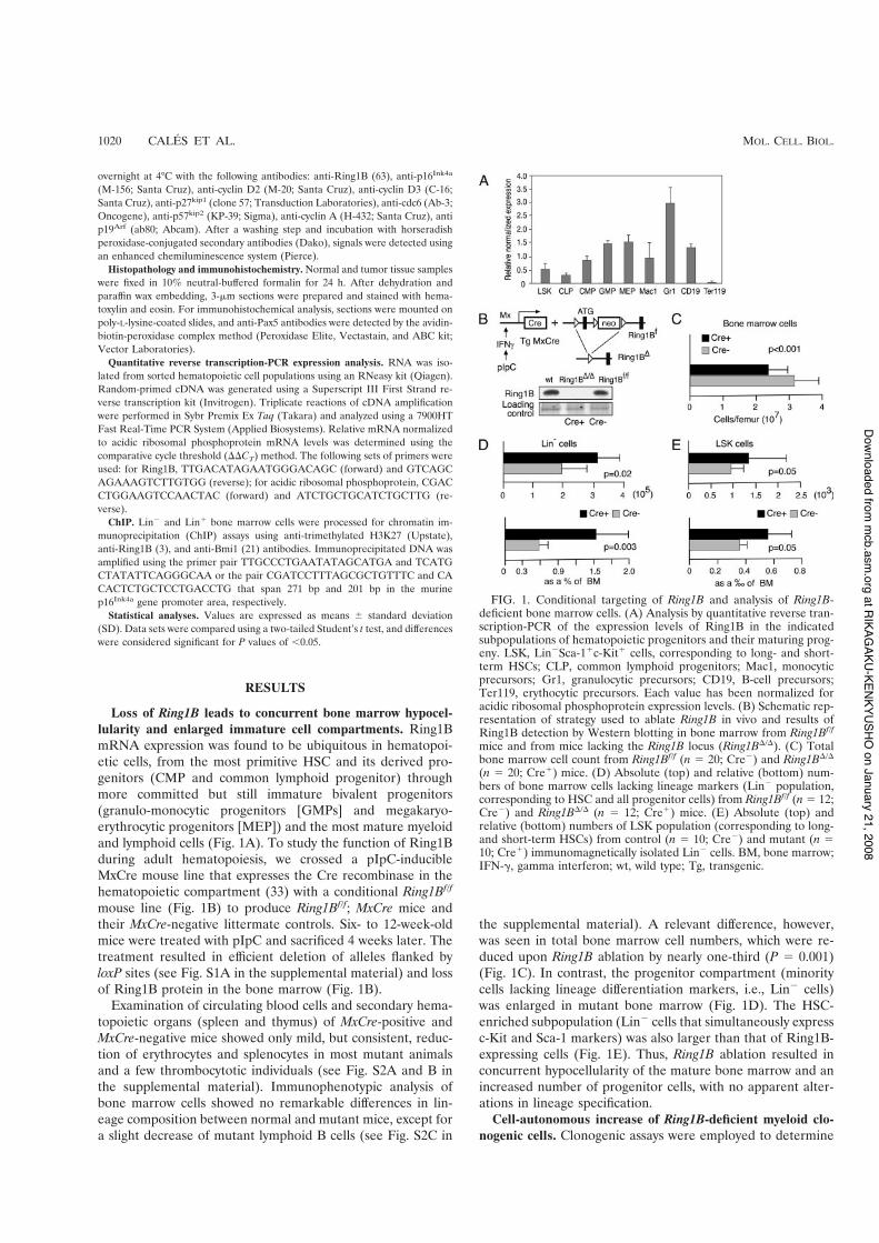
the abundance of cells (CFU) able to expand and give rise todifferentiated progeny in vitro. These include primitive, mul-tilineage progenitors and immediate unilineage precursors thatgive rise to characteristic, morphologically distinguishable col-onies. For lymphoid cells, clonogenic assays in the presence ofIL-7 revealed a severe reduction in the number and size ofRing1B-deficient pre-B-cell colonies (Fig. 2A). On the otherhand, ablation of Ring1B resulted in an almost twofold increaseof myeloid CFU (Fig. 2B). Whereas all types of supportedCFU were represented, their relative proportions were altered,particularly the mixed granulo-mono-erythromegakaryocitic(GEMM) colonies (sixfold increase; P ) 0.0001) (Fig. 2B)originated from the most immature progenitors. The GEMMcolonies derived from blast-like cell clusters, which in mutantbone marrow cultures appeared invariably denser (Fig. 2C,top) and also contained a higher proportion of the primitiveCMP (fivefold increase; P * 0.05) (Fig. 2C, bottom). We con-
clude that Ring1B clearly affected both the size and clonogenicactivity of the progenitors’ compartment.
Since extrinsic signals affect hematopoiesis, we aimed todetermine the cell-autonomous contribution to the above al-terations. To do this, we devised an ex vivo Ring1B inactivationsystem that used bone marrow cells from Ring1Bf/f; Cre-ERT2mice. These were obtained by crossing the conditionalRing1Bf/f mouse line with a RERTert mouse deleter line(Cre-ERT2::Polr2a [43]) (see Fig. S3A in the supplementalmaterial). The mouse deleter line ubiquitously expresses atamoxifen (4-HT)-inducible fusion protein between the Crerecombinase and a mutated ligand binding domain of the hu-man estrogen receptor. Bone marrow cells from these micewere cultured in parallel in the absence (Cre%) and in thepresence (Cre!) of 4-HT to generate control and Ring1B-deleted cells, respectively. Efficient Ring1B ablation wasachieved 2 days after 4-HT treatment (see Fig. S3B in thesupplemental material). As seen above for in vivo Ring1B-inactivated bone marrow cells (Fig. 2A and B), 4-HT treatmentresulted in a reduction of pre-B-cell colonies, an increase intotal Lin% cells, and a larger number of myeloid CFU (see Fig.S3C to E in the supplemental material). Consistent with this,mutant progenitors (!4-HT; Cre!) yielded more secondaryand tertiary colonies in a replicating assay than control pro-genitors (%4-HT; Cre%), indicating a higher self-renewal rateof Ring1B-deficient cells (Fig. 2D). These results indicate thatalteration of immature cell populations and of their clonogenicactivities are mostly due to a cell-autonomous defect resultingfrom the ablation of Ring1B.
Ring1B inactivation enhances the proliferative rate of my-eloid progenitors. We used ex vivo inactivation of Ring1B toinvestigate whether differences in cell proliferation rate, apop-tosis, or cell differentiation could account for the accumulationof Ring1B-deficient primitive hematopoietic cells. Pulse label-ing of Lin% cells with BrdU demonstrated that more cellsincorporated BrdU in 4-HT-treated (Cre!) than in nontreated(Cre%) cultures (Fig. 3A). We also used CFDA-SE fluores-cence decay as an additional test of the cell division rate. Thisassay is based on the dilution of the fluorescent dye resultingfrom its equal distribution among daughter cells upon celldivision. As shown in Fig. 3B, nearly all Ring1B-deficient Lin%
cells had completed one cell cycle at the same point when 30%of the Ring1B-expressing Lin% cells still remained undivided.Under these culture conditions, annexin V labeling (an indi-cator of apoptosis) showed no differences between treated andnontreated cultures (Fig. 3C). These results indicate that theenlargement of the mutant Lin% compartment is due to anincrease in the cell proliferation rate provoked by the loss ofRing1B.
Although the representation of the various lineages in bonemarrow and circulating cells in Ring1B mutant mice suggestedthat no major alterations in differentiation were occurring, wefurther tested this by immunophenotyping cells generated inmethylcellulose cultures of Lin% cells. As seen in Fig. 3D,levels of Gr-1, Mac-1, and CD-41 lineage markers (specific forgranulocytes, monocytes, and megakaryocytes, respectively)showed no differences between cultures from Ring1B-deletedand nondeleted Lin% cells. Moreover, FACS-sorted mutantCMPs retained the ability to give rise in vitro to more matureprogeny, MEPs and GMPs (Fig. 3E). Together, these data
FIG. 2. Clonogenic assays of Ring1B-deficient bone marrow cells.(A) Impaired pre-B-cell colony formation in mice in which the Ring1Blocus has been excised as a result of the expression of MxCre. Coloniesgenerated per 5 # 105 bone marrow cells from mutant (n * 14; Cre!)and control (n * 12; Cre%) mice. At right are images of representativecolonies at day !8 of culture. (B) Increased myeloid clonogenic activ-ity in MxCre! (excised Ring1B) mice (n * 14; Cre!) compared toMxCre% (nonexcised Ring1B) mice (n * 16; Cre%). Bar segmentsrepresent the average absolute number (CFU) of the various colonytypes: unilineage monocytic (M), granulocytic(G), and erythrocytic(BFU-E) and bilineage granulo-monocytic (GM) and GEMM (mixed)colonies. (C) Myeloid colonies. At top are photographs of represen-tative individual immature colonies picked at day !7 of culture; atbottom are contour plots of the flow cytometry analysis of these col-onies showing the myeloid progenitors MEPs, GMPs, and CMPs iden-tified as CD34% Fc&Rlo (lower left lobe), CD34! Fc&R! (upper rightlobe), and CD34! Fc&Rlo (lower right lobe; almost absent in controlCre% colonies), respectively. A representative experiment (out ofthree) is shown, indicating the CMP relative content value ( SD.(D) Replicating assay of ex vivo Ring1B-excised bone marrow cells.The results are shown as the mean ( SD (n * 6) of the total numberof secondary and tertiary colonies generated from identical numbers ofpooled primary control (%4-HT) and mutant (!4-HT) myeloid colo-nies.
VOL. 28, 2008 Ring1B CONTROL OF CELL PROLIFERATION 1021
at RIKAGAKU-KENKYUSHO
on January 21, 2008 m
cb.asm.org
Downloaded from

strongly suggest that the accumulation of early progenitor cellsresulting from Ring1B inactivation is mostly due to an en-hanced proliferation rate of myeloid progenitors and not toreduced apoptosis or impairment of their differentiatingability.
Ring1B negatively regulates cyclin D2 and p16Ink4a. Afterestablishing the role of Ring1B in proliferation of hematopoi-etic cells, we began to study the underlying mechanism(s) byanalyzing protein expression levels of known cell cycle regula-tors. The rate of cell proliferation in mammalian cells is de-termined mainly in the G1 phase of the cell cycle by the specificactivation of cyclin-dependent kinases, which are in turn neg-atively regulated by two families of cyclin-dependent kinaseinhibitors (CKIs) (66). First, we compared total bone marrowcell extracts from control MxCre-negative and in vivo deletedRing1B (MxCre positive) mice. Western blot analysis showedincreased levels of cyclin D2 (Fig. 4A) and also of the cell cycle
progression indicator cyclin A (see Fig. S4A in the supplemen-tal material) in extracts from Ring1B-deficient cells. Also, lev-els of the CKI p27 appeared slightly higher in mutant cells (seeFig. S4 in the supplemental material). However, the mostprominent difference was observed for the levels of p16Ink4a,another CKI which, while undetectable in extracts from non-deleted cells, gave a strong signal in Ring1B-deficient cell ex-tracts (Fig. 4A). No signals were detected for cdc6, a compo-nent of the replication licensing machinery, the CKI p57, orp19Arf in any of the extracts (see Fig. S4A in the supplementalmaterial).
Since the most immature cells (i.e., HSCs and progenitors)represent only a minor contribution to total bone marrow cells,we performed Western blot analysis on mutant and controlLin% cells (Fig. 3B). In contrast to whole bone marrow, theexpression pattern of cell cycle regulators in mutant Lin% cellsshowed an upregulation of proliferation activators cyclin D2
FIG. 3. Increased proliferation of Ring1B-deficient progenitors. (A to C) Proliferation and apoptosis of Lin% cells isolated from Ring1Bf/f;Cre-ERT2 bone marrow cells in the absence (Cre%; top) or the presence (Cre!; bottom) of 4-HT. (A) Solid plots correspond to cells labeled withanti-BrdU antibodies. Overlaid empty plots correspond to cells labeled with isotype-matching FITC antibody to define the BrdU-negativepopulation. Proliferating, BrdU-positive cells (indicated by the bar) are shown as the mean ( SD of two duplicate, independent experiments.(B) CFDA-SE fluorescence content of mutant (!4-HT; Cre!) (bottom) or control (%4-HT; Cre%) (top) Lin% cells, 18 h after immunoseparationand fluorescent dye incubation. The empty histogram shows CFDA-SE cell content immediately after dye loading. (C) FITC-conjugated annexinV labeling of propidium iodide-impermeable viable Lin% cells cultured with (Cre!; bottom) or without (Cre%; top) 4-HT. The overlaid emptyhistogram corresponds to cells treated under the same conditions but with no added annexin V. (D) Expression of lineage markers in cells frommutant (light empty plots) and control (dark solid plots) myeloid colonies harvested at day !8. Gr-1, Mac-1, and CD41 markers stain mature cellsof the granulocytic, monocytic, and megakaryocytic lineages, respectively. Bars indicate positively labeled cells. (E) Mutant CMPs retain theirdifferentiation potential. Sorted mutant CMPs (Lin% c-Kit! CD34! Fc&Rlo) cells were seeded into myeloid differentiation medium and individualcolonies appearing at day !7 (d !7)were labeled with antibodies to c-Kit, CD34, and Fc&R to determine MEP, CMP, and GMP content asindicated in the legend of Fig. 2.
1022 CALES ET AL. MOL. CELL. BIOL.
at RIKAGAKU-KENKYUSHO
on January 21, 2008 m
cb.asm.org
Downloaded from

(Fig. 4B, left), cyclin A, and cdc6 (see Fig. S4A in the supple-mental material) with a concomitant absence of proliferationinhibitors p16Ink4a (Fig. 4B, left), p27, and p57 (see Fig. S4A inthe supplemental material). However, the analysis of coloniesharvested from Lin% cell methylcellulose cultures at day 8showed a sustained upregulation of cyclin D2 together with a
dramatic increase of p16Ink4a levels in maturing Ring1B-defi-cient cells only (Fig. 4B, middle panel), thus resembling theexpression pattern seen in unfractionated bone marrow cells.A similar upregulation of p16Ink4a levels was observed in ex-tracts from cultured mutant pre-B cells (Fig. 4B, right). Onlycyclin D3, which is needed for the expansion of pre-B cells
FIG. 4. Cell cycle regulator levels and proliferation rate are differentially affected by Ring1B along differentiation. (A and B) RepresentativeWestern blots (3 to 15 independent experiments) from the indicated extracts. (A) Bone marrow extracts from mice in which the Ring1B locus hasbeen excised as a result of the expression of MxCre ('/'; Cre!) and from Ring1Bf/f (f/f; Cre%) mice. (B) Blots at left are extracts from Lin% cellsimmunomagnetically isolated from 4-HT-treated or untreated Ring1Bf/f; Cre-ERT2 bone marrow cells cultured for 48 h. Blots of extracts frommyeloid colonies harvested after culturing Lin% cells for 8 days in methylcellulose medium without or with 4-HT are shown in the middle. At rightare blots of extracts from pooled pre-B-cell colonies grown after 8-day cultures of Ring1Bf/f and Ring1B-deficient bone marrow cells. Ctl, extractsfrom mouse erythroleukemia cells; !, with 4-HT treatment; %, without 4-HT treatment. In panels A and B, Ponceau red-stained membranes wereused as a loading control. (C to F) Isolated Lin% cells were cultured in suspension for the indicated times in differentiation medium containingstem cell factor, IL-3, and IL-6 in the presence (!) or absence (%) of 4-HT. (C) Differential accumulation of mutant and control cells with timein culture. Bars represent the average ratio of the percentage of mutant to control cells, considering the value of control cells as 1. (D) Switch inthe values of CFDA-SE fluorescence of cells in cultures in which the Ring1B locus has been excised as a result of 4-HT treatment (Cre!) and inuntreated (nonexcised Ring1B; Cre%) cultures at the indicated times. (E) FITC-conjugated annexin V labeling of propidium iodide-impermeableviable cells present in day !6 cells cultured with (right) or without (left) 4-HT. Overlaid empty histogram corresponds to cells treated under thesame conditions but with no added annexin V. (F) Western blot analysis of cell extracts obtained from cells cultured in the presence (!) or theabsence (%) of 4-HT for the indicated times. Day !0 corresponds to extracts of Lin% cells isolated from the bone marrow of Ring1Bf/f; Cre-ERT2mice at the time of sacrifice. Controls and loading control are as described in panels A and B. (G) Ink4a promoter occupancy by Ring1B and Bmi1in progenitors and maturing hematopoietic cells. At top is a schematic representation of the p16Ink4a gene locus shows exons and coding (filled)sequences and the amplicon used for ChIP assays. Lin% and Lin! cell populations were immunomagnetically isolated from bone marrow andchromatin analyzed using the indicated antibodies (bottom). Ex, exon; H3K27me3, trimethylated H3K27; IgG, immunoglobulin G; d, day.
VOL. 28, 2008 Ring1B CONTROL OF CELL PROLIFERATION 1023
at RIKAGAKU-KENKYUSHO
on January 21, 2008 m
cb.asm.org
Downloaded from
(13), was detected in these cells, and its expression was hardlyaffected by the lack of Ring1B in either pre-B cells or myeloidcells (see Fig. S4C in the supplemental material), suggestingthat the effect on cyclin D2 is specific. Modest increases in p27and p57 levels were also observed in maturing mutant cells (seeFig. S4B in the supplemental material). These data show thatRing1B inactivation alters the expression pattern of cell cycleregulators in differentiating cells and suggest that the resultingpopulations of mature and immature cells are endowed withreduced and enhanced proliferation rates, respectively.
We investigated these cell populations by using liquid cul-tures of ex vivo Ring1B-ablated Lin% cells under conditionsthat recapitulate the differentiation events occurring in normalmyelopoiesis. Initially after treatment, the relative accumula-tion of cells was faster in mutant (!4-HT; Cre!) than incontrol (%4-HT; Cre%) cultures (Fig. 4C). However, by day 6after treatment, this ratio was inverted, and Ring1B-expressingcultures contained more cells than those of Ring1B-deficientcells. Analysis of CFDA-SE fluorescence decay confirmed theinversion of cell division rates between days 4 and 6 after 4-HT(Fig. 4D) treatment. The lower accumulation of mutant cellsdid not result from increased cell death, as determined byannexin V staining (Fig. 4E). Instead, the late upregulation ofp16Ink4a, detected only by day 6 after treatment (Fig. 4F),correlated with the switch in proliferation rate. This contrastedwith the prompt upregulation of the proliferation promotercyclin D2 in mutant cultures (Fig. 4F), which, in the absence ofthe inhibitor p16Ink4a, would explain the enhanced prolifera-tion observed before day !6. A possible explanation for thedifferential regulation of p16Ink4a in immature versus maturecells may be related to the decreasing levels of Bmi1 duringhematopoietic differentiation (26), which would ensurep16Ink4a silencing in progenitors, even in the absence ofRing1B. We tested this by analyzing promoter occupancy atthe Ink4a locus in ChIP assays and found that whereas Ring1Bwas detected in both immature Lin% and maturing Lin! cells,Bmi1 was seen associated predominantly in the immature com-partment (Fig. 4G).
Collectively, these results show that through the balancedexpression of positive and negative cell cycle regulators,Ring1B controls the expansion of differentiating hematopoi-etic cells both by restricting proliferation of progenitors and bycontributing to their expansion during maturation.
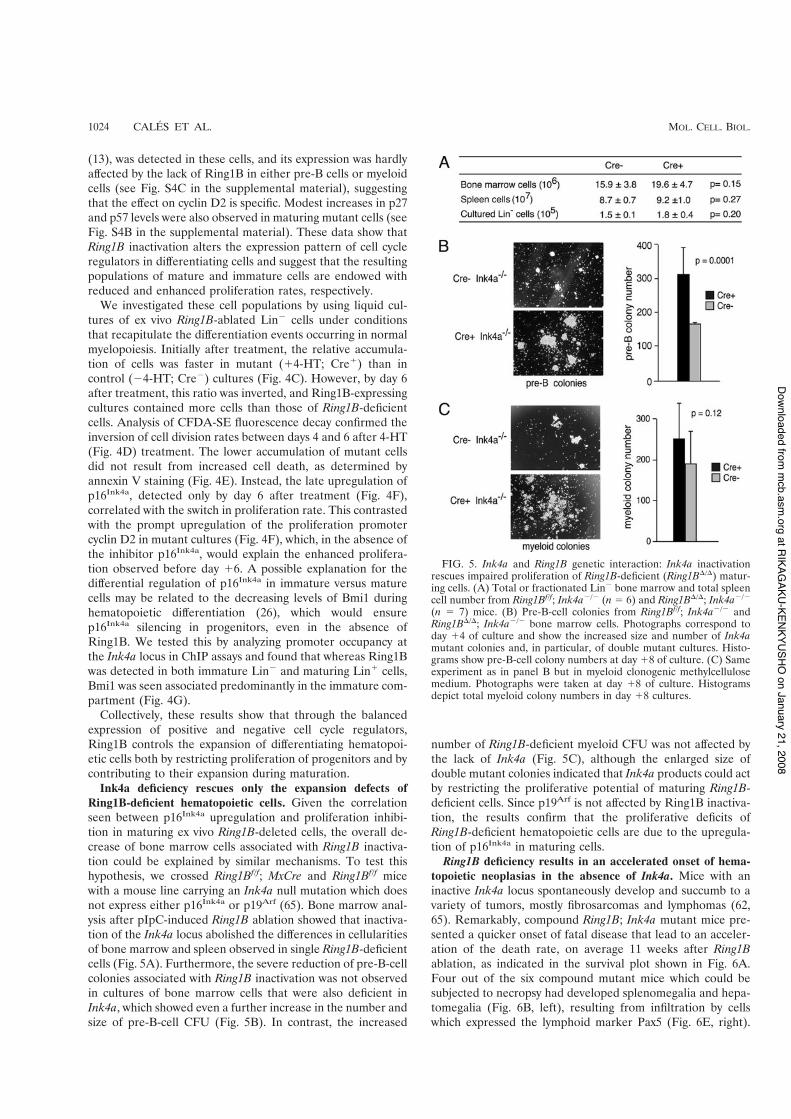
Ink4a deficiency rescues only the expansion defects ofRing1B-deficient hematopoietic cells. Given the correlationseen between p16Ink4a upregulation and proliferation inhibi-tion in maturing ex vivo Ring1B-deleted cells, the overall de-crease of bone marrow cells associated with Ring1B inactiva-tion could be explained by similar mechanisms. To test thishypothesis, we crossed Ring1Bf/f; MxCre and Ring1Bf/f micewith a mouse line carrying an Ink4a null mutation which doesnot express either p16Ink4a or p19Arf (65). Bone marrow anal-ysis after pIpC-induced Ring1B ablation showed that inactiva-tion of the Ink4a locus abolished the differences in cellularitiesof bone marrow and spleen observed in single Ring1B-deficientcells (Fig. 5A). Furthermore, the severe reduction of pre-B-cellcolonies associated with Ring1B inactivation was not observedin cultures of bone marrow cells that were also deficient inInk4a, which showed even a further increase in the number andsize of pre-B-cell CFU (Fig. 5B). In contrast, the increased
number of Ring1B-deficient myeloid CFU was not affected bythe lack of Ink4a (Fig. 5C), although the enlarged size ofdouble mutant colonies indicated that Ink4a products could actby restricting the proliferative potential of maturing Ring1B-deficient cells. Since p19Arf is not affected by Ring1B inactiva-tion, the results confirm that the proliferative deficits ofRing1B-deficient hematopoietic cells are due to the upregula-tion of p16Ink4a in maturing cells.
Ring1B deficiency results in an accelerated onset of hema-topoietic neoplasias in the absence of Ink4a. Mice with aninactive Ink4a locus spontaneously develop and succumb to avariety of tumors, mostly fibrosarcomas and lymphomas (62,65). Remarkably, compound Ring1B; Ink4a mutant mice pre-sented a quicker onset of fatal disease that lead to an acceler-ation of the death rate, on average 11 weeks after Ring1Bablation, as indicated in the survival plot shown in Fig. 6A.Four out of the six compound mutant mice which could besubjected to necropsy had developed splenomegalia and hepa-tomegalia (Fig. 6B, left), resulting from infiltration by cellswhich expressed the lymphoid marker Pax5 (Fig. 6E, right).
FIG. 5. Ink4a and Ring1B genetic interaction: Ink4a inactivationrescues impaired proliferation of Ring1B-deficient (Ring1B'/') matur-ing cells. (A) Total or fractionated Lin% bone marrow and total spleencell number from Ring1Bf/f; Ink4a%/% (n * 6) and Ring1B'/'; Ink4a%/%
(n * 7) mice. (B) Pre-B-cell colonies from Ring1Bf/f; Ink4a%/% andRing1B'/'; Ink4a%/% bone marrow cells. Photographs correspond today !4 of culture and show the increased size and number of Ink4amutant colonies and, in particular, of double mutant cultures. Histo-grams show pre-B-cell colony numbers at day !8 of culture. (C) Sameexperiment as in panel B but in myeloid clonogenic methylcellulosemedium. Photographs were taken at day !8 of culture. Histogramsdepict total myeloid colony numbers in day !8 cultures.
1024 CALES ET AL. MOL. CELL. BIOL.
at RIKAGAKU-KENKYUSHO
on January 21, 2008 m
cb.asm.org
Downloaded from
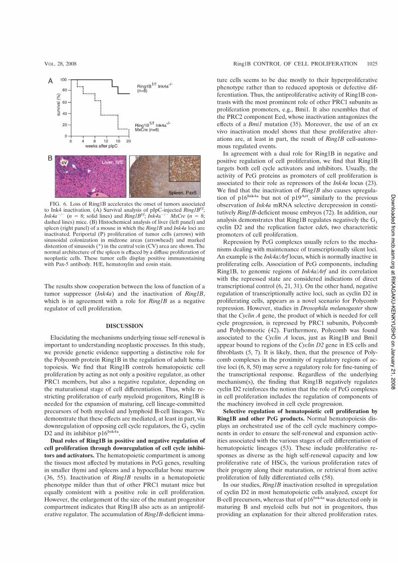
The results show cooperation between the loss of function of atumor suppressor (Ink4a) and the inactivation of Ring1B,which is in agreement with a role for Ring1B as a negativeregulator of cell proliferation.
DISCUSSION
Elucidating the mechanisms underlying tissue self-renewal isimportant to understanding neoplastic processes. In this study,we provide genetic evidence supporting a distinctive role forthe Polycomb protein Ring1B in the regulation of adult hema-topoiesis. We find that Ring1B controls hematopoietic cellproliferation by acting as not only a positive regulator, as otherPRC1 members, but also a negative regulator, depending onthe maturational stage of cell differentiation. Thus, while re-stricting proliferation of early myeloid progenitors, Ring1B isneeded for the expansion of maturing, cell lineage-committedprecursors of both myeloid and lymphoid B-cell lineages. Wedemonstrate that these effects are mediated, at least in part, viadownregulation of opposing cell cycle regulators, the G1 cyclinD2 and its inhibitor p16Ink4a.
Dual roles of Ring1B in positive and negative regulation ofcell proliferation through downregulation of cell cycle inhibi-tors and activators. The hematopoietic compartment is amongthe tissues most affected by mutations in PcG genes, resultingin smaller thymi and spleens and a hypocellular bone marrow(36, 55). Inactivation of Ring1B results in a hematopoieticphenotype milder than that of other PRC1 mutant mice butequally consistent with a positive role in cell proliferation.However, the enlargement of the size of the mutant progenitorcompartment indicates that Ring1B also acts as an antiprolif-erative regulator. The accumulation of Ring1B-deficient imma-
ture cells seems to be due mostly to their hyperproliferativephenotype rather than to reduced apoptosis or defective dif-ferentiation. Thus, the antiproliferative activity of Ring1B con-trasts with the most prominent role of other PRC1 subunits asproliferation promoters, e.g., Bmi1. It also resembles that ofthe PRC2 component Eed, whose inactivation antagonizes theeffects of a Bmi1 mutation (35). Moreover, the use of an exvivo inactivation model shows that these proliferative alter-ations are, at least in part, the result of Ring1B cell-autono-mous regulated events.
In agreement with a dual role for Ring1B in negative andpositive regulation of cell proliferation, we find that Ring1Btargets both cell cycle activators and inhibitors. Usually, theactivity of PcG proteins as promoters of cell proliferation isassociated to their role as repressors of the Ink4a locus (23).We find that the inactivation of Ring1B also causes upregula-tion of p16Ink4a but not of p19Arf, similarly to the previousobservation of Ink4a mRNA selective derepression in consti-tutively Ring1B-deficient mouse embryos (72). In addition, ouranalysis demonstrates that Ring1B regulates negatively the G1
cyclin D2 and the replication factor cdc6, two characteristicpromoters of cell proliferation.
Repression by PcG complexes usually refers to the mecha-nisms dealing with maintenance of transcriptionally silent loci.An example is the Ink4a/Arf locus, which is normally inactive inproliferating cells. Association of PcG components, includingRing1B, to genomic regions of Ink4a/Arf and its correlationwith the repressed state are considered indications of directtranscriptional control (6, 21, 31). On the other hand, negativeregulation of transcriptionally active loci, such as cyclin D2 inproliferating cells, appears as a novel scenario for Polycombrepression. However, studies in Drosophila melanogaster showthat the Cyclin A gene, the product of which is needed for cellcycle progression, is repressed by PRC1 subunits, Polycomband Polyhomeotic (42). Furthermore, Polycomb was foundassociated to the Cyclin A locus, just as Ring1B and Bmi1appear bound to regions of the Cyclin D2 gene in ES cells andfibroblasts (5, 7). It is likely, then, that the presence of Poly-comb complexes in the proximity of regulatory regions of ac-tive loci (6, 8, 50) may serve a regulatory role for fine-tuning ofthe transcriptional response. Regardless of the underlyingmechanism(s), the finding that Ring1B negatively regulatescyclin D2 reinforces the notion that the role of PcG complexesin cell proliferation includes the regulation of components ofthe machinery involved in cell cycle progression.
Selective regulation of hematopoietic cell proliferation byRing1B and other PcG products. Normal hematopoiesis dis-plays an orchestrated use of the cell cycle machinery compo-nents in order to ensure the self-renewal and expansion activ-ities associated with the various stages of cell differentiation ofhematopoietic lineages (53). These include proliferative re-sponses as diverse as the high self-renewal capacity and lowproliferative rate of HSCs, the various proliferation rates oftheir progeny along their maturation, or retrieval from activeproliferation of fully differentiated cells (58).
In our studies, Ring1B inactivation resulted in upregulationof cyclin D2 in most hematopoietic cells analyzed, except forB-cell precursors, whereas that of p16Ink4a was detected only inmaturing B and myeloid cells but not in progenitors, thusproviding an explanation for their altered proliferation rates.
FIG. 6. Loss of Ring1B accelerates the onset of tumors associatedto Ink4 inactivation. (A) Survival analysis of pIpC-injected Ring1Bf/f;Ink4a%/% (n * 8; solid lines) and Ring1Bf/f; Ink4a%/% MxCre (n * 8;dashed lines) mice. (B) Histochemical analysis of liver (left panel) andspleen (right panel) of a mouse in which the Ring1B and Ink4a loci areinactivated. Periportal (P) proliferation of tumor cells (arrows) withsinusoidal colonization in midzone areas (arrowhead) and markeddistention of sinusoids (*) in the central vein (CV) area are shown. Thenormal architecture of the spleen is effaced by a diffuse proliferation ofneoplastic cells. These tumor cells display positive immunostainingwith Pax-5 antibody. H/E, hematoxylin and eosin stain.
VOL. 28, 2008 Ring1B CONTROL OF CELL PROLIFERATION 1025
at RIKAGAKU-KENKYUSHO
on January 21, 2008 m
cb.asm.org
Downloaded from
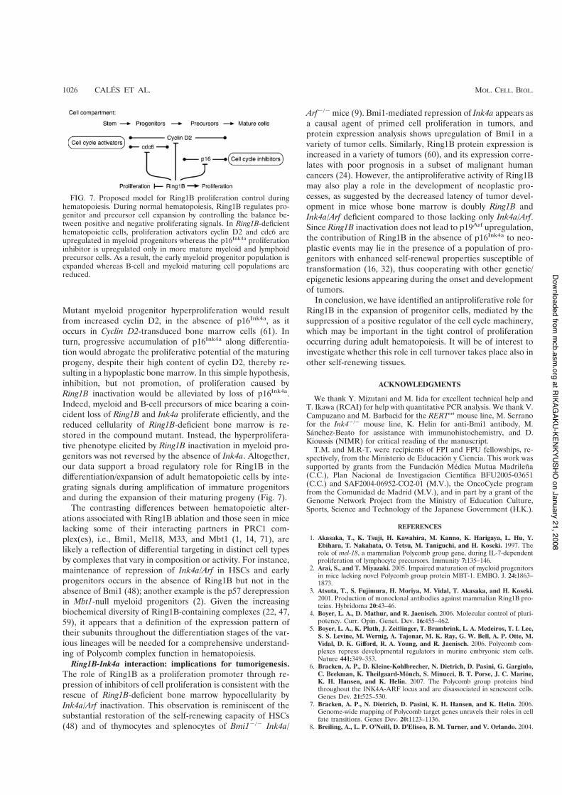
Mutant myeloid progenitor hyperproliferation would resultfrom increased cyclin D2, in the absence of p16Ink4a, as itoccurs in Cyclin D2-transduced bone marrow cells (61). Inturn, progressive accumulation of p16Ink4a along differentia-tion would abrogate the proliferative potential of the maturingprogeny, despite their high content of cyclin D2, thereby re-sulting in a hypoplastic bone marrow. In this simple hypothesis,inhibition, but not promotion, of proliferation caused byRing1B inactivation would be alleviated by loss of p16Ink4a.Indeed, myeloid and B-cell precursors of mice bearing a coin-cident loss of Ring1B and Ink4a proliferate efficiently, and thereduced cellularity of Ring1B-deficient bone marrow is re-stored in the compound mutant. Instead, the hyperprolifera-tive phenotype elicited by Ring1B inactivation in myeloid pro-genitors was not reversed by the absence of Ink4a. Altogether,our data support a broad regulatory role for Ring1B in thedifferentiation/expansion of adult hematopoietic cells by inte-grating signals during amplification of immature progenitorsand during the expansion of their maturing progeny (Fig. 7).
The contrasting differences between hematopoietic alter-ations associated with Ring1B ablation and those seen in micelacking some of their interacting partners in PRC1 com-plex(es), i.e., Bmi1, Mel18, M33, and Mbt1 (1, 14, 71), arelikely a reflection of differential targeting in distinct cell typesby complexes that vary in composition or activity. For instance,maintenance of repression of Ink4a/Arf in HSCs and earlyprogenitors occurs in the absence of Ring1B but not in theabsence of Bmi1 (48); another example is the p57 derepressionin Mbt1-null myeloid progenitors (2). Given the increasingbiochemical diversity of Ring1B-containing complexes (22, 47,59), it appears that a definition of the expression pattern oftheir subunits throughout the differentiation stages of the var-ious lineages will be needed for a comprehensive understand-ing of Polycomb complex function in hematopoiesis.
Ring1B-Ink4a interaction: implications for tumorigenesis.The role of Ring1B as a proliferation promoter through re-pression of inhibitors of cell proliferation is consistent with therescue of Ring1B-deficient bone marrow hypocellularity byInk4a/Arf inactivation. This observation is reminiscent of thesubstantial restoration of the self-renewing capacity of HSCs(48) and of thymocytes and splenocytes of Bmi1%/% Ink4a/
Arf%/% mice (9). Bmi1-mediated repression of Ink4a appears asa causal agent of primed cell proliferation in tumors, andprotein expression analysis shows upregulation of Bmi1 in avariety of tumor cells. Similarly, Ring1B protein expression isincreased in a variety of tumors (60), and its expression corre-lates with poor prognosis in a subset of malignant humancancers (24). However, the antiproliferative activity of Ring1Bmay also play a role in the development of neoplastic pro-cesses, as suggested by the decreased latency of tumor devel-opment in mice whose bone marrow is doubly Ring1B andInk4a/Arf deficient compared to those lacking only Ink4a/Arf.Since Ring1B inactivation does not lead to p19Arf upregulation,the contribution of Ring1B in the absence of p16Ink4a to neo-plastic events may lie in the presence of a population of pro-genitors with enhanced self-renewal properties susceptible oftransformation (16, 32), thus cooperating with other genetic/epigenetic lesions appearing during the onset and developmentof tumors.
In conclusion, we have identified an antiproliferative role forRing1B in the expansion of progenitor cells, mediated by thesuppression of a positive regulator of the cell cycle machinery,which may be important in the tight control of proliferationoccurring during adult hematopoiesis. It will be of interest toinvestigate whether this role in cell turnover takes place also inother self-renewing tissues.
ACKNOWLEDGMENTS
We thank Y. Mizutani and M. Iida for excellent technical help andT. Ikawa (RCAI) for help with quantitative PCR analysis. We thank V.Campuzano and M. Barbacid for the RERTert mouse line, M. Serranofor the Ink4%/% mouse line, K. Helin for anti-Bmi1 antibody, M.Sanchez-Beato for assistance with immunohistochemistry, and D.Kioussis (NIMR) for critical reading of the manuscript.
T.M. and M.R-T. were recipients of FPI and FPU fellowships, re-spectively, from the Ministerio de Educacion y Ciencia. This work wassupported by grants from the Fundacion Medica Mutua Madrilena(C.C.), Plan Nacional de Investigacion Cientıfica BFU2005-03651(C.C.) and SAF2004-06952-CO2-01 (M.V.), the OncoCycle programfrom the Comunidad de Madrid (M.V.), and in part by a grant of theGenome Network Project from the Ministry of Education Culture,Sports, Science and Technology of the Japanese Government (H.K.).
REFERENCES
1. Akasaka, T., K. Tsuji, H. Kawahira, M. Kanno, K. Harigaya, L. Hu, Y.Ebihara, T. Nakahata, O. Tetsu, M. Taniguchi, and H. Koseki. 1997. Therole of mel-18, a mammalian Polycomb group gene, during IL-7-dependentproliferation of lymphocyte precursors. Immunity 7:135–146.
2. Arai, S., and T. Miyazaki. 2005. Impaired maturation of myeloid progenitorsin mice lacking novel Polycomb group protein MBT-1. EMBO. J. 24:1863–1873.
3. Atsuta, T., S. Fujimura, H. Moriya, M. Vidal, T. Akasaka, and H. Koseki.2001. Production of monoclonal antibodies against mammalian Ring1B pro-teins. Hybridoma 20:43–46.
4. Boyer, L. A., D. Mathur, and R. Jaenisch. 2006. Molecular control of pluri-potency. Curr. Opin. Genet. Dev. 16:455–462.
5. Boyer, L. A., K. Plath, J. Zeitlinger, T. Brambrink, L. A. Medeiros, T. I. Lee,S. S. Levine, M. Wernig, A. Tajonar, M. K. Ray, G. W. Bell, A. P. Otte, M.Vidal, D. K. Gifford, R. A. Young, and R. Jaenisch. 2006. Polycomb com-plexes repress developmental regulators in murine embryonic stem cells.Nature 441:349–353.
6. Bracken, A. P., D. Kleine-Kohlbrecher, N. Dietrich, D. Pasini, G. Gargiulo,C. Beekman, K. Theilgaard-Monch, S. Minucci, B. T. Porse, J. C. Marine,K. H. Hansen, and K. Helin. 2007. The Polycomb group proteins bindthroughout the INK4A-ARF locus and are disassociated in senescent cells.Genes Dev. 21:525–530.
7. Bracken, A. P., N. Dietrich, D. Pasini, K. H. Hansen, and K. Helin. 2006.Genome-wide mapping of Polycomb target genes unravels their roles in cellfate transitions. Genes Dev. 20:1123–1136.
8. Breiling, A., L. P. O’Neill, D. D’Eliseo, B. M. Turner, and V. Orlando. 2004.
FIG. 7. Proposed model for Ring1B proliferation control duringhematopoiesis. During normal hematopoiesis, Ring1B regulates pro-genitor and precursor cell expansion by controlling the balance be-tween positive and negative proliferating signals. In Ring1B-deficienthematopoietic cells, proliferation activators cyclin D2 and cdc6 areupregulated in myeloid progenitors whereas the p16Ink4a proliferationinhibitor is upregulated only in more mature myeloid and lymphoidprecursor cells. As a result, the early myeloid progenitor population isexpanded whereas B-cell and myeloid maturing cell populations arereduced.
1026 CALES ET AL. MOL. CELL. BIOL.
at RIKAGAKU-KENKYUSHO
on January 21, 2008 m
cb.asm.org
Downloaded from

Epigenome changes in active and inactive Polycomb-group-controlled re-gions. EMBO Rep. 5:976–982.
9. Bruggeman, S. W., M. E. Valk-Lingbeek, P. P. van der Stoop, J. J. Jacobs, K.Kieboom, E. Tanger, D. Hulsman, C. Leung, Y. Arsenijevic, S. Marino, andM. van Lohuizen. 2005. Ink4a and Arf differentially affect cell proliferationand neural stem cell self-renewal in Bmi1-deficient mice. Genes Dev. 19:1438–1443.
10. Buchwald, G., P. van der Stoop, O. Weichenrieder, A. Perrakis, M. vanLohuizen, and T. K. Sixma. 2006. Structure and E3-ligase activity of theRing-Ring complex of Polycomb proteins Bmi1 and Ring1b. EMBO J. 25:2465–2474.
11. Buszczak, M., and A. C. Spradling. 2006. Searching chromatin for stem cellidentity. Cell 125:233–236.
12. Cao, R., L. Wang, H. Wang, L. Xia, H. Erdjument-Bromage, P. Tempst, R. S.Jones, and Y. Zhang. 2002. Role of histone H3 lysine 27 methylation inPolycomb-group silencing. Science 298:1039–1043.
13. Cooper, A. B., C. M. Sawai, E. Sicinska, S. E. Powers, P. Sicinski, M. R.Clark, and I. Aifantis. 2006. A unique function for cyclin D3 in early B celldevelopment. Nat. Immunol. 7:489–497.
14. Core, N., F. Joly, A. Boned, and M. Djabali. 2004. Disruption of E2F sig-naling suppresses the INK4a-induced proliferative defect in M33-deficientmice. Oncogene 23:7660–7668.
15. Core, N., S. Bel, S. J. Gaunt, M. Aurrand-Lions, J. Pearce, A. Fisher, and M.Djabali. 1997. Altered cellular proliferation and mesoderm patterning inPolycomb-M33-deficient mice. Development 124:721–729.
16. Cozzio, A., E. Passegue, P. M. Ayton, H. Karsunky, M. L. Cleary, and I. L.Weissman. 2003. Similar MLL-associated leukemias arising from self-renew-ing stem cells and short-lived myeloid progenitors. Genes Dev. 17:3029–3035.
17. Crosnier, C., D. Stamataki, and J. Lewis. 2006. Organizing cell renewal inthe intestine: stem cells, signals and combinatorial control. Nat. Rev. Genet.7:349–359.
18. Czermin, B., R. Melfi, D. McCabe, V. Seitz, A. Imhof, and V. Pirrotta. 2002.Drosophila enhancer of Zeste/ESC complexes have a histone H3 methyl-transferase activity that marks chromosomal Polycomb sites. Cell 111:185–196.
19. del Mar Lorente, M., C. Marcos-Gutierrez, C. Perez, J. Schoorlemmer, A.Ramırez, T. Magin, and M. Vidal. 2000. Loss- and gain-of-function muta-tions show a Polycomb group function for Ring1A in mice. Development127:5093–5100.
20. de Napoles, M., J. E. Mermoud, R. Wakao, Y. A. Tang, M. Endoh, R.Appanah, T. B. Nesterova, J. Silva, A. P. Otte, M. Vidal, H. Koseki, and N.Brockdorff. 2004. Polycomb group proteins Ring1A/B link ubiquitylation ofhistone H2A to heritable gene silencing and X inactivation. Dev. Cell 7:663–676.
21. Dietrich, N., A. P. Bracken, E. Trinh, C. K. Schjerling, H. Koseki, J.Rappsilber, K. Helin, and K. H. Hansen. 2007. Bypass of senescence by thepolycomb group protein CBX8 through direct binding to the INK4A-ARFlocus. EMBO J. 26:1637–1648.
22. Gearhart, M. D., C. M. Corcoran, J. A. Wamstad, and V. J. Bardwell. 2006.Polycomb group and SCF ubiquitin ligases are found in a novel BCORcomplex that is recruited to BCL6 targets. Mol. Cell. Biol. 26:6880–6889.
23. Gil, J., and G. Peters. 2006. Regulation of the INK4b-ARF-INK4a tumoursuppressor locus: all for one or one for all. Nat. Rev. Mol. Cell Biol. 7:667–677.
24. Glinsky, G. V., O. Berezovska, and A. B. Glinskii. 2005. Microarray analysisidentifies a death-from-cancer signature predicting therapy failure in pa-tients with multiple types of cancer. J. Clin. Investig. 115:1503–1521.
25. Hemenway, C. S., B. W. Halligan, and L. S. Levy. 1998. The Bmi-1 onco-protein interacts with dinG and MPh2: the role of RING finger domains.Oncogene 16:2541–2547.
26. Hosen, N., T. Yamane, M. Muijtjens, K. Pham, M. F. Clarke, and I. L.Weissman. 2007. Bmi-1-green fluorescent protein-knock-in mice reveal thedynamic regulation of bmi-1 expression in normal and leukemic hematopoi-etic cells. Stem Cells 25:1635–1644.
27. Jacobs, J. J., B. Scheijen, J. W. Voncken, K. Kieboom, A. Berns, and M. vanLohuizen. 1999. Bmi-1 collaborates with c-Myc in tumorigenesis by inhibitingc-Myc-induced apoptosis via INK4a/ARF. Genes Dev. 13:2678–2690.
28. Kajiume, T., Y. Ninomiya, H. Ishihara, R. Kanno, and M. Kanno. 2004.Polycomb group gene mel-18 modulates the self-renewal activity and cellcycle status of hematopoietic stem cells. Exp. Hematol. 32:571–578.
29. Kato, Y., H. Koseki, M. Vidal, H. Nakauchi, and A. Iwama. 2007. Uniquecomposition of polycomb repressive complex 1 in hematopoietic stem cells.Int. J. Hematol. 85:179–181.
30. Kondo, M., I. L. Weissman, and K. Akashi. 1997. Identification of clonogeniccommon lymphoid progenitors in mouse bone marrow. Cell 91:661–672.
31. Kotake, Y., R. Cao, P. Viatour, J. Sage, Y. Zhang, and Y. Xiong. 2007. pRBfamily proteins are required for H3K27 trimethylation and Polycomb repres-sion complexes binding to and silencing p16INK4a tumor suppressor gene.Genes Dev. 21:49–54.
32. Krivtsov, A. V., D. Twomey, Z. Feng, M. C. Stubbs, Y. Wang, J. Faber, J. E.Levine, J. Wang, W. C. Hahn, D. G. Gilliland, T. R. Golub, and S. A.
Armstrong. 2006. Transformation from committed progenitor to leukaemiastem cell initiated by MLL-AF9. Nature 442:818–822.
33. Kuhn, R., F. Schwenk, M. Aguet, and K. Rajewsky. 1995. Inducible genetargeting in mice. Science 269:1427–1429.
34. Kuzmichev, A., K. Nishioka, H. Erdjument-Bromage, P. Tempst, and D.Reinberg. 2002. Histone methyltransferase activity associated with a humanmultiprotein complex containing the Enhancer of Zeste protein. Genes Dev.16:2893–2905.
35. Lessard, J., A. Schumacher, U. Thorsteinsdottir, M. van Lohuizen, T.Magnuson, and G. Sauvageau. 1999. Functional antagonism of the Poly-comb-group genes Eed and Bmi1 in hemopoietic cell proliferation. GenesDev. 13:2691–2703.
36. Lessard, J., and G. Sauvageau. 2003. Polycomb group genes as epigeneticregulators of normal and leukemic hemopoiesis. Exp. Hematol. 31:567–585.
37. Lessard, J., and G. Sauvageau. 2003. Bmi-1 determines the proliferativecapacity of normal and leukaemic stem cells. Nature 423:255–260.
38. Leung, C., M. Lingbeek, O. Shakhova, J. Liu, E. Tanger, P. Saremaslani, M.Van Lohuizen, and S. Marino. 2004. Bmi1 is essential for cerebellar devel-opment and is overexpressed in human medulloblastomas. Nature 428:337–341.
39. Levine, S. S., A. Weiss, H. Erdjument-Bromage, Z. Shao, P. Tempst, andR. E. Kingston. 2002. The core of the Polycomb repressive complex iscompositionally and functionally conserved in flies and humans. Mol. Cell.Biol. 22:6070–6078.
40. Levine, S. S., I. F. King, and R. E. Kingston. 2004. Division of labor inPolycomb group repression. Trends Biochem. Sci. 29:478–485.
41. Martinez, A. M., and G. Cavalli. 2006. The role of Polycomb group proteinsin cell cycle regulation during development. Cell Cycle 5:1189–1197.
42. Martinez, A. M., S. Colomb, J. Dejardin, F. Bantignies, and G. Cavalli. 2006.Polycomb group-dependent cyclin A repression in Drosophila. Genes. Dev.20:501–513.
43. Mijimolle, N., J. Velasco, P. Dubus, C. Guerra, C. A. Weinbaum, P. J. Casey,V. Campuzano, and M. Barbacid. 2005. Protein farnesyltransferase in em-bryogenesis, adult homeostasis, and tumor development. Cancer Cell 7:313–324.
44. Molofsky, A. V., R. Pardal, T. Iwashita, I. K. Park, M. F. Clarke, and S. J.Morrison. 2003. Bmi-1 dependence distinguishes neural stem cell self-re-newal from progenitor proliferation. Nature 425:962–967.
45. Molofsky, A. V., S. He, M. Bydon, S. J. Morrison, and R. Pardal. 2005. Bmi-1promotes neural stem cell self-renewal and neural development but notmouse growth and survival by repressing the p16Ink4a and p19Arf senescencepathways. Genes Dev. 19:1432–1437.
46. Muller, J., C. M. Hart, N. J. Francis, M. L. Vargas, A. Sengupta, B. Wild,E. L. Miller, M. B. O’Connor, R. E. Kingston, and J. A. Simon. 2002. Histonemethyltransferase activity of a Drosophila Polycomb group repressor com-plex. Cell 111:197–208.
47. Ogawa, H., K. Ishiguro, S. Gaubatz, D. M. Livingston, and Y. Nakatani.2002. A complex with chromatin modifiers that occupies E2F- and Myc-responsive genes in G0 cells. Science 296:1132–1136.
48. Oguro, H., A. Iwama, Y. Morita, T. Kamijo, M. van Lohuizen, and H.Nakauchi. 2006. Differential impact of Ink4a and Arf on hematopoietic stemcells and their bone marrow microenvironment in Bmi1-deficient mice. J.Exp. Med. 203:2247–2253.
49. Ohta, H., A. Sawada, J. Y. Kim, S. Tokimasa, S. Nishiguchi, R. K.Humphries, J. Hara, and Y. Takihara. 2002. Polycomb group gene rae28 isrequired for sustaining activity of hematopoietic stem cells. J. Exp. Med.195:759–770.
50. Papp, B., and J. Muller. 2006. Histone trimethylation and the maintenanceof transcriptional ON and OFF states by TrxG and PcG proteins. Genes.Dev. 20:2041–2054.
51. Park, I. K., D. Qian, M. Kiel, M. W. Becker, M. Pihalja, I. L. Weissman, S. J.Morrison, and M. F. Clarke. 2003. Bmi-1 is required for maintenance ofadult self-renewing haematopoietic stem cells. Nature 423:302–305.
52. Pasini, D., A. P. Bracken, and K. Helin. 2004. Polycomb group proteins incell cycle progression and cancer. Cell Cycle 3:396–400.
53. Passegue, E., A. J. Wagers, S. Giuriato, W. C. Anderson, and I. L. Weissman.2005. Global analysis of proliferation and cell cycle gene expression in theregulation of hematopoietic stem and progenitor cell fates. J. Exp. Med.202:1599–1611.
54. Raaphorst, F. M. 2005. Deregulated expression of Polycomb-group onco-genes in human malignant lymphomas and epithelial tumors. Hum. Mol.Genet. 14(Suppl. 1):R93–R100.
55. Raaphorst, F. M., A. P. Otte, and C. J. Meijer. 2001. Polycomb-group genesas regulators of mammalian lymphopoiesis. Trends Immunol. 22:682–690.
56. Rando, T. A. 2006. Stem cells, ageing and the quest for immortality. Nature441:1080–1086.
57. Ringrose, L., and R. Paro. 2004. Epigenetic regulation of cellular memory bythe polycomb and trithorax group proteins. Annu. Rev. Genet. 38:413–443.
58. Rosenbauer, F., and D. G. Tenen. 2007. Transcription factors in myeloiddevelopment: balancing differentiation with transformation. Nat. Rev. Im-munol. 7:105–117.
59. Sanchez, C., I. Sanchez, J. A. Demmers, P. Rodriguez, J. Strouboulis, and M.
VOL. 28, 2008 Ring1B CONTROL OF CELL PROLIFERATION 1027
at RIKAGAKU-KENKYUSHO
on January 21, 2008 m
cb.asm.org
Downloaded from

Vidal. 2007. Proteomic analysis of Ring1B/Rnf2 interactors identifies a novelcomplex with the Fbxl10/ Jmjd1B histone demethylase and the BcoR core-pressor. Mol. Cell. Proteomics 6:820–834.
60. Sanchez-Beato, M., E. Sanchez, J. Gonzalez-Carrero, M. Morente, A. Dıez,L. Sanchez-Verde, M. C. Martın, J. C. Cigudosa, M. Vidal, and M. A. Piris.2006. Variability in the expression of polycomb proteins in different normaland tumoral tissues. A pilot study using tissue microarrays. Mod. Pathol.19:684–694.
61. Sasaki, Y., C. T. Jensen, S. Karlsson, and S. E. Jacobsen. 2004. Enforcedexpression of cyclin D2 enhances the proliferative potential of myeloidprogenitors, accelerates in vivo myeloid reconstitution, and promotes rescueof mice from lethal myeloablation. Blood 104:986–992.
62. Schmitt, C. A., M. E. McCurrach, E. de Stanchina, R. R. Wallace-Brodeur,and S. W. Lowe. 1999. INK4a/ARF mutations accelerate lymphomagenesisand promote chemoresistance by disabling p53. Genes. Dev. 13:2670–2677.
63. Schoorlemmer, J., C. Marcos-Gutierrez, F. Were, R. Martınez, E. Garcıa,D. P. Satijn, A. P. Otte, and M. Vidal. 1997. Ring1A is a transcriptionalrepressor that interacts with the Polycomb-M33 protein and is expressed atrhombomere boundaries in the mouse hindbrain. EMBO J. 16:5930–5942.
64. Schuettengruber, B., D. Chourrout, M. Vervoort, B. Leblanc, and G. Cavalli.2007. Genome regulation by polycomb and trithorax proteins. Cell 128:735–745.
65. Serrano, M., H. Lee, L. Chin, C. Cordon-Cardo, D. Beach, and R. A. De-Pinho. 1996. Role of the INK4a locus in tumor suppression and cell mor-tality. Cell 85:27–37.
66. Sherr, C. J., and J. M. Roberts. 1999. CDK inhibitors: positive and negativeregulators of G1-phase progression. Genes Dev. 13:1501–1512.
67. Soriano, P. 1997. The PDGF alpha receptor is required for neural crest cell
development and for normal patterning of the somites. Development 124:2691–2700.
68. Sparmann, A., and M. van Lohuizen. 2006. Polycomb silencers control cellfate, development and cancer. Nat. Rev. Cancer. 6:846–856.
69. Takihara, Y., D. Tomotsune, M. Shirai, Y. Katoh-Fukui, K. Nishii, M. A.Motaleb, M. Nomura, R. Tsuchiya, Y. Fujita, Y. Shibata, T. Higashinaka-gawa, and K. Shimada. 1997. Targeted disruption of the mouse homologueof the Drosophila polyhomeotic gene leads to altered anteroposterior pat-terning and neural crest defects. Development 124:3673–3682.
70. Tokimasa, S., H. Ohta, A. Sawada, Y. Matsuda, J. Y. Kim, S. Nishiguchi, J.Hara, and Y. Takihara. 2001. Lack of the Polycomb-group gene rae28 causesmaturation arrest at the early B-cell developmental stage. Exp. Hematol.29:93–103.
71. van der Lugt, N. M., J. Domen, K. Linders, M. van Roon, E. Robanus-Maandag, H. te Riele, M. van der Valk, J. Deschamps, M. Sofroniew, and M.van Lohuizen. 1994. Posterior transformation, neurological abnormalities,and severe hematopoietic defects in mice with a targeted deletion of thebmi-1 proto-oncogene. Genes Dev. 8:757–769.
72. Voncken, J. W., B. A. Roelen, M. Roefs, S. de Vries, E. Verhoeven, S. Marino,J. Deschamps, and M. van Lohuizen. 2003. Rnf2 (Ring1b) deficiency causesgastrulation arrest and cell cycle inhibition. Proc. Natl. Acad. Sci. USA100:2468–2473.
73. Wang, H., L. Wang, H. Erdjument-Bromage, M. Vidal, P. Tempst, R. S.Jones, and Y. Zhang. 2004. Role of histone H2A ubiquitination in Polycombsilencing. Nature 431:873–878.
74. Weissman, I. L. 2000. Stem cells: units of development, units of regenera-tion, and units in evolution. Cell 100:157–168.
1028 CALES ET AL. MOL. CELL. BIOL.
at RIKAGAKU-KENKYUSHO
on January 21, 2008 m
cb.asm.org
Downloaded from