INK4a/ARFLocus Alterations in Human Non-Hodgkin's Lymphomas Mainly Occur in Tumors with Wild-Type...
10
INK4a/ARF Locus Alterations in Human Non-Hodgkin’s Lymphomas Mainly Occur in Tumors with Wild-Type p53 Gene Magda Pinyol,* Luis Herna ´ ndez,* Antonio Martı ´nez,* Francesc Cobo, ² Silvia Herna ´ ndez,* Silvia Bea `,* Armando Lo ´ pez-Guillermo, ² Iracema Nayach,* Antonio Palacı ´n,* Alfons Nadal,* Pedro L. Ferna ´ ndez,* Emilio Montserrat, ² Antonio Cardesa,* and Elı ´as Campo* From the Laboratory of Pathology * and Department of Hematology, Hospital Clı ´nic, Institut d’Investigacions Biomediques August Pi i Sunyer, University of Barcelona, Barcelona, Spain INK4a/ARF locus codes for two different proteins , p16 INK4a and p14 ARF , involved in cell cycle regulation. p14 ARF is considered an upstream regulator of p53 function. To determine the role of these genes in the pathogenesis of human non-Hodgkin’s lymphomas we have analyzed exon 1b ,1a , and 2 of the INK4a/ ARF locus and p53 gene aberrations in 97 tumors previously characterized for p16 INK4a alterations. p53 alterations were detected in four of 51 (8%) indolent lymphomas but in 15 of 46 (33%) aggressive tumors. Inactivation of p14 ARF was always associated with p16 INK4a alterations. Exon 1b was concomitantly de- leted with exon 1a and 2 in eight tumors. One addi- tional lymphoblastic lymphoma showed deletion of exon 1a and 2 but retained exon 1b. No mutations were detected in exon 1a and 1b in any case. Two of the three mutations detected in exon 2 caused a non- sense mutation in the p16 INK4a reading frame and a missense mutation in the ARF reading frame involv- ing the nucleolar transport domain of the protein. The third mutation was a missense mutation in the p16 INK4a reading frame, but it was outside the coding region of p14 ARF . Aggressive lymphomas with p14 ARF inactivation and p53 wild type showed a significantly lower p53 protein expression than tumors with no alteration in any of these genes. In this series of tumors , inactivation of the INK4a/ARF locus mainly occurred in tumors with a wild-type p53 gene because only two lymphomas showed simultaneous aberra- tions in these genes. Tumors with concomitant alter- ations of p16 INK4a and p14 ARF /p53 genes seem to ex- hibit a worse clinical behavior than lymphomas with no alterations or isolated inactivation of any of these genes. These findings indicate that p14 ARF genetic alterations occur in a subset of aggressive NHLs, but they are always associated with p16 INK4a aberrations. Concomitant disruption of p16 INK4a and p14 ARF /p53 regulatory pathways may have a cooperative effect in the progression of these tumors. (Am J Pathol 2000, 156:1987–1996) Cytogenetic and molecular studies have identified a se- ries of gene alterations associated with particular types of non-Hodgkin’s lymphomas (NHLs). 1 Most of these alter- ations are chromosomal translocations involving specific oncogenes, the activation of which plays an important role in the pathogenesis of the tumor. On the other hand, inactivation of the p53 and p16 INK4a tumor suppressor genes is commonly found in different types of lymphomas mainly associated with primary aggressive and trans- formed variants. 2,3 The p53 and p16 INK4a genes are key elements in two different cell cycle regulatory pathways frequently disrupted in a broad range of human tumors. The p53 pathway induces growth arrest or apoptosis in response to DNA damage and other stimuli of cellular distress, 4 whereas p16 INK4a participates in the control of the G 1 phase by inhibiting the kinase activity of CDK4/6 and, consequently, the phosphorylation status of retino- blastoma protein (Rb). 5 The recent understanding of the peculiar genomic or- ganization of the INK4a locus has provided strong evi- dence for a common genetic link between the p53 and p16 INK4a /Rb pathways. In addition to p16 INK4a , this locus codifies for a second transcript, called p14 ARF in humans and p19 ARF in mice (ARF for alternative reading frame), which is derived from a different exon 1 (exon 1b) cen- tromeric to the first exon of p16 INK4a (exon 1a). 6,7 Exon 1b is spliced to the same exon 2 used by exon 1a but produces a different open reading frame coding for a protein that is structurally different from p16 INK4a . p14/ Supported by the Comision Interministerial de Ciencia y Tecnologia (CICYT) (SAF 99/20), Asociacio ´n Espan ˜ ola Contra el Cancer, and CIRIT, Generalitat de Catalunya (98SGR21). Accepted for publication February 16, 2000. The first two authors contributed equally to this study. Address reprint requests to Dr. Elias Campo, Laboratory of Pathology, Hospital Clinic, Villarroel 170, 08036 Barcelona, Spain. E-mail: campo@ medicina.ub.es. American Journal of Pathology, Vol. 156, No. 6, June 2000 Copyright © American Society for Investigative Pathology 1987
-
Upload
independent -
Category
Documents
-
view
1 -
download
0
Transcript of INK4a/ARFLocus Alterations in Human Non-Hodgkin's Lymphomas Mainly Occur in Tumors with Wild-Type...
INK4a/ARF Locus Alterations in HumanNon-Hodgkin’s Lymphomas Mainly Occur inTumors with Wild-Type p53 Gene
Magda Pinyol,* Luis Hernandez,*Antonio Martınez,* Francesc Cobo,†
Silvia Hernandez,* Silvia Bea,*Armando Lopez-Guillermo,† Iracema Nayach,*Antonio Palacın,* Alfons Nadal,*Pedro L. Fernandez,* Emilio Montserrat,†
Antonio Cardesa,* and Elıas Campo*From the Laboratory of Pathology * and Department of
Hematology,† Hospital Clınic, Institut d’Investigacions
Biomediques August Pi i Sunyer, University of Barcelona,
Barcelona, Spain
INK4a/ARF locus codes for two different proteins,p16INK4a and p14ARF, involved in cell cycle regulation.p14ARF is considered an upstream regulator of p53function. To determine the role of these genes in thepathogenesis of human non-Hodgkin’s lymphomaswe have analyzed exon 1b , 1a , and 2 of the INK4a/ARF locus and p53 gene aberrations in 97 tumorspreviously characterized for p16INK4a alterations. p53alterations were detected in four of 51 (8%) indolentlymphomas but in 15 of 46 (33%) aggressive tumors.Inactivation of p14ARF was always associated withp16INK4a alterations. Exon 1b was concomitantly de-leted with exon 1a and 2 in eight tumors. One addi-tional lymphoblastic lymphoma showed deletion ofexon 1a and 2 but retained exon 1b. No mutationswere detected in exon 1a and 1b in any case. Two ofthe three mutations detected in exon 2 caused a non-sense mutation in the p16INK4a reading frame and amissense mutation in the ARF reading frame involv-ing the nucleolar transport domain of the protein.The third mutation was a missense mutation in thep16INK4a reading frame, but it was outside the codingregion of p14ARF. Aggressive lymphomas with p14ARF
inactivation and p53 wild type showed a significantlylower p53 protein expression than tumors with noalteration in any of these genes. In this series oftumors, inactivation of the INK4a/ARF locus mainlyoccurred in tumors with a wild-type p53 gene becauseonly two lymphomas showed simultaneous aberra-tions in these genes. Tumors with concomitant alter-ations of p16INK4a and p14ARF/p53 genes seem to ex-hibit a worse clinical behavior than lymphomas withno alterations or isolated inactivation of any of these
genes. These findings indicate that p14ARF geneticalterations occur in a subset of aggressive NHLs, butthey are always associated with p16INK4a aberrations.Concomitant disruption of p16INK4a and p14ARF/p53regulatory pathways may have a cooperative effect inthe progression of these tumors. (Am J Pathol 2000,156:1987–1996)
Cytogenetic and molecular studies have identified a se-ries of gene alterations associated with particular types ofnon-Hodgkin’s lymphomas (NHLs).1 Most of these alter-ations are chromosomal translocations involving specificoncogenes, the activation of which plays an importantrole in the pathogenesis of the tumor. On the other hand,inactivation of the p53 and p16INK4a tumor suppressorgenes is commonly found in different types of lymphomasmainly associated with primary aggressive and trans-formed variants.2,3 The p53 and p16INK4a genes are keyelements in two different cell cycle regulatory pathwaysfrequently disrupted in a broad range of human tumors.The p53 pathway induces growth arrest or apoptosis inresponse to DNA damage and other stimuli of cellulardistress,4 whereas p16INK4a participates in the control ofthe G1 phase by inhibiting the kinase activity of CDK4/6and, consequently, the phosphorylation status of retino-blastoma protein (Rb).5
The recent understanding of the peculiar genomic or-ganization of the INK4a locus has provided strong evi-dence for a common genetic link between the p53 andp16INK4a/Rb pathways. In addition to p16INK4a, this locuscodifies for a second transcript, called p14ARF in humansand p19ARF in mice (ARF for alternative reading frame),which is derived from a different exon 1 (exon 1b) cen-tromeric to the first exon of p16INK4a (exon 1a).6,7 Exon 1bis spliced to the same exon 2 used by exon 1a butproduces a different open reading frame coding for aprotein that is structurally different from p16INK4a. p14/
Supported by the Comision Interministerial de Ciencia y Tecnologia(CICYT) (SAF 99/20), Asociacion Espanola Contra el Cancer, and CIRIT,Generalitat de Catalunya (98SGR21).
Accepted for publication February 16, 2000.
The first two authors contributed equally to this study.
Address reprint requests to Dr. Elias Campo, Laboratory of Pathology,Hospital Clinic, Villarroel 170, 08036 Barcelona, Spain. E-mail: [email protected].
American Journal of Pathology, Vol. 156, No. 6, June 2000
Copyright © American Society for Investigative Pathology
1987
p19ARF overexpression induces a cell cycle arrest in bothG1 and G2 without direct interaction with cyclin-depen-dent kinases (CDKs).7 However, its inhibitory effect isdependent on p53, indicating that p14/p19ARF may actas an upstream regulator of p53 function.7,8 Recent stud-ies have shown that p14/p19ARF interacts physically withMDM2 and stabilizes p53 protein in the nucleus of the cellby blocking its cytoplasmic transport and MDM2-medi-ated degradation.9–14 These observations have sug-gested that inactivation of p14/19ARF may lead to anincreased degradation of p53, and, consequently, it mayfunction as an alternative mechanism to p53 mutations inthe pathogenesis of tumors. Therefore, genetic alter-ations of the INK4a/ARF locus may impair both p14ARF/p53 and p16INK4a/Rb pathways, providing a selectiveadvantage in the development and progression of tu-mors.
Different studies have suggested a possible role ofp14/19ARF in tumorigenesis, independently of the inacti-vation of p16INK4a. Knockout mice lacking exon 1b de-velop a spectrum of malignant neoplasms similar to thosedescribed in animals deficient for INK4a exon 2,8,15,16 aswell as to the tumors described in p53 null mice.17 Inaddition, selective losses of exon 1b have been observedin other experimental murine lymphomas,18 occasionalhuman melanomas,19 and acute lymphoblastic leuke-mias.20,21 However, the presence of p14ARF alterations inhuman NHLs and their possible alternative or coordinateinactivation with p16INK4a and p53 genes are not wellknown. To determine the potential role of p14ARF in thepathogenesis of NHLs and its relationship with p16INK4a
and p53 inactivation, we have investigated the possiblep14ARF and p53 gene alterations in a large series of NHLspreviously characterized for p16INK4a abnormalities.22
Our results indicate that p14ARF alterations in humanNHLs are always associated with p16INK4a aberrationsand occur mainly in tumors with wild-type p53 gene.
Materials and Methods
Case Selection
Tumor specimens from 97 NHLs were obtained from theDepartment of Pathology of the Hospital Clinic, Universityof Barcelona, on the basis of the availability of frozensamples for molecular studies. These cases were classi-fied according to the Revised European-American Clas-sification of Lymphoid Neoplasms.23 The tumors weregrouped into indolent and aggressive categories for sta-tistical purposes. Indolent tumors included 18 chroniclymphocytic leukemias (CLLs), two hairy cell leukemias(HCLs), 14 follicular lymphomas (FLs), and 17 typicalmantle cell lymphomas (MCLs). Aggressive NHLs com-prised seven large cell lymphomas evolved from CLLs(Richter’s Syndrome), 10 diffuse large-cell lymphomas(LCLs) transformed from FLs, nine blastoid variants ofMCLs, 15 de novo diffuse B-cell LCLs, two lymphoblasticlymphomas (LBLs), one Burkitt’s lymphoma (BL), and twoanaplastic large cell lymphomas (ALCLs). All of thesetumors had previously been analyzed for p16INK4a exon 2deletions, exon 1a and 2 mutations, and hypermethyl-ation of the gene.22 Sixteen MCLs had been included ina previous study of exon 5–9 mutations of the p53 gene.24
p53 Gene Analysis
Mutational analysis of exons 5–9 was performed usingsingle-stranded conformation polymorphism (SSCP) anddirect sequencing as previously described.24 In addition,exon 4 was amplified by polymerase chain reaction (PCR),using the primers 59-TTTTCACCCATCTACAGTCCC-39 and59-CTCAGGGCAACTGACCGTG-39. Amplifications wereperformed with 0.5 mg of genomic DNA, 1.75 U of Taqpolymerase (Boehringer-Mannheim, Mannheim, Germany),
Table 1. INK4a/ARF and p53 Genetic Alterations in Human NHL
Diagnosis N p53
p16INK4a p14ARF
Exon 1a/2deletion Mutation Methylation
Exon 1b/2deletion Mutation
Indolent 51 4 (8%) 2 (4%) 1 (2%) 0 2 (4%) 0CLL 18 3 0 1* 0 0 0*HCL 2 0 0 0 0 0 0FL 14 0 1 0 0 1 0MCL 17 1 1 0 0 1 0
Aggressive 46 15 (33%) 7 (15%) 2 (4%) 5 (11%) 7 (15%) 2 (4%)RS 7 2 2 0 0 2 0FL-T 10 3 1 2† 0 1 2†
MCL-B 9 5‡ 2 0 0 2 0LCL 15 3 1 0 4 1 0Other 5 2 1§ 0 1 1§ 0
Total 97 19 (20%) 9 (9%) 3 (3%) 5 (5%) 9 (9%) 2 (2%)
CLL, chronic lymphocytic leukemia; HCL, hairy cell leukemia; FL, follicular lymphoma; MCL, typical mantle cell lymphoma; RS, large cell lymphomatransformed from a CLL (Richter’s syndrome); FL-T, large cell lymphoma transformed from follicular lymphoma; MCL-B, blastoid mantle cell lymphoma;LCL, B-cell large cell lymphoma; Other, includes one Burkitt’s lymphoma, two lymphoblastic lymphomas, and two anaplastic large cell lymphomas.
*This mutation in exon 2 is located outside the p14ARF open reading frame.†These two mutations in exon 2 produced a stop codon in p16INK4a and a missense mutation in the p14ARF open reading frame.‡Two cases had homozygous deletions of the p53 gene.§One case had exon 1a/2 deletion, but exon 1b was retained.
1988 Pinyol et alAJP June 2000, Vol. 156, No. 6
0.5 mmol/L of each primer, 100 mmol/L of deoxynucleosidetriphosphate, and PCR buffer in a final volume of 50 ml. Thereaction was performed for 35 cycles at 94°C for 30 sec-onds, 55°C for 30 seconds, and 72°C for 1 minute. Twomicroliters of amplified products were diluted sixfold in for-mamide dye loading buffer, incubated for 3 minutes at95°C, and immediately cooled on ice. Twenty microliterswas electrophoresed at 130 V for 20 hours at room temper-ature on a 12% nondenaturing polyacrylamide gel and at4°C on a 10% nondenaturing polyacrylamide gel. The gelswere developed using a previously described silver stainingprocedure.25
To confirm the possible p53 gene mutations, the sam-ples with altered migration in the SSCP analysis weresequenced using a commercial cycle sequencing kit(Perkin Elmer, Branchburg, NY) and [a-33P]deoxyade-nosine triphosphate. A total of 0.5 ml of the p53 gene PCRproduct was used as a template for sequencing. Theprimers and conditions used for exon 5–9 sequencingwere described elsewhere.24 The primers for exon 4 werethe same as those used in the PCR reaction described
above. Amplification conditions included an annealingtemperature of 68°C.
Southern blot analysis was performed with genomicDNA from frozen material in 55 cases, using proteinaseK/RNase treatment and phenol-chloroform extraction.DNA from each case (10 mg) was digested with EcoRIand HindIII, separated on 0.8% agarose gels, and trans-ferred to Quiabrane Nylon Plus (Quiagen, Hilden, Germa-ny). The membranes were prehybridized, hybridized with acDNA p53 probe, and washed as previously described.26
INK4a Locus Analysis
Deletions of exon 1a, exon 1b, and exon 2 of the INK4a/ARF locus were analyzed by multiplex PCR, using theamplification of the b-actin gene as an internal control.The primers used for b-actin were 59-TCCTTAATGT-CACGCACGATTTC-39 and 59-GTCACCCACACTGTGC-CCATCTA-39, the primers for exon 1b were 59-GGAG-GCGGCGAGAACAT-39 and 59-GGGCCTTTCCTACCTG-
Table 2. Genetic Alterations of the INK4a/ARF Locus and p53 in 34 NHLs
Case Diagnosis
INK4a locus p53 Altered pathway
Exon 1b Exon 1a/2 Gene IHQ p16 p53/p14
7003 CLL GL GL D151–154 (121 stop)* 0.1 2 18406 CLL GL GL R175G cgc-ggc 76 2 11764 CLL GL GL R174S agg-agt 28 2 12101 CLL GL MUT† WT 0 2 26016 RS GL GL H178D cac-gac ND 2 1321 RS DEL DEL D902–906 (303 stop)* 91 1 18324 RS DEL DEL WT 2 1 1291 FL DEL DEL WT 3 1 11072 FL-T GL GL A76G gca-gga 95 2 13834 FL-T GL MUT‡ WT 2 1 117000 FL-T GL MUT‡ WT 4 1 114760 FL-T GL GL K139Q aag-cag 81 2 11106 FL-T DEL DEL WT 20 1 114763 FL-T GL GL D281Y gac-tac 32 2 16473 MCL GL GL T155N acc-aac 60 2 1346 MCL DEL DEL WT 2 1 13858 MCL-B DEL DEL WT 1 1 1441 MCL-B GL GL DEL 0 2 14958 MCL-B GL GL P278L cct-ctt 76 2 11104 MCL-B GL GL R273H cgt-cat 98 2 111218 MCL-B GL GL G243S ggc-agc 76 2 115855 MCL-B GL GL DEL 2 2 115566 MCL-B DEL DEL WT 1 1 19630 LCL DEL DEL WT 0 1 113576 LCL GL M WT 8 1 215136 LCL GL M WT 6 1 22502 LCL GL GL Y236C tac-tcg 69 2 13591 LCL GL GL A83V gcg-gtg 75 2 114796 LCL GL M WT 4 1 212866 LCL GL M WT 16 1 22584 LCL GL GL R248Q cgg-cag 82 2 11765 BL GL GL R248W cgg-tgg 24 2 12286 LBL GL DEL R282W cgg-tgg 41 1 115345 ALCL GL M WT 11 1 2
*Deletions corresponding to the mentioned nucleotides. The number between parentheses represents the corresponding amino acid of thegenerated stop codon.
†This mutation in exon 2 (A143T) is outside the ARF reading frame.‡This mutation in exon 2 produces the changes R80ter in INK4a and P94L in ARF reading frames, respectively.ND, not done; DEL, homozygous deletion; GL, germ line; IHQ, immunohistochemistry, percentage of p53-positive cells; M, methylated; MUT,
mutated; WT, wild type.
INK4a/ARF and p53 Alterations in NHLs 1989AJP June 2000, Vol. 156, No. 6

GTCTT-39, the primers for exon 1a were 59-GAAGAAAG-AGGAGGGGCTG-39 and 59-GCGCTACCTGATTCC-AA-TTC-39, and the primers for exon 2 were 59-CTCTACA-CAAGCTTCCTTTC-39 and 59-CGGGCTGAACTTTCTGT-GCTGG39. We used a “touchdown” PCR strategy for theamplification. Conditions were one cycle at 95°C for 5minutes; four cycles at 94°C for 45 seconds, at 68°C for1 minute, and at 72°C for 1 minute; four cycles with anannealing temperature of 67°C; and 30 cycles with anannealing temperature of 66°C. PCR products were re-solved on a 2% agarose gel. DNA from laryngeal tumorspreviously analyzed for homozygous deletions by micro-satellite amplification were used as positive and negativecontrols.27 Homozygous deletions were considered inour samples when the normalized intensity was less than60% of the intensity of a normal control case.
Exon 1b mutational analysis was also performed byPCR-SSCP, using the same primers as described above.The PCR products were diluted in formamide-dye loadingbuffer and electrophoresed on a 15% nondenaturingpolyacrylamide gel with or without 10% glycerol at 150 Vfor 14 hours at room temperature. The gels were devel-
oped with a previously described silver staining proce-dure.25
p14ARF mRNA Expression
Total RNA was obtained from 20 tumors by guanidineisothiocyanate extraction and cesium chloride gradientcentrifugation. The reverse transcription reaction wasperformed with the SuperScript preamplification kit(Gibco BRL, Gaithersburg, MD) from 1 mg of total RNA,following the manufacturer specifications for a cDNA syn-thesis using an oligo (dT) primer. A 259-bp fragment ofp14ARF was amplified by PCR, using the primers 59-AGTGGCGCTGCTCACCTC-39 and 59-CTAGGAAGCG-GCTGCTGC-39. In each case the RPS14 ribosomalmRNA (143 bp) was amplified as positive control, usingthe primers 59-GGCAGACCGAGATGAATCCTCA-39 and59-CAGGTCCAGGGGTCTTGGTCC-39. The amplificationconditions were four cycles at 95°C for 45 seconds, 68°Cfor 1 minute, and 72°C for 1 minute, and 30 cycles with anannealing temperature of 66°C, with an additional elon-gation step at 72°C for 10 minutes. The amplified prod-ucts were separated by electrophoresis in 2% agarosegel and visualized by ethidium bromide staining.
p53 Immunohistochemical Analysis
p53 protein expression was assessed immunohisto-chemically on fixed and paraffin-embedded material, us-ing the anti-p53 clone BP53–12 monoclonal antibody(Novocastra, Newcastle on Tyne, UK) at a final dilution of1:50. The deparaffinized and rehydrated slides wereplaced in Dako ChemMate citrate solution (Dako,Glostrup, Denmark) in a pressure cooker before incuba-tion with the primary antibody. p53 antibody was incu-bated for 1 hour at room temperature. The immunoreac-tion was developed in an automated TechMate 500(Dako), using the Dako EnVision1 System, peroxidase(DAB) technique, with diaminobenzidine as the chromo-gen. The slides were counterstained with Mayer’s hema-toxylin. Quantification of positive cells was performedwith a computerized digital analyzer (MicroImage; Olym-pus Europe, Hamburg, Germany). To score the positive
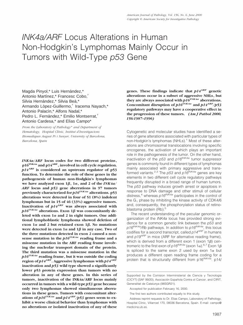
Figure 1. A: INK4a/ARF locus map. B: Analysis of p16 INK4a and p14 ARF genedeletions by PCR. p16 INK4a exon 1a and p14 ARF exon 1b deletions werealways associated with exon 2 losses. However, exon 1b was retained in onelymphoblastic lymphoma in which exon 1a and exon 2 were both deleted(case 2286). This case showed a p53 mutation. Case (321) showed concom-itant p16 INK4a and p14 ARF gene deletion associated with p53 mutation.
Table 3. Correlation between p16 INK4a and p14 ARF Alterationand p53 Gene Status in NHLs
INK4a/ARF n
p53 gene
Wild typeMutated/deleted
Wild type 80 63 17Exon 1a/2 deletion 9 7 2*Exon 1b alteration 8 7 1Exon 2 mutation 3† 3 0p16 INK4a hypermethylation 5 5 0
Total 97 78 19
*One lymphoblastic lymphoma with homozygous deletion of exons1a/2 but retaining exon 1b had a p53 mutation.
†One mutation in exon 2 was outside the p14ARF open readingframe.
1990 Pinyol et alAJP June 2000, Vol. 156, No. 6
nuclei, the percentage of the immunohistochemicallystained area was measured against the total nuclear area.Five or more fields of each tumor were analyzed, until aminimum of 1000 cells had been examined. The fields ineach slide were selected from the areas with greatest num-ber of stained cells. p53 expression was considered asnegative, low, or high when less than 5%, 5–30%, or morethan 30% of the cells showed nuclear immunostaining.
Statistical Analysis
Categorical data were compared using the Fisher’s exacttest (two-sided P value), whereas for ordinal data non-parametric tests were used. The actuarial survival analy-sis was performed according to the method described byKaplan and Meier, and the curves were compared by thelog-rank test.
Results
Analysis of the p53 Gene
p53 gene mutations in exons 4–9 were analyzed by PCR-SSCP, and cases with anomalous migrating bands weresequenced. Gene alterations were found in four (8%)indolent lymphomas and 15 (33%) aggressive tumors(indolent versus aggressive; P 5 0.01) (Table 1). Themutated indolent lymphomas were three CLLs and onetypical MCL. The CLLs showed two missense mutationsat exon 5 and one microdeletion of four nucleotides atexon 4, with a frameshift in the sequence leading to astop codon (Table 2). The typical MCL had a missensemutation in exon 5 (Table 2). The 15 aggressive lympho-mas with p53 alterations were two LCLs transformed fromCLL (Richter’s syndrome), three LCLs transformed fromFL, five blastoid MCLs, three de novo diffuse B-cell LCLs,one Burkitt’s lymphoma, and one T-cell lymphoblasticlymphoma (Tables 1 and 2). Twelve of these tumorsshowed missense mutations, two at exon 4, two at exon 5,four at exon 7, and four at exon 8 (Table 2). A LCLtransformed from a CLL showed a deletion of five nucle-otides at exon 8 with a frameshift in the sequence caus-ing a subsequent stop codon. The PCR amplificationsignal of the p53 gene was very low in two blastoid MCLs(cases 441 and 15855). Southern blot analysis of thesetwo cases showed a homozygous deletion of the p53gene (Table 2). The very faint band obtained in the PCRanalysis could be due to amplification of the gene from
the scarce normal or reactive cells present in the sample.No p53 gene alterations were detected in 53 additionaltumors that were also examined by Southern blot.
Analysis of p16INK4a and p14ARF Gene Deletions
Deletions of exons 1a, 1b, and 2 of the INK4a/ARF locuswere analyzed by multiplex PCR. Exon 2 deletions were
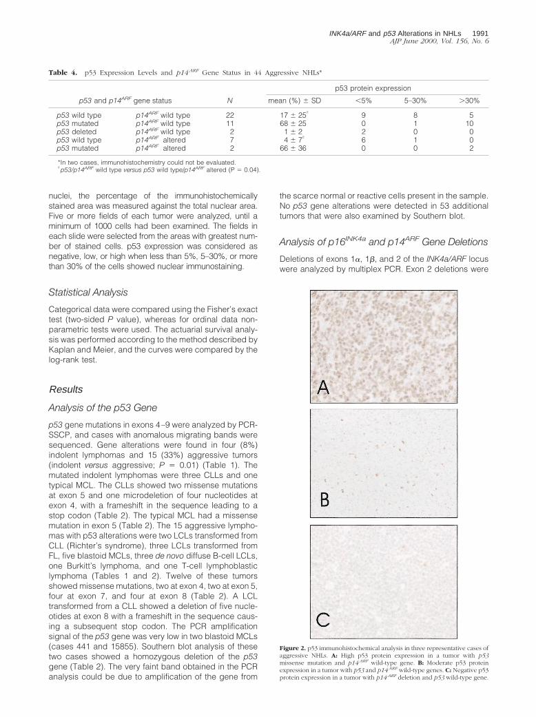
Figure 2. p53 immunohistochemical analysis in three representative cases ofaggressive NHLs. A: High p53 protein expression in a tumor with p53missense mutation and p14 ARF wild-type gene. B: Moderate p53 proteinexpression in a tumor with p53 and p14 ARF wild-type genes. C: Negative p53protein expression in a tumor with p14 ARF deletion and p53 wild-type gene.
Table 4. p53 Expression Levels and p14 ARF Gene Status in 44 Aggressive NHLs*
p53 and p14ARF gene status N
p53 protein expression
mean (%) 6 SD ,5% 5–30% .30%
p53 wild type p14ARF wild type 22 17 6 25† 9 8 5p53 mutated p14ARF wild type 11 68 6 25 0 1 10p53 deleted p14ARF wild type 2 1 6 2 2 0 0p53 wild type p14ARF altered 7 4 6 7† 6 1 0p53 mutated p14ARF altered 2 66 6 36 0 0 2
*In two cases, immunohistochemistry could not be evaluated.†p53/p14ARF wild type versus p53 wild type/p14ARF altered (P 5 0.04).
INK4a/ARF and p53 Alterations in NHLs 1991AJP June 2000, Vol. 156, No. 6
detected in nine cases, two indolent and seven aggres-sive lymphomas. The two indolent tumors were a typicalMCL and a follicular lymphoma. The seven aggressivelymphomas were two LCLs transformed from CLL (Rich-ter’s syndrome), one LCL transformed from FL, two blas-toid MCLs, one de novo diffuse B-cell LCL, and onelymphoblastic lymphoma of T-cell phenotype (Table 1).The results obtained in this PCR analysis of exon 2 wereconcordant with those obtained in the previous analysisof exon 2 by Southern blot.22
p16INK4a exon 1a and p14ARF exon 1b deletions werealways associated with exon 2 losses. However, exon 1bwas retained in one lymphoblastic lymphoma in whichexon 1a and exon 2 were both deleted (case 2286)(Figure 1). Interestingly, the Southern blot analysis of thiscase showed a deletion of p16INK4a exon 2, but p15INK4b
was in germ line configuration.22 p15INK4b was also de-leted in other seven tumors with exon 1b, 1a, and 2deletions but was in germ line configuration in one Rich-ter’s syndrome with deletion of exons 1b, 1a and 2.22 Noisolated deletions of exon 1b were detected in any case.
Mutational Analysis of the p14ARF Gene
Mutations of exons 1b and 2 were analyzed by PCR-SSCP followed by sequencing of cases with anomalousmigrating bands. No mutations were detected in exon 1bin any case (Table 1). Exon 2 mutations had been de-tected in one CLL and two transformed follicular lympho-mas.22 In the open reading frame of p16INK4a, the CLLmutation was at codon 143 (GCC3 ACC), resulting in achange of alanine to threonine. This mutation was locatedoutside the open reading frame of p14ARF. The two trans-formed FLs showed the same mutation. In the open read-ing frame of p16INK4a, this change was a nonsense mu-
tation at codon 80 CGA (Arg) 3 TGA (Stop). The samechange in the p14ARF open reading frame was at codon94, producing a missense mutation, CCG (Pro) 3 CTG(Leu). Western blot analysis of these two cases hadshown a lack of p16 protein expression.22
Correlation between p53, p14ARF, and p16INK4a
Gene Alterations
The correlation between p53, p14ARF, and p16INK4a genealterations is shown in Table 3. p53 inactivation wasdetected in 19 (20%) tumors: 15 missense mutations, twomicrodeletions leading to stop codons, and two homozy-gous deletions. p14ARF gene alterations were detected in11 (11%) tumors: eight showed deletions of exon 1b andexon 2, one case showed deletion of exon 2 but retainedexon 1b, and two tumors had point mutations in exon 2involving the nucleolar transport domain of the protein.p16INK4a alterations were detected in 17 (18%) tumors,including nine homozygous deletions involving both ex-ons 1a and 2, five hypermethylations, and three pointmutations.22
Genetic alterations of the p53 gene and the INK4a/ARFlocus mainly occurred in different tumors (Table 3). Sev-enteen cases with p53 gene alterations had a wild-typeINK4a/ARF locus, whereas 15 tumors with genetic alter-ations in this locus had a wild-type p53 gene. Concomi-tant alterations in the p53 gene and the INK4a/ARF locuswere detected in only two cases (Table 2). Interestingly,one of these cases was the lymphoblastic lymphoma inwhich the p14ARF exon 1b was retained, whereasp16INK4a exons 1a and 2 were deleted.
p53 and p14ARF Gene Expression
Experimental studies have shown that p14ARF stabilizesp53 protein in the nucleus of the cell by inhibiting itscytoplasmic transport and the MDM2-mediated degrada-tion.9–14 To determine whether p14ARF gene alterationsmay be associated with lower p53 levels in human tu-mors, p53 protein expression was examined by immuno-histochemistry, and its expression levels were comparedto the status of the p53 and p14ARF genes.
p53 protein expression in indolent lymphomas wasusually negative (,5%). Similarly, the CLL with a non-sense mutation in exon 4 was negative for p53 protein.The two CLLs and the typical MCL with p53 gene mis-sense mutations showed a high p53 protein expression.The typical MCL and the FL with p14ARF deletion showed
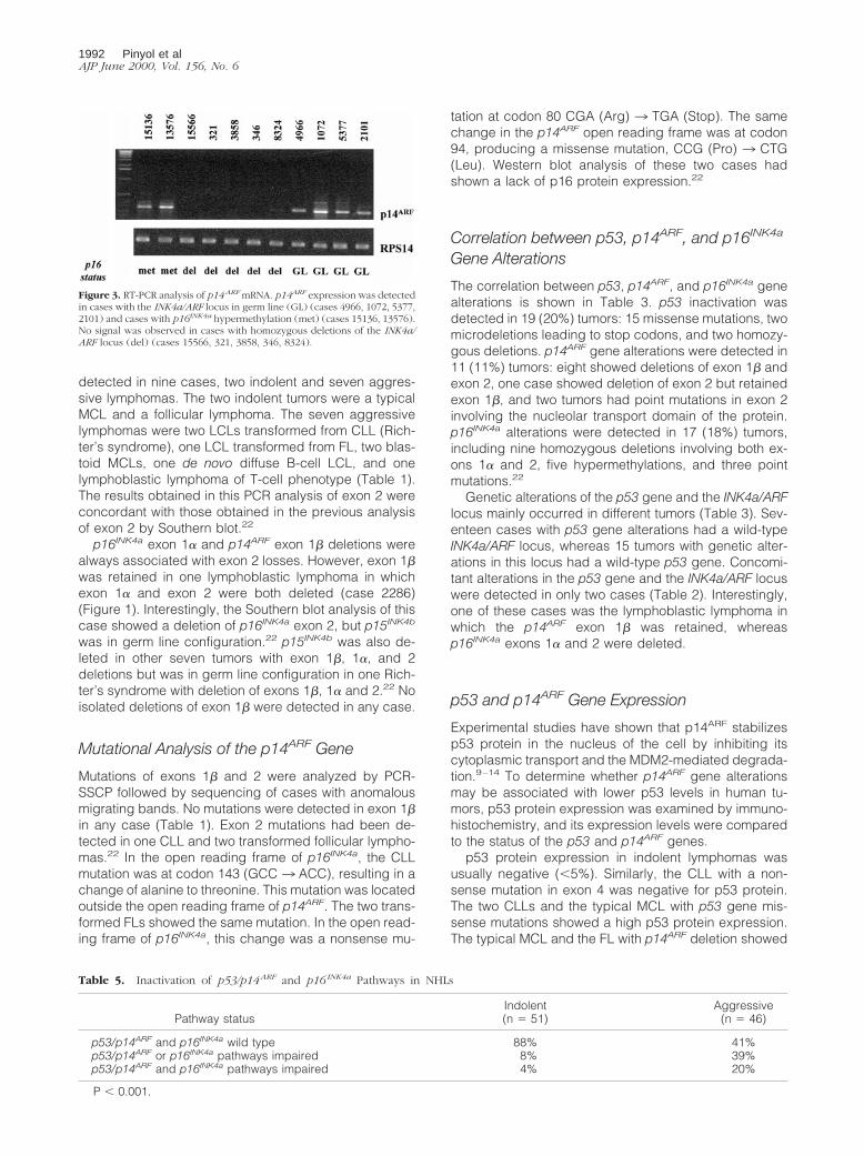
Figure 3. RT-PCR analysis of p14 ARF mRNA. p14ARF expression was detectedin cases with the INK4a/ARF locus in germ line (GL) (cases 4966, 1072, 5377,2101) and cases with p16INK4a hypermethylation (met) (cases 15136, 13576).No signal was observed in cases with homozygous deletions of the INK4a/ARF locus (del) (cases 15566, 321, 3858, 346, 8324).
Table 5. Inactivation of p53/p14 ARF and p16 INK4a Pathways in NHLs
Pathway statusIndolent(n 5 51)
Aggressive(n 5 46)
p53/p14ARF and p16INK4a wild type 88% 41%p53/p14ARF or p16INK4a pathways impaired 8% 39%p53/p14ARF and p16INK4a pathways impaired 4% 20%
P , 0.001.
1992 Pinyol et alAJP June 2000, Vol. 156, No. 6

negative levels similar to those of the remaining tumorswith wild-type p14ARF gene.
Contrary to the negative immunostaining in indolentlymphomas, aggressive tumors with wild-type p53 andp14ARF genes showed a broad range of p53 proteinexpression (mean 17% SD 6 25) (Table 4). Tumors withp53 missense mutations, including the two cases withconcomitant INK4a/ARF exon 2 deletions, showed highlevels of p53 protein expression, whereas the two tumorswith homozygous deletions of p53 were negative (Tables2 and 4). Interestingly, protein expression in aggressivetumors with p14ARF altered and wild-type p53 was signif-icantly lower than in tumors with no alteration in any ofthese genes (mean 4%, SD 6 7 versus mean 17%, SD 625; P 5 0.04) (Table 4 and Figure 2).
p14ARF mRNA expression was analyzed in 20 tumors,including five cases with homozygous deletions of thegene, two cases with p16INK4a hypermethylation but wild-type p14ARF, and 13 with no alterations of the INK4a/ARFlocus. p14ARF mRNA was present in all cases with wild-type INK4a/ARF locus and in the two cases with p16INK4a
hypermethylation. No expression was detected in thecases with deletion of the gene (Figure 3).
Correlation between p53/p14ARF and p16INK4a
Gene Alterations and the Outcome of thePatients
The p53/p14ARF and p16INK4a gene alterations accordingto the histological subgroup (indolent versus aggressive)are detailed in Table 5. p53 and p14ARF genes are con-sidered to be in the same regulatory pathway. Therefore,patients with alterations in either of these genes wereincluded in the same group. Patients with aggressivelymphomas showed one impaired pathway (either p53/p14ARF or p16INK4a) (39% versus 8%) and two impairedpathways (both p53/p14ARF and p16INK4a) more fre-quently (20% versus 4%; P , 0.001) than indolent lym-phomas.
To determine the possible correlation between thegene alterations and the outcome of the patients, 54patients with appropriate clinical data and all of the mo-lecular studies performed at diagnosis were included inthe analysis. For this purpose, patients with no alterationsin p53/p14ARF or p16INK4a (group A) were compared withpatients with alterations in either p53/p14ARF or p16INK4a
(group B), and patients with concomitant alterations inboth p53/p14ARF and p16INK4a (group C). In the group ofMCLs, whose clinical characteristics have previouslybeen published,28 patients with inactivation of both path-ways had a significantly worse prognosis than patientswith alterations in only one gene or no alterations in any ofthem. Thus, in these patients, the 36-month survival ratesof groups A, B, and C were 87%, 34%, and 0%, respec-tively (P 5 0.007) (Figure 4). In the whole series, patientsof group A showed a trend for better survival than thoseof groups B and C (median survival: 60, 24, and 17months, respectively; P 5 0.08).
Discussion
Inactivation of the two major human tumor suppressorgenes p53 and p16INK4a is a relatively common phenom-enon in primary and transformed high-grade lymphomas,occurring in 20–50% of these tumors.2,22,24,29 Most of theprevious studies have evaluated the two genes individu-ally, and, therefore, it is not known whether these alter-ations may function as cooperative or alternative mech-anisms in the pathogenesis of aggressive lymphomas.The recent understanding of the peculiar genomic struc-ture of the INK4a/ARF locus coding for two overlappinggenes, p16INK4a and p14ARF, and the function of p14ARF
as an upstream regulator of p53 activity, has raised thequestion of the relative importance of these genes in thedevelopment of human tumors.
In this study we have examined the status of p53 andp14ARF genes simultaneously in a large series of humanNHLs previously analyzed for p16INK4a. The frequency ofalterations in the p53 and p16INK4a genes was similar tothat observed in other series of lymphomas.29–33 Thusp53 aberrations were detected in 8% of indolent lympho-mas but in 33% of aggressive tumors, whereas p16INK4a
alterations were found in 6% and 30% of low- and high-grade lymphomas, respectively. Inactivation of p14ARF
was always associated with p16INK4a alterations. Exon 1bwas concomitantly deleted with exon 1a and 2 in almostall cases. Only one lymphoblastic lymphoma, which har-bored exon 1a and 2 deletions, retained exon 1b. Inaddition, no mutations were detected in exon 1b in anycase. Although some studies in murine models8,16,18 andhuman acute lymphoblastic leukemias20,21 have sug-gested that p14ARF may be selectively inactivated in lym-phoproliferative disorders, our findings suggest that in-dependent inactivation of p14ARF in human NHLs is a rarephenomenon.
p14/19ARF is considered to be an upstream regulatorof p53 function. Experimental studies have shown thatp14/19ARF interacts physically with MDM2, thus prevent-ing p53 degradation and promoting p53 stabilization andaccumulation.9,14,34,35 These observations suggest thattumors with inactivation of the ARF locus may have anincreased degradation of p53 protein and, consequently,
Figure 4. Overall survival of 24 patients with mantle cell lymphoma accord-ing to the gene alterations at diagnosis. A: p53/p14 ARF and p16 INK4a path-ways nonaltered (n 5 15; dead: 9). B: Alterations in the p53 pathway only(n 5 6; dead: 5). C: Concomitant alterations in both p53/p14 ARF andp16 INK4a pathways (n 5 3; dead: 3) (P 5 0.007).
INK4a/ARF and p53 Alterations in NHLs 1993AJP June 2000, Vol. 156, No. 6
a defective regulation of the p53 regulatory pathway.Consistent with these experimental studies, we observedthat p53 protein expression in aggressive lymphomaswith p14ARF exon 1b deletions was significantly lowerthan in other aggressive tumors with a wild-type INK4a/ARF locus.
Interestingly, two transformed FLs with mutations inexon 2 were also negative for p53 protein. Initial studiesof the functional domains of p19ARF showed that exon 1bwas necessary and sufficient to produce cell-cycle ar-rest, whereas exon 2 mutations seemed to specificallyinactivate the p16INK4a gene but not p19ARF.9,36 However,recent studies on human p14ARF have identified a C-terminal domain between residues 83 and 101 of exon 2that are required for its localization in the nucleolus andfor the formation of stable p14ARF-MDM2-p53 nuclearcomplexes.13 Mutations in these residues disrupt the nu-cleolar localization of p14ARF, leading to a cytoplasmicdiffusion of the protein and a reduced stabilization of p53protein.13 Interestingly, in contrast to human p14ARF, thenucleolar localization domain of murine p19ARF seems toreside in a region coded by exon 1b.34 These observa-tions suggest that mutations in the human exon 2 involv-ing this nucleolar domain may target both the p14ARF andp16INK4a genes. The exon 2 mutation detected in our twotransformed FLs was found in codon 80 of INK4a andcodon 94 of ARF reading frames, leading to a stop codonin p16INK4a and a missense mutation within the nucleolarlocalization domain of p14ARF. In fact, these two tumorsshowed a lack of expression of both p16INK4a and p53protein, suggesting that this mutation may target p14ARF
and p16INK4a genes simultaneously.We have also detected one exon 2 mutation in an
indolent CLL. This mutation was identified in p16INK4a
codon 143, which is outside the ankyrin repeat motifs in aregion that is not required for the protein to bind andinhibit CDK4,37 and it is outside the open reading frameof p14ARF. Therefore, it is possible that this mutation wasnot interfering with the function of either of these proteins.Concordantly, the clinical behavior of this patient wassimilar to that of other indolent CLL patients with anINK4a/ARF wild-type locus. In a review of the literature ofthe INK4a/ARF alterations in lymphoproliferative disor-ders, we have identified 21 mutations involving thep16INK4a gene, with eight occurring in indolent disorders(six CLLs and two FLs) and 13 in aggressive tumors (nineacute lymphoblastic leukemias, three LCLs, and one Bur-kitt’s lymphoma).20,22,29 Six of the 13 mutations in ag-gressive tumors, but only one of the eight indolent disor-ders, led to a change in the ARF open reading frame. Theremaining changes were downstream of the ARF stopcodon or did not cause an amino acid change in thesequence. These observations suggest that exon 2 mu-tations involving both genes may play a role in the devel-opment of aggressive tumors.
The recent understanding of the INK4a/ARF locus as aregulatory region of both p16INK4a/Rb and p14ARF/p53pathways has suggested that tumors with inactivation ofthis locus may harbor fewer p53 mutations than tumorswith wild-type INK4a/ARF genes.10 In our study p53 mu-tations and INK4a/ARF alterations appeared to occur in
different tumors because only two aggressive lympho-mas, one lymphoblastic lymphoma and one Richter’ssyndrome, harbored simultaneous p53 mutation and ho-mozygous deletion of the INK4a/ARF locus. Reciprocalalterations between p53 mutations and p16INK4a alter-ations have been observed in some tumors.25,30,38 How-ever, mutual exclusion between these alterations has notbeen detected in leukemia-lymphoma cell lines39 andother human tumors, including large B-cell lympho-mas.40,41 It has been suggested that the pattern of thesealterations in tumors may depend on the order ofevents.12 If deletion of the INK4a/ARF locus occurs earlyin the development of the tumor, the neoplastic cells mayremain wild type for p53. In contrast, an initial mutation inthe p53 gene may be followed by a strong pressureagainst p16INK4a that would lead to a concomitant dele-tion of p14ARF. However, tumors with inactivation of bothp53 and p14ARF may have a selective growth advantagebecause there is no complete overlap between the ARFand p53 functions.40 In fact, p14ARF does not seem toparticipate in the apoptotic response to DNA damage,and, consequently, cells with p53 loss may toleratehigher levels of genomic damage than tumor cells withwild-type p53.8,12
Different studies have demonstrated the prognosticvalue of p53 mutations in NHLs. However, the prognosticsignificance of INK4a/ARF alterations in these tumors isnot well known. Garcia-Sanz et al have indicated thatp16INK4a deletions and rearrangements are associatedwith poor prognosis in B-cell NHLs, but this study did notinclude p14ARF and p53 analysis.42 Similarly, we haverecently shown that DNA losses in 9pter are associatedwith a poor prognosis in MCL patients.26 Genetic alter-ations in the INK4a/ARF locus target both p14ARF/p53 andp16INK4a/Rb pathways and, therefore, may provide a se-lective growth advantage in the progression of tumors. Inour study, although the series was heterogeneous andthe number of patients relatively small, there was a trendindicating a poor prognosis for patients with inactivationof both p16INK4a and p14ARF/p53 genes. In the homoge-neous subset of MCLs, patients with genetic alterations inthe INK4a/ARF locus had a significantly worse prognosisthan patients with tumors with only p53 mutations or withno aberrations in any of these genes.
In this study, independent inactivation of p16INK4a onlyoccurred by hypermethylation. Similar to observations ofother tumors, in our lymphomas hypermethylation of thep16INK4a gene does not affect the expression ofp14ARF.43,44 On the other hand, different studies havealready shown that inactivation of p14ARF by hypermeth-ylation is extremely rare.44,45 The number of patients withisolated inactivation of p16INK4a in our series was toosmall to discern whether hypermethylation of p16INK4a
may have any particular influence on the survival of thepatients.
In conclusion, our findings indicate that genetic alter-ations in p14ARF occur in a subset of aggressive NHLs,but they are always associated with p16INK4a aberrations.Alterations in the INK4a/ARF locus seem to develop fre-quently in tumors with a wild-type p53 gene. The shortersurvival of patients with lymphomas harboring concomi-
1994 Pinyol et alAJP June 2000, Vol. 156, No. 6
tant alterations of the p16INK4a and p14ARF/p53 genessuggests that simultaneous inactivation of these two reg-ulatory pathways may have a cooperative effect on theprogression of the tumors.
Acknowledgments
We thank Dr. Manuel Serrano for his helpful comments onthe manuscript.
References
1. Nowell PC: Genetic alterations in leukemia and lymphomas: impres-sive progress and continuing complexity. Cancer Genet Cytogenet1997, 94:13–19
2. Imamura J, Miyoshi I, Koeffler HP: p53 in hematologic malignancies.Blood 1994, 84:2412–2421
3. Hirama T, Koeffler HP: Role of the cyclin-dependent kinase inhibitorsin the development of cancer. Blood 1995, 86:841–854
4. Levine AJ: p53, the cellular gatekeeper for growth and division. Cell1997, 88:323–331
5. Serrano M, Hannon GJ, Beach D: A new regulatory motif in cell-cyclecontrol causing specific inhibition of cyclin D/CDK4. Nature 1993,366:704–707
6. Mao L, Merlo A, Bedi G, Shapiro GI, Edwards CD, Rollins BJ, Sidran-sky D: A novel p16INK4a transcript. Cancer Res 1995, 55:2995–2997
7. Quelle DE, Zindy F, Ashmun RA, Sherr CJ: Alternative reading framesof the INK4a tumor suppressor gene encode two unrelated proteinscapable of inducing cell cycle arrest. Cell 1995, 83:993–1000
8. Kamijo T, Zindy F, Roussel MF, Quelle DE, Downing JR, Ashmun RA,Grosveld G, Sherr CJ: Tumor suppression at the mouse INK4a locusmediated by the alternative reading frame product p19ARF. Cell1997, 91:649–659
9. Zhang Y, Xiong Y, Yarbrough WG: ARF promotes MDM2 degradationand stabilizes p53: ARF-INK4a locus deletion impairs both the Rb andp53 tumor suppression pathways. Cell 1998, 92:725–734
10. Pomerantz J, Schreiber-Agus N, Liegeois NJ, Silverman A, Alland L,Chin L, Potes J, Chen K, Orlow I, Lee HW, Cordon-Cardo C, De PinhoRA: The INK4a tumor suppressor gene product, p19ARF, interactswith MDM2 and neutralizes MDM2’s inhibition of p53. Cell 1998,92:713–723
11. Kamijo T, Weber JD, Zambetti G, Zindy F, Roussel MF, Sherr CJ:Functional and physical interactions of the ARF tumor suppressorwith p53 and MDM2. Proc Natl Acad Sci USA 1998, 95:8292–8297
12. Stott FJ, Bates S, James MC, McConnell BB, Starborg M, Brookes S,Palmero I, Ryan K, Hara E, Vousden KH, Peters G: The alternativeproduct from the human CDKN2a locus, p14(ARF), participates in aregulatory feedback loop with p53 and MDM2. EMBO J 1998, 17:5001–5014
13. Zhang Y, Xiong Y: Mutations in human ARF exon 2 disrupt its nucle-olar localization and impair its ability to block nuclear export of MDM2and p53. Mol Cell 1999, 3:579–591
14. Tao W, Levine AJ: P19(ARF) stabilizes p53 by blocking nucleocyto-plasmic shuttling of MDM2. Proc Natl Acad Sci USA 1999, 96:6937–6941
15. Serrano M, Lee H, Chin L, Cordon-Cardo C, Beach D, De Pinho RA:Role of the INK4a locus in tumor suppression and cell mortality. Cell1996, 85:27–37
16. Kamijo T, Bodner S, van de Kamp E, Randle DH, Sherr CJ: Tumorspectrum in ARF-deficient mice. Cancer Res 1999, 59:2217–2222
17. Donehower LA, Harvey M, Slagle BL, McArthur MJ, Montgomery CA,Jr, Butel JS, Bradley A: Mice deficient for p53 are developmentallynormal but susceptible to spontaneous tumours. Nature 1992, 356:215–221
18. Zhuang SM, Schippert A, Haugen-Strano A, Wiseman RW, SoderkvistP: Inactivations of p16INK4a-alpha, p16INK4a-beta and p15INK4bgenes in 29,39-dideoxycytidine- and 1,3-butadiene-induced murinelymphomas. Oncogene 1998, 16:803–808
19. Kumar R, Lundh Rozell B, Louhelainen J, Hemminki K: Mutations in
the CDKN2a (p16INK4a) gene in microdissected sporadic primarymelanomas. Int J Cancer 1998, 75:193–198
20. Heyman M, Rasool O, Brandter LB, Liu Y, Grander D, Einhorn S,Soderhall S: Exclusive p15INK4B gene deletions in acute lympho-cytic leukemia include the E1 beta exon of the p16INK4 gene. Blood1996, 87:1657–1658
21. Gardie B, Cayuela JM, Martini S, Sigaux F: Genomic alterations of thep19ARF encoding exons in T-cell acute lymphoblastic leukemia.Blood 1998, 91:1016–1020
22. Pinyol M, Cobo F, Bea S, Jares P, Nayach I, Fernandez PL, Montser-rat E, Cardesa A, Campo E: p16INK4a gene inactivation by deletions,mutations and hypermethylation is associated with transformed andaggressive variants of non-Hodgkin’s lymphomas. Blood 1998, 91:2977–2984
23. Harris NL, Jaffe ES, Stein H, Banks PM, Chan JK, Cleary ML, DelsolG, De Wolf-Peeters C, Falini B, Gatter KC, Grogan TM, Isaacson P,Knowles DM, Mason DY, Muller-Hermelink HK, Pileri S, Piris MA,Ralfkiaer E, Warnke RA: A revised European-American classificationof lymphoid neoplasms: a proposal from the International LymphomaStudy Group. Blood 1994, 84:1361–1392
24. Hernandez L, Fest T, Cazorla M, Teruya-Feldstein J, Bosch F, Pei-nado MA, Piris MA, Montserrat E, Cardesa A, Jaffe ES, Campo E,Raffold M: p53 gene mutations and protein overexpression are asso-ciated with aggressive variants of mantle cell lymphomas. Blood1996, 87:3351–3359
25. Pinyol M, Hernandez L, Cazorla M, Balbin M, Jares P, Fernandez PL,Montserrat E, Cardesa A, Lopez-Otin C, Campo E: Deletions and lossof expression of p16INK4a and p21Waf1 genes are associated withaggressive variants of mantle cell lymphomas. Blood 1997, 89:272–280
26. Bea S, Ribas M, Hernandez JM, Bosch F, Pinyol M, Hernandez L,Garcıa JL, Flores T, Gonzalez M, Lopez-Guillermo A, Piris MA,Cardesa A, Montserrat E, Miro R, Campo E: Increased number ofchromosomal imbalances and high-level DNA amplifications in man-tle cell lymphoma are associated with blastoid variants. Blood 1999,93:4365–4374
27. Jares P, Nadal A, Fernandez PL, Pinyol M, Hernandez L, Cazorla M,Hernandez S, Bea S, Cardesa A, Campo E: Disregulation ofp16MTS1/CDK4I protein and mRNA expression is associated withgene alterations in squamous-cell carcinoma of the larynx. Int JCancer 1999, 81:705–711
28. Bosch F, Lopez-Guillermo A, Campo E, Ribera JM, Conde E, PirisMA, Vallespi T, Woessner S, Montserrat E: Mantle cell lymphoma:presenting features, response to therapy, and prognostic factors.Cancer 1998, 82:567–575
29. Ruas M, Peters G: The p16INK4a/CDKN2A tumor suppressor and itsrelatives. Biochim Biophys Acta 1998, 1378:F115–F177
30. Hangaishi A, Ogawa S, Imamura N, Miyawaki S, Miura Y, Uike N,Shimazaki C, Emi N, Takeyama K, Hirosawa S, Kamada N, KobayashiY, Takemoto Y, Kitani T, Toyama K, Ohtake S, Yazaki Y, Ueda R, HiraiH: Inactivation of multiple tumor-suppressor genes involved in nega-tive regulation of the cell cycle, MTS1/p16INK4a/CDKN2, MTS2/p15INK4b, p53, and Rb genes in primary lymphoid malignancies.Blood 1996, 87:4949–4958
31. Ogawa S, Hangaishi A, Miyawaki S, Hirosawa S, Miura Y, TakeyamaK, Kamada N, Ohtake S, Uike N, Shimazaki C: Loss of the cyclin-dependent kinase 4-inhibitor (p16; MTS1) gene is frequent in andhighly specific to lymphoid tumors in primary human hematopoieticmalignancies. Blood 1995, 86:1548–1556
32. Fizzotti M, Cimino G, Pisegna S, Alimena G, Quartarone C, MandelliF, Pelicci PG, Lo Coco F: Detection of homozygous deletions of thecyclin-dependent kinase 4 inhibitor (p16) gene in acute lymphoblas-tic leukemia and association with adverse prognostic features. Blood1995, 85:2685–2690
33. Stranks G, Height SE, Mitchell P, Jadayel D, Yuille MA, De Lord C,Clutterbuck RD, Treleaven JG, Powles RL, Nacheva E: Deletions andrearrangement of CDKN2 in lymphoid malignancy. Blood 1995, 85:893–901
34. Weber JD, Taylor LJ, Roussel MF, Sherr CJ, Bar-Sagi D: NucleolarARF sequesters Mdm2 and activates p53. Nat Cell Biol 1999,1:20–26
35. Honda R, Yasuda H: Association of p19ARF with Mdm2 inhibitsubiquitin ligase activity of Mdm2 for tumor suppressor p53. EMBO J1999, 18:22–27
INK4a/ARF and p53 Alterations in NHLs 1995AJP June 2000, Vol. 156, No. 6
36. Quelle DE, Cheng M, Ashmun RA, Sherr CJ: Cancer-associatedmutations at the INK4a locus cancel cell cycle arrest by p16INK4abut not by the alternative reading frame protein p19ARF. Proc NatlAcad Sci USA 1997, 94:669–673
37. Yang R, Gombart AF, Serrano M, Koeffler HP: Mutational effects onthe p16INK4a tumor suppressor protein. Cancer Res 1995, 55:2503–2506
38. Newcomb EW, Rao LS, Giknavorian SS, Lee SY: Alterations of multi-ple tumor suppressor genes (p53 (17p13), p16INK4 (9p21), andDBM (13q14)) in B-cell chronic lymphocytic leukemia. Mol Carcinog1995, 14:141–146
39. Drexler HG: Review of alterations of the cyclin-dependent kinaseinhibitor ink4 family genes p15, p16, p18 and p19 in human leukemia-lymphoma cells. Leukemia 1998, 12:845–859
40. Sharpless NE, DePinho RA: The INK4a/ARF locus and its two geneproducts. Curr Opin Genet Dev 1999, 9:22–30
41. Moller MB, Ino Y, Gerdes AM, Skdjodt K, Louis DN, Pedersen NT:Aberrations of the p53 pathway components p53, MDM2 and
CDKN2A appear independent in diffuse large B cell lymphoma.Leukemia 1999, 13:453–459
42. Garcia-Sanz R, Gonzalez M, Vargas M, Chillon MC, Balanzategui A,Barbon M, Flores MT, San Miguel JF: Deletions and rearrangementsof cyclin-dependent kinase 4 inhibitor gene p16 are associated withpoor prognosis in B cell non-Hodgkin’s lymphomas. Leukemia 1997,11:1915–1920
43. Della Valle V, Duro D, Bernard O, Larsen CJ: The human proteinp19ARF is not detected in hemopoietic human cell lines that abun-dantly express the alternative beta transcript of the p16INK4a/MTS1gene. Oncogene 1997, 15:2475–2481
44. Baur AS, Shaw P, Burri N, Delacretaz F, Bosman FT, Chaubert P:Frequent methylation silencing of p15(INK4b) (MTS2) andp16(INK4a) (MTS1) in B-cell and T-cell lymphomas. Blood 1999,94:1773–1781
45. Robertson KD, Jones PA: The human ARF cell cycle regulatory genepromoter is a CpG island which can be silenced by DNA methylationand down-regulated by wild-type p53. Mol Cell Biol 1998, 18:6457–6473
1996 Pinyol et alAJP June 2000, Vol. 156, No. 6