Electric Field-Induced Functional Reductions in the K + Channels Mainly Resulted from Supramembrane...
11
Electric Field-Induced Functional Reductions in the K 1 Channels Mainly Resulted from Supramembrane Potential-Mediated Electroconformational Changes Wei Chen,* Yu Han,* Yan Chen,* and Dean Astumian # *Departments of Dermatology and Physiology and Biophysics, The University of Illinois at Chicago, Chicago, Illinois 60612, and # Department of Plastic and Reconstructive Surgery, The University of Chicago, Chicago, Illinois 60637 USA ABSTRACT The goal of this study is to distinguish the supramembrane potential difference-induced electroconformational changes from the huge transmembrane current-induced thermal damages in the delayed rectifier K 1 channels. A double Vaseline-gap voltage clamp was used to deliver shock pulses and to monitor the channel currents. Three pairs of 4-ms shock pulses were used to mimic the electric shock by a power-line frequency electric field. Each pair consists of two pulses with the same magnitude, starting from 350 to 500 mV, but with opposite polarities. The shock pulse-generated transmembrane ion flux and the responding electric energy, the Joule heating, consumed in the cell membrane, as well as the effects on the K 1 channel currents, were obtained. Results showed that huge transmembrane currents are not necessary to cause damages in the K 1 channel proteins. In contrast, reductions in the K 1 channel currents are directly related to the field-induced supramembrane potential differences. By a comparison with the shock field-induced Joule heating effects on cell mem- branes, the field-induced supramembrane potential difference plays a dominant role in damaging the K 1 channels, resulting in electroconformational changes in the membrane proteins. In contrast, the shock field-induced huge transmembrane currents, therefore the thermal effects, play a secondary, trivial role. INTRODUCTION An electric field, as a tool, has been widely used in today’s medical diagnosis and treatments. For example, defibrilla- tion and pacemakers are common techniques used to rein- stall the normal cardiac cell functions (Lau and Camm, 1991; Raymond et al., 1991). Membrane electroporation has been widely used to increase the efficiency of gene trans- fection and cell fusion (Winegar et al., 1989; Zimmermann et al., 1980; Chang et al., 1991). Electroporation was also suggested for use on skin stratum corneum for transdermal drug delivery (Okino and Mohri, 1987; Mir et al., 1991; Bommannan et al., 1993; Prausnitz et al., 1993). Alternatively, the mechanisms involved in electrical trauma remain unclear. Better understanding of the mecha- nisms in electrical injury will significantly improve our diagnosis and treatment of electrically injured patients. The electric resistance of the cell’s phospholipid bilayer is six to eight orders of magnitude higher than those of cytoplasmic and extracellular electrolytes. When living cells are exposed to an external electric field, most of the field-induced voltage-drops fall on the cell membranes. In addition, since the cell dimension is a few orders of mag- nitude larger than the thickness of the cell membrane, the induced equivalent field strength inside the cell membrane is hundreds and thousands times higher than the apparent strength of the applied electric field (Cole, 1972). Under such a high field strength, the field-induced membrane potential difference is much higher than those in physiolog- ical condition. These supraphysiological potential differences or supramembrane potential differences may cause damage in membrane phospholipid bilayer and membrane proteins. One of the possible mechanisms involved in electrical injury is Joule heating. The direct effects of Joule heating in damaging cells and tissues has long been recognized as denaturation of the cell membrane and membrane proteins (Cravalho et al., 1992; Tropea and Lee, 1992). Toner et al. investigated thermal-mediated injury in isolated skeletal muscle fibers by study of quantitative microscopy of mem- brane permeability changes at supraphysiological temperatures in isolated muscle cells and fibroblasts (Bischof et al., 1995). Joule heating-induced membrane damage is known to be pro- portional to electric energy consumed in the cell membrane, which is directly related to the field-induced transmembrane currents (Rouge and Dimick, 1978; Lee et al., 1988). In addition, membrane electroporation was also consid- ered as one of the mechanisms of electrical trauma (Lee and Kolodney, 1987; Jones et al., 1987; O’Neill and Tung, 1991; Chen et al., 1992). In contrast to Joule heating caused by a huge transmembrane current, field-induced supramem- brane potential difference plays a significant role in mem- brane electroporation (Kinosita and Tsong, 1977). Mem- brane electroporation has been postulated mainly as the supramembrane potential difference-induced electroconfor- mational damages in the phospholipid bilayer (Tsong, 1991). Resealing of the membrane electropermeabilization under the control of cytoskeletal proteins is also considered as an alteration of the membrane organization or the con- sequence of the associated cytoplasmic content (Teissie and Rols, 1994). Received for publication 17 July 1997 and in final form 26 March 1998. Address reprint requests to Dr. Wei Chen, Department of Dermatology, The University of Illinois at Chicago, 808 South Wood St., Chicago, IL 60612. Tel.: 312-413-2448; Fax: 312-996-1188; E-mail: [email protected]. © 1998 by the Biophysical Society 0006-3495/98/07/196/11 $2.00 196 Biophysical Journal Volume 75 July 1998 196 –206
-
Upload
independent -
Category
Documents
-
view
2 -
download
0
Transcript of Electric Field-Induced Functional Reductions in the K + Channels Mainly Resulted from Supramembrane...
Electric Field-Induced Functional Reductions in the K1 Channels MainlyResulted from Supramembrane Potential-MediatedElectroconformational Changes
Wei Chen,* Yu Han,* Yan Chen,* and Dean Astumian#
*Departments of Dermatology and Physiology and Biophysics, The University of Illinois at Chicago, Chicago, Illinois 60612, and#Department of Plastic and Reconstructive Surgery, The University of Chicago, Chicago, Illinois 60637 USA
ABSTRACT The goal of this study is to distinguish the supramembrane potential difference-induced electroconformationalchanges from the huge transmembrane current-induced thermal damages in the delayed rectifier K1 channels. A doubleVaseline-gap voltage clamp was used to deliver shock pulses and to monitor the channel currents. Three pairs of 4-ms shockpulses were used to mimic the electric shock by a power-line frequency electric field. Each pair consists of two pulses withthe same magnitude, starting from 350 to 500 mV, but with opposite polarities. The shock pulse-generated transmembraneion flux and the responding electric energy, the Joule heating, consumed in the cell membrane, as well as the effects on theK1 channel currents, were obtained. Results showed that huge transmembrane currents are not necessary to cause damagesin the K1 channel proteins. In contrast, reductions in the K1 channel currents are directly related to the field-inducedsupramembrane potential differences. By a comparison with the shock field-induced Joule heating effects on cell mem-branes, the field-induced supramembrane potential difference plays a dominant role in damaging the K1 channels, resultingin electroconformational changes in the membrane proteins. In contrast, the shock field-induced huge transmembranecurrents, therefore the thermal effects, play a secondary, trivial role.
INTRODUCTION
An electric field, as a tool, has been widely used in today’smedical diagnosis and treatments. For example, defibrilla-tion and pacemakers are common techniques used to rein-stall the normal cardiac cell functions (Lau and Camm,1991; Raymond et al., 1991). Membrane electroporation hasbeen widely used to increase the efficiency of gene trans-fection and cell fusion (Winegar et al., 1989; Zimmermannet al., 1980; Chang et al., 1991). Electroporation was alsosuggested for use on skin stratum corneum for transdermaldrug delivery (Okino and Mohri, 1987; Mir et al., 1991;Bommannan et al., 1993; Prausnitz et al., 1993).
Alternatively, the mechanisms involved in electricaltrauma remain unclear. Better understanding of the mecha-nisms in electrical injury will significantly improve ourdiagnosis and treatment of electrically injured patients.
The electric resistance of the cell’s phospholipid bilayeris six to eight orders of magnitude higher than those ofcytoplasmic and extracellular electrolytes. When livingcells are exposed to an external electric field, most of thefield-induced voltage-drops fall on the cell membranes. Inaddition, since the cell dimension is a few orders of mag-nitude larger than the thickness of the cell membrane, theinduced equivalent field strength inside the cell membraneis hundreds and thousands times higher than the apparentstrength of the applied electric field (Cole, 1972). Undersuch a high field strength, the field-induced membrane
potential difference is much higher than those in physiolog-ical condition. These supraphysiological potential differencesor supramembrane potential differences may cause damage inmembrane phospholipid bilayer and membrane proteins.
One of the possible mechanisms involved in electricalinjury is Joule heating. The direct effects of Joule heating indamaging cells and tissues has long been recognized asdenaturation of the cell membrane and membrane proteins(Cravalho et al., 1992; Tropea and Lee, 1992). Toner et al.investigated thermal-mediated injury in isolated skeletalmuscle fibers by study of quantitative microscopy of mem-brane permeability changes at supraphysiological temperaturesin isolated muscle cells and fibroblasts (Bischof et al., 1995).Joule heating-induced membrane damage is known to be pro-portional to electric energy consumed in the cell membrane,which is directly related to the field-induced transmembranecurrents (Rouge and Dimick, 1978; Lee et al., 1988).
In addition, membrane electroporation was also consid-ered as one of the mechanisms of electrical trauma (Lee andKolodney, 1987; Jones et al., 1987; O’Neill and Tung,1991; Chen et al., 1992). In contrast to Joule heating causedby a huge transmembrane current, field-induced supramem-brane potential difference plays a significant role in mem-brane electroporation (Kinosita and Tsong, 1977). Mem-brane electroporation has been postulated mainly as thesupramembrane potential difference-induced electroconfor-mational damages in the phospholipid bilayer (Tsong,1991). Resealing of the membrane electropermeabilizationunder the control of cytoskeletal proteins is also consideredas an alteration of the membrane organization or the con-sequence of the associated cytoplasmic content (Teissie andRols, 1994).
Received for publication 17 July 1997 and in final form 26 March 1998.
Address reprint requests to Dr. Wei Chen, Department of Dermatology,The University of Illinois at Chicago, 808 South Wood St., Chicago, IL60612. Tel.: 312-413-2448; Fax: 312-996-1188; E-mail: [email protected].
© 1998 by the Biophysical Society
0006-3495/98/07/196/11 $2.00
196 Biophysical Journal Volume 75 July 1998 196–206
An intensive electric field also affects the membraneproteins, especially the voltage-sensitive proteins. In fact,structural features of many membrane proteins suggest alarge susceptibility to electric fields. Functions of theseelectric field-sensitive membrane proteins can therefore beaffected by the transmembrane potential (Eisenberg et al.,1987; Tsong and Astumian, 1987). A high-intensity electricfield may further cause electroconformational damages inthese membrane proteins. Tsong and his colleagues (Teissieand Tsong, 1980; Tsong and Astumian, 1986) showed thatan intensive shock field could induce leakage currentsthrough membrane proteins, the Na1/K1 ATPase of eryth-rocyte cell membranes. They attributed the field-inducedleakage as the membrane proteins’ electroconformationaldamages. Recently, electric field-induced damages in mem-brane proteins have been considered as a mechanism in-volved in electrical injury (Chen and Lee, 1994b, Abramovet al., 1997). We have found that a brief high-intensityelectric shock can cause not only membrane electropora-tion, but also reductions in the K1 channel conductance andionic selectivity.
A detailed study to identify whether the membrane pro-tein functional reductions result from the protein electro-conformational damages or from the field-induced Jouleheating has not been conducted yet. The former caused bya supramembrane potential may involve movements of thecharge particles, or reorientations of the equivalent dipolemoments in the channel proteins, while the latter caused bya huge transmembrane current may involve a local temper-ature increase leading to an electrical burn in the narrowestpores of the channel proteins.
In this paper we try to understand which among thesupramembrane potential difference-induced electroconfor-mational changes and the huge transmembrane current-mediated thermal effects is the dominant mechanism in-volved in membrane protein damages. We distinguishedthese two mechanisms by comparing the K1 channel func-tional reductions with the huge transmembrane current-induced Joule heating effects.
To perform these experiments, an improved double Va-seline-gap voltage clamp technique was used to mimicelectric shock by direct delivery of shock pulses to the cellmembrane. The evoked transmembrane currents were si-multaneously monitored during the shock so that the electricenergy consumed in the membrane could be studied. TheK1 channel functions were monitored before and after theelectric shocks. The shock-induced damages in the channelproteins were obtained and compared with the Joule heatingeffects. The results showed that shock field-induced reduc-tions in the K1 channel currents depend mainly on thefield-induced supramembrane potential difference, but notnecessarily on the supratransmembrane currents. The resultsclearly indicate that within a certain intensity range, thefield-induced electroconformational changes play a domi-nant role in the mechanism of electrical trauma.
MATERIAL AND METHODS
Skeletal muscle fiber preparation
The protocol for single fiber preparation was followed (Hille and Camp-bell, 1976; Kovacs et al., 1983; Irving et al., 1987; Hui and Chen, 1997)with some adjustments (Chen and Lee, 1994a–c). Briefly, skeletal twitchmuscles (semitendinosus) were separated from either English frogs,Ranatemporaria, or American frogs,Rana Pipe. Single fibers were hand-dissected and mounted in a custom-made chamber with two clips at theends for fiber attachment. The fibers were electrically isolated in threepools by two Vaseline-gap partitions. The width of the two partitions andthe width of the central pool are 100mm and 300mm, respectively. Thefibers were stretched up to the length of a sarcomere of 3mm to avoid fibercontraction during electrical stimulations.
The fiber segments in two endpools were treated with a solution with0.2% saponin for 2 min and then washed out with the internal solution,where the fiber segments were permeabilized electrically and ionically.The three pools were then connected to the voltage clamp by six agarbridges and three Ag/AgCl pallets. Resistance of these bridges and palletswas ,1 kV. Both stimulation pulses and shock pulses were deliveredthrough a computer-controlled voltage clamp (Dagon 8800 whole clamp).Data were sampled by an Axon data acquisition board (AC 1200) andstored in disks for further analysis.
Composition of solution
The composition of each solution was as follows (in mM): Relaxingsolution: 120 potassium glutamate; 1 MgSO4; 0.1 EGTA; 5 PIPES. Exter-nal solution: 120 NaCl; 2.5 KCl; 2.15 Na2HPO4; 0.85 NaH2PO4; 1.8CaCl2. OnemM TTX was added to block the sodium channels. Internalsolution: 45.5 potassium glutamate; 20 Tris-Creatine phosphate; 20 EGTA;6.8 MgSO4; 5 PIPES; 5 glucose; 5.5 Na2ATP.
Electric shock and channel function recording
We have suggested an improved configuration of the double Vaseline-gapvoltage clamp to study the electrical injured cell membranes in skeletalmuscle fibers (Chen and Lee, 1994a, b). Briefly, the two endpools wereconnected and a positive feedback circuit was used to compensate voltagedrops on the series resistance underneath the two Vaseline partitions. Theadvantages of the improved configuration include a more uniform mem-brane potential-distribution in the central pool and an elimination of thetransient overshock at the rising phase of the shock pulse. The protocol ofthe experiments was designed to investigate the shock-induced membraneelectroporation and damages in the voltage-gated K1 channels.
The membrane holding potential was290 mV. Two pulse groups wereused in this study. Stimulation pulses, a sequence of 30-ms-duration pulsesranging from 30 to 120 mV, held the membrane potential from260 to 30mV. Shock pulses within a supraphysiological membrane potential rangefrom 2200 mV to2600 mV were delivered by the voltage clamp to thecell membranes. The duration of all shock pulses was 4 ms to mimic theelectric shock by a field with power-line frequency. Before the applicationof a shock pulse, the stimulation sequence was applied to the cell mem-brane and the evoked K1 channel currents were recorded.
Then a supramembrane potential pulse was delivered to the membraneand the evoked transmembrane currents were simultaneously monitoredduring the shock. There are two advantages of simultaneously monitoringthe shock-induced transmembrane currents. First, we were able to quicklyidentify an occurrence of electroporation current. Second, the simultaneousmeasurements of transmembrane current and membrane potential differ-ence allowed us to study the electric energy consumed in the cell mem-brane, which is proportional to the transmembrane currents. Right after thepulsed shock the same stimulation sequence was delivered again to themembrane to distinguish the shock-induced effects on the K1 channel
Chen et al. Electroconformational Changes in K1 Channel Proteins 197
currents. A P/N method was used to identify channel currents and electro-poration currents. Briefly, four substimulation pre-pulses preceding eachstimulation pulse were employed to measure the linear transmembranecurrents. The magnitude of the substimulation pre-pulses was one-fourth ofthat of the immediately following stimulation pulse. Since the magnitude ofthe pre-pulses is far below the K1 channel open threshold, the elicitedcurrents were only the linear leakage currents and capacitance currents,which were proportional to the membrane potential differences. Theselinear currents were then scaled up and subtracted from the stimulationpulse-evoked total transmembrane current. The remaining currents werethe voltage-dependent K1 channel currents. With the same method, byusing a group of pre-shock subpulses, the shock pulse-induced electropo-ration current can be resolved. The only difference is that the number ofsubshock pre-pulses was higher than four, so that the magnitude of eachpre-pulse is far below the electroporation threshold.
RESULTS
Shock pulse-induced reductions in the K1
channel conductance
We started from a measurement of the delayed rectifier K1
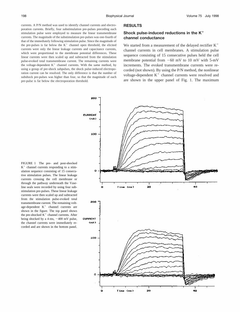
channel currents in cell membranes. A stimulation pulsesequence consisting of 15 consecutive pulses held the cellmembrane potential from260 mV to 10 mV with 5-mVincrements. The evoked transmembrane currents were re-corded (not shown). By using the P/N method, the nonlinearvoltage-dependent K1 channel currents were resolved andare shown in the upper panel of Fig. 1. The maximum
FIGURE 1 The pre- and post-shockedK1 channel currents responding to a stim-ulation sequence consisting of 15 consecu-tive stimulation pulses. The linear leakagecurrents crossing the cell membrane orthrough the pathway underneath the Vase-line seals were recorded by using four sub-stimulation pre-pulses. These linear leakagecurrents were then scaled up and subtractedfrom the stimulation pulse-evoked totaltransmembrane current. The remaining volt-age-dependent K1 channel currents areshown in the figure. The top panel showsthe pre-shocked K1 channel currents. Afterbeing shocked by a 4-ms,2400 mV pulse,the channel currents were immediately re-corded and are shown in the bottom panel.
198 Biophysical Journal Volume 75 July 1998
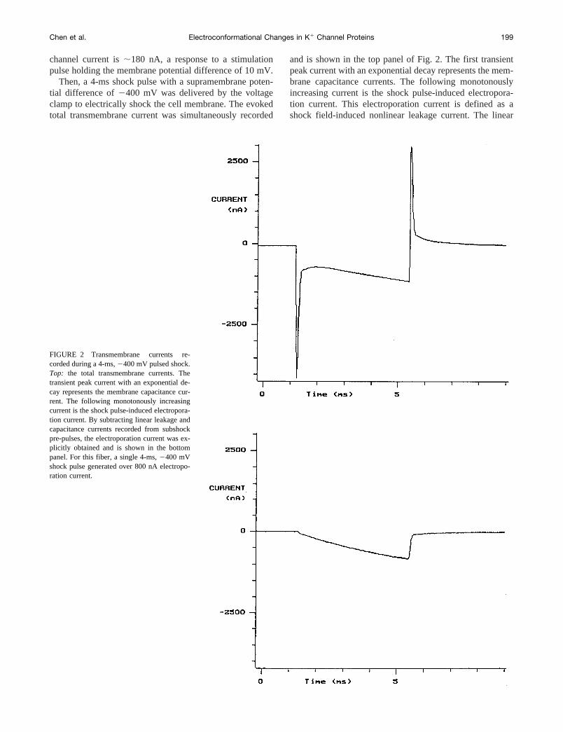
channel current is;180 nA, a response to a stimulationpulse holding the membrane potential difference of 10 mV.
Then, a 4-ms shock pulse with a supramembrane poten-tial difference of2400 mV was delivered by the voltageclamp to electrically shock the cell membrane. The evokedtotal transmembrane current was simultaneously recorded
and is shown in the top panel of Fig. 2. The first transientpeak current with an exponential decay represents the mem-brane capacitance currents. The following monotonouslyincreasing current is the shock pulse-induced electropora-tion current. This electroporation current is defined as ashock field-induced nonlinear leakage current. The linear
FIGURE 2 Transmembrane currents re-corded during a 4-ms,2400 mV pulsed shock.Top: the total transmembrane currents. Thetransient peak current with an exponential de-cay represents the membrane capacitance cur-rent. The following monotonously increasingcurrent is the shock pulse-induced electropora-tion current. By subtracting linear leakage andcapacitance currents recorded from subshockpre-pulses, the electroporation current was ex-plicitly obtained and is shown in the bottompanel. For this fiber, a single 4-ms,2400 mVshock pulse generated over 800 nA electropo-ration current.
Chen et al. Electroconformational Changes in K1 Channel Proteins 199
transmembrane currents were recorded by applying fivesubshock pre-pulses in a magnitude of280 mV, which wasfar below the membrane electroporation threshold of2300mV (Chen and Lee, 1994a). By subtracting these linearleakage and capacitance currents from the total shock cur-rent, the electroporation current could be explicitly obtainedand is shown in the bottom panel of Fig. 2. For this fiber, asingle 4-ms,2400 mV shock pulse generated up to over800 nA electroporation currents.
After the electric shock, the same stimulation pulse se-quence was reapplied to the cell membrane. With the samemethod the K1 channel currents were resolved and areshown in the bottom panel of Fig. 1. By comparing the pre-and post-shock channel currents, one can easily notice thatthe magnitudes of the K1 channel currents were signifi-cantly reduced after a brief electric shock. The maximumvalue of the K1 channel current for this fiber dropped from;180 nA to;125 nA, a.30% reduction from the originalvalue.
Damaging effects of the supramembranepotential difference on cellmembrane are directional
From the difference of the pre- and post-shock channelcurrents responding to the same stimulation pulses, wedemonstrated that after the cell membrane was shocked bya single 4-ms,2400 mV pulse, the K1 channel conductancereduced over 30%. The results also showed that the briefpulsed shock could damage the cell membranes generatinga huge transmembrane current. However, the uncertainty ishow this huge transmembrane current raised membranetemperature, thereby damaging the membrane proteins.
During the experiments, we had tried to use a microther-mistor to monitor the possible temperature changes causedby high-voltage shock pulses. Because of low temperature-resolution and slow time-constant, it is impossible to use athermistor to monitor local and transient temperaturechanges on the cell membranes. To investigate the field-induced thermal effects on cell membrane we need to studythe electrical Joule heating by identifying the electric energyconsumed in the cell membranes, by using our voltageclamp techniques to simultaneously measure membrane po-tential differences and transmembrane currents.
A new fiber was prepared by using the same methoddescribed above. The stimulation sequence was applied tothe cell membrane. By subtracting the linear capacitanceand leakage currents, the K1 channel currents were resolvedwith a maximum value of 168 nA (data not shown).
The fiber was then shocked by a series of pulse pairs,each pair consisting of two pulses with the same magnitudebut different polarities. The time interval between the twopulses in each pair was 5 min, and the pulse pairs wereseparated by 10 min to allow full relaxation of the fiber. Thepositive shock pulse in each pair was always applied to thefiber before the negative pulse, since a positive shock pulse
causes much less damage in channel proteins than a nega-tive pulse of the same magnitude (see the results). Thecorresponding transmembrane currents during the shockpulses were simultaneously recorded. After each shock, thesame stimulation sequence was applied to the cell mem-brane to monitor the shock pulse-induced reductions in thechannel activity.
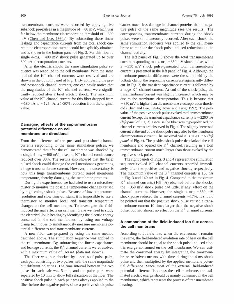
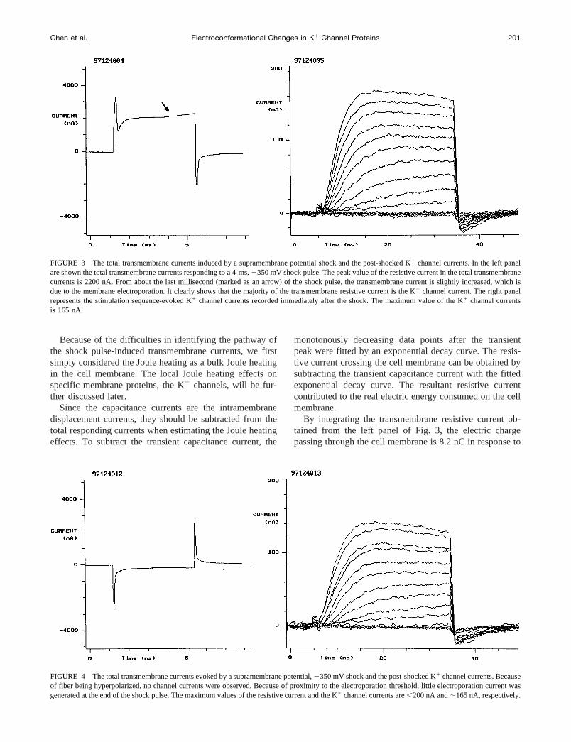
The left panel of Fig. 3 shows the total transmembranecurrent responding to a 4-ms,1350 mV shock pulse, whilea 2350 mV shock pulse-generated total transmembranecurrent is presented in the left panel of Fig. 4. Although themembrane potential differences were the same held by thevoltage clamp, the responding currents are significantly differ-ent. In Fig. 3, the transient capacitance current is followed bya huge K1 channel current. At end of the shock pulse, thetransmembrane current was slightly increased, which may bedue to the membrane electroporation. This is because that2350 mV is higher than the membrane electroporation thresh-old (Chen and Lee, 1994a; Tovar and Tung, 1992). The peakvalue of the positive shock pulse-evoked total transmembranecurrent (except the transient capacitance current) is;2200 nA(left panelof Fig. 3). Because the fiber was hyperpolarized, nochannel currents are observed in Fig. 4. The slightly increasedcurrent at the end of the shock pulse may also be the membraneelectroporation current. The maximal value is,200 nA (leftpanelof Fig. 4). The positive shock pulse depolarized the cellmembrane and opened the K1 channel, resulting in a totaltransmembrane current much larger than those evoked by thenegative shock pulse.
The right panels of Figs. 3 and 4 represent the stimulationsequence-evoked K1 channel currents recorded immedi-ately after the positive and negative shocks, respectively.The maximum value of the K1 channel currents is 165 nAin Fig. 3 and 140 nA in Fig. 4. Compared to the maximumK1 channel currents (168 nA) obtained before the shocks,the 1350 mV shock pulse had little, if any, effect on thechannel currents. However, the single 4-ms,2350 mVshock pulse reduced the channel currents;17%. It shouldbe pointed out that the positive shock pulse caused a trans-membrane current 10 times larger than the negative shockpulse, but had almost no effect on the K1 channel currents.
A comparison of the field-induced ion flux acrossthe cell membrane
According to Joule’s law, when the environment remainsthe same, the field-induced evolution rate of heat on the cellmembrane should be equal to the shock pulse-induced elec-tric energy consumed on the cell membrane. We can esti-mate the consumed energy by integrating the transmem-brane resistive currents with time during the 4-ms shockpulse and then multiplied by the applied membrane poten-tial difference. Since most of the external field-inducedpotential difference is across the cell membrane, the esti-mated electric energy should be mainly consumed in the cellmembranes, which represents the process of transmembraneheating.
200 Biophysical Journal Volume 75 July 1998
Because of the difficulties in identifying the pathway ofthe shock pulse-induced transmembrane currents, we firstsimply considered the Joule heating as a bulk Joule heatingin the cell membrane. The local Joule heating effects onspecific membrane proteins, the K1 channels, will be fur-ther discussed later.
Since the capacitance currents are the intramembranedisplacement currents, they should be subtracted from thetotal responding currents when estimating the Joule heatingeffects. To subtract the transient capacitance current, the
monotonously decreasing data points after the transientpeak were fitted by an exponential decay curve. The resis-tive current crossing the cell membrane can be obtained bysubtracting the transient capacitance current with the fittedexponential decay curve. The resultant resistive currentcontributed to the real electric energy consumed on the cellmembrane.
By integrating the transmembrane resistive current ob-tained from the left panel of Fig. 3, the electric chargepassing through the cell membrane is 8.2 nC in response to
FIGURE 3 The total transmembrane currents induced by a supramembrane potential shock and the post-shocked K1 channel currents. In the left panelare shown the total transmembrane currents responding to a 4-ms,1350 mV shock pulse. The peak value of the resistive current in the total transmembranecurrents is 2200 nA. From about the last millisecond (marked as an arrow) of the shock pulse, the transmembrane current is slightly increased, which isdue to the membrane electroporation. It clearly shows that the majority of the transmembrane resistive current is the K1 channel current. The right panelrepresents the stimulation sequence-evoked K1 channel currents recorded immediately after the shock. The maximum value of the K1 channel currentsis 165 nA.
FIGURE 4 The total transmembrane currents evoked by a supramembrane potential,2350 mV shock and the post-shocked K1 channel currents. Becauseof fiber being hyperpolarized, no channel currents were observed. Because of proximity to the electroporation threshold, little electroporation current wasgenerated at the end of the shock pulse. The maximum values of the resistive current and the K1 channel currents are,200 nA and;165 nA, respectively.
Chen et al. Electroconformational Changes in K1 Channel Proteins 201
a 1350 mV pulse. Only 0.9 nC charges passed through thecell membrane during the2350 mV shock pulse, obtainedfrom the left panel of Fig. 4. Although generating 8.2-nCcharges crossing the cell membranes, the1350 mV shockpulse caused little (3 of 168 nA) decrease in the K1 channelcurrents. In contrast, the2350 mV shock pulse, generatingonly 0.9-nC transmembrane charges, almost one-tenth ofthose induced by the positive-shock pulse, significantlydecreased the K1 channel currents, from 168 nA to 140 nA.This comparison clearly shows that electric field-inducedreduction in the K1 channel currents is not directly relatedto the amount of transmembrane currents.
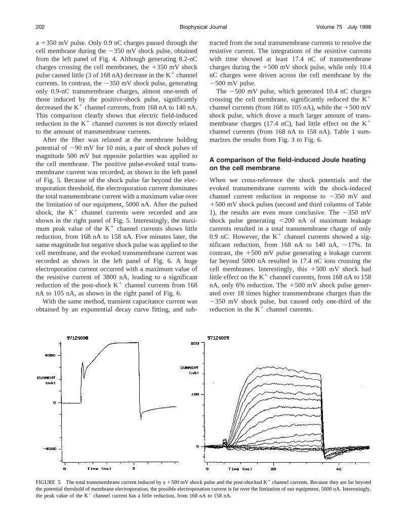
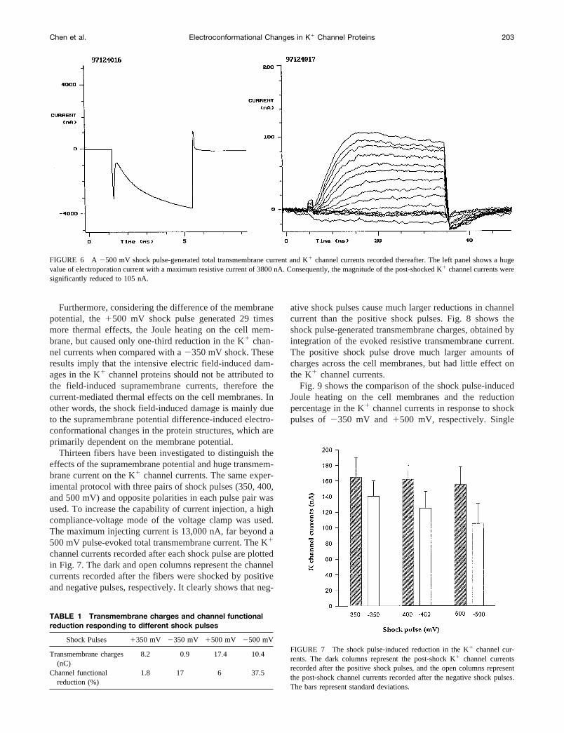
After the fiber was relaxed at the membrane holdingpotential of290 mV for 10 min, a pair of shock pulses ofmagnitude 500 mV but opposite polarities was applied tothe cell membrane. The positive pulse-evoked total trans-membrane current was recorded, as shown in the left panelof Fig. 5. Because of the shock pulse far beyond the elec-troporation threshold, the electroporation current dominatesthe total transmembrane current with a maximum value overthe limitation of our equipment, 5000 nA. After the pulsedshock, the K1 channel currents were recorded and areshown in the right panel of Fig. 5. Interestingly, the maxi-mum peak value of the K1 channel currents shows littlereduction, from 168 nA to 158 nA. Five minutes later, thesame magnitude but negative shock pulse was applied to thecell membrane, and the evoked transmembrane current wasrecorded as shown in the left panel of Fig. 6. A hugeelectroporation current occurred with a maximum value ofthe resistive current of 3800 nA, leading to a significantreduction of the post-shock K1 channel currents from 168nA to 105 nA, as shown in the right panel of Fig. 6.
With the same method, transient capacitance current wasobtained by an exponential decay curve fitting, and sub-
tracted from the total transmembrane currents to resolve theresistive current. The integrations of the resistive currentswith time showed at least 17.4 nC of transmembranecharges during the1500 mV shock pulse, while only 10.4nC charges were driven across the cell membrane by the2500 mV pulse.
The 2500 mV pulse, which generated 10.4 nC chargescrossing the cell membrane, significantly reduced the K1
channel currents (from 168 to 105 nA), while the1500 mVshock pulse, which drove a much larger amount of trans-membrane charges (17.4 nC), had little effect on the K1
channel currents (from 168 nA to 158 nA). Table 1 sum-marizes the results from Fig. 3 to Fig. 6.
A comparison of the field-induced Joule heatingon the cell membrane
When we cross-reference the shock potentials and theevoked transmembrane currents with the shock-inducedchannel current reduction in response to2350 mV and1500 mV shock pulses (second and third columns of Table1), the results are even more conclusive. The2350 mVshock pulse generating,200 nA of maximum leakagecurrents resulted in a total transmembrane charge of only0.9 nC. However, the K1 channel currents showed a sig-nificant reduction, from 168 nA to 140 nA,;17%. Incontrast, the1500 mV pulse generating a leakage currentfar beyond 5000 nA resulted in 17.4 nC ions crossing thecell membranes. Interestingly, this1500 mV shock hadlittle effect on the K1 channel currents, from 168 nA to 158nA, only 6% reduction. The1500 mV shock pulse gener-ated over 18 times higher transmembrane charges than the2350 mV shock pulse, but caused only one-third of thereduction in the K1 channel currents.
FIGURE 5 The total transmembrane current induced by a1500 mV shock pulse and the post-shocked K1 channel currents. Because they are far beyondthe potential threshold of membrane electroporation, the possible electroporation current is far over the limitation of our equipment, 5000 nA. Interestingly,the peak value of the K1 channel current has a little reduction, from 168 nA to 158 nA.
202 Biophysical Journal Volume 75 July 1998
Furthermore, considering the difference of the membranepotential, the1500 mV shock pulse generated 29 timesmore thermal effects, the Joule heating on the cell mem-brane, but caused only one-third reduction in the K1 chan-nel currents when compared with a2350 mV shock. Theseresults imply that the intensive electric field-induced dam-ages in the K1 channel proteins should not be attributed tothe field-induced supramembrane currents, therefore thecurrent-mediated thermal effects on the cell membranes. Inother words, the shock field-induced damage is mainly dueto the supramembrane potential difference-induced electro-conformational changes in the protein structures, which areprimarily dependent on the membrane potential.
Thirteen fibers have been investigated to distinguish theeffects of the supramembrane potential and huge transmem-brane current on the K1 channel currents. The same exper-imental protocol with three pairs of shock pulses (350, 400,and 500 mV) and opposite polarities in each pulse pair wasused. To increase the capability of current injection, a highcompliance-voltage mode of the voltage clamp was used.The maximum injecting current is 13,000 nA, far beyond a500 mV pulse-evoked total transmembrane current. The K1
channel currents recorded after each shock pulse are plottedin Fig. 7. The dark and open columns represent the channelcurrents recorded after the fibers were shocked by positiveand negative pulses, respectively. It clearly shows that neg-
ative shock pulses cause much larger reductions in channelcurrent than the positive shock pulses. Fig. 8 shows theshock pulse-generated transmembrane charges, obtained byintegration of the evoked resistive transmembrane current.The positive shock pulse drove much larger amounts ofcharges across the cell membranes, but had little effect onthe K1 channel currents.
Fig. 9 shows the comparison of the shock pulse-inducedJoule heating on the cell membranes and the reductionpercentage in the K1 channel currents in response to shockpulses of2350 mV and1500 mV, respectively. Single
FIGURE 6 A 2500 mV shock pulse-generated total transmembrane current and K1 channel currents recorded thereafter. The left panel shows a hugevalue of electroporation current with a maximum resistive current of 3800 nA. Consequently, the magnitude of the post-shocked K1 channel currents weresignificantly reduced to 105 nA.
TABLE 1 Transmembrane charges and channel functionalreduction responding to different shock pulses
Shock Pulses 1350 mV 2350 mV 1500 mV 2500 mV
Transmembrane charges(nC)
8.2 0.9 17.4 10.4
Channel functionalreduction (%)
1.8 17 6 37.5
FIGURE 7 The shock pulse-induced reduction in the K1 channel cur-rents. The dark columns represent the post-shock K1 channel currentsrecorded after the positive shock pulses, and the open columns representthe post-shock channel currents recorded after the negative shock pulses.The bars represent standard deviations.
Chen et al. Electroconformational Changes in K1 Channel Proteins 203
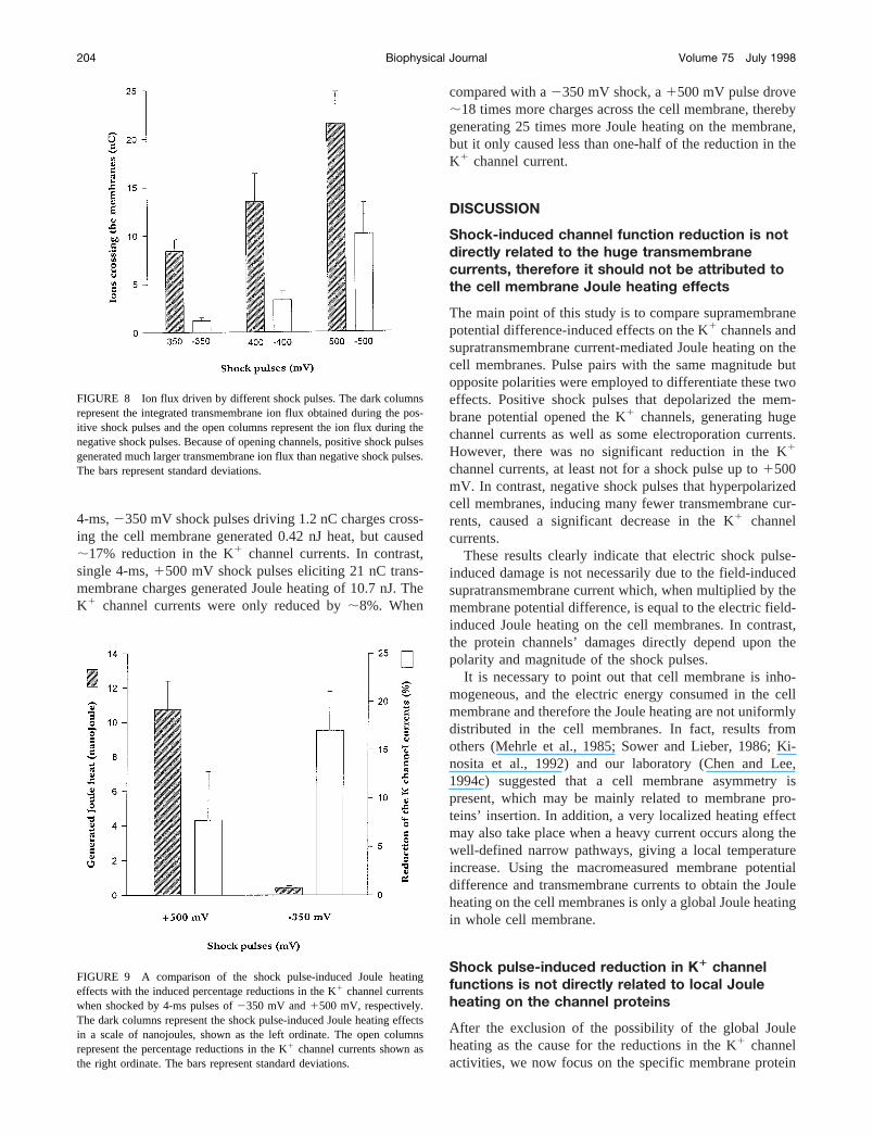
4-ms,2350 mV shock pulses driving 1.2 nC charges cross-ing the cell membrane generated 0.42 nJ heat, but caused;17% reduction in the K1 channel currents. In contrast,single 4-ms,1500 mV shock pulses eliciting 21 nC trans-membrane charges generated Joule heating of 10.7 nJ. TheK1 channel currents were only reduced by;8%. When
compared with a2350 mV shock, a1500 mV pulse drove;18 times more charges across the cell membrane, therebygenerating 25 times more Joule heating on the membrane,but it only caused less than one-half of the reduction in theK1 channel current.
DISCUSSION
Shock-induced channel function reduction is notdirectly related to the huge transmembranecurrents, therefore it should not be attributed tothe cell membrane Joule heating effects
The main point of this study is to compare supramembranepotential difference-induced effects on the K1 channels andsupratransmembrane current-mediated Joule heating on thecell membranes. Pulse pairs with the same magnitude butopposite polarities were employed to differentiate these twoeffects. Positive shock pulses that depolarized the mem-brane potential opened the K1 channels, generating hugechannel currents as well as some electroporation currents.However, there was no significant reduction in the K1
channel currents, at least not for a shock pulse up to1500mV. In contrast, negative shock pulses that hyperpolarizedcell membranes, inducing many fewer transmembrane cur-rents, caused a significant decrease in the K1 channelcurrents.
These results clearly indicate that electric shock pulse-induced damage is not necessarily due to the field-inducedsupratransmembrane current which, when multiplied by themembrane potential difference, is equal to the electric field-induced Joule heating on the cell membranes. In contrast,the protein channels’ damages directly depend upon thepolarity and magnitude of the shock pulses.
It is necessary to point out that cell membrane is inho-mogeneous, and the electric energy consumed in the cellmembrane and therefore the Joule heating are not uniformlydistributed in the cell membranes. In fact, results fromothers (Mehrle et al., 1985; Sower and Lieber, 1986; Ki-nosita et al., 1992) and our laboratory (Chen and Lee,1994c) suggested that a cell membrane asymmetry ispresent, which may be mainly related to membrane pro-teins’ insertion. In addition, a very localized heating effectmay also take place when a heavy current occurs along thewell-defined narrow pathways, giving a local temperatureincrease. Using the macromeasured membrane potentialdifference and transmembrane currents to obtain the Jouleheating on the cell membranes is only a global Joule heatingin whole cell membrane.
Shock pulse-induced reduction in K1 channelfunctions is not directly related to local Jouleheating on the channel proteins
After the exclusion of the possibility of the global Jouleheating as the cause for the reductions in the K1 channelactivities, we now focus on the specific membrane protein
FIGURE 9 A comparison of the shock pulse-induced Joule heatingeffects with the induced percentage reductions in the K1 channel currentswhen shocked by 4-ms pulses of2350 mV and1500 mV, respectively.The dark columns represent the shock pulse-induced Joule heating effectsin a scale of nanojoules, shown as the left ordinate. The open columnsrepresent the percentage reductions in the K1 channel currents shown asthe right ordinate. The bars represent standard deviations.
FIGURE 8 Ion flux driven by different shock pulses. The dark columnsrepresent the integrated transmembrane ion flux obtained during the pos-itive shock pulses and the open columns represent the ion flux during thenegative shock pulses. Because of opening channels, positive shock pulsesgenerated much larger transmembrane ion flux than negative shock pulses.The bars represent standard deviations.
204 Biophysical Journal Volume 75 July 1998
Joule heating; in this case, the K1 channels. First, we needto fully understand what our integrated ion flux means. Forthe currents responding to the positive shock pulses, aftersubtracting the curve-fitted capacitive currents, the remain-ing resistive currents include linear leakage current, channelcurrent, and electroporation current. For the currents re-sponding to the negative shock pulse, the remaining cur-rents include only the linear leakage current and the elec-troporation currents because the negative pulsehyperpolarized the cell membrane and closed most of theK1 channels. The ideal way to discuss local Joule heatingon the K1 channel proteins is to identify the shock-inducedchannel currents by subtracting the linear leakage currentand the membrane electroporation current from the totaltransmembrane currents. However, it is technically difficultto realize, since both channel current and membrane elec-troporation current are nonlinear currents. It is impossible touse the P/N method to distinguish two nonlinear components.
Even though we cannot quantitatively identify the purecurrent passing through the K1 channel during the shockpulses, it does not affect our qualitative analysis. As anexample, let’s go back to the comparison of the channelfunction reduction caused by either a1350 mV or a2350mV shock pulse. From Table 1 we showed that a1350 mVshock pulse generated.9 times more Joule heating in thecell membrane than a2350 mV shock pulse (current ra-tio 5 8.2:0.9 with the same magnitude of the membranepotential difference), while only causing a 1:9 (ratio51.8:17) reduction of the channel currents. Since the magni-tude of the shock pulses are the same, the induced linearleakage currents should be the same, which is proportionalto the membrane potential difference. We now only have todistinguish the channel currents from the membrane elec-troporation currents (through the phospholipid bilayer).
Voltage-dependent differences and current kinetic char-acteristics of each current component can be used to differ-entiate the channel currents from the membrane electropo-ration currents. Kinetic studies of the delayed rectifier K1
channels showed that the channel currents, after a shortdelay, have a long, flat plateau (Hille, 1992). Our previousstudies and this study (Fig. 2) showed that electroporationcurrent monotonously increases during the shock pulse(Chen and Lee, 1994a). From the left panel of Fig. 3, it isclearly shown that the majority of the resistive currentresponding to a1350 mV pulse was carried by the K1
channels, and the membrane electroporation current wasvery small, seen from the relative flat plateau of the resistivecurrent. We have estimated that.95% of the resistivecurrents are attributed to the channel current by fitting theearly part of the plateau to a horizontal straight line in orderto separate the channel currents and the electroporationcurrents. For the2350 mV shock pulse, we do not knowwhat percentage of the resistive current passes through theK1 channels. For the worst situation, let us assume all of theresistive currents pass through the K1 channels (eventhough it is impossible). In this case, we still have similarresults. In other words, a1350 mV shock pulse evoked a
current passing though the K1 channels at least 9 timeslarger than a2350 mV shock pulse would have evoked,therefore generating 9 times higher local Joule heating onthese specific membrane proteins. Yet this1350 mV pulseonly caused one-ninth of the reduction of channel activity.
This analysis clearly indicates that the post-shock reduc-tion of the K1 channel activity is not directly related to thehigh-intensity electric shock-induced transmembrane cur-rents through the K1 channel proteins. Furthermore, it is notrelated to the shock-induced local Joule heating on the K1
channels.In summary, our studies show that supramembrane po-
tential shock pulse-induced reductions in the K1 channelfunctions are not directly correlated to either the globaltransmembrane currents or the channel currents. Therefore,neither the global thermal effects (membrane Joule heating)caused by the huge transmembrane currents nor the localthermal effect (Joule heating on the K1 channels) mediatedby larger channel currents plays a significant role in dam-aging the channel proteins. In other words, these experi-mental results provide strong evidence that during an expo-sure to an intensive electric field, the field-inducedsupramembrane potential difference (magnitude and polar-ity) plays a dominant role in the level of protein damages.This supramembrane potential difference might cause elec-troconformational changes in the charged particles or equiv-alent dipole moments in the channel proteins, resulting inchannel dysfunction. Recently, we have succeeded in mea-suring the field-induced reductions in the intramembranecharge movement currents (Chen et al., 1998, submitted forpublication) and in predicting shock-medicated decrementsin the limiting charge particles in the K1 channel gatingsystem (Chen et al., 1997, submitted for publication).
It is necessary to point out that the magnitude of su-pramembrane potential differences that cause trivial thermaldamages in channel proteins are studied only up to 500 mV.We do not rule out the possibility that when the fieldintensity further increases, the thermal effects may play asignificantly more important role in damaging the mem-brane proteins.
This work was supported by National Institutes of Health Grant GM 50785(to W.C.)
REFERENCES
Abramov, G. S., M. Bier, M. Capelli-Schellpfeffer, and R. C. Lee. 1996.Alteration in sensory nerve function following electric shock.Burn.22:602–606.
Bischof, J. C., J. T. Padanilam, W. H. Holmes, R. M. Ezzell, R. C. Lee,E. G. Cravalho, R. G. Tompkins, M. L. Yarmush, and M. Toner. 1994.Dynamic of cell membrane permeability changes at supraphysiologicaltemperatures.Biophys. J.68:2608–2614.
Bommannan, D., L. Leung, J. Tamada, J. Sharifi, W. Abraham, and R.Potts. 1993. Transdermal delivery of luteinizing hormone releasinghormone: comparison between electroporation and iontophoresis invitro. Proc. Int. Symp. Control. Rel. Bioact. Mater.20:97.
Chen et al. Electroconformational Changes in K1 Channel Proteins 205
Chang, D. C., P. Q. Gao, and B. L. Maxwell. 1991. High efficiency genetransfection by electroporation using a radio-frequency electric field.Biochim. Biophys. Acta.1992:153–160.
Chen, W., and R. C. Lee. 1994a. An improved double Vaseline gap voltageclamp to study electroporated skeletal muscle fibers.Biophys. J.66:700–709.
Chen, W., and R. C. Lee. 1994b. Altered ion channel conductance andionic selectivity induced by large imposed membrane potential pulse.Biophys. J.67:603–612.
Chen, W., and R. C. Lee. 1994c. Electromediated permeabilization of frogskeletal muscle cell membrane: effect of voltage-gated ion channels.Bioelectrochem. Bioenerg.34:157–167.
Chen, W., P. Li, and R. C. Lee. 1992. Efficacy of non-ionic surfactant forsealing electropermeabilized skeletal muscle fibers.Biophys. J.61:2427.
Cole, K. S. 1972. Membrane, Ions and Impulses. University of CaliforniaPress, Berkeley, CA. 569–590.
Cravalho, E. G., M. Toner, D. C. Gaylor, and R. C. Lee. 1992. Responseof cells to supraphysiological temperatures: experimental measurementsand kinetic models.In Electrical Trauma: The Pathophysiology, Mani-festations, and Clinical Management. Cambridge University Press,London/New York. 281–300.
Ehrenberg, B., D. L. Farkas, E. N. Fluhler, Z. Lojewska, and L. M. Loew.1987. Membrane potential induced by external electric field pulses canbe followed with a potentiometric dye.Biophys. J. 51:833–837.
Hille, B. 1992. Ion Channels in Excitable Cell Membranes. Sinauer Asso-ciates, Inc., Sunderland, MA.
Hille, B., and D. T. Campbell. 1976. An improved Vaseline gap voltageclamp for skeletal muscle fibers.J. Gen. Physiol.67:265–293.
Hui, C. S., and W. Chen. 1997. Charge movement in cut fibers ofRanatemporariacontaining 0.1 mM EGTA.J. Physiol.503:563–570.
Irving, M., J. Maylie, N. L. Sizto, and W. K. Chandler. 1987. Intrinsicoptical and passive electrical properties of cut frog twitch fibers.J. Gen.Physiol. 89:1–40.
Jones, J. L., R. E. Jones, and G. Balasky. 1987. Microlesion formation inmyocardial cells by high-intensity electric field stimulation.Am. J. Physiol. 253:H480–H486.
Kinosita, K., Jr., M. Hibino, H. Itoh, M. Shigemori, K. Hirano, Y. Kirino,and T. Hayakawa. 1992. Events of membrane electroporation visualizedon a time scale from microsecond to seconds.In Guide to Electropora-tion and Electrofusion, D. C. Chang, B. M. Chassy, J. A. Saunders, andA. E. Sowers, editors. Academic Press, Inc., New York.
Kinosita, K., Jr., and T. Y. Tsong. 1977. Hemolysis of human erythrocytesby a transient electric field.Proc. Natl. Acad. Sci. USA.74:1923–1927.
Kovacs, L., E. Rios, and M. F. Schneider. 1983. Measurement and modi-fication of free calcium transients in frog skeletal muscle fibers by ametallochromic indicator dye.J. Physiol.343:161–196.
Lau, C. P., and A. J. Camm. 1991. Rate-responsive pacing: technical andclinical aspects.In Cardiac Pacing and Electrophysiology, N. El-Sherifand P. Samet, editors. W. B. Saunders Co., Philadelphia. 524–544,706–712.
Lee, R. C., D. G. Gaylor, D. L. Bhatt, and D. A. Israel. 1988. Role of cellmembrane rupture in the pathogenesis of electrical trauma.J. Surg. Res.44:709.
Lee, R. C., and M. S. Kolodney. 1987. Electrical injury mechanisms:electrical breakdown of cell membrane.Plast. Reconstr. Surg.80:672–679.
Mehrle, W., U. Zimmermann, and R. Hampp. 1985. Evidence for asym-metrical uptake of fluorescent dyes through electro-permeabilized mem-branes of Avena mesophyll protoplasts.FEBS Lett. 185:89–94.
Mir, L. M., S. Orlowski, J. Belehradek, and C. Paoletti. 1991.Electrochemotherapy: potentiation of antitumor effect of bleomycin bylocal electric pulses.Eur. J. Cancer.27:68.
Okino, M., and H. Mohri. 1987. Effects of a high voltage electrical impulseand an anticancer drug on in vivo growing tumors,Jpn. J. Cancer Res.78:1319.
O’Neill, R. J., and L. Tung. 1991. Cell-attached patch clamp study of theelectropermeabilization of amphibian cardiac cells.Biophys. J.59:1028–1039.
Prausnitz, M. R., V. G. Bose, R. Langer, and J. C. Weaver. 1993. Elec-troporation of mammalian skin: a mechanism to enhance transdermaldrug delivery.Proc. Natl. Acad. Sci. USA. 90:10504.
Raymond, E. I., A. S. L. Tang, D. W. Frazier, N. Shibata, P. S. Chen, andJ. M. Wharton. 1991. Ventricular defibrillation: basic concepts.In Car-diac Pacing and Electrophysiology. N. El-Sherif and P. Samet, editors.W. B. Saunders Co., Philadelphia. 706–712.
Rouge, R. G., and A. R. Dimick. 1978. The treatment of electrical injurycompared to burn injury: a review of pathophysiology and comparisonof patient management protocols.J. Trauma.18:43.
Sower, A. E., and M. R. Lieber. 1986. Electropore diameters, lifetimes,numbers, and locations in individual erythrocyte ghosts.FEBS Lett.205:179–184.
Teissie, J., and M. P. Rols. 1994. Manipulation of cell cytoskeleton affectsthe lifetime of cell membrane electropermeabilization.In ElectricalInjury: A Multidisciplinary Approach to Therapy, Prevention, and Re-habilitation.Ann. N.Y. Acad. Sci.720:98–100.
Teissie, J., and T. Y. Tsong. 1980. Evidence of voltage-induced channelopening in Na/K ATPase of human erythrocyte membrane.J. Membr.Biol. 55:133–140.
Tovar, O., and L. Tung. 1992. Electroporation and recovery of cardiac cellmembrane with rectangular voltage pulse.Am. J. Physiol. 92:H1128–H1136.
Tropea, B. I., and R. C. Lee. 1992. Thermal injury kinetics in electricaltrauma.J. Biomech. Eng.114:241–250.
Tsong, T. Y. 1991. Electroporation of cell membrane.Biophys. J.60:297–306.
Tsong, T. Y., and R. D. Astumian. 1986. Absorption and conversion ofelectric field energy by membrane-bounded ATPases.Bioelectrochem.Bioenerg.15:457–476.
Tsong, T. Y., and R. D. Astumian. 1987. Electroconformational couplingand membrane protein function.Prog. Biophys. Mol. Biol.50:1–45.
Winegar, R. A., J. W. Phillips, J. H. Yongblom, and W. F. Morgan. 1989.Cell electroporation is a highly efficient method for introducing restric-tion endonuclease into cells.Mutat. Res. 225:49–53.
Zimmermann, U., J. Vienken, and G. Pilwat. 1980. Development of drugcarrier systems: electric field induced effects in cell membranes.J. Elec-troanal. Chem. 116:553–574.
206 Biophysical Journal Volume 75 July 1998