Misfolded Proteins Recognition Strategies of E3 Ubiquitin Ligases and Neurodegenerative Diseases

Biophysical Journal Volume 97 December 2009 3029–3037 3029
Misfolded Amyloid Ion Channels Present Mobile b-Sheet Subunitsin Contrast to Conventional Ion Channels
Hyunbum Jang,†* Fernando Teran Arce,‡ Ricardo Capone,‡ Srinivasan Ramachandran,‡ Ratnesh Lal,‡
and Ruth Nussinov†§
†Center for Cancer Research Nanobiology Program, NCI-Frederick, SAIC-Frederick, Frederick, Maryland; ‡Center for Nanomedicine andDepartment of Medicine, University of Chicago, Chicago, Illinois; and §Sackler Institute of Molecular Medicine, Department of Human Geneticsand Molecular Medicine, Sackler School of Medicine, Tel Aviv University, Tel Aviv, Israel
ABSTRACT In Alzheimer’s disease, calcium permeability through cellular membranes appears to underlie neuronal cell death.It is increasingly accepted that calcium permeability involves toxic ion channels. We modeled Alzheimer’s disease ion channelsof different sizes (12-mer to 36-mer) in the lipid bilayer using molecular dynamics simulations. Our Ab channels consist of thesolid-state NMR-based U-shaped b-strand-turn-b-strand motif. In the simulations we obtain ion-permeable channels whosesubunit morphologies and shapes are consistent with electron microscopy/atomic force microscopy. In agreement with imagedchannels, the simulations indicate that b-sheet channels break into loosely associated mobile b-sheet subunits. The preferredchannel sizes (16- to 24-mer) are compatible with electron microscopy/atomic force microscopy-derived dimensions. Mobilesubunits were also observed for b-sheet channels formed by cytolytic PG-1 b-hairpins. The emerging picture from our large-scalesimulations is that toxic ion channels formed by b-sheets spontaneously break into loosely interacting dynamic units that asso-ciate and dissociate leading to toxic ionic flux. This sharply contrasts intact conventional gated ion channels that consist of tightlyinteracting a-helices that robustly prevent ion leakage, rather than hydrogen-bonded b-strands. The simulations suggest whyconventional gated channels evolved to consist of interacting a-helices rather than hydrogen-bonded b-strands that tend to breakin fluidic bilayers. Nature designs folded channels but not misfolded toxic channels.
doi: 10.1016/j.bpj.2009.09.014
INTRODUCTION
Alzheimer’s disease (AD), like many other protein conforma-
tional diseases, including Huntington’s disease, Parkinson’s
disease, type II diabetes, and cystic fibrosis result from protein
misfolding that alters their three-dimensional conformations
from native (generally soluble) to nonnative (insoluble)
folded structures (1). In AD, b-amyloid (Ab) peptides form
b-sheet-rich ordered aggregates (2) with Ab oligomers being
the toxic species (3,4). Although it is generally accepted
that toxicity relates to ion transport through the cellular
membrane, the mechanism has been a debated issue (5). On
the one hand, using a variety of methods, numerous studies
have consistently observed that Ab peptides assemble to
form ion channels in the cell membrane inducing selective
ion permeation (1,6–20). On the other hand, other studies
did not observe any channels. These studies (21,22) observed
nonselective ion leakage that they argued occurs through
membrane thinning and lowered dielectric barrier induced
by the interaction of the Ab oligomers with the membrane.
Recent investigations of the Ab-induced ion flux by current
recordings across the membranes seem to have resolved the
controversy (23). These studies reexamined the previous
postulate that Ab-induced membrane thinning caused gradual
increase in cellular ion flux (21,22). They showed that even in
the absence of Ab, trace amounts of the hexafluoro-isopropa-
nol remaining in the solvent used to prepare Ab oligomers led
Submitted June 1, 2009, and accepted for publication September 10, 2009.
*Correspondence: [email protected]
Editor: Gerhard Hummer.
� 2009 by the Biophysical Society
0006-3495/09/12/3029/9 $2.00
to membrane thinning causing a gradual increase in cellular
current. They further showed that elimination of the hexa-
fluoro-isopropanol solvent leads to the characteristic
Ab-induced stepwise ion flux across the planar lipid bilayer
(1,6,13,18), suggesting that Ab forms an ion channel.
Increasing evidence lends support to ion channel forma-
tion (24–26). Electron microscopy (EM) shows doughnut-
like Ab pores (27,28), and atomic force microscopy
(AFM) presents heterogeneous ion channels that directly
mediate ion conductance (1,16). The AFM-imaged Ab
channels in the 1,2-dioleoyl-sn-glycero-3-phosphocholine
(DOPC) membrane have a central pore of ~2.0 nm diameter
and 8–12 nm in outer diameter (1). The channels are subunit
assemblies, with shapes varying from rectangular with four
subunits to hexagonal with six, and show cation-selective
conductance. As AFM probes the topmost portion of the
channel, the observed structures may refer either to conduct-
ing pores or pores collapsed at the bottom, thus not providing
information about the functionality of the observed structure.
Our previous molecular dynamics simulations (19,20) of
nonamyloidogenic Ab peptides (29) in the bilayer predicted
ion-permeable channels formed by loosely attached mobile
subunits with shapes, morphologies, and dimensions similar
to the AFM-imaged channels (1,16). The simulations
focused on the Ab17–42 small protofibril (pentamer) coordi-
nate set based on two-dimensional NMR in combination
with solid state NMR (ssNMR), mutational data and EM
(30). In addition, Ab9–42 was also simulated, using the
ssNMR-based coordinates (31). In the modeled channels

3030 Jang et al.
the Ab peptides retained the U-shaped b-strand-turn-b-
strand motif observed in both coordinate sets. This motif
was first predicted by modeling the Ab16–35 (32). To date,
similar U-shaped motifs were observed in the ssNMR struc-
ture of a b2-microglobulin fragment (33) and in the CA150
WW domain (34), suggesting that U-shaped motifs are a
general feature of amyloid organization (35–37). These olig-
omers may also form channels in the membrane (38–40).
This motif associates into stacked b-sheets, with the intermo-
lecular b-strands H-bonded to each other.
Antimicrobial peptide protegrin-1 (PG-1), a b-hairpin
peptide, also forms channels in the membrane. The channel
is cytotoxic, leaking chloride ions. This unregulated leakage
leads to target cells death. The PG-1 b-hairpins associate to
form a b-sheet channel, also with loosely attached subunits
(41), thus sharing a common morphological motif with other
b-sheet channels (1,19,20).
This study aims to characterize toxic AD ion channels and
to obtain the preferred channel sizes and apparent molecular
mass. In particular, through this characterization and
comparison with experiment, it further aims to obtain some
of the hallmarks differentiating toxic amyloid channels
from ‘‘normally’’ folded gated (e.g., Naþ, Kþ, and Ca2þ)
ion channels that are optimized by evolution. Both misfolded
toxic amyloid channels and the normally folded channels are
gated; however, in the first the gating is unregulated and is
the outcome of the membrane dynamics, sharply contrasting
the second. Toxic amyloid channels possess features distinct
from the physiological gated ion channels that robustly regu-
late the ion flux (42,43). Functional gated channels i),
contain mostly a-helices; ii), fold into their native state;
iii), are stable and long-lived membrane proteins with mem-
brane supporting channel conformations; iv), have a fixed
number of subunits that are specifically associated; and v),
selectively transport ions that are essential to cell metabolism
via orchestrated conformational changes. In contrast, non-
physiological toxic amyloid channels i), can contain mostly
b-sheets; ii), are not present in a unique size; iii), can have
a different number of subunits that are nonspecifically asso-
ciated; iv), induce rapid ion leakage; v), are associated with
many diseases and lead to cell death; and vi), consist of mis-
folded states with the channels varying in size, shape, and
morphology in membranes (1,16). Our modeling captures
the distinct features of amyloid channels. These features
are further observed in the cytotoxic b-sheets channel of anti-
microbial peptide PG-1.
Using our previous protocols (19,20) we comprehensively
test a range of Ab channel sizes to characterize these on the
atomic scale. Without structural information from experi-
ments, modeling the three-dimensional structures of amyloid
ion channels is highly challenging. Our channel modeling
starts with the experimentally available Ab9–42 monomer
conformation in the oligomeric state (34 residues; ~3.6 kDa/
monomer; adding Ile41 and Ala42 (31)). Channels with in-
creasing number of Ab9–42 monomers (12-, 16-, 20-, 24-,
Biophysical Journal 97(11) 3029–3037
and 36-mer) were constructed and explicitly simulated in
the DOPC bilayer (see Fig. S1 in the Supporting Material).
Further, the 24-mer channel was also simulated in the anionic
bilayer containing palmitoyl-oleyl-phosphatidylcholine
(POPC) and palmitoyl-oleyl-phosphatidylglycerol (POPG)
(POPC/POPG ¼ 4:1, molar ratio) to see the lipid effects on
the channel formation. Because previous experimental data
relate to Ab1–42 and Ab1–40 (1,16), comprehensive experi-
ments were conducted to test the Ab9–42 model. Additional
ongoing work targets the Ab17–42, including AFM, calcium
uptake by APP deficient cells, single channel conductance,
and inhibition by zinc validating the candidate models
(H. Jang, F. Teran Arce, R. Capone, S. Ramachandran, R. Lal,
and R. Nussinov, unpublished). We note that as in any
modeling study, our work in this study can only suggest candi-
dates that would eventually need direct experimental tests.
MATERIALS AND METHODS
A monomer conformation of the Ab fragment Ab9–42 based on solid state
NMR of small Ab9–40 protofibril coordinates (31) was used to construct
channels with annular shapes adding Ile41 and Ala42. Annular shapes were
obtained through rotations of the monomer 12, 16, 20, 24, and 36 times
with respect to the pore axis (Fig. S1). Depending on the direction of the
rotation, two channel topologies were constructed (Fig. S2): i), CNpNC
(where C and N represent C- and N- terminal b-strands respectively, and
p denotes a central pore); and ii), NCpCN Ab channels. The former has
a central pore enclosed by the N-terminal b-strands, whereas the latter by
the C-terminal b-strands. In this study, we present only results for the
pore-preserving CNpNC topology because the NCpCN with the charged
N-terminal facing the bilayer and the hydrophobic C-terminal facing the
solvated pore collapse (19).
Each annular channel is minimized with a rigid body motion of the peptides
to enhance the formation of backbone hydrogen bonds (H-bonds) within a
b-sheet and then embedded in the DOPC bilayer. In particular, the 24-mer
channel is simulated in the anionic bilayer containing POPC and POPG
with a molar ratio of 4:1. A unit cell containing two layers of lipids with
90,000 to 200,000 atoms, depending on the channel size, is constructed (Table
S1). The bilayers containing 200–500 lipids constitute the unit cell with
TIP3P waters added at both sides. The system contains MgCl, KCl, CaCl,
and ZnCl at the same concentration of 25 mM to satisfy a total cations concen-
tration near 100 mM. The CHARMM program (44) was used to construct the
set of starting points and to relax the systems to a production-ready stage.
Simulations of the initial construction and the preequilibration were carried
out on the constant number of atoms, pressure, surface area, and temperature
ensemble. Production runs were 30 ns for the 12- to 24-mer channels and 50 ns
for the 36-mer channels. The NAMD code (45) on a Biowulf cluster at the
National Institutes of Health, Bethesda, MD (http://biowulf.nih.gov) was
used for the starting point. In the production simulations, the dynamics
were carried out on both the constant number of atoms, pressure, surface
area, and temperature and the constant number of atoms, pressure, surface
tension, and temperature ensembles with g ¼ 0. However, no significant
differences in the results were found for the different ensembles. Averages
were taken after 10 ns discarding initial transient conformations.
RESULTS
Optimal channel sizes
We use the experiment-based Ab oligomer coordinate set to
construct channels of different sizes. Our conceptual design
for the initial channel is based on a perfect annular shape.
Amyloid Ion Channels 3031
Depending on the direction of the monomer rotation, two
types of annular channel topologies are obtained (Fig. S2).
We focus only on the pore-preserving topology with the
hydrophobic C-terminal b-strands interfacing the bilayer
and the N-terminal b-strands containing polar and charged
residues facing into, and forming, the solvated pore
(19,20). Size exclusion chromatography indicated that
medium-to-large globulomers of (at least) 90–110 kDa
lead to membrane permeability (21). No channels were
observed for these particles. In our simulation, the molecular
mass of 36-mer Ab9–42 (130 kDa) channel is >90 kDa, the
upper limit of channel formation (Fig. S1). We classify the
channels into three groups: small (12-mer), intermediate
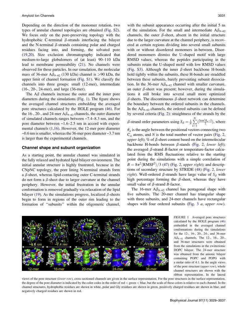
(16-, 20-, 24-mer), and large (36-mer).
The Ab channels increase the outer and the inner pore
diameters during the simulations (Fig. 1). The figure shows
the averaged channel structures embedding the averaged
pore structures calculated by the HOLE program (46). For
the 16-, 20-, and 24-mer Ab9–42 channels, the outer diameter
of simulated channels ranges between ~7.4–8.3 nm, and the
pore diameter between ~1.6–2.5 nm in accord with experi-
mental channels (1,16). However, the 12-mer pore diameter
~0.6 nm is smaller, whereas the 36-mer pore diameter ~3.7 nm
is larger than the experimental channels.
Channel shape and subunit organization
As a starting point, the annular channel was simulated in
the fully relaxed and hydrated lipid bilayer environment. The
initial annular structure is highly frustrated, because in the
CNpNC topology, the pore lining N-terminal strands form
a b-sheet, whereas lipid-contacting outer C-terminal strands
do not form a b-sheet due to larger curvature at the channel
periphery. However, the initial frustration in the annular
conformation is removed gradually via relaxation of the lipid
bilayer (19). As the simulations progress, localized b-sheets
begin to form in regions of the outer rim leading to the
formation of ‘‘subunits’’ within the oligomeric channel,
with the subunit appearance occurring after the initial 5 ns
of the simulation. For the small and intermediate Ab9–42
channels, the outer b-sheet, absent in the initial structure
due to the larger curvature at the channel periphery, is recov-
ered at certain regions dividing into several small subunits
with or without disordered monomers in-between. Disor-
dered monomers dismiss the U-shaped motif with large
RMSD values, whereas the peptides participating in the
subunits retain the U-shaped motif with low RMSD values
(Fig. S3). Although the inner b-sheet backbone H-bonds
hold tightly within the subunits, these H-bonds are straddled
between these subunits, barely preventing subunit dissocia-
tion. In the 36-mer Ab9–42 channel with smaller curvature,
an outer b-sheet was present; however, during the simula-
tions it still broke into several small more optimized
b-sheets. The discontinuous b-sheet network can determine
the boundary between the ordered subunits in the channels.
In the Ab9–42 channels, the ordered subunits can be defined
by several criteria (Fig. 2): straightness of the strands by the
b-strand order parameters using Sb ¼ 1N
PN
k¼1
ð3cos2qa�12Þ, where
qa is the angle between the positional vectors connecting two
Ca atoms, and N is the total number of vector pairs (Fig. 2,
upper left); % of b-sheet content based on the intermolecular
backbone H-bonds between b-stands (Fig. 2, lower left);the averaged b-strand B-factor or temperature-factor calcu-
lated from the RMS fluctuations relative to the starting
point during the simulations with a simple correlation of
B ¼ 8p2hRMSF2i=3 (47) (Fig. 2, upper right); and descrip-
tions of secondary structure by STRIDE (48) (Fig. 2, lowerright). Well-ordered b-strands have large value of Sb with
high percentage forming the b-sheet, whereas they have
small value of b-strand B-factor.
The 16-mer Ab9–42 channel has pentagonal shape with
five subunits. The 20-mer channel has triangular shape
with three subunits, and 24-mer channels have rectangular
shapes with four ordered subunits (Fig. 3 a, upper row).
~6.6 nm
~0.6 nm
~7.4 nm
~1.6 nm
~7.8 nm
~1.9 nm
~8.3 nm
~2.5 nm
~10.3 nm
~3.7 nm
12-mer 16-mer 20-mer 24-mer 36-mer
FIGURE 1 Averaged pore structures
calculated by the HOLE program (46)
embedded in the averaged channel
conformations during the simulations
for the 12-, 16-, 20-, 24-, and 36-mer
Ab9–42 channels. The 12-, 16-, 20-,
and 36-mer structures were obtained
from the simulations in the zwitterionic
DOPC bilayer. The 24-mer structure
was obtained from the anionic bilayer
containing POPC and POPG with
a molar ratio of 4:1. In the angle views
of the pore structure (upper row), whole
channel structures are shown with the
ribbon representation. In the lateral
views of the pore structure (lower row), cross-sectioned channels are given in the surface representation. For the pore structures in the surface representation,
the degree of the pore diameter is indicated by the color codes in the order of red< green< blue, but the scale of these colors is relative to each channel. In the
channel structures, hydrophobic residues are shown in white, polar and Gly residues are shown in green, positively charged residues are shown in blue, and
negatively charged residues are shown in red.
Biophysical Journal 97(11) 3029–3037

3032 Jang et al.
20-mer Aβ9-42 channel
β-strand order parameter Temperature factor
0.3
0.4
0.5
0.6
0.7
5 10 15 20
N-term
C-term
Sβ -
stra
nd
Peptide Number
0
40
80
120
160
200
5 10 15 20
N-term
C-term
<B> β
-stra
nd
Peptide Number
0
20
40
60
80
100
5 10 15 20
N-term
C-term
%
Peptide Number
% of β-sheet content
Secondary structure by STRIDE
Peptide Number2 4 6 8 10 12 14 16 18 20
Res
idue
Num
ber
2
4
6
8
10
12
14
16
18
20
22
24
26
28
30
32
Coil
Turn
β-strand
β-brigde
FIGURE 2 Parameters to define the ordered subunits for
the 20-mer Ab9–42 channel: the b-strand order parameter
Sb-strand (upper left), % of b-sheet content (lower left), the
averaged b-strand B-factor (upper right), and the description
of secondary structure by STRIDE (48) (lower right). The
b-strand order parameter, the b-sheet content based on the
intermolecular backbone H-bonds, and the averaged
b-strand B-factor from the b-strand RMSF are calculated
for the N-terminal (blue area) and C-terminal (green area)
b-strands separately as a function of peptide number. Peaks
in the Sb-strand and % of b-sheet content curves indicate well
ordered b-strand, whereas troughs in the b-strands B-factor
curves denote the ordered b-strand. The secondary structure
by STRIDE was calculated for the averaged structure.
The average positions of the b-strands calculated from the
contour map of the x,y coordinates of the b-strands projected
onto the two-dimensional bilayer surface verify the shapes
and number of subunits (Fig. 3 a, lower row). The 24-mer
channel obtained the subunits in the anionic bilayer
(POPC/POPG ¼ 4:1). Our previous 24-mer Ab9–42 channel
simulation obtained five subunits in the DOPC bilayer (20),
suggesting that even for the same channel, subunit forma-
tions strongly depend on the fluidic bilayer dynamics. The
12-mer Ab9–42 channel has two marginally defined subunits
with collapsed pore (Fig. 1), suggesting that at least three
subunits are required to form a water pore. The 36-mer
Ab9–42 channel has an ellipsoidal shape with two broken
parts (Fig. S4). A hemifission may take place at the disor-
dered narrowing neck. The disconnected end of the detached
subunits may migrate and fuse with nearby channels, with
the fission decaying spontaneously (49). The fission of the
36-mer channel suggests that the lipid bilayer does not
support such large channel formation. The subunit organiza-
tion observed for the Ab9–42 channels is shared by other
b-sheet channels (Fig. 3 b). Our recent simulations for the
PG-1 channels (41) have shown that the octameric PG-1
channels with both antiparallel and parallel b-sheet arrange-
ments divided into 4–5 subunits, indicating that although
sequence composition, lengths, and detailed monomer
conformations are different, the b-sheet channel formation
Biophysical Journal 97(11) 3029–3037
via subunits association-dissociation is determined by the
bilayer dynamics.
Comparative peptide interaction
The interaction of the individual peptide is inhomogeneous,
leading to heterogeneous Ab channel shapes (Fig. S5). Of
particular interest is the peptide interaction energy depending
on the channel size. We calculated the interaction energy for
each peptide with other peptides, lipids, and waters. The
peptide interaction energy was then averaged over the time
and the number of peptides in the channel (Fig. 4). For inter-
mediate Ab9–42 channels, well-balanced peptide interactions
with the environment support the channel conformations;
especially the channels have similar energy values for the
peptide-water interaction. The peptide interaction energies
suggest that in the channels the Ab peptides favor interaction
with the surroundings in the order of waters > other peptides
> lipids. Strong peptide-water interaction indicates that the
peptide has various ways of exposure to waters. In addition
to bulk water, these include the central water pore and the
hydrated cavity beneath each U-turn. For the peptide-peptide
interaction, the interaction energy mainly derives from inter-
action between neighboring peptides (Fig. S6); the peptides
have strong intermolecular backbone H-bonds between
the b-strands in the ordered subunits. The peptide-lipid

Amyloid Ion Channels 3033
a
12
3
1
2
3
45
1
3
4
2
16-mer 20-mer 24-mer
Aβ9-42 channels
x (nm)-4 -3 -2 -1 0 1 2 3 4
y (n
m)
-4
-3
-2
-1
0
1
2
3
4
x (nm)-4 -3 -2 -1 0 1 2 3 4
-4
-3
-2
-1
0
1
2
3
4
x (nm)-4 -3 -2 -1 0 1 2 3 4
-4
-3
-2
-1
0
1
2
3
4
b PG-1 channels
1
2
3
4
1
2
3
4
1
2
34
1
2
3
4 5
Antiparallel β-sheet channels Parallel β-sheet channels
FIGURE 3 Simulated channel struc-
tures with highlighted subunits (upperrow) and mapping of x,y coordinates
of the b-strands of each peptide onto
the x-y plane (lower row) for the (a)
Ab9-42 channels. The 12-, 16-, 20-,
and 36-mer structures were obtained
from the simulations in the zwitterionic
DOPC bilayer. The 24-mer structure
was obtained from the anionic bilayer
containing POPC and POPG with
a molar ratio of 4:1. The averaged chan-
nels in the surface representation are
shown in the view along the membrane
normal. In the b-strands mapping, the
contour lines enclose the high frequency
regions in the order of red < orange <
yellow < green < blue, but the scale
of these colors is relative to each
channel. (b) The simulated channel
structures with highlighted subunits for
the antiparallel and parallel b-sheet
channels of PG-1 (41).
interaction is relatively weaker than the peptide-peptide
interaction, suggesting that the subunits as rigid particles
are mobile in the fluidic lipid bilayer.
The small 12-mer Ab9–42 channel has relatively strong
peptide-water and peptide-lipid interactions. The peptides
in the 12-mer channels are mainly disordered; lacking the
intermolecular backbone H-bonds, they present increased
interactions with lipids and waters. The 12-mer channel
should have relatively weak peptide-peptide interaction
compared to intermediate channels, because the trend of
interaction energy decreases as the channels size decreases.
However, the relatively strong peptide-peptide interaction
in the 12-mer channel is due to the collapsed pore, inducing
additional peptide-peptide interaction for non-neighboring
pairs (Fig. 1). In contrast to the 12-mer channel, the
36-mer channel has relatively weak peptide-water and
peptide-lipid interactions, and relatively strong peptide-peptide
interaction. In the subunits of 36-mer channels, b-strand inter-
actions are fully saturated with the H-bonding producing
strong interactions between neighboring peptides. The ordered
subunits become fibril-like rigid oligomers. The relatively
weak interaction between peptides and lipids makes these
subunits mobile in the lipid bilayer, inducing fissile channels.
Ion permeable channels
The channels preserve a large pore, wide enough for con-
ducting water and ions. The Ab9–42 channels confirm that
cations are easily trapped by the negatively charged Glu22
side chains at the top bilayer leaflet, creating a cationic
ring (19). In the pore of the Ab9–42 channel, anions are at-
tracted to the positively charged His14 and Lys16 side chains
at the bottom bilayer leaflet. To observe the ions’ behavior in
the pore, the potential of mean force (PMF) representing the
relative free energy profile for each ion across the bilayer is
calculated (Fig. 5). The ion-density-based PMF calculation is
useful to estimate rough relative free energy changes for
ions, providing a general outline for ion permeation through
the pore (41,50,51). The details of the PMF calculation are
described in the Supporting Material.
In the simulations, the channel systems contain
four cations, Mg2þ, Ca2þ, Kþ, and Zn2þ at the same
Biophysical Journal 97(11) 3029–3037

3034 Jang et al.
concentration (25 mM), and an anion, Cl�. On the top
bilayer surface, the cations interact with the phosphate head-
groups at the lipid/water interface. Just below the interface,
the negatively charged Glu22 side chains in the pore form
cation binding sites. At the lower bilayer leaflet, the cations
interact with the phosphate headgroups and the Ab
C termini. In the pore, both Ca2þ and Zn2þ exhibit a low
mobility at the binding site, indicating that they are easily
trapped by the side chains, whereas Mg2þ and Kþ are very
mobile. Without Zn2þ, Ca2þ are dominantly trapped by the
side chains (19), but there are less trapped Ca2þ in the pres-
ence of Zn2þ. Zn2þ inhibits Ab toxicity, serving as channel
blocker by reducing calcium transport (1,6–17,19,20). The
16-, 20-, and 24-mer channels binding sites provide a rela-
tively low free energy profile for Ca2þ or Zn2þ compared
to other cations. However, in the 12-mer channel calcium
binding is not observed at the Glu22 side chains (Fig. S7),
because the collapsed pore blocks Ca2þ entering to the
pore with a high energy barrier. The 36-mer channel binding
sites provide a relatively low free energy profile for all
cations, suggesting nonselective ion leakiness due to the
large pore (Fig. S7). In the lower pore portion of Ab9–42
channel, the positively charged His14 and Lys16 side chains
provide low free energy profile for Cl�, creating anionic
binding sites.
DISCUSSION AND CONCLUSIONS
Explicit solvent molecular dynamics simulations of ssNMR-
based Ab oligomer coordinates (31) in the bilayer modeled
as perfectly annular structures obtain ion-permeable
channels. AFM images provide a consistent picture of ion
channels for a series of disease-related amyloid species (1).
The channels present a range of sizes and morphologies.
Remarkably, the 16-, 20-, and 24-mer simulations obtain
channels with similar subunit organization and dimensions
12-mer 16-mer 20-mer 24-mer 36-mer
<E> pe
ptid
e (kc
al/m
ol)
-600
-500
-400
-300
-200
-100
0
LipidPeptideWater
-700
-800
-900
-1000
-1100
-1200
-1300
FIGURE 4 Averaged peptide interaction energy for the Ab9-42 channels.
All peptide-lipid interactions were calculated for the peptides interacting
with DOPC lipids including 24-mer channels. The peptide-lipid (red bars)
and peptide-peptide (green bars) interaction use the scale of interaction
energy on the left (black labels). The peptide-water (blue) interaction uses
the scale of interaction energy on the right (blue labels).
Biophysical Journal 97(11) 3029–3037
-4 -3 -2 -1 0 1 2 3 4-4
-3
-2
-1
0
1
2
3
16-mer
-4 -3 -2 -1 0 1 2 3 4
Δ GPM
F (k
J/m
ol)
-4
-3
-2
-1
0
1
2
3
20-mer
Pore Axis (nm)-5 -4 -3 -2 -1 0 1 2 3 4 5
-4
-3
-2
-1
0
1
2
3
24-mer
FIGURE 5 PMF, DGPMF, calculated using the equation DGPMF ¼ �kBTln(rz/rbulk), where kB is the Boltzmann constant, T is the simulation temper-
ature, rz is the ion density at the position z along the pore axis, and rbulk is
the ion density in the bulk region, representing the relative free energy
profile for Mg2þ (green symbols and lines), Kþ (red), Ca2þ (blue), Zn2þ
(pink), and Cl� (black) as a function of the distance along the pore center
axis for Ab9–42 channels. The PMF results for the 12-, 16-, 20-, and
36-mer were obtained from the simulations in the zwitterionic DOPC
bilayer. The PMF result for the 24-mer was obtained from the anionic bilayer
containing POPC and POPG with a molar ratio of 4:1.

Amyloid Ion Channels 3035
Water pores
Hydro-
phobic
face
Polar/charged
face
a -sheet subunits
cMembrane pores
Antimicrobial peptides
b
Water pore
Subunits
α-helices
Gated ion channels
cMembrane pores
Antimicrobial peptides
Water pores
Hydro-
phobic
face
Polar/rr charged
facccccccccccccceeeeeeeeeeeeeee
a -sheet subunits
b
Water pore
Subunits
α-helices
Gated ion channels
FIGURE 6 (a) Cartoon representing the dynamics of Alzheimer ion
channel formation in the fluidic lipid bilayer. The mobile subunit has the
hydrophobic face (yellow) and polar/charged face (blue). A channel with
as imaged by AFM (1,16). The 24-mer channel was simu-
lated in both the zwitterionic DOPC bilayer and the anionic
bilayer containing POPC and POPG with a molar ratio of
4:1. It is expected that the channel interacts more strongly
with the anionic bilayer than the zwitterionic bilayer due to
the electrostatic interaction. However, no significant differ-
ences in the results of the subunit formation in the channel
conformation were found. The smaller (12-mer) channel
collapses and the larger (36-mer) channel is not supported
by the bilayer, allowing us to derive the apparent molecular
mass of the AFM-imaged channels that we confirm to be less
than what is predicted for large Ab oligomers (21). The range
of channel sizes observed for the Ab, 16-, 20-, and 24-mer,
contrasts the functional regulated native state gated channels
that have a single size, a precise number of subunits, and one
specific way of subunit organization.
Normally folded functional gated ion channels selectively
permeate ions across membranes with electrical currents in
two steady-state levels corresponding to the open and closed
states (42,43). In contrast, toxic Ab channels catalyze the
diffusion of ions across membranes presenting stepwise fluc-
tuations of electrical currents with typical ion channel
behavior; however, the fluctuating large amplitude spikes
in the current indicate distinct channels (23). This experi-
mentally observed behavior derives from Ab channel forma-
tion in the dynamic membrane: after insertion into the cell
membranes, small Ab oligomer subunits assemble to form
dynamic, leaky pore-like structures (Fig. 6 a). In the 16-,
20-, and 24-mer channel simulations we do not observe
a movement toward dissociation; what we observe are
dynamic structures. Such a dynamic leaky behavior is in
contrast to functional gated channels that fold into specific
native structures optimized by evolution (Fig. 6 b). In nor-
mally folded controlled gated channels good contacts are
maintained between the a-helical subunits, robustly prevent-
ing ion leakage. Ab channel formation is cytotoxic, similar to
the channels formed by b-cytolytic peptides such as PG-1
antimicrobial peptides (41), suggesting that amyloid channel
formation may induce membrane defects such as those
induced by PG-1 membrane pore formation (52). We note
that toxic a-antimicrobial peptides do not form channels.
Rather, the a-helices interactions are lipid-mediated (Fig. 6 c).
Our results suggest why controlled gated channels consist of
a-helices rather than the consistently observed breakage-
prone ion-leaking b-sheet channels.
water filled pore can be formed when at least three subunits assemble their
polar/charged faces together. The pore-preserving channels provide ion
conductance with selectivity across the membrane. Large pores with nonse-
lective ion conductance are not supported by the bilayer membranes and
tend to reduce the size (top left). Misassembled subunits do not conduct
ions due to the collapsed pore as denoted by the dotted circle. Cartoons rep-
resenting (b) functional gated ion channels and (c) membrane pore forma-
tions by toxic a-antimicrobial peptides. The gated ion channel diagram
was inspired by the Kþ channel with four identical subunits, but the number
of a-helices in the subunit diagram is arbitrary.
Biophysical Journal 97(11) 3029–3037

3036 Jang et al.
Our simulations suggest that the Ab9–42 channels induce
a barrel-stave membrane pore where the channel is parallel
to the lipid tails (Fig. S8). However, this is not necessarily
the case for all amyloid channels. Amyloid channels can
also induce other membrane pore types such as toroidal
depending on the outward-facing amino acid sequence inter-
acting with the bilayer (53,54). A charged/polar surface pref-
erentially interacting with lipid headgroups may induce such
a curved membrane surface (52). Under such circumstances,
ion leakage can be not only through the pore but via the
solvated interface between the channel and the bilayer. In
both toxic barrel-stave channels as observed here and
toroidal channels with subunit organizations, solvated inter-
subunits areas can also leak ions.
To conclude, our simulations provide atomic resolution
models for Ab channels based on the NMR b-strand-turn-
b-strand motif. We have simulated the channels in an explicit
membrane environment to obtain a range of potential sizes to
characterize, the apparent molecular mass and the preferred
channel size and organization and compared with experiment.
We have further simulated channels in different membrane
environments, in DOPC, and in an anionic bilayer. We note
that these models are consistent with experimental data;
however, as in all modeling studies (55), other models consis-
tent with available data are always possible. Moreover,
currently it is increasingly realized that although still present-
ing the b-strand-turn-b-strand motif, amyloid seeds and fibrils
are polymorphic (56–58), suggesting that small oligomers
penetrating the bilayer can also present a polymorphic vari-
ability affecting channel conformations. Nonetheless, we
believe that the dynamic toxic channel picture painted by
our models should hold for a polymorphic range. Our studies
provide what we believe are conformational details useful for
drug development (24–26). The Ab channels have b-sheet
structure that is reasonable for both amyloid formation
(36,37,59) and channel formation (40). It is interesting to
note that antimicrobial cytolytic b-sheet channels similarly
break into dynamic subunits (41), suggesting that the fluidic
membrane does not support intact cylindrical b-sheets chan-
nels, regardless of the sequence, length, and conformational
details. The H-bonds break in some regions leading to optimi-
zation of the H-bonds in the subunits. All these channels can
leak ions through the solvated channel pores. We propose that
this could be a main evolutionary reason why gated functional
channels tend to be made of tightly interacting a-helices rather
than of b-sheet organization.
SUPPORTING MATERIAL
Potential mean force calculations, references, a table, and eight figures are
available at http://www.biophysj.org/biophysj/supplemental/S0006-3495
(09)01460-X.
We thank Dr. Robert Tycko for providing us with the coordinates of the
Ab9–40 oligomer. This study used the high-performance computational capa-
bilities of the Biowulf PC/Linux cluster at the National Institutes of Health,
Biophysical Journal 97(11) 3029–3037
Bethesda, MD (http://biowulf.nih.gov). Initial coordinates of the channels
are available at http://protein3d.ncifcrf.gov/hbj/coor/.
This work was supported by the National Cancer Institute, National Insti-
tutes of Health (N01-CO-12400), and the Intramural Research Program of
the National Institutes of Health, National Cancer Institute, Center for
Cancer Research.
The content of this publication does not necessarily reflect the views or poli-
cies of the Department of Health and Human Services, nor does mention of
trade names, commercial products, or organizations imply endorsement by
the U.S. government.
REFERENCES
1. Quist, A., I. Doudevski, H. Lin, R. Azimova, D. Ng, et al. 2005.Amyloid ion channels: a common structural link for protein-misfoldingdisease. Proc. Natl. Acad. Sci. USA. 102:10427–10432.
2. Selkoe, D. J. 1991. Alzheimer’s disease. In the beginning. Nature.354:432–433.
3. Bucciantini, M., E. Giannoni, F. Chiti, F. Baroni, L. Formigli, et al.2002. Inherent toxicity of aggregates implies a common mechanismfor protein misfolding diseases. Nature. 416:507–511.
4. Walsh, D. M., I. Klyubin, J. V. Fadeeva, W. K. Cullen, R. Anwyl, et al.2002. Naturally secreted oligomers of amyloid b protein potently inhibithippocampal long-term potentiation in vivo. Nature. 416:535–539.
5. Kagan, B. L. 2005. Amyloidosis and protein folding. Science. 307:42–43, author reply 42–43.
6. Arispe, N., H. B. Pollard, and E. Rojas. 1993. Giant multilevel cationchannels formed by Alzheimer disease amyloid b-protein [AbP-(1–40)]in bilayer membranes. Proc. Natl. Acad. Sci. USA. 90:10573–10577.
7. Arispe, N., H. B. Pollard, and E. Rojas. 1994. b-Amyloid Ca(2þ)-channel hypothesis for neuronal death in Alzheimer disease. Mol.Cell. Biochem. 140:119–125.
8. Arispe, N., H. B. Pollard, and E. Rojas. 1996. Zn2þ interaction withAlzheimer amyloid b protein calcium channels. Proc. Natl. Acad. Sci.USA. 93:1710–1715.
9. Kawahara, M., N. Arispe, Y. Kuroda, and E. Rojas. 1997. Alzheimer’sdisease amyloid b-protein forms Zn(2þ)-sensitive, cation-selectivechannels across excised membrane patches from hypothalamic neurons.Biophys. J. 73:67–75.
10. Kawahara, M., and Y. Kuroda. 2000. Molecular mechanism of neurode-generation induced by Alzheimer’s b-amyloid protein: channel forma-tion and disruption of calcium homeostasis. Brain Res. Bull. 53:389–397.
11. Kawahara, M., Y. Kuroda, N. Arispe, and E. Rojas. 2000. Alzheimer’sb-amyloid, human islet amylin, and prion protein fragment evoke intra-cellular free calcium elevations by a common mechanism in a hypotha-lamic GnRH neuronal cell line. J. Biol. Chem. 275:14077–14083.
12. Rhee, S. K., A. P. Quist, and R. Lal. 1998. Amyloid b protein-(1–42)forms calcium-permeable, Zn2þ-sensitive channel. J. Biol. Chem.273:13379–13382.
13. Hirakura, Y., M. C. Lin, and B. L. Kagan. 1999. Alzheimer amyloidAb1–42 channels: effects of solvent, pH, and Congo Red. J. Neurosci.Res. 57:458–466.
14. Hirakura, Y., W. W. Yiu, A. Yamamoto, and B. L. Kagan. 2000.Amyloid peptide channels: blockade by zinc and inhibition by Congored (amyloid channel block). Amyloid. 7:194–199.
15. Lin, H., Y. J. Zhu, and R. Lal. 1999. Amyloid b protein (1–40) formscalcium-permeable, Zn2þ-sensitive channel in reconstituted lipid vesi-cles. Biochemistry. 38:11189–11196.
16. Lin, H., R. Bhatia, and R. Lal. 2001. Amyloid b protein forms ion chan-nels: implications for Alzheimer’s disease pathophysiology. FASEB J.15:2433–2444.
17. Zhu, Y. J., H. Lin, and R. Lal. 2000. Fresh and nonfibrillar amyloidb protein(1–40) induces rapid cellular degeneration in aged human

Amyloid Ion Channels 3037
fibroblasts: evidence for AbP-channel-mediated cellular toxicity.FASEB J. 14:1244–1254.
18. Kourie, J. I., C. L. Henry, and P. Farrelly. 2001. Diversity of amyloidb protein fragment [1–40]-formed channels. Cell. Mol. Neurobiol.21:255–284.
19. Jang, H., J. Zheng, and R. Nussinov. 2007. Models of b-amyloid ion-channels in the membrane suggest that channel formation in the bilayeris a dynamic process. Biophys. J. 93:1938–1949.
20. Jang, H., J. Zheng, R. Lal, and R. Nussinov. 2008. New structures helpthe modeling of toxic amyloid b ion channels. Trends Biochem. Sci.33:91–100.
21. Kayed, R., Y. Sokolov, B. Edmonds, T. M. McIntire, S. C. Milton, et al.2004. Permeabilization of lipid bilayers is a common conformation-dependent activity of soluble amyloid oligomers in protein misfoldingdiseases. J. Biol. Chem. 279:46363–46366.
22. Sokolov, Y., J. A. Kozak, R. Kayed, A. Chanturiya, C. Glabe, et al.2006. Soluble amyloid oligomers increase bilayer conductance byaltering dielectric structure. J. Gen. Physiol. 128:637–647.
23. Capone, R., F. G. Quiroz, P. Prangkio, I. Saluja, A. M. Sauer, et al.2009. Amyloid-b induced ion flux in artificial lipid bilayers andneuronal cells: resolving a controversy. Neurotox. Res. 16:1–13.
24. Diaz, J. C., O. Simakova, K. A. Jacobson, N. Arispe, and H. B. Pollard.2009. Small molecule blockers of the Alzheimer Ab calcium channelpotently protect neurons from Ab cytotoxicity. Proc. Natl. Acad. Sci.USA. 106:3348–3353.
25. Arispe, N., J. C. Diaz, and M. Flora. 2008. Efficiency of histidine-asso-ciating compounds for blocking the Alzheimer’s Ab channel activityand cytotoxicity. Biophys. J. 95:4879–4889.
26. Simakova, O., and N. J. Arispe. 2006. Early and late cytotoxic effects ofexternal application of the Alzheimer’s Ab result from the initial forma-tion and function of Ab ion channels. Biochemistry. 45:5907–5915.
27. Lashuel, H. A., D. Hartley, B. M. Petre, T. Walz, and P. T. Lansbury, Jr.2002. Neurodegenerative disease: amyloid pores from pathogenic muta-tions. Nature. 418:291.
28. Lashuel, H. A., D. M. Hartley, B. M. Petre, J. S. Wall, M. N. Simon,et al. 2003. Mixtures of wild-type and a pathogenic (E22G) form ofAb40 in vitro accumulate protofibrils, including amyloid pores.J. Mol. Biol. 332:795–808.
29. Thinakaran, G., and E. H. Koo. 2008. Amyloid precursor protein traf-ficking, processing, and function. J. Biol. Chem. 283:29615–29619.
30. Luhrs, T., C. Ritter, M. Adrian, D. Riek-Loher, B. Bohrmann, et al.2005. 3D structure of Alzheimer’s amyloid-b(1–42) fibrils. Proc.Natl. Acad. Sci. USA. 102:17342–17347.
31. Petkova, A. T., W. M. Yau, and R. Tycko. 2006. Experimentalconstraints on quaternary structure in Alzheimer’s b-amyloid fibrils.Biochemistry. 45:498–512.
32. Ma, B., and R. Nussinov. 2002. Stabilities and conformations of Alz-heimer’s b-amyloid peptide oligomers (Ab16–22, Ab16–35, and Ab10–35): sequence effects. Proc. Natl. Acad. Sci. USA. 99:14126–14131.
33. Iwata, K., T. Fujiwara, Y. Matsuki, H. Akutsu, S. Takahashi, et al. 2006.3D structure of amyloid protofilaments of b2-microglobulin fragmentprobed by solid-state NMR. Proc. Natl. Acad. Sci. USA. 103:18119–18124.
34. Ferguson, N., J. Becker, H. Tidow, S. Tremmel, T. D. Sharpe, et al.2006. General structural motifs of amyloid protofilaments. Proc. Natl.Acad. Sci. USA. 103:16248–16253.
35. Zheng, J., B. Ma, and R. Nussinov. 2006. Consensus features inamyloid fibrils: sheet-sheet recognition via a (polar or nonpolar) zipperstructure. Phys. Biol. 3:1–4.
36. Zheng, J., H. Jang, B. Ma, C. J. Tsai, and R. Nussinov. 2007. Modelingthe Alzheimer Ab17–42 fibril architecture: tight intermolecular sheet-sheet association and intramolecular hydrated cavities. Biophys J.93:3046–3057.
37. Zheng, J., H. Jang, B. Ma, and R. Nussinov. 2008. Annular structures asintermediates in fibril formation of Alzheimer Ab17–42. J. Phys. Chem.B. 112:6856–6865.
38. Lin, M. C., T. Mirzabekov, and B. L. Kagan. 1997. Channel formation
by a neurotoxic prion protein fragment. J. Biol. Chem. 272:44–47.
39. Hirakura, Y., R. Azimov, R. Azimova, and B. L. Kagan. 2000. Poly-
glutamine-induced ion channels: a possible mechanism for the neuro-
toxicity of Huntington and other CAG repeat diseases. J. Neurosci.Res. 60:490–494.
40. Hirakura, Y., and B. L. Kagan. 2001. Pore formation by b-2-microglo-
bulin: a mechanism for the pathogenesis of dialysis associated amyloid-
osis. Amyloid. 8:94–100.
41. Jang, H., B. Ma, R. Lal, and R. Nussinov. 2008. Models of toxic b-sheet
channels of protegrin-1 suggest a common subunit organization motif
shared with toxic Alzheimer b-amyloid ion channels. Biophys. J.95:4631–4642.
42. Roux, B. 2005. Ion conduction and selectivity in K(þ) channels. Annu.Rev. Biophys. Biomol. Struct. 34:153–171.
43. Tombola, F., M. M. Pathak, and E. Y. Isacoff. 2006. How does voltage
open an ion channel? Annu. Rev. Cell Dev. Biol. 22:23–52.
44. Brooks, B. R., R. E. Bruccoleri, B. D. Olafson, D. J. States, S. Swami-
nathan, et al. 1983. CHARMM—a program for macromolecular energy,
minimization, and dynamics calculations. J. Comput. Chem. 4:187–217.
45. Phillips, J. C., R. Braun, W. Wang, J. Gumbart, E. Tajkhorshid, et al.
2005. Scalable molecular dynamics with NAMD. J. Comput. Chem.26:1781–1802.
46. Smart, O. S., J. M. Goodfellow, and B. A. Wallace. 1993. The pore
dimensions of gramicidin A. Biophys. J. 65:2455–2460.
47. Wriggers, W., E. Mehler, F. Pitici, H. Weinstein, and K. Schulten. 1998.
Structure and dynamics of calmodulin in solution. Biophys. J. 74:1622–
1639.
48. Frishman, D., and P. Argos. 1995. Knowledge-based protein secondary
structure assignment. Proteins. 23:566–579.
49. Kozlovsky, Y., and M. M. Kozlov. 2003. Membrane fission: model for
intermediate structures. Biophys. J. 85:85–96.
50. de Groot, B. L., and H. Grubmuller. 2001. Water permeation across
biological membranes: mechanism and dynamics of aquaporin-1 and
GlpF. Science. 294:2353–2357.
51. Leontiadou, H., A. E. Mark, and S. J. Marrink. 2007. Ion transport
across transmembrane pores. Biophys. J. 92:4209–4215.
52. Brogden, K. A. 2005. Antimicrobial peptides: pore formers or metabolic
inhibitors in bacteria? Nat. Rev. Microbiol. 3:238–250.
53. Smith, P. E., J. R. Brender, and A. Ramamoorthy. 2009. Induction of
negative curvature as a mechanism of cell toxicity by amyloidogenic
peptides: the case of islet amyloid polypeptide. J. Am. Chem. Soc.131:4470–4478.
54. Wong, P. T., J. A. Schauerte, K. C. Wisser, H. Ding, E. L. Lee, et al.
2009. Amyloid-b membrane binding and permeabilization are distinct
processes influenced separately by membrane charge and fluidity.
J. Mol. Biol. 386:81–96.
55. Durell, S. R., H. R. Guy, N. Arispe, E. Rojas, and H. B. Pollard. 1994.
Theoretical models of the ion channel structure of amyloid b-protein.
Biophys. J. 67:2137–2145.
56. Meinhardt, J., C. Sachse, P. Hortschansky, N. Grigorieff, and M. Fan-
drich. 2009. Ab(1–40) fibril polymorphism implies diverse interaction
patterns in amyloid fibrils. J. Mol. Biol. 386:869–877.
57. Paravastu, A. K., R. D. Leapman, W. M. Yau, and R. Tycko. 2008.
Molecular structural basis for polymorphism in Alzheimer’s b-amyloid
fibrils. Proc. Natl. Acad. Sci. USA. 105:18349–18354.
58. Zhang, R., X. Hu, H. Khant, S. J. Ludtke, W. Chiu, et al. 2009. Inter-
protofilament interactions between Alzheimer’s Ab1–42 peptides in
amyloid fibrils revealed by cryoEM. Proc. Natl. Acad. Sci. USA.106:4653–4658.
59. Buchete, N. V., R. Tycko, and G. Hummer. 2005. Molecular dynamics
simulations of Alzheimer’s b-amyloid protofilaments. J. Mol. Biol.353:804–821.
Biophysical Journal 97(11) 3029–3037
Copyright © 2022 FDOKUMEN