Evolution of the class C GPCR Venus flytrap modules involved positive selected functional divergence
Upload
benthamscienceCategory
view
0download
0
Send Orders for Reprints to [email protected]
Current Medicinal Chemistry, 2014, 21, 3673-3686 3673
A Common Molecular Motif Characterizes Extracellular Allosteric Enhancers of GPCR Aminergic Receptors and Suggests Enhancer Mechanism of Action
Robert Root-Bernstein* and Patrick F. Dillon
Department of Physiology, Michigan State University, East Lansing, MI 48824 USA
Abstract: Several classes of compounds that have no intrinsic activity on aminergic systems nonetheless enhance the po-tency of aminergic receptor ligands three-fold or more while significantly increasing their duration of activity, preventing tachyphylaxis and reversing fade. Enhancer compounds include ascorbic acid, ethylenediaminetetraacetic acid, corticos-teroids, opioid peptides, opiates and opiate antagonists. This paper provides the first review of aminergic enhancement, demonstrating that all enhancers have a common, inobvious molecular motif and work through a common mechanism that is manifested by three common characteristics. First, aminergic enhancers bind directly to the amines they enhance, sug-gesting that the common structural motif is reflected in common binding targets. Second, one common target is the first extracellular loop of aminergic receptors. Third, at least some enhancers are antiphosphodiesterases. These observations suggest that aminergic enhancers act on the extracellular surface of aminergic receptors to keep the receptor in its high af-finity state, trapping the ligand inside the receptor. Enhancer binding produces allosteric modifications of the receptor structure that interfere with phosphorylation of the receptor, thereby inhibiting down-regulation of the receptor. The mechanism explains how enhancers potentiate aminergic activity and increase duration of activity and makes testable pre-dictions about additional compounds that should act as aminergic enhancers.
Keywords: aldosterone, allosteric, allostery, antiphosphodiesterase, ascorbic acid, corticosteroid, dopamine, down-regulation, EDTA, enhancement, enhances, epinephrine, GPCR, histamine, increased potency, molecular complementarity, morphine, naloxone, naltrexone, norepinephrine, opiate, opioid, phosphodiesterase inhibitors, potentiates, potentiation, serotonin, super-sensitivity, vitamin C.
INTRODUCTION
G protein-couple receptors (GPCR) comprise a class of seven-transmembrane proteins that retain a high degree of sequence similarity [1]. In consequence, it can be difficult to develop drugs that have a high degree of selectivity for one specific receptor subtype over others [2]. The recent discov-ery of extracellular allosteric sites that can be modulated in tandem with a specific GPCR subtypes represents useful drug development targets [3-5]. Until very recently, how-ever, extracellular allosteric sites in GPCR have been dem-onstrated only on muscarinic and adenosine receptors [3-7]. Since 2004, however, we have demonstrated that ascorbate enhances adrenergic and histaminergic ligand potency through an extracellular mechanism involving binding of the enhancer to the first extracellular loop of the relevant GPCR [8-11]. The existence of this extracellular binding site for ascorbic acid on aminergic GPCR, in conjunction with other evidence related to the effects of ascorbic acid and other aminergic enhancers on aminergic function, suggests a spe-cific mechanism for enhancer activity that may have implica-tions for drug development as profound as those associated with the discovery of the benzodiazepines. *Address correspondence to this author at the Department of Physiology, Michigan State University, East Lansing, MI 48824 USA; Tel: 1-517-884-5039; Fax: 1-517-335-5125; E-mail: [email protected]
A review of the literature on aminergic systems reveals that, in addition to ascorbic acid, several other classes of compounds have also been characterized as significantly enhancing aminergic GPCR activity. These compounds in-clude folic acid, ethylenediaminetetraacetic acid (EDTA), opiate drugs and their antagonists, opioid peptides, corticos-teroids, members of the citric acid cycle, and various flavenoids. All of these compounds have been demonstrated to produce significant (three- to ten-fold) enhancement of the potency and duration of activity of aminergic compounds while having no intrinsic activity themselves on aminergic systems. We define an aminergic enhancer, therefore, as a compound with no intrinsic pharmacological effect on the receptor or tissue upon which it is tested, but which increases the potency and/or duration of activity of an aminergic com-pound when present concurrently with that compound. En-hancers have two additional effects as well: they prevent tachyphylaxis (the down-regulation of receptors due to re-peated exposure to a compound) and fade (the loss of effi-cacy resulting from continuous exposure to a compound). All told, the effects of aminergic enhancers hold great therapeu-tic promise.
We report here that the seemingly disparate set of com-pounds that display aminergic enhancement share a simple molecular motif and most certainly work through one, com-mon mechanism involving binding to the first extracellular
1875-533X/14 $58.00+.00 © 2014 Bentham Science Publishers

3674 Current Medicinal Chemistry, 2014, Vol. 21, No. 32 Root-Bernstein and Dillon
loop of aminergic GPCR. The nature of the site suggests a novel, specific, allosteric mechanism of action by which en-hancement is effected through antiphosphodiesterase activ-ity. Finally, the relationship of structure and function be-tween enhancers and the compounds they enhance, as well as the way in which this structure-function relationship has been integrated into aminergic GPCR suggests the nature of the natural selection process that may have been at work during the evolution of this class of receptors. Understanding this natural selection process may provide clues as to the nature of enhancers for other classes of GPCR.
In short, by reviewing the forty year history of experi-ments regarding aminergic GPCR enhancement, we are able to suggest a specific mechanism of action for these en-hancers that makes a series of testable predictions concern-ing enhancer structure, an allosteric mechanism that retards and reverses phosphodiesterase activity, and how these struc-ture-function relationships evolved.
Ascorbic Acid
The best-characterized aminergic enhancer is ascorbic acid (vitamin C). Ascorbate has been shown to have no con-tractile effect on rabbit or porcine thoracic aortic rings, nor any relaxile effects on guinea pig or porcine trachealis at concentrations up to ten-fold higher than physiological nor-mal [8-12]. Ascorbate at physiological concentrations of 15 to 50 M enhances submaximal contractions, and at 150 to 500 M concentrations (about 3 to 10 times physiological normal for human beings) shifts the dose response curve of adrenergic agonists such as norepinephrine (NE), epineph-rine, ephedrine, albuterol, isoproterenol, and phenylpropano-lamine 0.5 to 1.0 log units to the left [8-13]. The effect is seen only at submaximal concentrations of adrenergic com-pounds and has no effect on the maximal contraction that can be achieved. In other words, ascorbate increases the potency but not the efficacy of these aminergic compounds. Under the same conditions, the duration of contraction is extended three- to ten-fold [8-13]. Ascorbate creates the same in-creased potency and duration of action when combined with histamine at any submaximal concentration [10].
Although it was originally hypothesized that aminergic enhancement was due to the antioxidant properties of ascor-bate [13], it has been demonstrated using multiple methods and protocols that oxidation is not a significant factor in these experiments, accounting for less than five percent of the observed enhancement [9, 10]. In fact, enhancement is observed in adrenergic compounds such as albuterol, isopro-terenol, ephedrine, phenylpropanolamine, and histamine that do not oxidize to a measurable extent over the time course of the experiments. Moreover, as we will show below, en-hancement can also be produced by classes of compounds such as opiates and corticosteroids that are not antioxidants.
Experiments demonstrating enhancement by ascorbate have been performed not only in vitro, but in vivo. Dillon, et al. [11] demonstrated increased potency of albuterol in horses with heaves (a model of chronic obstructive pulmo-nary disease). In a sheep model of asthma, the increase in potency of albuterol was more than ten-fold [11]. Houston, et al. [14] found that intraduodenal administration of ascorbate along with isoproterenol potentiated the isoproterenol effect
more than two-fold. Grossmann, et al. [15] reported that in human subjects, ascorbate enhances by almost three-fold the relaxation of veins induced by phenylephrine. Mak and New-ton [16] and Shinke, et al. [17] similarly observed a signifi-cant increase in the inotropic effects of dobutamine in human subjects when it was co-infused along with ascorbate. And Monahan, Eskurza and Seals [18, 19] reported that bringing the serum ascorbate level up from an average of 40 M to 1 mM in healthy human patients significantly increased car-diovagal baroreflex sensitivity to endogenous amines. These human studies suggest that ascorbate enhancement of adren-ergic and histaminergic drugs is safe and has real clinical potential.
EDTA
A second class of aminergic enhancing compounds is ex-emplified by the chelator ethylenediaminetetraacetic acid (EDTA). EDTA produces no contraction or relaxation of smooth muscle preparations at any dose thus far tested, but at micromolar concentrations has the same enhancing effects as ascorbate on adrenergic compounds [9, 12, 13]. The en-hancement effect of EDTA on aminergic receptors is a par-ticularly important finding since it is common practice for people isolating this class of receptors to use high concentra-tions of EDTA in their preparation of their receptors and these high concentrations of EDTA often remain during binding and second-messenger assays. The presence of EDTA during these assays may very likely increase apparent binding constants, drug potency, and duration of activity. The observation that EDTA enhances adrenergic compounds in a similar fashion to ascorbate also suggests that the mechanism of action of such enhancers must involve an ex-tracellular mechanism, since EDTA has no known receptor or transporter, and is so highly charged that it is unable to pass through cell membranes.
Opiates, Opiate Antagonists and Opioid Peptides
The likelihood that aminergic receptor enhancement in-volves an extracellular mechanism is further supported by evidence that opiates such as morphine, dextromethorphan and levorphanol and opiate antagonists such as naloxone also exhibit aminergic enhancing effects. These studies also prove that this enhancement is not mediated by opiate recep-tors, but by aminergic receptors.
Puri, Cochin and Volicer [20] found that a mixture of morphine with any submaximal dose of dopamine resulted in significantly increased dopamine activity in rat corpus stria-tum compared with dopamine alone. Morphine had no effect by itself. Marti [21] went on to demonstrate that pre-treatment of guinea pig ileum with morphine shifted the dose response curve to norepinephrine in guinea pig ileum about half a log unit to the left. This enhancement could not be blocked by the opiate antagonist naloxone. Rae and De Moraes [22] con-firmed all of Marti’s findings, demonstrating in addition that naloxone not only failed to block morphine’s effect, but could itself enhance norepinephrine. Akabori and Barraclough [23, 24] and He, Molnar and Barraclough [25] found that morphine enhanced norepinephrine-induced luteinizing hormone (LH) release compared with NE by itself but that morphine by itself had no effect on LH release. Kindman, Kates and Ginsburg

Common Molecular Motif Characterizes Extracellular Allosteric Enhancers Current Medicinal Chemistry, 2014, Vol. 21, No. 32 3675
[26] similarly demonstrated that 10 M morphine had no con-tractile effects on rabbit myocardium, but shifted the dose re-sponse curve of isoproterenol leftward between three- and five-fold. Like Rae and de Moraes [22], they found that opiate antagonists such as naloxone and naltrexone had an identical potentiating effect. Since both opiate agonists and antagonists are effective, they concluded that their enhancing effects were independent of specific stereochemistry and could not be opi-ate receptor-mediated.
The finding that opiate antagonists are as effective as opiates in enhancing adrenergic compounds has been repli-cated by many investigators, strongly suggesting that Kind-man, Kates and Ginsburg [26] were correct in concluding that adrenergic enhancement is not opiate-receptor mediated. Lechner, Gurll and Reynolds [27] may have been the first to recognize that naloxone potentiates both endogenous and exogenous monoamines, demonstrating that adrenalectomy eliminated circulatory responses to naloxone during treat-ment of hemorrhagic shock. They also demonstrated that naloxone increased the response to any given dose of exoge-nous alpha- or beta-agonist three-fold or more. Akabori and Barraclough [24] found that norepinephrine-induced release of luteinizing hormone could be enhanced by naloxone, which had no releasing effect by itself, but strangely, NE-stimulated release of luteinizing hormone-releasing hormone (LHRH) could be enhanced by morphine but not naloxone [28]. This is the only known instance of an enhancer poten-tiating one adrenergic system but not another. Allgood, Gurll and Reynolds [29] showed that the ability of naloxone to correct cardiovascular effects of endotoxic shock is depend-ent on circulating endogenous amines. The mechanism of this response was further explored by Caffrey, et al. [30, 31], who demonstrated that naloxone increases the response to epinephrine (EPI) in isolated canine arteries by more than 100% and that the response can selectively be eliminated by alpha antagonists. The inotropic potentiation of isoproterenol by naloxone was confirmed by Lechner [32], Gu, et al. [33] and Park, et al. [34]. McCubbin, et al. [35] found that naltrexone, another opiate antagonist, also significantly en-hanced physiological responses to exogenous epinephrine in a rat model of diabetes.
Despite the findings by Marti [21] and Kindman, Kates and Ginsburg [26] that naloxone does not block morphine-induced enhancement of adrenergic compounds, and despite the evidence just reviewed showing that naloxone and naltrexone are themselves enhancers, three methodologically flawed reports have suggested that opiate antagonists can block opiate enhancement of adrenergic compounds. One confusing report by Parra, et al. [36] argues that opiate an-tagonists block the enhancement effect of morphine on NE-induced contractions of rat aorta and that the enhancement effect is therefore mediated through opiate receptors. This finding appears to be a methodological artifact resulting from pretreating their tissues with enough opiate antagonists to maximize the response to any dose of adrenergic com-pounds before adding an opiate. In consequence, the opiate could not further enhance the adrenergic effect. In addition, Parra and his colleagues failed to run a control to test for the effects of opiate antagonists themselves on adrenergics, ap-parently assuming there would be no effect. A similar meth-odological problem calls into question two reports by Lee
and Berkowitz [37,38] that claim naloxone antagonism of the opiate pentazocine. Tissues were once again pretreated with opiate antagonists before addition of an opiate agonist, and no controls for the opiate antagonists were performed. The Lee and Berkowitz studies also suffer from having been per-formed at ED95 NE concentrations (10-7 M), which are two orders of magnitude higher than the NE concentrations util-ized by most of the other studies described here. Since en-hancement by ascorbate (see references above) has been found to increase the potency of drugs but not their efficacy, working near the maximal effective dose of an adrenergic or histaminergic compound will disguise any effects of poten-tial enhancers.
Opioid peptides also function as aminergic enhancers. As noted above, Deyo, Swift and Miller [39, 40] reported that Leu- and Met-enkephalins enhanced dopamine activity in the rat median eminence. Tagaya, et al. [41] also demonstrated that the -opiate receptor agonist, DAMGO ([D-Ala2, N-MePhe4, Gly-ol]-enkephalin), had no relaxant effect on ca-nine tracheal smooth muscle by itself, did not alter acetyl-choline-induced contraction, but did augment the dose re-sponse to isoproterenol 1.9 to 4.3-fold depending on the con-centration of DAMGO. DAMGO had no effect on relaxation caused by nitroprusside, verapamil, 8-cyclic-GMP, or methyl xanthenes, demonstrating that its enhancement effect is adrenergic-specific. Parra, et al. [36] found similarly that DAMGO had no effect on isolated rat aortic strips but in-creased the tension produced by any submaximal dose of NE. They also claim that opiate antagonists prevent DAMGO’s enhancing effects, but these experiments have the same methodological problems (pre-exposure of tissues to an opiate antagonist without antagonist controls for en-hancement) described above regarding their reports of an-tagonist elimination of morphine enhancement.
Corticosteroids
Some, though probably not all, corticosteroids may have aminergic enhancing effects as well, which may have impli-cations for understanding the well-known side effects of steroids including hypertension, mania, and increased ag-gression - all symptoms of increased aminergic activity.
Purdy, Weber, and Drayer [42-44] reported that aldoster-one alone had no contractile effect on rabbit ear arteries or thoracic aorta, but the addition of 1, 10 and 100 uM aldoster-one to rabbit thoracic aortic rings partially contracted with NE to a steady-state of 1.5-3.5 g caused a further contraction of 0.09, 0.47 and 0.80 g, respectively. Aldosterone also in-creased the duration of NE-induced contractions about 2.5-fold. This enhancement was blocked by the adrenergic an-tagonist phentolamine [42-44]. The fact that these experi-ments involve the use of aldosterone at several orders of magnitude above its physiological concentrations makes these results of questionable utility for drug therapy, but do not alter the utility of the results for understanding the struc-tural requirements for aminergic enhancer activity and may have some application to understanding renal physiology, where local concentrations of aldosterone may be signifi-cantly higher than blood or plasma levels.
Of greater clinical relevance, Gu, et al. [33] found that in human subjects, corticosterone enhanced epinephrine-

3676 Current Medicinal Chemistry, 2014, Vol. 21, No. 32 Root-Bernstein and Dillon
induced cardiac contractions, but had no effect on cardiac activity by itself. Corticosterone also decreased epinephrine uptake. And additional evidence supporting the role of some steroid compounds as aminergic enhancers comes from hu-man trials involving prednisolone, budesonide, and hydro-cortisone, all of which significantly potentiated the asthma drugs formoterol and salmeterol [45-47].
Folic Acid
Folic acid (vitamin B9) has also been shown to enhance adrenergic and serotonergic activity. Spector [48-50] found that while neither folic acid nor norepinephrine alone affect the respiration of rat brain synaptosomes, the combination produced a significant increase in respiration. As would be expected for a receptor enhancer, folic acid decreased reup-take of NE [51]. More recently, Brocardo, et al. [52] showed that folic acid alone has no psychostimulant effect, but co-administered orally or intravenously potentiates endogenous serotonin and norepinephrine. Doses of folic acid that have no effect on endogenous amines nonetheless potentiate ex-ogenous fluoxetine. This potentiation is eliminated by sero-tonin receptor antagonists (both 5HT1A and 5HT2A/2C) and alpha-1- and alpha-2-adrenoceptor receptor antagonists.
Ascorbate, opioids, opiates, opiate antagonists, aldoster-one and folic acid all satisfy the criteria for being aminergic enhancers. None has any independent effect on smooth mus-cle or other aminergically activated tissues or functions, but each potentiates any sub-maximal dose of amine or aminer-gic drug and increases its duration of action. The potentiation in each case is dependent on the presence of the amine and can be blocked by an appropriate aminergic receptor antago-nist demonstrating that the potentiation is receptor mediated.
Acetoacetate and Pyruvate
Two members of the citric acid cycle have also been found to enhance aminergic functions. Pyruvate and ace-toacetate have both been identified as enhancers of isopro-terenol [53-55]. Like other enhancers, pyruvate and ace-toacetate shift the dose response curve of isoproterenol activ-ity on myocardium to the left and very significantly increase the duration of its activity. As with ascorbate [9], the effects of pyruvate and acetoacetate continue even after it has been washed off of the tissue it has enhanced [55]. Squires has asserted [53-55] that this enhancement is due to the antioxi-dant effects of acetoacetate and pyruvate, but present no in-dependent tests of rates of oxidation to demonstrate this as-sertion. We have shown that isoproterenol does not oxidize measurably over the time course of these experiments [9], so that it is impossible that acetoacetate or pyruvate enhance adrenergic compounds by preventing oxidation of adrenergic compounds. Squires [53-55] suggests as an alternative hy-pothesis that pyruvate and acetoacetate may act indirectly by altering the redox potential of the ascorbate-glutathione sys-tem thereby altering phosphorylation of the adrenergic recep-tor, a point that we will take up below, but adds that their data demonstrate that non-redox mechanisms must also be at work. In light of the fact that other enhancers described here are not antioxidants, we believe that these non-redox mecha-nisms are likely to be of greater significance to understand-ing aminergic enhancement, though the role of phosphoryla-
tion in enhancement is certainly of great importance and we will take it up further below.
Flavonoids
One additional class of compounds may also function as aminergic receptor enhancers, but the data are less compel-ling than for the previous set of compounds. These additional compounds are flavonoids such as quercetin and fisetin. Kappusany and Das [56] demonstrated that quercetin and fisetin potentiated epinephrine- and isoproterenol- but not theophylline-stimulated phosphodiesterase activity in rat adipocytes. This potentiation could be blocked by addition of adrenergic antagonists. Naidu, et al. [57] showed that quer-cetin has an analgesic effect that is blocked by alpha-adrenergic and dopaminergic antagonists, and potentiated in the presence of alpha-adrenergic and dopaminergic agonists. And Kaur, et al. [58] demonstrated that quercetin has depres-sive effects that are mediated through presynaptic adrenergic receptors. The difficulty with these reports is that each sug-gests that, unlike the other enhancers just described, flavon-oids may have intrinsic pharmacological activity affecting the receptors and tissues upon which the tests were per-formed. The studies just cited do not provide sufficient con-trols to differentiate intrinsic pharmacological activity from enhancer effects and thus, while they are suggestive, do not prove such activity. Their roles as enhancers should be con-sidered merely possible in the discussion that follows.
Preventing Tachyphylaxis and Reversing Fade
Besides increasing the potency and the duration of activ-ity of aminergic compounds, enhancers can also affect aminergic activity in two other unusual and important ways: they prevent tachyphylaxis and reverse fade in muscle prepa-rations and in human subjects.
Tachyphylaxis is the decreasing response to repeated doses of a drug due to receptor down-regulation. Qin, et al. [59] found that ascorbate prevented desensitization of beta adrenergic receptors following induced myocardial infarction in a rabbit model. Tan, McFarlane and Lipworth [45,46,50] demonstrated that concurrent use of prednisolone and hydro-cortisone along with formoterol or salmeterol prevented the development of aminergic receptor subsensitivity (tachyphy-laxis). Lipworth and Aziz [47] then extended these findings to budesonide. Dillon, et al., [11] reported that ascorbate also prevents tachyphylaxis in vitro using porcine airway smooth muscle relaxed repeatedly by epinephrine.
Similarly, three classes of enhancers have been reported to reverse fade, the time-dependent loss of response to a drug that occurs when the drug is present continuously. Parra, et al. [36] found that the enkephalin compound DAMGO could restimulate an NE contraction after it had faded away. Tan, et al. [45, 46, 60] showed that the corti-costeroids prednisolone and hydrocortisone could reverse adrenergic subsensitivity (due either to fade or tachyphy-laxis) to formoterol and salmeterol in human airway. Shinke, et al. [17] report that ascorbate can reactivate dobu-tamine in human subjects after its cardiovascular effects have faded away and Dillon, et al. [11] similarly reported that ascorbate can restimulate an epinephrine contraction of guinea pig tracheal smooth muscle that has faded away. It is

Common Molecular Motif Characterizes Extracellular Allosteric Enhancers Current Medicinal Chemistry, 2014, Vol. 21, No. 32 3677
important to emphasize that in each of these cases, the en-hancer in question was shown to have no effect by itself on the tissue to which it was applied. Since reversal of fade is always on the order of seconds to minutes, it can only be explained by a reversal of short-term down-regulation mechanisms involved in aminergic receptor control.
The practical implications of prevention of tachyphylaxis and the reversal of fade are very significant for aminergic drug safety and activity. Tachyphylaxis results in the neces-sity to use increasing doses of drugs resulting in increased side effects and, in the most serious cases, loss of drug effi-cacy. Two striking examples are the development of resis-tance to both rescue and long-acting bronchodilators by pa-tients with asthma and the development of rhinitis medica-mentosa (or “rebound effect”) in the chronic use of decon-gestants in people with colds and chronic sinusitis. Similar effects have been found for many blood pressure medica-tions and neurotropics as well. Means to prevent the devel-opment of tachyphylaxis and reverse fade would have tre-mendous benefits for patients requiring repeated or chronic dosing with aminergic drugs.
Non-Enhancers and Non-Enhanced Compounds
Though the studies summarized above report on a wide range of compounds that can enhance aminergic activity, most studies report as well on compounds that have no en-hancing effects. These non-enhancers act as negative con-trols that help to define the boundaries of enhancer structure. For example, dehydroascorbic acid (DHA) does not enhance norepinephrine activity, nor does riboflavin [12]. Marti [21] demonstrated that reserpine antagonizes norepinephrine-induced contractions of guinea pig ileum. Indeed, all adren-ergic and aminergic receptor antagonists can be included as non-enhancing compounds since these have been used to block potentiation by enhancers (see below).
The studies summarized above also report enhancement of a very wide range of adrenergic and histaminergic agonists and antagonists. These include epinephrine, norepinephrine, dopamine, ephedrine, phenylpropanolamine, albuterol, isopro-terenol, phenylephrine, dobutamine, fluoxetine, histamine and antihistamines. This range of adrenergic compounds, in par-ticular, suggests that enhancement may be general to the entire class of adrenergic agonists and antagonists.
Despite the wide range of adrenergic and histaminergic compounds that can be enhanced, it is important to stress that the enhancers reviewed above are specific to particular classes of compounds and not others. Tagaya, et al. [41] demonstrated that the enhancer enkephalin was specific for adrenergic compounds but did not enhance acetylcholine, nitroprusside, verapamil, 8-cyclic-GMP, or methyl xanthe-nes. Dillon, et al. [9-12] have similarly demonstrated that ascorbate enhances adrenergic and histaminergic compounds but does not enhance contractions due to potassium, sero-tonin, angiotensin II, acetylcholine, or carbachol. These data are extremely important for limiting the possible mecha-nisms by which enhancers produce their common effects since all of these compounds produce contraction or relaxa-tion of smooth muscle, but only two classes - adrenergics and histaminergics - are potentiated by all of the enhancers reviewed here.
Common Molecular Motif Among Enhancers
Structural comparisons reveal that only one common mo-lecular motif is present among all known aminergic en-hancers (Fig. 1) and is absent among all known non-enhancers (Fig. 3). The common enhancer motif consists of at least three hydrogen-bonding moieties consisting of hy-droxyl (OH) groups and carbonyls (double-bonded O) lying within a distance of 4 to 6 angstroms (400 to 600 pm) of each other. The order of the carbonyls and hydroxyls is probably immaterial as the order can be varied in many of the compounds by tautomerization (Fig. 1). It is likely that the set of carbonyls and hydroxyls must be able to adopt a planar conformation with relation to each other, since the two hydroxyls and carbonyl in ascorbic acid, morphine, naloxone, naltrexone and quercetin are all constrained by their ring structures in this way. This constraint suggests that the binding site for enhancers must involve amino acids that can make hydrogen bonds to hydroxyls and carbonyls pre-sented by such ring structures. All of the other known en-hancers (enkephalins, folic acid, aldosterone and EDTA) are flexible enough to adopt a conformation of hydroxyl and carbonyl moieties that fits such a planar motif and so are at least theoretically capable of adapting to the same shape of binding site as, say, ascorbic acid (Fig. 2). No known non-enhancer has this motif or can adopt it. (Fig. 3).
The presence of a common molecular motif argues strongly for a single mechanism of action for all of the en-hancers and suggests that this mechanism involves binding to a common target. The common enhancer motif also makes it possible to predict other possible aminergic enhancers in advance of their testing. Some of these are shown in Fig. (4). A search for the simplest molecules that contain the enhancer motif has yielded several possibilities including malonic acid, malic acid, citric acid, isocitric acid, oxaloacetic acid, aconitic acid, oxoalosuccinic acid, and tartaric acid (Fig. 4). We have not found any reports of malonic acid enhancement of aminergic agonists, perhaps because it has never been tested. In any case, malonic acid acts as a competitive inhibi-tor of succinate dehydrogenase and would be unsafe to use as an aminergic enhancer. We can find no evidence that any of the other compounds have been characterized as en-hancers or potentiators of aminergic activities or functions, either. These predicted compounds therefore provide the possibility for testing the accuracy of the motif proposed here. Notably, these compounds are all part of the citric acid cycle and therefore likely to be extremely well tolerated physiologically even at relatively high concentrations, mak-ing them attractive starting points for drug development.
Mechanism of Enhancement: Entrapment and Allosteric G-Protein Inhibition
The recognition that several classes of relatively distinct compounds all produce very similar enhancing effects on adrenergic and histaminergic drug activity raises the obvious question of the mechanism or mechanisms by which these common effects are achieved. The problem can be subdi-vided into at least three more circumscribed questions. First, are there any features of the classes of compounds described above that can explain their common effects. Second, where are these effects initiated in the cells upon which they act: are they mediated by a single receptor, multiple receptors,

3678 Current Medicinal Chemistry, 2014, Vol. 21, No. 32 Root-Bernstein and Dillon
Fig. (1). Common Motif In Enhancer Structures. All known enhancers have a pair of hydroxyls and a carbonyl in a linear array within 4-6 angstroms (400-600 pm) of each other (circled). through a common second messenger, or some other mecha-nism? And third, how can the mechanism or mechanisms involved in answering the previous questions explain the increased potency and duration of activity of enhancers?
Fig. (2). Overlays of Enhancers Demonstrating Shared Arrays of Hydroxyls and Carbonyls. Left: Overlay of morphine (black) and aldosterone (green). Right: Overlay of naloxone (red) and EDTA (blue).
Fig. (3). The Common Motif Identified In Enhancers In Figure 1 Is Lacking In All Known Non-Enhancers.
Previous investigators have eliminated a number of pos-sible mechanisms that might have accounted for enhance-
Aldosterone Ascorbic Acid EDTA
Morphine Naloxone Naltrexone
Quercetin DAMGO ([D-Ala2.; N-MePhe4.; Gly-ol]-enkephalin)
Folic Acid
Riboflavin (Vit B2) Dehydroascorbic acid
Reserpine

Common Molecular Motif Characterizes Extracellular Allosteric Enhancers Current Medicinal Chemistry, 2014, Vol. 21, No. 32 3679
ment. For example, Gu, et al. [33] found that naloxone-induced enhancement of epinephrine could not be accounted for by increased uptake of epinephrine. They report that opi-ate compounds and their antagonists, in fact, decrease adren-ergic reuptake. The fact that EDTA works as an enhancer also seriously limits the possible mechanisms to extracellular ones, as EDTA has no known receptor or transporter and is far too polar to pass through a cell membrane. Thus, the mechanism of aminergic GPCR enhancement appears to be extracellular and probably receptor mediated.
Fig. (4). Predicted Aminergic Enhancers. Based on the common motif introduced in Fig. (1); we predict that the compounds illus-trated here will also enhance adrenergic compounds.
It is also very unlikely that enhancement is due to some sort of “cross-talk” between different classes of receptors or by effects of enhancers on intracellular mechanisms that then alter receptor function. One critical argument against such cross-talk again focuses on EDTA, which has no known re-ceptor and no known intracellular activity. The principle of Occam’s razor also argues against opiate receptors, corticos-teroid receptors, ascorbate transporters and folate transport-ers all sharing an equivalent interaction with aminergic GPCR involving a molecular mechanism also shared by py-ruvate, acetoacetate and flavonoids. Moreover, the fact that a compound made by tethering norepinephrine to ascorbate retains enhancer activity [8] makes it very hard to imagine that this compound can act via an ascorbate uptake system. Even if the EDTA and tethered compound data could be ex-plained and a mechanism that would enable such promiscu-ous cross-talk could be imagined, it would also have to ac-count for how both opiate agonists and antagonists result in enhancement of aminergic GPCR when they have opposite effects on the opiate receptors themselves. Rather than as-suming some sort of extremely complicated cross-talk be-tween various systems, it is much simpler to begin with the premise that since all known enhancers share a common mo-lecular motif, they all work through a single mechanism, and that the mechanism is mediated through the aminergic recep-tor itself.
Several lines of evidence strongly support an allosteric, aminergic receptor-mediated mechanism of enhancement. In the first place, the effects of several different classes of
aminergic enhancers can be blocked by adrenergic antago-nists. Enhancement of NE by aldosterone was blocked by the adrenergic antagonist phentolamine [42-44]. Caffrey, et al. [30,31] demonstrated that the naloxone potentiation of epi-nephrine can selectively be eliminated by alpha antagonists. Fluoxetine enhancement by folic acid could be blocked by both alpha-1- and alpha-2-adrenoceptor receptor antagonists [52].
Results of binding studies also argue for an aminergic re-ceptor-mediated enhancement mechanisms. Ascorbate in-creases about ten-fold the binding of (-)[125I]cyanopindolol (ICYP), a beta adrenergic-specific ligand, to beta adrenergic receptors [61,62] and similarly increases the binding of do-pamine to the dopamine D1 receptor [63]. Conversely, ascorbate blocks the binding of the D1-specific receptor agonists 125I-SCH 23982 [63] and [3H]-SKF R-38393 [64] and the antagonist 125I-SCH 23390 [65] suggesting that an ascorbate binding site may exist on the D1 receptor that is close enough to the agonist binding site to block the binding of compounds that are significantly larger than dopamine itself.
A receptor-mediated mechanism for enhancement is fur-ther supported by physicochemical experiments demonstrat-ing that ascorbate binds to the adrenergic receptor, specifi-cally at a site defined at least in part by the first extracellular loop [8-11]. Significantly, this loop shares very high homol-ogy with sequences of the sodium dependent vitamin C transport proteins supporting a shared affinity for vitamin C [10, 11]. Notably, the first extracellular loop of aminergic receptors has not previously been suggested to have any function other than purely structural, a conclusion we now challenge.
Further evidence that ascorbate potentiates aminergic compounds at an external site on the aminergic receptors has been provided by the synthesis of a novel tethered compound linking ascorbate to norepinephrine by means of a four-unit polyethylene tether. This tethered compound demonstrated in
vitro efficacy, producing the same type of long-duration con-tractions observed previously only when both norepinephrine and ascorbate were present simultaneously [8]. This tethered compound was also shown to bind to the first extracellular loop of the adrenergic receptor [8].
The binding of ascorbate to the external surface of aminergic receptors does not, of course, explain how en-hancers potentiate aminergic compounds, but it does provide clues that severely limit the possibilities. One possibility is essentially mechanical and the other allosteric, and both may operate simultaneously.
The mechanical possibility may involve trapping the aminergic compound in the receptor in its high affinity con-formation. In the absence of an enhancer, binding of aminer-gic agonists and antagonists will be reversible and dependent solely on the binding constant determined by the affinity of the receptor for the compound. Because the enhancer binding site is exterior to the aminergic ligand, the binding of an en-hancer to the site indicated by physicochemical studies could retard the release of aminergic compounds once bound into the receptor (Fig. 5). This trapping mechanism assumes that binding of enhancers to aminergic receptors is facilitated by
Citric Acid Malic Acid
Oxaloacetic acid Malonic acid Acetoacetic acid
Aconitic acid Oxalosuccinic acid Isocitric acid

3680 Current Medicinal Chemistry, 2014, Vol. 21, No. 32 Root-Bernstein and Dillon
prior binding of aminergic compounds into their receptor binding sites. Otherwise, one would expect the enhancers at high concentrations such as were used in most of the ex-periments reported above to antagonize aminergic activity. This mechanical mechanism should be testable by examining on- and off-rates of the orthosteric agonist in the presence and absence of enhancers.
Fig. (5). Binding Site of Enhancers. Probable binding site of en-hancers (orange) in relation to aminergic agonists (red) [based on studies by Dillon, et al. (8-11)].
Allostery represents the second mechanism by which en-hancers may potentiate aminergic receptor activity. The mechanism by which enhancement is mediated must involve allostery as it is possible to decrease the concentration of agonist and yet obtain increased potency in the presence of enhancers. This cross-over effect was first observed in allos-teric enzyme regulation and has since been recognized to be characteristic of all allosteric systems [66].
The specific means by which allostery is implemented in aminergic GPCR remains to be determined, but significant clues exist that constrain the possibilities. Several investiga-tors have proposed that aminergic receptors can exist in high affinity and low affinity states. Maintenance of the high af-finity state requires the formation of a disulphide bond be-tween the conserved cysteines of the first and second ex-tracellular loops of aminergic receptors (see Fig. 5) [67-69]. Enhancer binding could protect this disulphide bond. Bind-ing of aminergic agonists is then followed by changes in receptor conformation that result in the release of G-protein from the intracellular portion of the receptor. The release of G-protein permits phosphorylation of the receptor at multiple sites, desensitizing it and initiating a cascade of events re-sulting in the internalization of the receptor. We propose that enhancer binding to the exterior of the aminergic receptors prevents the release of the G protein, blocking phosphoryla-tion and thereby delaying the down-regulation cascade [11]. A strong argument can be made that the mechanism of en-hancer action must be mediated through such a G-protein modifying mechanism because of the data reviewed above concerning the prevention of tachyphylaxis (and thus of re-ceptor internalization) as well as the reversal of fade. The
reversal of fade is particularly telling as this reversal takes place on the order of seconds to minutes, and the only step involved in the down regulation of receptors that occurs on a similar time scale is release of the G protein leading to phos-phorylation.
The specific allosteric mechanism at work in aminergic enhancement is unknown. Various models of allostery exist. One is the Monod-Wyman-Changeux (MWC) model, which proposes that all allosteric proteins are oligomeric, have an axis of symmetry between the proteins forming the complex, have multiple ligand receptor sites, and exist in multiple sta-ble conformations in the absence of the ligand [70]. Allos-teric modulators in such a model increase the probability that the oligomeric complex will exist in either an active or inac-tive conformation [70] Aminergic GPCR can be oligomeric, forming transient dimers [71-73] but whether these dimers participate in allosteric regulation of agonist binding is a source of debate. As noted above, the second extracellular loop of aminergic GPCR may participate in enhancer bind-ing and this second extracellular loop has also been sug-gested to participate in dimerization [74] so that binding of enhancers might affect the stability of dimerization. This possibility is speculative, however. Even when dimers form, they do not produce complexes with an axis of symmetry between them. There do appear to be multiple ligand recep-tor sites, but these exist on the monomers (evidence pre-sented in this paper as well as [75]) and these monomers are able to adopt multiple stable conformations in the absence of the ligand, as will be discussed in more detail below. Finally, some investigators have predicted that, “Another important consequence of the MWC model that is often unappreciated in the GPCR field is the expectation that all ligands, whether they bind to orthosteric or allosteric sites, should display some degree of either agonism or inverse agonism depending on whether they select active or inactive receptor states.” [70] Not only do most GPCR allosteric modulators lack in-trinsic activity in the absence of endogenous ligands or ago-nists [75], the enhancers described in this paper also lack intrinsic activity. In short, models of allostery based on the MWC model [70] do not appear to be likely fits for the kinds of effects that aminergic enhancers display.
The information currently available about aminergic en-hancers is better explained within the framework recently suggested by Keov et al. [76] and Wooten, et al. [77], who categorize GPCR into four categories: 1) orthosteric agonism without allosteric modification; 2) positive allostery; 3) negative allostery; and 4) neutral allostery. In each of these cases, the allostery is mediated not through oligomeric GPCR forms (although this possibility is also allowed) but primarily through the existence of separate orthosteric and allosteric binding sites on the same protein chain. In general, binding of the allosteric modulator is significantly less than binding of the orthosteric agonist, which is certainly the case with all of the enhancers described here. In addition, the co-localization of the orthosteric and allosteric binding sites on a single chain makes it possible to design bitopic, linked compounds that can bind to both sites simultaneously, which is not possible if the orthosteric and allosteric sites are on separate chains in an oligomeric structure. As noted above, such a bitopic compound has successfully been synthesized by linking ascorbate to norepinephrine [8]. All of the en-

Common Molecular Motif Characterizes Extracellular Allosteric Enhancers Current Medicinal Chemistry, 2014, Vol. 21, No. 32 3681
hancers, including the bitopic compound, described in this review appear to work through positive allostery. Wooten, et al. [77] propose that such positive allostery may be mediated by either or both of two mechanisms, one of which increases the affinity of the orthosteric compound for its binding site, the other by increasing the efficacy of the orthosteric com-pound. The data summarized above clearly demonstrate that all of the aminergic enhancers increase efficacy; there is no relevant data as to whether any of the enhancers increase affinity. Kinetic binding studies will be needed to clarify this issue.
Another critical factor in characterizing allosteric mecha-nisms is the effect of allosteric modulators on probe depend-ence [76, 77]. Probe dependence is a phenomenon in which different orthosteric agonists or inverse agonists binding to the same GPCR produce a different array of second messen-ger effects. Allosteric modulators should produce biased second-messenger signaling in combination with orthosteric compounds that display probe dependence. There is no evi-dence of probe dependence in the enhancer data summarized here, but it is very important to emphasize that relevant stud-ies specifically designed to reveal probe dependence have not yet been carried out. Additionally, Wooten, et al. [77] note that differential metabolism and reuptake of orthosteric and allosteric compounds can also have very significant ef-fects on their interactions. Once again, there is no relevant data available in any of the studies reviewed here that is ap-propriate to address the metabolic issues, which would pro-vide yet another fruitful avenue for future research. Finally, several investigators [75-78] have noted that there are at least three, relatively distinct regions on GPCR to which allosteric modulators are known to bind: to the extracellar region; the transmembrane region; and the intracellular region. The data summarized here argue strongly for an extracellular allos-teric binding site. Many of the enhancers have been demon-strated to bind directly to peptides derived from the first and second extracellular loops of aminergic GPCR [8-12]; sev-eral of the enhancers (e.g., EDTA) do not enter cells directly and so must exert their allosteric effects from the extracellu-lar side of the receptor; and binding of the bitopic ascorbate-norepinephrine linked compound has been shown to involve the extracellular portion of the receptor [8]. Combined, the existing data on aminergic enhancers fits the general model of a positive allosteric modulator as characterized by Woo-ten, et al. [77], although a number of key experiments re-garding the possibilities of enhanced affinity, probe depend-ence, metabolic effects and binding site remain to be carried out. The possibility that enhancers modify dimerization of aminergic GPCR must also be considered.
In addition to acting through an allosteric mechanism, opiates, opiate antagonists and ascorbate have antiphos-phodiesterase activity that might also contribute to enhance-ment effects. Such a dual effect has also been reported for extracellular allosteric enhancers of adenine receptors [3]. In effect, G protein-mediated phosphorylation of GPCR re-quires a ready source of phosphates that are normally sup-plied by phosphodiesterases (PDE). Kinases add phosphates to the receptor, preparing it for internalization. The antiphos-phodiesterase activity of aminergic enhancers would de-crease the availability of kinase-mediated phosphorylation, thereby preventing down-regulation of receptors.
The most definitive study to date on the antiphosphodi-esterase activity of an enhancer is of naloxone’s effect on isoproterenol-induced contraction of guinea pig myocar-dium. Park, et al. [34] report that, “The enhancement of myocardial contractility by naloxone in the presence of stimulation of adenylyl cyclase activity appears to be medi-ated by inhibition of PDE, specifically PDE III.” Moffat, et al. [79] also reported that a number of opiate drugs including nalorphine, dextromethorphan, codeine and methadone all inhibited cAMP PDE activity in bovine heart preparations at about 1 mM concentrations. Although Moffat, et al. [80] also reported that morphine sulphate did not inhibit cAMP PED even at 10 mM concentrations, [80], Puri, et al. [20], and He, et al. [25] subsequently reported that morphine does display antiphosphodiesterase activity at micromolar concentrations in various adrenergic-stimulated tissue preparations. It is likely, therefore, that Moffat’s preparations were desensi-tized in some way, and possible that all of the compounds he reported to have inhibitory effects on PDE are more effective than his study suggests. Inhibition of cAMP PDE activity by ascorbate has also been reported at micromolar concentra-tions by Moffat, et al. [79], Buck and Zadunaisky [81], Tis-dale [82], Malamud and Kroll [83], and Shinohara, et al. [84]. As noted above, Squires and his colleagues [53-55] have proposed that pyruvate and acetoacetate may also alter the rate at which the adrenergic receptor is phosphorylated, but without providing evidence for any particular mecha-nism.
It is likely that a combination of the entrapment and the allosteric inhibition of G-protein activity, perhaps accompa-nied by antiphosphodiesterase activity as well, is responsible for the increased potency and duration of activity that char-acterizes aminergic receptors: 1) binding of the enhancer to the aminergic receptor keeps the receptor in its high-affinity state, decreasing the off-rate of the agonist, and thereby in-creasing drug potency; and 2) the binding of the enhancer to the receptor causes an allosteric modification of the receptor that prevents phosphorylation of the receptor, thereby retard-ing down-regulation and internalization of the receptor (Fig. 6). The result would be increased potency of any sub-maximal dose of aminergic compound accompanied by greater duration of activity - just what is observed.
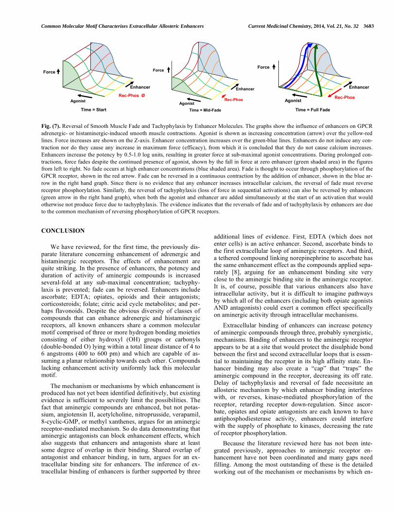
The mechanism proposed in Fig. (6) leads to an energetic model as well, which is illustrated in (Fig. 7). There are at least four factors that need to be considered in modeling the ability of enhancers to reverse fade and prevent tachyphy-laxis. One is the concentration of agonist. The second is the force of contraction (or relaxation) caused by the agonist at any given concentration. The third is the effect of any given concentration of enhancer. The enhancer effect will be, as proposed in (Fig. 6), opposite of the effect of phosphoryla-tion. A fourth effect is that of time. Fig. (7) illustrates how these four f actors interact to influence GPCR ardrenergic- or histaminergic-induced smooth muscle contractions (and by extrapolation, relaxations as well). As the agonist increases, the force increases to a maximum. Enhancers do not, them-selves, induce any contraction at any concentration, from which it is concluded that they do not cause calcium in-creases. Enhancers do increase the potency of agonists re-sulting in greater force at sub-maximal agonist concentra-tions. During prolonged contractions, in the absence of an

3682 Current Medicinal Chemistry, 2014, Vol. 21, No. 32 Root-Bernstein and Dillon
Fig. (6). Model of Enhancer Inhibition of GPCR Phosphorylation. Proposed model of enhancement of aminergic GPCR based on receptor allostery. Top Row: Binding of an amine to a GPCR normally results in allosteric changes in the receptor that initiate a series of molecular signals recruiting receptor kinases (indicated by the change of the intracellular loops from white to black and back again), which phosphory-late the receptor resulting in its down regulation and internalization. Bottom Row: We propose that binding of enhancers to GPCR results in prevention of aminergic release from the receptor accompanied by interference with the allosteric signaling (intracellular loops remain meta-phorically in the “black” state) that initiates receptor kinase recruitment. Retention of the amine in the binding site accompanied by retarda-tion of receptor phosphorylation results in prevention of tachyphylaxis. We also predict that during the first steps of receptor down-regulation binding of enhancers to the receptor can induce allosteric changes in the receptor that reverse receptor phosphorylation.; thereby also revers-ing fade. enhancer, force fades over time despite the continued pres-ence of agonist, but no fade occurs at high enhancer concen-trations. Since fade is thought to occur through phosphoryla-tion of the GPCR receptor, it may be presumed (as proposed above) that the enhancer blocks phosphorylation of the re-ceptor by some means. Notably, the energetic model shown in (Fig. 7) illustrates that there is no energetic barrier to re-versal of fade after a continuous contraction when an enhan-cer is added. The model also suggests that since there is no evidence that any enhancer increases intracellular calcium, the reversal of fade must reverse receptor phosphorylation, which is again consistent with the model proposed above. Similarly, the reversal of tachyphylaxis (loss of force in se-quential activations) can be reversed by enhancers when both the agonist and enhancer are added simultaneously at the
start of an activation that would otherwise not produce force. In sum, this energetic model is consistent with the evidence summarized above indicating that the reversals of fade and of tachyphylaxis by enhancers are due to the common mechanism of reversing phosphorylation of GPCR receptors.
The mechanism just proposed might also explain the oth-erwise counter-intuitive report that combinations of adrener-gic agonists and antagonists produce increased duration of activity and decreased tachyphylaxis [85]. As noted above, ascorbate is known to block several dopamine receptor an-tagonists, suggesting that these receptor antagonists bind at least partially into the enhancer site. Combinations of some adrenergic agonists and antagonists may therefore partially or wholly mimic the agonist-enhancer effect.
Common Molecular Motif Characterizes Extracellular Allosteric Enhancers Current Medicinal Chemistry, 2014, Vol. 21, No. 32 3683
Fig. (7). Reversal of Smooth Muscle Fade and Tachyphylaxis by Enhancer Molecules. The graphs show the influence of enhancers on GPCR adrenergic- or histaminergic-induced smooth muscle contractions. Agonist is shown as increasing concentration (arrow) over the yellow-red lines. Force increases are shown on the Z-axis. Enhancer concentration increases over the green-blue lines. Enhancers do not induce any con-traction nor do they cause any increase in maximum force (efficacy), from which it is concluded that they do not cause calcium increases. Enhancers increase the potency by 0.5-1.0 log units, resulting in greater force at sub-maximal agonist concentrations. During prolonged con-tractions, force fades despite the continued presence of agonist, shown by the fall in force at zero enhancer (green shaded area) in the figures from left to right. No fade occurs at high enhancer concentrations (blue shaded area). Fade is thought to occur through phosphorylation of the GPCR receptor, shown in the red arrow. Fade can be reversed in a continuous contraction by the addition of enhancer, shown in the blue ar-row in the right hand graph. Since there is no evidence that any enhancer increases intracellular calcium, the reversal of fade must reverse receptor phosphorylation. Similarly, the reversal of tachyphylaxis (loss of force in sequential activations) can also be reversed by enhancers (green arrow in the right hand graph), when both the agonist and enhancer are added simultaneously at the start of an activation that would otherwise not produce force due to tachyphylaxis. The evidence indicates that the reversals of fade and of tachyphylaxis by enhancers are due to the common mechanism of reversing phosphorylation of GPCR receptors.
CONCLUSION
We have reviewed, for the first time, the previously dis-parate literature concerning enhancement of adrenergic and histaminergic receptors. The effects of enhancement are quite striking. In the presence of enhancers, the potency and duration of activity of aminergic compounds is increased several-fold at any sub-maximal concentration; tachyphy-laxis is prevented; fade can be reversed. Enhancers include ascorbate; EDTA; opiates, opioids and their antagonists; corticosteroids; folate; citric acid cycle metabolites; and per-haps flavonoids. Despite the obvious diversity of classes of compounds that can enhance adrenergic and histaminergic receptors, all known enhancers share a common molecular motif comprised of three or more hydrogen bonding moieties consisting of either hydroxyl (OH) groups or carbonyls (double-bonded O) lying within a total linear distance of 4 to 6 angstroms (400 to 600 pm) and which are capable of as-suming a planar relationship towards each other. Compounds lacking enhancement activity uniformly lack this molecular motif.
The mechanism or mechanisms by which enhancement is produced has not yet been identified definitively, but existing evidence is sufficient to severely limit the possibilities. The fact that aminergic compounds are enhanced, but not potas-sium, angiotensin II, acetylcholine, nitroprusside, verapamil, 8-cyclic-GMP, or methyl xanthenes, argues for an aminergic receptor-mediated mechanism. So do data demonstrating that aminergic antagonists can block enhancement effects, which also suggests that enhancers and antagonists share at least some degree of overlap in their binding. Shared overlap of antagonist and enhancer binding, in turn, argues for an ex-tracellular binding site for enhancers. The inference of ex-tracellular binding of enhancers is further supported by three
additional lines of evidence. First, EDTA (which does not enter cells) is an active enhancer. Second, ascorbate binds to the first extracellular loop of aminergic receptors. And third, a tethered compound linking norepinephrine to ascorbate has the same enhancement effect as the compounds applied sepa-rately [8], arguing for an enhancement binding site very close to the aminergic binding site in the aminergic receptor. It is, of course, possible that various enhancers also have intracellular activity, but it is difficult to imagine pathways by which all of the enhancers (including both opiate agonists AND antagonists) could exert a common effect specifically on aminergic activity through intracellular mechanisms.
Extracellular binding of enhancers can increase potency of aminergic compounds through three, probably synergistic, mechanisms. Binding of enhancers to the aminergic receptor appears to be at a site that would protect the disulphide bond between the first and second extracellular loops that is essen-tial to maintaining the receptor in its high affinity state. En-hancer binding may also create a “cap” that “traps” the aminergic compound in the receptor, decreasing its off rate. Delay of tachyphylaxis and reversal of fade necessitate an allosteric mechanism by which enhancer binding interferes with, or reverses, kinase-mediated phosphorylation of the receptor, retarding receptor down-regulation. Since ascor-bate, opiates and opiate antagonists are each known to have antiphosphodiesterase activity, enhancers could interfere with the supply of phosphate to kinases, decreasing the rate of receptor phosphorylation.
Because the literature reviewed here has not been inte-grated previously, approaches to aminergic receptor en-hancement have not been coordinated and many gaps need filling. Among the most outstanding of these is the detailed working out of the mechanism or mechanisms by which en-
Force
Enhancer
Rec-Phos Ø Enhancer
Force
Rec-Phos Agonist
Force
Agonist Rec-Phos
Enhancer
Time = Start Time = Mid-Fade Time = Full Fade
Agonist

3684 Current Medicinal Chemistry, 2014, Vol. 21, No. 32 Root-Bernstein and Dillon
hancement is effected. No studies currently exist of the ef-fects of any of the enhancers described here on rates of adrenergic receptor internalization or phosphorylation. It is not known whether the initial steps of G-protein release fol-lowed by receptor phosphorylation can be reversed by addi-tion of enhancers, as is proposed here as a mechanism for reversing fade. It is not known whether the prevention of phosphorylation is the direct means by which enhancers re-tard tachyphylaxis. If these predictions are verified, it will still be necessary to work out how binding of an enhancer modifies changes in receptor structure and signaling to pre-vent phosphorylation. If these predictions are not verified, then another set of mechanisms consistent with the data re-viewed here will need to be postulated and tested. These are huge gaps to fill, but the research directions just suggested will be well worth the effort if they lead to the kinds of dra-matic breakthroughs in pharmacological treatment of dis-eases that followed the discovery of benzodiazapines.
CONFLICT OF INTEREST
The authors confirm that this article content has no con-flict of interest.
ACKNOWLEDGEMENTS
Declared none.
REFERENCES [1] Trumpp-Kallmeyer, S.; Hoflack , J.; Bruinvels, A.; Hibert, M.
Modeling of G-protein-coupled receptors: application to dopamine, adrenaline, serotonin, acetylcholine, and mammalian opsin recep-tors. J. Med. Chem., 1992, 35(19), 3448-3462.
[2] Bylund, D. B.; Eikenberg, D. C.; Hieble, J. P.; Langer, S. Z.; Lefkowitz, R. J.; Minneman, K. P.; Molinoff, P. B.; Ruffolo, R. R., Jr.; Trendelenburg, U. International Union of Pharmacology no-menclature of adrenoceptors. Pharmacol. Rev., 1994, 46, 121-136.
[3] Valant, C.; Aurelio, L.; Urmaliya, V. B.; White, P.; Scammells, P. J.; Sexton, P. M.; Christopoulos, A. Delineating the mode of action of adenosine A1 receptor allosteric modulators. Mol. Pharmacol., 2010, 78, 444-455.
[4] Gilchrist, A. Modulating G-protein-coupled receptors, from tradi-tional pharmacology to allosterics. Trends Pharmacol. Sci., 2007, 28, 431-437.
[5] Bridges, T. M.; Lindsley, C. W. G-protein-coupled receptors, from classical modes of modulation to allosteric mechanisms. A.C.S. Chem. Biol., 2008, 3, 530-541.
[6] Yanamala, N.; Klein-Seetharaman, J. Allosteric modulation of G protein coupled receptors by cytoplasmic, transmembrane and ex-tracellular ligands. Pharmaceuticals, 2010, 3, 3324-3342.
[7] May, L. T.; Avlani, V. A.; Sexton, P. M.; Christopoulos, A. Allos-teric modulation of G protein-coupled receptors. Curr. Pharm. Des., 2004, 10, 2003-2013.
[8] Root-Bernstein, R. S.; Dillon. P. F.; Hollingsworth, R. A tethered ascorbate-norepinephrine compound.; 4-UT.; displays long-acting adrenergic activity on rabbit aortic smooth muscle. Drug. Res. De-velop., 2008, 69, 242-250.
[9] Dillon, P. F.; Root-Bernstein, R. S.; Lieder, C. M. Antioxidant-independent ascorbate enhancement of catecholamine-induced con-tractions of vascular smooth muscle. Am. J. Physiol. Heart Circ. Physiol., 2004, 286, H2353-H2360.
[10] Dillon, P. F.; Root-Bernstein, R. S.; Lieder, C. M. Ascorbate en-hancement of H1 histamine receptor sensitivity coincides with ascorbate oxidation inhibition by histamine receptors. Am. J. Physiol. Cell. Physiol., 2006, 291, C977-C984.
[11] Dillon PF.; Root-Bernstein R.; Robinson NE.; Abraham WM.; Berney, C. Receptor-mediated enhancement of beta adrenergic drug activity by ascorbate in vitro and in vivo. PLoS One, 2010, 5, e15130.
[12] Root-Bernstein, R. S.; Dillon, P. F. Fostering adventure research. A case study of the discovery that ascorbic acid enhances adrenergic drug activity. Drug Develop. Res., 2002, 57, 58-74.
[13] Maxwell, L. C.; Herlihy, J. T.; Riedel, G.L. Effects of ascorbic acid and EDTA on vascular concentration-response to catecholamines. Microvasc. Res., 1983, 26, 81-88.
[14] Houston, J. B.; Wilkens, H. J.; Levy, G. Potentiation of isoprotere-nol effect by ascorbic acid. Res. Commun. Chem. Pathol. Pharma-col., 1976, 14, 643-650.
[15] Grossmann, M.; Dobrev, D.; Himmel, H. M.; Ravens, U.; Kirch, W. Ascorbic acid-induced modulation of venous tone in humans. Hypertension, 2001, 37, 949-954.
[16] Mak, S.; Newton, G. E. Vitamin C augments the inotropic response to dobutamine in humans with normal left ventricular function. Circulation, 2001, 103, 826-830.
[17] Shinke, T.; Shite, J.; Takaoka, H.; Hata, K.; Inoue, N.; Yoshikawa, R.; Matsumoto, H.; Masai, H.; Watanabe, S.; Ozawa, T.; Otake, H.; Matsumoto, D.; Hirata, K.; Yokoyama, M. Vitamin C restores the contractile response to dobutamine and improves myocardial effi-ciency in patients with heart failure after anterior myocardial in-farction. Am. Heart J., 2007, 154, 645.e1-645.e8.
[18] Monahan, K. D.; Eskurza, I.; Seals, D. R. Ascorbic acid increases cardiovagal baroreflex sensitivity in healthy older men. Am. J.
Physiol. Heart Circ. Physiol., 2004, 286, 6, H2113-H2117. [19] Jablonski, K. L.; Seals, D. R.; Eskurza, I.; Monahan, K. D.; Do-
nato, A. J. High-dose ascorbic acid infusion abolishes chronic vasoconstriction and restores resting leg blood flow in healthy older men. J. Appl. Physiol., 2007, 103, 1715-1721.
[20] Puri, S. K.; Cochin, J.; Volicer, L. Effect of morphine sulfate on adenylate cyclase and phosphodiesterase activities in rat corpus striatum. Life Sci., 1975, 16, 759-768.
[21] Marti, M. C. The effects of chronic morphine administration to mice on the contractile activity in vitro of the vas deferens without and with morphine. Eur. J. Pharmacol., 1982, 78, 439-447.
[22] Rae, G. A.; De Moraes, S. Supersensitivity to noradrenaline in vas deferens from morphine-dependent mice is confirmed. Eur. J. Pharmacol., 1983, 86, 347-352.
[23] Akabori, A.; Barraclough, C. A. Effects of morphine on LH secre-tion and catecholamine turnovers in the hypothalamus of estrogen-treated rats. Brain Res., 1986, 362, 221-226.
[24] Akabori, A.; Barraclough, C. A. Gonadotropin responses to naloxone may depend upon spontaneous activity in noradrenergic neurons at the time of treatment. Brain Res., 1986, 362, 55-62.
[25] He, J.-R.; Molnar, J.; Barraclough, C.A. Morphine amplifies nore-pinephrine , NE, -induced LH release but blocks NE-stimulated in-creases in LHRH mRNA levels, comparison of responses obtained in ovariectomized.; estrogen-treated normal and androgen-sterilized rats. Mol. Brain Res., 1993, 20, 71-78.
[26] Kindman, L/ A.; Kates, R. E.; Ginsburg, R. Opioids potentiate contractile response of rabbit myocardium to the beta adrenergic agonist isoproterenol. J. Cardiovasc. Pharmacol., 1991, 17, 61-67.
[27] Lechner, R. B.; Gurll, N. J.; Reynolds, D. G. Naloxone potentiates the cardiovascular effects of catecholamines in canine hemorrhagic shock. Circ. Shock , 1985, 16, 347-361.
[28] He, J.-R.; Barraclough, C. A. Morphine but not naloxone enhances luteinizing hormone-releasing hormone neuronal responsiveness to norepinephrine. J. Neuroendocrinol., 1992, 4, 92-99.
[29] Allgood, S. C.; Gurll, N. J.; Reynolds, D. G. Naloxone requires circulating catecholamines to attenuate the cardiovascular suppres-sion of endotoxic shock. J. Surg. Res., 1988, 44, 73-81.
[30] Caffrey, J. L.; Hathorne, L. F.; Carter, G. C.; Sinclair, R. J. Naloxone potentiates contractile responses to epinephrine in iso-lated canine arteries. Circ. Shock , 1990, 31, 317-332.
[31] Caffrey, J. L.; Stoll, S. T.; Sinclair, R. J.; Barron, B. A. +Naloxone enhances vascular contractile responses to added epinephrine. Prog. Clin. Biol. Res., 1990, 328, 375-378.
[32] Lechner , R. B. Naloxone potentiates inotropic but not chronotropic effects of isoproterenol in vitro. Circ. Shock, 1993, 39, 226-230.
[33] Gu, H.; Gaugl, J. F.; Barron, B. A.; Caffrey, J. L. Naloxone en-hances cardiac contractile responses to epinephrine without altering epinephrine uptake from plasma. Circ. Shock , 1990, 32, 257-271.
[34] Park, W. K.; Chang, C. H.; Chae, J. E.; Kim, M. H.; Cho, Y. L.; Ahn, D. S. Phosphodiesterase inhibition by naloxone augments the inotropic actions of beta-adrenergic stimulation. Acta Anaesthesiol. Scand., 2009, 53, 1043-1051.

Common Molecular Motif Characterizes Extracellular Allosteric Enhancers Current Medicinal Chemistry, 2014, Vol. 21, No. 32 3685
[35] McCubbin, J. A.; Surwit, R. S.; Kuhn, C. M.; Cochrane, C.; Fein-glos, M. N. Naltrexone potentiates glycemic responses during stress and epinephrine challenge in genetically obese mice. Psycho-
som. Med., 1989, 51, 441-448. [36] Parra, L.; Pérez-Vizcaíno, F.; Alsasua, A.; Martín, M. I.; Tamargo,
J. Mu- and delta-opioid receptor-mediated contractile effects on rat aortic vascular smooth muscle. Eur. J. Pharmacol., 1995, 277, 99-105.
[37] Lee, C. H.; Berkowitz, B. A. Stereoselective and calcium-dependent contractile effects of narcotic antagonist analgesics in the vascular smooth muscle of the rat. J. Pharmacol. Exp. Ther., 1976, 198, 347-356.
[38] Lee, C. H.; Berkowitz, B. A. Calcium antagonist activity of metha-done.; L-acetylmethadol and L-pentazocine in the rat aortic strip. J. Pharmacol. Exp. Ther., 1977, 202, 646-653.
[39] Deyo, S. N.; Swift, R. M.; Miller, R. J. Morphine and endorphins modulate dopamine turnover in rat median eminence. Proc. Natl.
Acad. Sci. U. S. A., 1979, 76, 3006-3009. [40] Deyo, S. N.; Swift, R. M.; Miller, R. J.; Fang, V. S. Development
of tolerance to the prolactin-releasing action of morphine and its modulation by hypothalamic dopamine. Endocrinology, 1980, 106, 1469-1474.
[41] Tagaya, E.; Tamaoki, J.; Chiyotani, A.; Konno, K. Stimulation of opioid mu-receptors potentiates beta adrenoceptor-mediated relaxa-tion of canine airway smooth muscle. J. Pharmacol. Exp. Ther., 1995, 275, 1288-1292.
[42] Purdy, R. E.; Weber, M. A. Enhancement and prolongation of vascular smooth muscle contraction by aldosterone. Blood Vessels, 1983, 20, 34-43.
[43] Purdy, R. E.; Weber, M. A.; Drayer, J.I. Vasoconstrictor effects of aldosterone in isolated vascular tissue. Clin. Exp. Hypertens. A., 1982, 4, 1583-1591.
[44] Weber, M. A.; Purdy, R. E.; Drayer, J. I. Interactions of mineralo-corticoids and pressor agents in vascular smooth muscle. Hypertension, 1983, 5, 141-146.
[45] Tan, K. S.; Grove, A.; McLean, A.; Gnosspelius, Y.; Hall, I. P.; Lipworth, B. J. , Systemic corticosteroid rapidly reverses bron-chodilator subsensitivity induced by formoterol in asthmatic pa-tients. Am. J. Respir. Crit. Care Med., 1997, 156, 28-35.
[46] Tan, K. S.; McFarlane, L. C.; Lipworth, B. J. Concomitant admini-stration of low-dose prednisolone protects against in vivo beta2-adrenoceptor subsensitivity induced by regular formoterol. Chest, 1998, 113, 34-41.
[47] Lipworth, B. J.; Aziz, I. Bronchodilator response to albuterol after formoterol and effects of acute corticosteroid administration. Chest, 2000, 117, 156-162.
[48] Spector, R. G. Influence of folic acid on noradrenaline stimulation of rat brain synaptosomes. Br. J. Pharmacol., 1971, 43, 438P.
[49] Spector, R. G. Effects of formyl tetrahydrofolic acid and noradrenaline on the oxygen consumption of rat brain synapto-some-mitochondrial preparations. Br. J. Pharmacol., 1972, 44, 279-85.
[50] Spector, R. G. Influence of folic acid on excitable tissues. Nat. New
Biol., 1972, 240, 247-249. [51] Bourne, M. G.; Sharma, S. K.; Spector, R. G. The effects of folate
on neurotransmitter uptake into rat cerebral cortex slices. Biochem. Pharmacol., 1976, 25, 1917-1918.
[52] Brocardo, P. S.; Budni, J.; Kaster, M. P.; Santos, A. R.; Rodrigues, A. L. Folic acid administration produces an antidepressant-like ef-fect in mice, evidence for the involvement of the serotenergic and noradrenergic systems. Neuropharmacology, 2008, 54, 464-473.
[53] Tejero-Taldo, M. I.; Sun, J.; Caffrey, J. L.; Mallet, R. T. Pyruvate potentiates -adrenergic inotropism of stunned guinea-pig myocar-dium. J. Mol. Cell. Cardiol., 1998, 30, 2327-2339.
[54] Tejero-Taldo, M. I.; Caffrey, J. L.; Sun, J.; Mallet, R. T. Antioxi-dant properties of pyruvate mediate its potentiation of -adrenergic inotropism in stunned myocardium. J. Mol. Cell. Cardiol., 1999, 31, 1863-1872.
[55] Squires, J. E.; Sun, J.; Caffrey, J. L.; Yoshishige, D.; Mallet, R. T. Acetoacetate augments adrenergic inotropism of stunned myocar-dium by an antioxidant mechanism. Am. J. Physiol. Heart Circ.
Physiol., 2003, 284, H1340-H1347. [56] Kuppusamy, U. R.; Das, N. P. Potentiation of beta-adrenoceptor
agonist-mediated lipolysis by quercetin and fisetin in isolated rat adipocytes. Biochem. Pharmacol., 1994, 47, 521-529.
[57] Naidu, P. S.; Singh, A.; Kulkarni, S. K. D2-dopamine receptor and alpha2-adrenoreceptor-mediated analgesic response of quercetin Indian J. Exp. Biol., 2003, 41, 1400-1404.
[58] Kaur, R.; Chopra, K.; Singh, D. Role of alpha2 receptors in quer-cetin-induced behavioral despair in mice. J. Med. Food, 2007, 10, 165-168.
[59] Qin, F.; Yan, C.; Patel, R.; Liu, W.; Dong, E. Vitamins C and E attenuate apoptosis.; beta-adrenergic receptor desensitization.; and sarcoplasmic reticular Ca2+ ATPase downregulation after myocar-dial infarction. Free Radic. Biol. Med., 2006, 40, 1827-1842.
[60] Aziz, I.; McFarlane, L. C.; Lipworth, B. J. Concomitant inhaled corticosteroid resensitises cardiac beta2-adrenoceptors in the pres-ence of long-acting beta2-agonist therapy. Eur. J. Clin. Pharma-
col., 1998, 54, 377-381. [61] Molenaar P.; Kompa A. R.; Roberts, S. J.; Pak, H. S.; Summers, R.
J. Localization of [125I]cyanopindolol binding in guinea-pig heart, characteristics of non-beta-adrenoceptor related binding in cardiac pacemaker and conducting regions. Neurosci. Lett., 1992, 136, 118-122.
[62] Self, G. J.; Rigby, P. J.; Passarelli, M. C.; Goldie, R. G. Localiza-tion of [125I]cyanopindolol binding in guinea-pig heart, characteris-tics of non- -adrenoceptor related binding in cardiac pacemaker and conducting regions. Eur. J. Pharmacol., 1990, 176, 169-176.
[63] Kimura, K.; Sidhu, A. Ascorbic acid inhibits 125I-SCH 23982 bind-ing but increases the affinity of dopamine for D1 dopamine recep-tors. J. Neurochem., 1994, 63, 2093-2098.
[64] Tolbert, L. C.; Morris, P. E., Jr.; Spollen, J. J.; Ashe, S. C. Stereo-specific effects of ascorbic acid and analogues on D.; and D.; ago-nist binding. Life Sci., 1992, 51, 921-930.
[65] Wiener, H. L.; Lajtha, A..; Sershen, H. Ascorbic acid inhibits I'H] SCH-23390 binding to striatal dopamine D.; receptors . J. Recept.
Res., 1989, 9, 331-339. [66] Newsholme, E. A.; Start, C. Regulation in Metabolism. John Wiley
& Sons, New York, 1981, pp. 16-17. [67] Rubenstein, L. A.; Lanzara, R. G. Activation of G protein-coupled
receptors entails cysteine modulation of agonist binding. J. Molec. Struc., 1998, 430, 57-71.
[68] Shi, L.; Javitch, J.A. The binding site of aminergic G protein-coupled receptors, the transmembrane segments and second ex-tracellular loop. Ann. Rev. Pharmacol. Toxicol., 2002, 42, 437-467.
[69] Rubenstein, L. A.; Zauhar, R. J.; Lanzara, R. G. Molecular dynam-ics of a biophysical model for beta2-adrenergic and G protein-coupled receptor activation. J. Mol. Graph. Model., 2006, 25, 396-409.
[70] Canals, M.; Sexton, P.M.; Christopoulos, A. Allostery in GPCRs: 'MWC' revisited. Trends Biochem. Sci., 2011, 36(12), 663-672.
[71] Johnston, J.M.; Wang, H.; Provasi, D.; Filizola, M. Assessing the relative stability of dimer interfaces in g protein-coupled receptors. PLoS Comput. Biol., 2012, 8(8), e1002649.
[72] Dorsch, S.; Klotz, K.N.; Engelhardt, S.; Lohse, M.J.; Bunemann, M. Analysis of receptor oligomerization by FRAP microscopy. Nat. Methods, 2009, 6, 225-230.
[73] Fung, J.J.; Deupi, X.; Pardo, L.; Yao, X.J.; Velez-Ruiz, G.A. Ligand regulated oligomerization of beta(2)-adrenoceptors in a model lipid bilayer. EMBO J., 2009, 28, 3315-3328.
[74] Huang, J.; Chen, S.; Zhang, J.J.; Huang, X.Y. Crystal structure of oligomeric 1-adrenergic G protein-coupled receptors in ligand-free basal state. Nat. Struct. Mol. Biol., 2013, 20(4), 419-425.
[75] May, L.T.; Leach, K.; Sexton, P.M.; Christopoulos, A. Allosteric modulation of G protein coupled receptors. Annu. Rev. Pharmacol. Toxicol., 2007, 47, 1-51.
[76] Keov, P.; Sexton, P.M.; Christopoulous, A. Allosteric modulation of G-protein coupled receptors: A pharmacological perspective. Neuropharmacology, 2011, 60, 24-35.
[77] Wooten, D.; Christopoulos, A.; Sexton, P.M. Emerging paradigms in GPCR allostery: implications for drug discovery. Nat. Rev. Drug Discov., 2013, 12, 630-643.
[78] Yanamala, N.; Klein-Seetharaman, J. Allosteric modulation of g protein coupled receptors by cytoplasmic, transmembrane and ex-tracellular ligands. Pharmaceuticals, 2010, 3, 3324-3342.
[79] Moffat, A. C.; Patterson, D. A.; Curry, A. S.; Owen, P. Inhibition in vitro of cyclic adenosine 3’4’-monophosphate phosphodiesterase activity by drugs. Eur. J. Toxicol., 1972, 5, 160-162.
[80] Gintzler, A. R.; Musacchio, J. M. Interactions of morphine.; adeno-sine.; adenosine triphosphate and phosphodiesterase inhibitors on
3686 Current Medicinal Chemistry, 2014, Vol. 21, No. 32 Root-Bernstein and Dillon
the field-stimulated guinea-pig ileum. J. Pharmacol. Exp. Ther., 1975, 194, 575-582.
[81] Buck, M. G.; Zadunaisky, J. A. Stimulation of ion transport by ascorbic acid through inhibition of 3', 5'-cyclic-AMP phosphodiesterase in the corneal epithelium and other tissues. Bio-
chim. Biophys. Acta, 1975, 389, 251-260. [82] Tisdale, M. J. Inhibition of cyclic adenosine 3’4’-monophosphate
phosphodiesterase from Walker carcinoma by ascorbic and dehy-droascorbic acid. Biochem. Biophys. Res. Commun., 1975, 62, 877-882.
[83] Malamud, D.; Kroll, Y. Ascorbic acid inhibition of cyclic nucleo-tide phosphodiesterase activity. Proc. Soc. Exp. Biol. Med., 1980,164, 534-536.
[84] Shinohara, K.; Fujiki, H.; Hidaka, Y.; Tseng, Y. K.; Murakami, H.; Omura, H. Effect of reductones on cyclic 3'.; 5'-adenosine mono-phosphate phosphodiesterase. Acta Vitaminol. Enzymol., 1985, 7, 99-108.
[85] Lanzara, R. Optimal agonist/antagonist combinations maintain receptor response by preventing rapid ß1-adrenergic receptor de-sensitization. Int. J. Pharmacol., 2005, 1, 122-131.
Received: October 16, 2013 Revised: April 08, 2014 Accepted: August 22, 2014
Copyright © 2022 FDOKUMEN