On the identification of FOXP2 gene enhancers and their role ...
243
On the identification of FOXP2 gene enhancers and their role in brain development Proefschrift ter verkrijging van de graad van doctor aan de Radboud Universiteit Nijmegen op gezag van de rector magnificus prof.dr. J.H.J.M. van Krieken, volgens besluit van het college van decanen in het openbaar te verdedigen op woensdag 26 oktober 2016 om 16:30 uur precies door Martin Becker geboren op 21 maart 1985 te Berlijn (Duitsland)
-
Upload
khangminh22 -
Category
Documents
-
view
1 -
download
0
Transcript of On the identification of FOXP2 gene enhancers and their role ...

On the identification of FOXP2 gene
enhancers and their role in brain
development
Proefschrift ter verkrijging van de graad van doctor
aan de Radboud Universiteit Nijmegen
op gezag van de rector magnificus prof.dr. J.H.J.M. van Krieken,
volgens besluit van het college van decanen
in het openbaar te verdedigen op
woensdag 26 oktober 2016
om 16:30 uur precies
door
Martin Becker geboren op 21 maart 1985
te Berlijn (Duitsland)

Promotor Prof. dr. Simon E. Fisher
Copromotor Dr. Sonja C. Vernes (Max Planck Instituut voor Psycholinguistiek)
Manuscriptcommissie Prof. dr. Hans H.L.M. van Bokhoven
Dr. Annette Schenck
Prof. dr. Wolfgang Enard (Ludwig-Maximilians- Universität München, Duitsland)

On the identification of FOXP2 gene
enhancers and their role in brain
development
Doctoral Thesis
to obtain the degree of doctor
from Radboud University Nijmegen
on the authority of the Rector Magnificus prof.dr. J.H.J.M. van Krieken,
according to the decision of the Council of Deans
to be defended in public on Wednesday, October 26, 2016
at 16:30 hours
by
Martin Becker Born on March 21, 1985
in Berlin (Germany)

Supervisor Prof. dr. Simon E. Fisher
Co-supervisor Dr. Sonja C. Vernes (Max Planck Institute for Psycholinguistics)
Doctoral Thesis Committee Prof. dr. Hans H.L.M. van Bokhoven
Dr. Annette Schenck
Prof. dr. Wolfgang Enard (Ludwig-Maximilians- Universität München, Germany)

"Take responsibility for making your own life beautiful."
By Timothy Leary, Your Brain Is God

© 2016 Martin Becker All rights reserved. No part of this thesis may be reproduced or printed in any form, by
any electronic or mechanical means, without written permission of the author.
Cover design: Antje Märtin, Martin Becker
Printed by: Ipskamp Drukkers, Enschede

1
Contents
Chapter 1:
General introduction……………………………………………………………………………………2
Chapter 2:
A chromosomal rearrangement in a child with severe speech and language disorder
separates FOXP2 from a functional enhancer …………………………………………………….. 31
Chapter 3:
Identification of neuronal FOXP2 enhancer contacts in human cell lines……………………….. 38
Chapter 4:
Upstream regulatory mechanisms acting at the promoters and enhancers of FOXP2………... 84
Chapter 5:
Activity of FOXP2 enhancers during development and in adult brains …………………………. 124
Chapter 6:
Early developmental gene enhancers affect subcortical volumes in the adult human brain….. 159
Chapter 7:
Summary and General discussion ………………………………………………………………….. 183
Appendix 1….……………………………………………………………………………………………… 207
Appendix 2…………………………………………………………………………………………………. 210
Dutch summary/Nederlandse samenvatting……………………………………………………………. 227
Curriculum Vitae…………………………………………………………………………………………… 231
List of publications…………………………………………………………………………………………. 232
Acknowledgements………………………………………………………………………………………... 233
MPI Series in Psycholinguistics………………………………………………………………………….. 234

Chapter 1: General introduction
2
Chapter 1
General introduction

Chapter 1: General introduction
3
Introduction
The study of genes in health and disease has dramatically increased our understanding of
human biology. The identification of mutated genes in diseases of the kidney, liver, muscle or
virtually any other human tissue enabled us to study the function of these tissues at molecular
and cellular levels. In this dissertation, I describe investigations of the FOXP2 gene, which has
been found to be mutated in people with speech and language problems. The relation between
FOXP2 and human language was first discovered nearly 15 years ago (Lai et al., 2001) and
has been described “as a molecular window into speech and language” (Fisher and Scharff,
2009). My aim was to change angles and look through that window at the molecular
mechanisms that precede FOXP2.
In this chapter I will introduce FOXP2, starting with the discovery of this gene in a family with
a speech and language disorder. I will describe the expression during development and in
adult brains and summarize the current knowledge regarding the downstream molecular and
cellular functions of FOXP2. Next, I will review the current literature on the upstream processes
that may regulate FOXP2 and show that this aspect of the FOXP2 story is not well understood.
At the end of this chapter, I will formulate the overarching question and aims of this dissertation.
Speech and language problems in people with FOXP2 mutations
In 1990, a large three-generation family was described, of which about 50 percent of the family
members presented with primary deficits in speech and language (Gopnik, 1990; Hurst et al.,
1990) (Figure 1). A major aspect of the disorder is developmental verbal dyspraxia (DVD), also
known as childhood apraxia of speech (CAS) (Hurst et al., 1990). The affected members of
this family, generally referred to as the KE family, have severe problems with articulation, as
well as poor processing and production of grammatical structures. The articulatory problems
involve difficulties in performing the rapid coordinated sequences of oral and facial movements
required for speech (Hurst et al., 1990; Vargha-Khadem et al., 1995). In addition to the orofacial
motor control problems, the affected members showed problems with language, including

Chapter 1: General introduction
4
reduced ability in applying grammatical rules governing tenses and plurals (Gopnik and Crago,
1991; Vargha-Khadem et al., 1995) and lower scores on tests for the understanding of
grammar (Vargha-Khadem et al., 1995). In terms of general cognition, both unaffected and
affected members scored on the lower side of the normal range of the general population.
However, a low IQ did not co-segregate with the speech and language disorder (Vargha-
Khadem et al., 1995). It has been argued by some that the speech and language problems in
this disorder stem from a core deficit affecting the coordination of orofacial motor-movements
(Vargha-Khadem et al., 2005).
Analyses of the phenotype of the KE family suggested that disrupted cognitive and neural
motor-control functions underlie the observed problems with speech and language
development. To identify affected brain regions in the KE family, brain imaging studies were
performed on healthy and affected members. In structural magnetic resonance imaging (MRI)
studies the grey matter volumes of the affected family members were compared to the
unaffected members and an unrelated control group, using voxel based morphometry (Vargha-
Khadem et al., 1998; Watkins et al., 2002; Belton et al., 2003). The volumes of the head of the
Figure 1: Pedigree of the KE family reproduced from Lai et al. 2001. Affected individuals are indicated by
filled symbols. Squares are males, circles are females, and a line through a symbol indicates that the person was
deceased in 2001.
Chapter 1: General introduction
5
caudate nucleus and cerebellar lobule VIIIB were consistently reduced in comparison to control
groups, whereas the volume of the putamen was increased. In addition, structural differences
were detected in cortical areas involved in motor control and language processing, including
increased volumes of the posterior superior temporal gyrus (including Wernicke’s area) and
angular gyrus, as well as decreased inferior frontal gyrus (including Broca’s area) and
precentral gyrus. The affected brain structures form cortico-striatal and cortico-cerebellar
circuits, which are important for planning and exerting motor movements (Middleton and Strick,
2000). In complementary functional brain studies, the caudate nucleus showed increased
activity in PET scans performed during word repetition tasks (Vargha-Khadem et al., 1998)
and the putamen reduced activity in fMRI scans during semantic language tasks, where the
subjects had to generate verbs in combination to heard nouns (Liegeois et al., 2003; Liegeois
et al., 2011). In addition, in these studies, under- and over-activation of cortical areas involved
in orofacial motor control, such as the precentral gyrus, and language-related cortical areas,
such as the inferior frontal gyrus, were detected (Vargha-Khadem et al., 1998; Liegeois et al.,
2003; Liegeois et al., 2011). Thus, structural and functional aberrations overlap in the basal
ganglia, cerebellum, motor-control area and language-related cortical areas in the affected KE
family members. Based on the observed effects in cortico-striatal and cortico-cerebellar
networks, it has been suggested that the speech and language phenotype result from problems
in planning and performing sequences of orofacial movements (Vargha-Khadem et al., 2005).
The mode of inheritance in the KE family suggested that a mutation in a single autosomal gene
might account for the speech and language disorder of this family. In 1998, the hypothesis of
an autosomal dominant locus was confirmed by molecular studies, and the location of the
affected gene was narrowed down to a region on chromosome 7 (Fisher et al., 1998). Using
genome-wide linkage analysis in the KE family, the authors identified a region on chromosomal
band 7q31 that perfectly segregated with affection status. Further clues to the location of the
likely damaged gene came from identification of an unrelated case, referred to as CS (Lai et
al., 2000), with a strikingly similar phenotype to that seen in the KE family. This child carried a

Chapter 1: General introduction
6
de novo balanced translocation involving reciprocal exchanges between chromosomes 7 and
5, with one breakpoint directly disrupting a newly identified gene on chromosome 7q31, in the
region of linkage identified in the KE family. This novel gene, given the name FOXP2, was
characterized and subsequently sequenced in the KE family, revealing a non-synonymous
mutation in all fifteen affected family members, which was not present in unaffected members
or healthy controls (Lai et al., 2001). The disruption of FOXP2 in CS and the KE family strongly
suggested that disruption of this gene was responsible for their speech and language deficits.
Indeed, the scientific literature now describes multiple independent cases with speech
problems and mutations affecting the coding sequence of FOXP2, including deletions of the
whole gene (Feuk et al., 2006; Zeesman et al., 2006; Lennon et al., 2007; Palka et al., 2012;
Rice et al., 2012; Zilina et al., 2012), chromosomal rearrangements disrupting the gene (Lai et
al., 2001; Feuk et al., 2006; Shriberg et al., 2006), missense mutations (Lai et al., 2001;
MacDermot et al., 2005), a nonsense mutation (MacDermot et al., 2005) and a two-nucleotide
intragenic deletion that yields a frameshift mutation and premature stop-codon (Turner et al.,
2013).
Neuronal expression of FOXP2 during development and in adult brains
The expression of the FOXP2 gene has been described in numerous species. To refer to
genes and gene products I will use the standardized nomenclature to distinguish between gene
orthologues and gene products (Wain et al., 2002). Genes, including their mRNA, are generally
written in italics and the proteins are referred to in non-italic style. Human gene symbols are
written in all capital letters (eg. FOXP2 gene, FOXP2 protein). According to standard
nomenclature, I will refer to mouse and rat genes with a starting capital letter (eg. Foxp2),
zebrafish genes with all lower case letters (foxp2) and zebra finch genes with a starting and
end capital letter (eg. FoxP2).
Foxp2 is expressed in multiple tissues including the central nervous system, gut, lung and
heart (Table 1) (Shu et al., 2001). Expression starts during embryonic development and

Chapter 1: General introduction
7
continues postnatally and into adulthood. Since the speech and language deficits in the KE
family relate to alterations in the central nervous system (as described above), I focus here on
reviewing the expression in the brain. First, I describe the expression pattern during embryonic
development and then continue on to detail the findings for postnatal development and in
adulthood. In addition I have summarized the findings of the systematic FOXP2/Foxp2
expression studies in mice and humans in table 1.
The earliest neural expression of FOXP2/Foxp2 can be detected in the developing medulla
oblongata at embryonic day 11.5 (E11.5) in mice and at Carnegie stage 23 (CS23) in humans
(Lai et al., 2003). At later embryonic stages the inferior olives of the medulla oblongata can be
identified as the FOXP2/Foxp2 positive nuclei. During further embryonic development Foxp2
mRNA was detected at E12.5 in the lateral ganglionic eminences (GE) and expression
remained in the GE-derived embryonic and postnatal striatum (Ferland et al., 2003). In
humans, FOXP2 mRNA can be detected at comparable developmental stages (CS23) in the
developing striatum (Lai et al., 2003). At E14.5 mRNA is detected in the cortical plate and
ventricular zone of mice (Ferland et al., 2003; Hisaoka et al., 2010). During embryonic and
post-natal development cortical mRNA expression can be detected in the cortical plate and
ventricular zone (Ferland et al., 2003; Lai et al., 2003). However, Foxp2 protein expression
was only detected in the cortical plate and was absent from the ventricular zone. Similarly, in
human prenatal brains FOXP2 expression can be detected in the subplate and inner cortical
plate (Miller et al., 2014). FOXP2/Foxp2 is not expressed in the hippocampal formation
(Ferland et al., 2003; Lai et al., 2003). Additional forebrain regions that showed strong
embryonic expression were the developing thalamus and hypothalamus in mouse embryos
(E13.5) and human foetuses (Ferland et al., 2003; Lai et al., 2003). Further expression was
detected in the amygdala and olfactory bulb and tubercule of mice (Ferland et al., 2003). In
addition to forebrain structures, expression was detected in developing mid- and hindbrain
regions (Ferland et al., 2003; Lai et al., 2003). Midbrain expression was detected in the
substantia nigra and tectum in mice (E16.5) and human at foetal stage 1 (FS1) (Lai et al.,

Chapter 1: General introduction
8
2003). In the hindbrain FOXP2/Foxp2 is expressed in the alar plate of the cerebellum in human
(CS23) and mice (E13.5) (Lai et al., 2003). With progressing embryonic development the
strong cerebellar expression was seen to be specific to the Purkinje cell layer in mice (Fujita
and Sugihara, 2012) and human (Lai et al., 2003). The deeper nuclei of the cerebellum show
weak FOXP2/Foxp2 expression (Lai et al., 2003; Fujita and Sugihara, 2012). In addition, Foxp2
expression was found in interneurons of the developing spinal cord (Morikawa et al., 2009).
Following the developmental expression, FOXP2/Foxp2 remains expressed during adulthood.
The adult cortical expression is mainly limited to the deeper cortical layer VI (Ferland et al.,
2003; Tsui et al., 2013). In mice, protein expression was also detected in layer V of the motor
and somatosensory cortex (Hisaoka et al., 2010). Foxp2 expression was absent from the
hippocampus and the three-layered paleocortex (Ferland et al., 2003). In the basal ganglia
Foxp2 is highly expressed in the caudate nucleus, putamen and globus pallidus of the striatum
(Ferland et al., 2003). Adult expression in the thalamus is strong in the auditory relay nucleus
of mice (Horng et al., 2009) and detectable in neuronal subtypes of the principal visual relay
nucleus (Iwai et al., 2013). Midbrain expression is detected in the substantia nigra and inferior
colliculi of the tectum (Ferland et al., 2003). Adult cerebral expression is specifically strong in
the Purkinje cells with weaker expression in deeper cerebellar nuclei (Ferland et al., 2003;
Fujita and Sugihara, 2012). Foxp2 expression in the brainstem is limited to the inferior olive
(Ferland et al., 2003; Fujita and Sugihara, 2012).

Chapter 1: General introduction
9
Table 1: FOXP2 expression studies in human and mice Structure Detail Species Age Molecule Ref.
Fore
brai
n/te
lenc
epha
lon
Cortex
inner intermediate zone of the neopallial cortex mouse
E12.5 to E16.5 mRNA 1
deeper layers of the cortical plate and subplate mouse E14.5 to birth
RNA, protein 2
cortical plate mouse newborn mRNA 3
restricted to cortical layer VI mouse postnatal RNA, protein 2
some layer V expression in very medial and posterior aspects of the cortex mouse postnatal
RNA, protein 2
Basal Ganglia
deep aspects of the ganglionic eminence mouse E12.5
RNA, protein 2
caudate nucleus mouse/human E13.5/CS23 mRNA 3 caudate putamen mouse/human E16.5/FS1 mRNA 3 substantia nigra mouse E16.5 mRNA 3 caudate nucleus mouse newborn mRNA 3 substantia nigra mouse newborn mRNA 3
striatum mouse adult RNA, protein 2
caudate-putamen, substantia nigra and ventral striatum mouse adult
RNA, protein 2
Olfactory system
olfactory bulb mouse adult RNA, protein 2
anterior olfactory nucleus mouse adult RNA, protein 2
olfactory tubercule mouse adult RNA, protein 2
septal nucleus mouse adult RNA, protein 2
Amygdala amygdala mouse adult
RNA, protein 2
Fore
brai
n/di
ence
phal
on
Thalamus
thalamus mouse/human E13.5/CS23 mRNA 3 thalamus mouse/human E16.5/FS1 mRNA 3 habenular nucleus mouse E16.5 mRNA 3 thalamic nuclei mouse newborn mRNA 3 thalamus (paraventricular, lateral posterior, dorsal thalamic nuclei, habenula, medial and lateral geniculate) mouse adult
RNA, protein 2
Hypothalamus
hypothalamus mouse/human E13.5/CS23 mRNA 3 hypothalamus mouse/human E16.5/FS1 mRNA 3 zona incerta mouse E16.5 mRNA 3 hypothalamus, paraventricular nucleus mouse adult
RNA, protein 2
Mid
brai
n
Tectum
inferior colliculus mouse E16.5 mRNA 3 inferior colliculus mouse newborn mRNA 3
superior and inferior colliculli mouse adult RNA, protein 2
Hin
dbra
in
Cerebellum
alar plate of the cerebellar primordium mouse/human E13.5/CS23 mRNA 3
cerebellum mouse E14.5 RNA, protein 2
developing cerebellum and cerebellar nuclei mouse/human E16.5/FS1 mRNA 3 piriform layer of cerebellum (PCs) mouse newborn mRNA 3

Chapter 1: General introduction
10
PCs of the cerebellum mouse mature cerebellum
RNA, protein 2
deep cerebellar nuclei mouse mature cerebellum
RNA, protein 2
Brainstem
precursor of medulla oblongata mouse E11.5 mRNA 3 midline of the hindbrain human CS18 mRNA 3 medullary raphe mouse/human E13.5/CS23 mRNA 3 medulla oblongata mouse/human E13.5/CS23 mRNA 3 medulla oblongata mouse/human E16.5/FS1 mRNA 3 lateral lemniscus nucleus mouse E16.5 mRNA 3 lemniscus nuclei mouse newborn mRNA 3 inferior olives of the medulla oblongata mouse newborn mRNA 3
inferior olive mouse adult RNA, protein 2
Spinal cord ventral interneurons of the spinal cord mouse
E12.5 to E16.5 mRNA 1
non-neuronal tissue
intestine mouse E12.5 to E16.5 mRNA 1
heart mouse E12.5 to E16.5 mRNA 1
lung epithelium mouse E12.5 to E16.5 mRNA 1
1: Shu et al. 2001
2: Ferland et al. 2003
3: Lai et al. 2003
The expression of FOXP2 overlaps with brain regions which have been noted to be structurally
and functionally aberrant in the KE family, including the basal ganglia, cerebellum and motor
cortical areas (Lai et al., 2003). Thus, the mutated variant of FOXP2 likely impairs the
development and function of these brain regions and associated neuronal networks. FOXP2
expression is present in additional brain regions, which did not show detectable changes in the
KE family. A single functional copy of FOXP2 may be sufficient for these structures to develop
and function normally or the influence of FOXP2 mutations may have been undetected by the
brain imaging studies. In addition, the affected KE family members have no reported problems
with the lung, heart or digestive tract in which FOXP2 is expressed, suggesting that one
functional copy of is sufficient in these tissues. In summary, FOXP2 is expressed in neuronal
subpopulations in multiple distinct brain regions, including the striatum, cerebellum and cortex.
The affected KE family members show structural and functional aberrations in these brain
regions, suggesting that mutations of FOXP2 impair the development of cortico-striatal and
cortico-cerebellar networks.

Chapter 1: General introduction
11
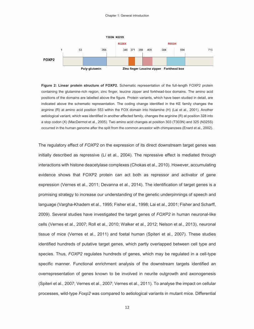
Protein structure and molecular function of FOXP2
The FOXP2 gene encodes a transcription factor (TF) that binds to DNA and regulates the
expression of other genes (Lai et al., 2001). The FOXP2 protein contains several known protein
domains: a glutamine-rich region, a zinc-finger, a leucine-zipper, a forkhead-box (FOX) DNA-
binding domain and a C-terminal acidic tail (Figure 2). The glutamine-rich domain of FOXP2
contains a short (10 glutamines) and a long (40 glutamines) stretch of contiguous glutamines;
such polyglutamine tracts are believed to mediate interactions with other proteins (Li et al.,
2004). The leucine-zipper domain of the FOXP2 protein enables it to form homo- and
heterodimers with other closely related TFs and the dimerized form of FOXP2 binds to DNA
(Li et al., 2004). Zinc-finger domains can mediate the interaction with a variety of molecules,
including DNA, RNA, protein and lipids (Laity et al., 2001). The function of the zinc-finger in
FOXP2 has not been studied in detail and, in one study, the deletion of this domain did not
affect the regulatory function in vitro (Li et al., 2004). The FOX domain is the defining feature
of the FOX family of TFs (Katoh and Katoh, 2004; Jackson et al., 2010) and binds to DNA in a
sequence-specific manner (Stroud et al., 2006; Vernes et al., 2007; Nelson et al., 2013). The
point mutation identified in the KE family affects the FOX domain and replaces an arginine at
the amino-acid position 553 with a histidine. The R553H variant of FOXP2 does not bind to the
consensus target DNA sequence (Lai et al., 2001; Vernes et al., 2006) or alternative motifs
(Nelson et al., 2013). Therefore, the mutation detected in the KE family disturbs the function
of the FOXP2 protein.
Chapter 1: General introduction
12
The regulatory effect of FOXP2 on the expression of its direct downstream target genes was
initially described as repressive (Li et al., 2004). The repressive effect is mediated through
interactions with histone deacetylase complexes (Chokas et al., 2010). However, accumulating
evidence shows that FOXP2 protein can act both as repressor and activator of gene
expression (Vernes et al., 2011; Devanna et al., 2014). The identification of target genes is a
promising strategy to increase our understanding of the genetic underpinnings of speech and
language (Vargha-Khadem et al., 1995; Fisher et al., 1998; Lai et al., 2001; Fisher and Scharff,
2009). Several studies have investigated the target genes of FOXP2 in human neuronal-like
cells (Vernes et al., 2007; Roll et al., 2010; Walker et al., 2012; Nelson et al., 2013), neuronal
tissue of mice (Vernes et al., 2011) and foetal human (Spiteri et al., 2007). These studies
identified hundreds of putative target genes, which partly overlapped between cell type and
species. Thus, FOXP2 regulates hundreds of genes, which may be regulated in a cell-type
specific manner. Functional enrichment analysis of the downstream targets identified an
overrepresentation of genes known to be involved in neurite outgrowth and axonogenesis
(Spiteri et al., 2007; Vernes et al., 2007; Vernes et al., 2011). To analyse the impact on cellular
processes, wild-type Foxp2 was compared to aetiological variants in mutant mice. Differential
Figure 2: Linear protein structure of FOXP2. Schematic representation of the full-length FOXP2 protein
containing the glutamine-rich region, zinc finger, leucine zipper and forkhead-box domains. The amino acid
positions of the domains are labelled above the figure. Protein variants, which have been studied in detail, are
indicated above the schematic representation. The coding change identified in the KE family changes the
arginine (R) at amino acid position 553 within the FOX domain into histamine (H) (Lai et al., 2001). Another
aetiological variant, which was identified in another affected family, changes the arginine (R) at position 328 into
a stop codon (X) (MacDermot et al., 2005). Two amino acid changes at position 303 (T303N) and 325 (N325S) occurred in the human genome after the split from the common ancestor with chimpanzees (Enard et al., 2002).

Chapter 1: General introduction
13
analysis of mouse primary neuronal cell cultures, homozygous for the R552H variant, which is
equivalent to the human R553H variant, revealed that the mutated variant impaired aspects of
neurite outgrowth, such as the number of branching sites and total neurite length (Vernes et
al., 2011). The human aetiological variant R328X, which was identified in another family
(MacDermot et al., 2005) is equivalent to S321X in mice and does not produce a protein
product, likely due to nonsense mediated-decay at the RNA level (Groszer et al., 2008). In
utero knock-down in the cortex of mouse embryos revealed that Foxp2 knock-down inhibits
neuronal differentiation and migration (Tsui et al., 2013). In addition, overexpression of FOXP2
in human neuronal-like cells increased neurite outgrowth and reduced cell migration (Devanna
et al., 2014). In conclusion, studies thus far suggest that FOXP2 is able to promote the
maturation of neurons through the support of neurite growth and reduced mobility. The
aetiological variants fail to regulate FOXP2 target genes, which results in impaired neuron
development.
The functions of FOXP2 orthologues in animal communication
The expression pattern of FOXP2 is highly conserved among vertebrates and has been
described in fish (Bonkowsky and Chien, 2005; Shah et al., 2006; Bonkowsky et al., 2008;
Itakura et al., 2008), crocodiles (Haesler et al., 2004), birds (Haesler et al., 2004; Teramitsu et
al., 2004; Chen et al., 2013), rodents (Ferland et al., 2003; Takahashi et al., 2003; Campbell
et al., 2009), carnivores (Rowell et al., 2010), monkeys (Takahashi et al., 2008b) and humans
(Lai et al., 2003). The conserved expression pattern makes animal studies a valuable strategy
to investigate the neuronal function of FOXP2. Foxp2 knockout and aetiological variants were
studied in mice to investigate the function of Foxp2 on brain development and behaviour (Shu
et al., 2005; French et al., 2007; Fujita et al., 2008; Groszer et al., 2008). Mutant mice have
been generated that carry either a conditional Foxp2 knockout (Foxp2-Flox) (French et al.,
2007), a complete knock-out (Foxp2-KO), a mouse equivalent of the aetiological R553H

Chapter 1: General introduction
14
mutation generated by a transgenic knock-in strategy (Foxp2-R552H-KI) (Fujita et al., 2008)
or mouse equivalents generated by chemical mutagenesis (Foxp2-R552H-ENU and Foxp2-
S321X) (Groszer et al., 2008). Homozygotes for aetiological mutations and homozygous
knock-out mice showed delayed development, reduced weight gain, severe motor problems
and died within 4 weeks after birth (Shu et al., 2005; French et al., 2007; Fujita et al., 2008;
Groszer et al., 2008). In addition, R552H and S321X homozygous mouse pups and the Foxp2-
flox, after global knock-out, presented with reduced cerebellar volume and intact cerebellar
cytoarchitecture (Fujita et al., 2008; Groszer et al., 2008). In contrast, the Foxp2-KO
homozygous mouse pups showed normal cerebellar volume, but abnormal cytoarchitecture of
the Purkinje cell layer and the external granular layer of the cerebellum (Shu et al., 2005). The
mouse studies demonstrate that complete loss of wild type Foxp2 is lethal at early post-natal
ages. In agreement with this finding, only heterozygous FOXP2 mutations have been detected
in humans.
Heterozygous mice were fully viable in all studies and did not show gross histological
anomalies (Shu et al., 2005; French et al., 2007; Fujita et al., 2008; Groszer et al., 2008).
Heterozygous Foxp2-KO mice presented with mild developmental delay with no differences
beyond postnatal day 15 (Shu et al., 2005). Mild developmental delay was also observed in
some Foxp2-R552H-KI heterozygotes (Fujita et al., 2008). However, the Foxp2-R552H-ENU
heterozygotes did not show developmental delay, but showed impaired long-term depression
in striatal neurons and reduced motor learning skills in comparison to wild-type littermates
(Groszer et al., 2008). Because human FOXP2 mutations affect speech and language, the
vocalizations of the mutant mice have been studied in more detail. Mice vocalize in the
ultrasonic frequency spectrum and ultrasonic vocalizations of homozygous and heterozygous
mice have been analysed in neonatal mouse pups, with debated results (French and Fisher,
2014). The innate nature of neonatal pup calls indicates that differences in mouse pup calls
are secondary to general developmental delays (Gaub et al., 2010). In male adult Foxp2-
R552H-ENU and Foxp2-S321X mice it was shown that disruption of Foxp2 produced altered
Chapter 1: General introduction
15
ultrasonic vocalizations in response to female/female urine (Gaub et al., 2016). In conclusion,
mouse studies indicate that heterozygous disruptions of Foxp2 have subtle effects on the
function and structure of Foxp2 positive brain regions. Furthermore, the aetiological variants
affect vocalization on cognitive and behavioural levels in adult mice.
In addition to the expression pattern, the amino-acid sequence of the protein encoded by
FOXP2 is highly conserved across mammals. The mouse and human orthologues of FOXP2
vary at three amino acid positions, two of which did arise in the human lineage after the split
from the chimpanzee (Enard et al., 2002; Zhang et al., 2002) and are present in Neanderthals
(Krause et al., 2007). The two substitutions are located in exon 7 outside of known protein
domains (Figure 2). Thus, the impact of the substitutions on protein function is unknown. The
presence of two amino-acid substitutions in this highly conserved protein suggested that the
substitutions may be of functional importance to human-specific traits (Enard et al., 2002). To
test the effect of human FOXP2 on mouse brain development, transgenic mice were generated
that carried the two human amino-acid changes within the endogenous Foxp2 locus (Enard et
al., 2009). Heterozygous and homozygous mice were both healthy and fertile. A set of
phenotypic tests revealed effects only on the explorative behaviour of the homozygous mice.
In Foxp2 positive brain regions, the dopamine levels were reduced and medium spiny neurons
in the striatum grew longer dendrites and exhibited increased long-term depression. In a follow-
up study, FOXP2 positive neurons in cortical layer VI and the thalamus also showed increased
neurite length (Reimers-Kipping et al., 2011). Thus, the human-specific protein-coding
changes of FOXP2 could have improved the connectivity of cortical basal-networks, which may
impact on cognition and behaviour. Indeed, it was further shown that the partially humanized
mice have improved declarative learning, as compared to wildtype littermates, which was
assessed in a T-maze paradigm with spatial cues (Schreiweis et al., 2014). The humanized
mice were also faster in switching from declarative to procedural learning. Taken together, the
findings suggest that the two amino acid changes, acquired during human evolution, alter the
connectivity of neuronal networks and may have improved sensorimotor learning processes.
Chapter 1: General introduction
16
The conserved expression pattern and protein structure of FOXP2 suggested that FOXP2
function is not only involved in human speech but may underlie animal communication. Results
supporting this hypothesis were obtained in studies conducted in zebra finch songbirds. Zebra
finches learn their songs from a tutor by imitation of the song structure. During the learning
process, the structure of the produced song varies during acquisition and stabilizes over time
to mimic the tutor song structure. The zebra finch orthologue of FOXP2, referred to as FoxP2,
is strongly expressed in striatal nucleus area X, which is important for song learning (Haesler
et al., 2004; Teramitsu et al., 2004). FoxP2 expression in this area was shown to change during
the vocal learning period in juvenile zebra finches (Haesler et al., 2004). Knockdown of FoxP2
in area X disrupted the ability of juvenile zebra finches to copy the tutor song, resulting in
incomplete and inaccurate production of song syllables (Haesler et al., 2007). To study the
effects of FoxP2 in adult zebra finches, the expression of FoxP2 in area X was compared
between males singing to a female (directed singing) and in the absence of a female
(undirected singing). The expression was found to be downregulated during undirected singing
of male zebra finches, as compared to non-singing males, indicating the regulation of FoxP2
expression during vocal behaviour (Teramitsu and White, 2006; Miller et al., 2008). In addition,
during directed singing the expression levels remained constant, indicating that the regulation
of FoxP2 also depends on the social context (Teramitsu and White, 2006). Analysis of the
neuronal song control circuits further indicated that FoxP2 knockdown alters the signalling
between area X and connected brain regions, which has been shown to be caused by
decreased dopamine signalling in area X (Murugan et al., 2013). Thus, striatal expression of
zebra finch FoxP2 is dynamically regulated during vocal communication, dependent on the
social context, and plays important functional roles in mediating plasticity of song structure.
Mouse studies investigating the functions of Foxp2 in auditory networks detected differential
expression in the auditory nucleus of the thalamus in response to auditory input (Horng et al.,
2009). Hence, dynamic expression of Foxp2 can be detected in mammals. Moreover,
aetiological Foxp2 mutations have been suggested to impair auditory-guided motor learning in

Chapter 1: General introduction
17
mice (Kurt et al., 2012). Taken together, the dynamic regulation of Foxp2 in the thalamus may
contribute to motor learning in mammals. As no techniques are available to study gene
expression in a non-invasive way, the active regulation of FOXP2 expression in post-natal
human brains has not been studied.
Regulation of FOXP2 expression
Signalling pathways upstream of FOXP2
FOXP2/Foxp2 can be used as a molecular marker for specific neuronal populations, such as
for cortical layer VI neurons (Chen et al., 2005; Hisaoka et al., 2010) or neurons derived from
induced pluripotent stem cells (iPSC) (Bickenbach et al., 2013; Espuny-Camacho et al., 2013;
Belinsky et al., 2014; Raitano et al., 2015). Consequently, temporal changes in Foxp2/FOXP2
expression have been described for these neuronal populations. One study investigated the
effect of histamine and expression of histamine type 1 receptor expression on the developing
rat cortex (Molina-Hernandez et al., 2013). Histamine-signalling is active during embryonic
neurodevelopment and regulates the proliferation of neuronal stem cells (Panula et al., 2014).
Panula et al. used Foxp2 as a marker for postmitotic deep layer cortical neurons and measured
the expression in cultured neurons isolated from rat embryos. The authors found that after
histamine treatment the proliferation of Foxp2-positive cells increased and that this effect was
dependent on histamine receptor 1.
Cultured neurons derived from human iPSCs can differentiate into FOXP2 positive neuronal
populations, using specific cell culture conditions and supplements (Bickenbach et al., 2013;
Espuny-Camacho et al., 2013; Chen et al., 2014; Raitano et al., 2015). For example, treatment
of cultured neurons with Noggin, a signalling protein important for embryonic development,
produced neurons with a cortical phenotype and a subpopulation of FOXP2 positive cortical
neurons (Espuny-Camacho et al., 2013). Thus, the upstream signalling pathways that may
activate FOXP2 expression can be studied in cultured human neurons. So far, the studies that

Chapter 1: General introduction
18
exploit human iPSC-derived neurons use FOXP2 as a neuronal marker and do not investigate
the signalling cascades that lead to FOXP2 expression. Accordingly, increased FOXP2
expression has been described in cultured neurons derived from patients with bipolar disorder
(Chen et al., 2014) and reduced expression in frontotemporal dementia (Raitano et al., 2015).
The nuclear factors and transcription factors that may have mediated the differential
expression, and possibly regulate FOXP2, were not investigated.
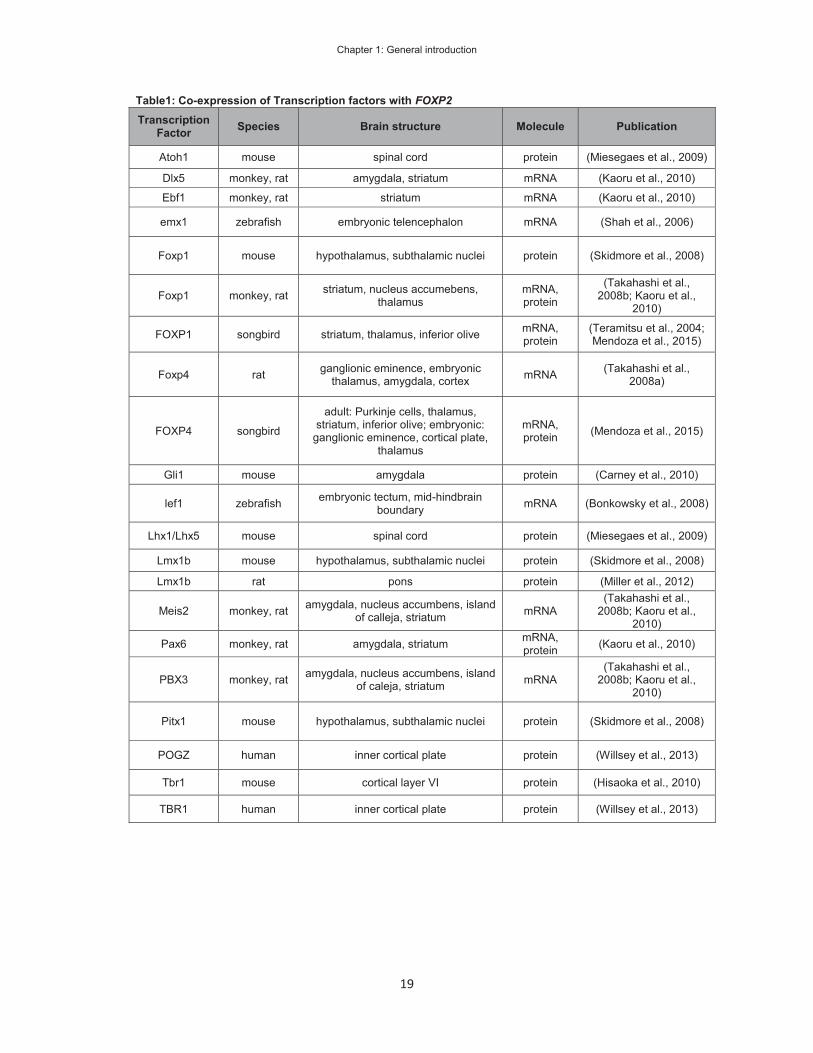
Transcription factors indicated to regulate FOXP2
Gene regulation in response to developmental programs or environmental cues is mediated
by TFs. The identification of upstream TFs is, therefore, a valuable approach to identify the
mechanisms preceding FOXP2 expression. The activation of FOXP2 expression during
development is of specific importance to understand the complex expression pattern. Co-
expression of TFs, which may indicate positive regulation, has been described in different
tissues and species. Table 1 shows a selection of co-expression studies, which include the
FOXP2 gene and other TFs. The co-expression of the listed TFs was determined on mRNA
and/or protein levels. The co-expression of two TFs can suggest hypotheses about their
function and relation, but experimental evidence for the interaction is required. The direct
regulation of FOXP2/foxp2 has been studied in detail for pax6 (Bonkowsky et al., 2008) and
lef1 in zebrafish (Coutinho et al., 2011) and for POU3F2 in human cell lines (Maricic et al.,
2013).
Chapter 1: General introduction
19
Table1: Co-expression of Transcription factors with FOXP2 Transcription
Factor Species Brain structure Molecule Publication
Atoh1 mouse spinal cord protein (Miesegaes et al., 2009)
Dlx5 monkey, rat amygdala, striatum mRNA (Kaoru et al., 2010)
Ebf1 monkey, rat striatum mRNA (Kaoru et al., 2010)
emx1 zebrafish embryonic telencephalon mRNA (Shah et al., 2006)
Foxp1 mouse hypothalamus, subthalamic nuclei protein (Skidmore et al., 2008)
Foxp1 monkey, rat striatum, nucleus accumebens, thalamus
mRNA, protein
(Takahashi et al., 2008b; Kaoru et al.,
2010)
FOXP1 songbird striatum, thalamus, inferior olive mRNA, protein
(Teramitsu et al., 2004; Mendoza et al., 2015)
Foxp4 rat ganglionic eminence, embryonic thalamus, amygdala, cortex mRNA (Takahashi et al.,
2008a)
FOXP4 songbird
adult: Purkinje cells, thalamus, striatum, inferior olive; embryonic:
ganglionic eminence, cortical plate, thalamus
mRNA, protein (Mendoza et al., 2015)
Gli1 mouse amygdala protein (Carney et al., 2010)
lef1 zebrafish embryonic tectum, mid-hindbrain boundary mRNA (Bonkowsky et al., 2008)
Lhx1/Lhx5 mouse spinal cord protein (Miesegaes et al., 2009)
Lmx1b mouse hypothalamus, subthalamic nuclei protein (Skidmore et al., 2008)
Lmx1b rat pons protein (Miller et al., 2012)
Meis2 monkey, rat amygdala, nucleus accumbens, island of calleja, striatum mRNA
(Takahashi et al., 2008b; Kaoru et al.,
2010)
Pax6 monkey, rat amygdala, striatum mRNA, protein (Kaoru et al., 2010)
PBX3 monkey, rat amygdala, nucleus accumbens, island of caleja, striatum mRNA
(Takahashi et al., 2008b; Kaoru et al.,
2010)
Pitx1 mouse hypothalamus, subthalamic nuclei protein (Skidmore et al., 2008)
POGZ human inner cortical plate protein (Willsey et al., 2013)
Tbr1 mouse cortical layer VI protein (Hisaoka et al., 2010)
TBR1 human inner cortical plate protein (Willsey et al., 2013)

Chapter 1: General introduction
20
One study investigated the regulation of foxp2 by pax6 in zebrafish (Coutinho et al., 2011).
PAX6 is a TF involved in early neurogenesis and brain patterning. The TF is expressed in the
developing neocortex, prethalamus and cerebellum (Osumi, 2001). In the zebrafish genome,
the foxp2 promoter was predicted to contain pax6 binding sites and Coutinho et al.
demonstrated the binding of pax6 to these sites (Coutinho et al., 2011). The element drove
reporter gene expression in foxp2-positive forebrain regions of zebrafish embryos. The in vivo
activity of this enhancer was dependent on pax6 expression. The results suggested that foxp2
is a target of pax6 and may suggest a link to the biological functions of pax6. Another study
investigated a link between lef1 and foxp2 in zebrafish (Bonkowsky et al., 2008). lef1 is a
transcription factor that regulates gene expression in response to WNT signalling. This
signalling pathway is important for cell-to-cell communication and the patterning of the central
nervous system during embryonic development. The authors characterized the expression of
lef1 and foxp2, and detected temporal overlap in the tectum and mid-hindbrain boundary in
developing zebrafish embryos, suggesting that lef1 might regulate foxp2. To test if there was
a direct regulatory effect, the authors ran in silico searches for lef1 binding sites near the foxp2
gene in the zebrafish genome and then determined if these elements drive gene expression in
embryonic zebrafish (Bonkowsky et al., 2008). Two of the predicted elements showed in vivo
enhancer activity and overlapped with foxp2 expression. These findings suggested that these
two enhancers regulated foxp2. The fact that foxp2 is a direct downstream target of lef1
suggests that foxp2 may be regulated by WNT-signalling.
Another regulatory relationship was suggested by investigating genetic changes, which
occurred in humans during recent evolutionary periods (Krause et al., 2007; Maricic et al.,
2013). Parts of the FOXP2 locus seem to have undergone positive selection after humans split
from Neanderthals. The positive selection was determined by the presence of modern human
alleles, which are fixed or occur at high frequency in humans and are absent in the Neanderthal
genome. Positive selection of these alleles could indicate that they are relevant for FOXP2
function in humans. One such human-specific substitution, which is located in intron 8 of
Chapter 1: General introduction
21
FOXP2 and occurred at an evolutionary conserved site, was predicted to reduce the binding
of POU3F2. Indeed, POU3F2 protein was shown to bind stronger to the Neanderthal allele
than to the human allele. In addition, the putative surrounding regulatory element carrying the
human allele showed reduced reporter gene expression in response to POU3F2
overexpression compared to the element carrying the Neanderthal allele. However, the
measured reporter gene expression was dependent on an additional viral enhancer,
suggesting that the tested regulatory element does not act as an independent enhancer. In
accordance to this, the element did not drive gene expression in transgenic mice. The study
by Maricic et al. suggests that POU3F2 regulates the expression of FOXP2 via an evolutionary
conserved site and that this regulatory interaction is reduced in humans.
Genetic variation and the regulation of FOXP2
Regulatory genomic elements control the timing and levels of gene expression. Consequently,
genetic changes in regulatory elements may alter gene expression and contribute to normal or
pathogenic phenotypic variation. The majority of trait-associated single nucleotide
polymorphisms (SNPs) identified in genome-wide association studies (GWAS) are located in
non-coding regions of the genome (Welter et al., 2014). Thus, associated SNPs near the
FOXP2 gene may be located within regulatory regions and affect the expression of this gene.
GWAS studies have identified associations between SNPs located near the FOXP2 gene with
a number of phenotypes, for example N-glycosylation of immunoglobin (Lauc et al., 2013),
Crohn’s disease (Julia et al., 2013), lymphoblast cell viability (de With et al., 2015), obesity
(Kim et al., 2013) smoking behaviour (Argos et al., 2014; Sung et al., 2015). These phenotypes
have no obvious link to motor-learning or communication. Thus, if the associated SNPs tag
regulatory regions, they do not seem to underlie FOXP2 regulation in relation to human speech
and language.
Candidate genetic association studies have investigated common genetic variants of FOXP2
in the context of human traits hypothesized to be related to FOXP2 function. To improve the

Chapter 1: General introduction
22
power to detect associations, most studies focused on a limited amount of genetic variants.
Studies have investigated associations of FOXP2 to auditory-visual hallucinations (Sanjuan et
al., 2006; McCarthy-Jones et al., 2014), schizophrenia (Tolosa et al., 2010; Spaniel et al.,
2011), ADHD (Ribases et al., 2012), autism (Park et al., 2014), dyslexia (Wilcke et al., 2012),
brain activation during reading tasks (Pinel et al., 2012) and enhanced language skills
(Chandrasekaran et al., 2015). The location of associated variants could indicate the presence
of regulatory elements. However, the low density of genetic markers would not allow for the
localization of regulatory elements. One study investigated genetic markers at the FOXP2
locus at high density, looking for association to brain volumes (Hoogman et al., 2014).
However, Hoogman et al. did not detect significant associations. In conclusion, candidate
association studies so far do not suggest the presence of regulatory SNPs near FOXP2. More
importantly, candidate association studies could benefit from including SNPs within regulatory
regions to improve the identification of genetic variants linked to normal variation in cognitive
traits.
Genetic variants in FOXP2 regulatory elements may also contribute to pathogenic variation.
Chromosomal rearrangements in the vicinity of FOXP2 have been described in clinical reports
of people presenting with speech impairments (Feuk et al., 2006; Moralli et al., 2015). These
genetic variants leave the protein coding sequence intact but may alter endogenous
expression. Chromosomal rearrangements have been detected in cases with speech
phenotypes similar to that observed in the KE family (Feuk et al., 2006; Adegbola et al., 2015;
Moralli et al., 2015). Moralli et al. described a child with late development of speech, who
carries an inversion of chromosome 7 and a breakpoint 200 kb downstream of FOXP2 (Moralli
et al., 2015). Feuk et al. described a patient with developmental delay with a deletion reported
to just start at the telomeric end of FOXP2 (Feuk et al., 2006). However, the authors do not
report the genomic location of the breakpoint. The assumed cause of the patient phenotypes
is an aberrant FOXP2 expression, caused by deleted or displaced regulatory elements.
However, the putative regulatory elements were not identified. The identification of these

Chapter 1: General introduction
23
regulatory sequences would help to clinically define these patients and investigate the
molecular aetiology of the observed phenotype.
Question and aims of this dissertation
Research into the function and the downstream molecular pathways of the FOXP2 gene has
proven to be highly valuable in investigating cognitive traits from molecule to behaviour.
Similarly, the pathways upstream of FOXP2 promise to increase our understanding of the
molecular mechanisms underlying speech and language. Consequently, my aim was to
investigate the regulation of FOXP2 expression in the brain and how the regulatory
mechanisms may contribute to human cognitive traits.
One of my aims, which is detailed in chapter 2, was to identify enhancers, which may have
contributed to the aetiology of delayed speech development in a child with a complex
chromosomal rearrangement (Moralli et al., 2015). The chromosomal rearrangement included
an inversion, with a breakpoint near the FOXP2 gene, suggesting this gene was involved in
the aetiology of the child’s speech phenotype. Using public data and molecular experiments I
located an enhancer just downstream of the inversion breakpoint, which would not be able to
regulate the FOXP2 gene in the child. This rare genetic variant allowed me to locate an
enhancer, which may regulate FOXP2 in healthy individuals.
In order for enhancers to regulate their target promoters they get into physical contact with the
promoter. Consequently, my aim in chapter 3 was to identify physical interactions of the
FOXP2 promoter with putative enhancers in FOXP2 expressing cells. I identified a number of
putative enhancers in the vicinity of FOXP2, as well as chromatin interactions to the
neighbouring gene promoter and the 3’UTR of FOXP2.
Enhancers, like promoters, are the integrative hubs for molecular mechanisms to control gene
expression during development or in response to environmental stimuli. Thus, my aim in
chapter 4 was to investigate the genetic pathways that regulate the FOXP2 enhancers. TFs,
as the effector proteins of signalling pathways, bind to promoters and enhancers. In a reporter

Chapter 1: General introduction
24
gene expression assay I determined the effect of TFs on the enhancers and promoters of
FOXP2.
The aim of chapter 5 was to characterize the activity of the most promising enhancers in
developing and adult brains. I created transgenic mice, carrying the human enhancers to
investigate the enhancers’ target brain regions and time of activity. In combination with the
previous chapters, the results combine aspects of the enhancer’s location, the enhancer-
interacting proteins, the brain structure in which these interactions may occur and the change
in enhancer activity during embryonic and post-natal development.
Following the results obtained in the earlier chapters, my aim in chapter 6 was to determine
how the effect of genetic variation within enhancers relates to normal variation in human traits.
To increase the statistical power of the analysis in this chapter, I analysed 296
neurodevelopmental enhancers from a public database, which were characterized with the
same method that I used to study the FOXP2 enhancers in chapter 5. In contrast to the
approach in chapter 2, where I investigated the rare genetic variation of a single case, chapter
6 investigated common genetic variants in 13,000 healthy individuals.
In chapter 7 I will summarize the findings from chapter 2 to 6 and put the results from each
chapter into relation to each other. The studies of FOXP2 enhancers shed light on the
upstream mechanism that control this gene and may contribute to the development of FOXP2
positive neurons and neuronal networks. The combination of the individual chapters enabled
me to generate new testable hypotheses on the regulation of human FOXP2.
Chapter 1: General introduction
25
References
Adegbola AA, Cox GF, Bradshaw EM, Hafler DA, Gimelbrant A, Chess A (2015) Monoallelic expression of the human FOXP2 speech gene. Proc Natl Acad Sci U S A 112:6848-6854.
Argos M, Tong L, Pierce BL, Rakibuz-Zaman M, Ahmed A, Islam T, Rahman M, Paul-Brutus R, Rahaman R, Roy S, Jasmine F, Kibriya MG, Ahsan H (2014) Genome-wide association study of smoking behaviours among Bangladeshi adults. Journal of medical genetics 51:327-333.
Belinsky GS, Rich MT, Sirois CL, Short SM, Pedrosa E, Lachman HM, Antic SD (2014) Patch-clamp recordings and calcium imaging followed by single-cell PCR reveal the developmental profile of 13 genes in iPSC-derived human neurons. Stem cell research 12:101-118.
Belton E, Salmond CH, Watkins KE, Vargha-Khadem F, Gadian DG (2003) Bilateral brain abnormalities associated with dominantly inherited verbal and orofacial dyspraxia. Human brain mapping 18:194-200.
Bickenbach JR, Tomanek-Chalkley A, Wiechert S, Winter MC (2013) Human skin keratinocytes can be reprogrammed to express neuronal genes and proteins after a single treatment with decitabine. BioResearch open access 2:217-221.
Bonkowsky JL, Chien CB (2005) Molecular cloning and developmental expression of foxP2 in zebrafish. Developmental dynamics : an official publication of the American Association of Anatomists 234:740-746.
Bonkowsky JL, Wang X, Fujimoto E, Lee JE, Chien CB, Dorsky RI (2008) Domain-specific regulation of foxP2 CNS expression by lef1. BMC developmental biology 8:103.
Campbell P, Reep RL, Stoll ML, Ophir AG, Phelps SM (2009) Conservation and diversity of Foxp2 expression in muroid rodents: functional implications. The Journal of comparative neurology 512:84-100.
Carney RS, Mangin JM, Hayes L, Mansfield K, Sousa VH, Fishell G, Machold RP, Ahn S, Gallo V, Corbin JG (2010) Sonic hedgehog expressing and responding cells generate neuronal diversity in the medial amygdala. Neural development 5:14.
Chandrasekaran B, Yi HG, Blanco NJ, McGeary JE, Maddox WT (2015) Enhanced procedural learning of speech sound categories in a genetic variant of FOXP2. The Journal of neuroscience : the official journal of the Society for Neuroscience 35:7808-7812.
Chen B, Schaevitz LR, McConnell SK (2005) Fezl regulates the differentiation and axon targeting of layer 5 subcortical projection neurons in cerebral cortex. Proc Natl Acad Sci U S A 102:17184-17189.
Chen HM, DeLong CJ, Bame M, Rajapakse I, Herron TJ, McInnis MG, O'Shea KS (2014) Transcripts involved in calcium signaling and telencephalic neuronal fate are altered in induced pluripotent stem cells from bipolar disorder patients. Transl Psychiat 4.
Chen Q, Heston JB, Burkett ZD, White SA (2013) Expression analysis of the speech-related genes FoxP1 and FoxP2 and their relation to singing behavior in two songbird species. The Journal of experimental biology 216:3682-3692.
Chokas AL, Trivedi CM, Lu MM, Tucker PW, Li S, Epstein JA, Morrisey EE (2010) Foxp1/2/4-NuRD interactions regulate gene expression and epithelial injury response in the lung via regulation of interleukin-6. J Biol Chem 285:13304-13313.
Coutinho P, Pavlou S, Bhatia S, Chalmers KJ, Kleinjan DA, van Heyningen V (2011) Discovery and assessment of conserved Pax6 target genes and enhancers. Genome Res 21:1349-1359.
de With SA, Pulit SL, Wang T, Staal WG, van Solinge WW, de Bakker PI, Ophoff RA (2015) Genome-wide association study of lymphoblast cell viability after clozapine exposure. American journal of medical genetics Part B, Neuropsychiatric genetics : the official publication of the International Society of Psychiatric Genetics 168B:116-122.
Devanna P, Middelbeek J, Vernes SC (2014) FOXP2 drives neuronal differentiation by interacting with retinoic acid signaling pathways. Frontiers in cellular neuroscience 8:305.
Enard W, Przeworski M, Fisher SE, Lai CS, Wiebe V, Kitano T, Monaco AP, Paabo S (2002) Molecular evolution of FOXP2, a gene involved in speech and language. Nature 418:869-872.
Enard W et al. (2009) A humanized version of Foxp2 affects cortico-basal ganglia circuits in mice. Cell 137:961-971.
Espuny-Camacho I, Michelsen KA, Gall D, Linaro D, Hasche A, Bonnefont J, Bali C, Orduz D, Bilheu A, Herpoel A, Lambert N, Gaspard N, Peron S, Schiffmann SN, Giugliano M, Gaillard A, Vanderhaeghen P (2013) Pyramidal neurons derived from human pluripotent stem cells integrate efficiently into mouse brain circuits in vivo. Neuron 77:440-456.
Chapter 1: General introduction
26
Ferland RJ, Cherry TJ, Preware PO, Morrisey EE, Walsh CA (2003) Characterization of Foxp2 and Foxp1 mRNA and protein in the developing and mature brain. The Journal of comparative neurology 460:266-279.
Feuk L et al. (2006) Absence of a paternally inherited FOXP2 gene in developmental verbal dyspraxia. Am J Hum Genet 79:965-972.
Fisher SE, Scharff C (2009) FOXP2 as a molecular window into speech and language. Trends Genet 25:166-177.
Fisher SE, Vargha-Khadem F, Watkins KE, Monaco AP, Pembrey ME (1998) Localisation of a gene implicated in a severe speech and language disorder. Nat Genet 18:168-170.
French CA, Fisher SE (2014) What can mice tell us about Foxp2 function? Current opinion in neurobiology 28:72-79.
French CA, Groszer M, Preece C, Coupe AM, Rajewsky K, Fisher SE (2007) Generation of mice with a conditional Foxp2 null allele. Genesis 45:440-446.
Fujita E, Tanabe Y, Shiota A, Ueda M, Suwa K, Momoi MY, Momoi T (2008) Ultrasonic vocalization impairment of Foxp2 (R552H) knockin mice related to speech-language disorder and abnormality of Purkinje cells. Proc Natl Acad Sci U S A 105:3117-3122.
Fujita H, Sugihara I (2012) FoxP2 expression in the cerebellum and inferior olive: development of the transverse stripe-shaped expression pattern in the mouse cerebellar cortex. The Journal of comparative neurology 520:656-677.
Gaub S, Fisher SE, Ehret G (2016) Ultrasonic vocalizations of adult male Foxp2-mutant mice: behavioral contexts of arousal and emotion. Genes Brain Behav 15:243-259.
Gaub S, Groszer M, Fisher SE, Ehret G (2010) The structure of innate vocalizations in Foxp2-deficient mouse pups. Genes Brain and Behavior 9:390-401.
Gopnik M (1990) Feature-blind grammar and dysphagia. Nature 344:715. Gopnik M, Crago MB (1991) Familial aggregation of a developmental language disorder. Cognition 39:1-
50. Groszer M et al. (2008) Impaired synaptic plasticity and motor learning in mice with a point mutation
implicated in human speech deficits. Current biology : CB 18:354-362. Haesler S, Rochefort C, Georgi B, Licznerski P, Osten P, Scharff C (2007) Incomplete and inaccurate
vocal imitation after knockdown of FoxP2 in songbird basal ganglia nucleus Area X. PLoS biology 5:e321.
Haesler S, Wada K, Nshdejan A, Morrisey EE, Lints T, Jarvis ED, Scharff C (2004) FoxP2 expression in avian vocal learners and non-learners. The Journal of neuroscience : the official journal of the Society for Neuroscience 24:3164-3175.
Hisaoka T, Nakamura Y, Senba E, Morikawa Y (2010) The forkhead transcription factors, Foxp1 and Foxp2, identify different subpopulations of projection neurons in the mouse cerebral cortex. Neuroscience 166:551-563.
Hoogman M, Guadalupe T, Zwiers MP, Klarenbeek P, Francks C, Fisher SE (2014) Assessing the effects of common variation in the FOXP2 gene on human brain structure. Frontiers in human neuroscience 8:473.
Horng S, Kreiman G, Ellsworth C, Page D, Blank M, Millen K, Sur M (2009) Differential gene expression in the developing lateral geniculate nucleus and medial geniculate nucleus reveals novel roles for Zic4 and Foxp2 in visual and auditory pathway development. The Journal of neuroscience : the official journal of the Society for Neuroscience 29:13672-13683.
Hurst JA, Baraitser M, Auger E, Graham F, Norell S (1990) An extended family with a dominantly inherited speech disorder. Developmental medicine and child neurology 32:352-355.
Itakura T, Chandra A, Yang Z, Xue X, Wang B, Kimura W, Hikosaka K, Inohaya K, Kudo A, Uezato T, Miura N (2008) The medaka FoxP2, a homologue of human language gene FOXP2, has a diverged structure and function. Journal of biochemistry 143:407-416.
Iwai L, Ohashi Y, van der List D, Usrey WM, Miyashita Y, Kawasaki H (2013) FoxP2 is a parvocellular-specific transcription factor in the visual thalamus of monkeys and ferrets. Cerebral cortex 23:2204-2212.
Jackson BC, Carpenter C, Nebert DW, Vasiliou V (2010) Update of human and mouse forkhead box (FOX) gene families. Human genomics 4:345-352.
Julia A et al. (2013) A genome-wide association study on a southern European population identifies a new Crohn's disease susceptibility locus at RBX1-EP300. Gut 62:1440-1445.
Kaoru T, Liu FC, Ishida M, Oishi T, Hayashi M, Kitagawa M, Shimoda K, Takahashi H (2010) Molecular characterization of the intercalated cell masses of the amygdala: implications for the relationship with the striatum. Neuroscience 166:220-230.

Chapter 1: General introduction
27
Katoh M, Katoh M (2004) Human FOX gene family (Review). International journal of oncology 25:1495-1500.
Kim HJ, Yoo YJ, Ju YS, Lee S, Cho SI, Sung J, Kim JI, Seo JS (2013) Combined linkage and association analyses identify a novel locus for obesity near PROX1 in Asians. Obesity 21:2405-2412.
Krause J, Lalueza-Fox C, Orlando L, Enard W, Green RE, Burbano HA, Hublin JJ, Hanni C, Fortea J, de la Rasilla M, Bertranpetit J, Rosas A, Paabo S (2007) The derived FOXP2 variant of modern humans was shared with Neandertals. Current biology : CB 17:1908-1912.
Kurt S, Fisher SE, Ehret G (2012) Foxp2 mutations impair auditory-motor association learning. PloS one 7:e33130.
Lai CS, Fisher SE, Hurst JA, Vargha-Khadem F, Monaco AP (2001) A forkhead-domain gene is mutated in a severe speech and language disorder. Nature 413:519-523.
Lai CS, Gerrelli D, Monaco AP, Fisher SE, Copp AJ (2003) FOXP2 expression during brain development coincides with adult sites of pathology in a severe speech and language disorder. Brain : a journal of neurology 126:2455-2462.
Lai CS, Fisher SE, Hurst JA, Levy ER, Hodgson S, Fox M, Jeremiah S, Povey S, Jamison DC, Green ED, Vargha-Khadem F, Monaco AP (2000) The SPCH1 region on human 7q31: genomic characterization of the critical interval and localization of translocations associated with speech and language disorder. Am J Hum Genet 67:357-368.
Laity JH, Lee BM, Wright PE (2001) Zinc finger proteins: new insights into structural and functional diversity. Current opinion in structural biology 11:39-46.
Lauc G et al. (2013) Loci associated with N-glycosylation of human immunoglobulin G show pleiotropy with autoimmune diseases and haematological cancers. PLoS Genet 9:e1003225.
Lennon PA, Cooper ML, Peiffer DA, Gunderson KL, Patel A, Peters S, Cheung SW, Bacino CA (2007) Deletion of 7q31.1 supports involvement of FOXP2 in language impairment: clinical report and review. American journal of medical genetics Part A 143A:791-798.
Li S, Weidenfeld J, Morrisey EE (2004) Transcriptional and DNA binding activity of the Foxp1/2/4 family is modulated by heterotypic and homotypic protein interactions. Molecular and cellular biology 24:809-822.
Liegeois F, Morgan AT, Connelly A, Vargha-Khadem F (2011) Endophenotypes of FOXP2: dysfunction within the human articulatory network. European journal of paediatric neurology : EJPN : official journal of the European Paediatric Neurology Society 15:283-288.
Liegeois F, Baldeweg T, Connelly A, Gadian DG, Mishkin M, Vargha-Khadem F (2003) Language fMRI abnormalities associated with FOXP2 gene mutation. Nat Neurosci 6:1230-1237.
MacDermot KD, Bonora E, Sykes N, Coupe AM, Lai CS, Vernes SC, Vargha-Khadem F, McKenzie F, Smith RL, Monaco AP, Fisher SE (2005) Identification of FOXP2 truncation as a novel cause of developmental speech and language deficits. Am J Hum Genet 76:1074-1080.
Maricic T, Gunther V, Georgiev O, Gehre S, Curlin M, Schreiweis C, Naumann R, Burbano HA, Meyer M, Lalueza-Fox C, de la Rasilla M, Rosas A, Gajovic S, Kelso J, Enard W, Schaffner W, Paabo S (2013) A recent evolutionary change affects a regulatory element in the human FOXP2 gene. Molecular biology and evolution 30:844-852.
McCarthy-Jones S, Green MJ, Scott RJ, Tooney PA, Cairns MJ, Wu JQ, Oldmeadow C, Carr V, Australian Schizophrenia Research B (2014) Preliminary evidence of an interaction between the FOXP2 gene and childhood emotional abuse predicting likelihood of auditory verbal hallucinations in schizophrenia. Journal of psychiatric research 50:66-72.
Mendoza E, Tokarev K, During DN, Retamosa EC, Weiss M, Arpenik N, Scharff C (2015) Differential coexpression of FoxP1, FoxP2, and FoxP4 in the Zebra Finch (Taeniopygia guttata) song system. The Journal of comparative neurology 523:1318-1340.
Middleton FA, Strick PL (2000) Basal ganglia and cerebellar loops: motor and cognitive circuits. Brain research Brain research reviews 31:236-250.
Miesegaes GR, Klisch TJ, Thaller C, Ahmad KA, Atkinson RC, Zoghbi HY (2009) Identification and subclassification of new Atoh1 derived cell populations during mouse spinal cord development. Developmental biology 327:339-351.
Miller JA et al. (2014) Transcriptional landscape of the prenatal human brain. Nature 508:199-206. Miller JE, Spiteri E, Condro MC, Dosumu-Johnson RT, Geschwind DH, White SA (2008) Birdsong
decreases protein levels of FoxP2, a molecule required for human speech. Journal of neurophysiology 100:2015-2025.
Miller RL, Knuepfer MM, Wang MH, Denny GO, Gray PA, Loewy AD (2012) Fos-activation of FoxP2 and Lmx1b neurons in the parabrachial nucleus evoked by hypotension and hypertension in conscious rats. Neuroscience 218:110-125.

Chapter 1: General introduction
28
Molina-Hernandez A, Rodriguez-Martinez G, Escobedo-Avila I, Velasco I (2013) Histamine up-regulates fibroblast growth factor receptor 1 and increases FOXP2 neurons in cultured neural precursors by histamine type 1 receptor activation: conceivable role of histamine in neurogenesis during cortical development in vivo. Neural development 8:4.
Moralli D, Nudel R, Chan MT, Green CM, Volpi EV, Benitez-Burraco A, Newbury DF, Garcia-Bellido P (2015) Language impairment in a case of a complex chromosomal rearrangement with a breakpoint downstream of FOXP2. Molecular cytogenetics 8:36.
Morikawa Y, Hisaoka T, Senba E (2009) Characterization of Foxp2-expressing cells in the developing spinal cord. Neuroscience 162:1150-1162.
Murugan M, Harward S, Scharff C, Mooney R (2013) Diminished FoxP2 levels affect dopaminergic modulation of corticostriatal signaling important to song variability. Neuron 80:1464-1476.
Nelson CS, Fuller CK, Fordyce PM, Greninger AL, Li H, DeRisi JL (2013) Microfluidic affinity and ChIP-seq analyses converge on a conserved FOXP2-binding motif in chimp and human, which enables the detection of evolutionarily novel targets. Nucleic Acids Res 41:5991-6004.
Osumi N (2001) The role of Pax6 in brain patterning. The Tohoku journal of experimental medicine 193:163-174.
Palka C, Alfonsi M, Mohn A, Cerbo R, Guanciali Franchi P, Fantasia D, Morizio E, Stuppia L, Calabrese G, Zori R, Chiarelli F, Palka G (2012) Mosaic 7q31 deletion involving FOXP2 gene associated with language impairment. Pediatrics 129:e183-188.
Panula P, Sundvik M, Karlstedt K (2014) Developmental roles of brain histamine. Trends in neurosciences 37:159-168.
Park Y, Won S, Nam M, Chung JH, Kwack K (2014) Interaction between MAOA and FOXP2 in association with autism and verbal communication in a Korean population. Journal of child neurology 29:NP207-211.
Pinel P, Fauchereau F, Moreno A, Barbot A, Lathrop M, Zelenika D, Le Bihan D, Poline JB, Bourgeron T, Dehaene S (2012) Genetic variants of FOXP2 and KIAA0319/TTRAP/THEM2 locus are associated with altered brain activation in distinct language-related regions. The Journal of neuroscience : the official journal of the Society for Neuroscience 32:817-825.
Raitano S, Ordovas L, De Muynck L, Guo W, Espuny-Camacho I, Geraerts M, Khurana S, Vanuytsel K, Toth BI, Voets T, Vandenberghe R, Cathomen T, Van Den Bosch L, Vanderhaeghen P, Van Damme P, Verfaillie CM (2015) Restoration of progranulin expression rescues cortical neuron generation in an induced pluripotent stem cell model of frontotemporal dementia. Stem cell reports 4:16-24.
Reimers-Kipping S, Hevers W, Paabo S, Enard W (2011) Humanized Foxp2 specifically affects cortico-basal ganglia circuits. Neuroscience 175:75-84.
Ribases M, Sanchez-Mora C, Ramos-Quiroga JA, Bosch R, Gomez N, Nogueira M, Corrales M, Palomar G, Jacob CP, Gross-Lesch S, Kreiker S, Reif A, Lesch KP, Cormand B, Casas M, Bayes M (2012) An association study of sequence variants in the forkhead box P2 (FOXP2) gene and adulthood attention-deficit/hyperactivity disorder in two European samples. Psychiatric genetics 22:155-160.
Rice GM, Raca G, Jakielski KJ, Laffin JJ, Iyama-Kurtycz CM, Hartley SL, Sprague RE, Heintzelman AT, Shriberg LD (2012) Phenotype of FOXP2 haploinsufficiency in a mother and son. American journal of medical genetics Part A 158A:174-181.
Roll P, Vernes SC, Bruneau N, Cillario J, Ponsole-Lenfant M, Massacrier A, Rudolf G, Khalife M, Hirsch E, Fisher SE, Szepetowski P (2010) Molecular networks implicated in speech-related disorders: FOXP2 regulates the SRPX2/uPAR complex. Human molecular genetics 19:4848-4860.
Rowell JJ, Mallik AK, Dugas-Ford J, Ragsdale CW (2010) Molecular analysis of neocortical layer structure in the ferret. The Journal of comparative neurology 518:3272-3289.
Sanjuan J, Tolosa A, Gonzalez JC, Aguilar EJ, Perez-Tur J, Najera C, Molto MD, de Frutos R (2006) Association between FOXP2 polymorphisms and schizophrenia with auditory hallucinations. Psychiatric genetics 16:67-72.
Schreiweis C, Bornschein U, Burguiere E, Kerimoglu C, Schreiter S, Dannemann M, Goyal S, Rea E, French CA, Puliyadi R, Groszer M, Fisher SE, Mundry R, Winter C, Hevers W, Paabo S, Enard W, Graybiel AM (2014) Humanized Foxp2 accelerates learning by enhancing transitions from declarative to procedural performance. Proc Natl Acad Sci U S A 111:14253-14258.
Shah R, Medina-Martinez O, Chu LF, Samaco RC, Jamrich M (2006) Expression of FoxP2 during zebrafish development and in the adult brain. The International journal of developmental biology 50:435-438.

Chapter 1: General introduction
29
Shriberg LD, Ballard KJ, Tomblin JB, Duffy JR, Odell KH, Williams CA (2006) Speech, prosody, and voice characteristics of a mother and daughter with a 7;13 translocation affecting FOXP2. Journal of speech, language, and hearing research : JSLHR 49:500-525.
Shu W, Yang H, Zhang L, Lu MM, Morrisey EE (2001) Characterization of a new subfamily of winged-helix/forkhead (Fox) genes that are expressed in the lung and act as transcriptional repressors. J Biol Chem 276:27488-27497.
Shu W, Cho JY, Jiang Y, Zhang M, Weisz D, Elder GA, Schmeidler J, De Gasperi R, Sosa MA, Rabidou D, Santucci AC, Perl D, Morrisey E, Buxbaum JD (2005) Altered ultrasonic vocalization in mice with a disruption in the Foxp2 gene. Proc Natl Acad Sci U S A 102:9643-9648.
Skidmore JM, Cramer JD, Martin JF, Martin DM (2008) Cre fate mapping reveals lineage specific defects in neuronal migration with loss of Pitx2 function in the developing mouse hypothalamus and subthalamic nucleus. Molecular and cellular neurosciences 37:696-707.
Spaniel F, Horacek J, Tintera J, Ibrahim I, Novak T, Cermak J, Klirova M, Hoschl C (2011) Genetic variation in FOXP2 alters grey matter concentrations in schizophrenia patients. Neuroscience letters 493:131-135.
Spiteri E, Konopka G, Coppola G, Bomar J, Oldham M, Ou J, Vernes SC, Fisher SE, Ren B, Geschwind DH (2007) Identification of the transcriptional targets of FOXP2, a gene linked to speech and language, in developing human brain. Am J Hum Genet 81:1144-1157.
Stroud JC, Wu Y, Bates DL, Han A, Nowick K, Paabo S, Tong H, Chen L (2006) Structure of the forkhead domain of FOXP2 bound to DNA. Structure 14:159-166.
Sung YJ, de Las Fuentes L, Schwander KL, Simino J, Rao DC (2015) Gene-smoking interactions identify several novel blood pressure loci in the Framingham Heart Study. American journal of hypertension 28:343-354.
Takahashi K, Liu FC, Hirokawa K, Takahashi H (2003) Expression of Foxp2, a gene involved in speech and language, in the developing and adult striatum. Journal of neuroscience research 73:61-72.
Takahashi K, Liu FC, Hirokawa K, Takahashi H (2008a) Expression of Foxp4 in the developing and adult rat forebrain. Journal of neuroscience research 86:3106-3116.
Takahashi K, Liu FC, Oishi T, Mori T, Higo N, Hayashi M, Hirokawa K, Takahashi H (2008b) Expression of FOXP2 in the developing monkey forebrain: comparison with the expression of the genes FOXP1, PBX3, and MEIS2. The Journal of comparative neurology 509:180-189.
Teramitsu I, White SA (2006) FoxP2 regulation during undirected singing in adult songbirds. The Journal of neuroscience : the official journal of the Society for Neuroscience 26:7390-7394.
Teramitsu I, Kudo LC, London SE, Geschwind DH, White SA (2004) Parallel FoxP1 and FoxP2 expression in songbird and human brain predicts functional interaction. The Journal of neuroscience : the official journal of the Society for Neuroscience 24:3152-3163.
Tolosa A, Sanjuan J, Dagnall AM, Molto MD, Herrero N, de Frutos R (2010) FOXP2 gene and language impairment in schizophrenia: association and epigenetic studies. BMC medical genetics 11:114.
Tsui D, Vessey JP, Tomita H, Kaplan DR, Miller FD (2013) FoxP2 regulates neurogenesis during embryonic cortical development. The Journal of neuroscience : the official journal of the Society for Neuroscience 33:244-258.
Turner SJ, Hildebrand MS, Block S, Damiano J, Fahey M, Reilly S, Bahlo M, Scheffer IE, Morgan AT (2013) Small intragenic deletion in FOXP2 associated with childhood apraxia of speech and dysarthria. American journal of medical genetics Part A 161A:2321-2326.
Vargha-Khadem F, Gadian DG, Copp A, Mishkin M (2005) FOXP2 and the neuroanatomy of speech and language. Nat Rev Neurosci 6:131-138.
Vargha-Khadem F, Watkins K, Alcock K, Fletcher P, Passingham R (1995) Praxic and nonverbal cognitive deficits in a large family with a genetically transmitted speech and language disorder. Proc Natl Acad Sci U S A 92:930-933.
Vargha-Khadem F, Watkins KE, Price CJ, Ashburner J, Alcock KJ, Connelly A, Frackowiak RS, Friston KJ, Pembrey ME, Mishkin M, Gadian DG, Passingham RE (1998) Neural basis of an inherited speech and language disorder. Proc Natl Acad Sci U S A 95:12695-12700.
Vernes SC, Spiteri E, Nicod J, Groszer M, Taylor JM, Davies KE, Geschwind DH, Fisher SE (2007) High-throughput analysis of promoter occupancy reveals direct neural targets of FOXP2, a gene mutated in speech and language disorders. Am J Hum Genet 81:1232-1250.
Vernes SC, Nicod J, Elahi FM, Coventry JA, Kenny N, Coupe AM, Bird LE, Davies KE, Fisher SE (2006) Functional genetic analysis of mutations implicated in a human speech and language disorder. Human molecular genetics 15:3154-3167.
Vernes SC, Oliver PL, Spiteri E, Lockstone HE, Puliyadi R, Taylor JM, Ho J, Mombereau C, Brewer A, Lowy E, Nicod J, Groszer M, Baban D, Sahgal N, Cazier JB, Ragoussis J, Davies KE,
Chapter 1: General introduction
30
Geschwind DH, Fisher SE (2011) Foxp2 regulates gene networks implicated in neurite outgrowth in the developing brain. PLoS Genet 7:e1002145.
Wain HM, Bruford EA, Lovering RC, Lush MJ, Wright MW, Povey S (2002) Guidelines for human gene nomenclature. Genomics 79:464-470.
Walker RM, Hill AE, Newman AC, Hamilton G, Torrance HS, Anderson SM, Ogawa F, Derizioti P, Nicod J, Vernes SC, Fisher SE, Thomson PA, Porteous DJ, Evans KL (2012) The DISC1 promoter: characterization and regulation by FOXP2. Human molecular genetics 21:2862-2872.
Watkins KE, Vargha-Khadem F, Ashburner J, Passingham RE, Connelly A, Friston KJ, Frackowiak RS, Mishkin M, Gadian DG (2002) MRI analysis of an inherited speech and language disorder: structural brain abnormalities. Brain : a journal of neurology 125:465-478.
Welter D, MacArthur J, Morales J, Burdett T, Hall P, Junkins H, Klemm A, Flicek P, Manolio T, Hindorff L, Parkinson H (2014) The NHGRI GWAS Catalog, a curated resource of SNP-trait associations. Nucleic Acids Res 42:D1001-1006.
Wilcke A, Ligges C, Burkhardt J, Alexander M, Wolf C, Quente E, Ahnert P, Hoffmann P, Becker A, Muller-Myhsok B, Cichon S, Boltze J, Kirsten H (2012) Imaging genetics of FOXP2 in dyslexia. European journal of human genetics : EJHG 20:224-229.
Willsey AJ et al. (2013) Coexpression networks implicate human midfetal deep cortical projection neurons in the pathogenesis of autism. Cell 155:997-1007.
Zeesman S, Nowaczyk MJ, Teshima I, Roberts W, Cardy JO, Brian J, Senman L, Feuk L, Osborne LR, Scherer SW (2006) Speech and language impairment and oromotor dyspraxia due to deletion of 7q31 that involves FOXP2. American journal of medical genetics Part A 140:509-514.
Zhang J, Webb DM, Podlaha O (2002) Accelerated protein evolution and origins of human-specific features: Foxp2 as an example. Genetics 162:1825-1835.
Zilina O, Reimand T, Zjablovskaja P, Mannik K, Mannamaa M, Traat A, Puusepp-Benazzouz H, Kurg A, Ounap K (2012) Maternally and paternally inherited deletion of 7q31 involving the FOXP2 gene in two families. American journal of medical genetics Part A 158A:254-256.

Chapter 2: A chromosomal rearrangement separates FOXP2 from a functional enhancer
31
Chapter 2
A chromosomal rearrangement in a child with
severe speech and language disorder
separates FOXP2 from a functional enhancer
This chapter has been published in
Becker M, Devanna P, Fisher SE, Vernes SC (2015) A chromosomal rearrangement in a
child with severe speech and language disorder separates FOXP2 from a functional
enhancer. Molecular cytogenetics 8:69.

Chapter 2: A chromosomal rearrangement separates FOXP2 from a functional enhancer
32
Abstract
Mutations of FOXP2 in 7q31 cause a rare disorder involving speech apraxia, accompanied by
expressive and receptive language impairments. A recent report described a child with speech
and language deficits, and a genomic rearrangement affecting chromosomes 7 and 11. One
breakpoint mapped to 7q31 and, although outside its coding region, was hypothesised to
disrupt FOXP2 expression. We identified an element 2kb downstream of this breakpoint with
epigenetic characteristics of an enhancer. We show that this element drives reporter gene
expression in human cell-lines. Thus, displacement of this element by translocation may
disturb gene expression, contributing to the observed language phenotype.

Chapter 2: A chromosomal rearrangement separates FOXP2 from a functional enhancer
33
Mutations and chromosomal rearrangements that disrupt the FOXP2 coding sequence cause
childhood apraxia of speech (CAS) [also known as developmental verbal dyspraxia (DVD)], as
well as expressive and receptive deficits in both spoken and written language (Lai et al., 2001;
MacDermot et al., 2005; Feuk et al., 2006; Shriberg et al., 2006; Zeesman et al., 2006; Laffin
et al., 2012; Palka et al., 2012; Rice et al., 2012; Zilina et al., 2012; Turner et al., 2013). Moralli
et al. recently described a child with a complex chromosomal rearrangement affecting
chromosome 7 and 11, showing severe speech and language problems, similar to the profile
typically seen for FOXP2 mutation cases (Moralli et al., 2015). For a detailed description of the
phenotype we refer to the original clinical report (Moralli et al., 2015).
The rearrangement in this child consists of a pericentric inversion of chromosome 7 (involving
7p15 and 7q31) and a translocation between chromosomes 7 and 11 (involving 7q21 and
11p12) (Moralli et al., 2015). The inversion and translocation breakpoints do not interrupt the
sequence of any protein-coding genes (Moralli et al., 2015). It is therefore likely that the
observed phenotype is caused by altered expression of nearby genes. FOXP2 was considered
the most promising candidate gene, given that haploinsufficency of this gene is known to cause
speech and language disorders with a similar phenotype (Rice et al., 2012). The chromosome
7q31 breakpoint was mapped to a position 205 kb downstream of the FOXP2 locus and 22 kb
upstream of the MDFIC gene (Figure 1A). MDFIC expression was not significantly different in
fibroblasts taken from the proband as compared to those from unaffected relatives. However,
Moralli et al. were not able to reliably determine if the breakpoint affected FOXP2 regulation,
because this gene shows very low expression in fibroblasts.

Chapter 2: A chromosomal rearrangement separates FOXP2 from a functional enhancer
34

Chapter 2: A chromosomal rearrangement separates FOXP2 from a functional enhancer
35
We hypothesized that the inversion in the Moralli et al. case would physically separate the
FOXP2 coding region from a genomic element with the potential to regulate expression of this
gene. In this letter we identify and characterize a functional regulatory element located >205
kb downstream of FOXP2. Our findings suggest a mechanism by which the breakpoint could
disrupt regulation of FOXP2 expression and provide support for the causative nature of this
rearrangement.
To determine if the 7q31 breakpoint disrupted a regulatory element we used functional
genomics data from the ENCODE project (ENCODE, 2012) to predict possible enhancer
regions that would drive gene expression. Although the reported breakpoint did not directly
disrupt any predicted enhancers, we identified two possible enhancers that are in close
proximity. One element, located 2.5 kb downstream of the breakpoint (Element 1), includes a
region of open chromatin (demonstrated via DNase hypersensitivity across multiple cell lines)
and carries histone modifications characteristic of an enhancer (H3K4Me1)(Hon et al., 2009;
Shlyueva et al., 2014) (Figure 1B). This genomic site has been shown to bind several
transcription factors (TF) in a colorectal carcinoma cell line (TF ChIP; Figure 1B), including
RNA polymerase II, which is found at transcriptional start sites and active enhancers (Louie et
al., 2003; Bonn et al., 2012). A second candidate region, located 12 kb downstream of the
breakpoint (Element 2), shows DNase hypersensitivity and TF binding, but no H3K4Me1
modifications (Figure 1B).
To test whether these regions could act as enhancer elements we cloned them into a reporter
construct in front of a minimal promoter and luciferase reporter gene (Figure 1C). The resulting
constructs were used to measure the ability of each element to drive increased expression of
the reporter gene in two human cell-lines; HEK293 and SK-N-MC (Materials and Methods see
Chapter 5). Since both cell lines endogenously express FOXP2 (Schroeder and Myers, 2008)

Chapter 2: A chromosomal rearrangement separates FOXP2 from a functional enhancer
36
they likely express TFs that are able to regulate this gene. Element 1, which had multiple
chromatin signatures characteristic of an enhancer, was able to act as a functional enhancer
in both cell-lines (Figure 1D). In HEK293 cells we observed a 3 fold increase of luciferase
expression in comparison to the empty vector control. In SK-N-MC cells the luciferase
expression increased nearly 7 fold as compared to the control. Element 2, which lacked histone
modifications characteristic of an enhancer, was not able to drive expression in either cell line.
Therefore, Element 1 is a functional enhancer capable of driving increased gene expression
in both human cell lines.
Thus, following fine-mapping of the breakpoint by Moralli et al. (Moralli et al., 2015), we were
able to identify an active enhancer that is displaced by the chromosome 7 inversion. Genome
wide structural mapping has shown that there are topological boundaries that regulatory
elements are unlikely to cross (Dixon et al., 2012). Enhancers usually regulate genes that lie
within the same topological domain, suggesting 4 genes to be potentially regulated by Element
1; PPP1R3A, FOXP2, MDFIC and TFEC. The inversion separates Element 1 from PPP1R3A
and FOXP2. Given that PPP1R3A is a muscle specific gene not thought to be expressed in
the brain (Tang et al., 1991), we consider the disrupted regulatory control of FOXP2 likely to
be a contributing factor to the phenotype found in this proband.
In sum, our functional data provide experimental support to the theory posited in Moralli et al.
(Moralli et al., 2015) that the chromosome 7 breakpoint carried by this patient contributed to
the speech and language phenotype by disrupting the regulation of FOXP2.
Chapter 2: A chromosomal rearrangement separates FOXP2 from a functional enhancer
37
References
Bonn S, Zinzen RP, Girardot C, Gustafson EH, Perez-Gonzalez A, Delhomme N, Ghavi-Helm Y, Wilczynski B, Riddell A, Furlong EE (2012) Tissue-specific analysis of chromatin state identifies temporal signatures of enhancer activity during embryonic development. Nat Genet 44:148-156.
Dixon JR, Selvaraj S, Yue F, Kim A, Li Y, Shen Y, Hu M, Liu JS, Ren B (2012) Topological domains in mammalian genomes identified by analysis of chromatin interactions. Nature 485:376-380.
ENCODE (2012) An integrated encyclopedia of DNA elements in the human genome. Nature 489:57-74.
Feuk L et al. (2006) Absence of a paternally inherited FOXP2 gene in developmental verbal dyspraxia. Am J Hum Genet 79:965-972.
Hon GC, Hawkins RD, Ren B (2009) Predictive chromatin signatures in the mammalian genome. Human molecular genetics 18:R195-201.
Laffin JJ, Raca G, Jackson CA, Strand EA, Jakielski KJ, Shriberg LD (2012) Novel candidate genes and regions for childhood apraxia of speech identified by array comparative genomic hybridization. Genetics in medicine : official journal of the American College of Medical Genetics 14:928-936.
Lai CS, Fisher SE, Hurst JA, Vargha-Khadem F, Monaco AP (2001) A forkhead-domain gene is mutated in a severe speech and language disorder. Nature 413:519-523.
Louie MC, Yang HQ, Ma AH, Xu W, Zou JX, Kung HJ, Chen HW (2003) Androgen-induced recruitment of RNA polymerase II to a nuclear receptor-p160 coactivator complex. Proc Natl Acad Sci U S A 100:2226-2230.
MacDermot KD, Bonora E, Sykes N, Coupe AM, Lai CS, Vernes SC, Vargha-Khadem F, McKenzie F, Smith RL, Monaco AP, Fisher SE (2005) Identification of FOXP2 truncation as a novel cause of developmental speech and language deficits. Am J Hum Genet 76:1074-1080.
Moralli D, Nudel R, Chan MT, Green CM, Volpi EV, Benitez-Burraco A, Newbury DF, Garcia-Bellido P (2015) Language impairment in a case of a complex chromosomal rearrangement with a breakpoint downstream of FOXP2. Molecular cytogenetics 8:36.
Palka C, Alfonsi M, Mohn A, Cerbo R, Guanciali Franchi P, Fantasia D, Morizio E, Stuppia L, Calabrese G, Zori R, Chiarelli F, Palka G (2012) Mosaic 7q31 deletion involving FOXP2 gene associated with language impairment. Pediatrics 129:e183-188.
Rice GM, Raca G, Jakielski KJ, Laffin JJ, Iyama-Kurtycz CM, Hartley SL, Sprague RE, Heintzelman AT, Shriberg LD (2012) Phenotype of FOXP2 haploinsufficiency in a mother and son. American journal of medical genetics Part A 158A:174-181.
Schroeder DI, Myers RM (2008) Multiple transcription start sites for FOXP2 with varying cellular specificities. Gene 413:42-48.
Shlyueva D, Stampfel G, Stark A (2014) Transcriptional enhancers: from properties to genome-wide predictions. Nat Rev Genet 15:272-286.
Shriberg LD, Ballard KJ, Tomblin JB, Duffy JR, Odell KH, Williams CA (2006) Speech, prosody, and voice characteristics of a mother and daughter with a 7;13 translocation affecting FOXP2. Journal of speech, language, and hearing research : JSLHR 49:500-525.
Tang PM, Bondor JA, Swiderek KM, DePaoli-Roach AA (1991) Molecular cloning and expression of the regulatory (RG1) subunit of the glycogen-associated protein phosphatase. J Biol Chem 266:15782-15789.
Turner SJ, Hildebrand MS, Block S, Damiano J, Fahey M, Reilly S, Bahlo M, Scheffer IE, Morgan AT (2013) Small intragenic deletion in FOXP2 associated with childhood apraxia of speech and dysarthria. American journal of medical genetics Part A 161A:2321-2326.
Zeesman S, Nowaczyk MJ, Teshima I, Roberts W, Cardy JO, Brian J, Senman L, Feuk L, Osborne LR, Scherer SW (2006) Speech and language impairment and oromotor dyspraxia due to deletion of 7q31 that involves FOXP2. American journal of medical genetics Part A 140:509-514.
Zilina O, Reimand T, Zjablovskaja P, Mannik K, Mannamaa M, Traat A, Puusepp-Benazzouz H, Kurg A, Ounap K (2012) Maternally and paternally inherited deletion of 7q31 involving the FOXP2 gene in two families. American journal of medical genetics Part A 158A:254-256.

Chapter 3: Identification of neuronal FOXP2 enhancer contacts
38
Chapter 3
Identification of neuronal FOXP2 enhancer
contacts in human cell lines

Chapter 3: Identification of neuronal FOXP2 enhancer contacts
39
Abstract
Mutations in the FOXP2 gene cause a speech and language disorder, involving developmental
verbal dyspraxia. The gene is expressed in distinct brain structures, some of which show
functional and structural aberrations in individuals with FOXP2 mutations. In general, gene
expression is regulated by genomic elements, such as enhancers, but in the case of FOXP2
gene, these are mostly unknown.
In this chapter, I describe the identification of enhancers of FOXP2. To accomplish this, I used
chromatin conformation capture (3C) in human neuronal-like cell lines. I measured the
chromatin interactions of the active promoter to distant genomic regions and detected contacts
to several genomic regions in cells with and without endogenous FOXP2 expression.
Quantitative analysis revealed that the majority of chromatin interactions were stronger in
FOXP2 positive neuronal-like cell lines. Using epigenetic data from the Roadmap
Epigenomics Project, I localized candidate enhancer elements with enhancer specific histone
marks in neuronal-derived tissue and cell-samples.
The chromatin interactions of the FOXP2 promoter strongly indicate that the expression of
FOXP2 is controlled by a number of regulatory genomic regions. I detected promoter-promoter
contacts to the MDFIC promoter, which suggest that this gene is co-regulated with FOXP2.
Increased interaction frequencies to the putative enhancers in FOXP2 expressing cell-lines
suggest that the interactions positively influence the FOXP2 expression. The molecular
mechanisms that regulate the putative enhancers and their activity in the brain were
investigated in the subsequent chapters.

Chapter 3: Identification of neuronal FOXP2 enhancer contacts
40
Introduction
The basic elements of transcription
The human diploid genome carries two copies of each gene, one on each autosomal
chromosome. Yet, the cell may contain thousands of RNA copies of each gene, which are
further translated into proteins. To achieve this, the genetic information is first converted from
DNA to RNA in the process of transcription. This process is tightly controlled to determine
precise levels of gene expression during development and in response to environmental cues.
In the previous chapter I have identified one enhancer, which may contribute to the expression
of FOXP2. I have noted that the detection of chromatin interactions between the identified
enhancer and the FOXP2 promoter would be required to validate this link. The focus of this
chapter lies on the identification of novel human enhancers of FOXP2, using a technique that
captures the three-dimensional interactions of chromatin.
In living eukaryotic cells, the genome is packaged in the form of chromatin, which is a complex
of DNA associated with proteins and RNA (Cairns, 2009). The functions of chromatin are to
condense the DNA molecule so that the genome fits into the cell nucleus, to stabilize and
protect the DNA from damage and to control gene expression (Berger, 2007; Bell et al., 2011).
At transcriptionally inactive genomic regions the chromatin is tightly packed so that the DNA
is inaccessible to the transcription machinery. For transcription to be initiated the chromatin
has to be in an open and accessible state. The exposed DNA sequence allows transcription
factors (TF), transcription initiation factors and RNA polymerases to bind to the nucleotides
(Cairns, 2009; Jiang and Pugh, 2009; Spitz and Furlong, 2012). Transcription is initiated at
gene promoters and the first nucleotide that is transcribed into RNA is called the transcriptional
start site (TSS). General transcription initiation factors bind to promoter sequences and
position the RNA polymerase at the TSS (Coulon et al., 2013). Promoter sequences typically
span 2kb around the TSS (Cairns, 2009). The promoters are bound by TFs, which in turn
recruit the transcription preinitiation complex and facilitate the positioning of RNA-polymerase
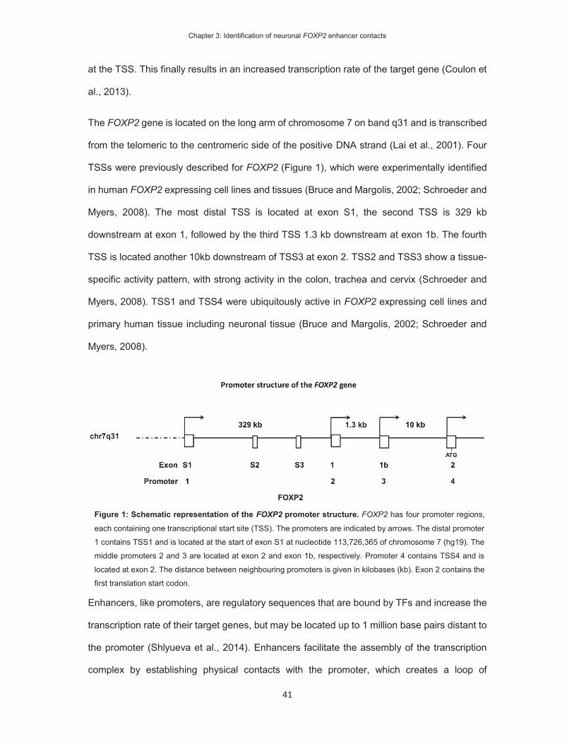
Chapter 3: Identification of neuronal FOXP2 enhancer contacts
41
at the TSS. This finally results in an increased transcription rate of the target gene (Coulon et
al., 2013).
The FOXP2 gene is located on the long arm of chromosome 7 on band q31 and is transcribed
from the telomeric to the centromeric side of the positive DNA strand (Lai et al., 2001). Four
TSSs were previously described for FOXP2 (Figure 1), which were experimentally identified
in human FOXP2 expressing cell lines and tissues (Bruce and Margolis, 2002; Schroeder and
Myers, 2008). The most distal TSS is located at exon S1, the second TSS is 329 kb
downstream at exon 1, followed by the third TSS 1.3 kb downstream at exon 1b. The fourth
TSS is located another 10kb downstream of TSS3 at exon 2. TSS2 and TSS3 show a tissue-
specific activity pattern, with strong activity in the colon, trachea and cervix (Schroeder and
Myers, 2008). TSS1 and TSS4 were ubiquitously active in FOXP2 expressing cell lines and
primary human tissue including neuronal tissue (Bruce and Margolis, 2002; Schroeder and
Myers, 2008).
Enhancers, like promoters, are regulatory sequences that are bound by TFs and increase the
transcription rate of their target genes, but may be located up to 1 million base pairs distant to
the promoter (Shlyueva et al., 2014). Enhancers facilitate the assembly of the transcription
complex by establishing physical contacts with the promoter, which creates a loop of
Figure 1: Schematic representation of the FOXP2 promoter structure. FOXP2 has four promoter regions,
each containing one transcriptional start site (TSS). The promoters are indicated by arrows. The distal promoter
1 contains TSS1 and is located at the start of exon S1 at nucleotide 113,726,365 of chromosome 7 (hg19). The
middle promoters 2 and 3 are located at exon 2 and exon 1b, respectively. Promoter 4 contains TSS4 and is
located at exon 2. The distance between neighbouring promoters is given in kilobases (kb). Exon 2 contains the
first translation start codon.
Chapter 3: Identification of neuronal FOXP2 enhancer contacts
42
interspacing DNA (Visel et al., 2009; Ong and Corces, 2011). Enhancer contacts to FOXP2
promoters have not been described in the literature. Following in silico predictions, two studies
report the identification of enhancer regions near Foxp2/foxp2 in zebrafish (Bonkowsky et al.,
2008; Coutinho et al., 2011) and mouse (Coutinho et al., 2011). In addition, I have identified a
human enhancer near FOXP2 in the previous chapter 2 (Becker et al., 2015). However, it has
not been shown that these enhancers make contact with FOXP2.
Challenges and possibilities to identify regulatory enhancer elements
The identification of enhancer elements is challenging for several reasons. Enhancer elements
are relatively small with a size of a hundred to a few thousand base pairs. TFs bind to short
sequence motifs of about 6-10 nucleotides within the enhancer sequence (Shlyueva et al.,
2014). The TF binding site (TFBS) for an individual TF is degenerate and usually represented
by one consensus motif. Therefore random sequence motifs that match a TFBS occur
frequently in the genome without being functional (Wasserman and Sandelin, 2004). The
functionality of any one enhancer is dependent on the combination of TFBSs. Thus, enhancers
consist of clusters of short redundant binding motifs, of unknown combinatorial complexity.
Additionally, enhancers can be distant from the genes that they regulate and the spacing DNA
between enhancer and gene can be of magnitudes bigger than the size of the enhancer
sequence itself (Ong and Corces, 2011; Spitz and Furlong, 2012). In short, the problem in
predicting enhancer sequences is that they are short, redundant and complex clusters of
sequence motifs, located at unknown distances from the protein-coding gene that they
regulate. Finally, enhancers are highly context-specific, being active in specific tissues,
developmental periods and in response to specific environmental stimuli. Therefore, the
prediction of enhancers is error prone and relies substantially on the incorporation of additional
data from molecular experiments or evolutionary conservation (Hardison and Taylor, 2012).
For example, an algorithm for whole-genome identification of enhancers incorporated
evolutionary constrained and clustered motif occurrence to achieve a predictive rate of only
Chapter 3: Identification of neuronal FOXP2 enhancer contacts
43
13% (Blanchette et al., 2006). As a consequence, functional molecular data are critical to
robustly identify enhancer elements.
Distinctive molecular properties enable the experimental identification of active enhancers.
One property is that active enhancer regions are in an open chromatin state and accessible
to TFs and chromatin remodelling complexes (Bell et al., 2011). Because these regions are
open, they are accessible to DNA degrading enzymes and can be identified as DNase
hypersensitivity sites (DHS). Another property of active enhancers is that the surrounding
chromatin is marked by specific histone modifications. Different chemical histone
modifications mark chromatin with specific functions, such as active enhancers, promoters or
transcribed regions (Roh et al., 2005; Heintzman et al., 2009; Hardison and Taylor, 2012).
Thus, genome-wide identification of enhancer-specific histone marks can be used to predict
enhancer regions. The third property is that active enhancers are in physical contact with the
promoter of their target genes. The genome-wide identification of active enhancers
incorporating these experimental data achieves a predictive power of 40 to 80% (Roh et al.,
2005; Heintzman et al., 2009; Cao et al., 2010; Hardison and Taylor, 2012). To reliably define
regulatory elements of FOXP2 I determined the physical contact of the FOXP2 promoter to
distant genomic regions as a proxy to identify enhancers.
Chromatin conformation at the FOXP2 gene locus
The interaction of distant genomic regions can be measured by chromatin conformation
capture (3C) (Figure 2) (Dekker et al., 2002). With this technique, the chromatin is chemically
fixated in its native state in living cells (Figure 2B). Interactions of distant genomic regions are
cross-linked via intermediate proteins. The chromatin is then isolated from the cells and
digested with restriction enzymes that recognize short cleavage sites (Figure 2C). The
digested chromatin is re-ligated within an increased volume to favour proximity ligation of DNA
strands within one chromatin complex (Figure 2D). The enhancer and promoter sequences
will form one linear strand of DNA (Figure 2E). As a consequence of this procedure the

Chapter 3: Identification of neuronal FOXP2 enhancer contacts
44
enhancer and promoter are spaced only by a few nucleotides instead of thousands of
kilobases. The sequence of the ligation products can be predicted to design specific PCR
primers, which anneal to the enhancer and promoter sequences. The ligation product can
finally be detected by regular PCR (Figure 2A, E). In this chapter, I described the use of 3C to
screen non-coding regions at the FOXP2 locus to identify long-distant enhancer loops at this
gene.

Chapter 3: Identification of neuronal FOXP2 enhancer contacts
45
The aims in this chapter were to i) measure the FOXP2 expression status in human neuronal-
like cell lines, ii) determine, which active FOXP2 promoter should act as the anchor point for
the 3C design, iii) analyse long-range chromatin interactions of this promoter and iv) narrow
down enhancer elements that can be further studied for their regulatory roles (as described in
Figure 2: Overview of chromatin conformation capture (3C). A) Regulatory enhancer elements (blue block)
are distant to their target promoters (red block and arrow) on the linear genome. Specific target primers (black
arrows) are designed at BglII restriction sites (black dotted lines). B) In the living cells the genome is folded by
proteins (brown spheres) and active enhancers contact their target promoters. The chromatin packed genome
is cross-linked in this state and digested with BglII restriction enzymes. B) The genomic fragments and DNA
overhangs remain in physical proximity. The DNA is diluted to favor proximity ligations between these genomic
fragments. D) Genomic fragments that were distant in the linear genome are ligated to form one short DNA
ligation product. E) The primers that were distant in genome amplify from the ligation product.

Chapter 3: Identification of neuronal FOXP2 enhancer contacts
46
chapters 4 and 5). The enhancer elements are the regulatory hubs that determine when,
where and how FOXP2 is expressed in the living organism. The identification of FOXP2
enhancers enabled me to study the genetic pathways that regulate FOXP2 expression
(Chapter 4), as well as the brain structures and developmental periods in which these
enhancers may act (Chapter 5).

Chapter 3: Identification of neuronal FOXP2 enhancer contacts
47
Materials and Methods
Cell Culture
HEK293, SH-SY5Y, IMR-32, SK-N-AS and KELLY cell lines were purchased from HPA
Culture Collections, SK-N-MC and PFSK-1 were purchased from ATCC. GM22647,
GM22671, GM22731 and GM22737 were obtained from Coriell Cell Repositories.
HEK293 cells were grown in DMEM (Invitrogen), supplemented with 10% fetal bovine serum
(FBS; Invitrogen) and 1% penicillin/streptavidin (P/S; Invitrogen). SK-N-MC and IMR-32 cells
were grown in MEM (Invitrogen), supplemented with 10% FBS, 1% P/S and 2 mM L-glutamine
(Invitrogen). SH-SY5Y and SK-N-AS were grown in DMEM:F12 (Invitrogen) supplemented
with 10% FBS, 1% P/S, 2 mM L-glutamine and 1% non-essential amino acids (NEAA;
Invitrogen). KELLY and PFSK-1 cell lines were grown in RPMI-160 (Invitrogen) medium
supplemented with 10% FBS and 1% P/S and 2mM glutamine. EBV-cell lines GM22647,
GM22671, GM22731 and GM22737 were grown in RPMI 1640 supplemented with 15% FBS,
1% P/S and 2mM L-glutamine.
Cells were cultured at 5% CO2 and 37°C. The adherent cells HEK293, SK-N-MC, PFSK-1,
SH-SY5Y, KELLY, IMR-32 and SK-N-AS were passaged at 90-100% confluency. To passage
cells, they were washed with PBS (Invitrogen) and detached from the cell culture surface using
trypsin (Invitrogen). Cells were spun at 300g for 4 min to remove the old medium and re-
suspended in fresh medium. Cells were routinely seeded into culture flasks for passaging or
culture dishes for experiments.
Expression analysis
Cells were grown to 80-90 % confluency in 6 cm cell culture dishes and lysed in 1 ml of cold
TRIzol (Life technologies). After repeatedly pipetting the TRIzol over the plate, the solution
was transferred into Eppendorf tubes and 200 μl of chloroform was added. The mixture was

Chapter 3: Identification of neuronal FOXP2 enhancer contacts
48
shaken vigorously for 2 min at RT and spun at 12,000g for 5 min at 4°C to separate the
aqueous from the organic phase. The aqueous phase was transferred to a clean Eppendorf
tube and mixed with 600 μl of 70% ethanol. The solution was then transferred to an RNeasy
Spin Column (Qiagen) and the RNA cleaned according to the Qiagen RNeasy Spin Colum
protocol. To remove genomic DNA on-column DNase digestion (Qiagen) was performed first,
followed by a membrane washing step and elution in 30 μl of RNase-free water. The amount
of RNA was quantified using Nanodrop (ThermoScientific) and the RNA quality determined on
a 1% agarose gel.
The reverse transcription PCR was performed on 2000ng of RNA using the High Capacity
cDNA Reverse Transcription Kit (Applied Biosystems). In short, the Master Mix (2 μl RT buffer,
0.8 μl 25x dNTP, 2 μl RT random primer, 1 μl MultiScribe reverse transcriptase and 4.2 μl
water) was prepared, 2000 ng of RNA added and the volume filled to 20 μl with RNase-free
water. The used RT-PCR program was 10 min at 25°C followed by 120 min at 37°C and 5 min
at 85°C.
To determine the relative amount of cDNA using real-time quantitative PCR (qPCR) the
sample was diluted 1:10. 2 μl of the cDNA solution (approx. 20 ng of cDNA) was added to 18
μl of SYBR green (BioRad) Master Mix (10 μl 2x SYBR green, 8.3 μl water, 0.6 μl 10 μM
forward primer, 0.6 μl 10 μM reverse primer). The primer sequences and coordinates are listed
in appendix 1, table 1. The 2-step real-time PCR protocol consisted of an initial 3 min 95°C
denaturation and enzyme activation step, followed by 40 cycles of 95°C denaturation for 15
sec and annealing/extension at 60°C for 30 sec. After 40 cycles the melt curve was recorded
in a range from 55°C to 95°C in 0.5°C increments of 10 sec each. The PCR reaction was
performed in the CFX96 PCR thermocycler (BioRad) and the fluorescence produced by the
PCR amplification was recorded using the CFX manager software (BioRad). All real-time PCR
reactions were performed in duplicate. The software automatically corrected for the
background fluorescence and determined the threshold fluorescence value to derive the
threshold cycle (Ct), at which logarithmic amplification was reached. The Ct values were

Chapter 3: Identification of neuronal FOXP2 enhancer contacts
49
recorded for each sample and the Ct values of a GAPDH control PCR were subtracted to
derive the delta Ct values.
Chromatin immunoprecipitation
Cells were grown to 80-90% confluency in T-75 cell culture flasks and trypsinized for 1-2 min.
The living cells were counted (Biorad TC-10) and diluted to 2.5 x 10⁶ cells per ml to a total
volume of 10 ml (25 x 106 cells total) using fresh medium. 135 μl of 37% formaldehyde was
added to cross-link cells for 10 min using 0.5% final formaldehyde concentration. The cells
were shaken for 10 min at room temperature (RT), after which the cross-linking reaction was
quenched by adding 125 mM freshly dissolved glycine. The solution was shaken for another
2 min to fully stop the reaction. The cells were spun down at 1600 rpm for 5 min at 4°C and
the supernatant removed. To clean the cell pellet, 10 ml of cold PBS were added and the
cross-linked cells re-suspended. The cells were spun again at 1600 rpm for 5 min at 4°C and
the supernatant decanted afterwards. The pellet was re-suspended in 10 ml lysis buffer (20
mM HEPES pH7.6; 10 mM EDTA; 0.5 mM EGTA, 0.5% triton-x100) and the solution shaken
for 10 min at 4°C. Following lysis, the cells were spun down at 1800 rpm for 5 min at 4°C and
the suspension was decanted. The cell pellet was re-suspended in 10 ml of stop buffer solution
(50 mM HEPES pH7.6; 150 mM NaCl; 1 mM EDTA; 0.5 mM EGTA) and shaken for 10 min at
4°C. The solution was spun down again at 1800 rpm for 5 min at 4°C and the suspension was
carefully removed. The pellet was finally resuspended in 2.1 ml of incubation buffer (10 mM
Tris pH8.0; 150 mM NaCl; 1 mM EDTA; 0.5 mM EGTA; 0.15% SDS; 1% Triton X-100) and
pipetted into aliquots of 300 μl. The aliquots were sonicated in 1.5 ml Eppendorf tubes in the
Bioruptor (Diagenode) set to “High” frequency power with cycles of 30 sec on and 30 sec off.
The number of sonication cycles was determined for each cell type to result in fragments of
300-800 bp in size.

Chapter 3: Identification of neuronal FOXP2 enhancer contacts
50
Before setting up the chromatin immunoprecipitation, 30 μl of protein G magnetic beads
(Invitrogen) were washed three times with incubation buffer, using magnetic racks. After
removing the supernatant of the third washing step, 40 μl of 5 x incubation buffer was added
to the beads. To block for unspecific binding 3 μl of a 10mg/μl bovine serum albumin solution
and to avoid protein degradation 10 μl protease inhibitor complex was added. 2 μl of Pol2RA
(Diagenode, AC-055-100) and molecular grade water was added to a final volume of 200 μl.
After adding 100 μl of sonicated chromatin, the immunoprecipitation was rotated overnight at
4°C.
The following day the samples were washed six times in total, using each 500 μl of different
washing buffers; two times using wash buffer 1 (10 mM Tris pH 8.0; 150 mM NaCl; 1 mM
EDTA; 0.5 mM EGTA; 0.1% SDS; 0.1% DOC; 1% Triton X-100), once using wash buffer 2 (10
mM Tris pH 8.0; 500 mM NaCl; 1 mM EDTA; 0.5 mM EGTA; 0.1% SDS; 0.1% DOC; 1% Triton
X-100), once using wash buffer 3 (10 mM Tris pH 8.0; 1 mM EDTA; 0.5 mM EGTA; 0.25 M
LiCl; 0.5% DOC; 0.5% NP-40) and two times using wash buffer 4 (10 mM Tris pH 8.0; 1 mM
EDTA; 0.5 mM EGTA). To elute the remaining DNA from the magnetic beads, 200 μl of fresh
elution buffer (1% SDS, 0.1 M NaHCO3) was added and the samples were rotated for 20 min
at RT. The eluate was taken off the magnetic beads and phenol extracted using 200 μl of
phenol: chloroform: isoamyl alcohol (SigmaAldrich). The DNA was further purified using
ethanol precipitation.
Target DNA fragments were amplified using the iQ SYBR Green Supermix (BioRad). 2 μl of
the pulled-down DNA sample was added to 18 μl of SYBR Green (BioRad) Master Mix (10 μl
2x SYBR Green, 8.3 μl water, 0.6 μl 10 μM forward primer, 0.6 μl 10 μM reverse primer).
Primer sequences for each target fragment are listed in appendix 1, table 2. The 2-step real-
time PCR protocol consisted of an initial 3 min 95°C denaturation and enzyme activation step,
followed by 40 cycles of 95°C denaturation for 15 sec and annealing/extension at 60°C for 30
sec. After 40 cycles the melt curve was recorded in a range from 55°C to 95°C in 0.5°C
increments of each 10 sec. The PCR reaction was performed in the CFX96 PCR thermocycler

Chapter 3: Identification of neuronal FOXP2 enhancer contacts
51
(BioRad) and the fluorescence produced by the PCR amplification was recorded using the
CFX manager software (BioRad). All real-time PCR reactions were performed in duplicate.
The delta Ct values were derived as described before. Input DNA samples were taken from
the sonicated aliquots, left at 4°C during the chromatin immunoprecipitation and included
again in the elution step. The delta Ct values were normalized to the promoter region of the
GAPDH housekeeping gene.
Chromatin conformation capture
The anchor point primer and TaqMan probe were designed to match the restriction fragment
that contains the first transcriptional start site (TSS) of the FOXP2 gene. Detection primers
were designed to be complementary to the 5’ end of restriction fragments so that the
amplicons of the ligation products were not bigger than 250 bp. Primers were designed to
match genomic fragments up to 106 kb upstream of TSS1 and 1,391 kb downstream of TSS1,
spanning a total of 1,497 kb around the FOXP2 gene locus. Within this genomic region, there
are 428 restriction fragments produced by BglII digestion. Primers were designed for 50
fragments. The quality of the primers was assessed using standard curves of the PCR reaction
on 3C samples with standardized concentrations (50ng, 125ng, 250ng, 500ng, 750ng and
1,000ng). 45 primers passed this quality control step, and they covered fragments of a total
size of 218 kb. All primers are listed in appendix 1, table 3.
Cells were grown to 80-90% confluency in T175 cell culture flasks. Cells were trypsinized and
the number of living cells was determined using TC-10 (BioRad) cell counter. 106 cells were
transferred into a clean 50 ml falcon tube and fresh medium was added to a total volume of
10 ml. 135 μl of 37% formaldehyde was added to cross-link cells at a final concentration of
0.5% formaldehyde. The cross-linking reaction was set to RT. After 10 min the reaction was
quenched by adding 1.41 ml of freshly dissolved 1 M glycine. The solution was inverted six
times. The cells were spun down at 1600 rpm for 5 min at 4°C and the supernatant decanted.

Chapter 3: Identification of neuronal FOXP2 enhancer contacts
52
The cell pellet was re-suspended in 2 ml of cold lysis buffer (50 mM tris, 150 mM NaCl, 5 mM
EDTA, 0.5% NP-40, 1% triton X-100 and 1x protease inhibitor complex) and set to shake for
10 min at 4°C. To improve cell lysis the cell solution was transferred to a dounce tissue grinder
and homogenized on ice with 2 times 15 piston strokes. The cell solution was again transferred
to a 50 ml falcon tube and pelleted at 1800 rpm for 5 min at 4°C. The supernatant was carefully
removed and the cell nuclei resuspended in 500 μl 1x restriction buffer 3 (NEB). The solution
was transferred to 1.5 ml Eppendorf tubes and spun at 600g, for 5 min at 4°C. The supernatant
was carefully removed and the pellet dissolved in 360 μl H2O and 60 μl of 10x restriction buffer
3. To loosen the chromatin structure, 15 μl of 10% was added and incubated for 10 min at
65°C, shaking at 900 rpm on a Thermomixer (Eppendorf). The SDS was quenched by adding
150 μl of 10% triton X-100 and incubated for 1h at 37°C, shaking at 900 rpm. To control the
digestion efficiency, 5 μl of the solution was separated as undigested control. To digest the
chromatin, 400 units of BglII (8 μl of 50,000 U/ml; NEB) were added and the digestion reaction
was incubated overnight at 37°C, shaking at 900 rpm. The next morning, another 200 units of
BglII restriction enzyme were added for another incubation period of 1h at 37°C to guarantee
proper digestion. Digestion efficiency was controlled on a 0.6% agarose gel. A barely visible
genomic band and high molecular weight smear were indicative of proper genomic digestion.
Before starting the re-ligation of genomic fragments, the restriction enzyme was inhibited by
adding 80 μl of 10% SDS and incubating for 30 min at 37°C. The solution was transferred to
a 50 ml tube and 4,860 μl of H2O and 700 μl of 10X ligation buffer were added. The ligation
reaction was done in a 10X higher volume to favour ligation events between cross-linked DNA
strands. The SDS was quenched with 750 μl of 10% triton X-100 and incubated for 30 min at
37°C. Finally, 50 Units of T4 Ligase (10 μl of 5 U/μl; Roche) were added and incubated at
16°C overnight.
To de-crosslink the samples 30μl Prot K (10 mg/ml) was added and incubated overnight at
65°C. The next morning, remaining RNA was digested by adding 30 μl of 10 mg/ml RNAse A
(Invitrogen) and incubating for 45 min at 37°C. The DNA was subsequently purified by phenol
Chapter 3: Identification of neuronal FOXP2 enhancer contacts
53
extraction using 50 ml PhaseLock Tubes (5 Prime). After ethanol precipitation, the DNA pellets
were recovered in 500 μl TE to give the final 3C library DNA. To determine the concentration
of the samples, a quantitative real-time PCR was performed, using SYBR green (BioRad).
The calibration curve was made using human genomic DNA in a range of concentrations from
0.5 to 250 ng per PCR reaction. The amplified region in the calibration curve is a genomic
fragment in the promoter region of GAPDH that does not contain a BglII restriction site.
The TaqMan real-time PCR of 3C ligation products was done using SsoFast Probes Supermix
(BioRad). Each PCR reaction mixture contained 2,000 ng of 3C library DNA, 1.5 μM TaqMan
probe, 0.5 μM anchor-point primer and 0.5 μM test primer in a 20 μl total reaction volume. All
real-time PCR reactions were performed in triplicates. Primer sequences are listed in appendix
1, table 3. The sequence of the MGB-TaqMan probe is 5’-
GATCTCTTAAACCACTGGGAATTCA-3’ and matches the sequence on chromosome 7 from
nucleotide 113,732,166 to 113,732,190 (reference genome hg19). The probe was ordered
from Applied Biosystems. The 2-step real-time PCR protocol consisted of an initial 3 min 95°C
denaturation and enzyme activation step, followed by 50 cycles of 95°C denaturation for 15sec
and annealing/extension at 60°C for 30 sec. The PCR reaction was performed in the CFX96
PCR thermocycler (BioRad) and the fluorescence produced by the PCR amplification was
recorded using the CFX manager software (BioRad).
The software automatically corrected for the background fluorescence and determined the
threshold fluorescence value to derive Ct. The Ct values were recorded for all ligation
products. The delta Ct value for each ligation product was calculated by subtracting the Ct
value from the average Ct value of same ligation product in the EBV-lymphocytes. In case that
the ligation product could not be detected in the EBV cell lines I subtracted a value of 40,
which is equal to the lowest detected ligation product in the EBV cell lines. From the Ct values,
I calculated the relative amount of starting ligation products by raising the negative Ct value to
the power of two, which is a measure of the interaction frequency. To normalize across the
different cell lines, I determined the ligation product with the lowest variation across all cell
Chapter 3: Identification of neuronal FOXP2 enhancer contacts
54
lines (primer at -10,574 bp) and normalized to the interaction frequency of this ligation product.
The final value is the relative interaction frequency.
Statistical analysis
Statistical analysis was performed using SPSS software. Statistical significance of the RNA
polymerase II ChIP-qPCR was assessed using pairwise ANOVA and posthoc Tukey test.
Statistical significance of the difference of 3C crosslinking frequencies to LBV-lymphoblast cell
crosslinking frequencies was assessed using two-tailed student t-test. P-values were
corrected for the amount of investigated interactions (45) using Bonferroni correction. To
determine interactions with statistically significantly increased crosslinking frequency in
FOXP2(+) cell lines, pairwise ANOVA was used between neuronal-like cell lines followed by
post-hoc LSD test.

Chapter 3: Identification of neuronal FOXP2 enhancer contacts
55
Results
FOXP2 is expressed in neuronal-like cell lines
Enhancers positively influence the expression of their target genes. Therefore, I expected to
FOXP2 enhancers to be active in cells that express this gene. I determined the expression of
this gene in human neuronal-like cell lines. Human cell lines were used to study enhancers in
the human genome. Also, cell lines were required to obtain sufficient amounts of chromatin
for subsequent 3C analysis. I used neuronal-like cells to identify enhancer elements that are
likely active in neuronal tissue, as enhancer interactions have been shown to be retained in
cell lines derived from the corresponding tissue (Jager et al., 2015). I screened seven cell lines
(SK-N-MC, PFSK-1, KELLY, IMR-32, SK-N-AS, SH-SY5Y and HEK293 cells), which are
commonly used in molecular studies of neuronal genes (Vernes et al., 2007; Schroeder and
Myers, 2008; Deriziotis et al., 2014). SK-N-AS, SH-SY5Y, IMR-32 and KELLY are
neuroblastoma cell lines that were derived from different sites of metastasis. SK-N-AS and
SH-SY5Y were isolated from the bone marrow; IMR-32 from an abdominal mass and for
KELLY the site of isolation is unpublished (Schwab et al., 1983). SK-N-MC and PFSK-1 are
malignant primitive neuroectodermal cancer cell lines. SK-N-MC was derived from the supra-
orbital area of a patient with neuroepithelioma (Biedler et al., 1973; Biedler et al., 1978; Dunn
et al., 1994) and PFSK-1 from a cerebral hemisphere (Fults et al., 1992). HEK293 cells have
been obtained from human embryonic kidney tissue using adenovirus infection (Graham et
al., 1977) and have previously been used for the study of FOXP2 and other related neuronal
genes (Vernes et al., 2006; Schroeder and Myers, 2008; Deriziotis et al., 2014). HEK293, SK-
N-MC and PFSK-1 cells have previously been shown to express FOXP2 (Schroeder and
Myers, 2008).
To measure endogenous FOXP2 expression, I harvested RNA from cultured cells and
performed reverse transcription using an oligo-d(T) primer. The produced cDNA was amplified
by quantitative PCR. The expression levels were first normalized to GAPDH and compared to
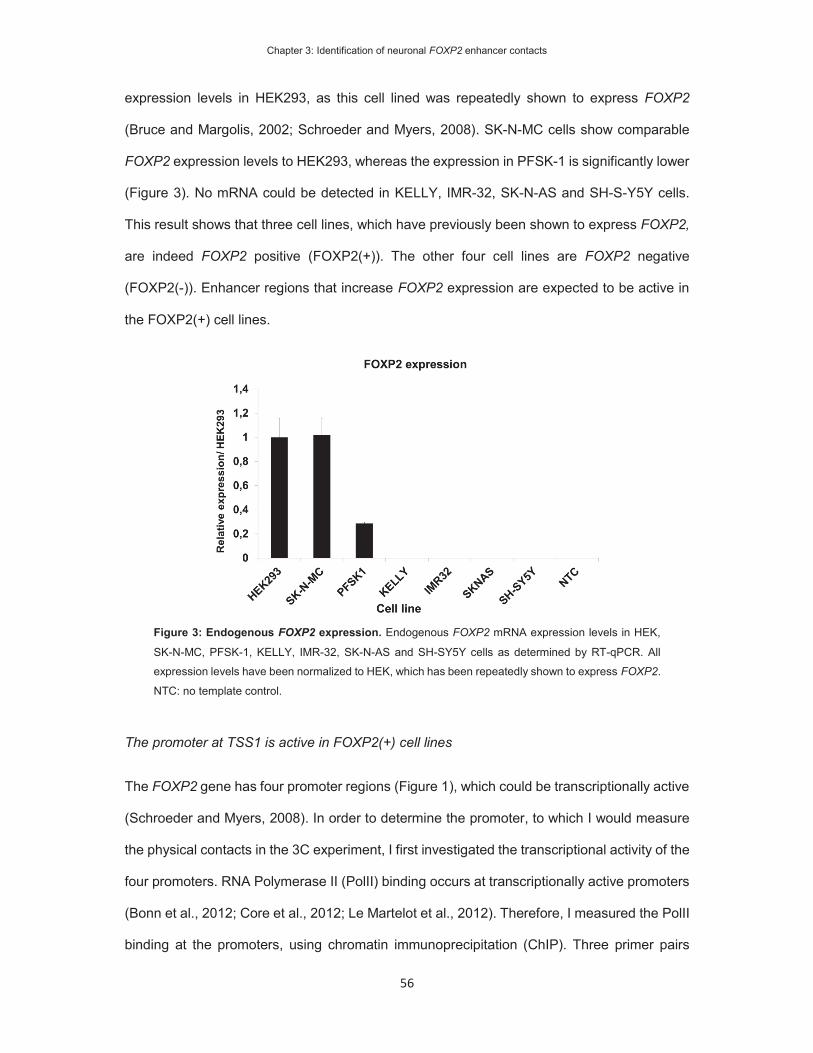
Chapter 3: Identification of neuronal FOXP2 enhancer contacts
56
expression levels in HEK293, as this cell lined was repeatedly shown to express FOXP2
(Bruce and Margolis, 2002; Schroeder and Myers, 2008). SK-N-MC cells show comparable
FOXP2 expression levels to HEK293, whereas the expression in PFSK-1 is significantly lower
(Figure 3). No mRNA could be detected in KELLY, IMR-32, SK-N-AS and SH-S-Y5Y cells.
This result shows that three cell lines, which have previously been shown to express FOXP2,
are indeed FOXP2 positive (FOXP2(+)). The other four cell lines are FOXP2 negative
(FOXP2(-)). Enhancer regions that increase FOXP2 expression are expected to be active in
the FOXP2(+) cell lines.
The promoter at TSS1 is active in FOXP2(+) cell lines
The FOXP2 gene has four promoter regions (Figure 1), which could be transcriptionally active
(Schroeder and Myers, 2008). In order to determine the promoter, to which I would measure
the physical contacts in the 3C experiment, I first investigated the transcriptional activity of the
four promoters. RNA Polymerase II (PolII) binding occurs at transcriptionally active promoters
(Bonn et al., 2012; Core et al., 2012; Le Martelot et al., 2012). Therefore, I measured the PolII
binding at the promoters, using chromatin immunoprecipitation (ChIP). Three primer pairs
Figure 3: Endogenous FOXP2 expression. Endogenous FOXP2 mRNA expression levels in HEK,
SK-N-MC, PFSK-1, KELLY, IMR-32, SK-N-AS and SH-SY5Y cells as determined by RT-qPCR. All
expression levels have been normalized to HEK, which has been repeatedly shown to express FOXP2.
NTC: no template control.
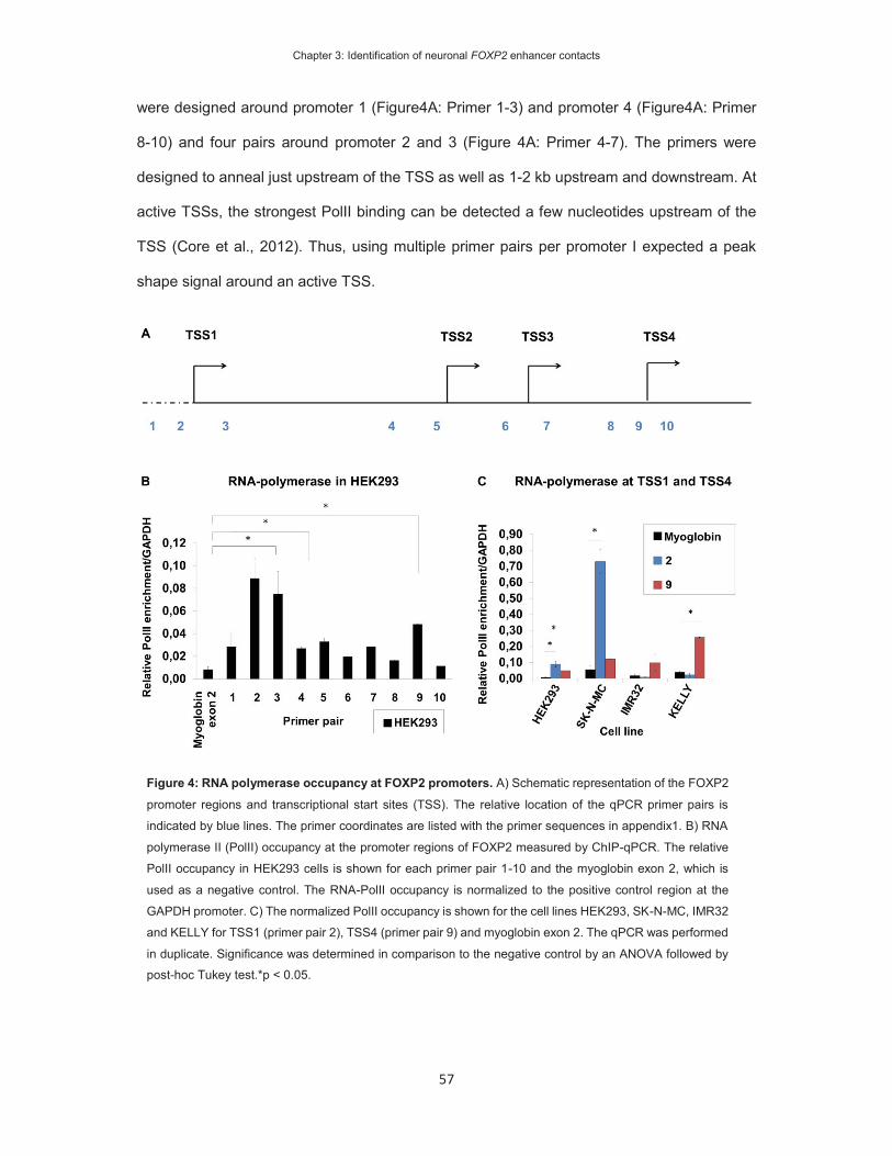
Chapter 3: Identification of neuronal FOXP2 enhancer contacts
57
were designed around promoter 1 (Figure4A: Primer 1-3) and promoter 4 (Figure4A: Primer
8-10) and four pairs around promoter 2 and 3 (Figure 4A: Primer 4-7). The primers were
designed to anneal just upstream of the TSS as well as 1-2 kb upstream and downstream. At
active TSSs, the strongest PolII binding can be detected a few nucleotides upstream of the
TSS (Core et al., 2012). Thus, using multiple primer pairs per promoter I expected a peak
shape signal around an active TSS.
Figure 4: RNA polymerase occupancy at FOXP2 promoters. A) Schematic representation of the FOXP2
promoter regions and transcriptional start sites (TSS). The relative location of the qPCR primer pairs is
indicated by blue lines. The primer coordinates are listed with the primer sequences in appendix1. B) RNA
polymerase II (PolII) occupancy at the promoter regions of FOXP2 measured by ChIP-qPCR. The relative
PolII occupancy in HEK293 cells is shown for each primer pair 1-10 and the myoglobin exon 2, which is
used as a negative control. The RNA-PolII occupancy is normalized to the positive control region at the
GAPDH promoter. C) The normalized PolII occupancy is shown for the cell lines HEK293, SK-N-MC, IMR32
and KELLY for TSS1 (primer pair 2), TSS4 (primer pair 9) and myoglobin exon 2. The qPCR was performed
in duplicate. Significance was determined in comparison to the negative control by an ANOVA followed by post-hoc Tukey test.*p < 0.05.

Chapter 3: Identification of neuronal FOXP2 enhancer contacts
58
First, I mapped the PolII binding at the promoter regions in the FOXP2(+) HEK293 cells. I used
the promoter region of the GAPDH housekeeping gene, which is active across different cell-
types to normalize the PolII signal between cell lines (Carter et al., 2010). As a negative control
I included a region at exon 2 of the myoglobin gene, which is inactive in tissue other than
muscle cells (Paschos et al., 2012; Tarrant-Elorza et al., 2014). I detected significant
enrichment of PolII with primer pair 2 and 3 (Figure 4B), which are located 130 bp upstream
and 1094 bp downstream of TSS1, respectively. No primer pair at TSS 2 or 3 was enriched
for PolII (Figure 4B: primers 4-7). I also observed enriched PolII binding at primer pair 9, which
is located 152bp upstream of TSS4 (Figure 4B). Thus, polymerase enrichment was detected
just upstream of TSS 1 and TSS 4, which indicates that they are active in HEK293. I further
determined PolII binding at promoter 1 and 4 in additional FOXP2(+) and FOXP2(-) cell lines,
using the primer pairs 2 and 9 as proxy for TSS1 and 4, respectively. At TSS1 -primer 2- the
two FOXP2(+) cell lines HEK293 and SKNMC showed, compared to the negative myoglobin
control, 9x and 12x enrichment of PolII binding, respectively (Figure 4C). The FOXP2(-) cell
lines IMR-32 and KELLY were not enriched for PolII at this promoter. At promoter 4, I detected
significant PolII enrichment in HEK293 and KELLY. Thus, PolII binding occurs at promoter 4
in both FOXP2(+) and FOXP2(-) cell lines. PolII occupancy at promoter 1 was only detected
in FOXP2(+) cells. This finding is in agreement with previous studies, which detected
expression from this promoter in HEK293 and SK-N-MC (Schroeder and Myers, 2008). In
conclusion, two FOXP2(+) cell lines show transcriptional activity at promoter 1. Therefore, I
examined the chromatin interactions to this promoter in the following 3C experiment.
The FOXP2 promoter is in contact with coding and non-coding regions in HEK293 cells
I designed the 3C experiment to capture chromatin contacts of promoter 1 –the anchor-point-
to distant genomic regions and thus determine enhancers that interact with this promoter. In
the 3C protocol, the chromatin is chemically fixed in living cells and subsequently digested

Chapter 3: Identification of neuronal FOXP2 enhancer contacts
59
with a site-specific restriction enzyme to produce genomic fragments (Figure 2). In silico
digestion of the genome predicts the produced restriction fragments, including their size and
sequence. Digestion sites within known functional elements could introduce a bias in the
digestion and ligation reactions (Naumova et al., 2012). Therefore, I chose the restriction
enzyme BglII, which produces a digestion fragment on which promoter 1 is located centrally.
I designed one primer, further referred to as anchor-point primer, to the 3’end of the promoter-
containing fragment (Figure 5B). In addition, I designed a TaqMan probe to match the
sequence between the anchor primer and the BglII digestion site. Thus, both the anchor-point
primer and TaqMan probe anneal on the promoter-containing restriction fragment. The use of
a fluorescent TaqMan probe allowed me to perform high sensitive PCR reactions (Navarro et
al., 2015).

Chapter 3: Identification of neuronal FOXP2 enhancer contacts
60
Target primers were designed to match the 3’ ends of the predicted restriction fragments. To
enable primer design with specific sequence recognition, the 3’ sequence was required to be
unique. On a genome-wide scale, the frequency of enhancer occurrences declines with the
distance to gene promoters in a symmetrical distribution (Stranger et al., 2007; Mifsud et al.,
2015). Therefore, I targeted candidate regions around the FOXP2 promoters and the adjacent
MDFIC promoter and designed several target primers to the genomic regions. One cluster of
target primers was designed in a 200kb window around promoter 1, spanning the intergenic
region between PPP1R3A and the first intron of FOXP2 (Figure 5A). The second cluster of
Figure 5: 3C design at the FOXP2 gene locus. A) The genomic location of the FOXP2 gene. The gene
structure of FOXP2 and the neighboring genes PPP13RA and MDFIC are shown in dark blue. The anchor point
primer is located at the distal promoter 1 and shown in red. The 3C target primers, depicted in light blue, were
designed to cluster at four locations of the gene locus: 1. around the anchor point promoter 1; 2. clustered
around the downstream promoters 2-4; 3. at the downstream intergenic region between the FOXP2 3’UTR and
MDFIC, 4. far downstream of the MDFIC gene. B) FOXP2 TSS1 is located at nucleotide 113,726,365 of the
human genome (hg19). The BglII restriction sites are depicted as black bars. The 3C primers (blue bars) have
been designed just upstream of the restriction sites. The viewpoint primer (red) is located on the restriction
fragment that contains TSS1. Note that not all BglII restriction sites are tagged by a 3C primer.
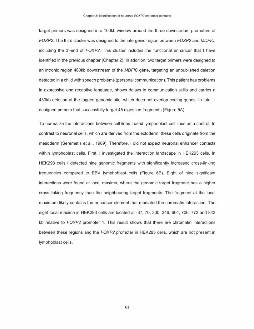
Chapter 3: Identification of neuronal FOXP2 enhancer contacts
61
target primers was designed in a 100kb window around the three downstream promoters of
FOXP2. The third cluster was designed to the intergenic region between FOXP2 and MDFIC,
including the 3’-end of FOXP2. This cluster includes the functional enhancer that I have
identified in the previous chapter (Chapter 2). In addition, two target primers were designed to
an intronic region 460kb downstream of the MDFIC gene, targeting an unpublished deletion
detected in a child with speech problems (personal communication). This patient has problems
in expressive and receptive language, shows delays in communication skills and carries a
430kb deletion at the tagged genomic site, which does not overlap coding genes. In total, I
designed primers that successfully target 45 digestion fragments (Figure 5A).
To normalize the interactions between cell lines I used lymphoblast cell lines as a control. In
contrast to neuronal cells, which are derived from the ectoderm, these cells originate from the
mesoderm (Seremetis et al., 1989). Therefore, I did not expect neuronal enhancer contacts
within lymphoblast cells. First, I investigated the interaction landscape in HEK293 cells. In
HEK293 cells I detected nine genomic fragments with significantly increased cross-linking
frequencies compared to EBV lymphoblast cells (Figure 6B). Eight of nine significant
interactions were found at local maxima, where the genomic target fragment has a higher
cross-linking frequency than the neighbouring target fragments. The fragment at the local
maximum likely contains the enhancer element that mediated the chromatin interaction. The
eight local maxima in HEK293 cells are located at -37, 70, 330, 346, 604, 706, 772 and 843
kb relative to FOXP2 promoter 1. This result shows that there are chromatin interactions
between these regions and the FOXP2 promoter in HEK293 cells, which are not present in
lymphoblast cells.
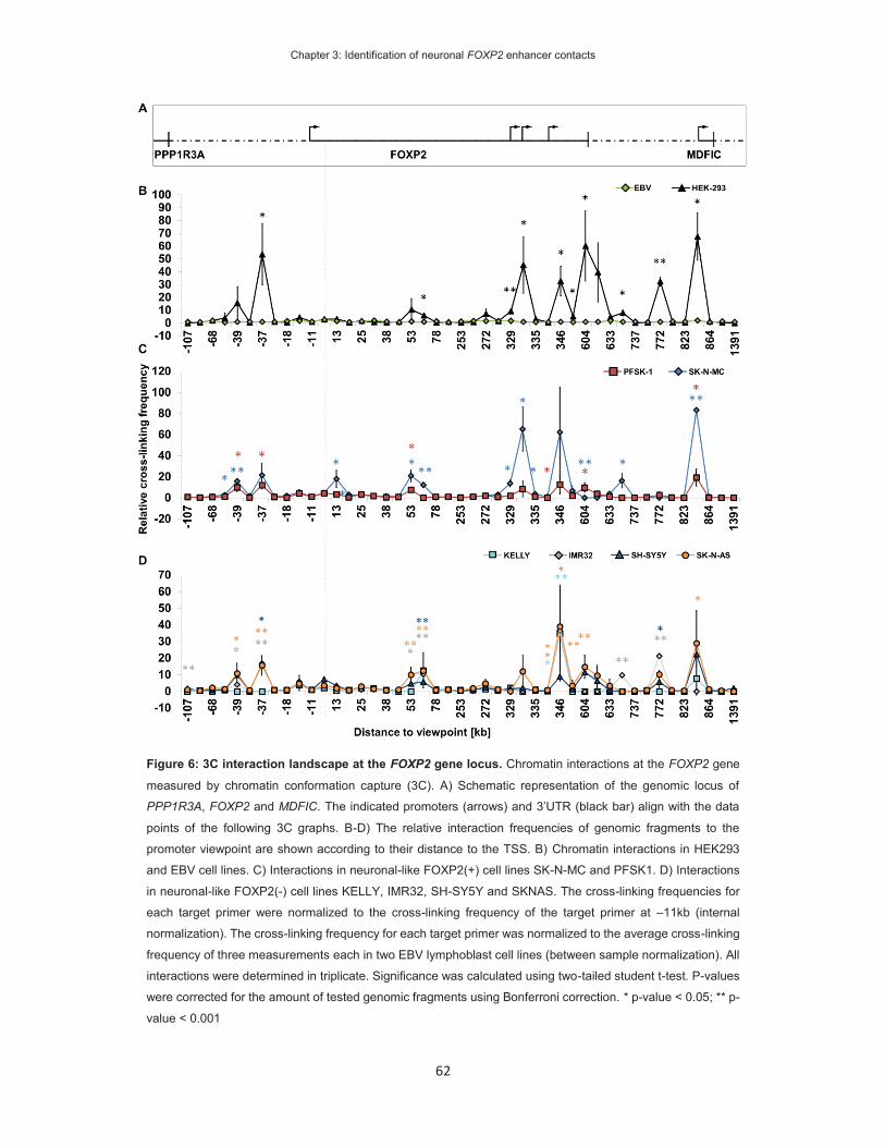
Chapter 3: Identification of neuronal FOXP2 enhancer contacts
62
Figure 6: 3C interaction landscape at the FOXP2 gene locus. Chromatin interactions at the FOXP2 gene
measured by chromatin conformation capture (3C). A) Schematic representation of the genomic locus of
PPP1R3A, FOXP2 and MDFIC. The indicated promoters (arrows) and 3’UTR (black bar) align with the data
points of the following 3C graphs. B-D) The relative interaction frequencies of genomic fragments to the
promoter viewpoint are shown according to their distance to the TSS. B) Chromatin interactions in HEK293
and EBV cell lines. C) Interactions in neuronal-like FOXP2(+) cell lines SK-N-MC and PFSK1. D) Interactions
in neuronal-like FOXP2(-) cell lines KELLY, IMR32, SH-SY5Y and SKNAS. The cross-linking frequencies for
each target primer were normalized to the cross-linking frequency of the target primer at –11kb (internal
normalization). The cross-linking frequency for each target primer was normalized to the average cross-linking
frequency of three measurements each in two EBV lymphoblast cell lines (between sample normalization). All
interactions were determined in triplicate. Significance was calculated using two-tailed student t-test. P-values
were corrected for the amount of tested genomic fragments using Bonferroni correction. * p-value < 0.05; ** p-
value < 0.001

Chapter 3: Identification of neuronal FOXP2 enhancer contacts
63
FOXP2(+) and (-) neuronal-like cells share regions of increased chromatin conformation
HEK293 cells were derived by viral transformation of embryonic kidney tissue, but their
morphology and transcriptome suggest a neuronal cell of origin (Shaw et al., 2002). They are
commonly used as a cellular system to study the function of neuronal genes. To obtain the
interaction landscape of additional FOXP2(+) cell lines, which originate from neuronal tissue,
I analysed the chromatin conformation in SK-N-MC and PFSK-1. In SK-N-MC cells I detected
ten genomic fragments with significantly increased cross-linking frequencies, of which five
mark local maxima at -39, 13, 53, 330, 706 and 843 kb (Figure 6C). All but one maximum at
13 kb were previously detected in HEK293 cells. The other significantly increased interactions
occur at the slope of these peaks at neighbouring genomic regions. Additionally, I detected a
significantly decreased cross-linking frequency at 604 kb, which showed increased interaction
in HEK293. In PFSK1 cells I detected four genomic regions with a significant increase in cross-
linking frequency (Figure 6B). Two of these mark local maxima that were detected in SK-N-
MC and HEK293 at 53 and 843 kb. The genomic region at 604 kb, which was significantly
decreased in SK-N-MC, is significantly increased in PFSK-1. I detected suggestive local
maxima at genomic regions that were significant in the other two FOXP2(+) cell lines. No
chromatin interaction was detected between promoter 1 and the gnomic fragment at 823kb
distance, which contains a previously identified enhancer (Chapter 2).
To determine differences in chromatin conformation between neuronal-like cells of different
FOXP2 expression status I measured the chromatin conformation in FOXP2(-) neuronal-like
cell lines KELLY, IM32, SH-SY5Y and SK-N-AS (Figure 6C). Eleven target fragments show
significantly increased cross-linking frequencies to the anchor-point fragment in one or several
cell lines. Eight of these interactions are located at local maxima and three interactions at
neighbouring genomic fragments. The maxima were located at -39, -37, 70, 346, 604, 706,
772 and 843 kb. This result shows that chromatin contacts between these regions and the
FOXP2 promoter are present in the absence of active transcription. The interactions of these

Chapter 3: Identification of neuronal FOXP2 enhancer contacts
64
regions were previously detected in FOXP2(+) cells, which indicates that chromatin contacts
are shared between FOXP2(+) and (-) neuronal-like cell lines.
Chromatin interactions are stronger in FOXP2 expressing cell lines
In total I identified eleven local maxima at which genomic regions contact the FOXP2 promoter
(Figure 7M). Two maxima, at 13 and 330 kb, were exclusively detected in FOXP2(+) cells and
nine were not shared with FOXP2(-) cell lines. To determine differences in chromatin
interactions between FOXP2(+) and (-) cells I compared the relative crosslinking frequencies
between the neuronal-like cell lines (Figure 7A-L). For each local maximum I calculated the
pairwise difference between cell lines and determined maxima that were significantly stronger
in FOXP2(+) cell lines. As expected, the cross-linking frequency at 13 kb is significantly
stronger in SK-N-MC cells than in any of the FOXP2(-) cells (Figure 7C). Similarly, the
interaction at region 330 kb is significantly stronger in HEK293 and SK-N-MC (Figure 7F). In
total, this analysis identified eight genomic regions at -37, 13, 53, 330, 604, 706, 772 and 843
kb with significantly increased chromatin interactions in at least one FOXP2 expressing cell
line (Figure 7M). These genomic fragments likely contain FOXP2 regulatory elements with
enhancer functions.

Chapter 3: Identification of neuronal FOXP2 enhancer contacts
65
Figure 7: Chromatin interactions at single peak regions. The chromatin interactions of the FOXP2 promoter
to the local maxima were re-plotted from figure 7B-D. The graphs compare the cross-linking frequencies for
individual maxima across the investigated cell lines. The cross-linking frequencies for EBV lymphoblast cells are
shown as black bars. The FOXP2(+) cell lines are shown as blue bars and the FOXP2(-) cell lines as white bars.
Peak regions are labelled according to their distance to promoter 1: A) -37 kb, B) -39 kb, C) 13 kb, D) 68 kb, E)
70 kb, F) 330 kb, G) 346 kb, H) 604 kb, I) 706 kb, K) 772 kb, L) 843 kb. Pairwise statistical difference was
determined using univariate ANOVA followed by post-hoc LSD test. Statistical significant local maxima show
stronger interaction in one FOXP2(+) cell lines compared to all FOXP2(-) cell lines. The reported p-value
represents the least significant difference between a FOXP2(+) and FOXP2(-) cell line. * p-value < 0.05; ** p-
value < 0.001. M) The peak regions are depicted in a schematic representation of the FOXP2 genomic locus.
Red: Regions with significantly higher interaction frequency in one or more FOXP2(+) cell lines.

Chapter 3: Identification of neuronal FOXP2 enhancer contacts
66
Local maxima map to regions with neuronal epigenetic marks
I detected eight chromatin interactions with increased cross-linking frequencies in FOXP2
expressing cell lines. The interactions occurred between promoter 1 and genomic fragments
located at a distance of -37, 13, 53, 330, 604, 706, 772 and 843 kb (Figure 7M). The restriction
digestion in the 3C method produced genomic fragments of 1 to 10 kb. The fragments likely
contain enhancer sequences, which have a size of several hundred nucleotides. To follow up
the enhancer function in molecular assays, it was necessary to define candidate enhancer
elements on the genomic fragments. The next step was to narrow down the enhancer
elements using histone marks.
The Roadmap Epigenomics Project mapped histone modifications in human tissue, including
samples from seven adult brain regions, two foetal brains (male and female), the foetal brain
germinal matrix, two neurosphere cultures derived from embryonic tissue (cortex and
ganglionic eminence), two neuronal progenitor cultures (derived from H1 and H9 embryonic
stem cells) and one neuron culture (derived from H9 embryonic stem cells). Twelve types of
epigenetic mark (eleven histone modifications and DNase hypersensitivity) were analysed
genome-wide and the combination of marks was used to predict the function of genomic
regions in the respective tissue. I used the predicted states in neuronal tissues to analyse the
genomic fragments with increased interaction frequencies and looked specifically for enhancer
marks. This allowed me to link the predictive properties of neuron-specific histone marks to
the enhancer interactions at the FOXP2 promoter.
I analysed the genomic regions that potentially contain enhancer elements (Figure 11-15).
The genomic fragment at -37 kb contains a site that is predicted to exert different functions in
the 15 analysed neuronal samples. In cultured neurospheres, derived from the ganglionic
eminence and tissue derived from the cingulate gyrus, angular gyrus and dorsolateral
prefrontal cortex this site was predicted to function as an enhancer (Figure 8: GE derived
neurosphere, cingulate gyrus, angular gyrus, dorsolateral prefrontal cortex). All predictions
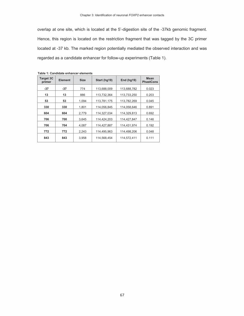
Chapter 3: Identification of neuronal FOXP2 enhancer contacts
67
overlap at one site, which is located at the 5’-digestion site of the -37kb genomic fragment.
Hence, this region is located on the restriction fragment that was tagged by the 3C primer
located at -37 kb. The marked region potentially mediated the observed interaction and was
regarded as a candidate enhancer for follow-up experiments (Table 1).
Table 1: Candidate enhancer elements Target 3C
primer Element Size Start (hg19) End (hg19) Mean PhastCons
-37 -37 774 113,688,009 113,688,782 0.023
13 13 886 113,732,364 113,733,250 0.203
53 53 1,094 113,781,175 113,782,269 0.045
330 330 1,801 114,056,845 114,058,646 0.891
604 604 2,779 114,327,034 114,329,813 0.692
706 700 3,645 114,424,203 114,427,847 0.146
706 704 4,087 114,427,887 114,431,974 0.192
772 772 2,243 114,495,963 114,498,206 0.048
843 843 3,958 114,568,454 114,572,411 0.111

Chapter 3: Identification of neuronal FOXP2 enhancer contacts
68
Figure 8: Detail of -39 to -37 kb peak region. The top panel shows the chromatin interactions in EBV-
lymphoblast and the representative SK-N-MC cell line. The alternative grey and white fields indicate the location
of genomic fragments produced by BglII digestion. The individual data points represent the interactions of
whole genomic fragments, as meassured with the respective target primers. The x-axis shows the distance of
the region to the FOXP2 TSS1. Below the graph are annotations aligned to the genomic position
(chr7:113,684,653-113,691,878 [hg19]). The BglII digestion sites (black lines) and 3C target primers (blue
lines) are shwon in the first two rows. The predicted functional states from the Epigenome Roadmaps are
shown for 5 human derived primary neuronal cell-types and 10 neuronal tissues. (light red = poised promoter,
red = active TSS, yellow = enhancer activity). The PhastCons Primate track shows the evolutionary
conservation between 10 primate genomes. The candidate enhancer annotation (black box) shows the
suggested enhancer element at this peak region.
Chapter 3: Identification of neuronal FOXP2 enhancer contacts
69
In total, I analysed eight genomic fragments for the presence of enhancer elements (Figure 8-
15) and identified nine candidate enhancers (Table 1). In addition to candidate enhancer
elements, two interacting fragments contained elements of known function. The genomic
region at 604 kb contains the 3’-UTR of FOXP2 (Figure 12) and the fragment at 843 kb spans
the promoter of the adjacent MDFIC gene (Figure 15). Therefore, the FOXP2 promoter seems
to interact with gene elements that typically function in transctiption initiation and mRNA
stability, but may also contribute to transcription regulation via long-distance interactions (Jash
et al., 2012; Sahlen et al., 2015). The evolutionary conservation, which may indicate functional
importance, of the candidate enhancers varies. The enhancer at 330kb is highly conserved
(Mean Primate PhastConservation Score: 0.891, Figure 11, Table 1) and may be functional in
a wide range of related species. The enhancer at -37kb shows low conservation (Mean
Primate Phast Cons: 0.023, Figure 8, Table 1) and the function may be more specific to
humans or primates. Thus, the 3C genomic fragments contain promising candidate enhancers
of variable evolutionary conservation, which may regulate the expression of FOXP2.

Chapter 3: Identification of neuronal FOXP2 enhancer contacts
70
Figure 9: Detail of 13 kb peak region. See figure 8. Genomic position (chr7:113731832-113743326 [hg19]).
Figure 10: Detail of 53 kb peak region. See figure 8. Genomic position (chr7:113,772,159-113,785,107
[hg19]).

Chapter 3: Identification of neuronal FOXP2 enhancer contacts
71
Figure 11: Detail of 330 kb peak region. See figure 8. Genomic position (chr7:114,053,511-114,061,726
[hg19]). The structure FOXP2 transcripts are shown below, indicating exon 1 at promoter 2 at which the
transcription of a short isoform starts.
Figure 12: Detail of 604 kb peak region. See figure 8. The crosslinking frequencies of the representative
HEK293 cell lines are shown. Genomic position (chr7:114320782-114348266 [hg19]). The structure of the
FOXP2 transcript is shown below, indicating the 3’-UTR at exon 17.
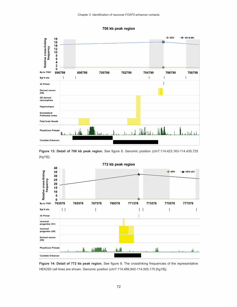
Chapter 3: Identification of neuronal FOXP2 enhancer contacts
72
Figure 14: Detail of 772 kb peak region. See figure 8. The crosslinking frequencies of the representative
HEK293 cell lines are shown. Genomic position (chr7:114,489,942-114,505,170 [hg19]).
Figure 13: Detail of 706 kb peak region. See figure 8. Genomic position (chr7:114,423,163-114,435,725
[hg19]).
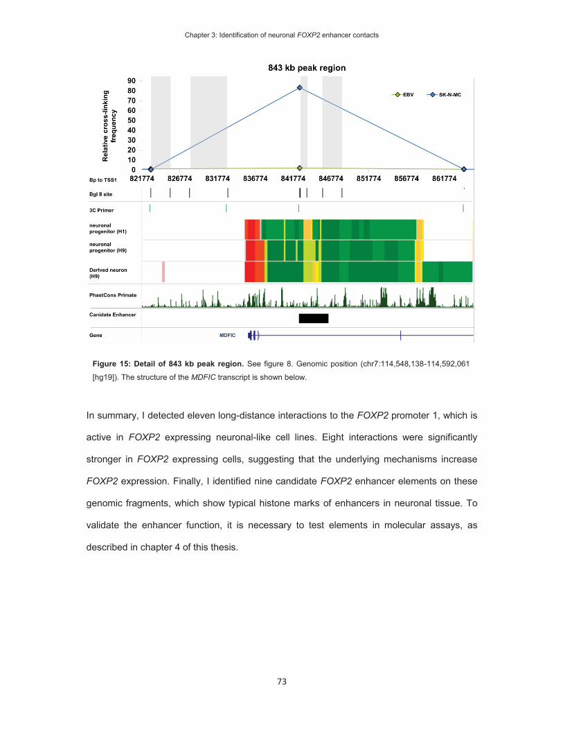
Chapter 3: Identification of neuronal FOXP2 enhancer contacts
73
In summary, I detected eleven long-distance interactions to the FOXP2 promoter 1, which is
active in FOXP2 expressing neuronal-like cell lines. Eight interactions were significantly
stronger in FOXP2 expressing cells, suggesting that the underlying mechanisms increase
FOXP2 expression. Finally, I identified nine candidate FOXP2 enhancer elements on these
genomic fragments, which show typical histone marks of enhancers in neuronal tissue. To
validate the enhancer function, it is necessary to test elements in molecular assays, as
described in chapter 4 of this thesis.
Figure 15: Detail of 843 kb peak region. See figure 8. Genomic position (chr7:114,548,138-114,592,061
[hg19]). The structure of the MDFIC transcript is shown below.
Chapter 3: Identification of neuronal FOXP2 enhancer contacts
74
Discussion
Four transcriptional start sites have been identified for FOXP2 and their activity has been
determined by a set of human cell lines and tissue samples (Schroeder and Myers, 2008).
Transcription from TSS1 and TSS4 was shown to be active in neuronal-like cell lines and
primary neuronal tissue, whereas TSS2 and TSS3 were active in the colon, trachea and cervix
(Schroeder and Myers, 2008). In agreement with this, I detected PolII occupancy at TSS1
and/or TSS4 in neuronal-like cell lines. PolII enrichment at TSS4 was detected in HEK293
cells, as well as in the FOXP2(-) KELLY cells. The PolII enrichment in FOXP2(-) cells could
indicate a poised polymerase, which may become active in response to external stimuli
(Gaertner et al., 2012). The published neuronal activity and potentially poised polymerase
make TSS4 an interesting target for future 3C studies. The promoter of TSS1 was occupied
exclusively in the investigated FOXP2 expressing cell lines. Therefore, I expected clear
differences for this promoter between FOXP2(+) and (-) cell lines and prioritized the
identification of long-range interactions with this promoter.
I detected an interaction between the FOXP2 promoter and a genomic fragment in the
proximity of the MDFIC promoter. Promoter-promoter interactions have been identified
between a substantial amount of co-regulated neighbouring genes in prior genome-wide
studies (Li et al., 2012; Sahlen et al., 2015). Thus, the promoter-promoter interaction to the
MDFIC gene suggests that this gene could be co-regulated with FOXP2. The MDFIC gene
encodes the MyoD family inhibitor domain containing protein, which modulates the
transcriptional regulation of the WNT and JNK/SAPK pathways (Kusano and Raab-Traub,
2002; Reiss-Sklan et al., 2009). Strong expression of this gene was described in T-cells
(Thebault et al., 2000), but the expression of this gene has not been studied in other tissues.
The Allen Brain Atlas indicates ubiquitous expression of Mdfic in adult mouse brains (Lein et
al., 2007) and the gene likely overlaps Foxp2 expression, which shows strong expression in
the basal ganglia, thalamus, cerebellum and neocortex (Chapter 1). Detailed studies of
MDFIC/Mdfic expression in adult and developing brain tissue would be required to identify

Chapter 3: Identification of neuronal FOXP2 enhancer contacts
75
neuronal co-regulation with FOXP2/Foxp2 in mouse or human. The co-regulation of genes
can be investigated in published weighted gene co-expression network analysis (WGCNA)
data. WGCNA data summarizes the co-regulation of genes across many samples of the same
origin (Fuller et al., 2007). One WGCNA study indicated that MDFIC and FOXP2 are co-
regulated in the human and primate frontal lobe (Konopka et al., 2012), which may suggest a
functional interaction in this tissue. MDFIC has established roles in WNT-signalling and prior
research has shown that downstream target genes of FOXP2 are enriched for function in the
WNT-signalling pathway (Vernes et al., 2007; Vernes et al., 2011). In addition, foxp2 in
zebrafish is likely regulated by lef1, a TF that regulates target gene expression in response to
activation of WNT-signalling. Thus, the co-regulation could further implicate FOXP2 in this
pathway.
In this chapter I described how the chromatin interactions were investigated in cell lines of
neuronal origin. Enhancer interactions have been shown to be retained in cell lines derived
from the corresponding tissue (Jager et al., 2015). This suggests that the identified interactions
occur in neuronal tissue. The interactions occurred between genomic elements that show
histone marks of active enhancers in one or several neuronal tissues (Roadmap Epigenomics
et al., 2015), further indicating that the identified enhancers show neuronal activity. The target
tissue and developmental period of the enhancer activity remain to be determined.
The Roadmap Epigenomics Consortium determined the histone marks in human derived
cultured neuronal progenitors, derived neurons, neurospheres, whole foetal brains and adult
neuronal tissue (Ernst and Kellis, 2015). I used these marks to determine regions of
overlapping function in several neuronal tissues. The example of the region at -37 kb (Figure
8) shows that histone marks overlap at one narrow region at this locus. Follow-up experiments
are important to validate the enhancers’ function and to determine tissue-specificity, as
discussed in subsequent chapters of this thesis. Transgenic mice are commonly used to study
tissue-specificity during development (Visel et al., 2007; Boyd et al., 2015) and in chapter 5 I
Chapter 3: Identification of neuronal FOXP2 enhancer contacts
76
used this approach to determine the developmental pattern and adult tissue-specificity of the
enhancer elements at -37 kb and 330 kb.
The evolutionary conservation of the candidate enhancers varies from an element that is well
conserved across vertebrates (Figure 11: 330 kb) to an element that shows low conservation
even across primates (Figure 8: -37 kb). The presence of conservation at a genomic site can
be regarded as the result of evolutionary constraint on sequence variety due to its functional
impact (Ureta-Vidal et al., 2003; Miller et al., 2004). Hence, highly conserved enhancers likely
drive target gene expression across related species. High conservation of FOXP2 enhancers
is in agreement with the conserved expression pattern of this gene across different vertebrate
species (Enard et al., 2002; Ferland et al., 2003; Lai et al., 2003; Teramitsu et al., 2004; Li et
al., 2007; Campbell et al., 2009). However, I also identified an evolutionary non-conserved
enhancer in the human gene locus, which may suggest that humans gained regulatory control
mechanisms. This may indicate that additional brain structures gained FOXP2 expression or
that the spatio-temporal dynamics of FOXP2 regulation in evolutionarily shared brain
structures has changed. The evolutionary conserved expression pattern would suggest that
regulation is more likely to change within shared structures. To explore this possibility, the
temporal expression changes of FOXP2 need to be studied in more detail across species.
Examples of dynamic adult expression changes have been found in the thalamus of mice
(Horng et al., 2009) and striatum of songbirds (Teramitsu and White, 2006). The evolutionary
conservation of these dynamic expression changes may indicate the presence of absence of
species-dependent regulatory enhancers.
On a molecular level, enhancer elements consist of nucleotide sequences that are bound by
TFs. Knowing the location and sequence of the potential enhancer elements, it is possible to
investigate TFs that bind to these enhancers. I investigated TF binding in chapter 4 and
discuss this aspect of the identified enhancer there.

Chapter 3: Identification of neuronal FOXP2 enhancer contacts
77
The aim of the current chapter was to identify long-distance interactions, which may exert an
enhancing effect on FOXP2 expression. However, in some cases long-distance interactions
have been reported to exert a repressive effect (Ogbourne and Antalis, 1998; Maston et al.,
2006). Therefore, the 3C approach could potentially also detect repressing elements, called
silencers. In comparison to enhancers, I would expect these elements to show strong
interaction in FOXP2(-) cell lines. However, I did not detect chromatin interactions that were
stronger in the absence of FOXP2 expression. Silencers may overlap with enhancer
sequences and the composition of TF binding determines the effect on transcription (Perissi
et al., 2004). I can therefore not exclude that the identified elements may repress transcription
under certain conditions. The interaction of the chromatin fragment at 604 kb was significantly
weaker in the FOXP2(+) SK-N-MC cells compared to the control EBV-lymphoblast cells. As
SK-N-MC cells express FOXP2, one explanation for the reduced interaction of this region
could be that a silencing element was actively repelled from the active promoter. However,
the same interaction was significantly increased in the FOXP2(+) HEK293 cells. Thus, the
expression status does not distinguish the presence or depletion of this interaction. The
fragment at 604 kb covers parts of the FOXP2 3’-UTR. At the RNA level, 3’-UTRs are known
to mediate mRNA transcript stability and translational efficiency (He and Hannon, 2004).
However, on the DNA level, 3’UTRs have also been found to establish long-distance
interactions with their gene promoter. The effect of this interaction on gene expression can be
repressive (Le Cam and Legraverend, 1995; Paul et al., 1998) or activating (Salerno et al.,
2000; Jash et al., 2012). My 3C results show that the 3’-UTR of FOXP2 interacts with the
promoter both in the absence and presence of expression. To determine the function of this
element on transcription, it will be necessary to study this element in isolation, using reporter
gene assays.
3C is a low-throughput method that requires the manual design for every investigated
interaction. Therefore, only a limited amount of genomic fragments at the FOXP2 gene locus
could be studied. As a consequence, long-range interactions to additional enhancers might

Chapter 3: Identification of neuronal FOXP2 enhancer contacts
78
have been missed. Recent research has shown that the genome is partitioned into topological
domains (TD) (Dixon et al., 2012). Dynamic enhancer contacts have been shown to occur
within TDs and not across the topological borders (TB). TBs are reported to be evolutionarily
conserved (Vietri Rudan et al., 2015) and stable during development (Dixon et al., 2012).
Therefore, the TBs determined in one cell-type limit enhancer interactions in all or most
differentiated tissues. All investigated genomic fragments and identified interactions were
located within the TD that contains the FOXP2 gene. However, the TD of FOXP2 further
covers the upstream PPP1R3A gene and the downstream genes MDFIC and TFIC. An
extension of the 3C design, which covers the complete TD of FOXP2, may identify further
enhancer elements.
Currently, a broad variety of 3C-based technologies are being developed to identify enhancer
interactions with higher specificity and sensitivity on a genome-wide scale (Dostie et al., 2006;
Dekker et al., 2013; Jin et al., 2013; Lefebvre et al., 2013; Nagano et al., 2013). For example,
capture Hi-C combines high-throughput sequencing technologies with the sensitivity of
targeted 3C approaches (Jager et al., 2015; Mifsud et al., 2015). Future experiments
investigating FOXP2 chromatin contacts could aim to identify additional or indeed all long-
distance enhancer contacts within a tissue. Additionally, one 3C based method is capable of
determining the genome-wide chromatin interaction landscape of single cells (Nagano et al.,
2013). This circumvents the requirement to use cultured cell lines, which I used to obtain
sufficient amount of chromatin. These approaches have so far focused on animal tissue, or
non-neuronal human tissue (Dekker et al., 2013; Jin et al., 2013; Nagano et al., 2013; Jager
et al., 2015; Mifsud et al., 2015; Vietri Rudan et al., 2015). Thus, future studies may identify
all enhancers of FOXP2 in a cell type-specific manner.
Enhancer elements have been shown to contain genetic variants that cause, or increase the
susceptibility to, human disease. For example, rare genetics variants in regulatory regions
have been linked to autism spectrum disorders (Poitras et al., 2010; Chen et al., 2014). The
investigation of the here identified enhancer sequences in people with speech and language

Chapter 3: Identification of neuronal FOXP2 enhancer contacts
79
disorders may reveal rare genetic variants underlying their deficits. In addition, common
genetic variants within these enhancers may be linked to normal variation in brain structure
and function (Chapter 6, (Becker et al.)). The catalogue of published genome-wide association
studies (GWAS) of the National Human Genome Research Institute (NHGRI) collects
genome-wide associations of common variants from published studies (Welter et al., 2014).
According to this catalogue associations have been found near the FOXP2 gene locus for
traits such as smoking behaviour (Argos et al., 2014; Sung et al., 2015), N-glycosylation of
immunoglobin (Lauc et al., 2013), Crohn’s disease (Julia et al., 2013), lymphoblast cell viability
(de With et al., 2015) and obesity (Kim et al., 2013). The associated single-nucleotide
polymorphisms (SNPs), however, are not located within the candidate FOXP2 enhancers or
the tagged genomic fragments. Thus, so far, no trait-associated SNP has been detected within
the enhancers. However, the identified enhancers are candidate regions for future association
studies of common genetic variants in language-related traits.
This chapter represents the first study investigating long-distance enhancer interactions at the
FOXP2 gene locus. The identified interactions occurred in neuronal-like cell lines and may
provide information about the regulatory mechanisms contributing to FOXP2 expression in the
human brain. The interactions occur between the active FOXP2 promoter to the 3’-UTR of
FOXP2, the promoter of MDFIC and candidate enhancer elements. The identification of
candidate enhancers facilitates the study of FOXP2 regulation and enables the identification
of regulatory genetic variants underlying normal or pathogenic variation.

Chapter 3: Identification of neuronal FOXP2 enhancer contacts
80
References
Argos M, Tong L, Pierce BL, Rakibuz-Zaman M, Ahmed A, Islam T, Rahman M, Paul-Brutus R, Rahaman R, Roy S, Jasmine F, Kibriya MG, Ahsan H (2014) Genome-wide association study of smoking behaviours among Bangladeshi adults. Journal of medical genetics 51:327-333.
Becker M, Devanna P, Fisher SE, Vernes SC (2015) A chromosomal rearrangement in a child with severe speech and language disorder separates FOXP2 from a functional enhancer. Molecular cytogenetics 8:69.
Bell O, Tiwari VK, Thoma NH, Schubeler D (2011) Determinants and dynamics of genome accessibility. Nat Rev Genet 12:554-564.
Berger SL (2007) The complex language of chromatin regulation during transcription. Nature 447:407-412.
Biedler JL, Helson L, Spengler BA (1973) Morphology and growth, tumorigenicity, and cytogenetics of human neuroblastoma cells in continuous culture. Cancer research 33:2643-2652.
Biedler JL, Roffler-Tarlov S, Schachner M, Freedman LS (1978) Multiple neurotransmitter synthesis by human neuroblastoma cell lines and clones. Cancer research 38:3751-3757.
Blanchette M, Bataille AR, Chen X, Poitras C, Laganiere J, Lefebvre C, Deblois G, Giguere V, Ferretti V, Bergeron D, Coulombe B, Robert F (2006) Genome-wide computational prediction of transcriptional regulatory modules reveals new insights into human gene expression. Genome Res 16:656-668.
Bonkowsky JL, Wang X, Fujimoto E, Lee JE, Chien CB, Dorsky RI (2008) Domain-specific regulation of foxP2 CNS expression by lef1. BMC developmental biology 8:103.
Bonn S, Zinzen RP, Girardot C, Gustafson EH, Perez-Gonzalez A, Delhomme N, Ghavi-Helm Y, Wilczynski B, Riddell A, Furlong EE (2012) Tissue-specific analysis of chromatin state identifies temporal signatures of enhancer activity during embryonic development. Nat Genet 44:148-156.
Boyd JL, Skove SL, Rouanet JP, Pilaz LJ, Bepler T, Gordan R, Wray GA, Silver DL (2015) Human-Chimpanzee Differences in a FZD8 Enhancer Alter Cell-Cycle Dynamics in the Developing Neocortex. Current biology : CB.
Bruce HA, Margolis RL (2002) FOXP2: novel exons, splice variants, and CAG repeat length stability. Human genetics 111:136-144.
Cairns BR (2009) The logic of chromatin architecture and remodelling at promoters. Nature 461:193-198.
Campbell P, Reep RL, Stoll ML, Ophir AG, Phelps SM (2009) Conservation and diversity of Foxp2 expression in muroid rodents: functional implications. The Journal of comparative neurology 512:84-100.
Cao Y, Yao Z, Sarkar D, Lawrence M, Sanchez GJ, Parker MH, MacQuarrie KL, Davison J, Morgan MT, Ruzzo WL, Gentleman RC, Tapscott SJ (2010) Genome-wide MyoD binding in skeletal muscle cells: a potential for broad cellular reprogramming. Dev Cell 18:662-674.
Carter CL, Lin C, Liu CY, Yang L, Liu ZR (2010) Phosphorylated p68 RNA helicase activates Snail1 transcription by promoting HDAC1 dissociation from the Snail1 promoter. Oncogene 29:5427-5436.
Chen CH, Huang CC, Cheng MC, Chiu YN, Tsai WC, Wu YY, Liu SK, Gau SS (2014) Genetic analysis of GABRB3 as a candidate gene of autism spectrum disorders. Molecular autism 5:36.
Core LJ, Waterfall JJ, Gilchrist DA, Fargo DC, Kwak H, Adelman K, Lis JT (2012) Defining the status of RNA polymerase at promoters. Cell Rep 2:1025-1035.
Coulon A, Chow CC, Singer RH, Larson DR (2013) Eukaryotic transcriptional dynamics: from single molecules to cell populations. Nat Rev Genet 14:572-584.
Coutinho P, Pavlou S, Bhatia S, Chalmers KJ, Kleinjan DA, van Heyningen V (2011) Discovery and assessment of conserved Pax6 target genes and enhancers. Genome Res 21:1349-1359.
de With SA, Pulit SL, Wang T, Staal WG, van Solinge WW, de Bakker PI, Ophoff RA (2015) Genome-wide association study of lymphoblast cell viability after clozapine exposure. American journal of medical genetics Part B, Neuropsychiatric genetics : the official publication of the International Society of Psychiatric Genetics 168B:116-122.
Dekker J, Marti-Renom MA, Mirny LA (2013) Exploring the three-dimensional organization of genomes: interpreting chromatin interaction data. Nat Rev Genet 14:390-403.
Dekker J, Rippe K, Dekker M, Kleckner N (2002) Capturing chromosome conformation. Science 295:1306-1311.

Chapter 3: Identification of neuronal FOXP2 enhancer contacts
81
Deriziotis P, O'Roak BJ, Graham SA, Estruch SB, Dimitropoulou D, Bernier RA, Gerdts J, Shendure J, Eichler EE, Fisher SE (2014) De novo TBR1 mutations in sporadic autism disrupt protein functions. Nature communications 5:4954.
Dixon JR, Selvaraj S, Yue F, Kim A, Li Y, Shen Y, Hu M, Liu JS, Ren B (2012) Topological domains in mammalian genomes identified by analysis of chromatin interactions. Nature 485:376-380.
Dostie J, Richmond TA, Arnaout RA, Selzer RR, Lee WL, Honan TA, Rubio ED, Krumm A, Lamb J, Nusbaum C, Green RD, Dekker J (2006) Chromosome Conformation Capture Carbon Copy (5C): a massively parallel solution for mapping interactions between genomic elements. Genome Res 16:1299-1309.
Dunn T, Praissman L, Hagag N, Viola MV (1994) ERG gene is translocated in an Ewing's sarcoma cell line. Cancer genetics and cytogenetics 76:19-22.
Enard W, Przeworski M, Fisher SE, Lai CS, Wiebe V, Kitano T, Monaco AP, Paabo S (2002) Molecular evolution of FOXP2, a gene involved in speech and language. Nature 418:869-872.
Ernst J, Kellis M (2015) Large-scale imputation of epigenomic datasets for systematic annotation of diverse human tissues. Nat Biotechnol 33:364-U374.
Ferland RJ, Cherry TJ, Preware PO, Morrisey EE, Walsh CA (2003) Characterization of Foxp2 and Foxp1 mRNA and protein in the developing and mature brain. The Journal of comparative neurology 460:266-279.
Fuller TF, Ghazalpour A, Aten JE, Drake TA, Lusis AJ, Horvath S (2007) Weighted gene coexpression network analysis strategies applied to mouse weight. Mammalian genome : official journal of the International Mammalian Genome Society 18:463-472.
Fults D, Pedone CA, Morse HG, Rose JW, McKay RD (1992) Establishment and characterization of a human primitive neuroectodermal tumor cell line from the cerebral hemisphere. Journal of neuropathology and experimental neurology 51:272-280.
Gaertner B, Johnston J, Chen K, Wallaschek N, Paulson A, Garruss AS, Gaudenz K, De Kumar B, Krumlauf R, Zeitlinger J (2012) Poised RNA polymerase II changes over developmental time and prepares genes for future expression. Cell Rep 2:1670-1683.
Graham FL, Smiley J, Russell WC, Nairn R (1977) Characteristics of a human cell line transformed by DNA from human adenovirus type 5. The Journal of general virology 36:59-74.
Hardison RC, Taylor J (2012) Genomic approaches towards finding cis-regulatory modules in animals. Nat Rev Genet 13:469-483.
He L, Hannon GJ (2004) MicroRNAs: small RNAs with a big role in gene regulation. Nat Rev Genet 5:522-531.
Heintzman ND et al. (2009) Histone modifications at human enhancers reflect global cell-type-specific gene expression. Nature 459:108-112.
Horng S, Kreiman G, Ellsworth C, Page D, Blank M, Millen K, Sur M (2009) Differential gene expression in the developing lateral geniculate nucleus and medial geniculate nucleus reveals novel roles for Zic4 and Foxp2 in visual and auditory pathway development. The Journal of neuroscience : the official journal of the Society for Neuroscience 29:13672-13683.
Jager R, Migliorini G, Henrion M, Kandaswamy R, Speedy HE, Heindl A, Whiffin N, Carnicer MJ, Broome L, Dryden N, Nagano T, Schoenfelder S, Enge M, Yuan Y, Taipale J, Fraser P, Fletcher O, Houlston RS (2015) Capture Hi-C identifies the chromatin interactome of colorectal cancer risk loci. Nature communications 6:6178.
Jash A, Yun K, Sahoo A, So JS, Im SH (2012) Looping mediated interaction between the promoter and 3' UTR regulates type II collagen expression in chondrocytes. PloS one 7:e40828.
Jiang C, Pugh BF (2009) Nucleosome positioning and gene regulation: advances through genomics. Nat Rev Genet 10:161-172.
Jin F, Li Y, Dixon JR, Selvaraj S, Ye Z, Lee AY, Yen CA, Schmitt AD, Espinoza CA, Ren B (2013) A high-resolution map of the three-dimensional chromatin interactome in human cells. Nature 503:290-294.
Julia A et al. (2013) A genome-wide association study on a southern European population identifies a new Crohn's disease susceptibility locus at RBX1-EP300. Gut 62:1440-1445.
Kim HJ, Yoo YJ, Ju YS, Lee S, Cho SI, Sung J, Kim JI, Seo JS (2013) Combined linkage and association analyses identify a novel locus for obesity near PROX1 in Asians. Obesity 21:2405-2412.
Konopka G, Friedrich T, Davis-Turak J, Winden K, Oldham MC, Gao F, Chen L, Wang GZ, Luo R, Preuss TM, Geschwind DH (2012) Human-specific transcriptional networks in the brain. Neuron 75:601-617.
Kusano S, Raab-Traub N (2002) I-mfa domain proteins interact with Axin and affect its regulation of the Wnt and c-Jun N-terminal kinase signaling pathways. Molecular and cellular biology 22:6393-6405.

Chapter 3: Identification of neuronal FOXP2 enhancer contacts
82
Lai CS, Fisher SE, Hurst JA, Vargha-Khadem F, Monaco AP (2001) A forkhead-domain gene is mutated in a severe speech and language disorder. Nature 413:519-523.
Lai CS, Gerrelli D, Monaco AP, Fisher SE, Copp AJ (2003) FOXP2 expression during brain development coincides with adult sites of pathology in a severe speech and language disorder. Brain : a journal of neurology 126:2455-2462.
Lauc G et al. (2013) Loci associated with N-glycosylation of human immunoglobulin G show pleiotropy with autoimmune diseases and haematological cancers. PLoS Genet 9:e1003225.
Le Cam A, Legraverend C (1995) Transcriptional repression, a novel function for 3' untranslated regions. European journal of biochemistry / FEBS 231:620-627.
Le Martelot G, Canella D, Symul L, Migliavacca E, Gilardi F, Liechti R, Martin O, Harshman K, Delorenzi M, Desvergne B, Herr W, Deplancke B, Schibler U, Rougemont J, Guex N, Hernandez N, Naef F, Cycli XC (2012) Genome-wide RNA polymerase II profiles and RNA accumulation reveal kinetics of transcription and associated epigenetic changes during diurnal cycles. PLoS biology 10:e1001442.
Lefebvre C, Vachon F, Grimault S, Thibault J, Guimond S, Peretz I, Zatorre RJ, Jolicoeur P (2013) Distinct electrophysiological indices of maintenance in auditory and visual short-term memory. Neuropsychologia 51:2939-2952.
Lein ES et al. (2007) Genome-wide atlas of gene expression in the adult mouse brain. Nature 445:168-176.
Li G, Wang J, Rossiter SJ, Jones G, Zhang S (2007) Accelerated FoxP2 evolution in echolocating bats. PloS one 2:e900.
Li G et al. (2012) Extensive promoter-centered chromatin interactions provide a topological basis for transcription regulation. Cell 148:84-98.
Maston GA, Evans SK, Green MR (2006) Transcriptional regulatory elements in the human genome. Annual review of genomics and human genetics 7:29-59.
Mifsud B, Tavares-Cadete F, Young AN, Sugar R, Schoenfelder S, Ferreira L, Wingett SW, Andrews S, Grey W, Ewels PA, Herman B, Happe S, Higgs A, LeProust E, Follows GA, Fraser P, Luscombe NM, Osborne CS (2015) Mapping long-range promoter contacts in human cells with high-resolution capture Hi-C. Nat Genet 47:598-606.
Miller W, Makova KD, Nekrutenko A, Hardison RC (2004) Comparative genomics. Annual review of genomics and human genetics 5:15-56.
Nagano T, Lubling Y, Stevens TJ, Schoenfelder S, Yaffe E, Dean W, Laue ED, Tanay A, Fraser P (2013) Single-cell Hi-C reveals cell-to-cell variability in chromosome structure. Nature 502:59-64.
Naumova N, Smith EM, Zhan Y, Dekker J (2012) Analysis of long-range chromatin interactions using Chromosome Conformation Capture. Methods 58:192-203.
Navarro E, Serrano-Heras G, Castano MJ, Solera J (2015) Real-time PCR detection chemistry. Clinica chimica acta; international journal of clinical chemistry 439:231-250.
Ogbourne S, Antalis TM (1998) Transcriptional control and the role of silencers in transcriptional regulation in eukaryotes. The Biochemical journal 331 ( Pt 1):1-14.
Ong CT, Corces VG (2011) Enhancer function: new insights into the regulation of tissue-specific gene expression. Nat Rev Genet 12:283-293.
Paschos K, Parker GA, Watanatanasup E, White RE, Allday MJ (2012) BIM promoter directly targeted by EBNA3C in polycomb-mediated repression by EBV. Nucleic Acids Res 40:7233-7246.
Paul C, Simar-Blanchet AE, Ro HS, Le Cam A (1998) Characterization of three transcriptional repressor sites within the 3' untranslated region of the rat serine protease inhibitor 2.3 gene. European journal of biochemistry / FEBS 254:538-546.
Perissi V, Aggarwal A, Glass CK, Rose DW, Rosenfeld MG (2004) A corepressor/coactivator exchange complex required for transcriptional activation by nuclear receptors and other regulated transcription factors. Cell 116:511-526.
Poitras L, Yu M, Lesage-Pelletier C, Macdonald RB, Gagne JP, Hatch G, Kelly I, Hamilton SP, Rubenstein JL, Poirier GG, Ekker M (2010) An SNP in an ultraconserved regulatory element affects Dlx5/Dlx6 regulation in the forebrain. Development 137:3089-3097.
Reiss-Sklan E, Levitzki A, Naveh-Many T (2009) The complex regulation of HIC (Human I-mfa domain containing protein) expression. PloS one 4:e6152.
Roadmap Epigenomics C et al. (2015) Integrative analysis of 111 reference human epigenomes. Nature 518:317-330.
Roh TY, Cuddapah S, Zhao K (2005) Active chromatin domains are defined by acetylation islands revealed by genome-wide mapping. Genes Dev 19:542-552.
Chapter 3: Identification of neuronal FOXP2 enhancer contacts
83
Sahlen P, Abdullayev I, Ramskold D, Matskova L, Rilakovic N, Lotstedt B, Albert TJ, Lundeberg J, Sandberg R (2015) Genome-wide mapping of promoter-anchored interactions with close to single-enhancer resolution. Genome Biol 16:156.
Salerno MS, Mordvinov VA, Sanderson CJ (2000) Binding of octamer factors to a novel 3'-positive regulatory element in the mouse interleukin-5 gene. J Biol Chem 275:4525-4531.
Schroeder DI, Myers RM (2008) Multiple transcription start sites for FOXP2 with varying cellular specificities. Gene 413:42-48.
Schwab M, Alitalo K, Klempnauer KH, Varmus HE, Bishop JM, Gilbert F, Brodeur G, Goldstein M, Trent J (1983) Amplified DNA with limited homology to myc cellular oncogene is shared by human neuroblastoma cell lines and a neuroblastoma tumour. Nature 305:245-248.
Seremetis S, Inghirami G, Ferrero D, Newcomb EW, Knowles DM, Dotto GP, Dalla-Favera R (1989) Transformation and plasmacytoid differentiation of EBV-infected human B lymphoblasts by ras oncogenes. Science 243:660-663.
Shaw G, Morse S, Ararat M, Graham FL (2002) Preferential transformation of human neuronal cells by human adenoviruses and the origin of HEK 293 cells. FASEB journal : official publication of the Federation of American Societies for Experimental Biology 16:869-871.
Shlyueva D, Stampfel G, Stark A (2014) Transcriptional enhancers: from properties to genome-wide predictions. Nat Rev Genet 15:272-286.
Spitz F, Furlong EE (2012) Transcription factors: from enhancer binding to developmental control. Nat Rev Genet 13:613-626.
Stranger BE, Nica AC, Forrest MS, Dimas A, Bird CP, Beazley C, Ingle CE, Dunning M, Flicek P, Koller D, Montgomery S, Tavare S, Deloukas P, Dermitzakis ET (2007) Population genomics of human gene expression. Nat Genet 39:1217-1224.
Sung YJ, de Las Fuentes L, Schwander KL, Simino J, Rao DC (2015) Gene-smoking interactions identify several novel blood pressure loci in the Framingham Heart Study. American journal of hypertension 28:343-354.
Tarrant-Elorza M, Rossetto CC, Pari GS (2014) Maintenance and replication of the human cytomegalovirus genome during latency. Cell host & microbe 16:43-54.
Teramitsu I, White SA (2006) FoxP2 regulation during undirected singing in adult songbirds. The Journal of neuroscience : the official journal of the Society for Neuroscience 26:7390-7394.
Teramitsu I, Kudo LC, London SE, Geschwind DH, White SA (2004) Parallel FoxP1 and FoxP2 expression in songbird and human brain predicts functional interaction. The Journal of neuroscience : the official journal of the Society for Neuroscience 24:3152-3163.
Thebault S, Gachon F, Lemasson I, Devaux C, Mesnard JM (2000) Molecular cloning of a novel human I-mfa domain-containing protein that differently regulates human T-cell leukemia virus type I and HIV-1 expression. J Biol Chem 275:4848-4857.
Ureta-Vidal A, Ettwiller L, Birney E (2003) Comparative genomics: genome-wide analysis in metazoan eukaryotes. Nat Rev Genet 4:251-262.
Vernes SC, Spiteri E, Nicod J, Groszer M, Taylor JM, Davies KE, Geschwind DH, Fisher SE (2007) High-throughput analysis of promoter occupancy reveals direct neural targets of FOXP2, a gene mutated in speech and language disorders. Am J Hum Genet 81:1232-1250.
Vernes SC, Nicod J, Elahi FM, Coventry JA, Kenny N, Coupe AM, Bird LE, Davies KE, Fisher SE (2006) Functional genetic analysis of mutations implicated in a human speech and language disorder. Human molecular genetics 15:3154-3167.
Vernes SC, Oliver PL, Spiteri E, Lockstone HE, Puliyadi R, Taylor JM, Ho J, Mombereau C, Brewer A, Lowy E, Nicod J, Groszer M, Baban D, Sahgal N, Cazier JB, Ragoussis J, Davies KE, Geschwind DH, Fisher SE (2011) Foxp2 regulates gene networks implicated in neurite outgrowth in the developing brain. PLoS Genet 7:e1002145.
Vietri Rudan M, Barrington C, Henderson S, Ernst C, Odom DT, Tanay A, Hadjur S (2015) Comparative Hi-C reveals that CTCF underlies evolution of chromosomal domain architecture. Cell Rep 10:1297-1309.
Visel A, Rubin EM, Pennacchio LA (2009) Genomic views of distant-acting enhancers. Nature 461:199-205.
Visel A, Minovitsky S, Dubchak I, Pennacchio LA (2007) VISTA Enhancer Browser--a database of tissue-specific human enhancers. Nucleic Acids Res 35:D88-92.
Wasserman WW, Sandelin A (2004) Applied bioinformatics for the identification of regulatory elements. Nat Rev Genet 5:276-287.
Welter D, MacArthur J, Morales J, Burdett T, Hall P, Junkins H, Klemm A, Flicek P, Manolio T, Hindorff L, Parkinson H (2014) The NHGRI GWAS Catalog, a curated resource of SNP-trait associations. Nucleic Acids Res 42:D1001-1006.

Chapter 4: Upstream regulatory mechanisms at the promoters and enhancers of FOXP2
84
Chapter 4
Upstream regulatory mechanisms acting at the
promoters and enhancers of FOXP2

Chapter 4: Upstream regulatory mechanisms at the promoters and enhancers of FOXP2
85
Abstract
Enhancers and promoters of FOXP2 control the expression of this gene during development
and in adult tissue. Transcription factors (TF) bind to such enhancers and promoters and
consequently regulate their activity. Thus, the TFs acting upstream of FOXP2 determine the
developmental and tissue-specific expression patterns of the gene. However, the TFs that
regulate the promoters and enhancers of human FOXP2 are largely unknown.
In this chapter I investigated the in vitro activity of FOXP2 promoters and candidate enhancers
in human cell lines using a reporter gene assay. One promoter and three enhancers increased
reporter gene expression in human neuronal-like cell lines. I further tested the effect of TFs
that had been linked to FOXP2 in prior literature. Using overexpression plasmids in
combination with the promoter and enhancer reporter constructs I detected regulation by
FOXP1, FOXP4, POU3F2, LEF1, PAX6, SOX5 and TBR1, as well as auto-regulation by
FOXP2.
The presented results demonstrate that FOXP2 is regulated by known TFs and suggest
upstream regulatory pathways. Auto-regulation of FOXP2 may explain the haploinsuficiency
effect observed for mono-allelic gene mutations and gives new insight into possible disease
mechanisms. One enhancer of interest (enhancer 330) is the target of TFs important for
cortical development and may contribute to FOXP2 expression during cortical development.
Chapter 4: Upstream regulatory mechanisms at the promoters and enhancers of FOXP2
86
Introduction
Transcription factors (TFs) bind to gene promoters and enhancers to activate target gene
expression. Promoters and enhancers of FOXP2 have been identified (as described in
Chapter 3), but the TFs that bind to them are unknown. Importantly, TFs are known to be
effector proteins of signalling pathways and modulate target gene expression in response to
developmental stimuli or environmental signals. Thus, the response of FOXP2 expression to
developmental programs and cellular processes is mediated by TFs. The aim of the current
chapter was validate candidate enhancers identified in the previous chapter 3 and to identify
TFs that regulate the enhancers and promoters of FOXP2 to help reveal the genetic pathways
acting upstream of this gene
Promoters and enhancers of FOXP2 are the target sites for transcription factor binding
Two promoters of FOXP2, promoter 1 and 4, are active in neuronal cell lines and neuronal
tissue (Bruce and Margolis, 2002; Schroeder and Myers, 2008) (Chapter 3, Figure 1). In the
prior work of this thesis I identified one enhancer downstream of an inversion breakpoint of a
complex chromosomal rearrangement in a child with speech and language problems matching
those seen in cases of FOXP2 mutation (Moralli et al., 2015). As described in Chapter 2, the
inversion separates the enhancer from the rest of the FOXP2 locus, suggesting that disrupted
regulation by this enhancer contributes to the observed phenotype in the affected child (Becker
et al., 2015) (Chapter 2). In addition, in Chapter 3 I used chromatin confirmation capture to
identify genomic regions, which interacted with promoter 1 in neuronal-like cell lines, and could
thus be considered as candidates for novel enhancers (Chapter 3). Enhancers and promoters
function independently from their genomic context. Their activity does not depend on their
orientation, distance to the target promoter and are thus able to regulate the expression of
reporter genes in vitro (Pennacchio et al., 2013). The activity of these elements can therefore
be studied in cultured cell lines (Deriziotis et al., 2014).

Chapter 4: Upstream regulatory mechanisms at the promoters and enhancers of FOXP2
87
Prioritizing putative upstream TFs of FOXP2
The study of enhancers in cell cultures further allows the investigation of their response to
transcription factors (TFs). TFs are expressed or activated under specific conditions and fulfil
varying functions. A group of TFs function in the general regulatory machinery of the cell, such
as EP300 (Chan and La Thangue, 2001) or CTCF (Ong and Corces, 2014). In contrast, some
TFs are expressed at specific developmental periods and regulate, for example, the body
patterning during embryonic development (Wellik, 2007). Other TFs are active in response to
environmental cues (Sheng and Greenberg, 1990) or environmental stress (DiDomenico et
al., 1982; Kothary et al., 1989) and regulate the rapid response to the changed conditions.
Certain TFs are expressed in a cell-type specific manner to establish the identity of
differentiated cells. The TFs that regulate FOXP2 can therefore shed light on the
developmental programs upstream of FOXP2 expression. However, the prediction of TFs that
regulate a gene is challenging, as the genome encodes about a thousand TFs, of which
approximately 300 are expressed in each foetal and neuronal tissue (Fulton et al., 2009;
Vaquerizas et al., 2009). The combination of TFs found in every cell is variable and determines
the final regulatory output. In the same way, the promoter and enhancer activity depends on
the combination of binding TFs. The co-expression of TFs with FOXP2 is a limiting factor for
positive regulatory interactions and has been described for a number of TFs (Introduction:
Figure 1: Schematic of FOXP2 promoter and potential enhancer elements. Schematic representation
of the FOXP2 promoters and enhancers in the human genome on chromosome 7 band q31.1. The figure
shows position 113,500,000 to 114,600,000 in reference human genome (build hg19). The candidate
enhancers, identified in Chapter 2 and 3 of this thesis, are depicted as red blocks and the promoters are
shown in blue. The FOXP2 and MDFIC genes are shown in black.

Chapter 4: Upstream regulatory mechanisms at the promoters and enhancers of FOXP2
88
Table1). I selected a number of TFs that were linked to FOXP2 in the literature to test their
regulation of the promoters and enhancers that I had identified.
Regulatory TFs described in the literature
In zebrafish embryos lef1 is co-expressed with foxp2 in the tectum, mid-hindbrain boundary
and hindbrain (Bonkowsky et al., 2008). Bonkowsky et al. further showed that knock down of
lef1 abolishes foxp2 expression in the tectum and mid-hindbrain boundary, suggesting that
foxp2 is regulated by lef1. LEF1 is the effector TF of the WNT-signalling pathway, which is
involved in neurogenesis and regulates neural differentiation in the neocortex, cerebral cortex,
hippocampus, olfactory bulb (OB) and thalamus (Oliva et al., 2013). In mouse embryos Lef1
is expressed in the mesencephalon and diencephalon and involved in brain patterning (van
Genderen et al., 1994). Strong adult expression of Lef1 is detected in the thalamus,
hypothalamus, hippocampus and OB (Shimogori et al., 2004; Oliva et al., 2013). If human
FOXP2 enhancers are regulated by LEF1, this could indicate the regulation of FOXP2 by
WNT-signalling pathways and associated biological processes. In addition, prior studies in
human and mouse foetal tissue indicate that FOXP2/Foxp2 regulates gene if the WNT-
signalling pathway (Spiteri et al., 2007; Vernes et al., 2011). Thus, the expression of FOXP2
may regulate the balance of WNT-signalling pathway.
Pax6 co-expression with Foxp2 was described in the amygdala and striatum of non-human
primates and rats (Kaoru et al., 2010). In zebrafish pax6 is co-expressed with foxp2 in the
embryonic telencephalon and knock-down of pax6 reduces levels of foxp2 in this brain region
(Coutinho et al., 2011). In addition, Coutinho et al. detected reduced Foxp2 expression in the
telencephalon of mouse embryos carrying a Pax6 null mutation, suggesting that this regulatory
interaction is evolutionary preserved. PAX6 has a dual role in maintaining neural stem cell
identity and promoting neural differentiation (Osumi, 2001; Osumi et al., 2008) and is
expressed in the developing and adult neocortex, amygdala, basal ganglia, thalamus, OB and
Chapter 4: Upstream regulatory mechanisms at the promoters and enhancers of FOXP2
89
cerebellum (Duan et al., 2013). In humans, PAX6 promotes the transition from embryonic stem
cells to the neuronal lineage (Zhang et al., 2010).
Another TF has been proposed to directly regulate FOXP2 expression in humans: POU3F2.
The regulation of FOXP2 by POU3F2 was tested on a putative regulatory element of FOXP2.
This element is located in an intron, within a bigger genomic region that carries a significant
excess of genetic variants that arose in the human genome after the split from a common
ancestor with Neanderthals (Maricic et al., 2013). The element did not show in vitro activity in
human cell lines or in vivo activity in transgenic mouse embryos (Chapter 1). However, a
human-specific genetic variant, which is conserved among vertebrates, was predicted to
decrease the binding of POU3F2. In agreement with the prediction, the human variant
decreased enhancer activity in response to POU3F2 overexpression, in comparison to the
Neanderthal variant. Thus, the study by Maricic et al. suggests that vertebrate FOXP2/Foxp2,
including that of the Neanderthals, was regulated by POU3F2 and this regulatory interaction
is reduced in humans. POU3F2 has been shown to regulate other TFs in retinoic acid-treated
embryonic stem cells and was suggested to be an upstream effector of the retinoic acid
signalling pathway (Urban et al., 2015). Similarly, FOXP2 can promotes neural differentiation
in combination with retinoic acid signalling in human cell lines (Devanna et al., 2014). Thus, it
is conceivable that FOXP2 could be one of the POU3F2 target genes that promote retinoic
acid induced neural differentiation.
Another candidate TF, which may regulate FOXP2, is TBR1. This TF has been shown to either
activate or repress the expression of its target genes (Hevner et al., 2001; Bedogni et al.,
2010; Han et al., 2011). TBR1 expression overlaps with FOXP2 expression in post-migratory
neurons of the embryonic cortical plate in humans (Willsey et al., 2013) and mice (Hisaoka et
al., 2010). In the adult mouse cortex Tbr1 is co-expressed with Foxp2 in cortical layer VI
(Hevner et al., 2001; Bedogni et al., 2010; Hisaoka et al., 2010). FOXP2 and TBR1 physically
interact with each other and may co-regulate target genes in a cooperative manner (Deriziotis
et al., 2014). In the cortex of Tbr1 knock-out mice the expression of Foxp2 is significantly

Chapter 4: Upstream regulatory mechanisms at the promoters and enhancers of FOXP2
90
decreased in the caudal area of layer VI (Bedogni et al., 2010). This could suggest that TBR1
enhances FOXP2 expression in the cortex. Another TF that is co-expressed with TBR1 in the
cortex is SOX5 (Rouaux and Arlotta, 2010). During embryonic development SOX5 is
expressed in the ganglionic eminence and cortical plate and regulates cortical neuron subtype
specification (Lai et al., 2008; Rouaux and Arlotta, 2010). Similar to TBR1, SOX5 can both
repress (Kwan et al., 2008) and activate target gene expression (Hao et al., 2014). SOX5 and
TBR1 together determine the subtype-specification of cortico-fugal neurons of cortical layer
VI (Lai et al., 2008; Greig et al., 2013). TBR1 and SOX5 are therefore promising candidates
for contributing to the layer specific expression of FOXP2.
FOXP2 autoregulation and overlapping expression with FOXP family members
The persistent expression of FOXP2 in adult tissue could suggest that FOXP2 positively
regulates its expression in a direct auto-regulatory loop (Takahashi et al., 2003; Alon, 2007).
Direct positive autoregulation is a common mechanism for TFs that maintain cellular identity
of differentiated cells (Crews and Pearson, 2009). Aberrant auto-regulation in a heterozygous
mutant could contribute to haploinsufficiency effects (Cook et al., 1998; Bhatia et al., 2013),
which is interesting in light of suggestions that FOXP2-associated language disorder may be
a haploinsufficiency syndrome (Zeesman et al., 2006; Palka et al., 2012; Rice et al., 2012).
The FOXP2 protein forms hetero-dimers with FOXP1 and FOXP4 proteins (Li et al., 2004; Sin
et al., 2015) and the combination of FOXPs may determine the directionality of the regulatory
output (Sin et al., 2015). Co-expression of FOXP1 and FOXP2 is well described in the
thalamus, hypothalamus and basal ganglia of mice, rats, songbirds and monkeys (Skidmore
et al., 2008; Takahashi et al., 2008b; Kaoru et al., 2010; Mendoza et al., 2015). In the cortex,
FOXP1 shows strong expression in layer III to layer V, whereas FOXP2 is most strongly
expressed in cortical layer VI (Ferland et al., 2003). However, some neurons at the transition
zone express both TFs in mice (Hisaoka et al., 2010). FOXP4 expression is strong during
development and overlaps with FOXP2 in the ganglionic eminence, cortical plate and
Chapter 4: Upstream regulatory mechanisms at the promoters and enhancers of FOXP2
91
thalamus of rats (Takahashi et al., 2008a). In the adult brain the co-expression between
FOXP2 and FOXP4 was described in Purkinje cells, thalamus, the inferior olive and the
striatum of rats and songbirds (Takahashi et al., 2008a; Mendoza et al., 2015). Thus, in
addition to the hypothesis that FOXP2 may autoregulate its own expression, I hypothesize
that the co-expressed FOXP family members also influence FOXP2 expression.
In silico prediction of TF motifs in enhancer and promoter sequences
TFs typically recognize motifs of 6 to 10 nucleotides via their DNA-binding domains (Shlyueva
et al., 2014). The DNA-binding domain has the capacity to bind motifs of variable sequence
compositions. Therefore, a TF binds to a range of short motifs of similar nucleotide sequence
composition. The set of sequence motifs and the frequency of each nucleotide per position
can be experimentally determined, often by genome-wide binding assays using chromatin
immunoprecipitation based techniques (Heinz et al., 2010; Zambelli et al., 2013). The TF
motifs can then be depicted as a consensus motif or position-weight matrix (PWM). The
consensus motif contains the most frequent nucleotide per nucleotide position. The PWM
provides a more informative representation of the redundant motif, because it includes the
frequencies of the four DNA nucleotides A, T, G and C per nucleotide position (Stormo, 2000).
In experimental studies, TF binding sites (TFBS) are commonly detected at hundreds to
thousands of regions in the genome. The high number of binding sites enables computational
algorithms to detect statistically significant motifs, which are represented as PWMs (Heinz et
al., 2010). The motifs of hundreds of TFs have been characterized in molecular experiments
and the PWMs are collected in databases, such as Transfac (Wingender et al., 2001),
HOCOMOCO (Kulakovskiy et al., 2013), Jaspar (Mathelier et al., 2014) and HT-SELEX (Jolma
et al., 2013).
The prediction of TFBS in the promoters and enhancers of FOXP2 may validate the presence
of TF motifs or identify novel TFBS. A number of computational methods aim to predict TF
motifs in DNA sequences (Wasserman and Sandelin, 2004; Hardison and Taylor, 2012).
Chapter 4: Upstream regulatory mechanisms at the promoters and enhancers of FOXP2
92
However, it is estimated that in a genome-wide scan 1,000 false positive predictions would
occur for every 1 functional TF binding event (Wasserman and Sandelin, 2004) and the
predictive power of these methods is dependent on additional information, such as epigenetic
marks (Roh et al., 2005; Visel et al., 2009), evolutionary conservation (Woolfe et al., 2005;
Stark et al., 2007; Visel et al., 2008) or prior molecular experiments (Grice et al., 2005;
Hardison and Taylor, 2012). The enhancers that I identified at the FOXP2 locus were narrowed
down by chromatin conformation capture and histone marks, such that the real positive
prediction rate is expected to be increased in these sequences. However, it remains crucial to
determine the direct interaction of the TF and the sequences.
The aim of this chapter was to identify the upstream regulatory TFs and pathways that regulate
FOXP2. To achieve this i) I characterized the basal in vitro activity of promoters and candidate
enhancers, defined in prior chapters, ii) tested the regulation of selected TFs from the
literature, and iii) and identified TF motifs in the enhancer and promoter sequences.

Chapter 4: Upstream regulatory mechanisms at the promoters and enhancers of FOXP2
93
Materials and Methods
Molecular cloning of FOXP2 regulatory sequences
I designed primers to amplify the regulatory sequences of FOXP2, as indicated in chapter 3.
Primer sequences are listed in appendix 2, table 1. The position and size of the cloned
sequences are listed in table 1 and table 2. For a detailed description of the included
sequences, conservation and functional annotations, see chapter 3. All primers were designed
using the Primer 3 software (Untergasser et al., 2012) and the hg19 reference genome
sequence. Promoter sequences were designed around the transcriptional start sites.
Downstream of the transcription start site (TSS), I included the 5’untranslated region of the
adjacent exon excluding the annotated start codon or splicing site. Upstream of the TSS, I
included evolutionarily conserved stretches, as a proxy for promoter proximal regulatory
sequences. .
The regulatory sequences were cloned from healthy human genomic DNA (Novagen) using
Advantage 2 Polymerase kit (Clontech). The PCR program started with a 95˚C activation step
and was followed by 10 cycles of denaturation (95˚C – 15 sec) and annealing/elongation
(64˚C) and another 25 cycles of denaturation (95˚C – 15 sec) and annealing/elongation (68˚C).
The annealing/elongation times were calculated and changed for each PCR product (1min
extension per 1 kb). The PCR program included a final elongation step of 68˚C for 7 min. The
PCR samples were run on a 1% agarose gel to identify the sizes of the products. PCR products
that had the correct nucleotide size were excised from the gel and extracted using the Wizard
SV Gel and PCR Clean-Up System (Promega). The DNA was eluted in 20 μl water and 1 μl
of the product was subsequently used in the TOPO TA Cloning Kit (Invitrogen) to insert the
PCR product into the pCR2.1-TOPO vector. After leaving the TOPO reaction for 1h at room
temperature, the plasmids were transformed into subcloning-efficient DH5α competent cells
(Invitrogen). The cells were incubated over night at 37˚C. Individual clones were picked the
next day and grown in liquid cultures for at last 12 h at 37˚C. The plasmids were harvested

Chapter 4: Upstream regulatory mechanisms at the promoters and enhancers of FOXP2
94
from the liquid cultures using the PureYield Plasmid MiniPrep System (Promega). At this stage
all inserts were sequenced, using the Sanger method, to confirm the sequence identity and to
select clones without amplification errors.
The primers were designed to add KpnI and XhoI restriction enzyme sites to the 5’ and 3’ends
of each PCR product, respectively. The regulatory sequence-containing pCR2.1-TOPO
plasmids and the pGL4.23 destination vector (Promega) were double-digested with KpnI and
XhoI high-fidelity digestion enzymes (New England Biolabs) for 1 h at 37˚C. The digestion
reactions were run on a 1% agarose gel to identify the DNA fragments with the desired product
sizes. The fragments were excised from the gel and extracted using the Wizard SV Gel and
PCR Clean-Up System (Promega). The digested destination vector and genomic fragments
were mixed for a ligation reaction using 25 ng of the destination plasmid and the insert
fragment in a 1:3 or 1:6 ratio. For the ligation I used 0.5 μl of 5U/μl T4 ligase (Fermentas) and
incubated the reaction over night at 16˚C. The next day 1 μl of ligation product was used to
transform subcloning-efficient DH5α cells (Invitrogen). The cells were incubated on LB agar
plates overnight at 37˚C and the next day single clones were picked and transferred into liquid
LB medium. The liquid cultures were incubated for at least 12 h at 37˚C and subsequently
harvested. Small liquid cultures (2ml) were harvested using the PureYield Plasmid MiniPrep
System (Promega) and big cultures (125 ml) were harvested using the PureYield Plasmid
MaxiPrep System (Promega). The eluates of each mini and maxi prep were sequenced to
confirm the sequence identity of the pGL4.23-cis-regulatory-element plasmids.
Luciferase assay of enhancer and promoter elements
HEK293 or SK-N-MC cells were seeded in 96-well cell-culture plates. Cell-culture conditions
and growth medium for both cell lines were as described in chapter 2. One day after seeding
the cells in the 96-well plates were transfected using Genejuice transfection reagent (Merck
Millipore). Each well was transfected with 48 ng of pGL4.23-CRE construct and 6 ng of pGL4-
hRLuc-TK control plasmid (Promega), which expresses renilla luciferase under the control of

Chapter 4: Upstream regulatory mechanisms at the promoters and enhancers of FOXP2
95
a herpes-simplex-virus thymidine kinase promoter element. After 2 days the cells were
washed once with PBS and lysed in 20 μl Passive Lysis buffer (Promega) and shaken for 20
min at room temperature. Luciferase enzymatic activity reactions and fluorescence recordings
were performed in a TECAN Infinite 2002 (TECAN) coupled with two software controlled
injector pumps using the iControl (TECAN) software package. The program was set up to
inject 50 μl of freshly prepared luciferase reaction reagent II (Promega), shake for 1 sec, wait
for 1 sec and then measure firefly luciferase fluorescence for 10 sec. To measure the control
renilla fluorescence the program was set up to inject 50 μl of freshly prepared Stop & Glo
reagent (Promega), shake for 1 sec, wait for 1 sec and measure fluorescence for 10 sec. The
automated filter settings of the software program were used to avoid detection overflow. The
fluorescence of the firefly luciferase was divided by the fluorescence of the renilla luciferase
to normalize the measurement and derive the relative luciferase activity. The promoter and
enhancer activities were each detected in triplicate.
Co-transfection experiments of pGL4.23-CRE and transcription factor over-expression
plasmids were done in HEK cells according to the above described protocol. Each well was
transfected with 2 ng of pGL4.23-CRE construct, 2 ng of pGL4-hRLuc-TK control plasmid
(Promega) and 10 ng of the individual TF plasmid. SOX5 was cloned into pcDNA4. pcYFP-
POU3F2, pcDNA4-TBR1 and pYFP-CASK plasmids were kindly made available by Pelagia
Deriziotis. pcDNA4-PAX6 was kindly made available by Dario Gajewski. pcDNA4-FOXP2,
pcDNA4-KE, pcDN4-FOXP1 and pcDNA4-FOXP4 were kindly made available by Sonja
Vernes (Vernes et al., 2006). pBABE-LEF1 was obtained from Addgene (Nguyen et al., 2009).
In silico motif scanning
I downloaded the PWMs from HOCOMOCO (Kulakovskiy et al., 2013), Transfac (Wingender
et al., 2000; Wingender et al., 2001), Jaspar (Mathelier et al., 2014) and HT-SELEX (Jolma et
al., 2013) and filtered those that were annotated to exist in humans. I retrieved the regulatory
sequences from the UCSC genome browser and loaded them into the MotifLab program

Chapter 4: Upstream regulatory mechanisms at the promoters and enhancers of FOXP2
96
environment (Klepper and Drablos, 2013). I scanned the regulatory sequences using
MotifScanner (Coessens et al., 2003). This algorithm was developed to identify TFBS in single
sequences. The algorithm calculates the probability that a TF motif is present above a
sequence background model. I used nucleotide-triplet frequencies generated from all human
promoter sequences as background model. Hence, I penalized motifs that bind frequently at
unrelated regulatory regions and favoured motifs that occur infrequently. The motif scan was
run, using a prior of 0.1 to allow for some degeneracy in the retrieved motifs. The prior can be
set between 0 and 1, where 0 does not allow for any variation from the consensus motif. The
predicted binding sites, including the motif short name, binding score and binding position
within the regulatory elements were extracted for analysis.
Statistical analysis
Statistical analysis was performed in SPSS software. To determine the basal activities, the
relative luciferase activities of the promoter/enhancer elements were compared to the
luciferase activity of the empty (minP) construct within the same cell lines. Statistical
significance of the basal enhancer and promoter activities was assessed using pairwise
ANOVA and posthoc LSD test. The luciferase activity after TF overexpression was compared
to luciferase activity after co-transfection with the empty pcDNA4 overexpression vector. The
difference between empty plasmid and TF overexpression was assessed per individual
element and the statistical significance was assessed using two-way ANOVA and post-hoc
LSD test.
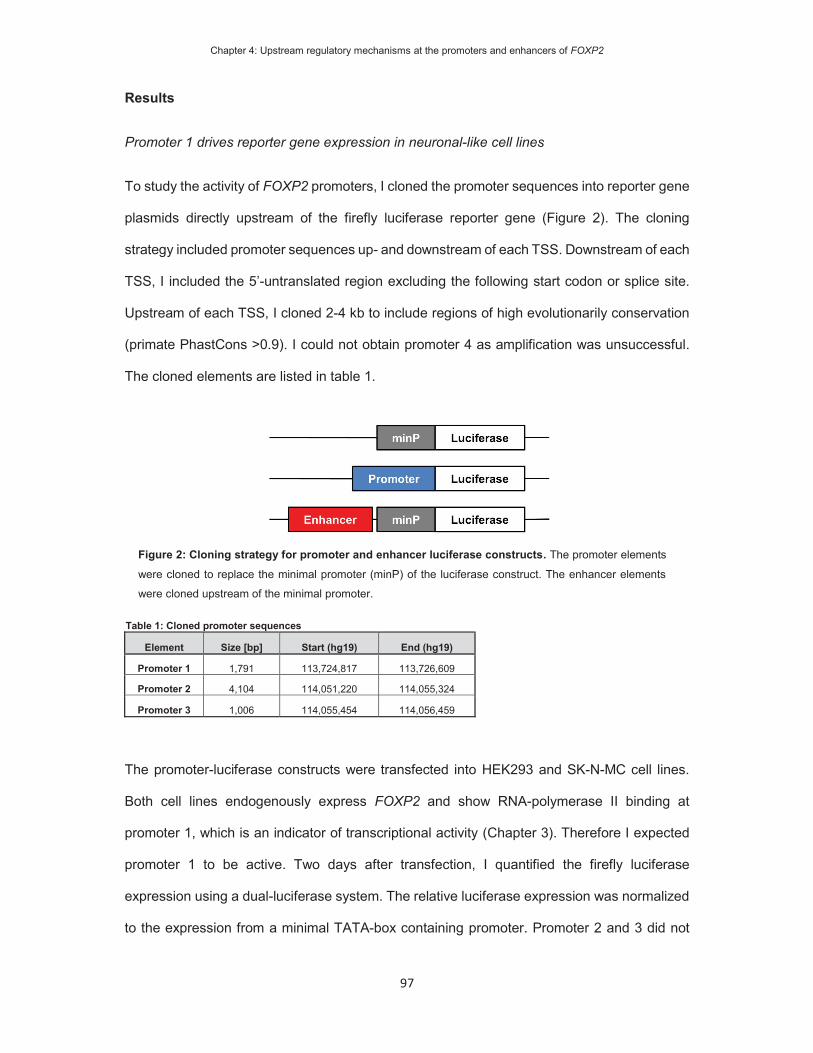
Chapter 4: Upstream regulatory mechanisms at the promoters and enhancers of FOXP2
97
Results
Promoter 1 drives reporter gene expression in neuronal-like cell lines
To study the activity of FOXP2 promoters, I cloned the promoter sequences into reporter gene
plasmids directly upstream of the firefly luciferase reporter gene (Figure 2). The cloning
strategy included promoter sequences up- and downstream of each TSS. Downstream of each
TSS, I included the 5’-untranslated region excluding the following start codon or splice site.
Upstream of each TSS, I cloned 2-4 kb to include regions of high evolutionarily conservation
(primate PhastCons >0.9). I could not obtain promoter 4 as amplification was unsuccessful.
The cloned elements are listed in table 1.
Table 1: Cloned promoter sequences
Element Size [bp] Start (hg19) End (hg19)
Promoter 1 1,791 113,724,817 113,726,609
Promoter 2 4,104 114,051,220 114,055,324
Promoter 3 1,006 114,055,454 114,056,459
The promoter-luciferase constructs were transfected into HEK293 and SK-N-MC cell lines.
Both cell lines endogenously express FOXP2 and show RNA-polymerase II binding at
promoter 1, which is an indicator of transcriptional activity (Chapter 3). Therefore I expected
promoter 1 to be active. Two days after transfection, I quantified the firefly luciferase
expression using a dual-luciferase system. The relative luciferase expression was normalized
to the expression from a minimal TATA-box containing promoter. Promoter 2 and 3 did not
Figure 2: Cloning strategy for promoter and enhancer luciferase constructs. The promoter elements
were cloned to replace the minimal promoter (minP) of the luciferase construct. The enhancer elements
were cloned upstream of the minimal promoter.

Chapter 4: Upstream regulatory mechanisms at the promoters and enhancers of FOXP2
98
show increased luciferase activity in HEK293 or SK-N-MC (Figure 3A), suggesting they are
inactive. This is in line with previous PolII binding assays (Chapter3: Figure 4). Promoter 1
significantly increased luciferase expression in HEK293 (5-fold) and in SK-N-MC cells (80-
fold). The stronger signal in SK-N-MC cells is in agreement with the earlier finding that PolII
binding at promoter 1 was more enriched in SK-N-MC (Chapter 3: Figure 4). Thus, the
luciferase results demonstrate that promoter 1 is active in both cell lines and may be more
active in SK-N-MC.
Figure 3: Basal promoter and enhancer activity in HEK293 and SK-N-MC. Relative luciferase activity
of A) promoter and B) enhancer elements driving firefly luciferase gene expression in HEK293 and SK-N-
MC cells. The promoter and enhancer constructs were each transfected with a control element expressing
the renilla luciferase under the control of a viral promoter. The firefly luciferase signal was divided by the
renilla signal to derive the relative luciferase activity. The activity for each luciferase construct was
normalized for the activity observed for the minimal promoter (minP). The constructs were measured in two
independent experiments in a total of six biological replicates, with the exception of elements 346, 700, 704
and 843 in SK-N-MC cells, which were measured in one experiment in three biological replicates. The
significance of the difference between each construct and minP was determined with two-way ANOVA and
post-hoc LSD testing. *p<0.05, ** p< 0.001. The significance in HEK293 is labeled with red asterisks; the
significance in SK-N-MC is labeled with blue asterisks.
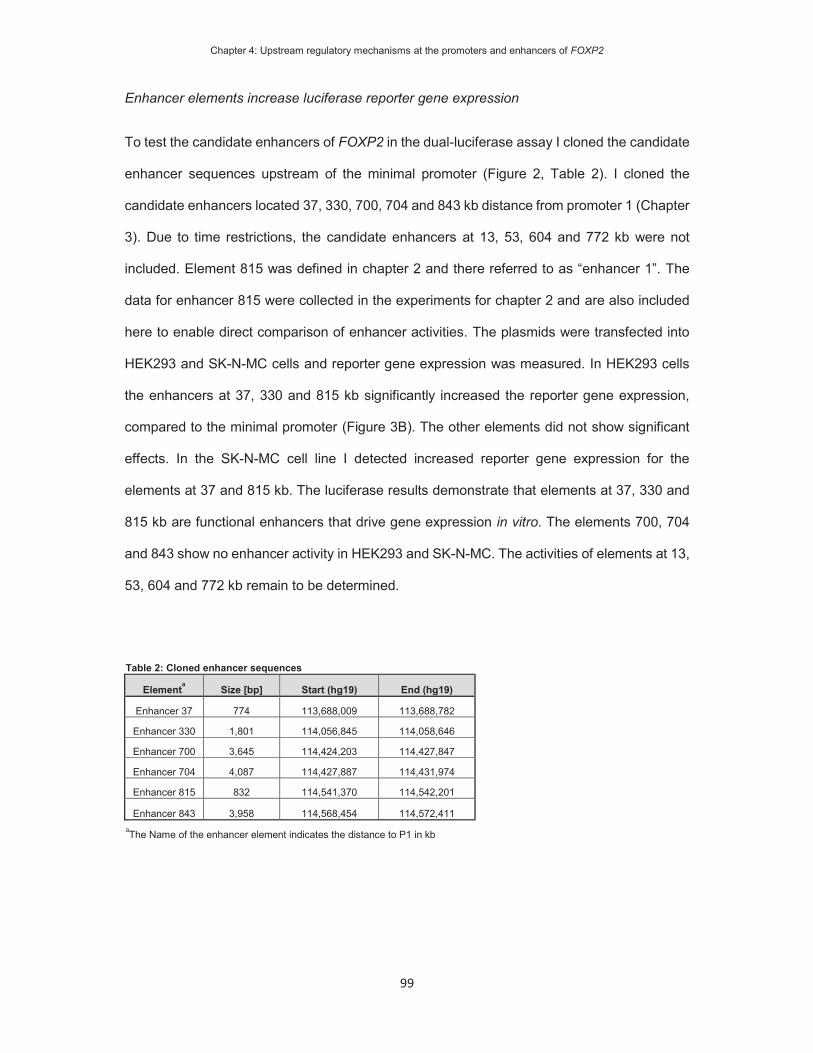
Chapter 4: Upstream regulatory mechanisms at the promoters and enhancers of FOXP2
99
Enhancer elements increase luciferase reporter gene expression
To test the candidate enhancers of FOXP2 in the dual-luciferase assay I cloned the candidate
enhancer sequences upstream of the minimal promoter (Figure 2, Table 2). I cloned the
candidate enhancers located 37, 330, 700, 704 and 843 kb distance from promoter 1 (Chapter
3). Due to time restrictions, the candidate enhancers at 13, 53, 604 and 772 kb were not
included. Element 815 was defined in chapter 2 and there referred to as “enhancer 1”. The
data for enhancer 815 were collected in the experiments for chapter 2 and are also included
here to enable direct comparison of enhancer activities. The plasmids were transfected into
HEK293 and SK-N-MC cells and reporter gene expression was measured. In HEK293 cells
the enhancers at 37, 330 and 815 kb significantly increased the reporter gene expression,
compared to the minimal promoter (Figure 3B). The other elements did not show significant
effects. In the SK-N-MC cell line I detected increased reporter gene expression for the
elements at 37 and 815 kb. The luciferase results demonstrate that elements at 37, 330 and
815 kb are functional enhancers that drive gene expression in vitro. The elements 700, 704
and 843 show no enhancer activity in HEK293 and SK-N-MC. The activities of elements at 13,
53, 604 and 772 kb remain to be determined.
Table 2: Cloned enhancer sequences
Elementa Size [bp] Start (hg19) End (hg19)
Enhancer 37 774 113,688,009 113,688,782
Enhancer 330 1,801 114,056,845 114,058,646
Enhancer 700 3,645 114,424,203 114,427,847
Enhancer 704 4,087 114,427,887 114,431,974
Enhancer 815 832 114,541,370 114,542,201
Enhancer 843 3,958 114,568,454 114,572,411aThe Name of the enhancer element indicates the distance to P1 in kb

Chapter 4: Upstream regulatory mechanisms at the promoters and enhancers of FOXP2
100
FOXP2 overexpression indicates autoregulation at promoters and enhancers
To test the autoregulation of FOXP2, I co-transfected the luciferase constructs with a plasmid
overexpressing the FOXP2 gene. As a control, the luciferase plasmids were co-transfected
with an empty pcDNA4 plasmid. Co-transfection experiments were conducted in HEK293 cells
because the transfection efficiency rate in SK-N-MC was below 20%, which did not guarantee
co-transfection of luciferase and expression plasmids in the same cells. FOXP2
overexpression increased the activity of promoter1, element 37 and 815 (Figure 4). Also, the
previously inactive promoter 2 and element 700 show significant increases of luciferase
expression. To determine if the increased enhancer activity is mediated by the direct binding
of FOXP2 to DNA, I tested two aetiological variants of FOXP2 that have been found in families
with monogenic forms of speech and language disorder (Lai et al., 2001; MacDermot et al.,
2005). The R553H variant of FOXP2 was found in the large multigenerational KE family and
has an amino-acid substitution in the DNA-binding domain (Lai et al., 2001). The R553H
variant is unable to bind to the consensus binding motif (Vernes et al., 2006). The other variant
is R328X, which was found in a smaller family and lacks the DNA-binding FOX domain due to
a premature stop codon (MacDermot et al., 2005) and cannot bind to DNA (Vernes et al.,
2006). As expected, the two variants fail to increase the activity of promoter 1, promoter 2 and
enhancers 37, 700, 815. The effects at promoter 1 and enhancer 37 are severely reduced for
the R328X variant, but remain significant in comparison to control. The reduced promoter and
enhancer activities suggest that the enhancing effects observed for wild-type FOXP2 were
mediated by direct DNA-binding. In agreement with this finding, a FOXP2 binding site within
promoter 1 has been detected by chromatin immunoprecipitation in human neuronal-like cell
lines SK-N-MC and PFSK-1 (Nelson et al., 2013). The aetiological variants increased the
activity of enhancer 330. The increase could be mediated by protein-protein interactions to
other DNA-binding factors, which in turn regulate the enhancer. In sum the luciferase results
suggest that FOXP2 is capable to auto-regulate via direct DNA-binding to its regulatory
elements.

Chapter 4: Upstream regulatory mechanisms at the promoters and enhancers of FOXP2
101
The FOXP family members regulate FOXP2 enhancers and promoters
FOXP2 can form hetero-dimers with the family members FOXP1 and FOXP4 to regulate target
gene expression (Li et al., 2004; Sin et al., 2015). Because FOXP2 regulates its enhancers
and promoters, it is conceivable that FOXP1 and FOXP4 have similar effects at the same
elements. After co-transfection of the luciferase constructs with a pcDNA4-FOXP1
overexpression plasmid, I detected increased activity for promoter 1, enhancer 37 and element
815 (Figure 5), which were also up-regulated by FOXP2 (Figure 4). Overexpression of FOXP4
increased the activity of enhancer 37 and decreased enhancer 330 (Figure 5). Thus, the three
FOXP family members increase enhancer 37. Promoter 1 and enhancer 815 are up-regulated
by both FOXP1 and FOXP2. The activity of enhancer 330, which was increased by the
aetiological FOXP2 variants, was repressed by FOXP4. The results suggest that the FOXPs
show regulatory effects at the same set of enhancers and the promoter 1.
Figure 4: Assessing auto-regulation of FOXP2 via promoter and enhancer constructs. Relative
luciferase activity of promoter and enhancer elements after co-transfection with FOXP2, FOXP2-R553H or
FOXP2-R328X in HEK293. The promoter and enhancer constructs were co-transfected with a control
construct expressing the renilla luciferase. The cells were additionally co-transfected with an empty pcDNA4
control, or FOXP2 overexpression plasmid. The firefly luciferase signal was divided by the renilla signal to
derive the relative luciferase activity. The activity for each luciferase construct was normalized for the activity
observed for the minimal promoter (minP). Each combination was tested in three biological replicates. The
statistical significant of the TF overexpression effect was determined with two-way ANOVA and post-hoc
LSD testing for each construct. P-value: *<0.05, ** < 0.001.

Chapter 4: Upstream regulatory mechanisms at the promoters and enhancers of FOXP2
102
The transcription factors LEF1 and PAX6, but not POU3F2, regulate human FOXP2 promoter and enhancers in vitro
I tested the promoter and enhancer constructs in combination with an overexpression vector
of LEF1. The WNT-signalling pathway is active under normal culture conditions in HEK293
cells (Oloumi et al., 2006). Therefore, I expected LEF1 to be active in the absence of further
stimulation of the WNT-pathway. I co-transfected the regulatory elements with a pBABE-LEF1
overexpression plasmid and determined the luciferase activity. I observed significantly
increased activity of promoter1, promoter 3, enhancer 37 and enhancer 330 (Figure 6). Thus,
LEF1 activates regulatory elements of FOXP2. Since LEF1 is the downstream effector of the
WNT-signalling pathway, these findings suggest that the promoter and enhancer activities
might be controlled by WNT-signalling.
Figure 5: Regulatory effect of FOXP1 and FOXP4 on promoter and enhancer constructs. Relative
luciferase activity of promoter and enhancer elements in HEK293 cells. The promoter and enhancer
constructs were each transfected with a control construct expressing the renilla luciferase under the control
of a viral promoter. The cells were also transfected with a control, FOXP1 or FOXP4 overexpression
plasmid. The firefly luciferase signal was divided for the renilla signal to derive the relative luciferase activity.
The activity for each luciferase construct was normalized for the activity observed for the minimal promoter
(minP). Each combination was tested in three biological replicates. The statistical significant of the TF
overexpression effect was determined with two-way ANOVA and post-hoc LSD testing for each construct.
P-value: *<0.05, ** < 0.001.

Chapter 4: Upstream regulatory mechanisms at the promoters and enhancers of FOXP2
103
Next, I tested the overexpression of PAX6, which reduced the activity of promoter 1 and
element 815 (Figure 7A). The luciferase activity remained stable for the other regulatory
elements. The luciferase results of promoter 1 and element 815 indicate a repressive effect of
PAX6 on FOXP2 expression. This is inconsistent with published results from mouse and
zebrafish embryos, where Pax6/pax6 increased Foxp2/foxp2 expression (Coutinho et al.,
2011). Despite the difference in direction of effect, my luciferase findings do support the
hypothesis that FOXP2 is under control of PAX6 in humans.
I went on to investigate a prior suggested link between POU3F2 and FOXP2 by testing the
regulatory elements that I had identified, in combination with POU3F2 overexpression. I co-
transfected the luciferase constructs with pYFP-POU3F2 and an empty pcDNA4 plasmid as
Figure 6: Regulatory effect of the WNT-signalling TF LEF1 on promoter and enhancer constructs.
Relative luciferase activity of promoter and enhancer elements in HEK293 cells. The promoter and
enhancer constructs were each transfected with a control construct expressing the renilla luciferase under
the control of a viral promoter. The cells were also transfected with a pcDNA4, or LEF1 overexpression
plasmid. The firefly luciferase signal was divided by the renilla signal to derive the relative luciferase activity.
The activity for each luciferase construct was normalized for the activity observed for the minimal promoter
(minP). Each combination was tested in three biological replicates. The statistical significance of the TF
overexpression effect was determined with two-way ANOVA and post-hoc LSD testing for each construct.
P-value: *p<0.05, **p< 0.001.

Chapter 4: Upstream regulatory mechanisms at the promoters and enhancers of FOXP2
104
control. The in vitro activity of all promoters and enhancers remained constant (Figure 7B),
indicating that POU3F2 does not regulate the tested FOXP2 enhancers and promoters.
SOX5 and combinations of TBR1 with CASK regulate FOXP2 promoter and enhancer
activity
Next, I tested the regulation of the FOXP2 regulatory elements by TBR1. Co-transfection with
TBR1 reduced the activity of enhancer 330 and 37. TBR1 has been shown to exert both
Figure 7: Testing regulatory effects of PAX6 and POU3F2 on promoter and enhancer constructs. Relative luciferase activity of promoter and enhancer elements in HEK293 cells for experiments with A)
PAX6, and B) POU3F2. The promoter and enhancer constructs were each transfected with a control
construct expressing the renilla luciferase under the control of a viral promoter and the relevant TF
overexpression construct or a pcDNA4 control plasmid. The firefly luciferase signal was divided by the
renilla signal to derive the relative luciferase activity. The activity for each luciferase construct was
normalized for the activity observed for the minimal promoter (minP). Each combination was tested in three
biological replicates. The statistical significance for each construct was assessed using two-way ANOVA
and post-hoc LSD testing. P-value: * p<0.05, ** p< 0.001.

Chapter 4: Upstream regulatory mechanisms at the promoters and enhancers of FOXP2
105
activating and repressive effects on target gene expression (Bedogni et al., 2010; Han et al.,
2011), which may dependent on the co-activator CASK (Hsueh et al., 2000; Wang et al.,
2004). I therefore co-transfected the promoter and enhancer constructs with TBR1, CASK or
a combination of both. The combination increased the activity of the enhancer 37 and promoter
1 (Figure 7). Overexpression of CASK alone had a repressive effect on enhancer 37. As
predicted, the activating effect of TBR1 was detectable at promoter 1 and enhancer 37 only in
combination with the co-activator CASK. As CASK does not bind DNA, the repressive effect,
observed in the absence of TBR1, must be mediated via protein-protein interactions to another
DNA-binding factor.
Finally, I tested the regulatory elements in combination with a pcDNA4-SOX5 overexpression
plasmid. Promoter 1, elements 37 and 704 increased luciferase gene expression in response
to SOX5 overexpression (Figure 5C). The activity of enhancer 330 however decreased. Thus,
SOX5 exerts repressive and activating effects. Repressive and activating effects of SOX5
have been reported before (Kwan et al., 2008; Aza-Carmona et al., 2011). This suggests that
SOX5 may enhance or decrease FOXP2 expression levels, depending on the regulatory
element. To my best knowledge, opposing regulatory effects of single TFs at regulatory
elements of the same target gene have not been reported.
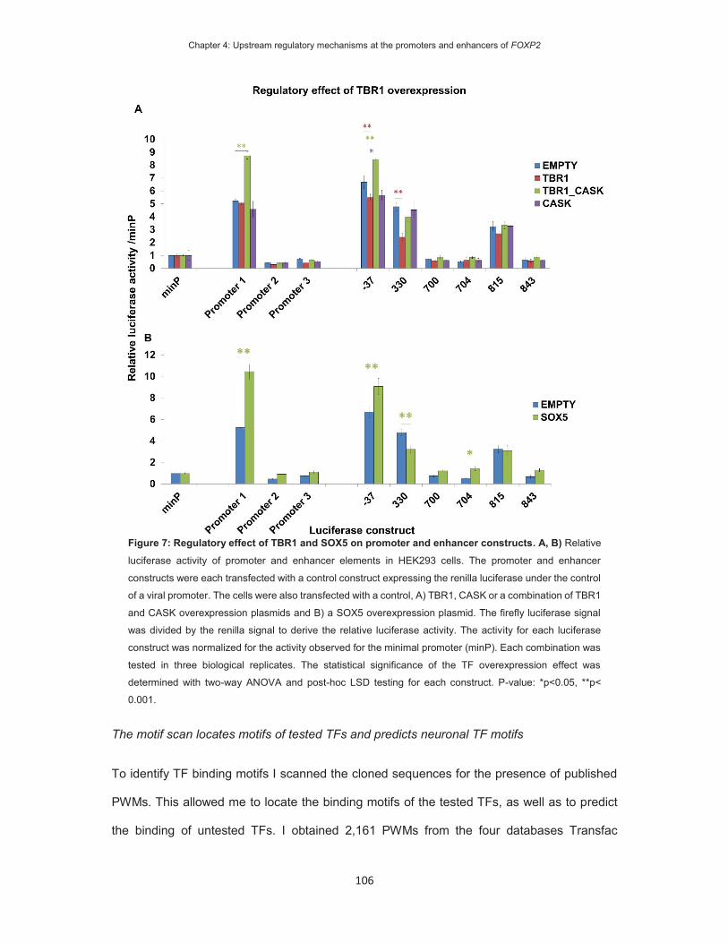
Chapter 4: Upstream regulatory mechanisms at the promoters and enhancers of FOXP2
106
The motif scan locates motifs of tested TFs and predicts neuronal TF motifs
To identify TF binding motifs I scanned the cloned sequences for the presence of published
PWMs. This allowed me to locate the binding motifs of the tested TFs, as well as to predict
the binding of untested TFs. I obtained 2,161 PWMs from the four databases Transfac
Figure 7: Regulatory effect of TBR1 and SOX5 on promoter and enhancer constructs. A, B) Relative
luciferase activity of promoter and enhancer elements in HEK293 cells. The promoter and enhancer
constructs were each transfected with a control construct expressing the renilla luciferase under the control
of a viral promoter. The cells were also transfected with a control, A) TBR1, CASK or a combination of TBR1
and CASK overexpression plasmids and B) a SOX5 overexpression plasmid. The firefly luciferase signal
was divided by the renilla signal to derive the relative luciferase activity. The activity for each luciferase
construct was normalized for the activity observed for the minimal promoter (minP). Each combination was
tested in three biological replicates. The statistical significance of the TF overexpression effect was
determined with two-way ANOVA and post-hoc LSD testing for each construct. P-value: *p<0.05, **p<
0.001.

Chapter 4: Upstream regulatory mechanisms at the promoters and enhancers of FOXP2
107
(Wingender et al., 2000), HOCOMOCO (Kulakovskiy et al., 2013), HT-Selex (Jolma et al.,
2013) and Jaspar (Mathelier et al., 2014). These PWMs were used within the MotifScanner
algorithm to scan the cloned sequences (Aerts et al., 2003; Coessens et al., 2003). The
databases included the PWMs for all investigated TFs (FOXP1, FOXP2, LEF1, PAX6,
POU3F2, TBR1 and SOX5), except FOXP4. Therefore, I could not identify potential FOXP4
binding sites in the enhancer and promoter sequences.
The motif scan predicted between 373 (Enhancer 815) to 855 (Enhancer 330) motifs in the 9
analysed sequences (3 promoters, 6 enhancers) (Table 3, Appendix 2: Table S2). The
predictions located putative motifs of the previously tested TFs (Table 4). FOXP2 motifs were
predicted within seven cloned elements (promoter 1-3, enhancer 330, enhancer 700,
enhancer 815 and enhancer 843). However, in response to FOXP2 overexpression, I only
detected changes in reporter gene expression at five regulatory elements (Promoter 1,
promoter 2, enhancer 37, enhancer 700 and enhancer 815). Therefore, the results of the motif
prediction and luciferase are concordant at four elements (Promoter1, promoter 2, enhancer
700 and enhancer 815). The FOXP2 motifs at promoter 3, enhancer 330 and enhancer 843
represent false positive predictions, possibly caused by non-functional binding sites. In
addition, no FOXP2 motif was predicted for enhancer 37. Explanations for the false negative
prediction may be a too stringent detection threshold in the motif scan or that the observed
regulation is due to an indirect interaction. In total I identified 19 TF motifs at concordant
promoters and enhancers (Table 5). To verify that changes in luciferase expression were
mediated by direct TF - DNA interactions it would be necessary to mutate or delete the
predicted TF motifs.
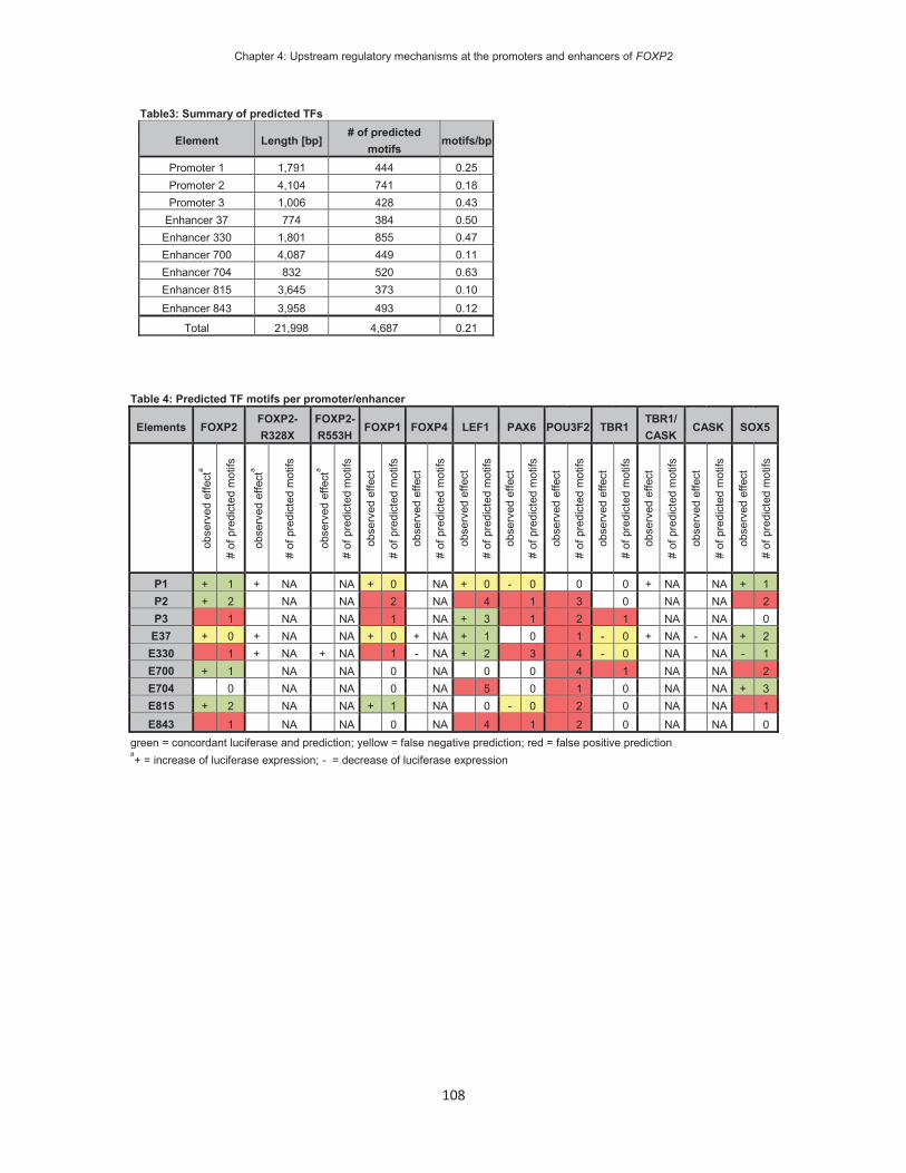
Chapter 4: Upstream regulatory mechanisms at the promoters and enhancers of FOXP2
108
Table3: Summary of predicted TFs Element Length [bp] # of predicted
motifsmotifs/bp
Promoter 1 1,791 444 0.25Promoter 2 4,104 741 0.18Promoter 3 1,006 428 0.43
Enhancer 37 774 384 0.50Enhancer 330 1,801 855 0.47Enhancer 700 4,087 449 0.11Enhancer 704 832 520 0.63Enhancer 815 3,645 373 0.10Enhancer 843 3,958 493 0.12
Total 21,998 4,687 0.21
Table 4: Predicted TF motifs per promoter/enhancer
Elements FOXP2 FOXP2-R328X FOXP2-
R553H FOXP1 FOXP4 LEF1 PAX6 POU3F2 TBR1 TBR1/ CASK
CASK SOX5
obse
rved
effe
cta
# of
pre
dict
ed m
otifs
obse
rved
effe
cta
# of
pre
dict
ed m
otifs
obse
rved
effe
cta
# of
pre
dict
ed m
otifs
obse
rved
effe
ct
# of
pre
dict
ed m
otifs
obse
rved
effe
ct
# of
pre
dict
ed m
otifs
obse
rved
effe
ct
# of
pre
dict
ed m
otifs
obse
rved
effe
ct
# of
pre
dict
ed m
otifs
obse
rved
effe
ct
# of
pre
dict
ed m
otifs
obse
rved
effe
ct
# of
pre
dict
ed m
otifs
obse
rved
effe
ct
# of
pre
dict
ed m
otifs
obse
rved
effe
ct
# of
pre
dict
ed m
otifs
obse
rved
effe
ct
# of
pre
dict
ed m
otifs
P1 + 1 + NA NA + 0 NA + 0 - 0 0 0 + NA NA + 1 P2 + 2 NA NA 2 NA 4 1 3 0 NA NA 2 P3 1 NA NA 1 NA + 3 1 2 1 NA NA 0
E37 + 0 + NA NA + 0 + NA + 1 0 1 - 0 + NA - NA + 2 E330 1 + NA + NA 1 - NA + 2 3 4 - 0 NA NA - 1 E700 + 1 NA NA 0 NA 0 0 4 1 NA NA 2 E704 0 NA NA 0 NA 5 0 1 0 NA NA + 3 E815 + 2 NA NA + 1 NA 0 - 0 2 0 NA NA 1 E843 1 NA NA 0 NA 4 1 2 0 NA NA 0
green = concordant luciferase and prediction; yellow = false negative prediction; red = false positive prediction a+ = increase of luciferase expression; - = decrease of luciferase expression

Chapter 4: Upstream regulatory mechanisms at the promoters and enhancers of FOXP2
109
Table 5: Location of concordant TF motifs Element TF Start [hg18] End [hg18] PWM ID Motif
Promoter 1 FOXP2 113,513,165 113,513,175 MA0593 GTTGTTTACATPromoter 1 SOX5 113,513,220 113,513,227 HM00343 GAACAATAPromoter 2 FOXP2 113,841,749 113,841,759 MA0593 ATTGTTTACTTPromoter 2 FOXP2 113,840,403 113,840,413 MA0593 ATGTAAACAGAPromoter 3 LEF1 113,843,051 113,843,056 M00805 GTTTGAPromoter 3 LEF1 113,843,147 113,843,161 HT00135 AAACATGAAAGGAGTPromoter 3 LEF1 113,843,177 113,843,183 HM00191 CTTTGTA
Enhancer 37 LEF1 113,475,432 113,475,438 HM00191 CTTTGATEnhancer 37 SOX5 113,475,604 113,475,611 HM00343 CATTGTTG
Enhancer 300 LEF1 113,844,283 113,844,289 HM00191 CTTTGTAEnhancer 300 LEF1 113,844,761 113,844,770 M01022 CACTTTGAAAEnhancer 300 LEF1 113,844,980 113,844,994 HT00135 AGTGCTTTCATCTTTEnhancer 300 SOX5 113,844,505 113,844,512 HM00343 CATTGTTCEnhancer 704 SOX5 114,215,241 114,215,250 M00042 TTAACAATATEnhancer 704 SOX5 114,216,589 114,216,598 M00042 CAATTGTTAGEnhancer 704 SOX5 113,513,220 113,513,227 HM00343 GAACAATAEnhancer 815 FOXP2 114,328,727 114,328,737 MA0593 CAGTAAACACAEnhancer 815 FOXP2 114,328,816 114,328,824 HM00111 ATGTTTACTEnhancer 815 FOXP1 114,328,724 114,328,738 MA0481 TATCAGTAAACACAC
A promising approach to select novel TFs that may regulate FOXP2 is to prioritize TFs that
are expressed in the brain. Therefore, I filtered the list of predicted TFs using the DAVID
bioinformatics web tool to filter for TFs within the brain expressed category (UniProt_Tissue:
brain) (Huang da et al., 2009) and detected 145 TFs within the brain expressed category
(Appendix 2: Table S3). As a complementary alternative, I prioritized TFs that are collected in
the gene ontology category of neuron differentiation (Appendix 2: Table S4) because FOXP2
function in neuron differentiation is well established (Vernes et al., 2006; Vernes et al., 2011;
Tsui et al., 2013; Devanna et al., 2014). The “neuron differentiation” list contained 86 TFs, of
which 54 overlap with the “brain expressed” category. The absence of some “neuron
differentiation” genes from the “brain expressed” list may be caused by incorrect manual
annotation of the GO-terms. An alternative explanation may be the absence of TFs, which are
expressed during brain development in the “brain expressed” list. Both lists include several
TFs that I studied experimentally in this chapter, such as SOX5, POU3F2, and PAX6. In
addition the neuron differentiation list includes TFs which have been shown to be co-
expressed with FOXP2, such as ATOH1, DLX5, EMX1, LMX1B (as described in Chapter 1).

Chapter 4: Upstream regulatory mechanisms at the promoters and enhancers of FOXP2
110
The motif scan could be used to prioritize additional TFs for further luciferase experiments to
investigate the regulation of FOXP2 by these TFs.
Chapter 4: Upstream regulatory mechanisms at the promoters and enhancers of FOXP2
111
Discussion
FOXP2 enhancer and promoters are active in neuronal-like cell lines
Promoter 1 is transcriptionally active in HEK293 and SK-N-MC cells, which is in agreement
with the report that endogenous FOXP2 expression is driven by this promoter in these cell
lines (Schroeder and Myers, 2008). HEK293 and SK-N-MC cells express comparable levels
of FOXP2 mRNA, yet the promoter activity in HEK293 cells was substantially lower than in
SK-N-MC. Similarly, I previously detected lower RNA polymerase II binding at this promoter
in the HEK293 cell line (Chapter 3). These findings suggest that regulation of the promoter
does not solely determine endogenous FOXP2 expression in HEK293 and that enhancers
increase expression levels. In accordance with this hypothesis, three enhancers were active
in HEK293 cells. One of the enhancers, enhancer 330, was inactive in SK-N-MC, which may
contribute to the similarity in expression levels of FOXP2 in HEK293 and SK-N-MC. However,
additional regulatory elements, such as promoter 4 and four untested candidate enhancers,
may further contribute to the total endogenous FOXP2 expression (Chapter 3).
Promoter 2 and 3 did not drive luciferase expression in SK-N-MC or HEK293 cells, which is in
accordance with the previous observation that these promoters are not bound by RNA
polymerase II in these cells (Chapter 3). In addition, this is in agreement with a previous study,
which did not detect mRNA transcripts originating from promoter 2 or 3 in these cell lines
(Schroeder and Myers, 2008). However, Schroeder and Myers detected tissue-specific
expression in non-neuronal tissue, such as the cervix, liver and trachea. Luciferase
experiments in cell lines derived from these tissues may reveal promoter 2 and/or promoter 3
activities. Because I wanted to gain insight into the neuronal function of FOXP2, its regulation
in non-neuronal tissue was outside the scope of this thesis.
I demonstrated that the enhancers at 37 and 330 kb increase reporter gene expression in
vitro. The results support the hypothesis that the measured chromatin interactions represent
functional promoter-enhancer loops (Chapter3). The basal activity of the elements was tested

Chapter 4: Upstream regulatory mechanisms at the promoters and enhancers of FOXP2
112
in the same FOXP2(+) cell lines which had been used to study the chromatin interactions. The
activity of enhancer 330 was detected in HEK293 and absent in SK-N-MC. However, the
chromatin interactions were comparable between both cell lines (see Chapter 3: Figure 5).
The chromatin interaction is therefore not proportional to the in vitro activity of the enhancer
element. The absence of enhancer 330 activity in SK-N-MC cells suggests that the enhancer
may be “poised” in this cell line. Poised enhancers regulate rapid changes in target gene
expression in response to developmental or environmental stimuli (Zentner et al., 2011; Jin et
al., 2013; Shlyueva et al., 2014). As I observed repressing effects on enhancer 330 by TBR1,
SOX5 and FOXP4, the inactivity of this enhancer in SK-N-MC could be caused by the
presence of these TFs. The knock-down of these TFs in SK-N-MC cells might potentially
activate enhancer 330. An alternative explanation could be the absence of an activating TF.
A promising candidate could be LEF1, which increased enhancer 330 activity in HEK293 cells.
As LEF1 activity is dependent on WNT-signalling, this hypothesis could be tested by activation
of this pathway in SK-N-MC cells.
Positive direct auto-regulation of FOXP2 likely contributes to neuronal expression
I hypothesized that FOXP2 could auto-regulate via its promoters or enhancers, and detected
increased activity of five regulatory elements in response to FOXP2 overexpression. Positive
auto-regulatory loops enable rapid amplification of the TF and to maintain expression at stable
plateau levels (Bateman, 1998). Auto-regulation has been described for developmental
(Bateman, 1998; di Gennaro et al., 2013; Mead et al., 2013) and neurodevelopmental TFs
(Meredith et al., 2009). Heterozygous mutations in positive auto-regulatory TFs could
decrease the expression of the wildtype protein from the healthy allele. Consequently, the
expression of wildtype protein would be severely reduced and below 50% compared to the
non-mutated situation. Thus, heterogeneous mutations of positive auto-regulatory TFs may
lead to haploinsufficiency, where the absence of one healthy allele is sufficient to cause a
disease phenotype (Lamb et al., 2012; di Gennaro et al., 2013; Mead et al., 2013). Mutations
of FOXP2 or the loss of one FOXP2 allele would be predicted to result in reduced activation
Chapter 4: Upstream regulatory mechanisms at the promoters and enhancers of FOXP2
113
of FOXP2 expression and subsequently reduce the expression of the healthy allele. Indeed,
heterogeneous whole-gene deletions of FOXP2 have been detected in people with speech
impairments, strongly indicating that haploinsufficiency underlies the molecular aetiology of
the FOXP2-related speech phenotype in some cases (Zeesman et al., 2006; Palka et al.,
2012; Rice et al., 2012). In addition, studies in mouse models of aetiological FOXP2 mutations
indicate that heterozygous mice are overtly normal with specific effects on cortico-striatal
neural networks (Groszer et al., 2008; Gaub et al., 2016). Positive auto-regulation could
explain the occurrence of the haploinsufficiency effect in a subset of FOXP2-positive tissues,
as this effect would be present only in target tissue of the enhancers.
I have shown that both FOXP1 and FOXP4 can regulate the enhancers and promoters of
FOXP2 in vitro, suggesting that they may also enhance FOXP2 expression in vivo. The FOXP
family members have been shown to form functional hetero-dimers (Li et al., 2004; Sin et al.,
2015; Spaeth et al., 2015) and hetero-dimerization could influence the regulation of FOXP2.
HEK293 cells do express FOXP1, FOXP2 and FOXP4 (Gierman et al., 2007; Uhlen et al.,
2015). It is thus possible that endogenous expression of these TFs influenced the luciferase
results and that the observed effects are caused by FOXP dimers. Co-transfection of FOXP
family members may further investigate the role of hetero- and homodimers in the regulation
of FOXP2 enhancers.
Regulatory TFs of promoters/enhancers suggest upstream pathways for FOXP2 expression
I detected that LEF1 is capable of increasing the activity of promoter 1, promoter 3 and two
enhancer elements. In prior work LEF1 has been shown to activate expression of target genes
(Arce et al., 2006), which is consistent with the observed effects in my experiments. Moreover,
the increase of enhancer and promoter activity after LEF1 overexpression is consistent with
previously described effects of Lef1 in zebrafish, where knockdown of lef1 abolishes foxp2
expression in the tectum, mid-hindbrain boundary and hindbrain (Bonkowsky et al., 2008).
Thus, the luciferase results indicate that LEF1 regulates FOXP2 also in human cells. It remains

Chapter 4: Upstream regulatory mechanisms at the promoters and enhancers of FOXP2
114
to be determined if regulation by LEF1 occurs in the same regions as observed for the
zebrafish. The activities of promoter 1, promoter 3, enhancer 37 and enhancer 330 during
development and adulthood should be characterized to address this question. As the protein
structures of TFs are evolutionary more stable than enhancer sequences (Domene et al.,
2013; Rubinstein and de Souza, 2013) the human enhancers are likely recognized by
orthologous TFs in related species. Thus, the human enhancer activities could be tested in
transgenic mice (Visel et al., 2008), which I did for the most promising enhancers in chapter
5.
I found that in human cells PAX6 overexpression decreased promoter 1 and enhancer 815
activity. A previous report showed in zebrafish that pax6 knockdown reduced foxp2 expression
in the developing telencephalon, suggesting that it activates foxp2 expression (Coutinho et al.,
2011). The conflicting results between my data and the study by Coutinho et al. could be
caused by inter-species differences. Also, my data was obtained within neuronal-like cell lines,
which are unlikely to resemble the transcriptional program of embryonic telencephalon.
However, my luciferase data indicates that FOXP2 is a target gene of PAX6 in humans. Co-
expression of both TFs was shown in the amygdala and striatum of non-human primates and
rats (Kaoru et al., 2010) and the embryonic (E12) cortical plate of mice (Tsui et al., 2013). In
the developing cortex of Pax6 knock-out mice, cortico-fugal axons show aberrant migratory
pathways (Jones et al., 2002). FOXP2 is expressed specifically in cortico-fugal neurons of
cortical layer VI (Hisaoka et al., 2010) and regulates neurite outgrowth (Vernes et al., 2011).
Thus, aberrant Foxp2 regulation in Pax6 knock-out mice may contribute to the defect in axon
pathfinding.
In a previous report, POU3F2 has been suggested to regulate the expression of FOXP2 via a
putative enhancer element in intron 8 (Maricic et al., 2013). Maricic et al. suggested that the
regulation by POU3F2 was reduced in modern humans, due to a genetic variant that arose in
humans after the split from a common ancestor with Neanderthals. However, the element
proposed by Maricic et al. did not show independent in vitro or in vivo activity in their study
Chapter 4: Upstream regulatory mechanisms at the promoters and enhancers of FOXP2
115
and was not included here. In my experiments, POU3F2 did not increase or decrease the
activity of the promoters and enhancers that I tested. The presented results cannot exclude
the possibility that POU3F2 is capable of regulating FOXP2 expression via untested regulatory
elements or indirect interactions.
TF important for cortical neuron subtype specification may contribute to FOXP2 expression
I identified regulatory effects on FOXP2 promoters and enhancers by SOX5, which is
expressed in post-migratory neurons of the developing cortex and is involved in subtype
specification (Kwan et al., 2008; Lai et al., 2008). Similar to SOX5, TBR1 is involved in the
development and maturation of cortical neurons (Hevner et al., 2001; Han et al., 2011). FOXP2
is involved in embryonic cortical development (Tsui et al., 2013) and restricted to cortical layer
VI (Lai et al., 2003). Thus, in the adult cortex expression of TBR1 and SOX5 overlaps with
FOXP2 expression in cortical layer VI. I showed that TBR1, in absence of the co-activator
CASK, represses the activity of enhancer 330, while in presence of CASK it promotes activity
of enhancer 37 and promoter 1. CASK is expressed throughout the cortex with the highest
expression in cortical layers III and IV (Kristiansen et al., 2010). SOX5 and TBR1 in
combination with CASK increase the activity of enhancer 37. This could indicate that enhancer
37 is active in cortical layer VI, where FOXP2 is co-expressed with these TFs. However,
enhancer 330 activity is decreased by TBR1 and SOX5. It is therefore unlikely that both
enhancer 37 and 330 are active in the same neurons. The enhancer activity pattern and an
assessment of the overlap with FOXP2 in the brain are thus important to fully evaluate the
effects observed in my experiments. As there are no methods to study enhancer activity in
human brains it is required to test the enhancers in transgenic animals (Visel et al., 2007).
Mutations in components of the upstream regulatory network reported in disease phenotypes

Chapter 4: Upstream regulatory mechanisms at the promoters and enhancers of FOXP2
116
Some TFs that regulate the FOXP2 enhancers and promoters may cause disease phenotypes
if mutated. Rare de novo mutations of TBR1 have been implicated in the aetiology of autism
spectrum disorder (ASD) (O'Roak et al., 2012b; O'Roak et al., 2012a). Patients with ASD have
impaired communication skills and language disruption is a common phenotype shared
between patients with TBR1 and FOXP2 mutations (Deriziotis et al., 2014). Haploinsufficiency
of SOX5 causes intellectual disability with prominent speech delay (Lamb et al., 2012;
Schanze et al., 2013). Mutations of PAX6 and chromosomal rearrangements at the PAX6
gene have been shown to cause aniridia, a disorder that is characterized by the absence of
the iris in affected cases (Prosser and van Heyningen, 1998). In rare cases mutations of PAX6
have been found in patients with intellectual disability (ID) and autism (Ticho et al., 2006;
Graziano et al., 2007; Davis et al., 2008). The possibility of an underlying molecular link
between language phenotypes observed in for mutations in FOXP2, PAX6, TBR1 or SOX5
need to be addressed in future studies.
In this chapter, I included enhancer 815, which I have identified in chapter 2 downstream of a
de novo inversion breakpoint found in a child with a complex rearrangement and language
deficits (Becker et al., 2015; Moralli et al., 2015). The proximity to FOXP2 suggested that the
de-regulation of this gene might contribute to the observed phenotype in this child. In the
present chapter, I determined that this element is responsive to the overexpression of PAX6,
FOXP1 and FOXP2. FOXP1 mutations have been found in cases of autism spectrum
disorders (Bacon et al., 2015), which are partly characterized by communication problems.
The language problems of people with FOXP1 and PAX6 mutations could be secondary to
major problems in unrelated molecular pathways. PAX6 mutations commonly cause
developmental problems, which are unrelated to cognition and behaviour (Prosser and van
Heyningen, 1998). It will be important to identify the patterns of tissue activity of element 815
to determine whether and how these overlap with sites of expression of FOXP1 and PAX6.
The brain regions that are affected in the child described by Moralli et al. have not been
reported. The overlap of affected brain regions and the expression of FOXP1 or PAX6, as well

Chapter 4: Upstream regulatory mechanisms at the promoters and enhancers of FOXP2
117
as the activity of element 815 could provide an explanation for the molecular aetiology of the
observed phenotype.
HEK293 cells have been shown to express FOXP1, FOXP4, SOX5, CASK, LEF1 and PAX6
(Gierman et al., 2007; Uhlen et al., 2015). In an alternative experiment the endogenous TF
levels could be knocked down, using RNA silencing techniques, to assess the effects on
promoters and enhancers. Ideally, follow-up experiments should employ cells obtained from
tissue and developmental periods in which the regulatory effect is expected to occur. It will be
useful to switch to more advanced cellular systems, such as primary neurons from mouse
embryos or induced pluripotent stem (iPS) cells. In developmental protocols of iPS cells,
FOXP2 is used as a marker for development stages (Molina-Hernandez et al., 2013; Belinsky
et al., 2014). Therefore the longitudinal expression pattern of FOXP2 has been described for
this system (Belinsky et al., 2014). This makes iPS cells an ideal system to manipulate the
expression of potential regulatory TFs and to analyse the effect on FOXP2 expression during
their development.
In this chapter I demonstrated the in vitro activity of previously identified enhancer elements
and one promoter of FOXP2. The enhancer and promoter activities respond to the
overexpression of several TFs that had previously been linked to FOXP2 in the literature.
However, changes of endogenous FOXP2 mRNA in response to TFs still need to be
determined. In silico motif scan partially predicted the DNA binding motifs in the responding
regulatory sequences and suggested additional TFs that may regulate the
enhancers/promoters. The activity patterns of the enhancer and promoter elements need to
be characterized to address the developmental period and target tissue of the observed
regulatory effects, which I describe in the following chapter. Overall, the results of the current
chapter indicated the influence of known developmental programs and signalling pathways on
the expression of FOXP2 and open new directions to investigate the biological role of this TF
in normal and pathogenic development.

Chapter 4: Upstream regulatory mechanisms at the promoters and enhancers of FOXP2
118
References
Aerts S, Thijs G, Coessens B, Staes M, Moreau Y, De Moor B (2003) Toucan: deciphering the cis-regulatory logic of coregulated genes. Nucleic Acids Res 31:1753-1764.
Alon U (2007) Network motifs: theory and experimental approaches. Nat Rev Genet 8:450-461. Arce L, Yokoyama NN, Waterman ML (2006) Diversity of LEF/TCF action in development and disease.
Oncogene 25:7492-7504. Aza-Carmona M, Shears DJ, Yuste-Checa P, Barca-Tierno V, Hisado-Oliva A, Belinchon A, Benito-
Sanz S, Rodriguez JI, Argente J, Campos-Barros A, Scambler PJ, Heath KE (2011) SHOX interacts with the chondrogenic transcription factors SOX5 and SOX6 to activate the aggrecan enhancer. Human molecular genetics 20:1547-1559.
Bacon C, Schneider M, Le Magueresse C, Froehlich H, Sticht C, Gluch C, Monyer H, Rappold GA (2015) Brain-specific Foxp1 deletion impairs neuronal development and causes autistic-like behaviour. Molecular psychiatry 20:632-639.
Bateman E (1998) Autoregulation of eukaryotic transcription factors. Progress in nucleic acid research and molecular biology 60:133-168.
Becker M, Devanna P, Fisher SE, Vernes SC (2015) A chromosomal rearrangement in a child with severe speech and language disorder separates FOXP2 from a functional enhancer. Molecular cytogenetics 8:69.
Bedogni F, Hodge RD, Elsen GE, Nelson BR, Daza RA, Beyer RP, Bammler TK, Rubenstein JL, Hevner RF (2010) Tbr1 regulates regional and laminar identity of postmitotic neurons in developing neocortex. Proc Natl Acad Sci U S A 107:13129-13134.
Belinsky GS, Rich MT, Sirois CL, Short SM, Pedrosa E, Lachman HM, Antic SD (2014) Patch-clamp recordings and calcium imaging followed by single-cell PCR reveal the developmental profile of 13 genes in iPSC-derived human neurons. Stem cell research 12:101-118.
Bhatia S, Bengani H, Fish M, Brown A, Divizia MT, de Marco R, Damante G, Grainger R, van Heyningen V, Kleinjan DA (2013) Disruption of Autoregulatory Feedback by a Mutation in a Remote, Ultraconserved PAX6 Enhancer Causes Aniridia. Am J Hum Genet 93:1126-1134.
Bonkowsky JL, Wang X, Fujimoto E, Lee JE, Chien CB, Dorsky RI (2008) Domain-specific regulation of foxP2 CNS expression by lef1. BMC developmental biology 8:103.
Bruce HA, Margolis RL (2002) FOXP2: novel exons, splice variants, and CAG repeat length stability. Human genetics 111:136-144.
Chan HM, La Thangue NB (2001) p300/CBP proteins: HATs for transcriptional bridges and scaffolds. J Cell Sci 114:2363-2373.
Coessens B, Thijs G, Aerts S, Marchal K, De Smet F, Engelen K, Glenisson P, Moreau Y, Mathys J, De Moor B (2003) INCLUSive: A web portal and service registry for microarray and regulatory sequence analysis. Nucleic Acids Res 31:3468-3470.
Cook DL, Gerber LN, Tapscott SJ (1998) Modeling stochastic gene expression: Implications for haploinsufficiency. Proc Natl Acad Sci U S A 95:15641-15646.
Coutinho P, Pavlou S, Bhatia S, Chalmers KJ, Kleinjan DA, van Heyningen V (2011) Discovery and assessment of conserved Pax6 target genes and enhancers. Genome Res 21:1349-1359.
Crews ST, Pearson JC (2009) Transcriptional autoregulation in development. Current biology : CB 19:R241-246.
Davis LK, Meyer KJ, Rudd DS, Librant AL, Epping EA, Sheffield VC, Wassink TH (2008) Pax6 3' deletion results in aniridia, autism and mental retardation. Human genetics 123:371-378.
Deriziotis P, O'Roak BJ, Graham SA, Estruch SB, Dimitropoulou D, Bernier RA, Gerdts J, Shendure J, Eichler EE, Fisher SE (2014) De novo TBR1 mutations in sporadic autism disrupt protein functions. Nature communications 5:4954.
Devanna P, Middelbeek J, Vernes SC (2014) FOXP2 drives neuronal differentiation by interacting with retinoic acid signaling pathways. Frontiers in cellular neuroscience 8:305.
di Gennaro A, Spadaro O, Baratta MG, De Felice M, Di Lauro R (2013) Functional analysis of the murine Pax8 promoter reveals autoregulation and the presence of a novel thyroid-specific DNA-binding activity. Thyroid : official journal of the American Thyroid Association 23:488-496.
DiDomenico BJ, Bugaisky GE, Lindquist S (1982) The heat shock response is self-regulated at both the transcriptional and posttranscriptional levels. Cell 31:593-603.
Domene S, Bumaschny VF, de Souza FS, Franchini LF, Nasif S, Low MJ, Rubinstein M (2013) Enhancer turnover and conserved regulatory function in vertebrate evolution. Philosophical transactions of the Royal Society of London Series B, Biological sciences 368:20130027.
Chapter 4: Upstream regulatory mechanisms at the promoters and enhancers of FOXP2
119
Duan D, Fu Y, Paxinos G, Watson C (2013) Spatiotemporal expression patterns of Pax6 in the brain of embryonic, newborn, and adult mice. Brain structure & function 218:353-372.
Ferland RJ, Cherry TJ, Preware PO, Morrisey EE, Walsh CA (2003) Characterization of Foxp2 and Foxp1 mRNA and protein in the developing and mature brain. The Journal of comparative neurology 460:266-279.
Fulton DL, Sundararajan S, Badis G, Hughes TR, Wasserman WW, Roach JC, Sladek R (2009) TFCat: the curated catalog of mouse and human transcription factors. Genome Biol 10:R29.
Gaub S, Fisher SE, Ehret G (2016) Ultrasonic vocalizations of adult male Foxp2-mutant mice: behavioral contexts of arousal and emotion. Genes Brain Behav 15:243-259.
Gierman HJ, Indemans MH, Koster J, Goetze S, Seppen J, Geerts D, van Driel R, Versteeg R (2007) Domain-wide regulation of gene expression in the human genome. Genome Res 17:1286-1295.
Graziano C, D'Elia AV, Mazzanti L, Moscano F, Guidelli Guidi S, Scarano E, Turchetti D, Franzoni E, Romeo G, Damante G, Seri M (2007) A de novo nonsense mutation of PAX6 gene in a patient with aniridia, ataxia, and mental retardation. American journal of medical genetics Part A 143A:1802-1805.
Greig LC, Woodworth MB, Galazo MJ, Padmanabhan H, Macklis JD (2013) Molecular logic of neocortical projection neuron specification, development and diversity. Nat Rev Neurosci 14:755-769.
Grice EA, Rochelle ES, Green ED, Chakravarti A, McCallion AS (2005) Evaluation of the RET regulatory landscape reveals the biological relevance of a HSCR-implicated enhancer. Human molecular genetics 14:3837-3845.
Groszer M et al. (2008) Impaired synaptic plasticity and motor learning in mice with a point mutation implicated in human speech deficits. Current biology : CB 18:354-362.
Han W, Kwan KY, Shim S, Lam MM, Shin Y, Xu X, Zhu Y, Li M, Sestan N (2011) TBR1 directly represses Fezf2 to control the laminar origin and development of the corticospinal tract. Proc Natl Acad Sci U S A 108:3041-3046.
Hao H, Li Y, Tzatzalos E, Gilbert J, Zala D, Bhaumik M, Cai L (2014) Identification of a transient Sox5 expressing progenitor population in the neonatal ventral forebrain by a novel cis-regulatory element. Developmental biology 393:183-193.
Hardison RC, Taylor J (2012) Genomic approaches towards finding cis-regulatory modules in animals. Nat Rev Genet 13:469-483.
Heinz S, Benner C, Spann N, Bertolino E, Lin YC, Laslo P, Cheng JX, Murre C, Singh H, Glass CK (2010) Simple combinations of lineage-determining transcription factors prime cis-regulatory elements required for macrophage and B cell identities. Molecular cell 38:576-589.
Hevner RF, Shi L, Justice N, Hsueh Y, Sheng M, Smiga S, Bulfone A, Goffinet AM, Campagnoni AT, Rubenstein JL (2001) Tbr1 regulates differentiation of the preplate and layer 6. Neuron 29:353-366.
Hisaoka T, Nakamura Y, Senba E, Morikawa Y (2010) The forkhead transcription factors, Foxp1 and Foxp2, identify different subpopulations of projection neurons in the mouse cerebral cortex. Neuroscience 166:551-563.
Hsueh YP, Wang TF, Yang FC, Sheng M (2000) Nuclear translocation and transcription regulation by the membrane-associated guanylate kinase CASK/LIN-2. Nature 404:298-302.
Huang da W, Sherman BT, Lempicki RA (2009) Bioinformatics enrichment tools: paths toward the comprehensive functional analysis of large gene lists. Nucleic Acids Res 37:1-13.
Jin F, Li Y, Dixon JR, Selvaraj S, Ye Z, Lee AY, Yen CA, Schmitt AD, Espinoza CA, Ren B (2013) A high-resolution map of the three-dimensional chromatin interactome in human cells. Nature 503:290-294.
Jolma A, Yan J, Whitington T, Toivonen J, Nitta KR, Rastas P, Morgunova E, Enge M, Taipale M, Wei G, Palin K, Vaquerizas JM, Vincentelli R, Luscombe NM, Hughes TR, Lemaire P, Ukkonen E, Kivioja T, Taipale J (2013) DNA-binding specificities of human transcription factors. Cell 152:327-339.
Jones L, Lopez-Bendito G, Gruss P, Stoykova A, Molnar Z (2002) Pax6 is required for the normal development of the forebrain axonal connections. Development 129:5041-5052.
Kaoru T, Liu FC, Ishida M, Oishi T, Hayashi M, Kitagawa M, Shimoda K, Takahashi H (2010) Molecular characterization of the intercalated cell masses of the amygdala: implications for the relationship with the striatum. Neuroscience 166:220-230.
Klepper K, Drablos F (2013) MotifLab: a tools and data integration workbench for motif discovery and regulatory sequence analysis. BMC bioinformatics 14:9.

Chapter 4: Upstream regulatory mechanisms at the promoters and enhancers of FOXP2
120
Kothary R, Clapoff S, Darling S, Perry MD, Moran LA, Rossant J (1989) Inducible expression of an hsp68-lacZ hybrid gene in transgenic mice. Development 105:707-714.
Kristiansen LV, Bakir B, Haroutunian V, Meador-Woodruff JH (2010) Expression of the NR2B-NMDA receptor trafficking complex in prefrontal cortex from a group of elderly patients with schizophrenia. Schizophrenia research 119:198-209.
Kulakovskiy IV, Medvedeva YA, Schaefer U, Kasianov AS, Vorontsov IE, Bajic VB, Makeev VJ (2013) HOCOMOCO: a comprehensive collection of human transcription factor binding sites models. Nucleic Acids Res 41:D195-202.
Kwan KY, Lam MM, Krsnik Z, Kawasawa YI, Lefebvre V, Sestan N (2008) SOX5 postmitotically regulates migration, postmigratory differentiation, and projections of subplate and deep-layer neocortical neurons. Proc Natl Acad Sci U S A 105:16021-16026.
Lai CS, Fisher SE, Hurst JA, Vargha-Khadem F, Monaco AP (2001) A forkhead-domain gene is mutated in a severe speech and language disorder. Nature 413:519-523.
Lai CS, Gerrelli D, Monaco AP, Fisher SE, Copp AJ (2003) FOXP2 expression during brain development coincides with adult sites of pathology in a severe speech and language disorder. Brain : a journal of neurology 126:2455-2462.
Lai T, Jabaudon D, Molyneaux BJ, Azim E, Arlotta P, Menezes JR, Macklis JD (2008) SOX5 controls the sequential generation of distinct corticofugal neuron subtypes. Neuron 57:232-247.
Lamb AN et al. (2012) Haploinsufficiency of SOX5 at 12p12.1 is associated with developmental delays with prominent language delay, behavior problems, and mild dysmorphic features. Human mutation 33:728-740.
Li S, Weidenfeld J, Morrisey EE (2004) Transcriptional and DNA binding activity of the Foxp1/2/4 family is modulated by heterotypic and homotypic protein interactions. Molecular and cellular biology 24:809-822.
MacDermot KD, Bonora E, Sykes N, Coupe AM, Lai CS, Vernes SC, Vargha-Khadem F, McKenzie F, Smith RL, Monaco AP, Fisher SE (2005) Identification of FOXP2 truncation as a novel cause of developmental speech and language deficits. Am J Hum Genet 76:1074-1080.
Maricic T, Gunther V, Georgiev O, Gehre S, Curlin M, Schreiweis C, Naumann R, Burbano HA, Meyer M, Lalueza-Fox C, de la Rasilla M, Rosas A, Gajovic S, Kelso J, Enard W, Schaffner W, Paabo S (2013) A recent evolutionary change affects a regulatory element in the human FOXP2 gene. Molecular biology and evolution 30:844-852.
Mathelier A, Zhao X, Zhang AW, Parcy F, Worsley-Hunt R, Arenillas DJ, Buchman S, Chen CY, Chou A, Ienasescu H, Lim J, Shyr C, Tan G, Zhou M, Lenhard B, Sandelin A, Wasserman WW (2014) JASPAR 2014: an extensively expanded and updated open-access database of transcription factor binding profiles. Nucleic Acids Res 42:D142-147.
Mead TJ, Wang Q, Bhattaram P, Dy P, Afelik S, Jensen J, Lefebvre V (2013) A far-upstream (-70 kb) enhancer mediates Sox9 auto-regulation in somatic tissues during development and adult regeneration. Nucleic Acids Res 41:4459-4469.
Mendoza E, Tokarev K, During DN, Retamosa EC, Weiss M, Arpenik N, Scharff C (2015) Differential coexpression of FoxP1, FoxP2, and FoxP4 in the Zebra Finch (Taeniopygia guttata) song system. The Journal of comparative neurology 523:1318-1340.
Meredith DM, Masui T, Swift GH, MacDonald RJ, Johnson JE (2009) Multiple transcriptional mechanisms control Ptf1a levels during neural development including autoregulation by the PTF1-J complex. The Journal of neuroscience : the official journal of the Society for Neuroscience 29:11139-11148.
Molina-Hernandez A, Rodriguez-Martinez G, Escobedo-Avila I, Velasco I (2013) Histamine up-regulates fibroblast growth factor receptor 1 and increases FOXP2 neurons in cultured neural precursors by histamine type 1 receptor activation: conceivable role of histamine in neurogenesis during cortical development in vivo. Neural development 8:4.
Moralli D, Nudel R, Chan MT, Green CM, Volpi EV, Benitez-Burraco A, Newbury DF, Garcia-Bellido P (2015) Language impairment in a case of a complex chromosomal rearrangement with a breakpoint downstream of FOXP2. Molecular cytogenetics 8:36.
Nelson CS, Fuller CK, Fordyce PM, Greninger AL, Li H, DeRisi JL (2013) Microfluidic affinity and ChIP-seq analyses converge on a conserved FOXP2-binding motif in chimp and human, which enables the detection of evolutionarily novel targets. Nucleic Acids Res 41:5991-6004.
Nguyen DX, Chiang AC, Zhang XH, Kim JY, Kris MG, Ladanyi M, Gerald WL, Massague J (2009) WNT/TCF signaling through LEF1 and HOXB9 mediates lung adenocarcinoma metastasis. Cell 138:51-62.
O'Roak BJ et al. (2012a) Sporadic autism exomes reveal a highly interconnected protein network of de novo mutations. Nature 485:246-250.

Chapter 4: Upstream regulatory mechanisms at the promoters and enhancers of FOXP2
121
O'Roak BJ et al. (2012b) Multiplex targeted sequencing identifies recurrently mutated genes in autism spectrum disorders. Science 338:1619-1622.
Oliva CA, Vargas JY, Inestrosa NC (2013) Wnts in adult brain: from synaptic plasticity to cognitive deficiencies. Frontiers in cellular neuroscience 7:224.
Oloumi A, Syam S, Dedhar S (2006) Modulation of Wnt3a-mediated nuclear beta-catenin accumulation and activation by integrin-linked kinase in mammalian cells. Oncogene 25:7747-7757.
Ong CT, Corces VG (2014) CTCF: an architectural protein bridging genome topology and function. Nat Rev Genet 15:234-246.
Osumi N (2001) The role of Pax6 in brain patterning. The Tohoku journal of experimental medicine 193:163-174.
Osumi N, Shinohara H, Numayama-Tsuruta K, Maekawaa M (2008) Concise review: Pax6 transcription factor contributes to both embryonic and adult neurogenesis as a multifunctional regulator. Stem cells 26:1663-1672.
Palka C, Alfonsi M, Mohn A, Cerbo R, Guanciali Franchi P, Fantasia D, Morizio E, Stuppia L, Calabrese G, Zori R, Chiarelli F, Palka G (2012) Mosaic 7q31 deletion involving FOXP2 gene associated with language impairment. Pediatrics 129:e183-188.
Pennacchio LA, Bickmore W, Dean A, Nobrega MA, Bejerano G (2013) Enhancers: five essential questions. Nat Rev Genet 14:288-295.
Prosser J, van Heyningen V (1998) PAX6 mutations reviewed. Human mutation 11:93-108. Rice GM, Raca G, Jakielski KJ, Laffin JJ, Iyama-Kurtycz CM, Hartley SL, Sprague RE, Heintzelman
AT, Shriberg LD (2012) Phenotype of FOXP2 haploinsufficiency in a mother and son. American journal of medical genetics Part A 158A:174-181.
Roh TY, Cuddapah S, Zhao K (2005) Active chromatin domains are defined by acetylation islands revealed by genome-wide mapping. Genes Dev 19:542-552.
Rouaux C, Arlotta P (2010) Fezf2 directs the differentiation of corticofugal neurons from striatal progenitors in vivo. Nat Neurosci 13:1345-1347.
Rubinstein M, de Souza FS (2013) Evolution of transcriptional enhancers and animal diversity. Philosophical transactions of the Royal Society of London Series B, Biological sciences 368:20130017.
Schanze I, Schanze D, Bacino CA, Douzgou S, Kerr B, Zenker M (2013) Haploinsufficiency of SOX5, a member of the SOX (SRY-related HMG-box) family of transcription factors is a cause of intellectual disability. European journal of medical genetics 56:108-113.
Schroeder DI, Myers RM (2008) Multiple transcription start sites for FOXP2 with varying cellular specificities. Gene 413:42-48.
Sheng M, Greenberg ME (1990) The regulation and function of c-fos and other immediate early genes in the nervous system. Neuron 4:477-485.
Shimogori T, VanSant J, Paik E, Grove EA (2004) Members of the Wnt, Fz, and Frp gene families expressed in postnatal mouse cerebral cortex. The Journal of comparative neurology 473:496-510.
Shlyueva D, Stampfel G, Stark A (2014) Transcriptional enhancers: from properties to genome-wide predictions. Nat Rev Genet 15:272-286.
Sin C, Li H, Crawford DA (2015) Transcriptional regulation by FOXP1, FOXP2, and FOXP4 dimerization. Journal of molecular neuroscience : MN 55:437-448.
Skidmore JM, Cramer JD, Martin JF, Martin DM (2008) Cre fate mapping reveals lineage specific defects in neuronal migration with loss of Pitx2 function in the developing mouse hypothalamus and subthalamic nucleus. Molecular and cellular neurosciences 37:696-707.
Spaeth JM, Hunter CS, Bonatakis L, Guo M, French CA, Slack I, Hara M, Fisher SE, Ferrer J, Morrisey EE, Stanger BZ, Stein R (2015) The FOXP1, FOXP2 and FOXP4 transcription factors are required for islet alpha cell proliferation and function in mice. Diabetologia 58:1836-1844.
Spiteri E, Konopka G, Coppola G, Bomar J, Oldham M, Ou J, Vernes SC, Fisher SE, Ren B, Geschwind DH (2007) Identification of the transcriptional targets of FOXP2, a gene linked to speech and language, in developing human brain. Am J Hum Genet 81:1144-1157.
Stark A et al. (2007) Discovery of functional elements in 12 Drosophila genomes using evolutionary signatures. Nature 450:219-232.
Stormo GD (2000) DNA binding sites: representation and discovery. Bioinformatics 16:16-23. Takahashi K, Liu FC, Hirokawa K, Takahashi H (2003) Expression of Foxp2, a gene involved in speech
and language, in the developing and adult striatum. Journal of neuroscience research 73:61-72.
Takahashi K, Liu FC, Hirokawa K, Takahashi H (2008a) Expression of Foxp4 in the developing and adult rat forebrain. Journal of neuroscience research 86:3106-3116.

Chapter 4: Upstream regulatory mechanisms at the promoters and enhancers of FOXP2
122
Takahashi K, Liu FC, Oishi T, Mori T, Higo N, Hayashi M, Hirokawa K, Takahashi H (2008b) Expression of FOXP2 in the developing monkey forebrain: comparison with the expression of the genes FOXP1, PBX3, and MEIS2. The Journal of comparative neurology 509:180-189.
Ticho BH, Hilchie-Schmidt C, Egel RT, Traboulsi EI, Howarth RJ, Robinson D (2006) Ocular findings in Gillespie-like syndrome: association with a new PAX6 mutation. Ophthalmic genetics 27:145-149.
Tsui D, Vessey JP, Tomita H, Kaplan DR, Miller FD (2013) FoxP2 regulates neurogenesis during embryonic cortical development. The Journal of neuroscience : the official journal of the Society for Neuroscience 33:244-258.
Uhlen M et al. (2015) Proteomics. Tissue-based map of the human proteome. Science 347:1260419. Untergasser A, Cutcutache I, Koressaar T, Ye J, Faircloth BC, Remm M, Rozen SG (2012) Primer3--
new capabilities and interfaces. Nucleic Acids Res 40:e115. Urban S, Kobi D, Ennen M, Langer D, Le Gras S, Ye T, Davidson I (2015) A Brn2-Zic1 axis specifies
the neuronal fate of retinoic-acid-treated embryonic stem cells. J Cell Sci 128:2303-2318. van Genderen C, Okamura RM, Farinas I, Quo RG, Parslow TG, Bruhn L, Grosschedl R (1994)
Development of several organs that require inductive epithelial-mesenchymal interactions is impaired in LEF-1-deficient mice. Genes Dev 8:2691-2703.
Vaquerizas JM, Kummerfeld SK, Teichmann SA, Luscombe NM (2009) A census of human transcription factors: function, expression and evolution. Nat Rev Genet 10:252-263.
Vernes SC, Nicod J, Elahi FM, Coventry JA, Kenny N, Coupe AM, Bird LE, Davies KE, Fisher SE (2006) Functional genetic analysis of mutations implicated in a human speech and language disorder. Human molecular genetics 15:3154-3167.
Vernes SC, Oliver PL, Spiteri E, Lockstone HE, Puliyadi R, Taylor JM, Ho J, Mombereau C, Brewer A, Lowy E, Nicod J, Groszer M, Baban D, Sahgal N, Cazier JB, Ragoussis J, Davies KE, Geschwind DH, Fisher SE (2011) Foxp2 regulates gene networks implicated in neurite outgrowth in the developing brain. PLoS Genet 7:e1002145.
Visel A, Minovitsky S, Dubchak I, Pennacchio LA (2007) VISTA Enhancer Browser--a database of tissue-specific human enhancers. Nucleic Acids Res 35:D88-92.
Visel A, Prabhakar S, Akiyama JA, Shoukry M, Lewis KD, Holt A, Plajzer-Frick I, Afzal V, Rubin EM, Pennacchio LA (2008) Ultraconservation identifies a small subset of extremely constrained developmental enhancers. Nat Genet 40:158-160.
Visel A, Blow MJ, Li Z, Zhang T, Akiyama JA, Holt A, Plajzer-Frick I, Shoukry M, Wright C, Chen F, Afzal V, Ren B, Rubin EM, Pennacchio LA (2009) ChIP-seq accurately predicts tissue-specific activity of enhancers. Nature 457:854-858.
Wang GS, Hong CJ, Yen TY, Huang HY, Ou Y, Huang TN, Jung WG, Kuo TY, Sheng M, Wang TF, Hsueh YP (2004) Transcriptional modification by a CASK-interacting nucleosome assembly protein. Neuron 42:113-128.
Wasserman WW, Sandelin A (2004) Applied bioinformatics for the identification of regulatory elements. Nat Rev Genet 5:276-287.
Wellik DM (2007) Hox patterning of the vertebrate axial skeleton. Developmental dynamics : an official publication of the American Association of Anatomists 236:2454-2463.
Willsey AJ et al. (2013) Coexpression networks implicate human midfetal deep cortical projection neurons in the pathogenesis of autism. Cell 155:997-1007.
Wingender E, Chen X, Hehl R, Karas H, Liebich I, Matys V, Meinhardt T, Pruss M, Reuter I, Schacherer F (2000) TRANSFAC: an integrated system for gene expression regulation. Nucleic Acids Res 28:316-319.
Wingender E, Chen X, Fricke E, Geffers R, Hehl R, Liebich I, Krull M, Matys V, Michael H, Ohnhauser R, Pruss M, Schacherer F, Thiele S, Urbach S (2001) The TRANSFAC system on gene expression regulation. Nucleic Acids Res 29:281-283.
Woolfe A, Goodson M, Goode DK, Snell P, McEwen GK, Vavouri T, Smith SF, North P, Callaway H, Kelly K, Walter K, Abnizova I, Gilks W, Edwards YJ, Cooke JE, Elgar G (2005) Highly conserved non-coding sequences are associated with vertebrate development. PLoS biology 3:e7.
Zambelli F, Pesole G, Pavesi G (2013) Motif discovery and transcription factor binding sites before and after the next-generation sequencing era. Briefings in bioinformatics 14:225-237.
Zeesman S, Nowaczyk MJ, Teshima I, Roberts W, Cardy JO, Brian J, Senman L, Feuk L, Osborne LR, Scherer SW (2006) Speech and language impairment and oromotor dyspraxia due to deletion of 7q31 that involves FOXP2. American journal of medical genetics Part A 140:509-514.
Zentner GE, Tesar PJ, Scacheri PC (2011) Epigenetic signatures distinguish multiple classes of enhancers with distinct cellular functions. Genome Res 21:1273-1283.

Chapter 4: Upstream regulatory mechanisms at the promoters and enhancers of FOXP2
123
Zhang X, Huang CT, Chen J, Pankratz MT, Xi J, Li J, Yang Y, Lavaute TM, Li XJ, Ayala M, Bondarenko GI, Du ZW, Jin Y, Golos TG, Zhang SC (2010) Pax6 is a human neuroectoderm cell fate determinant. Cell stem cell 7:90-100.

Chapter 5: Activity of FOXP2 enhancers during development and in adult brains
124
Chapter 5
Activity of FOXP2 enhancers during
development and in adult brains

Chapter 5: Activity of FOXP2 enhancers during development and in adult brains
125
Abstract
Mutations in FOXP2 affect the structure and function of the striatum, cortex and cerebellum.
During development FOXP2 is expressed in these brain areas, as well as other neural sites,
and it remains expressed during adulthood. The mechanisms that initiate developmental
FOXP2 expression in these different brain structures are unknown. Enhancers of FOXP2 and
their upstream regulatory processes have been identified (as described in prior chapters of this
thesis), but their tissue-specific activity has not been investigated.
In this chapter I aimed to characterize the neural sites of action of a number of human FOXP2
enhancer elements defined from my earlier work. I created transgenic mice that expresses the
LacZ reporter gene under control of the enhancer 37 and enhancer 330, and analysed the
reporter gene expression in embryonic and adult mouse brains. Adult mice carrying enhancer
37 showed LacZ expression in the hippocampus, the ventricles and the olfactory bulb (OB).
The ventricular expression of LacZ started at early post-natal age (P7) and remained during
adulthood. Expression in OB began during late embryogenesis (E18.5) and faded in adult
animals (P280+). By contrast, activity of enhancer 330 in adulthood was detected in the cortex
and the Purkinje cell (PC) layer of the cerebellum. The enhancer activity in the cortex started
in the ventricular zone during embryonic development and was found in all cortical layers at
early post-natal ages (P8) and early adult ages (P88). During adulthood the enhancer activity
weakened in the deeper cortical layers (P150).
The tissue-specific activity of enhancer 37 may link FOXP2 to adult neurogenesis. The in vivo
activity of enhancer 330 suggests that it activates FOXP2 early during cortical development.
The regulatory mechanisms of this enhancer may contribute to the cortical layer specific
expression of FOXP2.
Chapter 5: Activity of FOXP2 enhancers during development and in adult brains
126
Introduction
The expression of human and mouse FOXP2/Foxp2 starts during embryonic development in
sub-structures of the forebrain, midbrain and hindbrain, including the thalamus, basal ganglia,
cortex and cerebellum (Ferland et al., 2003; Lai et al., 2003). This complex expression pattern
persists into adulthood within developed brain structures (Ferland et al., 2003). I have identified
enhancer elements which may regulate the expression of human FOXP2 during development
or adulthood (Becker et al., 2015) (Chapter 3). However, the tissue-specific activity of the
enhancers needs to be identified, and this is the focus of the current chapter.
To characterize enhancer tissue-specific activity, transgenic mice can be created that carry a
certain enhancer sequence and express a reporter gene under the control of the enhancer
(Visel et al., 2007; Boyd et al., 2015). The enhancer sequence is commonly cloned upstream
of a minimal promoter and a reporter such as the beta-galactosidase (LacZ) gene (Kothary et
al., 1989), which can be visualized by enzymatic staining in tissue samples to identify the
tissue-specific activity of the enhancer (Visel et al., 2007). The preferred minimal promoter for
these studies is typically derived from the mouse heat-shock 68 (Hsp68) gene and inactive
under normal conditions (Kothary et al., 1989). Therefore, the reporter gene expression is
dependent on the upstream enhancer and induced in endogenous tissue relevant for the
activity of the enhancer (Visel et al., 2007; Visel et al., 2008; Visel et al., 2009; Visel et al.,
2013).
The study of human FOXP2 enhancers in mice is a promising approach to characterize human
enhancer activity in homologous mammalian brain regions (Visel et al., 2013; Boyd et al.,
2015). Comparative studies of TFs and enhancers strongly suggest that enhancers are subject
to evolutionary changes whereas the protein structure of TFs and their binding motifs remain
more stable during evolution (Rubinstein and de Souza, 2013; Villar et al., 2015). Furthermore,
the expression patterns of many TFs also tend to change little during evolution (Rubinstein and
de Souza, 2013). Similarly, the expression pattern of FOXP2/Foxp2 is conserved between
humans and mice (Ferland et al., 2003; Lai et al., 2003). One example of the cross-species
Chapter 5: Activity of FOXP2 enhancers during development and in adult brains
127
activity of gene enhancers can be seen for two mouse enhancers of the Pomc gene, which
drive gene expression in the hypothalamus (Young et al., 1998). The Pomc enhancer
sequences evolved de novo in mammals, but are capable to drive reporter gene expression in
the hypothalamus of zebrafish (Domene et al., 2013). Thus, the tissue-specific activity of
enhancers can be conserved across species and the activity of human enhancers in transgenic
mice is assumed to mimic the activity pattern in humans.
The hypothesis for the work carried out in this chapter was that the FOXP2 enhancers would
drive gene expression within known regions of FOXP2/Foxp2 expression. The expression of
human and mouse FOXP2/Foxp2 is detailed in the general introduction (Chapter 1). In short,
FOXP2/Foxp2 is mainly expressed in the neocortex, thalamus, hypothalamus, basal ganglia,
amygdala, olfactory system, midbrain, cerebellum and medulla oblongata (Figure 1, 2) (Shu et
al., 2001; Ferland et al., 2003; Lai et al., 2003). The expression in the cortex of mice starts at
embryonic day 12.5 (E12.5) (Shu et al., 2001). In the developed cortex the Foxp2 expression
is mostly restricted to the deep layer VI (Figure 2) (Ferland et al., 2003) with some expression
extending into layer V (Campbell et al., 2009; Hisaoka et al., 2010). The expression in the
thalamus and hypothalamus was detected at E13.5 in mice and Carnegie Stage (CS) 23 in
human (Figure 1C) (Lai et al., 2003). The ganglionic eminence, the developmental origin of the
basal ganglia, expresses FOXP2/Foxp2 at E12.5 in mice (Ferland et al., 2003) and CS23 in
human (Figure 1D) (Lai et al., 2003). Midbrain expression of FOXP2/Foxp2 is detected in the
superior and inferior colliculi of the tectum and substantia nigra of mice (Ferland et al., 2003)
and human (Figure 1B) (Lai et al., 2003). The cerebellar primordium shows FOXP2/Foxp2
expression at CS23/E14.5 (Figure 1E, F) and the precursor of the medulla oblongata shows
the first expression of Foxp2 at E11.5 in mice and CS18 in human (Figure 1C) (Lai et al., 2003).
Adult cerebellar expression is restricted to the Purkinje cells (PCs) and weak expression is
detected in some cerebellar nuclei in mice (Ferland et al., 2003; Fujita and Sugihara, 2012).
Expression in the medulla oblongata is specific to the inferior olive (Figure 1G, H) (Lai et al.,
2003; Fujita et al., 2012). I focused on the neuronal expression, as mutations in FOXP2 affect

Chapter 5: Activity of FOXP2 enhancers during development and in adult brains
128
the structure and function of neurons and neuronal networks, yielding disorders of speech and
language (Vargha-Khadem et al., 1998; Watkins et al., 2002; Lai et al., 2003; Vernes et al.,
2011).
Figure 1: Reproduced from Lai et al. 2003, Brain. FOXP2 mRNA expression in the developing human brain. In situ hybridization to FOXP2 mRNA in transverse sections of human fetal brain slices (~9 weeks post-
fertilization). Boxed areas B, C, D, F and H in pictures A, E and G are magnified in adjacent pictures. FOXP2
expression is shown in the tectum (B), thalamus (C), caudate nucleus (D), cerebellum (F) and inferior olives (H).
CN: caudate nucleus, Th: thalamus, CB: cerebellum, IO: inferior olivary complex, Tm: tectum, CT-f: cortico-thalamic tract. Scale bars: (A,E and G) 1mm, (B-D, F and H) 0.5 mm.

Chapter 5: Activity of FOXP2 enhancers during development and in adult brains
129
In earlier work described in this thesis, I identified enhancer elements of the human FOXP2
gene (Chapter 3) and TFs that may regulate the activity of these enhancers (Chapter 4). The
enhancer elements were identified based on the demonstration that they contact the active
FOXP2 promoter in neuronal-like cells using chromatin conformation capture (3C). In addition
two of the enhancers, enhancer 37 and enhancer 330, activated the expression of a luciferase
reporter gene in vitro (Chapter 4). Activity of enhancer 37 was increased by overexpression of
LEF1, SOX5, FOXP1, FOXP2, FOXP4 and TBR1 in the presence of the CASK co-activator.
Enhancer 330 was increased by LEF1 and repressed by TBR1 alone, SOX5 and FOXP4.
Figure 2: Reproduced from Ferland et al. 2003, J Comp Neurol. Post-natal expression of Foxp2 mRNA in the mouse brain. Representative photomicrographs of in situ hybridizations to Foxp2 mRNA in mouse brain
slices at different postnatal time points (P1.5, P3.5, P6.5, P9.5, P15.5 and adult). Foxp2 expression is visualized
as dark staining and series of photomicrographs are shown for the cortex, striatum, hippocampus and cerebellum.
L1-6: cortical layer 1-6, MZ: marginal zone, STR: striatum, V.STR: ventral striatum, THAL: thalamus.

Chapter 5: Activity of FOXP2 enhancers during development and in adult brains
130
However, the enhancer’s tissue-specific activities (i.e. the sites where they are active) are
unknown. The in vivo characterization can therefore link the previously identified regulatory
interactions to the brain regions and developmental periods they occur in.
In a genome-wide analysis of ultra-conserved non-coding regions, defined by long stretches
(>200 bp) of identical nucleotide sequences between human and mouse, the tissue activity of
hundreds of human enhancers were characterized in transgenic mice and the data made
publicly available in the VISTA Enhancer Browser (Visel et al., 2008). The tissue activity was
determined in E11.5 mouse embryos using the Hsp68-LacZ reporter gene construct (Visel et
al., 2008), the same construct I used for analyses in this chapter. The regions that were tested
in the VISTA project included enhancer 330 (VISTA ID: hs218), because of the high
evolutionary conservation across vertebrate species (Figure 3A). Enhancer 330 is annotated
in the VISTA Browser as being active in the forebrain. Here I followed the activity between
early post-natal (P8) and adult mice (P200).
The aim of this chapter was to characterize the neural regions of FOXP2 enhancer 330 (Figure
3A) and enhancer 37 activity (Figure 3B). First, I determined the tissue-specificity activity of
the enhancers in brains of adult animals. The aim was to then follow the enhancer activity
during late embryonic and early post-natal development. In combination with the previously
described FOXP2/Foxp2 expression pattern, these results will help us understand the
regulatory mechanisms that guide expression of FOXP2 in specific brain regions.

Chapter 5: Activity of FOXP2 enhancers during development and in adult brains
131
Figure 3: Evolutionary and common human genetic variation of FOXP2 enhancer elements. The alignment of human genomic sequences to primate, mammal and genomes of evolutionary more distant species is shown in the below the enhancer. Evolutionary conservation of each nucleotide across primate species is shown within the Primate Conservation by PhastCons track. In addition, common single/simple nucleotide polymorphisms (SNPs), which are found in more than 1% of the human population, are shown in the last track. The A) Genomic position of enhancer 330 (chr7: 114056845-114058646, hg19) and 1kb of flanking sequences. The enhancer sequence is highly conserved across vertebrates. The enhancer, including the flanking sequences contains 12 common human SNPs B) Genomic position of enhancer 37 (ch7: 113688009- 113688782, hg19) and 1kb of flanking sequence. The enhancer sequence shows low conservation across primates and is mostly absent from non-primate vertebrates. The enhancer, including flanking sequences contains 8 common human SNPs.
Chapter 5: Activity of FOXP2 enhancers during development and in adult brains
132
Materials and Methods
Molecular cloning of LacZ constructs
I created DNA constructs that contain the enhancer sequence in a cassette with a minimal
promoter (Hsp68) and the beta-galactosidase (LacZ) gene (Kothary et al., 1989). Enhancer 37
and 330 were amplified from genomic DNA of a healthy individual and inserted into TOPO
vectors, as described before (Chapter 3). The enhancer sequences were further amplified
using primers, tagged with attB gateway-cloning sequences. The resulting PCR products were
run on a 1% agarose gel and extracted from the gel using Wizard SV Geld and PCR Clean-
Up System (Promega). The attB-tagged PCR product together with pDONR 221 vector was
mixed with a BP clonase to insert the PCR product into the plasmid (Gateway BP cloning
reaction). The resulting pDONR/Enhancer plasmids were incubated with pHsp68-
LacZ.Gateway and an LR clonase to produce the final pHsp68-LacZ.Enhancer37 and pHsp68-
LacZ.Enhancer330 plasmids (Gateway LR cloning reaction).
Pronuclear injection of enhancer constructs
The Enhancer constructs were linearized using SalI restriction enzymes (NEB) in 2h digestion
at 37˚C. The digestion sample was run on a 0.6% agarose gel (TAE buffer), loading 200 ng of
product on individual lanes. The gel was run in TAE buffer and the desired product was
extracted from the gel using QIAEX II (QIAGEN). This was necessary to remove the plasmid
backbone, which reduces the success rate of the genomic integration. The final products were
eluted and stored at 4˚C.
The transgenic mice were generated in the laboratory of Friedemann Kiefer, Max Planck
Institute for Molecular Biomedicine, Münster, Germany using the C57BL/6 mouse strain.
Pronuclear injection (PNI) was done as described before (Ittner and Gotz, 2007). With this
technique the enhancer-hsp68-LacZ construct is injected into fertilized mouse oocytes at the
one-cell stage where it integrates randomly into the mouse genome. Upon successful
integration, the enhancer-reporter cassette is present in all cells of the developing mouse.
Chapter 5: Activity of FOXP2 enhancers during development and in adult brains
133
To validate genomic integration, I genotyped the mouse pups for the presence of the LacZ
reporter gene. Mouse tails were lysed in 100 μl lysis buffer containing 50 μg/ml fresh
Proteinase K for 10 min at 100˚C. The samples were constantly shaken at 1000RPM and
vortexed for several seconds after 5 min of incubation and at the end of the incubation. The
samples were spun down for 5 min at 12,000g. The supernatant was transferred to a clean
Eppendorf tube and the eluted DNA precipitated with ice-cold ethanol. After ethanol
precipitation the pelleted DNA was dissolved in 20 μl water. To amplify the LacZ gene, 1μl was
used in a standard titanium PCR reaction (Clontech) and a LacZ specific primer pair (Forward:
5’- CGATGAGCGTGGTGGTTATGCC-3’; Reverse: 5’- GACGATTCATTGGCACCATGC-3’).
The band pattern was analysed in a 1% agarose gel. The expected band size for presence of
the LacZ reporter gene was 440 bp.
Positively genotyped mice were transported from the Max Planck Institute for Molecular
Biomedicine in Münster, Germany to the central animal facility (Centraal Dierenlaboratorium)
in Nijmegen, The Netherlands according to regulations. The mice were housed for 1-2 months
in quarantine to ensure that the mice were pathogen free.
Staining of mouse brain tissue
Whole brains were dissected from sacrificed mice and washed in PBS. The brains were first
incubated in 4% formalin (4% formaldehyde, 10% methanol) for 30 min at RT and then cut. All
brains were cut at the sagittal midline. Embryonic day 14 brains were not cut further. One
hemisphere of embryonic day 18/19 and early post-natal brains were cut at two coronal planes;
one cut through the frontal cortex and ganglionic eminences and another cut at the dorsal end
of the cortex. Adult (P15+) brains were cut at an additional coronal plane at the medial cortical
line. The brain blocks were incubated in 4% formalin for another 30 min at RT to further fixate
the tissue. The brain was then transferred into staining buffer (10 mM phosphate buffer pH 7,
150 mM NaCl, 1 mM MgCl2, 0.1 % deoxycholate, 0.02% Igepal and 0.01 % SDS) with fresh
added 50 mg/ml X-Gal, 200 mM ferrocyanide and 200 mM ferricyanide. The tissue was

Chapter 5: Activity of FOXP2 enhancers during development and in adult brains
134
incubated over night at 37˚C. The protein product of LacZ, beta-galactosidase, is enzymatically
active and able to hydrolyze X-Gal (5-Brom-4-chlor-3-indoxyl-β-D-galactopyranosid) to
galactose and a blue water-insoluble indigo-dye (Juers et al., 2012). The next morning the
tissue was washed three times in PBS and further fixed in 4% formaldehyde for 1 day. Finally
the tissue was transferred to 0.05% azine in PBS solution and stored at 4˚C. The staining was
analysed under the S2000C (Zeiss) stereo microscope and documented using the powershot
G7 (Canon) digital camera at 3.2x magnification.

Chapter 5: Activity of FOXP2 enhancers during development and in adult brains
135
Results
Enhancer 330 drives cortical and cerebellar expression
In order to investigate the in vivo activity of the enhancer elements I cloned the sequences
previously used in chapter 4, into a plasmid that contains the E.coli derived LacZ gene
downstream of the Hsp68 minimal promoter. The enhancer constructs integrated randomly
into the mouse genome (Ittner and Gotz, 2007). I generated several mice to search for
reproducible reporter gene expression across random insertion events. According to the
VISTA enhancer browser, a brain structure with reporter gene expression can be defined as
“reproducibly staining brain region” if it is found in at least three founder animals (Visel et al.,
2007). In addition to this criterion, I defined a region as reproducibly staining if it is found in
50% or more of the analysed founder animals. Reproducible staining is believed to represent
the endogenous enhancer activity.
Pronuclear injections (PNI) of enhancer 330 yielded 108 mice, of which seven mice were
positively genotyped for LacZ. One of the seven mice was excluded from the analysis because
it showed staining across the whole brain, which is a potential source of false-positive
replication. The initial assessment of reporter gene expression was done on adult brains,
because, the transgenic mice were generated in a separate institute and for ethical reasons,
the pregnant foster mothers could not be transported to our facility. In the adult (P108+) mice
I detected reproducible staining in the cortex (4 of 6 mice) and cerebellum (3 of 6 mice) (Figure
4). I further detected non-reproducible staining in the thalamus (2 of 6 mice). Two founder mice
did not show any staining. The cortical staining was distributed in patches with no recurrent
pattern (Figure 5). The staining was detectable in all cortical layers and the brain with the
strongest overall staining showed strong staining in the upper layers and weak staining in the
deeper layers (Figure 5 E, F; Figure 4A, B). Prior work has established that FOXP2/Foxp2
cortical expression is largely limited to the deeper cortical layer VI. Therefore, it is surprising
that the enhancer shows a tendency to stain the upper cortical layers. Positive LacZ staining
was strong within the cell bodies of the PC layer (Figure 4C, D; Figure 6). One brain showed

Chapter 5: Activity of FOXP2 enhancers during development and in adult brains
136
strong staining within all lobules (Figure 6A), whereas two brains showed more prominent
staining in the lobules VIII-X (Figure 6A, C). FOXP2/Foxp2 shows strong expression in PCs
within most lobules, with moderate to weak expression in lobule X of the mouse cerebellum
(Fujita and Sugihara, 2012). The observed enhancer activity thus overlaps known expression
patterns for FOXP2/Foxp2 in the cerebellum. Immunohistochemistry (IHC) and/or in situ
hybridization (ISH) experiments would be necessary to determine the exact overlap. The
reporter gene expression suggests that enhancer 330 regulates human FOXP2 expression in
the cortex and PCs.
Figure 4: Tissue activity of enhancer 330 in adult mouse brain. Representative stereomicroscopic
pictures of adult (P108) mouse brains stained for LacZ expression. Boxed areas B and D in pictures
A and C are manual zooms of adjacent pictures. A,B) The upper cortical layers of the neocortex show
strong staining and scattered positive cells are visible in the lower layers; C-E) The Purkinje cell layer
in the cerebellum shows strong and specific staining. CC: corpus callosum, Ctx: cortex, I-VI: cortical
layer I-VI, Str: striatum, Cb: cerebellum, gl: granular layer, PC: Purkinje cell layer, ml: molecular layer.
Scale bars: 1 mm.
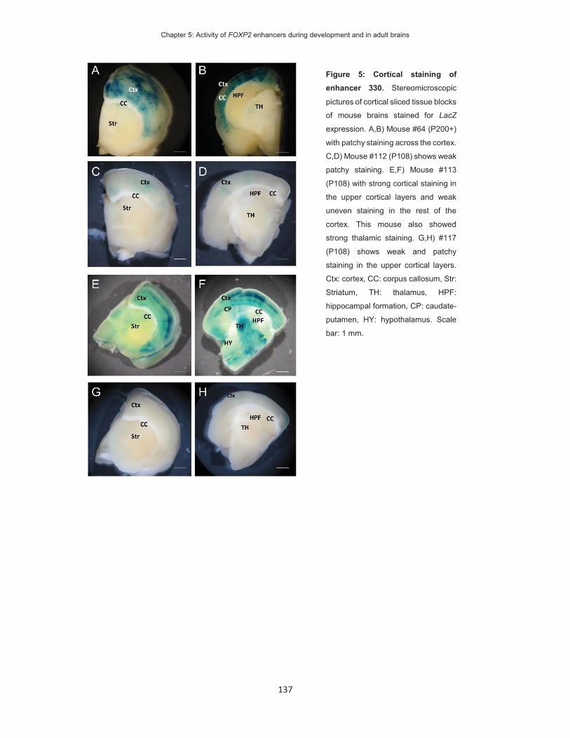
Chapter 5: Activity of FOXP2 enhancers during development and in adult brains
137
Figure 5: Cortical staining of enhancer 330. Stereomicroscopic
pictures of cortical sliced tissue blocks
of mouse brains stained for LacZ
expression. A,B) Mouse #64 (P200+)
with patchy staining across the cortex.
C,D) Mouse #112 (P108) shows weak
patchy staining. E,F) Mouse #113
(P108) with strong cortical staining in
the upper cortical layers and weak
uneven staining in the rest of the
cortex. This mouse also showed
strong thalamic staining. G,H) #117
(P108) shows weak and patchy
staining in the upper cortical layers.
Ctx: cortex, CC: corpus callosum, Str:
Striatum, TH: thalamus, HPF:
hippocampal formation, CP: caudate-
putamen, HY: hypothalamus. Scale
bar: 1 mm.

Chapter 5: Activity of FOXP2 enhancers during development and in adult brains
138
Cortical activity of enhancer 330 during development overlaps published FOXP2 expression sites
The most reproducible staining for enhancer 330 was detected in the neocortex, which is in
agreement with data from the VISTA enhancer browser (Visel et al., 2007). According to the
prior VISTA data, enhancer 330 is active in the neocortex of the forebrain at E11.5 (Figure 7A-
C). In the cortical sections from the VISTA project the staining was present in the ventricular
zone of the pallium, which is the developmental precursor of the neocortex, (Figure 7B, C)
along the anterior to posterior line (Figure 7A). These findings are consistent with the
hypothesis that the enhancer activates Foxp2 in the embryonic cortex. However, the VISTA
enhancer data is limited to the early embryonic development and further enhancer mapping
was necessary to determine the activity in post-natal and adult cortices.
Figure 6: Cerebellar staining of enhancer 330. Stereomicroscopic pictures of the cerebellum mouse brains
stained for LacZ expression. A) Mouse #112 shows specific staining in the cell body of the PCs. The majority of
stained PCs are located in lobule IX. B) Mouse #113 shows strong staining in the PC layer of the whole
cerebellum, C) Mouse #117 shows weak staining in lobules X and IX. gl: granular layer, ml: molecular layer, PC:
Purkinje cell layer. Scale bar: 1 mm.

Chapter 5: Activity of FOXP2 enhancers during development and in adult brains
139
Figure 7: Embryonic, neo-natal and adult cortical staining of enhancer 330. Enhancer 330 tissue activity in mouse brains, visualized by LacZ staining. Pictures C,F,I ,L and O are manual zooms of the adjacent pictures. A-C) Reproduced and adapted pictures of enhancer 330 in E11.5 mouse embryos downloaded from the VISTA Enhancer Browser (http://enhancer.lbl.gov/cgi-bin/imagedb3.pl?form=presentation&show=1&experiment_id=218&organism_id=1). A) Lateral view of the embryo and B-C) Microscopic picture of LacZ staining in cortical slices of the same embryo. D-O) Mouse brains from mouse line #64. (D, G, J, M) lateral view of mouse brains, showing intense staining across the forebrain and (E, F, H, I, K, L, N, O) cortical sliced brain tissue blocks, showing LacZ staining across the cortical layers. HB: hindbrain, MB: midbrain, FB: forebrain, Pal: pallium, LGE: lateral ganglionic eminence: MGE: medial ganglionic eminence, PallV: ventricular zone of pallium, PallM: mantle zone of pallium, Ctx: cortex, OB: olfactory bulb, HPF: hippocampal formation, I-VI: cortical layer I-VI. Scale bar: 1mm, no scale bars available for pictures A-C.

Chapter 5: Activity of FOXP2 enhancers during development and in adult brains
140
I established a mouse line of founder mouse #64, chosen because it showed strongest
neocortical staining. To establish this mouse line, I crossed the founder mouse with a WT
C57BL/6 mouse to produce heterozygote offspring. The heterozygotes were further crossed
with WT or heterozygote offspring of the same founder animal. I did not observe differences in
staining intensity or staining pattern for the homozygous of heterozygotes state. Due to limited
access to the experimental mice, timed mating was not possible and I determined only post-
natal reporter gene expression of this mouse line. I analysed enhancer activity at post-natal
day 8 (P8), in adult mice (P88 and P150) and at a late adult time point (P200). At P8 the cortical
layers of the neocortex are established and the enhancer activity in all layers appeared equally
strong (Figure 7E, F). In the coronal plane the cortical staining was present along the rostral-
ventral line in the medial and lateral parts of cortex. At age P150 the staining decreased in the
deeper layers of the cortex (Figure 7J-L) and at age P200 the staining intensities further
decreased in both the deeper and upper cortical layers (Figure 7M-O). The activity pattern
along the rostral-ventral line remained stable through adulthood, for the different ages that I
studied. Some patches of weaker staining were visible within the stained cortical structures,
but the location of these patches was inconsistent across post-natal time points. At all
developmental stages the reporter gene staining was absent from the dorsal side of the cortex,
which forms the three-layered paleocortex. In summary, enhancer 330 is activated in the
ventricular zone of the pallium, remains active across the cortical plate in early post-natal mice
and in adult mice the enhancer activity decreases. I could not study the cerebellar activity of
enhancer 330, as the mouse line did not show cerebellar staining.
Enhancer 37 is active in regions both with and without known FOXP2 expression
PNI of the enhancer 37 construct yielded 87 mice, of which 13 mice had positive genotypes
for the reporter gene. The mice were sacrificed at adult age (P145+) and the brains were
dissected for LacZ staining. Two mice showed unspecific whole-brain staining and were
excluded from the analysis as potential source for false-positive replication. In sum, I analysed
the brain staining pattern of eleven mice (Mouse #2, 49, 41, 38, 45, 48, 57 58, 59, 32 and 33).

Chapter 5: Activity of FOXP2 enhancers during development and in adult brains
141
I detected LacZ staining in six brain regions. Reproducible staining was observed in only two
regions the hippocampus (7/11) and ventricles (6/11). In the hippocampus I observed staining
in the pyramidal neurons of the dentate gyrus and CA1 (Figure 8A, B; Figure 9). Expression
was visible in the dentate gyrus of 5 brains (Figure 9A, D-G), whereas weak CA1 expression
was detected only in three brains. Thus, according to my definition the staining in individual
pyramidal regions of the hippocampus is not reproducible. The ventricular staining was found
in a thin layer of the lateral ventricles (Figure 8C, D; Figure 10) and the third ventricle (Figure
8C, E). The staining seems to appear in the outermost cell layer, which consist of specialist
ependymal cells. IHC of ISH for cell-type specific markers would be required to validate the
cell identity. The staining pattern in hippocampus and ventricles was surprising as FOXP2 has
not been reported to be expressed in these regions in either the mouse or human.

Chapter 5: Activity of FOXP2 enhancers during development and in adult brains
142
Figure 8: Tissue activity of enhancer 37 in adult mouse brain at P200+. Representative stereomicroscopic pictures of
mouse brains stained for LacZ expression. Boxed areas B, D, E, G, I and J in pictures A, C, F and H are manual zooms of
adjacent pictures. LacZ staining shown A,B) in the pyramidal neurons of the hippocampus; C-E) in the ventricular system of
the left (D) and third ventricle (E); E,G) in the olfactory bulb and H-J) in the neocortex. DG: dentate gyrus; CA1: cornu
ammonis area 1; LV: left ventricle; 3rdV; third ventricle; OB: olfactory bulb, Str: striatum, CC: corpus callosum, I-VI: cortical
layer I-VI.

Chapter 5: Activity of FOXP2 enhancers during development and in adult brains
143
Figure 9: Hippocampal staining of enhancer 37 mouse lines. A-G)
Stereomicroscopic pictures of mouse brains stained for LacZ
expression. All mice carried the enhancer 37 construct at random
genomic positions. Mice A) #32 (P145), D) #45 (P200+) and G) #58
(P200+) show intense staining in the DG. Mice B) #33 (P145), C) #41
(P200+), E) #48 (P200+) and F) #49 (P200+) show various degrees of
staining intensities in the pyramidal cells of the HPF. Ctx: cortex, HPF:
hippocampal formation, TH: thalamus. Scale bar: 1 mm.

Chapter 5: Activity of FOXP2 enhancers during development and in adult brains
144
The OB and neocortex stained positive in five out of eleven founder mice (Figure 11 and 12).
These staining meet the criteria of being stained in more or equal to 3 mice, but do not meet
the strict criteria for reproducible staining in 50% of the analysed animals. Because
FOXP2/Foxp2 is expressed in these regions I determined their activity pattern. In the OB I saw
consistent staining in a lateral region (Figure 8F, G; Figure 11), which does not overlap with a
defined region from the Allen Brain Reference Atlas (Hawrylycz et al., 2012). The OB staining
could suggest overlap with FOXP2 expression. The cortical staining was distributed in clusters
with dense staining in the upper cortical layers and 4 brains showed staining in the allocortex
(Figure 8 H-J). However, the allocortex, as well as the upper layers of the neocortex, do not
express Foxp2 (Ferland et al., 2003). The staining therefore suggests that the activity of the
enhancer does not overlap with known FOXP2 expression patterns in the cortex.
Figure 10: Lateral ventricle staining of enhancer 37 mouse lines. A-F) Stereomicroscopic pictures of mouse brains stained
for LacZ expression. All mice carried the enhancer 37 construct at random genomic positions. Mouse A) #2 (P129), B) #32
(P145), C) #33 (P145), D) #45 (P200+), E) #48 (P200+) and F) #57 (P200+). A,B,E) Strong staining in the LV. C,D,F) weak
staining in the LV. CC: corpus callosum, Ctx: cortex, Str: striatum, LV: lateral ventricle. Scale bar: 1 mm

Chapter 5: Activity of FOXP2 enhancers during development and in adult brains
145
Figure 11: Olfactory bulb staining of enhancer 37 mouse lines. A-E) Stereomicroscopic pictures of mouse brains stained
for LacZ expression. All mice carried the enhancer 37 construct at random genomic positions. Mice A) #2 (P200+), B) #32
(P145), C) #33 (P135), D) #41 (P200+) and E) #49 (P200+) show staining of a ventral region in the olfactory bulb. Ctx: cortex,
OB: olfactory bulb. Scale bars: 1 mm

Chapter 5: Activity of FOXP2 enhancers during development and in adult brains
146
Enhancer 37 activity changes during embryonic and adult development
The most promising overlap of known FOXP2 expression patterns and enhancer 37 activity
was in the OB. Therefore, I established a stable mouse line of founder mouse #2, which
showed staining in the adult ventricles and OB. I harvested mice at embryonic stages (E13.5,
E18.5), neo-natal (P5, P7), juvenile (P18), late adolescent (P39), adult (P129) and late adult
age (P218). In the early embryo (E13.5) I did not detect LacZ staining (Figure 13A). At E18.5
I observed staining in the OB (Figure 13B), which was also observed in adolescent mice (P39)
(Figure 13C). The late adult individual (P281) did not show OB staining (Figure 13D).
Figure 12: Neocortical staining of enhancer 37 mouse lines. A-E) Stereomicroscopic pictures of mouse brains stained for
LacZ expression. All mice carried the enhancer 37 construct at random genomic positions. A) Mouse #38 (P200+) shows
scattered positive stained cells across the cortex. B) Mouse #49 (P200+) shows patchy staining across the cortex and OB.
C) Mouse #57 (P200+), D) #58 (P200+) and E) #59 (P200+) with weak and patchy staining across the cortex. CC: corpus
callosum, Ctx: cortex, Str: striatum, OB: olfactory bulb. Scale bar: 1 mm.

Chapter 5: Activity of FOXP2 enhancers during development and in adult brains
147
Ventricular staining was first visible at post-natal age P7 (Figure 14A). In late adolescent (P39)
and late adult (P281) mice, I observed staining in the lateral and third ventricles (Figure 14B,
C). Similar to the adult, the staining is observable in a thin lining of the ventricular walls. The
regulatory control of enhancer 37 in the ventricular system seems to start after birth and
remains throughout adulthood. In the OB, the enhancer becomes active during late embryonic
development and declines in late adult stages. To determine the overlap of this enhancer with
Foxp2 expression it will be crucial to determine the Foxp2 mRNA and protein levels at these
sites.
Figure 13: Development of olfactory bulb staining of enhancer 37 in mouse line 2. Stereomicroscopic pictures of mouse
brains stained for LacZ expression. All mice are offspring of the same founder mouse. (A) LacZ staining shown in a sagittal
section along the midbrain line at embryonic day 13.5 is not present; (B). OB staining is first visible at age E18.5; (C) OB
staining continues to be present in late adolescent mice at P39; (D) No OB staining is not detectable in mature adult mice.
A-D) are lateral views of the olfactory bulb of a tissue block that was cut along the frontal cortex. HB: hindbrain, MB:
midbrain, Di: diencephalon, Tel: Telencephalon, Ctx: cortex, OB: olfactory bulb. Scale bars: 1 mm

Chapter 5: Activity of FOXP2 enhancers during development and in adult brains
148
Figure 14: Development of ventricular staining of enhancer 37 in mouse line 2. Stereomicroscopic pictures of mouse brains
stained for LacZ expression. All mice are offspring of the same founder animal. The staining of the lateral ventricle is first
visible at age P7 (A) and is present in late adolescent mice (B). The staining remains intense in mature adult mice (C). fi:
fimbria, LV: lateral ventricle, CC: corpus callosum, Ctx: cortex, Str: striatum, 3rdV: third ventricle, HPF: hippocampal
formation, TH: thalamus. Scale bar: 1 mm.

Chapter 5: Activity of FOXP2 enhancers during development and in adult brains
149
Discussion
In this chapter I analysed the activity patterns of two putative FOXP2 enhancers in the
developing and adult central nervous system. I used PNI to insert enhancer-reporter gene
constructs into fertilized mouse oocytes. With this method the transgene inserts randomly into
the genome. It was therefore crucial to use criteria that distinguish endogenous enhancer
activity from spurious activity caused by positional effects (i.e. the impact of neighbouring
regulatory regions at the points of genomic insertion). The VISTA enhancer browser collects
the results for thousands of enhancers tested with the same PNI-based method (Visel et al.,
2007). In the VISTA method, at least five transgenic mice were generated for each enhancer,
carrying the sequence at different genomic positions. The staining pattern was analysed in
every animal and endogenous activity was defined for staining that is observed in the same
tissue in at least three animals (Visel et al., 2007). Both enhancers drive expression in two or
more distinct brain regions. The finding that enhancers are active in multiple tissues is in line
with data from previously reported enhancers (Visel et al., 2007; Bonn et al., 2012). As both
enhancers have been shown to physically contact the transcriptionally active FOXP2 promoter
in human cell lines (Chapter 3) and drive gene expression in reporter assays, they could
contribute to FOXP2 expression in the detected brain regions.
Enhancer 37 activity is present in the OB, which has been reported to express FOXP2. The
enhancer activity was detected in the outer layers of the OB, but did not overlap with a defined
anatomical region. FOXP2 has been previously found in glomerular and granular cells of the
OB (Campbell et al., 2009; Zegary, 2009), of which the glomerular cells form the external
neuronal layer. Therefore, enhancer 37 activity and FOXP2 expression likely overlap in this
layer. In the glomerular cells, FOXP2 expression marks one of three defined subtypes (Zegary,
2009). Thus, enhancer 37 may contribute to the expression of FOXP2 in the OB. Co-
localization of enhancer 37 activity with Foxp2 expression in the transgenic mice is necessary
to determine the exact overlap of targeted cell types.

Chapter 5: Activity of FOXP2 enhancers during development and in adult brains
150
Enhancer 37 was also active in the hippocampus and ventricles, which do not show FOXP2
expression according to prior research. Enhancers may regulate more than one gene. Thus,
enhancer 37 could regulate another gene that is expressed in these structures (Bonn et al.,
2012). Enhancer contacts are dynamic between different cell types, but limited to genes within
a topological domain (Dixon et al., 2012). Therefore, genes of the same topological domain as
FOXP2 may be targeted by enhancer 37. Next to FOXP2, the surrounding topological domain
contains the MDFIC, TFEC and PPP1R3A genes (Dixon et al., 2012). In neuronal-like cell lines
the enhancer interacted with the FOXP2 promoter (Chapter3). The promoter was in contact
with additional genomic regions, including the promoter of the MDFIC gene. It is therefore
possible that enhancer 37 interacts with the MDFIC promoter and regulates the expression of
this gene. MDFIC encodes the MyoD family inhibitor domain-containing protein, which
regulates the WNT and JNK pathways (Kusano and Raab-Traub, 2002). The Allen Brain Atlas
suggests strong expression of MDFIC in the CA1 and dentate gyrus, which matches the
observed enhancer activity in the hippocampus (Lein et al., 2007). To further assess the
hypothesis that enhancer 37 regulates MDFIC, it would be required to determine the direct
interaction of enhancer 37 to the MDFIC promoter. In addition, co-localization of enhancer 37
with Mdfic and Foxp2 in transgenic mice could further indicate the target gene of this enhancer
in the hippocampus.
The tissue, in which enhancer 37 is active has been linked to adult neurogenesis in prior
studies (Eriksson et al., 1998; Jessberger and Gage, 2014). Adult neurogenesis occurs in the
hippocampus and is involved in memory formation in mammals and humans (Eriksson et al.,
1998; Jessberger and Gage, 2014; Ernst and Frisen, 2015). A second site of adult
neurogenesis is the subventricular zone (SVZ) of the ventricles. In rodents, the new-born
neurons migrate from the SVZ towards the OB, where they differentiate into interneurons and
integrate in the existing cellular architecture (Lois et al., 1996; Bergmann et al., 2015). To
demonstrate a link between enhancer 37 and neurogenesis it would be necessary to co-label
enhancer 37 reporter staining with markers of neurogenesis (Young et al., 2007). Mouse Foxp2

Chapter 5: Activity of FOXP2 enhancers during development and in adult brains
151
has been implicated in embryonic neurogenesis and contributes to the transformation from
neural progenitor to differentiated neurons (Tsui et al., 2013). Targets of FOXP2 in human
neuronal-like cells and mouse embryonic brain are enriched for genes involved in
neurogenesis (Spiteri et al., 2007; Vernes et al., 2007; Vernes et al., 2011). In adult zebra
finch, FoxP2 expression has been detected in new-born neurons, originating from the SVZ
(Rochefort et al., 2007). In zebra finches the SVZ born neurons migrated to and integrated into
the striatal nucleus area X. FOXP2 expression was not observed in the SVZ itself, suggesting
that FOXP2 expression is activated in post-migratory neurons (Rochefort et al., 2007).
However, it remains to be shown whether or not FOXP2 is involved in adult neurogenesis in
mammals. Research shows that, in humans, SVZ born neurons migrate and integrate into the
striatum (Ernst et al., 2014; Ernst and Frisen, 2015), suggesting that the neurons from the
rostral migratory stream could migrate towards the striatum. Enhancer 37 shows low
evolutionary conservation across primates and is absent from non-primate species (Figure
3A), suggesting that the function of this enhancer was obtained during more recent
evolutionary events. Species-specific genetic variation in enhancers has been shown to
contribute to human-specific neuronal changes (Boyd et al., 2015). However, from the
presented data it remains speculative whether or not this enhancer could be linked to the
human specific traits, such as a changed migratory pathway of new-born neurons. Migration
of SVZ neurons to the striatum can be induced in mice after blocking the Notch-signalling
pathway (Magnusson et al., 2014). Inducing this pathway in enhancer transgenic mice could
be used to assess whether enhancer 37 is active in SVZ-to-striatal migratory neurons.
The other investigated enhancer, enhancer 330, was previously shown to drive forebrain
expression at embryonic day E11.5 (Visel et al., 2007). In other studies, Foxp2 mRNA
expression in the ventricular zone and cortical plate has been detected at E14.5 (Ferland et
al., 2003). Indeed, after knock-out of the Dicer gene, which encodes the enzyme responsible
for processing immature micro RNAs (miRNA), cortical expression of Foxp2 can be detected
at E13.5 (Clovis et al., 2012). In the early post-natal cortex, I detected activity of enhancer 330

Chapter 5: Activity of FOXP2 enhancers during development and in adult brains
152
in all cortical layers, at a time when Foxp2 is expression is mostly restricted to the deeper
cortical layer VI (Ferland et al., 2003). In the post-natal brain the activity of enhancer 330
overlaps FOXP2 expression in the deeper cortical layers. Thus, enhancer330 activity and
FOXP2 expression could overlap in the embryonic and post-natal cortex suggesting that this
enhancer activates FOXP2 expression during cortical development. Enhancer 330 activity was
specific to the six-layered neocortex and absent in the three-layered paleocortex. This is in
agreement with the absence of FOXP2 expression in the paleocortex (Ferland et al., 2003).
In adult brain tissue, enhancer 330 is active in the upper cortical layers, whereas FOXP2 is
expressed specifically in the deeper cortical layer VI (Ferland et al., 2003; Hisaoka et al., 2010).
The difference in enhancer 330 activity and FOXP2 expression may be explained by post-
transcriptional and/or post-translational regulation. At the post-transcriptional level FOXP2
mRNA has been shown to be a target of miRNAs (Clovis et al., 2012; Fu et al., 2014). The
miRNAs miR-9 and miR-132 repress FOXP2 expression specifically in the upper cortical layers
(Clovis et al., 2012). In addition, the FOXP2 protein is post-translationally modified and may
be targeted for protein degradation. For example, the post-translational addition of a SUMO
protein to FOXP2 has been shown, but this modification has not been shown to target FOXP2
for degradation (Meredith et al., 2015; Estruch et al., 2016). The LacZ reporter gene does not
contain the FOXP2 amino-acid sequences required for protein degradation or 3’-UTR for
miRNA-mediated silencing. Therefore, it is likely that staining positive for the reporter gene is
observed in cells which endogenously reduce FOXP2 protein levels via post-transcriptional
and post-translational mechanisms.
People with mutations of FOXP2 show structural and functional abnormalities in some cortical
areas, such as the classical language associated Wenicke’s and Broca’s area, the angular
gyrus and the precentral gyrus (Vargha-Khadem et al., 1998; Watkins et al., 2002; Belton et
al., 2003; Liegeois et al., 2003; Liegeois et al., 2011). Genetic variation of enhancer 330 could
in principle impact on enhancer function and affect the cortical expression of FOXP2. To my
best knowledge, no case has been described with a selective deletion or mutation of this

Chapter 5: Activity of FOXP2 enhancers during development and in adult brains
153
element and high-throughput sequencing studies of clinical cases commonly focus on genetic
variants within the exonic part of the genome and would not detect variation in the enhancer
(Rabbani et al., 2014). The common single nucleotide polymorphism (SNP) rs12533005, which
is located within 1kb of the upstream flanking sequence (Figure 3A), showed association to
human traits in candidate association studies. One study investigated 12 SNPs and detected
an association of this SNP to ADHD in a German cohort, which did not replicate in a Spanish
cohort (Ribases et al., 2012). Another study investigated the association of 9 SNPs with
dyslexia and detected a nominal association to rs12533005 (Wilcke et al., 2012). Wilcke et al.
further conducted functional magnetic resonance imaging in 25 individuals carrying the risk-
conferring minor allele and 8 non-carrier individuals. They detected that carriers of the minor
allele showed reduced activity in the angular gyrus and supramarginal gyri during phonological
processing tasks. Thus, enhancer 330 activity and genetic effects of rs12533005 coincide at
cortical areas that show anomalies in patients with FOXP2 mutations. This could suggest that
rs12533005 influences the activity of enhancer 330 or tags another functional variant within
this enhancer. A third study detected an association of rs12533005 (out of 6 tested variants)
to increased left-hemispheric speech perception in healthy individuals (Ocklenburg et al.,
2013), further indicating that this SNP may affect cortical functioning. However, the link
between this SNP and enhancer 330 requires additional evidence. For example, genotyping
of additional SNPs within this enhancer may show that rs12533005 is a proxy of an association
to a variant within enhancer 330. Alternatively, a longer version of enhancer 330 could be
investigated in reporter gene studies to determine if the sequence at rs12533005 contributes
to enhancer function. In addition, the screening of enhancer 330 for mutations in cohorts of
people with speech/language disorders is a promising approach to detect pathological
variation.
FOXP2 has been recognized as a marker gene for cortical differentiation of cultured neuronal
stem cells (Bickenbach et al., 2013; Espuny-Camacho et al., 2013; Belinsky et al., 2014;
Raitano et al., 2015). During the in vitro differentiation of induced pluripotent stem cells (iPSC)
Chapter 5: Activity of FOXP2 enhancers during development and in adult brains
154
to cortical neurons, endogenous FOXP2 expression is switched on (Espuny-Camacho et al.,
2013; Belinsky et al., 2014). The activity of the FOXP2 enhancer 330 in developing cortex
could suggest that this enhancer would be activated in the cultured neurons. Chromatin
conformation (Chapter 3) and reporter gene experiments (Chapter 4) in cultured stem cells
would help to test this hypothesis. Human iPSCs and derived neurons are promising systems
to study the neurodevelopmental function of enhancer 330 in human genetic background.
I also detected enhancer 330 activity in the adult cerebellum. The cerebellum develops after
E11.5 and enhancer activity at this time may be undetected by the analysis performed for the
VISTA enhancer browser. The cerebellar anlage is formed at E12.5 and the cerebellar
hemisphere can be distinguished from other anatomical regions at embryonic day E14 (Millen
et al., 1994; Fujita and Sugihara, 2012). FOXP2 is expressed in the majority of neuronal nuclei
of the cerebellar hemisphere at E14.5 (Fujita and Sugihara, 2012). The founder mouse and
subsequent mice of the established mouse line, which were used to study the longitudinal
activity of enhancer 330 did not show cerebellar staining. Therefore, it remains to be
determined at what developmental stage FOXP2 expression and enhancer 330 activity start
in the cerebellar anlage/cerebellar hemisphere.
Several FOXP2 positive brain structures did not show enhancer activity for either enhancer 37
or 330. I therefore expect that additional enhancers regulate the expression of FOXP2. In the
previous chapters I identified enhancers that were not tested in this chapter and could be
promising candidates for recapitulating further parts of the FOXP2 expression pattern.
In summary, in this chapter I characterized the pattern of activity of two FOXP2 enhancers in
the brain and followed this activity during development. The enhancers recapitulated some
parts of the complex FOXP2 expression pattern. Enhancer 37 is active in the OB, a known
region of FOXP2 expression, and coincides with sites of adult neurogenesis. Enhancer 330
may be involved in modulating FOXP2 expression in the cortex and PCs. In addition, the

Chapter 5: Activity of FOXP2 enhancers during development and in adult brains
155
proximity of enhancer 330 to rs12533005 may explain the observed associations of this SNP
to language-related traits.
Chapter 5: Activity of FOXP2 enhancers during development and in adult brains
156
References
Becker M, Devanna P, Fisher SE, Vernes SC (2015) A chromosomal rearrangement in a child with severe speech and language disorder separates FOXP2 from a functional enhancer. Molecular cytogenetics 8:69.
Belinsky GS, Rich MT, Sirois CL, Short SM, Pedrosa E, Lachman HM, Antic SD (2014) Patch-clamp recordings and calcium imaging followed by single-cell PCR reveal the developmental profile of 13 genes in iPSC-derived human neurons. Stem cell research 12:101-118.
Belton E, Salmond CH, Watkins KE, Vargha-Khadem F, Gadian DG (2003) Bilateral brain abnormalities associated with dominantly inherited verbal and orofacial dyspraxia. Human brain mapping 18:194-200.
Bergmann O, Spalding KL, Frisen J (2015) Adult Neurogenesis in Humans. Cold Spring Harbor perspectives in biology 7.
Bickenbach JR, Tomanek-Chalkley A, Wiechert S, Winter MC (2013) Human skin keratinocytes can be reprogrammed to express neuronal genes and proteins after a single treatment with decitabine. BioResearch open access 2:217-221.
Bonn S, Zinzen RP, Girardot C, Gustafson EH, Perez-Gonzalez A, Delhomme N, Ghavi-Helm Y, Wilczynski B, Riddell A, Furlong EE (2012) Tissue-specific analysis of chromatin state identifies temporal signatures of enhancer activity during embryonic development. Nat Genet 44:148-156.
Boyd JL, Skove SL, Rouanet JP, Pilaz LJ, Bepler T, Gordan R, Wray GA, Silver DL (2015) Human-Chimpanzee Differences in a FZD8 Enhancer Alter Cell-Cycle Dynamics in the Developing Neocortex. Current biology : CB.
Campbell P, Reep RL, Stoll ML, Ophir AG, Phelps SM (2009) Conservation and diversity of Foxp2 expression in muroid rodents: functional implications. The Journal of comparative neurology 512:84-100.
Clovis YM, Enard W, Marinaro F, Huttner WB, De Pietri Tonelli D (2012) Convergent repression of Foxp2 3'UTR by miR-9 and miR-132 in embryonic mouse neocortex: implications for radial migration of neurons. Development 139:3332-3342.
Dixon JR, Selvaraj S, Yue F, Kim A, Li Y, Shen Y, Hu M, Liu JS, Ren B (2012) Topological domains in mammalian genomes identified by analysis of chromatin interactions. Nature 485:376-380.
Domene S, Bumaschny VF, de Souza FS, Franchini LF, Nasif S, Low MJ, Rubinstein M (2013) Enhancer turnover and conserved regulatory function in vertebrate evolution. Philosophical transactions of the Royal Society of London Series B, Biological sciences 368:20130027.
Eriksson PS, Perfilieva E, Bjork-Eriksson T, Alborn AM, Nordborg C, Peterson DA, Gage FH (1998) Neurogenesis in the adult human hippocampus. Nature medicine 4:1313-1317.
Ernst A, Frisen J (2015) Adult neurogenesis in humans- common and unique traits in mammals. PLoS biology 13:e1002045.
Ernst A, Alkass K, Bernard S, Salehpour M, Perl S, Tisdale J, Possnert G, Druid H, Frisen J (2014) Neurogenesis in the striatum of the adult human brain. Cell 156:1072-1083.
Espuny-Camacho I, Michelsen KA, Gall D, Linaro D, Hasche A, Bonnefont J, Bali C, Orduz D, Bilheu A, Herpoel A, Lambert N, Gaspard N, Peron S, Schiffmann SN, Giugliano M, Gaillard A, Vanderhaeghen P (2013) Pyramidal neurons derived from human pluripotent stem cells integrate efficiently into mouse brain circuits in vivo. Neuron 77:440-456.
Estruch SB, Graham SA, Deriziotis P, Fisher SE (2016) The language-related transcription factor FOXP2 is post-translationally modified with small ubiquitin-like modifiers. Sci Rep 6.
Ferland RJ, Cherry TJ, Preware PO, Morrisey EE, Walsh CA (2003) Characterization of Foxp2 and Foxp1 mRNA and protein in the developing and mature brain. The Journal of comparative neurology 460:266-279.
Fu LJ, Shi ZM, Luo GZ, Tu WH, Wang XJ, Fang ZD, Li XC (2014) Multiple microRNAs regulate human FOXP2 gene expression by targeting sequences in its 3 ' untranslated region. Molecular brain 7.
Fujita E, Tanabe Y, Momoi MY, Momoi T (2012) Cntnap2 expression in the cerebellum of Foxp2(R552H) mice, with a mutation related to speech-language disorder. Neuroscience letters 506:277-280.
Fujita H, Sugihara I (2012) FoxP2 expression in the cerebellum and inferior olive: development of the transverse stripe-shaped expression pattern in the mouse cerebellar cortex. The Journal of comparative neurology 520:656-677.
Hawrylycz MJ et al. (2012) An anatomically comprehensive atlas of the adult human brain transcriptome. Nature 489:391-399.
Chapter 5: Activity of FOXP2 enhancers during development and in adult brains
157
Hisaoka T, Nakamura Y, Senba E, Morikawa Y (2010) The forkhead transcription factors, Foxp1 and Foxp2, identify different subpopulations of projection neurons in the mouse cerebral cortex. Neuroscience 166:551-563.
Ittner LM, Gotz J (2007) Pronuclear injection for the production of transgenic mice. Nature protocols 2:1206-1215.
Jessberger S, Gage FH (2014) Adult neurogenesis: bridging the gap between mice and humans. Trends Cell Biol 24:558-563.
Juers DH, Matthews BW, Huber RE (2012) LacZ beta-galactosidase: structure and function of an enzyme of historical and molecular biological importance. Protein science : a publication of the Protein Society 21:1792-1807.
Kothary R, Clapoff S, Darling S, Perry MD, Moran LA, Rossant J (1989) Inducible expression of an hsp68-lacZ hybrid gene in transgenic mice. Development 105:707-714.
Kusano S, Raab-Traub N (2002) I-mfa domain proteins interact with Axin and affect its regulation of the Wnt and c-Jun N-terminal kinase signaling pathways. Molecular and cellular biology 22:6393-6405.
Lai CS, Gerrelli D, Monaco AP, Fisher SE, Copp AJ (2003) FOXP2 expression during brain development coincides with adult sites of pathology in a severe speech and language disorder. Brain : a journal of neurology 126:2455-2462.
Lein ES et al. (2007) Genome-wide atlas of gene expression in the adult mouse brain. Nature 445:168-176.
Liegeois F, Morgan AT, Connelly A, Vargha-Khadem F (2011) Endophenotypes of FOXP2: dysfunction within the human articulatory network. European journal of paediatric neurology : EJPN : official journal of the European Paediatric Neurology Society 15:283-288.
Liegeois F, Baldeweg T, Connelly A, Gadian DG, Mishkin M, Vargha-Khadem F (2003) Language fMRI abnormalities associated with FOXP2 gene mutation. Nat Neurosci 6:1230-1237.
Lois C, Garcia-Verdugo JM, Alvarez-Buylla A (1996) Chain migration of neuronal precursors. Science 271:978-981.
Magnusson JP, Goritz C, Tatarishvili J, Dias DO, Smith EM, Lindvall O, Kokaia Z, Frisen J (2014) A latent neurogenic program in astrocytes regulated by Notch signaling in the mouse. Science 346:237-241.
Meredith LJ, Wang CM, Nascimento L, Liu R, Wang L, Yang WH (2015) The Key Regulator for Language and Speech Development, FOXP2, is a Novel Substrate for SUMOylation. Journal of cellular biochemistry.
Millen KJ, Wurst W, Herrup K, Joyner AL (1994) Abnormal embryonic cerebellar development and patterning of postnatal foliation in two mouse Engrailed-2 mutants. Development 120:695-706.
Ocklenburg S, Arning L, Gerding WM, Epplen JT, Gunturkun O, Beste C (2013) FOXP2 variation modulates functional hemispheric asymmetries for speech perception. Brain Lang 126:279-284.
Rabbani B, Tekin M, Mahdieh N (2014) The promise of whole-exome sequencing in medical genetics. J Hum Genet 59:5-15.
Raitano S, Ordovas L, De Muynck L, Guo W, Espuny-Camacho I, Geraerts M, Khurana S, Vanuytsel K, Toth BI, Voets T, Vandenberghe R, Cathomen T, Van Den Bosch L, Vanderhaeghen P, Van Damme P, Verfaillie CM (2015) Restoration of progranulin expression rescues cortical neuron generation in an induced pluripotent stem cell model of frontotemporal dementia. Stem cell reports 4:16-24.
Ribases M, Sanchez-Mora C, Ramos-Quiroga JA, Bosch R, Gomez N, Nogueira M, Corrales M, Palomar G, Jacob CP, Gross-Lesch S, Kreiker S, Reif A, Lesch KP, Cormand B, Casas M, Bayes M (2012) An association study of sequence variants in the forkhead box P2 (FOXP2) gene and adulthood attention-deficit/hyperactivity disorder in two European samples. Psychiatric genetics 22:155-160.
Rochefort C, He X, Scotto-Lomassese S, Scharff C (2007) Recruitment of FoxP2-expressing neurons to area X varies during song development. Developmental neurobiology 67:809-817.
Rubinstein M, de Souza FS (2013) Evolution of transcriptional enhancers and animal diversity. Philosophical transactions of the Royal Society of London Series B, Biological sciences 368:20130017.
Shu W, Yang H, Zhang L, Lu MM, Morrisey EE (2001) Characterization of a new subfamily of winged-helix/forkhead (Fox) genes that are expressed in the lung and act as transcriptional repressors. J Biol Chem 276:27488-27497.
Spiteri E, Konopka G, Coppola G, Bomar J, Oldham M, Ou J, Vernes SC, Fisher SE, Ren B, Geschwind DH (2007) Identification of the transcriptional targets of FOXP2, a gene linked to speech and language, in developing human brain. Am J Hum Genet 81:1144-1157.

Chapter 5: Activity of FOXP2 enhancers during development and in adult brains
158
Tsui D, Vessey JP, Tomita H, Kaplan DR, Miller FD (2013) FoxP2 regulates neurogenesis during embryonic cortical development. The Journal of neuroscience : the official journal of the Society for Neuroscience 33:244-258.
Vargha-Khadem F, Watkins KE, Price CJ, Ashburner J, Alcock KJ, Connelly A, Frackowiak RS, Friston KJ, Pembrey ME, Mishkin M, Gadian DG, Passingham RE (1998) Neural basis of an inherited speech and language disorder. Proc Natl Acad Sci U S A 95:12695-12700.
Vernes SC, Spiteri E, Nicod J, Groszer M, Taylor JM, Davies KE, Geschwind DH, Fisher SE (2007) High-throughput analysis of promoter occupancy reveals direct neural targets of FOXP2, a gene mutated in speech and language disorders. Am J Hum Genet 81:1232-1250.
Vernes SC, Oliver PL, Spiteri E, Lockstone HE, Puliyadi R, Taylor JM, Ho J, Mombereau C, Brewer A, Lowy E, Nicod J, Groszer M, Baban D, Sahgal N, Cazier JB, Ragoussis J, Davies KE, Geschwind DH, Fisher SE (2011) Foxp2 regulates gene networks implicated in neurite outgrowth in the developing brain. PLoS Genet 7:e1002145.
Villar D, Berthelot C, Aldridge S, Rayner TF, Lukk M, Pignatelli M, Park TJ, Deaville R, Erichsen JT, Jasinska AJ, Turner JM, Bertelsen MF, Murchison EP, Flicek P, Odom DT (2015) Enhancer evolution across 20 mammalian species. Cell 160:554-566.
Visel A, Minovitsky S, Dubchak I, Pennacchio LA (2007) VISTA Enhancer Browser--a database of tissue-specific human enhancers. Nucleic Acids Res 35:D88-92.
Visel A, Prabhakar S, Akiyama JA, Shoukry M, Lewis KD, Holt A, Plajzer-Frick I, Afzal V, Rubin EM, Pennacchio LA (2008) Ultraconservation identifies a small subset of extremely constrained developmental enhancers. Nat Genet 40:158-160.
Visel A, Blow MJ, Li Z, Zhang T, Akiyama JA, Holt A, Plajzer-Frick I, Shoukry M, Wright C, Chen F, Afzal V, Ren B, Rubin EM, Pennacchio LA (2009) ChIP-seq accurately predicts tissue-specific activity of enhancers. Nature 457:854-858.
Visel A et al. (2013) A high-resolution enhancer atlas of the developing telencephalon. Cell 152:895-908.
Watkins KE, Vargha-Khadem F, Ashburner J, Passingham RE, Connelly A, Friston KJ, Frackowiak RS, Mishkin M, Gadian DG (2002) MRI analysis of an inherited speech and language disorder: structural brain abnormalities. Brain : a journal of neurology 125:465-478.
Wilcke A, Ligges C, Burkhardt J, Alexander M, Wolf C, Quente E, Ahnert P, Hoffmann P, Becker A, Muller-Myhsok B, Cichon S, Boltze J, Kirsten H (2012) Imaging genetics of FOXP2 in dyslexia. European journal of human genetics : EJHG 20:224-229.
Young JI, Otero V, Cerdan MG, Falzone TL, Chan EC, Low MJ, Rubinstein M (1998) Authentic cell-specific and developmentally regulated expression of pro-opiomelanocortin genomic fragments in hypothalamic and hindbrain neurons of transgenic mice. The Journal of neuroscience : the official journal of the Society for Neuroscience 18:6631-6640.
Young KM, Fogarty M, Kessaris N, Richardson WD (2007) Subventricular zone stem cells are heterogeneous with respect to their embryonic origins and neurogenic fates in the adult olfactory bulb. The Journal of neuroscience : the official journal of the Society for Neuroscience 27:8286-8296.
Zegary JA (2009) Transcription Factor Regulation of Olfactory Bulb Interneuron Heterogeneity. Doctoral dissertation of the College of Medicine at the University of Cincinnati

Chapter 6: Gene enhancers affect subcortical volumes in the adult human brain
159
Chapter 6
Early developmental gene enhancers affect
subcortical volumes in the adult human brain
This chapter has been published in
Becker M, Guadalupe T, Franke B, Hibar DP, Renteria ME, Stein JL, Thompson PM,
Francks C, Vernes SC, Fisher SE (2016) Early developmental gene enhancers affect
subcortical volumes in the adult human brain. Human Brain Mapping 37:1788-1800.
The supplementary information is available online on the website of the Human Brain
Mapping journal.

Chapter 6: Gene enhancers affect subcortical volumes in the adult human brain
160
Abstract
Genome-wide association screens (GWAS) aim to identify common genetic variants
contributing to the phenotypic variability of complex traits, such as human height or brain
morphology. The identified genetic variants are mostly within non-coding genomic regions and
the biology of the genotype-phenotype association typically remains unclear. In this paper, we
propose a complementary targeted strategy to reveal the genetic underpinnings of variability
in subcortical brain volumes, by specifically selecting genomic loci that are experimentally
validated forebrain enhancers, active in early embryonic development. We hypothesized that
genetic variation within these enhancers may affect the development and ultimately the
structure of subcortical brain regions in adults. We tested whether variants in forebrain
enhancer regions showed an overall enrichment of association with volumetric variation in
subcortical structures of >13,000 healthy adults. We observed significant enrichment of
genomic loci that affect the volume of the hippocampus within forebrain enhancers (empirical
p = 0.0015), a finding which robustly passed the adjusted threshold for testing of multiple brain
phenotypes (cut-off of p < 0.0083 at an alpha of 0.05). In analyses of individual SNPs, we
identified an association upstream of the ID2 gene with rs7588305 and variation in
hippocampal volume. This SNP-based association survived multiple-testing correction for the
number of SNPs analyzed but not for the number of subcortical structures. Targeting known
regulatory regions offers a way to understand the underlying biology that connects genotypes
to phenotypes, particularly in the context of neuroimaging genetics. This biology-driven
approach generates testable hypotheses regarding the functional biology of identified
associations.
Chapter 6: Gene enhancers affect subcortical volumes in the adult human brain
161
Introduction
Global research efforts are underway to determine how genomic variants contribute to
variation in brain volume in the general population. Altered volumes of brain regions are
associated with neurodegenerative (Jack et al., 2011) and neuropsychiatric disorders
(Videbech and Ravnkilde, 2004; van Erp et al., 2015). Current GWAS studies connect
common genetic variants known as single nucleotide polymorphisms (SNPs) with phenotypic
variations in brain imaging data in cohorts of normal individuals (Bis et al., 2012; Stein et al.,
2012; Cai et al., 2014; Guadalupe et al., 2014b). However, most SNPs identified by GWAS
are within non-coding regions and their biological functions remain unclear. We employed a
hypothesis-driven strategy involving pre-selection of SNPs within genomic regions of
experimentally defined biological function. We found that the selected SNPs are significantly
enriched for association with hippocampal volume. This approach can help to generate
testable follow-up hypotheses about the underlying genotype-phenotype correlation.
It is challenging to predict and test the consequences of single nucleotide changes in genomic
regions that do not encode protein-coding genes or regulatory RNA molecules (Visel et al.,
2009a). Nevertheless, a vast amount of non-coding DNA is thought to play some functional
role, for example by regulating gene expression as promoters or enhancers. These regions
are bound by regulatory proteins such as transcription factors (TFs) to directly influence gene
expression and single nucleotide changes within these regions can affect gene expression
(Spitz and Furlong, 2012).
A complementary strategy to genome-wide association screening is to look specifically for
association within genomic regions that are predicted to be functional; for example by targeting
regions known to be bound by a particular TF or marked by a given epigenetic modification.
The approach was adopted in a study focusing on the genomic distribution of cell-type specific
regulatory regions in hematopoietic cells. These regions were enriched for the presence of
genetic variants associated with hematological traits such as platelet count, while no enriched

Chapter 6: Gene enhancers affect subcortical volumes in the adult human brain
162
association was detected for unrelated traits such as body-mass index (BMI) (Paul et al.,
2013). Another study applied a similar approach to publicly available functional datasets and
found enriched association across a range of human traits and disorders, including Crohn’s
disease and height (Pickrell, 2014). Thus, a priori knowledge of the functional characteristics
of genomic regions may help in selecting subsets of genomic variants for association testing.
To our knowledge, no prior study has systematically integrated information about functional
brain enhancers with genetic data in human individuals. In the current study, we analyzed
publicly available data from the ENIGMA (Enhancing Neuroimaging Genetics through Meta-
analysis) consortium, a neuroimaging genetics initiative that combines GWAS statistics from
50 cohorts spread world-wide, with a total combined sample size of 30,717 individuals
(Thompson et al., 2014). The ENIGMA consortium recently presented GWAS findings on
seven subcortical volumes and intracranial volume (Hibar et al., 2015). Five genetic variants
were significantly associated with the volumes of the hippocampus and putamen. These SNPs
mapped within non-coding regions of the genome and their functional roles are largely
unknown. We hypothesized that polymorphisms located within experimentally validated brain
enhancers, with established impacts on early neural development may be enriched for
association with volumetric changes in subcortical brain structures. A valuable source of
enhancers is the VISTA browser, which reports the tissue-specificity of about two thousand
potential enhancers. Genomic elements tested in VISTA were chosen for their conservation
across species and/or enhancer-specific chromatin marks in brain tissue (Visel et al., 2008;
Visel et al., 2009b). Their potential to reproducibly drive expression of a reporter gene was
tested at an early developmental timepoint in the mouse brain (Visel et al., 2007). VISTA
contains 309 enhancers that drive expression in the embryonic forebrain; the developmental
origin of the subcortical regions analyzed in adults by the ENIGMA consortium. Here, we
integrate ENIGMA meta-analysis data with information on VISTA forebrain enhancer regions
to assess for genetic effects on normal variation in subcortical brain volumes, using gene-set
and SNP-based approaches.

Chapter 6: Gene enhancers affect subcortical volumes in the adult human brain
163
Methods and Materials
VISTA enhancer browser of experimentally validated enhancer fragments
The VISTA enhancer browser is a collection of experimentally validated non-coding fragments
of the human and mouse genomes that exhibit enhancer activity (Visel et al., 2007). Potential
enhancers were predicted based on ChIP-seq experiments or evolutionary conservation. The
selected non-coding fragments were cloned in front of a minimal promoter and the LacZ
reporter gene. The resulting constructs were injected into the pro-nucleus of mouse embryos
and re-implanted into foster mothers. Developing embryos were stained for reporter gene
expression at embryonic day 11.5 (E11.5). Non-coding fragments that drive reproducible
reporter gene expression in multiple (≥5) embryos are defined as positive enhancers. If no
reproducible staining was detected in seven individual embryos, the non-coding fragment was
regarded as negative. Thus, the database represents a collection of functional enhancers and
their tissue of activity in developing embryos.
ENIGMA meta-analyzed neuroimaging GWAS of subcortical brain volumes
The ENIGMA consortium conducts harmonized analyses that combine neuroimaging genetics
data from over 50 cohorts world-wide to search for common gene variants that are associated
with brain structure and function. ENIGMA2 describes the second project of this consortium,
which tested associations of 8.5 million SNPs with the variance in volumes of the nucleus
accumbens, amygdala, caudate nucleus, hippocampus, pallidum, putamen, thalamus, and the
intracranial volume, measured from brain MRI (Hibar et al., 2015). The discovery sample for
ENIGMA2 consisted of 13,171 subjects of European ancestry. The ENIGMA consortium also
had access to a replication set of 17,546 subjects and reported on a total of 30,717 individuals.
Subjects were scanned at the individual sites using brain MRI and the resulting images were
processed in a standard way across all sites. The volumetric measures of all subcortical
structures were corrected for age, sex, genetic homogeneity and intracranial volume. At each

Chapter 6: Gene enhancers affect subcortical volumes in the adult human brain
164
site, genotypes were obtained from commercially available platforms and imputed to the
reference panel 1000genomes v1.3 using scripts provided by the ENIGMA working group.
GWAS was performed at each site, and the meta-analysis of all 50 cohorts was performed
centrally using the software package METAL (Willer et al., 2010). Here, we use the meta-
analyzed summary p-values of the 13,171 subjects obtained from the discovery sample. The
remaining 17,000 subjects from ENIGMA2 lack meta-analyzed genome-wide statistics and
were thus unavailable for use in the present study.
Selecting SNPs within human enhancers
A list of enhancers, including their genomic coordinates (human genome build hg19) and their
tissue of activity, was downloaded from the VISTA enhancer browser (Version 19.Sept 2014)
(Visel et al., 2007). We filtered all human enhancer elements with a reproducible forebrain
activity at embryonic stage E11.5. The forebrain is the developmental origin of the subcortical
regions analyzed by ENIGMA. We removed nine enhancer elements located on chromosome
X, as this chromosome was not included in the ENIGMA2 data. Four enhancers (hs322,
hs1354, hs998, and hs1597) contained within larger enhancers were deleted to obtain a non-
redundant list of 296 loci with a mean size of 1936 bp (standard deviation of 941 bp). Enhancer
boundaries are not well defined, and flanking regions can be critical for the stability of an
enhancers activity (Ludwig et al., 2011). We intersected the genomic enhancer regions,
including 500 bp flanking regions with the genomic positions of all markers present in the
meta-analyzed GWAS files of ICV-adjusted subcortical volumes (nucleus accumbens,
amygdala, caudate nucleus, hippocampus, pallidum, putamen, and thalamus) from ENIGMA2.
From this, we derived a list of 2082 SNPs and indels, which represent non-coding genetic
variants with a potential impact on enhancer activity. We refer to these variants as enhancer
SNPs.
Chapter 6: Gene enhancers affect subcortical volumes in the adult human brain
165
Testing enrichment of association with subcortical volumes
ENIGMA2 tested for the association of 8.5 million genome-wide markers against volumes of
7 subcortical brain structures. 2,082 of these markers were located within forebrain enhancer
regions. For each of these markers we converted their association p-values into z-scores.
Note that we could not specify a priori whether a particular allele of a SNP within an enhancer
should lead to an increase or decrease in the activity of the relevant enhancer. Moreover,
increases in gene expression may be correlated with either an increase or decrease in volume
of a structure, depending on the nature of the gene product; both directions of effect are
possible in principle, given what is known about mechanisms by which genes influence brain
morphology. Thus, because we did not have prior hypotheses regarding direction of effect, we
converted the p-values to right-tailed z-scores. These z-scores were then summed to calculate
a "forebrain enhancer" test statistic. This was repeated for each of the tested brain
phenotypes.
The probability of observing a similar (or greater) test statistic was determined by sampling
2,082 markers from 296 random genomic regions from the rest of the ENIGMA2 results. For
each of the 296 forebrain enhancer regions, a randomly sampled genomic region of similar
length was selected (with a difference no greater than 1000 bp), which included at least the
same number of markers as the original one. We chose the generated regions to be of similar
size to avoid repetitive parts of the genome, such as telomeres or centromeres. We note that
since the ENIGMA results are obtained via a large-scale meta-analysis of GWAS statistics
from many different populations, we are unable to determine the precise LD structure that
would most appropriately match the enrichment analyses. Thus, our permutation approach
assumes that the LD structure of the enhancer regions is broadly characteristic of the rest of
the genome. From this set of new selected regions, 2,082 markers were used to calculate the
new randomly-generated set statistic. This was repeated ten thousand times. The p-value for
the enrichment is the result of dividing the number of randomly-generated test statistics that
are higher than the observed test statistic by the number of permutations (10,000). The seven

Chapter 6: Gene enhancers affect subcortical volumes in the adult human brain
166
phenotypes are correlated, and we determined that the effective number of independent
phenotypes is six, using the Matrix Spectral Decomposition, matSpD software. We accounted
for multiple testing by Bonferroni correction for this effective number of independent
phenotypes. Thus, at an alpha of 0.05, the p-value threshold for significant enrichment was
0.0083.
Testing enrichment of association with unrelated human traits as a control
As noted above, our permutation approach for assessing enrichment assumes that enhancer
regions do not deviate substantially in LD structure, as compared to the rest of the genome.
Therefore, we performed additional control experiments to discount the possibility that
evidence of enrichment might be an artefact of unusual LD patterns. We downloaded the
meta-analyzed GWAS summary statistics for BMI (Speliotes et al., 2010), height (Lango Allen
et al., 2010) and waist-to-hip ratio (WHR) (Heid et al., 2010) from the Genetic Investigation of
Anthropometric Traits (GIANT) consortium. We used the Galaxy web-based platform
(Giardine et al., 2005; Blankenberg et al., 2010; Goecks et al., 2010) to add the SNP’s genomic
locations (hg19) and carried out enrichment analysis as described above. Based on Bonferroni
correction for use of three control phenotypes, at an alpha of 0.05 the p-value threshold for
significant enrichment in these analyses was 0.017.
Association of individual enhancer SNPs with subcortical volumes
SNPs within the same enhancer are in close proximity, and often in linkage disequilibrium
(LD), so not independent from each other. We therefore wanted to determine the effective
number of independent SNPs in the SNP-based association tests. Since the original
genotypes for all subjects were not centrally collected by ENIGMA they were unavailable for
the present study. Therefore, we calculated the effective number of tested SNPs from a large

Chapter 6: Gene enhancers affect subcortical volumes in the adult human brain
167
subset of ENIGMA for which genotypes were available to us: the Brain Imaging Genetics (BIG)
cohort of the Cognomics initiative (Guadalupe et al., 2014a). This cohort contributed
approximately 10% of the subjects to the ENIGMA2 discovery sample and consists of healthy
subjects of European ancestry. Using the Genetic Type I Error Calculator (Li et al., 2012), we
determined that the number of effectively independent tests performed when assessing
associations for the 2,082 enhancer SNPs was 770.
We evaluated the evidence for association between enhancer SNPs (spread across 296
enhancers) and variation in the seven subcortical volumes analyzed in ENIGMA2. As noted
above, analyses with matSpD (Nyholt, 2004) indicated that the effective number of
independent phenotypes is six. We derived an appropriate significance threshold based on
Bonferroni correction, taking into consideration both the effective number of tested SNPs and
the effective number of independent phenotypes. At an alpha of 0.05, the significance
threshold for a SNP was 1.08x10⁻⁵.
Analysis of eQTL effects in adult brain samples
We used the BRAINEAC eQTL database (Ramasamy et al., 2014) to investigate if rs7588305
is associated with expression changes in adult human hippocampal samples. Using the
webtool we obtained eQTL association p-values for the queried SNP to expression probes of
surrounding genes in different brain areas. We corrected the p-values for the number of probes
that the rs7588305 SNP was correlated with to determine if the SNP represents an eQTL for
one of the surrounding genes.
Predicting transcription factor binding events
To predict the effect of rs7588305 alleles on TF binding events, we performed a motif analysis
using the MATCH algorithm, set to minimize false positives (Kel et al., 2003). As the source

Chapter 6: Gene enhancers affect subcortical volumes in the adult human brain
168
for position weight matrices we used the commercially available Transfac2014.4 motif library.
We predicted the binding events for a 51-nucleotide long genomic fragment centered on
rs7588305, which was downloaded from the UCSC Genome Browser human genome version
19 (hg19). We predicted binding events for both the major and minor alleles.

Chapter 6: Gene enhancers affect subcortical volumes in the adult human brain
169
Results
Developmental brain enhancers are significantly enriched for association with hippocampal
volume
The a priori selection of functional genomic regions (Figure 1, Supporting table 1) allowed us
to test for enriched genetic association among all enhancer elements as a group. We
hypothesized that the group of enhancers contains a significant number of SNPs with sub-
threshold associations, which we could detect by comparing the enhancer regions to random
genomic regions. Using a permutation approach, we detected significantly enriched
association of the enhancer region set with hippocampal volume (empirical p = 0.0015,
passing our predesignated threshold of p < 0.0083 based on Bonferroni correction for multiple
brain phenotypes). Additionally, we detected nominally significant enrichment in the nucleus
accumbens, putamen, and thalamus (p = 0.016, p=0.048, p=0.016, respectively) that did not
pass multiple testing correction for the number of subcortical volumes that were analyzed
(Table 1). This result supports the hypothesis that experimentally validated forebrain enhancer
regions from early developmental timepoints contain a significant number of genetic variants
associated with the size of adult subcortical brain structures.

Chapter 6: Gene enhancers affect subcortical volumes in the adult human brain
170
Figure 1: Overview of enhancer SNP association study. In this study we used two independent datasets. (A)
Genomic regions of forebrain enhancers as determined in transgenic E11.5 mouse embryos and (B) association
of common genetic variants with the volumetric variation of subcortical brain structures, determined by MRI. (C)
Preselecting SNPs within enhancer regions allowed us to investigate associations that do not pass genome-wide
significance. We used two complementary strategies testing for enriched occurrence of association signals within
the group of enhancer regions, and looking for association of individual enhancer SNPs.
Table 1: Enrichment of enhancer SNP association Brain structure Enrichment p-value
Accumbens 0.016
Amygdala 0.068
Caudate 0.076
Hippocampus 0.0015
Pallidum 0.075
Putamen 0.048
Thalamus 0.016
Within each permutation the SNPs were randomly generated, but had to be located in genomic
regions of similar SNP density to the enhancer regions. Genomic regions with low SNP density
may have a different overall LD structure from that of the enhancer regions and may skew the

Chapter 6: Gene enhancers affect subcortical volumes in the adult human brain
171
observed enrichment. Indeed, if we did not limit the genomic distribution of the randomly
generated SNPs, we detect a more significant enrichment for the forebrain enhancer regions
within the hippocampus (p < 0.0006).
Our hypothesis was that specifically forebrain enhancers are enriched for association with
subcortical volumes. To assess if the detected enrichment could instead be due to a more
general enrichment in enhancer regions, we generated a list of VISTA enhancers that are
active in tissues other than the brain. We obtained a comparable number of non-brain
enhancers (320 enhancers, Supporting table 2). We calculated the enrichment of association
with hippocampal volume using the same approach as for the forebrain enhancer set and
found that the control enhancer regions were not significantly enriched for association with
hippocampal volume (p = 0.95). These findings suggest that the enrichment we detected
within forebrain enhancers reflect functional links between these enhancer sequences and
their tissue of activity.
Enhancers may function in more than one tissue and many of the forebrain enhancers are
also active in other parts of the developing embryo, such as the neural tube (79/296 enhancer
regions), limbs (33/296), eye (19/296) or heart (5/296) (Supporting table 3). However, we
wanted to determine if the forebrain enhancers are specifically enriched for neuronal traits and
exclude that the enrichment is caused by factors other than the biological link. We downloaded
GWAS summary statistics for BMI, height and WHR from the GIANT consortium and repeated
the enrichment analysis. We detected no enrichment for BMI, height or WHR (p = 0.06, p =
0.99, p = 0.97 respectively, none approaching predesignated threshold of p < 0.017 for control
phenotypes). This finding supports the view that the significant enrichment of forebrain
enhancers with hippocampal volume reflects specific effects, rather than being an artefact e.g.
of the particular LD structure of the regions being studied.

Chapter 6: Gene enhancers affect subcortical volumes in the adult human brain
172
Suggestive association of an enhancer SNP upstream of ID2 with variation in hippocampal
volume
In parallel we analyzed the individual enhancer SNPs for association with the seven
subcortical volumes. After adjusting our significance threshold for the effective number of
tested SNPs and the effective number of independent phenotypes, no individual SNP met the
strictest significant threshold (Table 2, Supporting table 4). Nonetheless, the strongest
evidence of association was between hippocampus volume and SNP rs7588305, which had
an unadjusted p-value of 2.9x10⁻⁵ (Figure 2a, Table 2). This association remained significant
after correcting for the effective number of tested SNPs (p = 0.022 after Bonferroni correction),
but not after further correction for the number of subcortical brain regions investigated
(adjusted p = 0.13). Rs7588305 has a minor allele frequency (MAF) of 0.45. The SNP is
located in the enhancer hs1527, which according to the VISTA enhancer browser is active in
the forebrain, hindbrain, midbrain, and neural tube (Figure 2b). In the genome this enhancer
is approximately 38 kb upstream of the closest gene ID2 (Figure 2d), which is involved in
transcriptional regulation (Ferrer-Vicens et al., 2014). ID2 expression overlaps hs1527
enhancer activity in the forebrain and midbrain at the same embryonic period (Figure 2c).
However, we could not find supportive expression quantitative trait loci (eQTL) association
between rs7588305 and ID2 in a dataset of human adult hippocampal expression data
(Ramasamy et al., 2014).
Table 2: Most significant individual SNP associations in each subcortical brain structure Brain
structure Enhancer
ID Enhancer
SNP Allele 1
[A1] Allele 2
[A2] P-value Effect of A2 [mm3/allele]
StdErr effect
Accumbens hs1578 rs139118507 A C 5.65E-04 -18.16 5.27 Amygdala hs1300 rs149826134 A G 9.50E-04 -31.75 9.61 Caudate hs1362 rs17710617 T C 8.79E-05 20.66 5.27
Hippocampus hs1527 rs7588305 C G 2.88E-05 -21.73 5.20 Pallidum hs1636 rs36058915 - T 1.18E-04 7.73 2.01 Putamen hs1300 rs149826134 A G 1.33E-04 -88.06 23.04 Thalamus hs200 rs34415491 A - 1.29E-04 -26.76 6.99
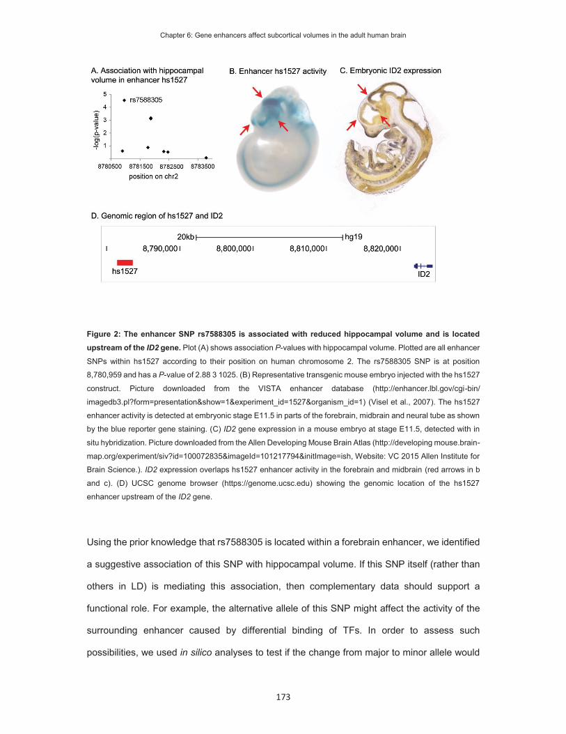
Chapter 6: Gene enhancers affect subcortical volumes in the adult human brain
173
Figure 2: The enhancer SNP rs7588305 is associated with reduced hippocampal volume and is located upstream of the ID2 gene. Plot (A) shows association P-values with hippocampal volume. Plotted are all enhancer
SNPs within hs1527 according to their position on human chromosome 2. The rs7588305 SNP is at position
8,780,959 and has a P-value of 2.88 3 1025. (B) Representative transgenic mouse embryo injected with the hs1527
construct. Picture downloaded from the VISTA enhancer database (http://enhancer.lbl.gov/cgi-bin/
imagedb3.pl?form=presentation&show=1&experiment_id=1527&organism_id=1) (Visel et al., 2007). The hs1527
enhancer activity is detected at embryonic stage E11.5 in parts of the forebrain, midbrain and neural tube as shown
by the blue reporter gene staining. (C) ID2 gene expression in a mouse embryo at stage E11.5, detected with in
situ hybridization. Picture downloaded from the Allen Developing Mouse Brain Atlas (http://developing mouse.brain-
map.org/experiment/siv?id=100072835&imageId=101217794&initImage=ish, Website: VC 2015 Allen Institute for
Brain Science.). ID2 expression overlaps hs1527 enhancer activity in the forebrain and midbrain (red arrows in b
and c). (D) UCSC genome browser (https://genome.ucsc.edu) showing the genomic location of the hs1527
enhancer upstream of the ID2 gene.
Using the prior knowledge that rs7588305 is located within a forebrain enhancer, we identified
a suggestive association of this SNP with hippocampal volume. If this SNP itself (rather than
others in LD) is mediating this association, then complementary data should support a
functional role. For example, the alternative allele of this SNP might affect the activity of the
surrounding enhancer caused by differential binding of TFs. In order to assess such
possibilities, we used in silico analyses to test if the change from major to minor allele would

Chapter 6: Gene enhancers affect subcortical volumes in the adult human brain
174
affect known TF binding sites. The differential motif analysis of the surrounding genomic
fragment predicted a gain of six new motifs and a loss of five motifs for the minor allele
(Supporting table 5). Among these predicted differences we found two ubiquitously expressed
general TFs that are involved in chromatin structure and looping, CTCF and p300 (Supporting
figure 1). The minor allele of rs7588305 gained a binding motif for CTCF - a general TF that
stabilizes chromatin loops and is generally found in structural chromatin loops that either block
enhancer promoter interactions or are active enhancer loops (Ong and Corces, 2014). The
second change for the minor allele was the loss of a p300-binding motif. P300 is a
transcriptional coactivator that is found at active enhancer-promoter loops and increases
transcription of the target gene (Chan and La Thangue, 2001). The motif analysis of the
rs7588305 SNP is thus consistent with the view that the allelic state of this SNP could be
associated with changed activity of the enhancer. The variant could potentially decrease the
activity of the enhancer, given that the binding site for the co-activator p300 is lost. However,
CTCF function is highly dependent on context, and the effect of a gained CTCF binding site is
difficult to predict. Thus, to determine the direction of effect of the minor allele would require
future experiments, for example using cellular models.
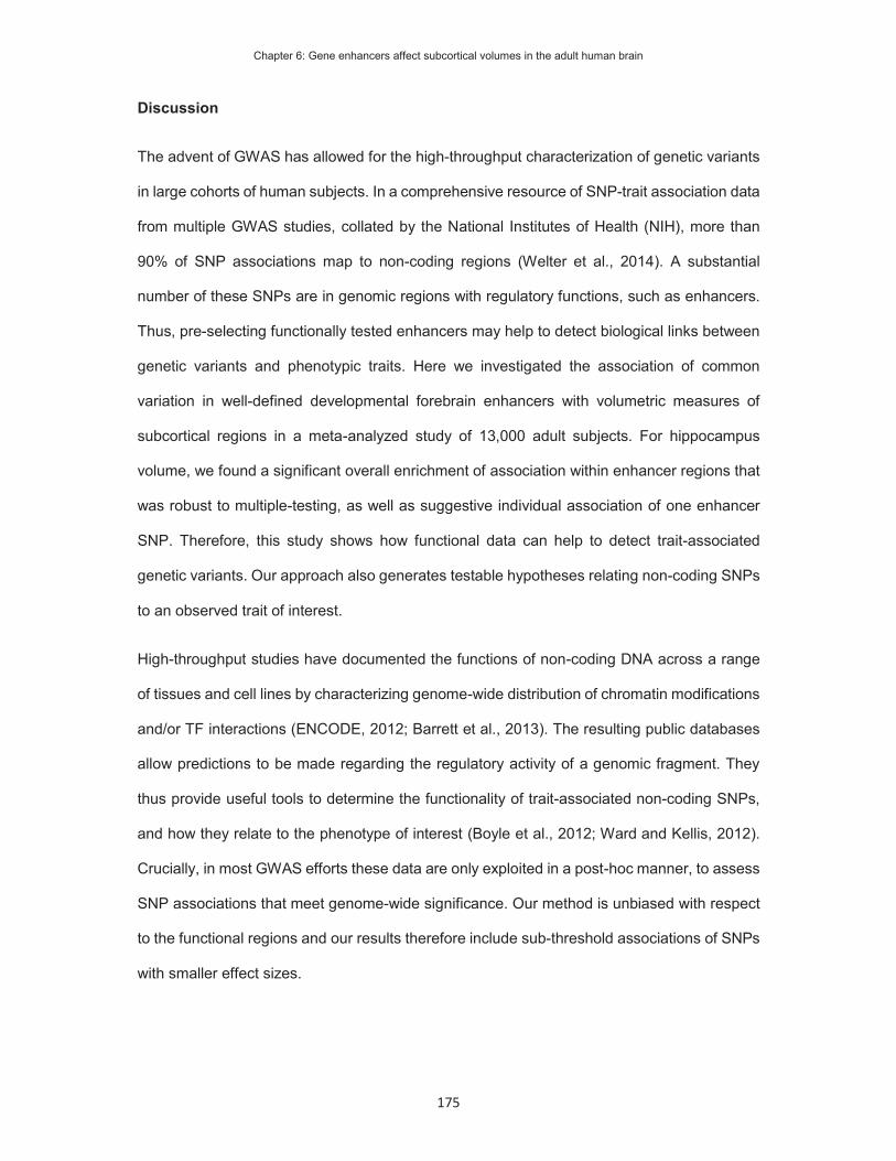
Chapter 6: Gene enhancers affect subcortical volumes in the adult human brain
175
Discussion
The advent of GWAS has allowed for the high-throughput characterization of genetic variants
in large cohorts of human subjects. In a comprehensive resource of SNP-trait association data
from multiple GWAS studies, collated by the National Institutes of Health (NIH), more than
90% of SNP associations map to non-coding regions (Welter et al., 2014). A substantial
number of these SNPs are in genomic regions with regulatory functions, such as enhancers.
Thus, pre-selecting functionally tested enhancers may help to detect biological links between
genetic variants and phenotypic traits. Here we investigated the association of common
variation in well-defined developmental forebrain enhancers with volumetric measures of
subcortical regions in a meta-analyzed study of 13,000 adult subjects. For hippocampus
volume, we found a significant overall enrichment of association within enhancer regions that
was robust to multiple-testing, as well as suggestive individual association of one enhancer
SNP. Therefore, this study shows how functional data can help to detect trait-associated
genetic variants. Our approach also generates testable hypotheses relating non-coding SNPs
to an observed trait of interest.
High-throughput studies have documented the functions of non-coding DNA across a range
of tissues and cell lines by characterizing genome-wide distribution of chromatin modifications
and/or TF interactions (ENCODE, 2012; Barrett et al., 2013). The resulting public databases
allow predictions to be made regarding the regulatory activity of a genomic fragment. They
thus provide useful tools to determine the functionality of trait-associated non-coding SNPs,
and how they relate to the phenotype of interest (Boyle et al., 2012; Ward and Kellis, 2012).
Crucially, in most GWAS efforts these data are only exploited in a post-hoc manner, to assess
SNP associations that meet genome-wide significance. Our method is unbiased with respect
to the functional regions and our results therefore include sub-threshold associations of SNPs
with smaller effect sizes.
Chapter 6: Gene enhancers affect subcortical volumes in the adult human brain
176
Targeted studies of single genomic regions indicate that variants in non-coding DNA are
related to variation in normal brain volume and risk for psychiatric illness. For example, a rare
variant implicated in autism was found in an enhancer that is active during forebrain
development. The variant that may increase risk for autism alters binding of regulatory TFs
and reduces enhancer activity in the developing forebrain (Poitras et al., 2010). Another study
focused on an enhancer that underwent accelerated evolution on the human lineage, after
splitting from that of chimpanzees. In mouse models, the human version of this enhancer
prolongs forebrain growth and increases the size of the developing neocortex (Boyd et al.,
2015). This supports our finding that human gene enhancers help explain how non-coding
SNPs contribute to variation in brain volume.
We identified an enhancer SNP that has a within-phenotype significant association with
hippocampal volume. The SNP is contained within the enhancer hs1527 and positioned
upstream of the inhibitor of DNA binding 2 (ID2) gene. ID2 is expressed in the embryonic
mouse forebrain and in regions of adult rat brain, including the hippocampus (Kitajima et al.,
2006). The expression pattern of ID2 overlaps with the regions of hs1527 enhancer activity
(Figure 2b-c). ID2 enhances cell proliferation (Iavarone et al., 1994) and promotes axon growth
in primary rat neurons (Lasorella et al., 2006). In human neuronal cell lines, ID2 is directly
regulated by MECP2. Disruptions of the MECP2 gene cause Rett syndrome, a
neurodevelopmental disorder characterized by severe intellectual disability and microcephaly
(Amir et al., 1999). Accordingly, ID2 expression was higher in post-mortem brain samples from
subjects with Rett Syndrome (Peddada et al., 2006). The absence of association between
rs7588305 and ID2 expression in adult human hippocampal samples, as measured by the
Braineac eQTL database, does not discount an effect on enhancer activity in embryonic tissue.
Differential ID2 expression during embryonic development may be sufficient to lead to
volumetric changes in the adult, which agrees with the role of the gene in cell-proliferation and
neurogenesis. Future studies of ID2 function in neurodevelopment may link this gene to
hippocampal development.
Chapter 6: Gene enhancers affect subcortical volumes in the adult human brain
177
A benefit of pre-selecting genetic variants based on functional information is that specific
testable hypotheses are generated for follow-up studies. For example, motif analysis using
the major and minor alleles of rs7588305 predicted differential binding of CTCF and p300, two
ubiquitously expressed proteins involved in chromatin loop formation and transcriptional
regulation. The minor allele lacks the motif for p300, a general mediator for enhancer activity,
and gains a motif for CTCF, which may affect enhancer loop formation. The shifted binding of
these two general chromatin associated factors could conceivably alter the activity of the
hs1527 enhancer, a hypothesis that can be tested with future experiments in cellular models,
which would also enable the direction of effect to be determined.
Here, we determined if a set of functionally validated enhancers, active during early brain
development, was enriched for association with volumetric differences in subcortical brain
structures in human adults. We found significant enrichment with hippocampal volume and
nominal significance for the nucleus accumbens, pallidum, and thalamus. The selected
forebrain enhancers contained an increased number of common genetic variants with sub-
threshold associations. The observed enrichment in the hippocampus was found with
forebrain enhancers, but was not detectable for non-brain enhancers. In order to exclude that
the effects reflected unusual LD structure of the forebrain enhancers, or some other artefact,
we performed additional control analyses of other human traits, including BMI, height and
WHR, and did not observe enrichment. This finding implies that early developmental enhancer
activity has an impact on adult hippocampal volume. During the human lifespan more than
80% of all genes (≥ 20,000) are expressed in the developing and adult human brain (Kang et
al., 2011), and each active gene promoter is in contact with several enhancers (Sanyal et al.,
2012; Jin et al., 2013). Tens of thousands of enhancers may guide the coordinated expression
of genes during development. Indeed, more than 80,000 non-coding genomic regions show
histone marks indicative of active enhancers in brain tissue (Vermunt et al., 2014). The VISTA
enhancer database likely represents only a fraction of enhancers that act during human brain
development, and genetic variants in unsampled enhancers may also contribute to subcortical

Chapter 6: Gene enhancers affect subcortical volumes in the adult human brain
178
volumes. This could likely have limited our ability to detect enrichment beyond the
hippocampus. Future testing of all existing brain enhancers may increase the power to identify
enrichment of associations.
Another current limitation is that the VISTA database characterizes enhancer activity at E11.5
and anatomically annotates this activity only to the general forebrain region. An annotation
with respect to specific adult subcortical areas is not possible at this early embryonic period.
It is crucial to look at the most relevant tissue for association studies of enhancers, so more
detailed anatomical annotations of enhancer activities at later time points would improve the
approach. This may enable mapping of more direct connections from enhancer activity to adult
neuroimaging data.
In this study we focused on experimentally validated enhancer elements, because these give
a robust set of regions with clear functional links to defined human tissues. The method we
have employed may be expanded to study tissue-specific histone modifications as they
become more readily available (Vermunt et al., 2014; Roadmap Epigenomics et al., 2015). A
benefit of these datasets may be their more fine-grained anatomical description and sampling
across several developmental time points.
In summary, we demonstrated genotype-phenotype connections between early
neurodevelopmental enhancers and the volumes of adult subcortical brain regions in a dataset
of over 13,000 people. As with all genetic association studies, replication of these findings
should be sought in independent samples of a similar size where matched genome-wide
genotyping and brain measures are available. Comparable analyses of enhancer SNPs in
cohorts of patients with neurodevelopmental disorders has the potential to link early embryonic
enhancer activity to disease processes. Our study illustrates a novel method for integrating
functional genomic and phenotypic data to identify the biological underpinnings of highly
heterogeneous traits, including neuropsychological traits. Our method can be applied to other

Chapter 6: Gene enhancers affect subcortical volumes in the adult human brain
179
sets of functional non-coding genomic elements, to connect the function of the regions to
studied phenotypes of interest.

Chapter 6: Gene enhancers affect subcortical volumes in the adult human brain
180
References
Amir RE, Van den Veyver IB, Wan M, Tran CQ, Francke U, Zoghbi HY (1999) Rett syndrome is caused by mutations in X-linked MECP2, encoding methyl-CpG-binding protein 2. Nat Genet 23:185-188.
Barrett T, Wilhite SE, Ledoux P, Evangelista C, Kim IF, Tomashevsky M, Marshall KA, Phillippy KH, Sherman PM, Holko M, Yefanov A, Lee H, Zhang N, Robertson CL, Serova N, Davis S, Soboleva A (2013) NCBI GEO: archive for functional genomics data sets--update. Nucleic Acids Res 41:D991-995.
Bis JC et al. (2012) Common variants at 12q14 and 12q24 are associated with hippocampal volume. Nat Genet 44:545-551.
Blankenberg D, Von Kuster G, Coraor N, Ananda G, Lazarus R, Mangan M, Nekrutenko A, Taylor J (2010) Galaxy: a web-based genome analysis tool for experimentalists. Current protocols in molecular biology / edited by Frederick M Ausubel [et al] Chapter 19:Unit 19 10 11-21.
Boyd JL, Skove SL, Rouanet JP, Pilaz LJ, Bepler T, Gordan R, Wray GA, Silver DL (2015) Human-Chimpanzee Differences in a FZD8 Enhancer Alter Cell-Cycle Dynamics in the Developing Neocortex. Current biology : CB.
Boyle AP, Hong EL, Hariharan M, Cheng Y, Schaub MA, Kasowski M, Karczewski KJ, Park J, Hitz BC, Weng S, Cherry JM, Snyder M (2012) Annotation of functional variation in personal genomes using RegulomeDB. Genome Res 22:1790-1797.
Cai DC, Fonteijn H, Guadalupe T, Zwiers M, Wittfeld K, Teumer A, Hoogman M, Arias-Vasquez A, Yang Y, Buitelaar J, Fernandez G, Brunner HG, van Bokhoven H, Franke B, Hegenscheid K, Homuth G, Fisher SE, Grabe HJ, Francks C, Hagoort P (2014) A genome-wide search for quantitative trait loci affecting the cortical surface area and thickness of Heschl's gyrus. Genes Brain Behav 13:675-685.
Chan HM, La Thangue NB (2001) p300/CBP proteins: HATs for transcriptional bridges and scaffolds. J Cell Sci 114:2363-2373.
ENCODE (2012) An integrated encyclopedia of DNA elements in the human genome. Nature 489:57-74.
Ferrer-Vicens I, Riffo-Campos AL, Zaragoza R, Garcia C, Lopez-Rodas G, Vina JR, Torres L, Garcia-Trevijano ER (2014) In vivo genome-wide binding of Id2 to E2F4 target genes as part of a reversible program in mice liver. Cell Mol Life Sci 71:3583-3597.
Giardine B, Riemer C, Hardison RC, Burhans R, Elnitski L, Shah P, Zhang Y, Blankenberg D, Albert I, Taylor J, Miller W, Kent WJ, Nekrutenko A (2005) Galaxy: a platform for interactive large-scale genome analysis. Genome Res 15:1451-1455.
Goecks J, Nekrutenko A, Taylor J, Galaxy T (2010) Galaxy: a comprehensive approach for supporting accessible, reproducible, and transparent computational research in the life sciences. Genome Biol 11:R86.
Guadalupe T, Zwiers MP, Teumer A, Wittfeld K, Vasquez AA, Hoogman M, Hagoort P, Fernandez G, Buitelaar J, Hegenscheid K, Volzke H, Franke B, Fisher SE, Grabe HJ, Francks C (2014a) Measurement and genetics of human subcortical and hippocampal asymmetries in large datasets. Human brain mapping 35:3277-3289.
Guadalupe T, Zwiers MP, Wittfeld K, Teumer A, Vasquez AA, Hoogman M, Hagoort P, Fernandez G, Buitelaar J, van Bokhoven H, Hegenscheid K, Volzke H, Franke B, Fisher SE, Grabe HJ, Francks C (2014b) Asymmetry within and around the human planum temporale is sexually dimorphic and influenced by genes involved in steroid hormone receptor activity. Cortex.
Heid IM et al. (2010) Meta-analysis identifies 13 new loci associated with waist-hip ratio and reveals sexual dimorphism in the genetic basis of fat distribution. Nat Genet 42:949-960.
Hibar DP et al. (2015) Common genetic variants influence human subcortical brain structures. Nature. Iavarone A, Garg P, Lasorella A, Hsu J, Israel MA (1994) The helix-loop-helix protein Id-2 enhances
cell proliferation and binds to the retinoblastoma protein. Genes Dev 8:1270-1284. Jack CR, Jr. et al. (2011) Steps to standardization and validation of hippocampal volumetry as a
biomarker in clinical trials and diagnostic criterion for Alzheimer's disease. Alzheimer's & dementia : the journal of the Alzheimer's Association 7:474-485 e474.
Jin F, Li Y, Dixon JR, Selvaraj S, Ye Z, Lee AY, Yen CA, Schmitt AD, Espinoza CA, Ren B (2013) A high-resolution map of the three-dimensional chromatin interactome in human cells. Nature 503:290-294.
Kang HJ et al. (2011) Spatio-temporal transcriptome of the human brain. Nature 478:483-489.

Chapter 6: Gene enhancers affect subcortical volumes in the adult human brain
181
Kel AE, Gossling E, Reuter I, Cheremushkin E, Kel-Margoulis OV, Wingender E (2003) MATCH: A tool for searching transcription factor binding sites in DNA sequences. Nucleic Acids Res 31:3576-3579.
Kitajima K, Takahashi R, Yokota Y (2006) Localization of Id2 mRNA in the adult mouse brain. Brain Res 1073-1074:93-102.
Lango Allen H et al. (2010) Hundreds of variants clustered in genomic loci and biological pathways affect human height. Nature 467:832-838.
Lasorella A, Stegmuller J, Guardavaccaro D, Liu G, Carro MS, Rothschild G, de la Torre-Ubieta L, Pagano M, Bonni A, Iavarone A (2006) Degradation of Id2 by the anaphase-promoting complex couples cell cycle exit and axonal growth. Nature 442:471-474.
Li MX, Yeung JM, Cherny SS, Sham PC (2012) Evaluating the effective numbers of independent tests and significant p-value thresholds in commercial genotyping arrays and public imputation reference datasets. Human genetics 131:747-756.
Ludwig MZ, Manu, Kittler R, White KP, Kreitman M (2011) Consequences of eukaryotic enhancer architecture for gene expression dynamics, development, and fitness. PLoS Genet 7:e1002364.
Nyholt DR (2004) A simple correction for multiple testing for single-nucleotide polymorphisms in linkage disequilibrium with each other. Am J Hum Genet 74:765-769.
Ong CT, Corces VG (2014) CTCF: an architectural protein bridging genome topology and function. Nat Rev Genet 15:234-246.
Paul DS, Albers CA, Rendon A, Voss K, Stephens J, van der Harst P, Chambers JC, Soranzo N, Ouwehand WH, Deloukas P (2013) Maps of open chromatin highlight cell type-restricted patterns of regulatory sequence variation at hematological trait loci. Genome Res 23:1130-1141.
Peddada S, Yasui DH, LaSalle JM (2006) Inhibitors of differentiation (ID1, ID2, ID3 and ID4) genes are neuronal targets of MeCP2 that are elevated in Rett syndrome. Human molecular genetics 15:2003-2014.
Pickrell JK (2014) Joint analysis of functional genomic data and genome-wide association studies of 18 human traits. Am J Hum Genet 94:559-573.
Poitras L, Yu M, Lesage-Pelletier C, Macdonald RB, Gagne JP, Hatch G, Kelly I, Hamilton SP, Rubenstein JL, Poirier GG, Ekker M (2010) An SNP in an ultraconserved regulatory element affects Dlx5/Dlx6 regulation in the forebrain. Development 137:3089-3097.
Ramasamy A, Trabzuni D, Guelfi S, Varghese V, Smith C, Walker R, De T, Coin L, de Silva R, Cookson MR, Singleton AB, Hardy J, Ryten M, Weale ME (2014) Genetic variability in the regulation of gene expression in ten regions of the human brain. Nat Neurosci 17:1418-1428.
Roadmap Epigenomics C et al. (2015) Integrative analysis of 111 reference human epigenomes. Nature 518:317-330.
Sanyal A, Lajoie BR, Jain G, Dekker J (2012) The long-range interaction landscape of gene promoters. Nature 489:109-113.
Speliotes EK et al. (2010) Association analyses of 249,796 individuals reveal 18 new loci associated with body mass index. Nat Genet 42:937-948.
Spitz F, Furlong EE (2012) Transcription factors: from enhancer binding to developmental control. Nat Rev Genet 13:613-626.
Stein JL et al. (2012) Identification of common variants associated with human hippocampal and intracranial volumes. Nat Genet 44:552-561.
Thompson PM et al. (2014) The ENIGMA Consortium: large-scale collaborative analyses of neuroimaging and genetic data. Brain Imaging Behav 8:153-182.
van Erp TG et al. (2015) Subcortical brain volume abnormalities in 2028 individuals with schizophrenia and 2540 healthy controls via the ENIGMA consortium. Molecular psychiatry.
Vermunt MW, Reinink P, Korving J, de Bruijn E, Creyghton PM, Basak O, Geeven G, Toonen PW, Lansu N, Meunier C, van Heesch S, Clevers H, de Laat W, Cuppen E, Creyghton MP (2014) Large-scale identification of coregulated enhancer networks in the adult human brain. Cell Rep 9:767-779.
Videbech P, Ravnkilde B (2004) Hippocampal volume and depression: a meta-analysis of MRI studies. Am J Psychiatry 161:1957-1966.
Visel A, Rubin EM, Pennacchio LA (2009a) Genomic views of distant-acting enhancers. Nature 461:199-205.
Visel A, Minovitsky S, Dubchak I, Pennacchio LA (2007) VISTA Enhancer Browser--a database of tissue-specific human enhancers. Nucleic Acids Res 35:D88-92.

Chapter 6: Gene enhancers affect subcortical volumes in the adult human brain
182
Visel A, Prabhakar S, Akiyama JA, Shoukry M, Lewis KD, Holt A, Plajzer-Frick I, Afzal V, Rubin EM, Pennacchio LA (2008) Ultraconservation identifies a small subset of extremely constrained developmental enhancers. Nat Genet 40:158-160.
Visel A, Blow MJ, Li Z, Zhang T, Akiyama JA, Holt A, Plajzer-Frick I, Shoukry M, Wright C, Chen F, Afzal V, Ren B, Rubin EM, Pennacchio LA (2009b) ChIP-seq accurately predicts tissue-specific activity of enhancers. Nature 457:854-858.
Ward LD, Kellis M (2012) HaploReg: a resource for exploring chromatin states, conservation, and regulatory motif alterations within sets of genetically linked variants. Nucleic Acids Res 40:D930-934.
Welter D, MacArthur J, Morales J, Burdett T, Hall P, Junkins H, Klemm A, Flicek P, Manolio T, Hindorff L, Parkinson H (2014) The NHGRI GWAS Catalog, a curated resource of SNP-trait associations. Nucleic Acids Res 42:D1001-1006.
Willer CJ, Li Y, Abecasis GR (2010) METAL: fast and efficient meta-analysis of genomewide association scans. Bioinformatics 26:2190-2191.

Chapter 7: Summary and General Discussion
183
Chapter 7
Summary and
General discussion

Chapter 7: Summary and General Discussion
184
Summary
DNA sequence variants affecting protein-coding genes may cause Mendelian disorders or
increase the likelihood to develop a certain phenotype. FOXP2 mutations cause a condition
that is characterized by severe difficulties in performing the oral motor movements required for
speech (developmental verbal dyspraxia), as well as reduced receptive and expressive
language skills. The expression pattern of FOXP2 in the brain suggests that this gene supports
the development of neuronal circuits involved in sensorimotor integration and learning of motor
skills. Thus, the upstream pathways that determine this complex expression pattern are key
for understanding its biological effects. In this dissertation, I investigated the regulation of the
FOXP2 gene, from the molecular level, through cultured cell-lines, to the whole organism. I
identified regulatory elements of FOXP2 implicated in disorder (Chapter2), as well as novel
enhancers that make direct contact with promoters of the gene (Chapter3). I studied the
upstream mechanisms that regulate these elements (Chapter 4) and characterized the tissue
activity of the most promising enhancers in the developing mouse brain (Chapter 5). Finally I
demonstrated how this type of enhancer information can generate novel insights into the
underlying biology of genotype-phenotype associations in relation to human brain development
(Chapter 6).
The aim of this thesis was to shed light on the mechanisms of neuronal FOXP2 regulation in
humans and to investigate the contribution of regulatory genetic variants to phenotypic
variation in health and disease. In this final chapter, I will summarize the findings of each
experimental chapter and highlight the different levels at which I approached the overarching
aims of this dissertation. I will combine the results from the single chapters and discuss the
findings in a bigger picture. Furthermore, I will discuss follow-up strategies to gain further
insight into the genetics and biology of speech and language.

Chapter 7: Summary and General Discussion
185
What can we learn from the genome?
Variability in neurodevelopmental enhancers are known to contribute to both normal variation
and disease phenotypes (Visel et al., 2013; Ramasamy et al., 2014; Boyd et al., 2015). In
chapter 2, I identified an enhancer near the FOXP2 gene that is displaced in a child with
delayed speech development (Becker et al., 2015). The affected individual carries a complex
chromosomal rearrangement, including an inversion of chromosome 7. One breakpoint of the
inversion is located downstream of FOXP2 and was mapped to the exact nucleotide position
by Moralli et al. (Moralli et al., 2015). The published information, in combination with predicted
functional states of the genome, was necessary and sufficient to locate the functional
enhancer. The phenotype overlaps that of people with high-penetrance FOXP2 mutations,
which suggested that misregulation of this gene could be involved in the aetiology of the
disorder. Thus, the displacement of the identified enhancer could cause aberrant FOXP2
expression and consequently disrupt speech development.
Moving beyond the linear sequence of the genome, I used the three-dimensional folding of the
DNA molecule to help uncover further novel regulatory elements that might regulate FOXP2
expression. In chapter 3, my approach to identify enhancers was to investigate chromosomal
interactions of the FOXP2 promoter. To achieve this, I identified the transcriptionally active
promoter in neuronal-like cells and then measured the chromatin interactions of this promoter
with genomic elements located upstream and downstream of the FOXP2 locus. In total, I
identified eleven genomic regions that interacted with the promoter in neuronal-like cells
(Figure 1). Promoter-promoter interactions between FOXP2 and MDFIC suggested that these
genes could be co-regulated. Indeed, transcriptome sequencing studies of primate forebrain
clustered both genes in one group of co-regulated genes (Konopka et al., 2012). However, not
much is known about MDIFC function and expression. Follow-up studies of this gene with
respect to FOXP2 function may further establish a link between FOXP2 and the WNT-
signalling pathway. I also detected an interaction of the FOXP2 promoter with the 3’-UTR of
the FOXP2. Similar interactions have been described for other genes and were reported to

Chapter 7: Summary and General Discussion
186
both decrease (Le Cam and Legraverend, 1995; Paul et al., 1998) and increase gene
expression (Salerno et al., 2000; Jash et al., 2012). The interaction to the 3’-UTR in FOXP2
expressing cells was either increased or depleted and thus, the influence of this interaction on
FOXP2 expression remains unclear. Within the eleven interacting regions I detected nine
candidate enhancer elements that were characterized by enhancer-specific histone marks in
neuronal tissue and increased chromatin interactions in FOXP2 expressing cell lines.
What can we learn from enhancers?
The candidate enhancers of FOXP2 enabled me to study the regulatory mechanism of this
gene at different levels. On the molecular level, enhancers are active within genetic networks
and respond to specific signaling pathways. On the cellular level, enhancers are active in
specific tissues and at developmental periods. On the human population level, variation in
enhancer sequences may contribute to the variation in structure and function of the target
Figure 1: Chromatin interactions at the FOXP2 promoter. I detected chromatin interactions of the active
FOXP2 promoter in human neuronal-like cells (Chapter 3). The distal FOXP2 promoter interacts with the MDFIC
promoter, the 3’-UTR and neurodevelopmental enhancers. Interactions to two representative enhancers (red) are
depicted. Promoter-promoter interactions, 3’-UTR and enhancer contacts may regulate the expression of FOXP2
in response to developmental or environmental cues.

Chapter 7: Summary and General Discussion
187
tissue. In the following section I summarized what I have learned from studying enhancers on
the different levels.
Upstream regulatory networks of FOXP2 identified in molecular studies
In chapter 4 I determined the basal activity of the FOXP2 promoters and enhancers in human
neuronal-like cells. I detected the activity of promoter 1 and demonstrated the in vitro enhancer
function of three enhancer elements. In combination with the enhancers, I studied the
regulatory potential of eight TFs: LEF1, POU3F2, PAX6, TBR1, SOX5, FOXP1, FOXP2 and
FOXP4. All TFs except POU3F2 modulated the activity of one or more regulatory element.
I demonstrated an enhancing effect of FOXP2 on its regulatory elements, which suggested a
positive auto-regulatory loop. Auto-regulation is found for TFs that function to maintain the
cellular identity of differentiated cells (Bateman, 1998; Meredith et al., 2009) and for some TFs
has been suggested as a mechanism to explain haploinsufficiency in disorders (Kleinjan et al.,
2004). I detected WNT-signalling TF motifs in all active regulatory elements and consequently
determined regulation by the WNT effector LEF1 (Figure 2). The effects of this experiment
suggested that FOXP2 might be actively regulated in response to WNT-signalling. This
pathway is important in neurodevelopment and adult neurogenesis (Piccin and Morshead,
2011; Dickins and Salinas, 2013; Oliva et al., 2013). It will be important to determine the
developmental period and brain tissue in which FOXP2 could be regulated by WNT signalling.
Additionally, I demonstrated the regulation of the candidate elements by SOX5 and TBR1,
which overlap FOXP2 expression in cortico-fugal interneurons of the cortical layer VI and
regulate subtype specification in post-mitotic neurons (Kwan et al., 2008; Lai et al., 2008;
Bedogni et al., 2010; Hisaoka et al., 2010; Han et al., 2011; McKenna et al., 2011; Hao et al.,
2014). Both TFs increased promoter 1 and enhancer 37 activity, which suggest an activating
effect on FOXP2 expression in cortical layer VI.

Chapter 7: Summary and General Discussion
188
FOXP2 enhancers drive in vivo expression in specific neuronal tissue
In chapter 5, I mapped the activity of two FOXP2 enhancers in the developing mouse brain. I
created transgenic mice that expressed a reporter gene under the control of these enhancers
and visualized reporter gene expression in embryonic, postnatal and adult brains. Enhancer
37 activated reporter gene expression in the hippocampus, ventricles and olfactory bulb (OB).
The activity of enhancer 37 overlapped known FOXP2 expression sites only in the OB. The
ventricular activity of this enhancer started after birth and remained stable throughout
adulthood, whereas the LacZ expression in OB was detectable at embryonic day 18.5 and
decreased in adult mice. The activity pattern of this enhancer could suggest that it is activated
in adult new-born neurons in the ventricles and hippocampus, and remains active in neurons
that migrate to the OB (Doetsch and Alvarez-Buylla, 1996; Lois et al., 1996). In humans and
songbirds, the ventricular born neurons have been shown to migrate into the striatum (Alvarez-
Buylla et al., 1994; Bergmann et al., 2015; Ernst and Frisen, 2015). In songbirds, these neurons
Figure 2: Promoter and enhancers are regulated by transcription factors. Co-transfection luciferase
experiments reveal regulatory interactions of the regulatory elements in human neuronal-like cell lines (Chapter
4). The schematic shows effect of LEF1 (blue) on promoter 1, enhancer 37 and enhancer 330. Enhancer 815
activity was not affected by LEF1. In chapter 4 I additionally studied the effects of PAX6, POU3F2, FOXP1,
FOXP2, FOXP4, SOX5 and TBR1.

Chapter 7: Summary and General Discussion
189
express FoxP2 and migrate into in the striatal nucleus area X (Rochefort et al., 2007). The
increased expression of FoxP2 in area X was linked to singing behaviour in adult zebra finches
(Haesler et al., 2004; Teramitsu and White, 2006; Haesler et al., 2007). It remains to be shown
whether or not this gene is active during adult neurogenesis in a mammalian model system. In
particular, it is unknown if FOXP2 is expressed in the migratory stream of adult new-born
neurons in humans.
The transgenic mice carrying enhancer 330 showed reporter gene expression in the neocortex
and Purkinje Cells (PCs) of the cerebellum (Figure 3). The enhancer was active in the
ventricular zone of the embryo and cortex of neonatal brains. Thus, enhancer 330 activity
overlaps with known sites of FOXP2 expression during development. These results suggest
that enhancer 330 might be involved in driving early cortical expression of FOXP2. In adult
mice, the activity of enhancer 330 decreased in deeper cortical layers, but remained active in
the upper layers. Interestingly, prior studies have shown that FOXP2 expression is localized
specifically to the deeper cortical layer VI (Ferland et al., 2003). The absence of FOXP2 mRNA
in the upper layers can be explained by micro RNA mediated down-regulation, as the two micro
RNAs mir-9 and mir-132 have been shown to repress FOXP2 expression specifically in the
upper cortical layers (Clovis et al., 2012). The lack of enhancer 330 activity in the adult deeper
cortical layers indicates that FOXP2 transcription in this layer is maintained by regulatory
mechanisms that only act on the promoter or on other enhancers. In summary, the enhancer
330 activity overlapped FOXP2 expression in the adult PCs and developing neocortex, which
strongly suggests that this enhancer contributes to FOXP2 expression in these structures.

Chapter 7: Summary and General Discussion
190
Genetic variants in neurodevelopmental enhancers explain structural variation in brain tissue
In chapter 6 I studied common genetic variants in a set of neurodevelopmental enhancers. The
hypothesis was that genetic variants in enhancers affect the structure of the enhancer’s target
tissue in the adult brain. To test this hypothesis I selected common genetic variants located
within experimentally validated regulatory elements. Because I expected subtle effects for
single genetic enhancer variants, I analysed a set of 296 enhancers of the prior VISTA study,
publically available through the VISTA enhancer browser (Visel et al., 2007). The results of
this chapter are therefore not specific to the FOXP2 enhancers. I focused on enhancers that
had been shown to be active in the embryonic forebrain, which develops into the cortex and
most of the subcortical brain regions. I hypothesized that variants in these enhancers influence
the enhancer activity and consequently the expression of their target genes. Differential
expression of neural genes could potentially alter the function and/or structure of the forebrain.
I selected the enhancer variants from the summary data of the ENIGMA2 genome-wide
Figure 3: Neurodevelopmental enhancers drive tissue-specific expression in vivo. I determined the in vivo
enhancer activity of two elements in transgenic mice expressing the LacZ reporter gene under control of the
enhancer elements. In the schematic, enhancer 330 is depicted as active enhancer in the endogenous genomic
loci controlling FOXP2 in the cortex and PCs.

Chapter 7: Summary and General Discussion
191
association study (GWAS) (Hibar et al., 2015). The ENIMGA2 study aimed to identify
associations between genetic variants and variation in subcortical brain volumes of 30,000
healthy human adults, of which I analysed the discovery sample (13,000) in chapter 6. Within
the group of forebrain enhancer variants I detected significant enrichment for variants
associated with hippocampal volume. This enrichment was specific to subcortical phenotypes
and not detectable in control phenotypes, such as body-mass index. The results of this chapter
demonstrate how targeting experimentally-defined enhancers may help identify the underlying
molecular aetiology of genotype-phenotype associations.
Integration of results across the chapters
Point mutations in FOXP2 cause a developmental speech and language disorder (Lai et al.,
2001). People with FOXP2 mutations show functional and structural differences in brain
structures related to motor control and language (Vargha-Khadem et al., 2005; Liegeois et al.,
2011). The aim of this thesis was to identify the upstream regulatory networks that control
FOXP2 expression, which might help shed further light on genetic pathways involved in the
development of neuronal circuits supporting human speech and language. Each chapter
produced independent results and provided insight into components of the regulatory
mechanisms. I obtained results on the molecular, cellular, tissue and human population levels.
In order to understand the complex regulatory mechanisms and their possible implications for
pathogenic and normal variation it is important to discuss the results across the chapters.
Purkinje cells maintenance mediated by WNT-signalling
In this thesis, I demonstrated that enhancer 330 is a neurodevelopmental enhancer and active
in the PCs of the cerebellum (Chapter 5). The enhancer activity could be increased by LEF1
(Chapter 4), which is known to be expressed in the developing cerebellum and in adult PCs
Chapter 7: Summary and General Discussion
192
(Wisniewska et al., 2010). LEF1 is the effector TF of the WNT-signalling pathway, which is
active in a subset of adult PCs (Selvadurai and Mason, 2011). Thus, my findings raise the
possibility that LEF1 might regulate FOXP2 expression during cerebellar development and in
adult PCs (Figure 4). However, I did not study the embryonic activity pattern of enhancer 330
in the cerebellum, which is necessary to determine the overlap with WNT-signalling. In a prior
study of zebrafish embryos, lef1 knock-down was reported to abolish foxp2 expression in the
hindbrain (Bonkowsky et al., 2008), which is the developmental origin of the cerebellum. It has
been established that WNT-signalling in the cerebellum controls axon maintenance and
synapse formation (Salinas, 1999; Dickins and Salinas, 2013) and that PCs express different
components of the WNT-signalling pathway (Salinas et al., 1994; Patapoutian and Reichardt,
2000). Mouse Foxp2 has been shown to promote neuronal differentiation, neurite formation
and axon guidance in the striatum (Vernes et al., 2011). It can therefore be hypothesized that
LEF1 may potentially control FOXP2 expression to regulate aspects of axon growth and
synapse plasticity in the developing cerebellum (Figure 1), something that could be specifically
tested with future functional investigations. Notably, mice homozygous for aetiological Foxp2
mutations develop a smaller cerebellum (Groszer et al., 2008). The hypothesis that a Lef1-
Foxp2 pathway is important for normal cerebellar development could be followed up by
conditional knock-out of Foxp2 or Lef1 in PCs. Homozygous knock-out mice of Lef1 die before
weening, whereas heterozygotes appear normal with no effect on cerebellar development
(Vangenderen et al., 1994; Galceran et al., 2000). Conditional knock-outs of Lef1, where the
gene is disrupted specifically in the PCs, may be required to determine subtle effects on
cerebellar development, which cannot be detected with conventional knock-out. In conclusion,
Foxp2 is important for normal cerebellar development and my findings suggest that its
cerebellar expression may be regulated by enhancer 330. The human enhancer 330 is itself
regulated by LEF1, which may indicate a LEF1-FOXP2 pathway in the developing cerebellum.

Chapter 7: Summary and General Discussion
193
Is cortical neuron subtype specificity mediated by enhancer 330?
I detected reproducible activity of enhancer 330 in the developing and adult neocortex (Chapter
5). This enhancer was previously shown to reproducibly drive cortical expression in mouse
embryos at day E11.5 (Visel et al., 2007). In earlier studies, cortical Foxp2 mRNA expression
has been detected as early as E14.5 in the ventricular zone and in the postnatal cortex (Ferland
et al., 2003). In the cortex of neonatal mice, enhancer 330 is active in all cortical layers. FOXP2
expression is known to be primarily limited to the cortical layer VI in the adult cortex, whereas
I found that enhancer 330 showed the strongest activity in the higher cortical layers (Chapter
5). Thus, enhancer 330 and FOXP2 overlap in embryonic and neonatal brains, but not in the
adult cortex.
Figure 4: Integration of FOXP2 in cell-specific signaling pathways. Enhancer 330 (red) is located within the
FOXP2 gene body. This enhancer contacts the FOXP2 promoter (Chapter 3) and increases gene expression
(Chapter 4). The enhancer is, among others, active in developing and adult PCs (Chapter 5). PCs express LEF1
(blue), which is the transcription factor downstream of the WNT-signaling pathway and activates genes
responsible for synapse development and axon maintenance. LEF1 increases the activity of enhancer 330
(Chapter 4). The hypothesis is that WNT-signaling via LEF1 regulates FOXP2 expression in the PCs to control
axon and synapse plasticity.

Chapter 7: Summary and General Discussion
194
I detected reduced enhancer 330 activity in response to TBR1 and SOX5 overexpression in
human neuronal-like cell lines (Chapter 4) and enhancer 330 activity in the deeper cortical
layers of adult brains (Figure 5). The results from both chapters together suggest that TBR1
and SOX5 downregulate the expression of FOXP2 in the developing cortex. The expression
of SOX5 and TBR1 is present in the developing cortical subplate and cortical layer VI (Kwan
et al., 2008; McKenna et al., 2011). Enhancer 330 activity, SOX5 and TBR1 expression thus
likely overlap in embryonic and prenatal neurons. Immunohistochemistry or in situ hybridization
of SOX5/TBR1 with FOXP2 is required to determine the exact expression overlap of these
TFs. Cortical development of cortico-thalamic projection neurons is regulated by SOX5 and
TBR1, which establish the cellular-identity by repressing TFs that are found in other cortical
subtypes, such as FEZF2 and CTIP (Kwan et al., 2008; Lai et al., 2008; McKenna et al., 2011;
Greig et al., 2013). In the adult cortex, SOX5 and TBR1 expression is limited to layer VI (Lai
et al., 2008) and enhancer 330 activity in this layer decreases in adult animals. This suggests
that SOX5 and TBR1 may repress enhancer 330 in adult cortical layer VI. However, FOXP2
remains expressed in adult cortical layer VI neurons and all FOXP2 positive cortical neurons
in the postnatal cortex have been reported to be TBR1 positive (Hisaoka et al., 2010). Thus,
the adult activity of enhancer 330 cannot explain adult FOXP2 expression in the cortical layers.

Chapter 7: Summary and General Discussion
195
Downregulation of FOXP2 in upper cortical layers by post-transcriptional mechanisms may
explain the missing FOXP2 expression in these layers. Indeed, mir-9 and mir-132 have been
shown to specifically repress FOXP2 mRNA in the upper cortical layers (Clovis et al., 2012).
Thus, enhancer 330 may activate expression in all cortical progenitor cells and the layer VI
specificity is achieved by the post-transcriptional down-regulation. The expression in the
deeper cortical layers could potentially be maintained by FOXP2 itself, via autoregulation at
other regulatory elements (Chapter 4). Thus, enhancer 330 may only be required for embryonic
and early postnatal expression of FOXP2. In conclusion, my data suggest that FOXP2
expression in the cortex could be initiated by enhancer 330. SOX5 and TBR1 could be involved
in the regulation of enhancer 330, but the timing of the regulatory interaction will have to be
investigated in model systems of neural development.
Figure 5: Integration of FOXP2 in cortical laminar development. Enhancer 330 (red) is located within the
FOXP2 gene body. This enhancer contacts the FOXP2 promoter (Chapter 3) and increases gene expression
(Chapter 4). The enhancer is active in the developing cortex and upper cortical layers in the adult (Chapter
5). FOXP2 expression overlaps enhancer 330 activity in the developing cortex. In the adult cortex FOXP2 is
specifically expressed in the deeper cortical layer VI. TBR1 and SOX5 are expressed in the cortical layer VI
and decrease the activity of enhancer 330 (Chapter 4).
Chapter 7: Summary and General Discussion
196
Could enhancer 37 regulate the expression of FOXP2 in adult neurogenesis?
I mapped the in vivo activity of enhancer 37 to the hippocampus, ventricles and OB. Adult
neurogenesis occurs in the hippocampus and ventricles (Eriksson et al., 1998; Ernst et al.,
2014; Bergmann et al., 2015). In mice, the ventricular born neurons migrate to the OB in the
rostral migratory stream (Doetsch and Alvarez-Buylla, 1996; Wichterle et al., 1997). The
ventricular and OB activity of enhancer 37 could suggest that this enhancer is active in neurons
that migrate in the rostral migratory stream. I also detected that TF motifs of the WNT-signalling
pathway are located in enhancer 37 and that it is upregulated by LEF1 (Chapter 4). LEF1 in
complex with TCF-1 regulates the expression of WNT-signalling target genes (Dickins and
Salinas, 2013; Oliva et al., 2013). WNT signalling has been shown to regulate neurogenesis
in the ventricles (Piccin and Morshead, 2011), the hippocampus (Lie et al., 2005) and OB
(Booker-Dwyer et al., 2008). Enhancer 37 could thus be a target regulatory element of WNT-
signalling during adult neurodevelopment (Figure 6).

Chapter 7: Summary and General Discussion
197
In humans, the ventricular born neurons do not migrate to the OB but have been shown to
migrate into the striatum, where they differentiate into interneurons (Ernst et al., 2014; Ernst
and Frisen, 2015). Similarly, in songbirds ventricular born neurons migrate into the striatal
song-learning nucleus called Area X, where they differentiate and integrate into the existing
neuronal architecture (Alvarez-Buylla et al., 1994). The regulation of FoxP2 in Area X has been
shown to be linked to singing behaviour in the songbirds (Chen et al., 2013; Mendoza et al.,
2015). The increase of FoxP2 expression is partly due to new-born neurons in adult animals
that migrated into the striatum (Rochefort et al., 2007). Thus, adult neurogenesis may be linked
to FoxP2 expression and singing behaviour in songbirds (Schulz et al., 2010). However, in
songbirds, the new-born neurons form medium spiny neurons, whereas in humans they form
interneurons (Ernst and Frisen, 2015). The new-born human interneurons are DARPP32
Figure 6: Integration of FOXP2 in adult neurogenesis. Enhancer 37 (red) is located upstream of the FOXP2
gene. This enhancer and the promoter region of MDFIC contact the FOXP2 promoter (Chapter 3). Enhancer 37
increases gene expression (Chapter 4). The enhancer is active in the OB, the outer layer of the ventricles, the
dentate gyrus and CA1 neurons of the hippocampus (Chapter 5). Ventricular born neurons migrate to OB in
mice; and to the striatum in human and songbirds. The canonical WNT-signaling pathway, with the downstream
activators LEF1 and TCF-1 regulate neural differentiation in adult new-born neurons. LEF1 increases the activity
of enhancer 37. The hypothesis is that WNT-signaling via LEF1 regulates FOXP2 expression in adult new-born
neurons in the ventricles and the neurons migrate to the species-specific target tissue.
Chapter 7: Summary and General Discussion
198
negative and express parvalbumin and calbindin. The co-expression of FOXP2 with these
marker genes in striatal interneurons would, therefore, be crucial to determine a possible
relation between FOXP2 and human adult neurogenesis. Molecular studies of adult
neurogenesis in humans are not feasible, because of the lack of neuronal tissue for
experimental investigations. As new-born neurons in mice migrate into the OB, this animal is
not a suitable system to study the link between FOXP2 and neurogenesis. Possibly, this link
could be studied in other mammals, such as bats. Recently bats obtained attention as a model
system for mammalian vocal and communication system (Rodenas-Cuadrado et al., 2015).
Ventricular born neurons in bats at least partially migrate towards the OB (Amrein et al., 2007),
but the migratory route to the striatum has not yet been investigated.
Signalling pathways involved in language-related phenotypes
I identified enhancer 815 based on the information about a child with delayed speech
development, described in the literature (Chapter 2). The child carries an inversion that
separates FOXP2 from this enhancer, suggesting that misregulation of the gene contributes
to the disorder. My in vitro experiments suggested that activity of enhancer 815 may be
regulated by PAX6, FOXP1 and FOXP2 (Chapter 4), which suggests that, in the affected child,
the FOXP2 gene lacks enhancer 815 mediated regulatory input from these TFs. Similarly, loss-
of-function mutations in these TFs could affect the regulation of FOXP2 in other clinical cases.
The study of phenotypes associated with PAX6 and FOXP1 mutations and their overlap with
phenotypes seen for people with FOXP2 mutations may shed light on an overlapping molecular
aetiology. Genetic disruptions of FOXP1 can cause autism spectrum disorder, intellectual
disability, motor delay and expressive language problems (Pariani et al., 2009; Carr et al.,
2010; Hamdan et al., 2010; Horn et al., 2010; O'Roak et al., 2011; Bacon and Rappold, 2012;
Talkowski et al., 2012). The phenotypic spectra associated with FOXP1 and FOXP2
disruptions were previously described in a review and show overlap for language problems

Chapter 7: Summary and General Discussion
199
(Bacon and Rappold, 2012). PAX6 mutations generally cause aniridia and other malformations
of the eye (Hanson, 2003). A splice site mutation in PAX6 was linked to Gillespie syndrome,
which among other problems includes impairments in orofacial motor control and dysarthric
speech (Ticho et al., 2006). While dysarthria has been suggested to be a phenotype associated
with FOXP2 disruptions at least in some cases (Shriberg et al., 2006), it was not reported for
the child with the chromosomal rearrangement that separates FOXP2 from enhancer 815
(Moralli et al., 2015). Another patient carrying a de novo nonsense mutation in PAX6 presented
with the typical aniridia phenotype, but further included coordination impairment, general
dyspraxia, intellectual disability and poor speech (Graziano et al., 2007). Detailed comparisons
between people with PAX6 and FOXP2 mutations would be required to determine a possible
common overlapping phenotype.
Co-expression of FOXP2 with FOXP1 has been described in the basal ganglia, thalamus and
inferior olive (Teramitsu et al., 2004; Takahashi et al., 2008; Kaoru et al., 2010; Mendoza et
al., 2015) and with PAX6 in the amygdala and striatum (Kaoru et al., 2010). Notably, co-
expression of all three TFs has been seen in the striatum of monkeys and rats (Kaoru et al.,
2010). Thus, the observed regulation of enhancer 815 by FOXP1 and PAX6 could suggest that
this enhancer is active in the striatum (Figure 7). Perhaps the overlap in speech/language-
related phenotypes between cases of PAX6, FOXP1 and FOXP2 mutation can be partially
explained by regulatory interactions in the striatum. Overlapping functional or structural
aberrations in the striatum of the child that led to the discovery of enhancer 815 would further
support this hypothesis. Brain imaging studies of this child have not been reported, and
comparison to findings in additional clinical cases would be required to thoroughly evaluate
this hypothesis.

Chapter 7: Summary and General Discussion
200
Future direction
In the previous section, I presented models of FOXP2 regulation that emerged from analysing
results across the individual chapters and suggested follow-up strategies to investigate these.
In the following section, I address additional future directions that follow from my results.
Enhancers as tools to study FOXP2
The experiments in each chapter investigated a limited amount of the possible search space.
Future findings on FOXP2 regulation may highlight the importance of genomic regions which
have not been tested in chapter 3, and TFs which have not been investigated in chapter 4.
Clinically relevant copy number variants near FOXP2 may justify the extension of the chromatin
conformation capture design to more distant genomic regions. My in silico analyses detected
hundreds of TF motifs in the putative regulatory elements that I identified. Many of the relevant
Figure 7: Disruption of enhancer 815 regulation in language development. Enhancer 815 (red) is located
downstream of the FOXP2 gene. This enhancer is separated from FOXP2 in a patient with language problems
(Chapter 2). Enhancer 815 is repressed by PAX6 and activated by FOXP1 and FOXP2 (Chapter 4). Mutations
in PAX6 and FOXP2 can cause language problems. The hypothesis is that the language phenotypes caused by
mutations of FOXP1, FOXP2 and PAX6 are the result of aberrant FOXP2 regulation. This regulatory interaction
possibly occurs in the developing striatum.

Chapter 7: Summary and General Discussion
201
TFs are expressed in the brain and known to be involved in neuronal development (chapter
4). For example, the overlapping expression of foxp2 with emx1 in the zebrafish forebrain could
be an interesting link to follow up (Shah et al., 2006). Also retinoic acid binding receptor alpha
was detected in the motif analysis and accumulating evidence shows that retinoic acid
signalling is linked to FOXP2 function (Devanna et al., 2014) and language-related neural
networks (van Rhijn and Vernes, 2015). Furthermore, I selected the most promising enhancers
to map the in vivo activity (Chapter 5) and did not investigate enhancer 815 using these
techniques. As previously discussed, this enhancer is likely involved in the pathogenesis of a
child with delayed speech development. Uncovering the tissue-specific activity of this enhancer
will be important to determine the molecular aetiology underlying the phenotype of this case.
Genetic variation underlying language-related phenotypes
The identification of FOXP2 mutations in speech and language disorders has sparked
speculations on the potential contributions of this gene to other pathological and normal human
cognitive phenotypes. Consequently, researchers have investigated the FOXP2 gene locus for
genetic association to various traits. Such studies investigated the association of common
variants within the FOXP2 gene to auditory-visual hallucinations (Sanjuan et al., 2006;
McCarthy-Jones et al., 2014), schizophrenia (Tolosa et al., 2010; Spaniel et al., 2011), ADHD
(Ribases et al., 2012), autism (Park et al., 2014), enhanced language skills (Chandrasekaran
et al., 2015) or brain volumes (Hoogman et al., 2014). However, most of these studies report
negative findings or have not been replicated. Genetic variants that affect the identified
regulatory elements from this thesis could potentially alter FOXP2 expression and disrupting
the development of FOXP2 positive neuronal circuits. According to this hypothesis, in chapter
6 I demonstrated on a broader scale that neurodevelopmental enhancers are enriched for
association to subtle common phenotypes. Thus future candidate gene association studies of
FOXP2 would benefit from targeting genetic variants within the identified regulatory enhancer
elements from this thesis. Similarly, next generation sequencing of samples of people with
speech- and language-related disorders has been limited to the exome, focusing on coding

Chapter 7: Summary and General Discussion
202
sequences (Villanueva et al., 2015). The regulatory regions of FOXP2 are promising candidate
regions to contain rare variants, which would remain undetected with exome-sequencing. As
the field of clinical genetics moves rapidly from exome-sequencing to whole-genome
sequencing it will be crucial to filter the variants in a justified and meaningful way. Thus, the
incorporation of enhancer and other regulatory sequences will be necessary for all genes. The
field of language genetics is well equipped to identify genetic variants that may affect the
regulation of FOXP2 and contribute to language-related traits.
Conclusion
In my thesis, I investigated the regulation of FOXP2 at the molecular, cellular, tissue and
phenotypic level. I identified the first human enhancers of the FOXP2 gene, which led to the
identification of upstream signalling pathways of FOXP2. I mapped the enhancer activity in the
developing and adult brain, which relates the identified upstream pathways to the neuronal
tissue in which they regulate FOXP2. I linked a set of neurodevelopmental enhancers,
unrelated to FOXP2, to normal variation in brain structure and pathologic variation of language
skills. The integration of results across these levels created new hypotheses regarding the
mechanisms underlying the neurobiology of FOXP2.

Chapter 7: Summary and General Discussion
203
References
Alvarez-Buylla A, Ling CY, Yu WS (1994) Contribution of neurons born during embryonic, juvenile, and adult life to the brain of adult canaries: regional specificity and delayed birth of neurons in the song-control nuclei. The Journal of comparative neurology 347:233-248.
Amrein I, Dechmann DK, Winter Y, Lipp HP (2007) Absent or low rate of adult neurogenesis in the hippocampus of bats (Chiroptera). PloS one 2:e455.
Bacon C, Rappold GA (2012) The distinct and overlapping phenotypic spectra of FOXP1 and FOXP2 in cognitive disorders. Human genetics 131:1687-1698.
Bateman E (1998) Autoregulation of eukaryotic transcription factors. Progress in nucleic acid research and molecular biology 60:133-168.
Becker M, Devanna P, Fisher SE, Vernes SC (2015) A chromosomal rearrangement in a child with severe speech and language disorder separates FOXP2 from a functional enhancer. Molecular cytogenetics 8:69.
Bedogni F, Hodge RD, Elsen GE, Nelson BR, Daza RA, Beyer RP, Bammler TK, Rubenstein JL, Hevner RF (2010) Tbr1 regulates regional and laminar identity of postmitotic neurons in developing neocortex. Proc Natl Acad Sci U S A 107:13129-13134.
Bergmann O, Spalding KL, Frisen J (2015) Adult Neurogenesis in Humans. Cold Spring Harbor perspectives in biology 7.
Bonkowsky JL, Wang X, Fujimoto E, Lee JE, Chien CB, Dorsky RI (2008) Domain-specific regulation of foxP2 CNS expression by lef1. BMC developmental biology 8:103.
Booker-Dwyer T, Hirsh S, Zhao H (2008) A unique cell population in the mouse olfactory bulb displays nuclear beta-catenin signaling during development and olfactory sensory neuron regeneration. Developmental neurobiology 68:859-869.
Boyd JL, Skove SL, Rouanet JP, Pilaz LJ, Bepler T, Gordan R, Wray GA, Silver DL (2015) Human-Chimpanzee Differences in a FZD8 Enhancer Alter Cell-Cycle Dynamics in the Developing Neocortex. Current biology : CB.
Carr CW, Moreno-De-Luca D, Parker C, Zimmerman HH, Ledbetter N, Martin CL, Dobyns WB, Abdul-Rahman OA (2010) Chiari I malformation, delayed gross motor skills, severe speech delay, and epileptiform discharges in a child with FOXP1 haploinsufficiency. European journal of human genetics : EJHG 18:1216-1220.
Chandrasekaran B, Yi HG, Blanco NJ, McGeary JE, Maddox WT (2015) Enhanced procedural learning of speech sound categories in a genetic variant of FOXP2. The Journal of neuroscience : the official journal of the Society for Neuroscience 35:7808-7812.
Chen Q, Heston JB, Burkett ZD, White SA (2013) Expression analysis of the speech-related genes FoxP1 and FoxP2 and their relation to singing behavior in two songbird species. The Journal of experimental biology 216:3682-3692.
Clovis YM, Enard W, Marinaro F, Huttner WB, De Pietri Tonelli D (2012) Convergent repression of Foxp2 3'UTR by miR-9 and miR-132 in embryonic mouse neocortex: implications for radial migration of neurons. Development 139:3332-3342.
Devanna P, Middelbeek J, Vernes SC (2014) FOXP2 drives neuronal differentiation by interacting with retinoic acid signaling pathways. Frontiers in cellular neuroscience 8:305.
Dickins EM, Salinas PC (2013) Wnts in action: from synapse formation to synaptic maintenance. Frontiers in cellular neuroscience 7:162.
Doetsch F, Alvarez-Buylla A (1996) Network of tangential pathways for neuronal migration in adult mammalian brain. Proc Natl Acad Sci U S A 93:14895-14900.
Eriksson PS, Perfilieva E, Bjork-Eriksson T, Alborn AM, Nordborg C, Peterson DA, Gage FH (1998) Neurogenesis in the adult human hippocampus. Nature medicine 4:1313-1317.
Ernst A, Frisen J (2015) Adult neurogenesis in humans- common and unique traits in mammals. PLoS biology 13:e1002045.
Ernst A, Alkass K, Bernard S, Salehpour M, Perl S, Tisdale J, Possnert G, Druid H, Frisen J (2014) Neurogenesis in the striatum of the adult human brain. Cell 156:1072-1083.
Ferland RJ, Cherry TJ, Preware PO, Morrisey EE, Walsh CA (2003) Characterization of Foxp2 and Foxp1 mRNA and protein in the developing and mature brain. The Journal of comparative neurology 460:266-279.
Galceran J, Miyashita-Lin EM, Devaney E, Rubenstein JLR, Grosschedl R (2000) Hippocampus development and generation of dentate gyrus granule cells is regulated by LEF1. Development 127:469-482.

Chapter 7: Summary and General Discussion
204
Graziano C, D'Elia AV, Mazzanti L, Moscano F, Guidelli Guidi S, Scarano E, Turchetti D, Franzoni E, Romeo G, Damante G, Seri M (2007) A de novo nonsense mutation of PAX6 gene in a patient with aniridia, ataxia, and mental retardation. American journal of medical genetics Part A 143A:1802-1805.
Greig LC, Woodworth MB, Galazo MJ, Padmanabhan H, Macklis JD (2013) Molecular logic of neocortical projection neuron specification, development and diversity. Nat Rev Neurosci 14:755-769.
Groszer M et al. (2008) Impaired synaptic plasticity and motor learning in mice with a point mutation implicated in human speech deficits. Current biology : CB 18:354-362.
Haesler S, Rochefort C, Georgi B, Licznerski P, Osten P, Scharff C (2007) Incomplete and inaccurate vocal imitation after knockdown of FoxP2 in songbird basal ganglia nucleus Area X. PLoS biology 5:e321.
Haesler S, Wada K, Nshdejan A, Morrisey EE, Lints T, Jarvis ED, Scharff C (2004) FoxP2 expression in avian vocal learners and non-learners. The Journal of neuroscience : the official journal of the Society for Neuroscience 24:3164-3175.
Hamdan FF, Daoud H, Rochefort D, Piton A, Gauthier J, Langlois M, Foomani G, Dobrzeniecka S, Krebs MO, Joober R, Lafreniere RG, Lacaille JC, Mottron L, Drapeau P, Beauchamp MH, Phillips MS, Fombonne E, Rouleau GA, Michaud JL (2010) De novo mutations in FOXP1 in cases with intellectual disability, autism, and language impairment. Am J Hum Genet 87:671-678.
Han W, Kwan KY, Shim S, Lam MM, Shin Y, Xu X, Zhu Y, Li M, Sestan N (2011) TBR1 directly represses Fezf2 to control the laminar origin and development of the corticospinal tract. Proc Natl Acad Sci U S A 108:3041-3046.
Hanson IM (2003) PAX6 and congenital eye malformations. Pediatr Res 54:791-796. Hao H, Li Y, Tzatzalos E, Gilbert J, Zala D, Bhaumik M, Cai L (2014) Identification of a transient Sox5
expressing progenitor population in the neonatal ventral forebrain by a novel cis-regulatory element. Developmental biology 393:183-193.
Hibar DP et al. (2015) Common genetic variants influence human subcortical brain structures. Nature 520:224-229.
Hisaoka T, Nakamura Y, Senba E, Morikawa Y (2010) The forkhead transcription factors, Foxp1 and Foxp2, identify different subpopulations of projection neurons in the mouse cerebral cortex. Neuroscience 166:551-563.
Hoogman M, Guadalupe T, Zwiers MP, Klarenbeek P, Francks C, Fisher SE (2014) Assessing the effects of common variation in the FOXP2 gene on human brain structure. Frontiers in human neuroscience 8:473.
Horn D et al. (2010) Identification of FOXP1 deletions in three unrelated patients with mental retardation and significant speech and language deficits. Human mutation 31:E1851-1860.
Jash A, Yun K, Sahoo A, So JS, Im SH (2012) Looping mediated interaction between the promoter and 3' UTR regulates type II collagen expression in chondrocytes. PloS one 7:e40828.
Kaoru T, Liu FC, Ishida M, Oishi T, Hayashi M, Kitagawa M, Shimoda K, Takahashi H (2010) Molecular characterization of the intercalated cell masses of the amygdala: implications for the relationship with the striatum. Neuroscience 166:220-230.
Kleinjan DA, Seawright A, Childs AJ, van Heyningen V (2004) Conserved elements in Pax6 intron 7 involved in (auto)regulation and alternative transcription. Developmental biology 265:462-477.
Konopka G, Friedrich T, Davis-Turak J, Winden K, Oldham MC, Gao F, Chen L, Wang GZ, Luo R, Preuss TM, Geschwind DH (2012) Human-specific transcriptional networks in the brain. Neuron 75:601-617.
Kwan KY, Lam MM, Krsnik Z, Kawasawa YI, Lefebvre V, Sestan N (2008) SOX5 postmitotically regulates migration, postmigratory differentiation, and projections of subplate and deep-layer neocortical neurons. Proc Natl Acad Sci U S A 105:16021-16026.
Lai CS, Fisher SE, Hurst JA, Vargha-Khadem F, Monaco AP (2001) A forkhead-domain gene is mutated in a severe speech and language disorder. Nature 413:519-523.
Lai T, Jabaudon D, Molyneaux BJ, Azim E, Arlotta P, Menezes JR, Macklis JD (2008) SOX5 controls the sequential generation of distinct corticofugal neuron subtypes. Neuron 57:232-247.
Le Cam A, Legraverend C (1995) Transcriptional repression, a novel function for 3' untranslated regions. European journal of biochemistry / FEBS 231:620-627.
Lie DC, Colamarino SA, Song HJ, Desire L, Mira H, Consiglio A, Lein ES, Jessberger S, Lansford H, Dearie AR, Gage FH (2005) Wnt signalling regulates adult hippocampal neurogenesis. Nature 437:1370-1375.

Chapter 7: Summary and General Discussion
205
Liegeois F, Morgan AT, Connelly A, Vargha-Khadem F (2011) Endophenotypes of FOXP2: dysfunction within the human articulatory network. European journal of paediatric neurology : EJPN : official journal of the European Paediatric Neurology Society 15:283-288.
Lois C, Garcia-Verdugo JM, Alvarez-Buylla A (1996) Chain migration of neuronal precursors. Science 271:978-981.
McCarthy-Jones S, Green MJ, Scott RJ, Tooney PA, Cairns MJ, Wu JQ, Oldmeadow C, Carr V, Australian Schizophrenia Research B (2014) Preliminary evidence of an interaction between the FOXP2 gene and childhood emotional abuse predicting likelihood of auditory verbal hallucinations in schizophrenia. Journal of psychiatric research 50:66-72.
McKenna WL, Betancourt J, Larkin KA, Abrams B, Guo C, Rubenstein JL, Chen B (2011) Tbr1 and Fezf2 regulate alternate corticofugal neuronal identities during neocortical development. The Journal of neuroscience : the official journal of the Society for Neuroscience 31:549-564.
Mendoza E, Tokarev K, During DN, Retamosa EC, Weiss M, Arpenik N, Scharff C (2015) Differential coexpression of FoxP1, FoxP2, and FoxP4 in the Zebra Finch (Taeniopygia guttata) song system. The Journal of comparative neurology 523:1318-1340.
Meredith DM, Masui T, Swift GH, MacDonald RJ, Johnson JE (2009) Multiple transcriptional mechanisms control Ptf1a levels during neural development including autoregulation by the PTF1-J complex. The Journal of neuroscience : the official journal of the Society for Neuroscience 29:11139-11148.
Moralli D, Nudel R, Chan MT, Green CM, Volpi EV, Benitez-Burraco A, Newbury DF, Garcia-Bellido P (2015) Language impairment in a case of a complex chromosomal rearrangement with a breakpoint downstream of FOXP2. Molecular cytogenetics 8:36.
O'Roak BJ, Deriziotis P, Lee C, Vives L, Schwartz JJ, Girirajan S, Karakoc E, Mackenzie AP, Ng SB, Baker C, Rieder MJ, Nickerson DA, Bernier R, Fisher SE, Shendure J, Eichler EE (2011) Exome sequencing in sporadic autism spectrum disorders identifies severe de novo mutations. Nat Genet 43:585-589.
Oliva CA, Vargas JY, Inestrosa NC (2013) Wnts in adult brain: from synaptic plasticity to cognitive deficiencies. Frontiers in cellular neuroscience 7:224.
Pariani MJ, Spencer A, Graham JM, Jr., Rimoin DL (2009) A 785kb deletion of 3p14.1p13, including the FOXP1 gene, associated with speech delay, contractures, hypertonia and blepharophimosis. European journal of medical genetics 52:123-127.
Park Y, Won S, Nam M, Chung JH, Kwack K (2014) Interaction between MAOA and FOXP2 in association with autism and verbal communication in a Korean population. Journal of child neurology 29:NP207-211.
Patapoutian A, Reichardt LF (2000) Roles of Wnt proteins in neural development and maintenance. Current opinion in neurobiology 10:392-399.
Paul C, Simar-Blanchet AE, Ro HS, Le Cam A (1998) Characterization of three transcriptional repressor sites within the 3' untranslated region of the rat serine protease inhibitor 2.3 gene. European journal of biochemistry / FEBS 254:538-546.
Piccin D, Morshead CM (2011) Wnt signaling regulates symmetry of division of neural stem cells in the adult brain and in response to injury. Stem cells 29:528-538.
Ramasamy A, Trabzuni D, Guelfi S, Varghese V, Smith C, Walker R, De T, Coin L, de Silva R, Cookson MR, Singleton AB, Hardy J, Ryten M, Weale ME (2014) Genetic variability in the regulation of gene expression in ten regions of the human brain. Nat Neurosci 17:1418-1428.
Ribases M, Sanchez-Mora C, Ramos-Quiroga JA, Bosch R, Gomez N, Nogueira M, Corrales M, Palomar G, Jacob CP, Gross-Lesch S, Kreiker S, Reif A, Lesch KP, Cormand B, Casas M, Bayes M (2012) An association study of sequence variants in the forkhead box P2 (FOXP2) gene and adulthood attention-deficit/hyperactivity disorder in two European samples. Psychiatric genetics 22:155-160.
Rochefort C, He X, Scotto-Lomassese S, Scharff C (2007) Recruitment of FoxP2-expressing neurons to area X varies during song development. Developmental neurobiology 67:809-817.
Rodenas-Cuadrado P, Chen XS, Wiegrebe L, Firzlaff U, Vernes SC (2015) A novel approach identifies the first transcriptome networks in bats: a new genetic model for vocal communication. BMC genomics 16:836.
Salerno MS, Mordvinov VA, Sanderson CJ (2000) Binding of octamer factors to a novel 3'-positive regulatory element in the mouse interleukin-5 gene. J Biol Chem 275:4525-4531.
Salinas PC (1999) Wnt factors in axonal remodelling and synaptogenesis. Biochemical Society symposium 65:101-109.

Chapter 7: Summary and General Discussion
206
Salinas PC, Fletcher C, Copeland NG, Jenkins NA, Nusse R (1994) Maintenance of Wnt-3 expression in Purkinje cells of the mouse cerebellum depends on interactions with granule cells. Development 120:1277-1286.
Sanjuan J, Tolosa A, Gonzalez JC, Aguilar EJ, Perez-Tur J, Najera C, Molto MD, de Frutos R (2006) Association between FOXP2 polymorphisms and schizophrenia with auditory hallucinations. Psychiatric genetics 16:67-72.
Schulz SB, Haesler S, Scharff C, Rochefort C (2010) Knockdown of FoxP2 alters spine density in Area X of the zebra finch. Genes Brain Behav 9:732-740.
Selvadurai HJ, Mason JO (2011) Wnt/beta-catenin signalling is active in a highly dynamic pattern during development of the mouse cerebellum. PloS one 6:e23012.
Shah R, Medina-Martinez O, Chu LF, Samaco RC, Jamrich M (2006) Expression of FoxP2 during zebrafish development and in the adult brain. The International journal of developmental biology 50:435-438.
Shriberg LD, Ballard KJ, Tomblin JB, Duffy JR, Odell KH, Williams CA (2006) Speech, prosody, and voice characteristics of a mother and daughter with a 7;13 translocation affecting FOXP2. Journal of speech, language, and hearing research : JSLHR 49:500-525.
Spaniel F, Horacek J, Tintera J, Ibrahim I, Novak T, Cermak J, Klirova M, Hoschl C (2011) Genetic variation in FOXP2 alters grey matter concentrations in schizophrenia patients. Neuroscience letters 493:131-135.
Takahashi K, Liu FC, Hirokawa K, Takahashi H (2008) Expression of Foxp4 in the developing and adult rat forebrain. Journal of neuroscience research 86:3106-3116.
Talkowski ME et al. (2012) Sequencing chromosomal abnormalities reveals neurodevelopmental loci that confer risk across diagnostic boundaries. Cell 149:525-537.
Teramitsu I, White SA (2006) FoxP2 regulation during undirected singing in adult songbirds. The Journal of neuroscience : the official journal of the Society for Neuroscience 26:7390-7394.
Teramitsu I, Kudo LC, London SE, Geschwind DH, White SA (2004) Parallel FoxP1 and FoxP2 expression in songbird and human brain predicts functional interaction. The Journal of neuroscience : the official journal of the Society for Neuroscience 24:3152-3163.
Ticho BH, Hilchie-Schmidt C, Egel RT, Traboulsi EI, Howarth RJ, Robinson D (2006) Ocular findings in Gillespie-like syndrome: association with a new PAX6 mutation. Ophthalmic genetics 27:145-149.
Tolosa A, Sanjuan J, Dagnall AM, Molto MD, Herrero N, de Frutos R (2010) FOXP2 gene and language impairment in schizophrenia: association and epigenetic studies. BMC medical genetics 11:114.
van Rhijn JR, Vernes SC (2015) Retinoic Acid Signaling: A New Piece in the Spoken Language Puzzle. Frontiers in psychology 6:1816.
Vangenderen C, Okamura RM, Farinas I, Quo RG, Parslow TG, Bruhn L, Grosschedl R (1994) Development of Several Organs That Require Inductive Epithelial-Mesenchymal Interactions Is Impaired in Lef-1-Deficient Mice. Genes Dev 8:2691-2703.
Vargha-Khadem F, Gadian DG, Copp A, Mishkin M (2005) FOXP2 and the neuroanatomy of speech and language. Nat Rev Neurosci 6:131-138.
Vernes SC, Oliver PL, Spiteri E, Lockstone HE, Puliyadi R, Taylor JM, Ho J, Mombereau C, Brewer A, Lowy E, Nicod J, Groszer M, Baban D, Sahgal N, Cazier JB, Ragoussis J, Davies KE, Geschwind DH, Fisher SE (2011) Foxp2 regulates gene networks implicated in neurite outgrowth in the developing brain. PLoS Genet 7:e1002145.
Villanueva P et al. (2015) Exome sequencing in an admixed isolated population indicates NFXL1 variants confer a risk for specific language impairment. PLoS Genet 11:e1004925.
Visel A, Minovitsky S, Dubchak I, Pennacchio LA (2007) VISTA Enhancer Browser--a database of tissue-specific human enhancers. Nucleic Acids Res 35:D88-92.
Visel A et al. (2013) A high-resolution enhancer atlas of the developing telencephalon. Cell 152:895-908.
Wichterle H, Garcia-Verdugo JM, Alvarez-Buylla A (1997) Direct evidence for homotypic, glia-independent neuronal migration. Neuron 18:779-791.
Wisniewska MB, Misztal K, Michowski W, Szczot M, Purta E, Lesniak W, Klejman ME, Dabrowski M, Filipkowski RK, Nagalski A, Mozrzymas JW, Kuznicki J (2010) LEF1/beta-catenin complex regulates transcription of the Cav3.1 calcium channel gene (Cacna1g) in thalamic neurons of the adult brain. The Journal of neuroscience : the official journal of the Society for Neuroscience 30:4957-4969.

Appendix 1
207
Appendix 1
Appendix table 1: Expression primer Gene Exon Direction Sequence
FOXP2 5 Forward ACAGCATCCTGGAAAGCAAG FOXP2 6 Reverse ATGGAGATGAGTCCCTGACG GAPDH 2 Forward AAGGTGAAGGTCGGAGTCAAC GAPDH 3 Reverse GGGGTCATTGATGGCAACAATA
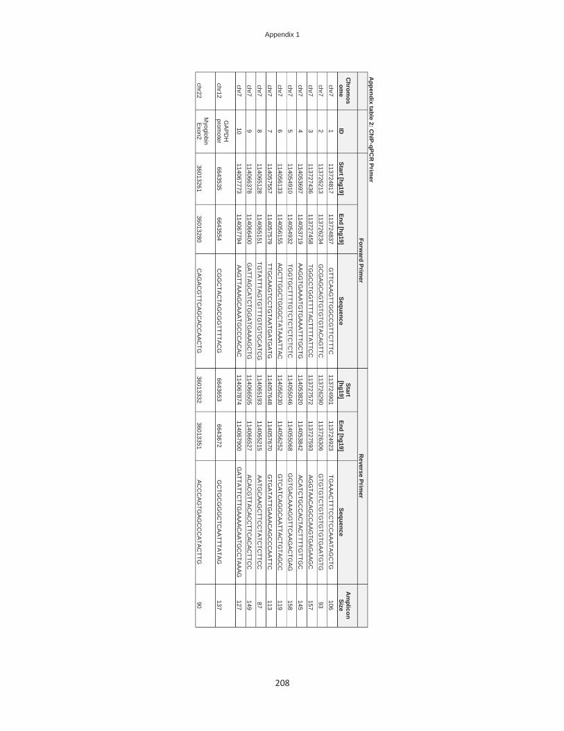
Appendix 1
208
Appendix table 2: C
hIP-qPCR
Primer
Forward Prim
er R
everse Primer
Chrom
osom
e ID
Start [hg19]
End [hg19] Sequence
Start [hg19]
End [hg19] Sequence
Am
plicon Size
chr7 1
113724817 113724837
GTTC
AA
GTTG
GC
CG
TTCTTTC
113724901
113724923 TG
AA
AC
TTTCC
TCC
AA
ATAG
CTG
106
chr7 2
113726213 113726234
GC
GA
GC
AG
TGTG
TGTA
CA
GTTC
113726290
113726306 G
TGTG
TCTG
TGTG
TGTG
AA
TGTG
93
chr7 3
113727436 113727458
TGG
CC
TGG
TTTTAC
TTTTATTC
C
113727572 113727593
AG
GTA
AC
AG
CC
AA
GTG
AG
AA
GC
157
chr7 4
114053697 114053719
AA
GG
TGA
AA
TGTG
AA
ATTTG
CTG
114053820
114053842 A
CA
TCTG
CC
AC
TAC
TTTTGTTG
C
145
chr7 5
114054910 114054932
TGG
TGC
TTTTGTC
TCTC
TCTC
TC
114055046 114055068
GG
TGA
CA
AA
GG
TTCA
AG
AC
TGA
G
158
chr7 6
114056133 114056155
AG
CTTG
GC
TGG
GC
TATA
AA
TTAC
114056230
114056252 G
TCA
TCA
GG
CA
ATTA
CTG
TAG
CC
119
chr7 7
114057557 114057579
TTGC
AA
GTC
CTG
TAA
TGA
TGA
TG
114057648 114057670
GTG
ATA
TTGA
AA
CA
GC
CC
AA
TTC
113
chr7 8
114065128 114065151
TGTA
TTTAG
TGTTTG
TGTG
CA
TCG
114065193
114065215 A
ATG
CA
AG
CTTC
CTA
TCTC
TTCC
87
chr7 9
114066378 114066400
GA
TTAG
CA
TCTG
GA
TGA
AA
GC
TG
114066505 114066527
AC
AC
GTTA
CA
CC
TTCA
CA
CTTC
C
149
chr7 10
114067773 114067794
AA
GTTA
AA
GC
AA
ATG
CC
CA
CA
C
114067874 114067900
GA
TTATTC
TTGA
AA
AC
AA
TGC
CTA
AAG
127
chr12 G
AP
DH
prom
oter 6643535
6643554 C
GG
CTA
CTA
GC
GG
TTTTAC
G
6643653 6643672
GC
TGC
GG
GC
TCA
ATTTA
TAG
137
chr22 M
yoglobin E
xon2 36013261
36013280 C
AG
AC
GTTC
AG
CA
CC
AA
CTG
36013332
36013351 A
CC
CA
GTG
AG
CC
CA
TAC
TTG
90
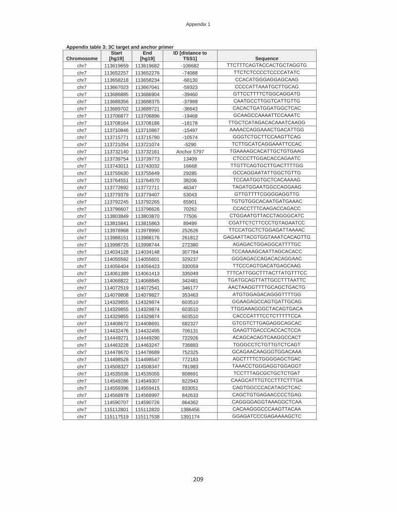
Appendix 1
209
Appendix table 3: 3C target and anchor primer
Chromosome Start
[hg19] End
[hg19] ID [distance to
TSS1] Sequence chr7 113619659 113619682 -106682 TTCTTTCAGTACCACTGCTAGGTG chr7 113652257 113652276 -74088 TTCTCTCCCCTCCCCATATC chr7 113658218 113658234 -68130 CCACATGGGAGGAGCAAG chr7 113667023 113667041 -59323 CCCCATTAAATGCTTGCAG chr7 113686885 113686904 -39460 GTTCCTTTTCTGGCAGGATG chr7 113688356 113688375 -37989 CAATGCCTTGGTCATTGTTG chr7 113689702 113689721 -36643 CACACTGATGGATGGCTCAC chr7 113706877 113706896 -19468 GCAAGCCAAAATTCCAAATC chr7 113708164 113708186 -18178 TTGCTCATAGACACAAATCAAGG chr7 113710846 113710867 -15497 AAAACCAGGAAACTGACATTGG chr7 113715771 113715790 -10574 GGGTCTGCTTCCAAGTTCAG chr7 113721054 113721074 -5290 TCTTGCATCAGGAAATTCCAC chr7 113732140 113732161 Anchor 5797 TGAAAAGCACATTGCTGTGAAG chr7 113739754 113739773 13409 CTCCCTTGGACACCAGAATC chr7 113743011 113743032 16668 TTGTTCAGTGCTTGACTTTTGG chr7 113755630 113755649 29285 GCCAGGAATATTGGCTGTTG chr7 113764551 113764570 38206 TCCAATGGTGCTCACAAAAG chr7 113772692 113772711 46347 TAGATGGAATGGCCAGGAAG chr7 113779379 113779407 53043 GTTGTTTTCGGGGAGGTTG chr7 113792245 113792265 65901 TGTGTGGCACAATGATGAAAC chr7 113796607 113796626 70262 CCACCTTTCAAGACCAGACC chr7 113803849 113803870 77506 CTGGAATGTTACCTAGGGCATC chr7 113815841 113815863 89499 CGATTCTCTTCCCTGTAGAATCC chr7 113978968 113978990 252626 TTCCATGCTCTGGAGATTAAAAC chr7 113988151 113988176 261812 GAGAATTACGTGGTAAATCACAGTTG chr7 113998725 113998744 272380 AGAGACTGGAGGCATTTTGC chr7 114034128 114034148 307784 TCCAAAAGCAATTAGCACACC chr7 114055582 114055601 329237 GGGAGACCAGACACAGGAAC chr7 114056404 114056423 330059 TTCCCAGTGACATGAGCAAG chr7 114061389 114061413 335049 TTTCATTGGCTTTACTTATGTTTCC chr7 114068822 114068845 342481 TGATGCAGTTATTGCCTTTAATTC chr7 114072519 114072541 346177 AACTAAGGTTTTGCAGCTGACTG chr7 114079808 114079827 353463 ATGTGGAGACAGGGTTTTGG chr7 114329855 114329874 603510 GGAAGAGCCAGTGATTGCAG chr7 114329855 114329874 603510 TTGGAAAGGGCTACAGTGACA chr7 114329855 114329874 603510 CACCCATTTCCTCTTTTTCCA chr7 114408672 114408691 682327 GTCGTCTTGAGAGGCAGCAC chr7 114432476 114432495 706131 GAAGTTGACCCACCACTCCA chr7 114449271 114449290 722926 ACAGCACAGTCAAGGCCACT chr7 114463228 114463247 736883 TGGGCCTCTGTTGTCTCAGT chr7 114478670 114478689 752325 GCAGAACAAGGGTGGACAAA chr7 114498528 114498547 772183 AGCTTTTCTGGGGAGCTGAC chr7 114508327 114508347 781983 TAAACCTGGGAGGTGGAGGT chr7 114535036 114535055 808691 TCCTTTAGCGCTGCTCTGAT chr7 114549286 114549307 822943 CAAGCATTTGTCCTTTCTTTGA chr7 114559396 114559415 833051 CAGTGGCCCACATAGCTCAC chr7 114568978 114568997 842633 CAGCTGTGAGAACCCCTGAG chr7 114590707 114590726 864362 CAGGGGAGGTAAAGGCTCAA chr7 115112801 115112820 1386456 CACAAGGGCCCAAGTTACAA chr7 115117519 115117538 1391174 GGAGATCCCGAGAAAAGCTC

Appendix 2
210
Appendix 2
Table S1: Cloning Prim
er
Reg. Elem
ent O
rientation Start [hg19]
End [hg19] Sequence
Size E
lement 37
Forward
113688009 113688029
ATC
AG
GTA
CC
CA
CA
GG
GG
AA
CC
TCTG
CA
AAA
CTG
C
20 E
lement 37
Reverse
113475999 113476018
ATC
AC
TCG
AG
CA
CA
ATTTGTG
GTG
GG
TGG
GTG
GG
19
Elem
ent 330 Forw
ard 114056847
114056867 A
TCA
GG
TAC
CG
GTTTTC
AC
GG
AAA
GC
AG
AG
T 20
Elem
ent 330 R
everse 114058626
114058647 A
TCA
GA
GC
TCG
AC
GTC
TGTTC
TGTG
AG
TTTCC
21
Elem
ent 815 Forw
ard 114541369
114541389 A
TCA
GG
TAC
CA
GTATTG
GC
AC
ACC
CA
GC
AA
20
Elem
ent 815 R
everse 114542182
114542201 A
TCA
GA
GC
TCTG
TTCC
TAGG
CG
TGTC
TGTG
19
Elem
ent 700 Forw
ard 114424203
114424224 A
TCA
GA
GC
TCA
CC
AG
AGG
AAAC
CC
TTCTTA
CC
21
Elem
ent 700 R
everse 114427828
114427847 A
TCA
CTC
GA
GA
GAA
GC
ATGTC
CC
AG
AAA
GC
19
Elem
ent 704 Forw
ard 114427887
114427907 A
TCA
GG
TAC
CTC
AG
AC
AC
AAC
CG
AC
ATTC
C
20 E
lement 704
Reverse
114431955 114431974
ATC
AC
TCG
AG
AC
CTC
GA
GC
AC
ATG
TTTTCC
19
Elem
ent 843 Forw
ard 114568454
114568473 A
TCA
GG
TAC
CA
GTG
TGA
TGG
CA
GAA
CA
TGG
19
Elem
ent 843 R
everse 114572392
114572411 A
TCA
CTC
GA
GA
CA
AC
TCC
CC
AATTTC
TCG
C
19 P
romoter 1
Forward
113724817 113724838
ATC
AG
AG
CTC
GTTC
AAG
TTGG
CC
GTTC
TTTCC
21
Prom
oter 1 R
everse 113726588
113726609 A
TCA
CTC
GA
GA
AG
CC
CC
CA
AACA
AC
AC
AC
C
21 P
romoter 2
Forward
114051220 114051242
ATC
AG
AG
CTC
CTC
AC
ATC
GC
CAA
AG
GTA
TAC
C
22 P
romoter 2
Reverse
114055298 114055324
ATC
AC
TCG
AG
GC
AC
AG
AAA
AC
AGTA
TAAA
AAG
AA
GG
G
26 P
romoter 3
Forward
114055454 114055475
ATC
AG
AG
CTC
TAG
AGA
GG
GG
TGG
GA
TTTTAC
G
21 P
romoter 3
Reverse
114056438 114056459
ATC
AC
TCG
AG
GG
TCA
GA
GG
AAAAA
GC
TGTTG
G
21

Appendix 2
211
Table S2: Predicted TF motifs Enhancer 37, 330 and 700 Enhancer 704, 815 and 843 Promoter 1, 2 and 3
Reg. Element Transcription Factor
# of motifs/ element Reg. Element
Transcription Factor
# of motifs/ element
Reg. Element
Transcription Factor
# of motifs/ element
Enhancer 37 AML 1 Enhancer 704 ALX3 4 Promoter 1 AHR 2 Enhancer 37 AML1 7 Enhancer 704 AML1 4 Promoter 1 AHRHIF 1 Enhancer 37 AP1 24 Enhancer 704 AP1 18 Promoter 1 AML 1 Enhancer 37 AP1FJ 3 Enhancer 704 AP1FJ 2 Promoter 1 AP2 5 Enhancer 37 AP2A 1 Enhancer 704 ARI3A 1 Promoter 1 AP2ALPHA 4 Enhancer 37 AP2C 1 Enhancer 704 ARX 6 Promoter 1 AP2B 1 Enhancer 37 AP3 1 Enhancer 704 Atoh1 2 Promoter 1 AP2D 1 Enhancer 37 AP4 3 Enhancer 704 BARHL1 1 Promoter 1 AP2GAMMA 1 Enhancer 37 ARP1 1 Enhancer 704 BARX1 1 Promoter 1 AREB6 1 Enhancer 37 ATF3 1 Enhancer 704 BARX2 2 Promoter 1 ARNT2 1 Enhancer 37 BACH1 1 Enhancer 704 BATF 2 Promoter 1 Ascl2 1 Enhancer 37 BACH2 1 Enhancer 704 BCL6 5 Promoter 1 ATF 2 Enhancer 37 BARX1 3 Enhancer 704 BHLHA15 2 Promoter 1 ATF1 3 Enhancer 37 BARX2 4 Enhancer 704 BHLHE23 1 Promoter 1 ATF2+ATF4 2 Enhancer 37 BATF 2 Enhancer 704 BPTF 1 Promoter 1 ATF3 1 Enhancer 37 BATF::JUN 2 Enhancer 704 BRCA1 1 Promoter 1 ATF4 2 Enhancer 37 BCL6 9 Enhancer 704 CART1 4 Promoter 1 BHLHE22 1 Enhancer 37 BLIMP1 2 Enhancer 704 CDP 2 Promoter 1 BHLHE23 2 Enhancer 37 CART1 4 Enhancer 704 CDX2 3 Promoter 1 BPTF 3 Enhancer 37 CEBP 4 Enhancer 704 CEBPGAMMA 1 Promoter 1 BRCA 1 Enhancer 37 CEBPA 3 Enhancer 704 CEBPZ 1 Promoter 1 BRCA1 1 Enhancer 37 CEBPB 5 Enhancer 704 COT2 2 Promoter 1 CEBPZ 1 Enhancer 37 CEBPD 1 Enhancer 704 COUP 1 Promoter 1 CETS1P54 1 Enhancer 37 CEBPDELTA 2 Enhancer 704 COUPTF 1 Promoter 1 CHCH 1 Enhancer 37 CEBPE 1 Enhancer 704 CPEB1 2 Promoter 1 CKROX 2 Enhancer 37 CEBPG 1 Enhancer 704 CTCF 1 Promoter 1 COE1 1
Enhancer 37 CEBPGAMMA 2 Enhancer 704 DEC 1 Promoter 1 CP2 5
Enhancer 37 CMYB 1 Enhancer 704 DLX1 2 Promoter 1 CREB 10 Enhancer 37 COUPTF 2 Enhancer 704 DLX2 2 Promoter 1 CREB1 4 Enhancer 37 CP2 2 Enhancer 704 DLX3 2 Promoter 1 CREB3 1 Enhancer 37 CPEB1 5 Enhancer 704 DLX7 1 Promoter 1 CREBATF 2 Enhancer 37 DBP 2 Enhancer 704 DR1 1 Promoter 1 CREBP1 2 Enhancer 37 EBOX 1 Enhancer 704 DR3 1 Promoter 1 CREBP1CJUN 2 Enhancer 37 EGR2 4 Enhancer 704 EAR2 1 Promoter 1 CREM 2 Enhancer 37 EHF 2 Enhancer 704 EFC 1 Promoter 1 CXXC1 1 Enhancer 37 ELF1 6 Enhancer 704 EHF 1 Promoter 1 E12 1 Enhancer 37 ELF3 2 Enhancer 704 ELF1 1 Promoter 1 E2A 1 Enhancer 37 Elf5 3 Enhancer 704 EMX2 1 Promoter 1 E2F 4 Enhancer 37 ELK3 2 Enhancer 704 EN1 2 Promoter 1 E2F1 6 Enhancer 37 EPAS1 1 Enhancer 704 EN2 2 Promoter 1 E2F6 1 Enhancer 37 ERR3 1 Enhancer 704 ERR1 2 Promoter 1 E47 1 Enhancer 37 ESE1 2 Enhancer 704 ERR2 1 Promoter 1 EBF 1 Enhancer 37 ESR1 2 Enhancer 704 ESX1 3 Promoter 1 EBF1 1 Enhancer 37 ESR2 4 Enhancer 704 EVI1 3 Promoter 1 EGR 1 Enhancer 37 ESRRA 1 Enhancer 704 EVX1 1 Promoter 1 EGR1 1 Enhancer 37 ESRRG 1 Enhancer 704 FEV 1 Promoter 1 EGR2 2 Enhancer 37 ETS 2 Enhancer 704 FOS 3 Promoter 1 EGR3 2 Enhancer 37 ETS1 3 Enhancer 704 FOSB 3 Promoter 1 EGR4 2 Enhancer 37 ETS2 2 Enhancer 704 FOSL1 2 Promoter 1 EHF 1 Enhancer 37 ETV4 1 Enhancer 704 FOSL2 6 Promoter 1 ELF2 1 Enhancer 37 ETV5 1 Enhancer 704 FOXB1 7 Promoter 1 ELF5 2 Enhancer 37 FEV 5 Enhancer 704 FOXC1 5 Promoter 1 ELK1 3 Enhancer 37 FOS 5 Enhancer 704 FOXC2 3 Promoter 1 ELK3 1 Enhancer 37 FOSB 2 Enhancer 704 FOXI1 3 Promoter 1 ELK4 1 Enhancer 37 FOSL1 5 Enhancer 704 FOXJ2 8 Promoter 1 EPAS1 2 Enhancer 37 FOSL2 5 Enhancer 704 Foxj3 6 Promoter 1 ER 1 Enhancer 37 FOXH1 1 Enhancer 704 FOXL1 6 Promoter 1 ERF 1 Enhancer 37 Foxj3 7 Enhancer 704 FOXM1 4 Promoter 1 ERG 2 Enhancer 37 FOXO4 1 Enhancer 704 FOXO1 5 Promoter 1 ESR2 1 Enhancer 37 FRA1 3 Enhancer 704 FOXO3 4 Promoter 1 ETF 2
Enhancer 37 GABP1+GABP2 1 Enhancer 704 FRA1 2 Promoter 1 ETS 1
Enhancer 37 GATA3 6 Enhancer 704 GATA1 2 Promoter 1 ETS1 2 Enhancer 37 GCM1 5 Enhancer 704 GATA3 5 Promoter 1 ETV1 1 Enhancer 37 GLI1 1 Enhancer 704 GATA4 2 Promoter 1 ETV2 1 Enhancer 37 GLI3 2 Enhancer 704 GATA5 2 Promoter 1 ETV4 1 Enhancer 37 HBP1 3 Enhancer 704 GATA6 2 Promoter 1 ETV5 2 Enhancer 37 HELIOSA 2 Enhancer 704 GBX1 1 Promoter 1 EVI1 4 Enhancer 37 HES1 2 Enhancer 704 GBX2 4 Promoter 1 FAC1 2 Enhancer 37 HLTF 1 Enhancer 704 GCR 1 Promoter 1 FEV 1 Enhancer 37 HMGA2 1 Enhancer 704 GFI1 2 Promoter 1 FIGLA 2 Enhancer 37 HMGIY 2 Enhancer 704 GFI1B 1 Promoter 1 FLI1 2 Enhancer 37 HNF1 3 Enhancer 704 GKLF 2 Promoter 1 FOS 3 Enhancer 37 HNF4 2 Enhancer 704 GLI 1 Promoter 1 FOXA1 7 Enhancer 37 HNF4G 1 Enhancer 704 GLI3 6 Promoter 1 FOXA2 4 Enhancer 37 HOXA4 1 Enhancer 704 GR 1 Promoter 1 FOXA3 4 Enhancer 37 HOXA5 1 Enhancer 704 GSX1 1 Promoter 1 FOXB1 6 Enhancer 37 HOXD8 1 Enhancer 704 HAND1 2 Promoter 1 FOXC1 12
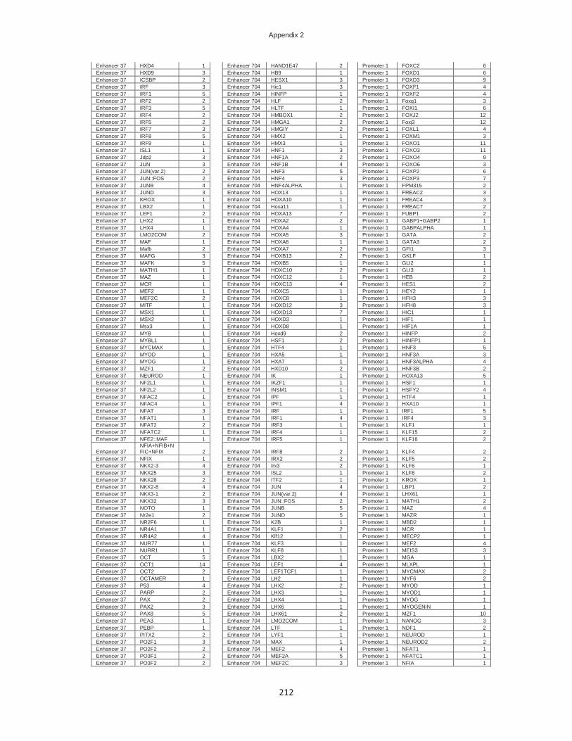
Appendix 2
212
Enhancer 37 HXD4 1 Enhancer 704 HAND1E47 2 Promoter 1 FOXC2 6 Enhancer 37 HXD9 3 Enhancer 704 HB9 1 Promoter 1 FOXD1 6 Enhancer 37 ICSBP 2 Enhancer 704 HESX1 3 Promoter 1 FOXD3 9 Enhancer 37 IRF 3 Enhancer 704 Hic1 3 Promoter 1 FOXF1 4 Enhancer 37 IRF1 5 Enhancer 704 HINFP 1 Promoter 1 FOXF2 4 Enhancer 37 IRF2 2 Enhancer 704 HLF 2 Promoter 1 Foxg1 3 Enhancer 37 IRF3 5 Enhancer 704 HLTF 1 Promoter 1 FOXI1 6 Enhancer 37 IRF4 2 Enhancer 704 HMBOX1 2 Promoter 1 FOXJ2 12 Enhancer 37 IRF5 2 Enhancer 704 HMGA1 2 Promoter 1 Foxj3 12 Enhancer 37 IRF7 3 Enhancer 704 HMGIY 2 Promoter 1 FOXL1 4 Enhancer 37 IRF8 5 Enhancer 704 HMX2 1 Promoter 1 FOXM1 3 Enhancer 37 IRF9 1 Enhancer 704 HMX3 1 Promoter 1 FOXO1 11 Enhancer 37 ISL1 1 Enhancer 704 HNF1 3 Promoter 1 FOXO3 11 Enhancer 37 Jdp2 3 Enhancer 704 HNF1A 2 Promoter 1 FOXO4 9 Enhancer 37 JUN 3 Enhancer 704 HNF1B 4 Promoter 1 FOXO6 3 Enhancer 37 JUN(var.2) 2 Enhancer 704 HNF3 5 Promoter 1 FOXP2 6 Enhancer 37 JUN::FOS 2 Enhancer 704 HNF4 3 Promoter 1 FOXP3 7 Enhancer 37 JUNB 4 Enhancer 704 HNF4ALPHA 1 Promoter 1 FPM315 2 Enhancer 37 JUND 3 Enhancer 704 HOX13 1 Promoter 1 FREAC2 3 Enhancer 37 KROX 1 Enhancer 704 HOXA10 1 Promoter 1 FREAC4 3 Enhancer 37 LBX2 1 Enhancer 704 Hoxa11 1 Promoter 1 FREAC7 2 Enhancer 37 LEF1 2 Enhancer 704 HOXA13 7 Promoter 1 FUBP1 2 Enhancer 37 LHX2 1 Enhancer 704 HOXA2 2 Promoter 1 GABP1+GABP2 1 Enhancer 37 LHX4 1 Enhancer 704 HOXA4 1 Promoter 1 GABPALPHA 1 Enhancer 37 LMO2COM 2 Enhancer 704 HOXA5 3 Promoter 1 GATA 2 Enhancer 37 MAF 1 Enhancer 704 HOXA6 1 Promoter 1 GATA3 2 Enhancer 37 Mafb 2 Enhancer 704 HOXA7 2 Promoter 1 GFI1 3 Enhancer 37 MAFG 3 Enhancer 704 HOXB13 2 Promoter 1 GKLF 1 Enhancer 37 MAFK 5 Enhancer 704 HOXB5 1 Promoter 1 GLI2 1 Enhancer 37 MATH1 1 Enhancer 704 HOXC10 2 Promoter 1 GLI3 1 Enhancer 37 MAZ 1 Enhancer 704 HOXC12 1 Promoter 1 HEB 2 Enhancer 37 MCR 1 Enhancer 704 HOXC13 4 Promoter 1 HES1 2 Enhancer 37 MEF2 1 Enhancer 704 HOXC5 1 Promoter 1 HEY2 1 Enhancer 37 MEF2C 2 Enhancer 704 HOXC8 1 Promoter 1 HFH3 3 Enhancer 37 MITF 1 Enhancer 704 HOXD12 3 Promoter 1 HFH8 3 Enhancer 37 MSX1 1 Enhancer 704 HOXD13 7 Promoter 1 HIC1 1 Enhancer 37 MSX2 1 Enhancer 704 HOXD3 1 Promoter 1 HIF1 1 Enhancer 37 Msx3 1 Enhancer 704 HOXD8 1 Promoter 1 HIF1A 1 Enhancer 37 MYB 1 Enhancer 704 Hoxd9 2 Promoter 1 HINFP 2 Enhancer 37 MYBL1 1 Enhancer 704 HSF1 2 Promoter 1 HINFP1 1 Enhancer 37 MYCMAX 1 Enhancer 704 HTF4 1 Promoter 1 HNF3 5 Enhancer 37 MYOD 1 Enhancer 704 HXA5 1 Promoter 1 HNF3A 3 Enhancer 37 MYOG 1 Enhancer 704 HXA7 1 Promoter 1 HNF3ALPHA 4 Enhancer 37 MZF1 2 Enhancer 704 HXD10 2 Promoter 1 HNF3B 2 Enhancer 37 NEUROD 1 Enhancer 704 IK 1 Promoter 1 HOXA13 5 Enhancer 37 NF2L1 1 Enhancer 704 IKZF1 1 Promoter 1 HSF1 1 Enhancer 37 NF2L2 1 Enhancer 704 INSM1 1 Promoter 1 HSFY2 4 Enhancer 37 NFAC2 1 Enhancer 704 IPF 1 Promoter 1 HTF4 1 Enhancer 37 NFAC4 1 Enhancer 704 IPF1 4 Promoter 1 HXA10 1 Enhancer 37 NFAT 3 Enhancer 704 IRF 1 Promoter 1 IRF1 5 Enhancer 37 NFAT1 1 Enhancer 704 IRF1 4 Promoter 1 IRF4 3 Enhancer 37 NFAT2 2 Enhancer 704 IRF3 1 Promoter 1 KLF1 1 Enhancer 37 NFATC2 1 Enhancer 704 IRF4 1 Promoter 1 KLF15 2 Enhancer 37 NFE2::MAF 1 Enhancer 704 IRF5 1 Promoter 1 KLF16 2
Enhancer 37 NFIA+NFIB+NFIC+NFIX 2 Enhancer 704 IRF8 2 Promoter 1 KLF4 2
Enhancer 37 NFIX 1 Enhancer 704 IRX2 2 Promoter 1 KLF5 2 Enhancer 37 NKX2-3 4 Enhancer 704 Irx3 2 Promoter 1 KLF6 1 Enhancer 37 NKX25 3 Enhancer 704 ISL2 1 Promoter 1 KLF8 2 Enhancer 37 NKX28 2 Enhancer 704 ITF2 1 Promoter 1 KROX 1 Enhancer 37 NKX2-8 4 Enhancer 704 JUN 4 Promoter 1 LBP1 2 Enhancer 37 NKX3-1 2 Enhancer 704 JUN(var.2) 4 Promoter 1 LHX61 1 Enhancer 37 NKX32 3 Enhancer 704 JUN::FOS 2 Promoter 1 MATH1 2 Enhancer 37 NOTO 1 Enhancer 704 JUNB 5 Promoter 1 MAZ 4 Enhancer 37 Nr2e1 2 Enhancer 704 JUND 5 Promoter 1 MAZR 1 Enhancer 37 NR2F6 1 Enhancer 704 K2B 1 Promoter 1 MBD2 1 Enhancer 37 NR4A1 1 Enhancer 704 KLF1 2 Promoter 1 MCR 1 Enhancer 37 NR4A2 4 Enhancer 704 Klf12 1 Promoter 1 MECP2 1 Enhancer 37 NUR77 1 Enhancer 704 KLF3 1 Promoter 1 MEF2 4 Enhancer 37 NURR1 1 Enhancer 704 KLF8 1 Promoter 1 MEIS3 3 Enhancer 37 OCT 5 Enhancer 704 LBX2 1 Promoter 1 MGA 1 Enhancer 37 OCT1 14 Enhancer 704 LEF1 4 Promoter 1 MLXPL 1 Enhancer 37 OCT2 2 Enhancer 704 LEF1TCF1 1 Promoter 1 MYCMAX 2 Enhancer 37 OCTAMER 1 Enhancer 704 LH2 1 Promoter 1 MYF6 2 Enhancer 37 P53 4 Enhancer 704 LHX2 2 Promoter 1 MYOD 1 Enhancer 37 PARP 2 Enhancer 704 LHX3 1 Promoter 1 MYOD1 1 Enhancer 37 PAX 2 Enhancer 704 LHX4 1 Promoter 1 MYOG 1 Enhancer 37 PAX2 3 Enhancer 704 LHX6 1 Promoter 1 MYOGENIN 1 Enhancer 37 PAX8 5 Enhancer 704 LHX61 2 Promoter 1 MZF1 10 Enhancer 37 PEA3 1 Enhancer 704 LMO2COM 1 Promoter 1 NANOG 3 Enhancer 37 PEBP 1 Enhancer 704 LTF 1 Promoter 1 NDF1 2 Enhancer 37 PITX2 2 Enhancer 704 LYF1 1 Promoter 1 NEUROD 1 Enhancer 37 PO2F1 3 Enhancer 704 MAX 1 Promoter 1 NEUROD2 2 Enhancer 37 PO2F2 2 Enhancer 704 MEF2 4 Promoter 1 NFAT1 1 Enhancer 37 PO3F1 2 Enhancer 704 MEF2A 5 Promoter 1 NFATC1 1 Enhancer 37 PO3F2 2 Enhancer 704 MEF2C 3 Promoter 1 NFIA 1
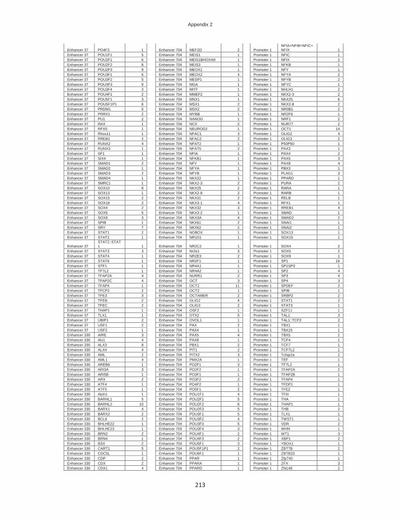
Appendix 2
213
Enhancer 37 PO4F2 1 Enhancer 704 MEF2D 2 Promoter 1 NFIA+NFIB+NFIC+NFIX 1
Enhancer 37 POU1F1 5 Enhancer 704 MEIS1 1 Promoter 1 NFIC 1 Enhancer 37 POU2F1 6 Enhancer 704 MEIS1BHOXA9 1 Promoter 1 NFIX 2 Enhancer 37 POU2F2 8 Enhancer 704 MEIS3 1 Promoter 1 NFKB 1 Enhancer 37 POU2F3 9 Enhancer 704 MEOX1 1 Promoter 1 NFY 1 Enhancer 37 POU3F1 6 Enhancer 704 MEOX2 4 Promoter 1 NFYA 2 Enhancer 37 POU3F2 5 Enhancer 704 MESP1 1 Promoter 1 NFYB 2 Enhancer 37 POU3F3 9 Enhancer 704 MGA 1 Promoter 1 NFYC 1 Enhancer 37 POU3F4 3 Enhancer 704 MITF 1 Promoter 1 NHLH1 2 Enhancer 37 POU4F1 2 Enhancer 704 MMEF2 1 Promoter 1 NKX2-3 2 Enhancer 37 POU5F1 3 Enhancer 704 MNX1 1 Promoter 1 NKX25 6 Enhancer 37 POU5F1P1 6 Enhancer 704 MSX1 2 Promoter 1 NKX2-8 2 Enhancer 37 PRDM1 5 Enhancer 704 MSX2 2 Promoter 1 NR0B1 2 Enhancer 37 PRRX1 2 Enhancer 704 MYBB 1 Promoter 1 NR2F6 1 Enhancer 37 PU1 2 Enhancer 704 NANOG 1 Promoter 1 NRF1 2 Enhancer 37 RAX 1 Enhancer 704 NCX 2 Promoter 1 NUR77 2 Enhancer 37 RFX5 1 Enhancer 704 NEUROD2 1 Promoter 1 OCT1 14 Enhancer 37 Rhox11 1 Enhancer 704 NFAC1 3 Promoter 1 OLIG2 4 Enhancer 37 RREB1 2 Enhancer 704 NFAC2 2 Promoter 1 OLIG3 2 Enhancer 37 RUNX2 4 Enhancer 704 NFAT2 1 Promoter 1 P50P50 1 Enhancer 37 RUNX3 1 Enhancer 704 NFAT5 2 Promoter 1 PAX3 1 Enhancer 37 SF1 1 Enhancer 704 NFIA 1 Promoter 1 PAX4 2 Enhancer 37 SIX4 1 Enhancer 704 NFKB1 1 Promoter 1 PAX5 3 Enhancer 37 SMAD1 2 Enhancer 704 NFY 1 Promoter 1 PAX8 4 Enhancer 37 SMAD2 1 Enhancer 704 NFYA 1 Promoter 1 PBX3 1 Enhancer 37 SMAD3 2 Enhancer 704 NFYB 1 Promoter 1 PLAG1 3 Enhancer 37 SMAD4 1 Enhancer 704 NKX22 1 Promoter 1 PPARD 1 Enhancer 37 SMRC1 1 Enhancer 704 NKX2-3 2 Promoter 1 PURA 2 Enhancer 37 SOX10 8 Enhancer 704 NKX25 2 Promoter 1 RARA 1 Enhancer 37 SOX13 1 Enhancer 704 NKX2-8 2 Promoter 1 RARB 1 Enhancer 37 SOX15 2 Enhancer 704 NKX31 2 Promoter 1 RELB 1 Enhancer 37 SOX18 2 Enhancer 704 NKX3-1 4 Promoter 1 RFX1 1 Enhancer 37 SOX4 2 Enhancer 704 NKX32 3 Promoter 1 RREB1 4 Enhancer 37 SOX5 5 Enhancer 704 NKX3-2 1 Promoter 1 SMAD 1 Enhancer 37 SOX9 3 Enhancer 704 NKX3A 4 Promoter 1 SMAD2 2 Enhancer 37 SPIB 1 Enhancer 704 NKX61 2 Promoter 1 SNAI1 1 Enhancer 37 SRY 7 Enhancer 704 NKX62 2 Promoter 1 SNAI2 1 Enhancer 37 STAT1 2 Enhancer 704 NOBOX 1 Promoter 1 SOX13 1 Enhancer 37 STAT2 1 Enhancer 704 NR1D1 1 Promoter 1 SOX15 1
Enhancer 37 STAT2::STAT1 1 Enhancer 704 NR2C2 1 Promoter 1 SOX4 2
Enhancer 37 STAT3 3 Enhancer 704 Nr2e1 3 Promoter 1 SOX5 2 Enhancer 37 STAT4 1 Enhancer 704 NR2E3 2 Promoter 1 SOX9 1 Enhancer 37 STAT6 1 Enhancer 704 NR2F1 1 Promoter 1 SP1 16 Enhancer 37 STF1 1 Enhancer 704 NR4A1 1 Promoter 1 SP1SP3 1 Enhancer 37 TF7L2 1 Enhancer 704 NR4A2 1 Promoter 1 SP2 4 Enhancer 37 TFAP2A 4 Enhancer 704 NURR1 2 Promoter 1 SP3 4 Enhancer 37 TFAP2C 4 Enhancer 704 OCT 3 Promoter 1 SP4 3 Enhancer 37 TFAP4 1 Enhancer 704 OCT1 11 Promoter 1 SPDEF 2 Enhancer 37 TFCP2 2 Enhancer 704 OCT2 1 Promoter 1 SPIB 1 Enhancer 37 TFE3 3 Enhancer 704 OCTAMER 2 Promoter 1 SRBP2 2 Enhancer 37 TFEB 2 Enhancer 704 OLIG2 4 Promoter 1 STAT1 2 Enhancer 37 TFEC 2 Enhancer 704 OLIG3 2 Promoter 1 STAT3 1 Enhancer 37 THAP1 1 Enhancer 704 OSF2 1 Promoter 1 SZF11 1 Enhancer 37 TLX1 1 Enhancer 704 OTX2 5 Promoter 1 TAL1 3 Enhancer 37 UBIP1 2 Enhancer 704 OVOL1 1 Promoter 1 TAL1::TCF3 2 Enhancer 37 USF1 2 Enhancer 704 PAX 2 Promoter 1 TBX1 1 Enhancer 37 USF2 1 Enhancer 704 PAX4 1 Promoter 1 TBX15 1 Enhancer 330 AIRE 3 Enhancer 704 PAX5 4 Promoter 1 TBX5 2 Enhancer 330 Alx1 4 Enhancer 704 PAX8 1 Promoter 1 TCF4 1 Enhancer 330 ALX3 8 Enhancer 704 PBX1 2 Promoter 1 TCF7 1 Enhancer 330 ALX4 4 Enhancer 704 PIT1 2 Promoter 1 TCF7L2 1 Enhancer 330 AML 2 Enhancer 704 PITX2 3 Promoter 1 Tcfap2a 2 Enhancer 330 AML1 4 Enhancer 704 PMX2A 1 Promoter 1 TEF 1 Enhancer 330 AREB6 1 Enhancer 704 PO2F1 3 Promoter 1 TF7L2 1 Enhancer 330 ARI3A 3 Enhancer 704 PO2F2 1 Promoter 1 TFAP2A 2 Enhancer 330 ARI5B 1 Enhancer 704 PO3F1 1 Promoter 1 TFAP2B 1 Enhancer 330 ARX 2 Enhancer 704 PO3F2 2 Promoter 1 TFAP4 1 Enhancer 330 ATF4 1 Enhancer 704 PO4F2 1 Promoter 1 TFDP1 1 Enhancer 330 ATF5 1 Enhancer 704 PO5F1 2 Promoter 1 TFE2 1 Enhancer 330 Atoh1 1 Enhancer 704 POU1F1 4 Promoter 1 TFIII 1 Enhancer 330 BARHL1 5 Enhancer 704 POU2F1 5 Promoter 1 THA 1 Enhancer 330 BARHL2 10 Enhancer 704 POU2F2 6 Promoter 1 THAP1 1 Enhancer 330 BARX1 4 Enhancer 704 POU2F3 5 Promoter 1 THB 2 Enhancer 330 BARX2 2 Enhancer 704 POU3F1 5 Promoter 1 TLX1 1 Enhancer 330 BCL6 2 Enhancer 704 POU3F2 4 Promoter 1 TWST1 1 Enhancer 330 BHLHE22 1 Enhancer 704 POU3F3 5 Promoter 1 VDR 2 Enhancer 330 BHLHE23 1 Enhancer 704 POU3F4 3 Promoter 1 WHN 1 Enhancer 330 BRN2 2 Enhancer 704 POU4F1 2 Promoter 1 WT1 3 Enhancer 330 BRN4 1 Enhancer 704 POU4F3 2 Promoter 1 XBP1 2 Enhancer 330 BSX 2 Enhancer 704 POU5F1 3 Promoter 1 YBOX1 1 Enhancer 330 CART1 5 Enhancer 704 POU5F1P1 2 Promoter 1 ZBT7B 2 Enhancer 330 CDC5L 1 Enhancer 704 POU6F1 1 Promoter 1 ZBTB33 1 Enhancer 330 CDP 2 Enhancer 704 PPAR 1 Promoter 1 Zfp740 1 Enhancer 330 CDX 2 Enhancer 704 PPARA 1 Promoter 1 ZFX 3 Enhancer 330 CDX1 4 Enhancer 704 PPARD 1 Promoter 1 ZN148 2

Appendix 2
214
Enhancer 330 CDX2 7 Enhancer 704 PR 2 Promoter 1 ZN219 2 Enhancer 330 CEBP 8 Enhancer 704 PRGR 1 Promoter 1 ZNF219 2 Enhancer 330 CEBPA 3 Enhancer 704 PRRX1 2 Promoter 1 ZNF263 2 Enhancer 330 CEBPB 1 Enhancer 704 PRRX2 1 Promoter 1 ZNF354C 1 Enhancer 330 CEBPD 1 Enhancer 704 PU1 1 Promoter 1 ZNF740 3 Enhancer 330 CEBPE 1 Enhancer 704 PXRRXR 2 Promoter 2 AIRE 2 Enhancer 330 CEBPG 1 Enhancer 704 RAX 1 Promoter 2 ALX1 2 Enhancer 330 CEBPZ 1 Enhancer 704 RBPJK 1 Promoter 2 ALX3 3 Enhancer 330 CHX10 1 Enhancer 704 RFX1 1 Promoter 2 ALX4 2 Enhancer 330 CREB 1 Enhancer 704 RORA 4 Promoter 2 AP2ALPHA 3 Enhancer 330 CREBP1 1 Enhancer 704 RORA1 1 Promoter 2 AP3 1 Enhancer 330 CRX 3 Enhancer 704 RORA2 1 Promoter 2 ARI3A 4 Enhancer 330 CUX1 1 Enhancer 704 RORBETA 1 Promoter 2 ARI5B 1 Enhancer 330 DBP 2 Enhancer 704 RORG 1 Promoter 2 ATF5 2 Enhancer 330 DLX1 5 Enhancer 704 RUNX2 1 Promoter 2 Barhl1 4 Enhancer 330 DLX2 7 Enhancer 704 RXRB 1 Promoter 2 BARHL2 7 Enhancer 330 DLX3 4 Enhancer 704 RXRG 1 Promoter 2 BARX1 5 Enhancer 330 DLX4 2 Enhancer 704 SATB1 1 Promoter 2 BARX2 2 Enhancer 330 DLX5 4 Enhancer 704 SF1 3 Promoter 2 BCL6 3 Enhancer 330 DLX6 2 Enhancer 704 SMAD1 3 Promoter 2 BPTF 1 Enhancer 330 DMRT2 1 Enhancer 704 SMAD3 2 Promoter 2 BRN4 2 Enhancer 330 DMRT3 1 Enhancer 704 SMAD4 2 Promoter 2 BSX 1 Enhancer 330 DRGX 2 Enhancer 704 SMRC1 3 Promoter 2 CDC5 1 Enhancer 330 DRI1 1 Enhancer 704 SOX 1 Promoter 2 CDC5L 1 Enhancer 330 DUX4 1 Enhancer 704 SOX10 6 Promoter 2 CDX1 2 Enhancer 330 E2F1 1 Enhancer 704 SOX13 1 Promoter 2 CDX2 3 Enhancer 330 E4BP4 1 Enhancer 704 SOX15 2 Promoter 2 CEBP 2 Enhancer 330 EGR1 1 Enhancer 704 SOX2 1 Promoter 2 CEBPA 2 Enhancer 330 EMX1 3 Enhancer 704 SOX4 1 Promoter 2 CEBPB 1 Enhancer 330 EMX2 6 Enhancer 704 SOX5 4 Promoter 2 CEBPGAMMA 1 Enhancer 330 EN1 8 Enhancer 704 SOX9 4 Promoter 2 CHOP 1 Enhancer 330 EN2 7 Enhancer 704 SOX91 1 Promoter 2 COT2 1 Enhancer 330 ER 1 Enhancer 704 SRF 2 Promoter 2 CP2 1 Enhancer 330 ESX1 4 Enhancer 704 SRY 6 Promoter 2 CPEB1 1 Enhancer 330 EVX1 4 Enhancer 704 STA5A 3 Promoter 2 CRX 3 Enhancer 330 EVX2 2 Enhancer 704 STA5B 2 Promoter 2 CUX1 1 Enhancer 330 FOS 1 Enhancer 704 STAT 1 Promoter 2 DBP 1 Enhancer 330 FOXA1 3 Enhancer 704 STAT1 6 Promoter 2 DLX1 3 Enhancer 330 FOXA2 1 Enhancer 704 STAT1STAT1 1 Promoter 2 DLX2 6 Enhancer 330 FOXA3 2 Enhancer 704 STAT2 2 Promoter 2 DLX3 3 Enhancer 330 FOXB1 2 Enhancer 704 STAT2::STAT1 1 Promoter 2 DLX4 1 Enhancer 330 FOXC1 6 Enhancer 704 STAT3 4 Promoter 2 DLX5 2 Enhancer 330 FOXC2 3 Enhancer 704 STAT4 3 Promoter 2 DLX6 1 Enhancer 330 FOXD1 2 Enhancer 704 STAT5A 3 Promoter 2 DLX7 1 Enhancer 330 FOXD2 2 Enhancer 704 STAT5B 2 Promoter 2 DMRT1 2 Enhancer 330 FOXD3 4 Enhancer 704 SUH 1 Promoter 2 DMRT2 1 Enhancer 330 FOXF1 1 Enhancer 704 TAL1::TCF3 1 Promoter 2 DMRT3 1 Enhancer 330 FOXF2 2 Enhancer 704 TBP 3 Promoter 2 DPRX 1 Enhancer 330 Foxg1 1 Enhancer 704 TBX1 1 Promoter 2 DRI1 1 Enhancer 330 FOXI1 3 Enhancer 704 TBX15 1 Promoter 2 EGR3 1 Enhancer 330 FOXJ2 3 Enhancer 704 TBX20 1 Promoter 2 EMX1 2 Enhancer 330 FOXJ3 2 Enhancer 704 TBX4 1 Promoter 2 EMX2 2 Enhancer 330 Foxk1 1 Enhancer 704 TBX5 1 Promoter 2 EN1 6 Enhancer 330 FOXL1 4 Enhancer 704 TCF11 1 Promoter 2 EN2 3 Enhancer 330 FOXM1 1 Enhancer 704 TCF3 2 Promoter 2 EPAS1 1 Enhancer 330 FOXO1 6 Enhancer 704 TCF4 1 Promoter 2 ERR1 1 Enhancer 330 FOXO3 5 Enhancer 704 TEAD1 11 Promoter 2 ERR3 1 Enhancer 330 FOXO3A 1 Enhancer 704 TEAD3 5 Promoter 2 ESE1 1 Enhancer 330 FOXO4 5 Enhancer 704 TEAD4 4 Promoter 2 ESX1 2 Enhancer 330 FOXO6 1 Enhancer 704 TEF 8 Promoter 2 ETV7 1 Enhancer 330 FOXP1 1 Enhancer 704 TEF1 2 Promoter 2 EVI1 3 Enhancer 330 FOXP2 2 Enhancer 704 TFE 1 Promoter 2 FAC1 1 Enhancer 330 FOXP3 3 Enhancer 704 TFE2 2 Promoter 2 FOS 1 Enhancer 330 FREAC7 1 Enhancer 704 TFEB 1 Promoter 2 FOX 2 Enhancer 330 FUBP1 1 Enhancer 704 TFEC 1 Promoter 2 FOXA1 4 Enhancer 330 GATA1 2 Enhancer 704 TGIF1 1 Promoter 2 FOXA2 2 Enhancer 330 GATA2 2 Enhancer 704 THA 2 Promoter 2 FOXA3 2 Enhancer 330 GATA3 2 Enhancer 704 TR4 1 Promoter 2 FOXB1 4 Enhancer 330 GATA4 1 Enhancer 704 TWST1 1 Promoter 2 FOXC1 8 Enhancer 330 GATA5 2 Enhancer 704 UBIP1 1 Promoter 2 FOXC2 4 Enhancer 330 GATA6 2 Enhancer 704 Uncx 3 Promoter 2 FOXD1 4 Enhancer 330 Gbx1 4 Enhancer 704 USF 1 Promoter 2 FOXD2 4 Enhancer 330 GBX2 9 Enhancer 704 USF1 2 Promoter 2 FOXD3 7 Enhancer 330 GFI1 2 Enhancer 704 USF2 1 Promoter 2 FOXF1 2 Enhancer 330 GR 2 Enhancer 704 VAX1 1 Promoter 2 FOXF2 3 Enhancer 330 GSC 1 Enhancer 704 VBP 2 Promoter 2 Foxg1 2 Enhancer 330 GSX1 2 Enhancer 704 VSX1 1 Promoter 2 FOXI1 3 Enhancer 330 GSX2 2 Enhancer 704 VSX2 1 Promoter 2 FOXJ2 7 Enhancer 330 HAND1 1 Enhancer 704 ZABC1 2 Promoter 2 Foxj3 6 Enhancer 330 HAND1E47 1 Enhancer 704 ZEB1 1 Promoter 2 Foxk1 2 Enhancer 330 HB9 1 Enhancer 704 ZFHX3 1 Promoter 2 FOXL1 3 Enhancer 330 HBP1 1 Enhancer 704 ZNF333 2 Promoter 2 FOXM1 2 Enhancer 330 HELIOSA 1 Enhancer 704 ZNF354C 1 Promoter 2 FOXO1 6 Enhancer 330 HESX1 2 Enhancer 815 AFP1 2 Promoter 2 FOXO3 6 Enhancer 330 HFH3 1 Enhancer 815 Alx1 5 Promoter 2 FOXO3A 1 Enhancer 330 HFH4 1 Enhancer 815 ALX3 3 Promoter 2 FOXO4 4 Enhancer 330 HLF 6 Enhancer 815 ALX4 6 Promoter 2 FOXO6 2

Appendix 2
215
Enhancer 330 HMBOX1 1 Enhancer 815 AP1 13 Promoter 2 FOXP1 2 Enhancer 330 HMGA1 2 Enhancer 815 AP1FJ 1 Promoter 2 FOXP2 4 Enhancer 330 HMGIY 4 Enhancer 815 AP4 1 Promoter 2 FOXP3 5 Enhancer 330 HMX1 1 Enhancer 815 ARI5B 1 Promoter 2 FREAC2 2 Enhancer 330 HMX2 1 Enhancer 815 ARX 4 Promoter 2 FREAC4 2 Enhancer 330 HMX3 2 Enhancer 815 ATF3 1 Promoter 2 FREAC7 1 Enhancer 330 HNF1 4 Enhancer 815 ATF5 3 Promoter 2 FUBP1 1 Enhancer 330 HNF1A 1 Enhancer 815 BACH2 1 Promoter 2 GATA 1 Enhancer 330 HNF1B 3 Enhancer 815 BARHL2 1 Promoter 2 GATA1 4 Enhancer 330 HNF3 3 Enhancer 815 BATF 2 Promoter 2 GATA2 1 Enhancer 330 HNF3A 1 Enhancer 815 BATF::JUN 1 Promoter 2 GATA3 1 Enhancer 330 HNF3ALPHA 2 Enhancer 815 BCL6 4 Promoter 2 GATA4 1 Enhancer 330 HNF3B 2 Enhancer 815 CART1 2 Promoter 2 GATA5 2 Enhancer 330 HNF4 2 Enhancer 815 CDX 2 Promoter 2 GATA6 1 Enhancer 330 HNF6 3 Enhancer 815 CDX1 1 Promoter 2 Gbx1 2 Enhancer 330 HOX13 2 Enhancer 815 CDX2 3 Promoter 2 GBX2 3 Enhancer 330 HOXA1 3 Enhancer 815 CEBP 8 Promoter 2 GFI1 2 Enhancer 330 HOXA10 5 Enhancer 815 CEBPA 6 Promoter 2 GMEB2 1 Enhancer 330 Hoxa11 3 Enhancer 815 CEBPB 6 Promoter 2 GR 1 Enhancer 330 HOXA13 2 Enhancer 815 CEBPD 2 Promoter 2 GSC 1 Enhancer 330 HOXA2 6 Enhancer 815 CEBPDELTA 1 Promoter 2 GSC2 1 Enhancer 330 HOXA3 2 Enhancer 815 COT2 3 Promoter 2 GSX1 1 Enhancer 330 HOXA4 4 Enhancer 815 CRX 3 Promoter 2 GSX2 1 Enhancer 330 HOXA6 2 Enhancer 815 DBP 2 Promoter 2 HB9 1 Enhancer 330 HOXA9 2 Enhancer 815 DMBX1 1 Promoter 2 HELIOSA 1 Enhancer 330 HOXB2 2 Enhancer 815 DMRT3 1 Promoter 2 HESX1 2 Enhancer 330 HOXB3 2 Enhancer 815 DPRX 1 Promoter 2 HFH3 1 Enhancer 330 HOXB5 4 Enhancer 815 DRGX 3 Promoter 2 HFH4 1 Enhancer 330 HOXB6 2 Enhancer 815 EPAS1 1 Promoter 2 HFH8 2 Enhancer 330 HOXB9 1 Enhancer 815 EVI1 7 Promoter 2 HIF2A 2 Enhancer 330 HOXC10 8 Enhancer 815 FOS 2 Promoter 2 HMGA1 1 Enhancer 330 HOXC11 4 Enhancer 815 FOSB 3 Promoter 2 HMGIY 4 Enhancer 330 HOXC12 2 Enhancer 815 FOSL1 1 Promoter 2 HMX1 1 Enhancer 330 HOXC4 1 Enhancer 815 FOSL2 4 Promoter 2 HMX2 1 Enhancer 330 HOXC5 1 Enhancer 815 FOX 3 Promoter 2 HMX3 2 Enhancer 330 HOXC8 2 Enhancer 815 FOXA1 5 Promoter 2 HNF1 3 Enhancer 330 HOXC9 1 Enhancer 815 FOXA2 3 Promoter 2 HNF1A 2 Enhancer 330 HOXD11 2 Enhancer 815 FOXA3 3 Promoter 2 HNF1B 4 Enhancer 330 HOXD12 3 Enhancer 815 FOXB1 6 Promoter 2 HNF3 3 Enhancer 330 HOXD13 3 Enhancer 815 FOXC1 7 Promoter 2 HNF3A 2 Enhancer 330 HOXD3 4 Enhancer 815 FOXC2 5 Promoter 2 HNF3ALPHA 2 Enhancer 330 HOXD8 3 Enhancer 815 FOXD1 5 Promoter 2 HNF3B 1 Enhancer 330 Hoxd9 3 Enhancer 815 FOXD2 4 Promoter 2 HOX13 1 Enhancer 330 HSF1 2 Enhancer 815 FOXD3 8 Promoter 2 HOXA1 1 Enhancer 330 HSF2 2 Enhancer 815 FOXF1 3 Promoter 2 HOXA10 4 Enhancer 330 HXA10 1 Enhancer 815 FOXF2 5 Promoter 2 Hoxa11 2 Enhancer 330 HXA13 1 Enhancer 815 Foxg1 2 Promoter 2 HOXA13 4 Enhancer 330 HXA5 1 Enhancer 815 FOXI1 4 Promoter 2 HOXA2 2 Enhancer 330 HXD10 1 Enhancer 815 FOXJ2 7 Promoter 2 HOXA3 2 Enhancer 330 HXD4 3 Enhancer 815 FOXJ3 5 Promoter 2 HOXA4 1 Enhancer 330 HXD9 3 Enhancer 815 Foxk1 2 Promoter 2 HOXA6 2 Enhancer 330 INSM1 1 Enhancer 815 FOXL1 7 Promoter 2 HOXA7 1 Enhancer 330 IPF 1 Enhancer 815 FOXM1 5 Promoter 2 HOXA9 1 Enhancer 330 IPF1 14 Enhancer 815 FOXO1 5 Promoter 2 HOXB13 1 Enhancer 330 IRF 4 Enhancer 815 FOXO3 5 Promoter 2 HOXB2 1 Enhancer 330 IRF1 4 Enhancer 815 FOXO3A 1 Promoter 2 HOXB3 1 Enhancer 330 IRF3 4 Enhancer 815 FOXO4 5 Promoter 2 HOXB5 2 Enhancer 330 IRF4 4 Enhancer 815 FOXO6 2 Promoter 2 HOXB6 2 Enhancer 330 IRF7 4 Enhancer 815 FOXP1 1 Promoter 2 HOXB9 1 Enhancer 330 IRX2 2 Enhancer 815 FOXP2 5 Promoter 2 HOXC10 5 Enhancer 330 IRX4 2 Enhancer 815 FOXP3 4 Promoter 2 HOXC11 4 Enhancer 330 IRX5 2 Enhancer 815 FRA1 3 Promoter 2 HOXC12 1 Enhancer 330 IRXB3 1 Enhancer 815 FREAC2 2 Promoter 2 HOXC13 1 Enhancer 330 ISL1 2 Enhancer 815 FREAC3 2 Promoter 2 HOXC4 1 Enhancer 330 ISL2 1 Enhancer 815 FUBP1 2 Promoter 2 HOXC5 2 Enhancer 330 ISRE 1 Enhancer 815 GAF 1 Promoter 2 HOXC8 2 Enhancer 330 ISX 4 Enhancer 815 GATA1 3 Promoter 2 HOXC9 1 Enhancer 330 JUN 3 Enhancer 815 GATA3 7 Promoter 2 HOXD11 2 Enhancer 330 JUND(var.2) 1 Enhancer 815 GATA5 3 Promoter 2 HOXD12 2 Enhancer 330 K2B 1 Enhancer 815 GATA6 4 Promoter 2 HOXD13 3 Enhancer 330 LBX2 4 Enhancer 815 GFI1 4 Promoter 2 HOXD3 3 Enhancer 330 LEF1 6 Enhancer 815 GKLF 1 Promoter 2 Hoxd9 2 Enhancer 330 LH2 2 Enhancer 815 GLI2 1 Promoter 2 HSF2 1 Enhancer 330 LHX2 6 Enhancer 815 GLI3 3 Promoter 2 HXA13 1 Enhancer 330 LHX3 1 Enhancer 815 GSC 1 Promoter 2 HXA5 1 Enhancer 330 LHX4 5 Enhancer 815 GSC2 1 Promoter 2 HXC8 1 Enhancer 330 LHX5 1 Enhancer 815 HESX1 2 Promoter 2 HXD10 2 Enhancer 330 LHX6 2 Enhancer 815 HFH3 4 Promoter 2 HXD13 1 Enhancer 330 LHX61 3 Enhancer 815 HFH8 2 Promoter 2 HXD4 2 Enhancer 330 Lhx8 2 Enhancer 815 Hic1 3 Promoter 2 HXD9 2 Enhancer 330 LHX9 2 Enhancer 815 HIC2 2 Promoter 2 ID4 1 Enhancer 330 LMX1 1 Enhancer 815 HLF 1 Promoter 2 IPF 1 Enhancer 330 LMX1A 2 Enhancer 815 HMBOX1 1 Promoter 2 IPF1 4 Enhancer 330 LMX1B 4 Enhancer 815 HMGA1 2 Promoter 2 IRF 1 Enhancer 330 MAFG 4 Enhancer 815 HMGIY 4 Promoter 2 IRF1 4 Enhancer 330 MEF2A 1 Enhancer 815 HNF3 6 Promoter 2 IRF2 1 Enhancer 330 MEF2C 2 Enhancer 815 HNF3A 3 Promoter 2 IRF4 2
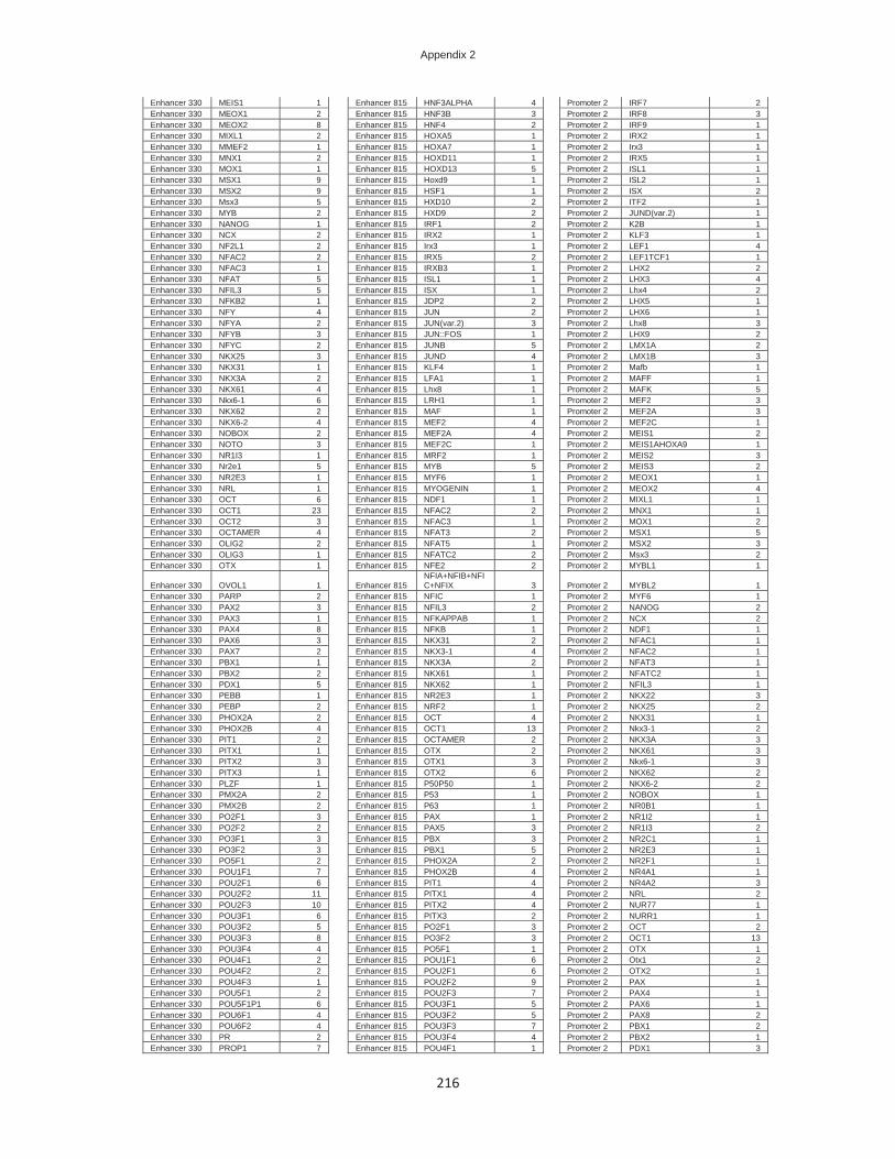
Appendix 2
216
Enhancer 330 MEIS1 1 Enhancer 815 HNF3ALPHA 4 Promoter 2 IRF7 2 Enhancer 330 MEOX1 2 Enhancer 815 HNF3B 3 Promoter 2 IRF8 3 Enhancer 330 MEOX2 8 Enhancer 815 HNF4 2 Promoter 2 IRF9 1 Enhancer 330 MIXL1 2 Enhancer 815 HOXA5 1 Promoter 2 IRX2 1 Enhancer 330 MMEF2 1 Enhancer 815 HOXA7 1 Promoter 2 Irx3 1 Enhancer 330 MNX1 2 Enhancer 815 HOXD11 1 Promoter 2 IRX5 1 Enhancer 330 MOX1 1 Enhancer 815 HOXD13 5 Promoter 2 ISL1 1 Enhancer 330 MSX1 9 Enhancer 815 Hoxd9 1 Promoter 2 ISL2 1 Enhancer 330 MSX2 9 Enhancer 815 HSF1 1 Promoter 2 ISX 2 Enhancer 330 Msx3 5 Enhancer 815 HXD10 2 Promoter 2 ITF2 1 Enhancer 330 MYB 2 Enhancer 815 HXD9 2 Promoter 2 JUND(var.2) 1 Enhancer 330 NANOG 1 Enhancer 815 IRF1 2 Promoter 2 K2B 1 Enhancer 330 NCX 2 Enhancer 815 IRX2 1 Promoter 2 KLF3 1 Enhancer 330 NF2L1 2 Enhancer 815 Irx3 1 Promoter 2 LEF1 4 Enhancer 330 NFAC2 2 Enhancer 815 IRX5 2 Promoter 2 LEF1TCF1 1 Enhancer 330 NFAC3 1 Enhancer 815 IRXB3 1 Promoter 2 LHX2 2 Enhancer 330 NFAT 5 Enhancer 815 ISL1 1 Promoter 2 LHX3 4 Enhancer 330 NFIL3 5 Enhancer 815 ISX 1 Promoter 2 Lhx4 2 Enhancer 330 NFKB2 1 Enhancer 815 JDP2 2 Promoter 2 LHX5 1 Enhancer 330 NFY 4 Enhancer 815 JUN 2 Promoter 2 LHX6 1 Enhancer 330 NFYA 2 Enhancer 815 JUN(var.2) 3 Promoter 2 Lhx8 3 Enhancer 330 NFYB 3 Enhancer 815 JUN::FOS 1 Promoter 2 LHX9 2 Enhancer 330 NFYC 2 Enhancer 815 JUNB 5 Promoter 2 LMX1A 2 Enhancer 330 NKX25 3 Enhancer 815 JUND 4 Promoter 2 LMX1B 3 Enhancer 330 NKX31 1 Enhancer 815 KLF4 1 Promoter 2 Mafb 1 Enhancer 330 NKX3A 2 Enhancer 815 LFA1 1 Promoter 2 MAFF 1 Enhancer 330 NKX61 4 Enhancer 815 Lhx8 1 Promoter 2 MAFK 5 Enhancer 330 Nkx6-1 6 Enhancer 815 LRH1 1 Promoter 2 MEF2 3 Enhancer 330 NKX62 2 Enhancer 815 MAF 1 Promoter 2 MEF2A 3 Enhancer 330 NKX6-2 4 Enhancer 815 MEF2 4 Promoter 2 MEF2C 1 Enhancer 330 NOBOX 2 Enhancer 815 MEF2A 4 Promoter 2 MEIS1 2 Enhancer 330 NOTO 3 Enhancer 815 MEF2C 1 Promoter 2 MEIS1AHOXA9 1 Enhancer 330 NR1I3 1 Enhancer 815 MRF2 1 Promoter 2 MEIS2 3 Enhancer 330 Nr2e1 5 Enhancer 815 MYB 5 Promoter 2 MEIS3 2 Enhancer 330 NR2E3 1 Enhancer 815 MYF6 1 Promoter 2 MEOX1 1 Enhancer 330 NRL 1 Enhancer 815 MYOGENIN 1 Promoter 2 MEOX2 4 Enhancer 330 OCT 6 Enhancer 815 NDF1 1 Promoter 2 MIXL1 1 Enhancer 330 OCT1 23 Enhancer 815 NFAC2 2 Promoter 2 MNX1 1 Enhancer 330 OCT2 3 Enhancer 815 NFAC3 1 Promoter 2 MOX1 2 Enhancer 330 OCTAMER 4 Enhancer 815 NFAT3 2 Promoter 2 MSX1 5 Enhancer 330 OLIG2 2 Enhancer 815 NFAT5 1 Promoter 2 MSX2 3 Enhancer 330 OLIG3 1 Enhancer 815 NFATC2 2 Promoter 2 Msx3 2 Enhancer 330 OTX 1 Enhancer 815 NFE2 2 Promoter 2 MYBL1 1
Enhancer 330 OVOL1 1 Enhancer 815 NFIA+NFIB+NFIC+NFIX 3 Promoter 2 MYBL2 1
Enhancer 330 PARP 2 Enhancer 815 NFIC 1 Promoter 2 MYF6 1 Enhancer 330 PAX2 3 Enhancer 815 NFIL3 2 Promoter 2 NANOG 2 Enhancer 330 PAX3 1 Enhancer 815 NFKAPPAB 1 Promoter 2 NCX 2 Enhancer 330 PAX4 8 Enhancer 815 NFKB 1 Promoter 2 NDF1 1 Enhancer 330 PAX6 3 Enhancer 815 NKX31 2 Promoter 2 NFAC1 1 Enhancer 330 PAX7 2 Enhancer 815 NKX3-1 4 Promoter 2 NFAC2 1 Enhancer 330 PBX1 1 Enhancer 815 NKX3A 2 Promoter 2 NFAT3 1 Enhancer 330 PBX2 2 Enhancer 815 NKX61 1 Promoter 2 NFATC2 1 Enhancer 330 PDX1 5 Enhancer 815 NKX62 1 Promoter 2 NFIL3 1 Enhancer 330 PEBB 1 Enhancer 815 NR2E3 1 Promoter 2 NKX22 3 Enhancer 330 PEBP 2 Enhancer 815 NRF2 1 Promoter 2 NKX25 2 Enhancer 330 PHOX2A 2 Enhancer 815 OCT 4 Promoter 2 NKX31 1 Enhancer 330 PHOX2B 4 Enhancer 815 OCT1 13 Promoter 2 Nkx3-1 2 Enhancer 330 PIT1 2 Enhancer 815 OCTAMER 2 Promoter 2 NKX3A 3 Enhancer 330 PITX1 1 Enhancer 815 OTX 2 Promoter 2 NKX61 3 Enhancer 330 PITX2 3 Enhancer 815 OTX1 3 Promoter 2 Nkx6-1 3 Enhancer 330 PITX3 1 Enhancer 815 OTX2 6 Promoter 2 NKX62 2 Enhancer 330 PLZF 1 Enhancer 815 P50P50 1 Promoter 2 NKX6-2 2 Enhancer 330 PMX2A 2 Enhancer 815 P53 1 Promoter 2 NOBOX 1 Enhancer 330 PMX2B 2 Enhancer 815 P63 1 Promoter 2 NR0B1 1 Enhancer 330 PO2F1 3 Enhancer 815 PAX 1 Promoter 2 NR1I2 1 Enhancer 330 PO2F2 2 Enhancer 815 PAX5 3 Promoter 2 NR1I3 2 Enhancer 330 PO3F1 3 Enhancer 815 PBX 3 Promoter 2 NR2C1 1 Enhancer 330 PO3F2 3 Enhancer 815 PBX1 5 Promoter 2 NR2E3 1 Enhancer 330 PO5F1 2 Enhancer 815 PHOX2A 2 Promoter 2 NR2F1 1 Enhancer 330 POU1F1 7 Enhancer 815 PHOX2B 4 Promoter 2 NR4A1 1 Enhancer 330 POU2F1 6 Enhancer 815 PIT1 4 Promoter 2 NR4A2 3 Enhancer 330 POU2F2 11 Enhancer 815 PITX1 4 Promoter 2 NRL 2 Enhancer 330 POU2F3 10 Enhancer 815 PITX2 4 Promoter 2 NUR77 1 Enhancer 330 POU3F1 6 Enhancer 815 PITX3 2 Promoter 2 NURR1 1 Enhancer 330 POU3F2 5 Enhancer 815 PO2F1 3 Promoter 2 OCT 2 Enhancer 330 POU3F3 8 Enhancer 815 PO3F2 3 Promoter 2 OCT1 13 Enhancer 330 POU3F4 4 Enhancer 815 PO5F1 1 Promoter 2 OTX 1 Enhancer 330 POU4F1 2 Enhancer 815 POU1F1 6 Promoter 2 Otx1 2 Enhancer 330 POU4F2 2 Enhancer 815 POU2F1 6 Promoter 2 OTX2 1 Enhancer 330 POU4F3 1 Enhancer 815 POU2F2 9 Promoter 2 PAX 1 Enhancer 330 POU5F1 2 Enhancer 815 POU2F3 7 Promoter 2 PAX4 1 Enhancer 330 POU5F1P1 6 Enhancer 815 POU3F1 5 Promoter 2 PAX6 1 Enhancer 330 POU6F1 4 Enhancer 815 POU3F2 5 Promoter 2 PAX8 2 Enhancer 330 POU6F2 4 Enhancer 815 POU3F3 7 Promoter 2 PBX1 2 Enhancer 330 PR 2 Enhancer 815 POU3F4 4 Promoter 2 PBX2 1 Enhancer 330 PROP1 7 Enhancer 815 POU4F1 1 Promoter 2 PDX1 3

Appendix 2
217
Enhancer 330 PRRX1 7 Enhancer 815 POU4F3 1 Promoter 2 PIT1 4 Enhancer 330 PRRX2 6 Enhancer 815 POU5F1 3 Promoter 2 PITX1 3 Enhancer 330 RAX 5 Enhancer 815 POU5F1P1 5 Promoter 2 PITX2 2 Enhancer 330 RAXL1 2 Enhancer 815 PROP1 7 Promoter 2 PITX3 2 Enhancer 330 RFX2 1 Enhancer 815 PRRX1 3 Promoter 2 PKNX1 1 Enhancer 330 Rhox11 2 Enhancer 815 RHOXF1 2 Promoter 2 PO2F1 2 Enhancer 330 RORA 1 Enhancer 815 SMAD1 3 Promoter 2 PO2F2 1 Enhancer 330 RUNX1 1 Enhancer 815 SMAD2 1 Promoter 2 PO3F1 2 Enhancer 330 RUNX2 5 Enhancer 815 SMAD3 1 Promoter 2 PO3F2 1 Enhancer 330 SHOX 2 Enhancer 815 SMAD4 1 Promoter 2 POU1F1 4 Enhancer 330 Shox2 4 Enhancer 815 SMRC1 3 Promoter 2 POU2F1 4 Enhancer 330 SMAD1 1 Enhancer 815 SOX10 2 Promoter 2 POU2F2 8 Enhancer 330 SOX10 4 Enhancer 815 SOX17 2 Promoter 2 POU2F3 3 Enhancer 330 SOX13 1 Enhancer 815 SOX5 1 Promoter 2 POU3F1 4 Enhancer 330 SOX15 2 Enhancer 815 SP1 1 Promoter 2 POU3F2 5 Enhancer 330 SOX2 3 Enhancer 815 SRF 1 Promoter 2 POU3F3 5 Enhancer 330 SOX5 1 Enhancer 815 SRY 4 Promoter 2 POU3F4 4 Enhancer 330 SOX9 5 Enhancer 815 STA5A 1 Promoter 2 POU4F1 2 Enhancer 330 SOX91 1 Enhancer 815 STAT1 1 Promoter 2 POU4F3 1 Enhancer 330 SRF 2 Enhancer 815 STAT3 2 Promoter 2 POU5F1 1 Enhancer 330 SRY 5 Enhancer 815 STAT4 1 Promoter 2 POU5F1P1 3 Enhancer 330 STAT1STAT1 1 Enhancer 815 STAT5A 2 Promoter 2 POU6F2 1
Enhancer 330 STAT2::STAT1 1 Enhancer 815 STAT6 2 Promoter 2 PPARA 1
Enhancer 330 STAT3 1 Enhancer 815 TAL1::GATA1 2 Promoter 2 PPARG 1 Enhancer 330 STAT4 1 Enhancer 815 TAL1BETAITF2 1 Promoter 2 PROP1 1 Enhancer 330 STAT5A 1 Enhancer 815 TBP 1 Promoter 2 PRRX1 3 Enhancer 330 STAT6 2 Enhancer 815 TCF3 1 Promoter 2 PRRX2 2 Enhancer 330 TAL1 1 Enhancer 815 TEAD1 7 Promoter 2 PXR 1 Enhancer 330 TATA 2 Enhancer 815 TEAD3 3 Promoter 2 PXRRXR 2 Enhancer 330 TBP 1 Enhancer 815 TEAD4 4 Promoter 2 RARA 1 Enhancer 330 TCF11 1 Enhancer 815 TEF 7 Promoter 2 RARG 1 Enhancer 330 TCF3 2 Enhancer 815 TFE2 1 Promoter 2 RAX 1 Enhancer 330 Tcf7 1 Enhancer 815 TFIII 3 Promoter 2 RAXL1 1 Enhancer 330 TCF7L2 1 Enhancer 815 UNCX 2 Promoter 2 RFX1 2 Enhancer 330 TEAD1 2 Enhancer 815 VBP 1 Promoter 2 RFX2 6 Enhancer 330 TEAD3 1 Enhancer 815 YBOX1 1 Promoter 2 Rfx3 4 Enhancer 330 TEAD4 1 Enhancer 815 ZBTB4 2 Promoter 2 RFX4 2 Enhancer 330 TEF 3 Enhancer 815 ZN589 1 Promoter 2 RFX5 7 Enhancer 330 TEF1 1 Enhancer 815 ZNF333 1 Promoter 2 Rhox11 1 Enhancer 330 TGIF1 1 Enhancer 843 AFP1 1 Promoter 2 RHOXF1 3 Enhancer 330 TITF1 1 Enhancer 843 Alx1 3 Promoter 2 RORA 1 Enhancer 330 TLX1 1 Enhancer 843 ALX3 1 Promoter 2 RORA1 1 Enhancer 330 UBIP1 2 Enhancer 843 ALX4 2 Promoter 2 RORBETA 1 Enhancer 330 UNCX 8 Enhancer 843 AML 1 Promoter 2 RSRFC4 2 Enhancer 330 VAX1 2 Enhancer 843 AML1 3 Promoter 2 RUSH1A 1 Enhancer 330 VAX2 4 Enhancer 843 AP1 6 Promoter 2 SHOX 1 Enhancer 330 VBP 2 Enhancer 843 AR 3 Promoter 2 SHOX2 2 Enhancer 330 VENTX 1 Enhancer 843 AREB6 3 Promoter 2 SIX1 1 Enhancer 330 VSX1 6 Enhancer 843 ARI3A 1 Promoter 2 SIX4 2 Enhancer 330 VSX2 4 Enhancer 843 ATF5 2 Promoter 2 SIX6 3 Enhancer 330 YY1 1 Enhancer 843 BATF 2 Promoter 2 SMAD 1 Enhancer 330 ZFHX3 1 Enhancer 843 BCL6 4 Promoter 2 SMAD1 1
Enhancer 330 ZNF333 2 Enhancer 843 BPTF 1 Promoter 2 SMAD2::SMAD3::SMAD4 1
Enhancer 330 ZNF354C 1 Enhancer 843 CART1 1 Promoter 2 SNAI2 1 Enhancer 700 AFP1 1 Enhancer 843 CDC5 2 Promoter 2 SOX10 3 Enhancer 700 AIRE 2 Enhancer 843 CDP 3 Promoter 2 SOX13 1 Enhancer 700 AP1 10 Enhancer 843 CDX 1 Promoter 2 SOX15 1 Enhancer 700 AP1FJ 2 Enhancer 843 CDX2 4 Promoter 2 SOX17 1 Enhancer 700 AP4 1 Enhancer 843 CEBP 7 Promoter 2 SOX18 1 Enhancer 700 ARI3A 4 Enhancer 843 CEBPA 7 Promoter 2 SOX21 1 Enhancer 700 ATF5 1 Enhancer 843 CEBPB 5 Promoter 2 SOX4 2 Enhancer 700 Atoh1 4 Enhancer 843 CEBPD 3 Promoter 2 SOX5 2 Enhancer 700 BARX2 3 Enhancer 843 CEBPG 3 Promoter 2 SOX9 3 Enhancer 700 BCL6 8 Enhancer 843 CEBPGAMMA 1 Promoter 2 SOX91 1 Enhancer 700 BEN 1 Enhancer 843 COT1 1 Promoter 2 SRF 7 Enhancer 700 BHLHA15 4 Enhancer 843 COT2 2 Promoter 2 SRY 5 Enhancer 700 BHLHE22 2 Enhancer 843 CPEB1 2 Promoter 2 STAT1STAT1 1 Enhancer 700 BHLHE23 2 Enhancer 843 CTF1 1 Promoter 2 STAT2 1 Enhancer 700 BLIMP1 1 Enhancer 843 CUX1 2 Promoter 2 STAT2::STAT1 1 Enhancer 700 BRN2 1 Enhancer 843 CUX2 1 Promoter 2 STAT4 1 Enhancer 700 CDX 2 Enhancer 843 DBP 3 Promoter 2 TAL1 3 Enhancer 700 CDX2 6 Enhancer 843 DELTAEF1 1 Promoter 2 TATA 1 Enhancer 700 CEBPDELTA 1 Enhancer 843 DLX3 3 Promoter 2 TBP 4 Enhancer 700 CEBPZ 2 Enhancer 843 DMRT2 1 Promoter 2 TBX20 1 Enhancer 700 CETS1P54 1 Enhancer 843 DRGX 1 Promoter 2 TBX5 1 Enhancer 700 COT1 2 Enhancer 843 DRI1 1 Promoter 2 TCF3 2 Enhancer 700 COT2 2 Enhancer 843 E12 1 Promoter 2 TCF4 2 Enhancer 700 CP2 1 Enhancer 843 E2F6 2 Promoter 2 TCF7 1 Enhancer 700 CPEB1 4 Enhancer 843 EFC 1 Promoter 2 TCF7L2 2 Enhancer 700 CUX1 2 Enhancer 843 EHF 2 Promoter 2 Tcfap2a 2 Enhancer 700 CUX2 1 Enhancer 843 ELF1 3 Promoter 2 TEAD1 1 Enhancer 700 DEC 2 Enhancer 843 ELF3 3 Promoter 2 TEAD3 2 Enhancer 700 DLX2 3 Enhancer 843 ELF4 1 Promoter 2 TEAD4 1 Enhancer 700 DLX3 3 Enhancer 843 Elf5 3 Promoter 2 TEF 4 Enhancer 700 DMRT3 1 Enhancer 843 ELK1 5 Promoter 2 TF7L2 2

Appendix 2
218
Enhancer 700 DR1 2 Enhancer 843 ELK3 2 Promoter 2 TFAP2A 4 Enhancer 700 E2F6 1 Enhancer 843 ELK4 1 Promoter 2 TFAP2B 2 Enhancer 700 EAR2 2 Enhancer 843 ERR1 2 Promoter 2 TFAP2C 4 Enhancer 700 EGR1 2 Enhancer 843 ERR2 3 Promoter 2 TFE2 1 Enhancer 700 EGR2 3 Enhancer 843 ERR3 1 Promoter 2 TFIIA 1 Enhancer 700 EGR3 2 Enhancer 843 ESE1 1 Promoter 2 TGIF1 2 Enhancer 700 EGR4 1 Enhancer 843 Esrra 4 Promoter 2 TITF1 1 Enhancer 700 ELF1 4 Enhancer 843 ESRRG 1 Promoter 2 UNCX 3 Enhancer 700 ELF3 1 Enhancer 843 ETS 2 Promoter 2 VAX1 1 Enhancer 700 ELK3 1 Enhancer 843 ETS2 2 Promoter 2 VAX2 1 Enhancer 700 EMX2 2 Enhancer 843 ETV6 1 Promoter 2 VDR 1 Enhancer 700 EN1 5 Enhancer 843 EVI1 6 Promoter 2 VENTX 1 Enhancer 700 EOMES 4 Enhancer 843 EVX1 1 Promoter 2 Vsx1 2 Enhancer 700 ER 1 Enhancer 843 FEV 1 Promoter 2 YY1 1 Enhancer 700 ESE1 1 Enhancer 843 FLI1 2 Promoter 2 ZBTB7B 1 Enhancer 700 ESR2 1 Enhancer 843 FOS 1 Promoter 2 ZFHX3 2 Enhancer 700 ETS1 1 Enhancer 843 FOSB 2 Promoter 2 ZN384 1 Enhancer 700 EVI1 3 Enhancer 843 FOX 2 Promoter 3 ALX1 1 Enhancer 700 FEV 3 Enhancer 843 FOXA1 4 Promoter 3 AML1 1 Enhancer 700 FOXA1 2 Enhancer 843 FOXA2 3 Promoter 3 AP1 5 Enhancer 700 FOXA3 1 Enhancer 843 FOXA3 3 Promoter 3 AP1FJ 1 Enhancer 700 FOXB1 6 Enhancer 843 FOXB1 5 Promoter 3 AP3 1 Enhancer 700 FOXC1 5 Enhancer 843 FOXC1 9 Promoter 3 AR 1 Enhancer 700 FOXC2 4 Enhancer 843 FOXC2 4 Promoter 3 AREB6 2 Enhancer 700 FOXD1 2 Enhancer 843 FOXD1 3 Promoter 3 ATF3 2 Enhancer 700 FOXD2 1 Enhancer 843 FOXD2 4 Promoter 3 Atf4 4 Enhancer 700 FOXD3 2 Enhancer 843 FOXD3 5 Promoter 3 Barhl1 1 Enhancer 700 FOXF1 1 Enhancer 843 FOXF1 4 Promoter 3 BARHL2 2 Enhancer 700 FOXI1 3 Enhancer 843 FOXF2 5 Promoter 3 BARX1 5 Enhancer 700 FOXJ2 6 Enhancer 843 FOXH1 2 Promoter 3 BARX2 3 Enhancer 700 FOXJ3 6 Enhancer 843 FOXI1 3 Promoter 3 BATF 1 Enhancer 700 FOXM1 3 Enhancer 843 FOXJ2 8 Promoter 3 BATF::JUN 1 Enhancer 700 FOXO1 2 Enhancer 843 Foxj3 8 Promoter 3 BCL6 1 Enhancer 700 FOXO3A 1 Enhancer 843 FOXL1 4 Promoter 3 BSX 1 Enhancer 700 FOXP2 1 Enhancer 843 FOXM1 4 Promoter 3 CDP 1 Enhancer 700 FOXP3 2 Enhancer 843 FOXO1 7 Promoter 3 CDX1 1 Enhancer 700 FUBP1 1 Enhancer 843 FOXO3 4 Promoter 3 CDX2 2 Enhancer 700 FXR 1 Enhancer 843 FOXO4 5 Promoter 3 CEBP 4 Enhancer 700 GATA 2 Enhancer 843 FOXP2 3 Promoter 3 CEBPA 4 Enhancer 700 GATA1 2 Enhancer 843 FPM315 1 Promoter 3 CEBPB 3 Enhancer 700 GATA2 2 Enhancer 843 FRA1 3 Promoter 3 CEBPD 2 Enhancer 700 GATA3 4 Enhancer 843 FREAC2 3 Promoter 3 CEBPDELTA 2 Enhancer 700 GBX2 5 Enhancer 843 FREAC7 3 Promoter 3 CEBPG 2 Enhancer 700 GCM1 2 Enhancer 843 FUBP1 2 Promoter 3 COUPTF 1 Enhancer 700 GFI1B 2 Enhancer 843 GABP1+GABP2 1 Promoter 3 CREM 3 Enhancer 700 GLI3 4 Enhancer 843 GABPA 2 Promoter 3 DBP 2 Enhancer 700 HELIOSA 1 Enhancer 843 GABPALPHA 1 Promoter 3 DLX1 2 Enhancer 700 HFH3 2 Enhancer 843 GADP 1 Promoter 3 DLX2 5 Enhancer 700 HFH8 1 Enhancer 843 GATA 5 Promoter 3 DLX3 2 Enhancer 700 Hic1 3 Enhancer 843 GATA1 2 Promoter 3 DLX4 1 Enhancer 700 HMGA1 3 Enhancer 843 GATA3 7 Promoter 3 DLX5 3 Enhancer 700 HMX3 2 Enhancer 843 GATA4 4 Promoter 3 DLX6 1 Enhancer 700 HNF1 5 Enhancer 843 GATA5 4 Promoter 3 DLX7 1 Enhancer 700 HNF1A 4 Enhancer 843 GATA6 5 Promoter 3 ELF3 1 Enhancer 700 HNF1B 7 Enhancer 843 GCM 1 Promoter 3 EMX2 1 Enhancer 700 HNF3 3 Enhancer 843 GFI1 5 Promoter 3 EN1 5 Enhancer 700 HNF3ALPHA 2 Enhancer 843 GFI1B 5 Promoter 3 EOMES 1 Enhancer 700 HNF3B 2 Enhancer 843 GRHL1 4 Promoter 3 ERR2 1 Enhancer 700 HNF4 2 Enhancer 843 HEB 1 Promoter 3 ETS2 1 Enhancer 700 HOXA10 2 Enhancer 843 HELIOSA 2 Promoter 3 EVI1 2 Enhancer 700 Hoxa11 2 Enhancer 843 HESX1 2 Promoter 3 FOSB 1 Enhancer 700 HOXA13 13 Enhancer 843 HFH3 3 Promoter 3 FOSL1 1 Enhancer 700 HOXB13 3 Enhancer 843 HFH4 1 Promoter 3 FOSL2 2 Enhancer 700 HOXC10 5 Enhancer 843 HFH8 3 Promoter 3 FOX 1 Enhancer 700 HOXC11 3 Enhancer 843 Hic1 2 Promoter 3 FOXA1 6 Enhancer 700 HOXC12 3 Enhancer 843 HIC2 1 Promoter 3 FOXA2 4 Enhancer 700 HOXC13 7 Enhancer 843 HMGA1 1 Promoter 3 FOXA3 4 Enhancer 700 HOXD11 1 Enhancer 843 HMGIY 6 Promoter 3 FOXB1 4 Enhancer 700 HOXD12 5 Enhancer 843 HMX2 1 Promoter 3 FOXC1 9 Enhancer 700 Hoxd13 8 Enhancer 843 HNF1 4 Promoter 3 FOXC2 3 Enhancer 700 Hoxd9 2 Enhancer 843 HNF1B 1 Promoter 3 FOXD1 3 Enhancer 700 HSF1 6 Enhancer 843 HNF3 5 Promoter 3 FOXD2 2 Enhancer 700 HSF2 3 Enhancer 843 HNF3A 1 Promoter 3 FOXD3 4 Enhancer 700 HSF4 1 Enhancer 843 HNF3ALPHA 4 Promoter 3 FOXF1 4 Enhancer 700 HXD10 2 Enhancer 843 HNF3B 2 Promoter 3 FOXF2 3 Enhancer 700 HXD9 2 Enhancer 843 HNF4ALPHA 2 Promoter 3 FOXG1 3 Enhancer 700 ICSBP 1 Enhancer 843 HNF6 1 Promoter 3 FOXH1 1 Enhancer 700 IPF1 7 Enhancer 843 HOXA10 1 Promoter 3 FOXI1 4 Enhancer 700 IRF 2 Enhancer 843 HOXA13 3 Promoter 3 FOXJ2 10 Enhancer 700 IRF1 5 Enhancer 843 HOXB13 1 Promoter 3 Foxj3 12 Enhancer 700 IRF3 3 Enhancer 843 HOXC13 1 Promoter 3 Foxk1 2 Enhancer 700 IRF4 2 Enhancer 843 HOXD13 4 Promoter 3 FOXL1 3 Enhancer 700 IRF5 2 Enhancer 843 HXA7 1 Promoter 3 FOXM1 2 Enhancer 700 IRF8 3 Enhancer 843 HXA9 3 Promoter 3 FOXO1 11 Enhancer 700 IRX2 2 Enhancer 843 HXD10 3 Promoter 3 FOXO3 7 Enhancer 700 Irx3 2 Enhancer 843 HXD13 3 Promoter 3 FOXO4 9 Enhancer 700 IRX5 1 Enhancer 843 HXD4 2 Promoter 3 FOXP1 1
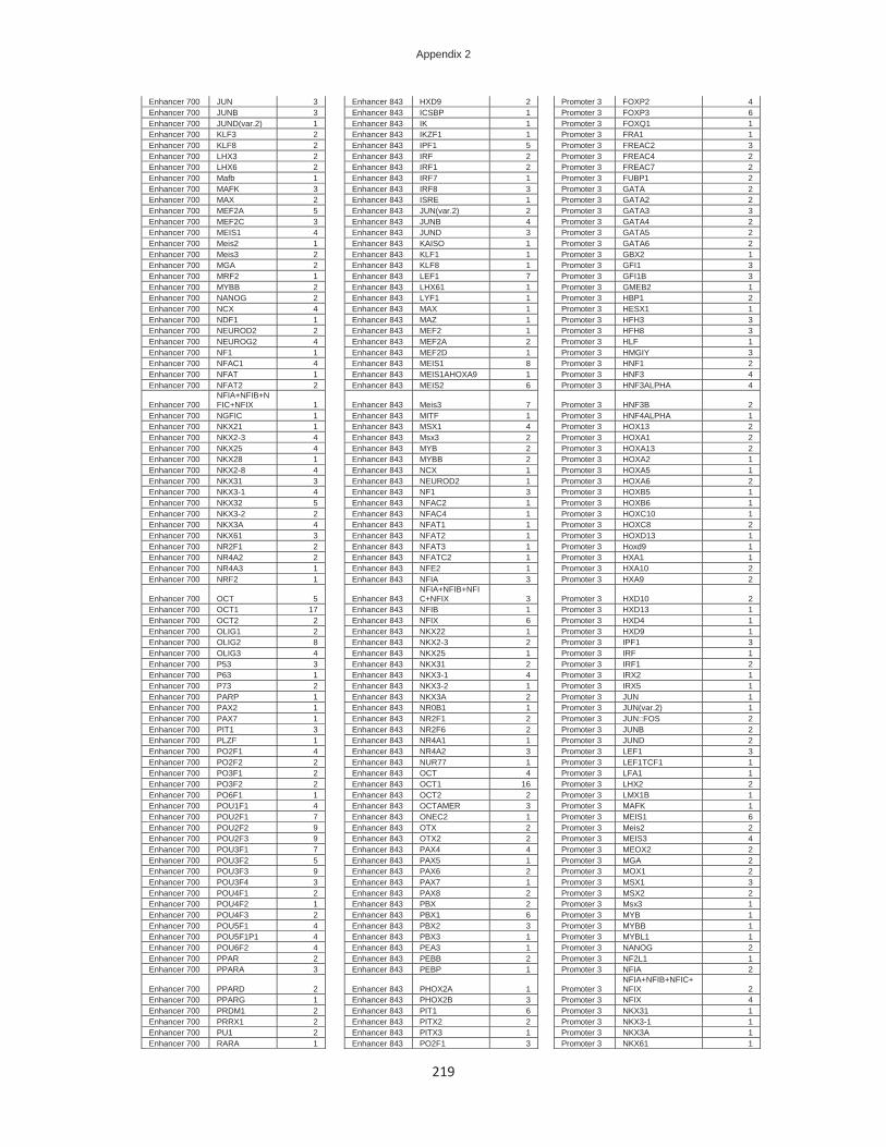
Appendix 2
219
Enhancer 700 JUN 3 Enhancer 843 HXD9 2 Promoter 3 FOXP2 4 Enhancer 700 JUNB 3 Enhancer 843 ICSBP 1 Promoter 3 FOXP3 6 Enhancer 700 JUND(var.2) 1 Enhancer 843 IK 1 Promoter 3 FOXQ1 1 Enhancer 700 KLF3 2 Enhancer 843 IKZF1 1 Promoter 3 FRA1 1 Enhancer 700 KLF8 2 Enhancer 843 IPF1 5 Promoter 3 FREAC2 3 Enhancer 700 LHX3 2 Enhancer 843 IRF 2 Promoter 3 FREAC4 2 Enhancer 700 LHX6 2 Enhancer 843 IRF1 2 Promoter 3 FREAC7 2 Enhancer 700 Mafb 1 Enhancer 843 IRF7 1 Promoter 3 FUBP1 2 Enhancer 700 MAFK 3 Enhancer 843 IRF8 3 Promoter 3 GATA 2 Enhancer 700 MAX 2 Enhancer 843 ISRE 1 Promoter 3 GATA2 2 Enhancer 700 MEF2A 5 Enhancer 843 JUN(var.2) 2 Promoter 3 GATA3 3 Enhancer 700 MEF2C 3 Enhancer 843 JUNB 4 Promoter 3 GATA4 2 Enhancer 700 MEIS1 4 Enhancer 843 JUND 3 Promoter 3 GATA5 2 Enhancer 700 Meis2 1 Enhancer 843 KAISO 1 Promoter 3 GATA6 2 Enhancer 700 Meis3 2 Enhancer 843 KLF1 1 Promoter 3 GBX2 1 Enhancer 700 MGA 2 Enhancer 843 KLF8 1 Promoter 3 GFI1 3 Enhancer 700 MRF2 1 Enhancer 843 LEF1 7 Promoter 3 GFI1B 3 Enhancer 700 MYBB 2 Enhancer 843 LHX61 1 Promoter 3 GMEB2 1 Enhancer 700 NANOG 2 Enhancer 843 LYF1 1 Promoter 3 HBP1 2 Enhancer 700 NCX 4 Enhancer 843 MAX 1 Promoter 3 HESX1 1 Enhancer 700 NDF1 1 Enhancer 843 MAZ 1 Promoter 3 HFH3 3 Enhancer 700 NEUROD2 2 Enhancer 843 MEF2 1 Promoter 3 HFH8 3 Enhancer 700 NEUROG2 4 Enhancer 843 MEF2A 2 Promoter 3 HLF 1 Enhancer 700 NF1 1 Enhancer 843 MEF2D 1 Promoter 3 HMGIY 3 Enhancer 700 NFAC1 4 Enhancer 843 MEIS1 8 Promoter 3 HNF1 2 Enhancer 700 NFAT 1 Enhancer 843 MEIS1AHOXA9 1 Promoter 3 HNF3 4 Enhancer 700 NFAT2 2 Enhancer 843 MEIS2 6 Promoter 3 HNF3ALPHA 4
Enhancer 700 NFIA+NFIB+NFIC+NFIX 1 Enhancer 843 Meis3 7 Promoter 3 HNF3B 2
Enhancer 700 NGFIC 1 Enhancer 843 MITF 1 Promoter 3 HNF4ALPHA 1 Enhancer 700 NKX21 1 Enhancer 843 MSX1 4 Promoter 3 HOX13 2 Enhancer 700 NKX2-3 4 Enhancer 843 Msx3 2 Promoter 3 HOXA1 2 Enhancer 700 NKX25 4 Enhancer 843 MYB 2 Promoter 3 HOXA13 2 Enhancer 700 NKX28 1 Enhancer 843 MYBB 2 Promoter 3 HOXA2 1 Enhancer 700 NKX2-8 4 Enhancer 843 NCX 1 Promoter 3 HOXA5 1 Enhancer 700 NKX31 3 Enhancer 843 NEUROD2 1 Promoter 3 HOXA6 2 Enhancer 700 NKX3-1 4 Enhancer 843 NF1 3 Promoter 3 HOXB5 1 Enhancer 700 NKX32 5 Enhancer 843 NFAC2 1 Promoter 3 HOXB6 1 Enhancer 700 NKX3-2 2 Enhancer 843 NFAC4 1 Promoter 3 HOXC10 1 Enhancer 700 NKX3A 4 Enhancer 843 NFAT1 1 Promoter 3 HOXC8 2 Enhancer 700 NKX61 3 Enhancer 843 NFAT2 1 Promoter 3 HOXD13 1 Enhancer 700 NR2F1 2 Enhancer 843 NFAT3 1 Promoter 3 Hoxd9 1 Enhancer 700 NR4A2 2 Enhancer 843 NFATC2 1 Promoter 3 HXA1 1 Enhancer 700 NR4A3 1 Enhancer 843 NFE2 1 Promoter 3 HXA10 2 Enhancer 700 NRF2 1 Enhancer 843 NFIA 3 Promoter 3 HXA9 2
Enhancer 700 OCT 5 Enhancer 843 NFIA+NFIB+NFIC+NFIX 3 Promoter 3 HXD10 2
Enhancer 700 OCT1 17 Enhancer 843 NFIB 1 Promoter 3 HXD13 1 Enhancer 700 OCT2 2 Enhancer 843 NFIX 6 Promoter 3 HXD4 1 Enhancer 700 OLIG1 2 Enhancer 843 NKX22 1 Promoter 3 HXD9 1 Enhancer 700 OLIG2 8 Enhancer 843 NKX2-3 2 Promoter 3 IPF1 3 Enhancer 700 OLIG3 4 Enhancer 843 NKX25 1 Promoter 3 IRF 1 Enhancer 700 P53 3 Enhancer 843 NKX31 2 Promoter 3 IRF1 2 Enhancer 700 P63 1 Enhancer 843 NKX3-1 4 Promoter 3 IRX2 1 Enhancer 700 P73 2 Enhancer 843 NKX3-2 1 Promoter 3 IRX5 1 Enhancer 700 PARP 1 Enhancer 843 NKX3A 2 Promoter 3 JUN 1 Enhancer 700 PAX2 1 Enhancer 843 NR0B1 1 Promoter 3 JUN(var.2) 1 Enhancer 700 PAX7 1 Enhancer 843 NR2F1 2 Promoter 3 JUN::FOS 2 Enhancer 700 PIT1 3 Enhancer 843 NR2F6 2 Promoter 3 JUNB 2 Enhancer 700 PLZF 1 Enhancer 843 NR4A1 1 Promoter 3 JUND 2 Enhancer 700 PO2F1 4 Enhancer 843 NR4A2 3 Promoter 3 LEF1 3 Enhancer 700 PO2F2 2 Enhancer 843 NUR77 1 Promoter 3 LEF1TCF1 1 Enhancer 700 PO3F1 2 Enhancer 843 OCT 4 Promoter 3 LFA1 1 Enhancer 700 PO3F2 2 Enhancer 843 OCT1 16 Promoter 3 LHX2 2 Enhancer 700 PO6F1 1 Enhancer 843 OCT2 2 Promoter 3 LMX1B 1 Enhancer 700 POU1F1 4 Enhancer 843 OCTAMER 3 Promoter 3 MAFK 1 Enhancer 700 POU2F1 7 Enhancer 843 ONEC2 1 Promoter 3 MEIS1 6 Enhancer 700 POU2F2 9 Enhancer 843 OTX 2 Promoter 3 Meis2 2 Enhancer 700 POU2F3 9 Enhancer 843 OTX2 2 Promoter 3 MEIS3 4 Enhancer 700 POU3F1 7 Enhancer 843 PAX4 4 Promoter 3 MEOX2 2 Enhancer 700 POU3F2 5 Enhancer 843 PAX5 1 Promoter 3 MGA 2 Enhancer 700 POU3F3 9 Enhancer 843 PAX6 2 Promoter 3 MOX1 2 Enhancer 700 POU3F4 3 Enhancer 843 PAX7 1 Promoter 3 MSX1 3 Enhancer 700 POU4F1 2 Enhancer 843 PAX8 2 Promoter 3 MSX2 2 Enhancer 700 POU4F2 1 Enhancer 843 PBX 2 Promoter 3 Msx3 1 Enhancer 700 POU4F3 2 Enhancer 843 PBX1 6 Promoter 3 MYB 1 Enhancer 700 POU5F1 4 Enhancer 843 PBX2 3 Promoter 3 MYBB 1 Enhancer 700 POU5F1P1 4 Enhancer 843 PBX3 1 Promoter 3 MYBL1 1 Enhancer 700 POU6F2 4 Enhancer 843 PEA3 1 Promoter 3 NANOG 2 Enhancer 700 PPAR 2 Enhancer 843 PEBB 2 Promoter 3 NF2L1 1 Enhancer 700 PPARA 3 Enhancer 843 PEBP 1 Promoter 3 NFIA 2
Enhancer 700 PPARD 2 Enhancer 843 PHOX2A 1 Promoter 3 NFIA+NFIB+NFIC+NFIX 2
Enhancer 700 PPARG 1 Enhancer 843 PHOX2B 3 Promoter 3 NFIX 4 Enhancer 700 PRDM1 2 Enhancer 843 PIT1 6 Promoter 3 NKX31 1 Enhancer 700 PRRX1 2 Enhancer 843 PITX2 2 Promoter 3 NKX3-1 1 Enhancer 700 PU1 2 Enhancer 843 PITX3 1 Promoter 3 NKX3A 1 Enhancer 700 RARA 1 Enhancer 843 PO2F1 3 Promoter 3 NKX61 1

Appendix 2
220
Enhancer 700 RARG 1 Enhancer 843 PO2F2 2 Promoter 3 Nkx6-1 1 Enhancer 700 RBPJK 2 Enhancer 843 PO3F1 2 Promoter 3 NKX62 1 Enhancer 700 RFX 2 Enhancer 843 PO3F2 3 Promoter 3 NKX6-2 1 Enhancer 700 RFX1 2 Enhancer 843 POU1F1 7 Promoter 3 NOBOX 1 Enhancer 700 RORA 5 Enhancer 843 POU2F1 6 Promoter 3 NR1I2 1 Enhancer 700 RORA1 2 Enhancer 843 POU2F2 12 Promoter 3 NR1I3 1 Enhancer 700 RREB1 1 Enhancer 843 POU2F3 8 Promoter 3 OCT 1 Enhancer 700 RSRFC4 1 Enhancer 843 POU3F1 6 Promoter 3 OCT1 7 Enhancer 700 RUSH1A 1 Enhancer 843 POU3F2 4 Promoter 3 OCTAMER 2 Enhancer 700 RXRB 3 Enhancer 843 POU3F3 9 Promoter 3 OTX 1 Enhancer 700 SMAD 1 Enhancer 843 POU3F4 3 Promoter 3 OTX1 1 Enhancer 700 SMAD1 2 Enhancer 843 POU5F1 4 Promoter 3 P50RELAP65 1 Enhancer 700 SMAD3 2 Enhancer 843 POU5F1P1 6 Promoter 3 P53 2 Enhancer 700 SNAI1 1 Enhancer 843 PPARA 2 Promoter 3 P63 1 Enhancer 700 SOX 2 Enhancer 843 PR 1 Promoter 3 PAX2 3 Enhancer 700 SOX10 7 Enhancer 843 PROP1 2 Promoter 3 PAX4 4 Enhancer 700 SOX15 3 Enhancer 843 PRRX1 2 Promoter 3 PAX6 1 Enhancer 700 SOX18 1 Enhancer 843 PTF1A 1 Promoter 3 PBX1 2 Enhancer 700 SOX2 2 Enhancer 843 PU1 1 Promoter 3 PBX2 1 Enhancer 700 SOX21 3 Enhancer 843 Rarb 1 Promoter 3 PEBB 1 Enhancer 700 SOX4 2 Enhancer 843 RARG 2 Promoter 3 PHOX2B 1 Enhancer 700 SOX5 7 Enhancer 843 RBPJK 3 Promoter 3 PIT1 3 Enhancer 700 SOX9 6 Enhancer 843 RORA1 1 Promoter 3 PKNX1 1 Enhancer 700 SOX91 2 Enhancer 843 RUNX1 2 Promoter 3 PO2F1 1 Enhancer 700 SRBP2 1 Enhancer 843 RUNX2 2 Promoter 3 PO2F2 1 Enhancer 700 SREBF2 1 Enhancer 843 RUNX3 1 Promoter 3 PO3F1 1 Enhancer 700 SREBP 1 Enhancer 843 RUSH1A 1 Promoter 3 PO3F2 1 Enhancer 700 SRF 3 Enhancer 843 SF1 2 Promoter 3 PO5F1 1 Enhancer 700 SRY 11 Enhancer 843 SIX1 1 Promoter 3 POU1F1 4 Enhancer 700 T3R 2 Enhancer 843 SIX4 1 Promoter 3 POU2F1 2 Enhancer 700 TAL1 1 Enhancer 843 SMAD1 3 Promoter 3 POU2F2 5 Enhancer 700 TBP 4 Enhancer 843 SMRC1 1 Promoter 3 POU2F3 2 Enhancer 700 TBR1 1 Enhancer 843 SOX 1 Promoter 3 POU3F1 3 Enhancer 700 TBX1 2 Enhancer 843 SOX10 4 Promoter 3 POU3F2 2 Enhancer 700 TBX2 2 Enhancer 843 SOX15 2 Promoter 3 POU3F3 4 Enhancer 700 TBX20 3 Enhancer 843 SOX17 1 Promoter 3 POU3F4 1 Enhancer 700 TBX21 2 Enhancer 843 SOX18 1 Promoter 3 POU4F1 1 Enhancer 700 TBX3 1 Enhancer 843 SOX2 1 Promoter 3 POU4F2 1 Enhancer 700 TBX4 4 Enhancer 843 SOX4 1 Promoter 3 POU4F3 1 Enhancer 700 TBX5 4 Enhancer 843 SOX9 3 Promoter 3 POU5F1 2 Enhancer 700 TCF11 2 Enhancer 843 Spic 1 Promoter 3 POU5F1P1 2 Enhancer 700 TCF11MAFG 1 Enhancer 843 SRY 7 Promoter 3 PRRX2 1 Enhancer 700 TCF7L1 1 Enhancer 843 STAT 2 Promoter 3 PXRRXR 1 Enhancer 700 Tcfap2a 1 Enhancer 843 STAT1STAT1 1 Promoter 3 RUNX1 1 Enhancer 700 TEAD1 5 Enhancer 843 STAT2 1 Promoter 3 SMAD1 2 Enhancer 700 TERALPHA 1 Enhancer 843 STAT3 5 Promoter 3 SMAD3 1 Enhancer 700 TFAP2A 2 Enhancer 843 STAT5A 3 Promoter 3 SMAD4 1 Enhancer 700 TFAP2B 1 Enhancer 843 SUH 1 Promoter 3 SOX10 3 Enhancer 700 TFAP2C 2 Enhancer 843 TAL1 2 Promoter 3 SOX15 2 Enhancer 700 TFE 2 Enhancer 843 TAL1::GATA1 1 Promoter 3 SOX9 2 Enhancer 700 TFIII 1 Enhancer 843 TATA 1 Promoter 3 SPZ1 1 Enhancer 700 TGIF1 2 Enhancer 843 TBX5 5 Promoter 3 SREBP1 1 Enhancer 700 THA 3 Enhancer 843 TCF4 3 Promoter 3 SRY 4 Enhancer 700 TP63 1 Enhancer 843 Tcf7 2 Promoter 3 STA5A 1 Enhancer 700 Tp73 2 Enhancer 843 TCF7L2 4 Promoter 3 STA5B 2 Enhancer 700 TWST1 3 Enhancer 843 TEAD4 1 Promoter 3 STAT 1 Enhancer 700 UBIP1 2 Enhancer 843 TEF 3 Promoter 3 STAT1 7 Enhancer 700 VAX1 2 Enhancer 843 TEF1 1 Promoter 3 STAT3 5 Enhancer 700 VAX2 1 Enhancer 843 TF7L2 3 Promoter 3 STAT4 1 Enhancer 700 VSX2 2 Enhancer 843 TFAP2A 1 Promoter 3 STAT5A 2 Enhancer 700 ZABC1 3 Enhancer 843 TFAP2C 1 Promoter 3 STAT5B 2 Enhancer 700 ZBTB49 1 Enhancer 843 TFE 1 Promoter 3 TAL1 4 Enhancer 700 ZBTB6 1 Enhancer 843 TFEB 1 Promoter 3 TAL1::TCF3 3 Enhancer 700 ZFHX3 2 Enhancer 843 TFIII 2 Promoter 3 TAL1ALPHAE47 1 Enhancer 700 ZN333 1 Enhancer 843 TGIF1 4 Promoter 3 TBP 3 Enhancer 700 ZN384 1 Enhancer 843 TLX1::NFIC 1 Promoter 3 TBR1 2 Enhancer 700 ZNF263 1 Enhancer 843 TR4 1 Promoter 3 TBX1 2 Enhancer 700 ZSCAN4 1 Enhancer 843 TWST1 1 Promoter 3 TBX15 2 Enhancer 843 USF 2 Promoter 3 TBX2 1 Enhancer 843 USF1 2 Promoter 3 TBX20 2 Enhancer 843 USF2 1 Promoter 3 TBX21 2 Enhancer 843 VSX2 1 Promoter 3 TBX4 1 Enhancer 843 YY1 2 Promoter 3 TBX5 6 Enhancer 843 ZNF263 2 Promoter 3 TCF3 1 Enhancer 843 ZNF524 2 Promoter 3 TCF4 3 Promoter 3 Tcf7 2 Promoter 3 TCF7L2 3 Promoter 3 TEF 2 Promoter 3 TF7L2 3 Promoter 3 TFE2 2 Promoter 3 TFE3 1 Promoter 3 TGIF 3 Promoter 3 TGIF1 2 Promoter 3 TP53 1 Promoter 3 TP63 2 Promoter 3 UBIP1 1 Promoter 3 UNCX 1

Appendix 2
221
Promoter 3 WHN 2 Promoter 3 ZN384 1 Promoter 3 ZNF333 1 Promoter 3 ZNF75A 1
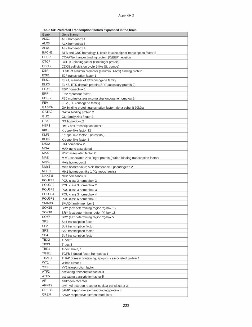
Appendix 2
222
Table S3: Predicted Transcription factors expressed in the brain Gene Gene Name ALX1 ALX homeobox 1 ALX3 ALX homeobox 3 ALX4 ALX homeobox 4 BACH2 BTB and CNC homology 1, basic leucine zipper transcription factor 2 CEBPE CCAAT/enhancer binding protein (C/EBP), epsilon CTCF CCCTC-binding factor (zinc finger protein) CDC5L CDC5 cell division cycle 5-like (S. pombe) DBP D site of albumin promoter (albumin D-box) binding protein E2F1 E2F transcription factor 1 ELK1 ELK1, member of ETS oncogene family ELK3 ELK3, ETS-domain protein (SRF accessory protein 2) ESX1 ESX homeobox 1 ERF Ets2 repressor factor FOSB FBJ murine osteosarcoma viral oncogene homolog B FEV FEV (ETS oncogene family) GABPA GA binding protein transcription factor, alpha subunit 60kDa GATA2 GATA binding protein 2 GLI2 GLI family zinc finger 2 GSX2 GS homeobox 2 HBP1 HMG-box transcription factor 1 Klf12 Kruppel-like factor 12 KLF5 Kruppel-like factor 5 (intestinal) KLF8 Kruppel-like factor 8 LHX2 LIM homeobox 2 MGA MAX gene associated MAX MYC associated factor X MAZ MYC-associated zinc finger protein (purine-binding transcription factor) Meis2 Meis homeobox 2 Meis3 Meis homeobox 3; Meis homeobox 3 pseudogene 2 MIXL1 Mix1 homeobox-like 1 (Xenopus laevis) NKX2-8 NK2 homeobox 8 POU2F3 POU class 2 homeobox 3 POU3F2 POU class 3 homeobox 2 POU3F3 POU class 3 homeobox 3 POU3F4 POU class 3 homeobox 4 POU6F1 POU class 6 homeobox 1 SMAD3 SMAD family member 3 SOX15 SRY (sex determining region Y)-box 15 SOX18 SRY (sex determining region Y)-box 18 SOX5 SRY (sex determining region Y)-box 5 SP1 Sp1 transcription factor SP2 Sp2 transcription factor SP3 Sp3 transcription factor SP4 Sp4 transcription factor TBX2 T-box 2 TBX3 T-box 3 TBR1 T-box, brain, 1 TGIF1 TGFB-induced factor homeobox 1 THAP1 THAP domain containing, apoptosis associated protein 1 WT1 Wilms tumor 1 YY1 YY1 transcription factor ATF3 activating transcription factor 3 ATF5 activating transcription factor 5 AR androgen receptor ARNT2 aryl-hydrocarbon receptor nuclear translocator 2 CREB3 cAMP responsive element binding protein 3 CREM cAMP responsive element modulator

Appendix 2
223
CTF1 cardiotrophin 1 CPEB1 cytoplasmic polyadenylation element binding protein 1 DLX1 distal-less homeobox 1 DLX2 distal-less homeobox 2 DMRT1 doublesex and mab-3 related transcription factor 1 EGR3 early growth response 3 EGR4 early growth response 4 EMX1 empty spiracles homeobox 1 EMX2 empty spiracles homeobox 2 EN2 engrailed homeobox 2 EOMES eomesodermin homolog (Xenopus laevis) ESR1 estrogen receptor 1 ESRRA estrogen-related receptor alpha ESRRG estrogen-related receptor gamma ETV1 ets variant 1 ETV7 ets variant 7 FUBP1 far upstream element (FUSE) binding protein 1 FIGLA folliculogenesis specific basic helix-loop-helix Foxg1 forkhead box G1 Foxj3 forkhead box J3 Foxk1 forkhead box K1 FOXP2 forkhead box P2 GBX2 gastrulation brain homeobox 2 GMEB2 glucocorticoid modulatory element binding protein 2 GFI1B growth factor independent 1B transcription repressor HAND1 heart and neural crest derivatives expressed 1 HSF1 heat shock transcription factor 1 HSF2 heat shock transcription factor 2 HLTF helicase-like transcription factor HLF hepatic leukemia factor Hoxd3 homeobox D3 HMBOX1 homeobox containing 1 HIC2 hypermethylated in cancer 2 IRF3 interferon regulatory factor 3 IRF8 interferon regulatory factor 8 MECP2 methyl CpG binding protein 2 (Rett syndrome) MZF1 myeloid zinc finger 1 MEF2A myocyte enhancer factor 2A MEF2D myocyte enhancer factor 2D NHLH1 nescient helix loop helix 1 NF1 neurofibromin 1 NEUROD2 neurogenic differentiation 2 NEUROG2 neurogenin 2 NFIA nuclear factor I/A NFIX nuclear factor I/X (CCAAT-binding transcription factor) NFAT5 nuclear factor of activated T-cells 5, tonicity-responsive NFATC1 nuclear factor of activated T-cells, cytoplasmic, calcineurin-dependent 1 NR2E1 nuclear receptor subfamily 2, group E, member 1 NR4A2 nuclear receptor subfamily 4, group A, member 2 NFYC nuclear transcription factor Y, gamma OLIG2 oligodendrocyte lineage transcription factor 2 OLIG1 oligodendrocyte transcription factor 1 OTX1 orthodenticle homeobox 1 PAX6 paired box 6 PHOX2A paired-like homeobox 2a PHOX2B paired-like homeobox 2b PLAG1 pleiomorphic adenoma gene 1 PBX1 pre-B-cell leukemia homeobox 1 PURA purine-rich element binding protein A

Appendix 2
224
RREB1 ras responsive element binding protein 1 RFX4 regulatory factor X, 4 (influences HLA class II expression) RARA retinoic acid receptor, alpha RXRB retinoid X receptor, beta SRF serum response factor (c-fos serum response element-binding transcription factor) STAT1 signal transducer and activator of transcription 1, 91kDa STAT4 signal transducer and activator of transcription 4 STAT5A signal transducer and activator of transcription 5A STAT5B signal transducer and activator of transcription 5B IK similar to CG18005; IK cytokine, down-regulator of HLA II SPZ1 spermatogenic leucine zipper 1 SF1 splicing factor 1 TEF thyrotrophic embryonic factor TFAP4 transcription factor AP-4 (activating enhancer binding protein 4) TFEB transcription factor EB TFEC transcription factor EC TP53 tumor protein p53 ETS2 v-ets erythroblastosis virus E26 oncogene homolog 2 (avian) Mafb v-maf musculoaponeurotic fibrosarcoma oncogene homolog B (avian) MAFK v-maf musculoaponeurotic fibrosarcoma oncogene homolog K (avian) MYBL2 v-myb myeloblastosis viral oncogene homolog (avian)-like 2 VAX1 ventral anterior homeobox 1 ZEB1 zinc finger E-box binding homeobox 1 ZBTB4 zinc finger and BTB domain containing 4 ZNF219 zinc finger protein 219 ZNF333 zinc finger protein 333 ZBTB49 zinc finger protein 509 ZNF524 zinc finger protein 524

Appendix 2
225
Table S4: Predicted Transcription factors in the "Neuron differentiation" category Gene Gene Name BARHL2 BarH-like homeobox 2 CEBPB CCAAT/enhancer binding protein (C/EBP), beta GATA2 GATA binding protein 2 GLI2 GLI family zinc finger 2 GLI3 GLI family zinc finger 3 GSX1 GS homeobox 1 GSX2 GS homeobox 2 ISL1 ISL LIM homeobox 1 ISL2 ISL LIM homeobox 2 LHX2 LIM homeobox 2 LHX4 LIM homeobox 4 LHX5 LIM homeobox 5 LHX6 LIM homeobox 6 Lhx8 LIM homeobox 8 LMX1A LIM homeobox transcription factor 1, alpha LMX1B LIM homeobox transcription factor 1, beta NKX2-8 NK2 homeobox 8 NKX6-1 NK6 homeobox 1 NKX6-2 NK6 homeobox 2 POU3F2 POU class 3 homeobox 2 POU3F4 POU class 3 homeobox 4 POU4F1 POU class 4 homeobox 1 POU4F2 POU class 4 homeobox 2 POU4F3 POU class 4 homeobox 3 RORA RAR-related orphan receptor A SMAD4 SMAD family member 4 SOX2 SRY (sex determining region Y)-box 2 SOX5 SRY (sex determining region Y)-box 5 TBR1 T-box, brain, 1 TLX1 T-cell leukemia homeobox 1 ARX aristaless related homeobox Atoh1 atonal homolog 1 (Drosophila) BHLHE22 basic helix-loop-helix family, member e22 CREB1 cAMP responsive element binding protein 1 CTF1 cardiotrophin 1 CUX1 cut-like homeobox 1 DLX1 distal-less homeobox 1 DLX2 distal-less homeobox 2 DLX5 distal-less homeobox 5 EGR2 early growth response 2 EMX1 empty spiracles homeobox 1 EMX2 empty spiracles homeobox 2 EN1 engrailed homeobox 1 EN2 engrailed homeobox 2 ETV4 ets variant 4 EVX1 even-skipped homeobox 1 FOXA1 forkhead box A1 FOXA2 forkhead box A2 Foxg1 forkhead box G1 GBX2 gastrulation brain homeobox 2 GFI1 growth factor independent 1 transcription repressor HES1 hairy and enhancer of split 1, (Drosophila) HOXA1 homeobox A1 HOXA2 homeobox A2 HOXC10 homeobox C10 HOXC8 homeobox C8 Hoxd9 homeobox D9

Appendix 2
226
ID4 inhibitor of DNA binding 4, dominant negative helix-loop-helix protein IRX5 iroquois homeobox 5 MNX1 motor neuron and pancreas homeobox 1 NRL neural retina leucine zipper NEUROD2 neurogenic differentiation 2 NEUROG2 neurogenin 2 NR2E1 nuclear receptor subfamily 2, group E, member 1 NR2E3 nuclear receptor subfamily 2, group E, member 3 NR2F6 nuclear receptor subfamily 2, group F, member 6 NR4A2 nuclear receptor subfamily 4, group A, member 2 OLIG2 oligodendrocyte lineage transcription factor 2 OLIG1 oligodendrocyte transcription factor 1 OTX2 orthodenticle homeobox 2 PAX2 paired box 2 PAX3 paired box 3 PAX6 paired box 6 PAX7 paired box 7 PHOX2A paired-like homeobox 2a PITX3 paired-like homeodomain 3 PTF1A pancreas specific transcription factor, 1a PBX3 pre-B-cell leukemia homeobox 3 RUNX3 runt-related transcription factor 3 SRF serum response factor (c-fos serum response element-binding transcription factor) STAT3 signal transducer and activator of transcription 3 (acute-phase response factor) Tp73 tumor protein p73 VAX1 ventral anterior homeobox 1 VAX2 ventral anterior homeobox 2 VSX1 visual system homeobox 1 VSX2 visual system homeobox 2

227
Nederlandse samenvatting
Mutaties in het FOXP2-gen veroorzaken problemen bij de ontwikkeling van spraak en taal.
Het gen codeert voor een eiwit uit de transcriptiefactor (TF)-familie, dat met het DNA
interageert en daardoor honderden andere genen aan en uit kan zetten. De mutaties in
FOXP2 zorgen ervoor dat het eiwit niet aan het DNA kan binden of niet goed geproduceerd
kan worden. Onder normale omstandigheden wordt FOXP2 geproduceerd in de longen, het
hart, de darm en de hersenen. In de hersenen wordt het gen aangezet in bepaalde
structuren, zoals de hersenenschors, de kleine hersenen en de basale gangliën. De
taalproblemen bij mensen met FOXP2-mutaties worden vooral veroorzaakt door het
ontbreken van het normale FOXP2-eiwit in de hersenen.
De effecten van de mutatie op moleculair en cellulair niveau zijn intensief bestudeerd en zij
leveren veel kennis op over de rol van het gen in de ontwikkeling van neuronen en de
formatie van neurale netwerken die de menselijke taalvaardigheden beïnvloeden. In
hoofdstuk 1 van deze PhD-thesis beschrijf ik hoe de eerste mutaties in FOXP2 ontdekt zijn,
wat voor aandoeningen mensen met FOXP2-mutaties precies hebben en wat er al is ontdekt
over de functie van FOXP2 in menselijke en dierlijke hersencellen. Hoewel al veel bekend is
over de processen die het FOXP2-eiwit reguleert, weten wij vrij weinig over de processen die
de productie van het FOXP2-gen zelf reguleren. Daarom was het doel van dit PhD-project
om te onderzoeken hoe het FOXP2-gen wordt aangezet, wat voor processen daar een rol bij
spelen en wat er gebeurt als de regulatie misgaat.
Mijn onderzoek uit hoofdstuk 2 is gebaseerd op een eerdere beschrijving van een kind met
een vertraagde taalontwikkeling. In het genoom van het kind werd aangetoond dat sommige
delen van het DNA in volgorde zijn omgedraaid. Eén van de breukpunten, waar delen van
het genoom zijn omgedraaid, ligt in de buurt van het FOXP2-gen en het gen zelf werd niet
veranderd. Het idee is dus, dat door de omgedraaide volgorde een regulerend DNA-element
verplaatst werd en het FOXP2-eiwit niet goed wordt aangemaakt. Ik beschrijf in hoofdstuk 2

228
hoe ik een regulerend element, enhancer genoemd, met behulp van het bovengenoemde
breukpunt kon identificeren. Bij het bovengenoemde kind is de enhancer verplaatst en zal
het FOXP2-gen niet kunnen reguleren, met als gevolg dat FOXP2 niet goed wordt
aangemaakt.
Enhancers reguleren de productie in bepaalde weefsels en cellen. Genen met een complexe
expressie, zoals FOXP2, hebben meestal meerdere enhancers. In hoofdstuk 3 was het mijn
doel om verdere enhancers van het gen op te sporen. Een eigenschap van enhancers is dat
ze de productie van genen reguleren door contact te maken met het beginpunt van het gen,
de zogenaamde promoter. Om FOXP2-enhancers op te sporen heb ik de contacten van de
FOXP2-promoter met andere delen van het genoom bepaald. Ik verwachtte in cellen die
FOXP2 produceren contacten te ontdekken die afwezig -of minder sterk- zijn in cellen zonder
FOXP2-productie. Inderdaad kon ik een aantal van deze contacten detecteren en daardoor
potentiële enhancers ontdekken.
De activiteit van enhancers en promoters wordt mede bepaald door TFs – waarvan FOXP2
er éé is-, die aan specifieke DNA-motieven binden. In hoofdstuk 4 heb ik de activiteit van
enhancers in menselijke cellen getest en vervolgens bepaald of de activiteit afhankelijk is
van bepaalde TFs. In totaal heb ik de invloed van 8 TFs bestudeerd op basis van vermoede
associaties uit de literatuur. Inderdaad zag ik voor 7 TFs dat ze de activiteit van de
enhancers of promoters verhogen of verlagen. Ik kon bijvoorbeeld zien dat FOXP2 zelf de
activiteit van zijn promoter en een enhancer verhoogt. Het gen zou dus zijn eigen productie
positief kunnen beïnvloeden. Deze positieve autoregulatie zou een bijdrage kunnen leveren
aan het feit dat mutaties in één van de twee FOXP2-kopieën voldoende is om de bekende
aandoening in taalontwikkeling te veroorzaken.
Zoals eerder genoemd, zijn enhancers slechts in bepaalde weefsels en cellen actief. Zij
dragen dus bij aan de productie van het gen in een beperkt aantal cellen. Om te begrijpen
waar een enhancer door TFs wordt aangestuurd is het dus noodzakelijk om de weefsel-

229
specifieke activiteit van de enhancers te onderzoeken. In het vijfde hoofdstuk heb ik twee
enhancers in transgene muizen bestudeerd. Deze muizen produceren een reportergen onder
de controle van de ontdekte enhancer. Het reportergen is dan eenvoudig te visualiseren in
de weefsels en cellen waar de enhancer actief is. Ik zag dat een enhancer actief was in de
cortex, en juist rond de geboorte activiteit vertoonde in het specifieke weefsel waar ook
FOXP2 wordt geproduceerd. Deze enhancer zal dus FOXP2 tijdens de ontwikkeling aan
kunnen sturen. Verdere overeenstemming tussen enhanceractiviteit en FOXP2-productie
was te zien in de cerebellum. De enhancer stuurt dus de productie van FOXP2 tijdens een
belangrijke fase van de hersenontwikkeling. Genetische variatie in deze enhancer zou
kunnen veroorzaken dat FOXP2 in deze weefsels minder goed wordt geproduceerd en zou
dus problemen in taal en spraak kunnen veroorzaken. Verder onderzoek naar mutaties in
deze enhancer bij mensen met specifieke taalproblemen zou dus een genetische verklaring
van deze specifieke aandoeningen kunnen opleveren.
Voor de zesde hoofdstuk was mijn doel om aan te tonen dat normale genetische
veranderingen in enhancers de structuur van de hersenen kan beïnvloeden. Normale
genetische veranderingen zijn variaties van enkele DNA-bouwstenen die bij iedereen
aanwezig kunnen zijn. Er zijn miljoenen posities in het genoom bekend waar mensen van
elkaar verschillen. De effecten van enkele variaties zullen dus heel klein zijn. Om toch een
effect te ontdekken, heb ik zo’n 300 enhancers uit de wetenschappelijke literatuur
onderzocht, waarvan bekend is dat ze in de hersenen actief zijn. Ik wilde weten of normale
genetische verandering in één van de enhancers invloed op de hersenenstructuur van
gezonde mensen heeft. Een vergelijkbare vraag werd eerder onderzocht door een
wereldwijd consortium, Enhancing Neuro Imaging Genetics by Meta-Analysis genaamd,
kortweg ENIGMA. Dit consortium heeft de normale genetische variatie en hersenenstructuur
van 30,000 mensen bestudeerd en voor meer dan 8 miljoen variaties statistisch berekend
wat de invloed op de structuren in het midden van de hersenen zou kunnen zijn. Bij 8 miljoen
bekeken variaties is het heel moeilijk om statistisch aan te tonen dat het effect van één

230
variant echt de hersenenstructuur beïnvloedt, of dat de berekende invloed door toeval is
ontstaan. Omdat ik niet naar het hele genoom keek, maar naar een beperkt aantal
interessante posities, de 300 enhancers, was het mogelijk om statistische effecten in deze
enhancers aan te tonen. In hoofdstuk zes laat ik zien dat normale genetische variatie in
enhancers, die in de hersenen actief zijn, de normale variatie in de hersenstructuur
beïnvloeden. Verder onderzoek naar genetische variatie in enhancers van FOXP2 zou
mogelijk effecten op kunnen leveren in de hersendelen waar FOXP2 geproduceerd wordt of
in de neuronale functies van deze hersengebieden.
Samenvattend, ik heb de basiselementen van genregulatie op moleculair en cellulair niveau
onderzocht en de mogelijke effecten van mis-regulatie bestudeerd. Mijn onderzoek was
speciaal gericht op het FOXP2-gen, dat bekend is als veroorzaker van mogelijke
ontwikkelingsstoornissen van taal en spraak. De resultaten van mijn project tonen genetische
elementen aan die de productie van FOXP2 sturen en factoren die de productie verder
kunnen versterken of verminderen. Bovendien heb ik voor twee enhancers de activiteit in de
hersenen zien laten zien. De afwezigheid van deze enhancers zou een
taalontwikkelingsstoornis kunnen veroorzaken. Normale genetische variatie in deze
enhancers zou ook een invloed kunnen hebben op de normale variatie in de hersenstructuur.
Met behulp van mijn resultaten kunnen toekomstige studies onderzoeken hoe de productie
van het FOXP2-gen de hersenontwikkeling verandert.
231
Curriculum Vitae
Martin Becker was born on the 21st of March, 1985 in Berlin, Germany and obtained his
Abitur from the Alfred-Krupp-Schule in Essen, Germany in 2004. The next two years he
visited the Berufskolleg Hilden and graduated in 2006 as biological research assistant
(Biologisch-technischer Assistant) with a specialisation in molecular biological techniques
obtained from the chamber of industry and commerce (Industrie und Handelskammer)
Düsseldorf.
In 2009 he finished his Bachelor of Science in the Molecular Lifescience program of the
Radboud University Nijmegen, The Netherlands. In 2011, Martin obtained the Master of
Science degree from the medical faculty of the Radboud University after completing the
Molecular Mechanism of Disease program. During that time, he studied gene enhancer
variants associated with colorectal cancer at the Wellcome Trust Centre for Human Genetics,
Oxford University, UK and chromosomal rearrangements in patients with intellectual disability
at the Human Genetics department of the Radboud University Medical Centre, Nijmegen.
In September 2011, Martin started his PhD project at the Max Planck Institute for
Psycholinguistics, Nijmegen under the supervision of Prof. Dr. Simon Fisher and Dr. Sonja
Vernes. Martin investigated the transcriptional regulation of the FOXP2 gene and
neurodevelopmental gene enhancers in health and disease and obtained his doctoral degree
in October 2016 from the Radboud Univserity Nijmegen.
Since March 2016, Martin is working in Dr. Kristiina Tammimies group at the Karolinska
Institute Centre for Neurodevelopmental Disorders in Stockholm, Sweden and investigates
the interaction of genetic variants and environmental exposure in the development of autism.

232
List of publications
Becker, M., Guadalupe, T., Franke B., Hibar, D.P., Renteria, M.E., Stein, J.L., Thompson,
P.M., Francks, C., Vernes, S.C., Fisher, S.E. (2016) Early developmental gene enhancers
affect subcortical volumes in the adult human brain. Human Brain Mapping, 37:1788-1800.
Becker, M., Devanna P., Fisher, S.E. & Vernes, S.C. (2015). A chromosomal rearrangement
in a child with severe speech and language disorder separates FOXP2 from a functional
enhancer. Molecular Cytogenetics, 8:69.
Lewis, A., Freeman-Mills, L., de la Calle-Mustienes, E., Giráldez-Pérez, R.M., Davis, H.,
Jaeger, E., Becker, M., Hubner, N.C., Nguyen, L.N., Zeron-Medina, J., Bond, G.,
Stunnenberg, H.G., Carvajal, J.J., Gomez-Skarmeta, J.L., Leedham, S., Tomlinson, I. (2014).
A polymorphic enhancer near GREM1 influences bowel cancer risk through differential CDX2
and TCF7L2 binding. Cell Reports, 8, 983-990.
Jaeger, E., Leedham, S., Lewis, A., Segditsas, S., Becker, M., Rodenas-Cuadrado, P.,
Davis, H., Kaur, K., Heinimann, K., Howarth, K., East, J., Taylor, J., Thomas, H., &
Tomlinson, I. (2012). Hereditary mixed polyposis syndrome is caused by a 40-kb upstream
duplication that leads to increased and ectopic expression of the BMP antagonist GREM1.
Nature Genetics, 44, 699-703.

233
Acknowledgments
First of all, I would like to thank my promoter Simon Fisher for giving me the opportunity to
pursue my PhD project in his department and for taking his time in giving me so much
valuable feedback. I want to thank my co-promoter Sonja Vernes for challenging me and my
ideas to constantly improve my critical thinking and experimental approaches.
Further I want to thank everyone in the lab! All colleagues who created a great lab
atmosphere and have all helped me a lot to get around in the lab. Pela for being my favourite
Greek. Sarah for showing me what really good organization skills are. Pedro for sharing the
sunset over Elafonisos with me. Paolo for keeping me awake with amazing coffee and
teaching me some of his lab magician skills. Jasper for his magnificent lab management
skills that made lab work so much smoother. Joses for showing me how to hug people. Sara
for being hilarious company as a car passenger. Kai for inspiring my T-shirt selection. Elliot
for understanding my jokes. Arianna for keeping Paolo in check and Moritz for helping me
out with the mouse brains.
Many thanks go to the computational side of the department: Clyde for his initial support to
pursue the idea underlying chapter 6. Tulio for the great exchange of ideas and explaining
concepts in statistical genetics. Rick for teaching me in Python. Dan for always asking smart
questions and Amaia for fun talks about science.
I further want to thank all my friends in Nijmegen, who have significantly improved my life in
the Netherlands. On a similar note, I want to thank my friends from Essen, Germany, who
are the reason that this city will keep on being the anchor point of my life.
My biggest thanks go to Antje for having my back during the last year, helping me to stay
sane, on track and to move forward in life. Finally, I want to thank my sister Maria for being a
leading example throughout my life and my parents for raising a little scientist in me.

234
MPI Series in Psycholinguistics
1. The electrophysiology of speaking: Investigations on the time course of semantic, syntactic, and phonological processing. Miranda van Turennout
2. The role of the syllable in speech production: Evidence from lexical statistics, metalinguistics, masked priming, and electromagnetic midsagittal articulography. Niels O. Schiller
3. Lexical access in the production of ellipsis and pronouns. Bernadette M. Schmitt
4. The open-/closed-class distinction in spoken-word recognition. Alette Haveman
5. The acquisition of phonetic categories in young infants: A self-organising artificial neural network approach. Kay Behnke
6. Gesture and speech production. Jan-Peter de Ruiter
7. Comparative intonational phonology: English and German. Esther Grabe
8. Finiteness in adult and child German. Ingeborg Lasser
9. Language input for word discovery. Joost van de Weijer
10. Inherent complement verbs revisited: Towards an understanding of argument structure in Ewe. James Essegbey
11. Producing past and plural inflections. Dirk Janssen
12. Valence and transitivity in Saliba: An Oceanic language of Papua New Guinea. Anna Margetts
13. From speech to words. Arie van der Lugt
14. Simple and complex verbs in Jaminjung: A study of event categorisation in an Australian language. Eva Schultze-Berndt
15. Interpreting indefinites: An experimental study of children’s language comprehension. Irene Krämer
16. Language-specific listening: The case of phonetic sequences. Andrea Weber
17. Moving eyes and naming objects. Femke van der Meulen
18. Analogy in morphology: The selection of linking elements in Dutch compounds. Andrea Krott
19. Morphology in speech comprehension. Kerstin Mauth
20. Morphological families in the mental lexicon. Nivja H. de Jong
21. Fixed expressions and the production of idioms. Simone A. Sprenger
22. The grammatical coding of postural semantics in Goemai (a West Chadic language of Nigeria). Birgit Hellwig
23. Paradigmatic structures in morphological processing: Computational and cross-linguistic experimental studies. Fermín Moscoso del Prado Martín
24. Contextual influences on spoken-word processing: An electrophysiological approach. Daniëlle van den Brink
25. Perceptual relevance of prevoicing in Dutch. Petra M. van Alphen
26. Syllables in speech production: Effects of syllable preparation and syllable frequency. Joana Cholin
27. Producing complex spoken numerals for time and space. Marjolein Meeuwissen
28. Morphology in auditory lexical processing: Sensitivity to fine phonetic detail and insensitivity to suffix reduction. Rachèl J. J. K. Kemps
29. At the same time...: The expression of simultaneity in learner varieties. Barbara Schmiedtová
30. A grammar of Jalonke argument structure. Friederike Lüpke
31. Agrammatic comprehension: An electrophysiological approach. Marlies Wassenaar
32. The structure and use of shape-based noun classes in Miraña (North West Amazon). Frank Seifart
33. Prosodically-conditioned detail in the recognition of spoken words. Anne Pier Salverda

235
34. Phonetic and lexical processing in a second language. Mirjam Broersma
35. Retrieving semantic and syntactic word properties. Oliver Müller
36. Lexically-guided perceptual learning in speech processing. Frank Eisner
37. Sensitivity to detailed acoustic information in word recognition. Keren B. Shatzman
38. The relationship between spoken word production and comprehension. Rebecca Özdemir
39. Disfluency: Interrupting speech and gesture. Mandana Seyfeddinipur
40. The acquisition of phonological structure: Distinguishing contrastive from non-contrastive variation. Christiane Dietrich
41. Cognitive cladistics and the relativity of spatial cognition. Daniel B.M. Haun
42. The acquisition of auditory categories. Martijn Goudbeek
43. Affix reduction in spoken Dutch. Mark Pluymaekers
44. Continuous-speech segmentation at the beginning of language acquisition: Electrophysiological evidence. Valesca Kooijman
45. Space and iconicity in German Sign Language (DGS). Pamela Perniss
46. On the production of morphologically complex words with special attention to effects of frequency. Heidrun Bien
47. Crosslinguistic influence in first and second languages: Convergence in speech and gesture. Amanda Brown
48. The acquisition of verb compounding in Mandarin Chinese. Jidong Chen
49. Phoneme inventories and patterns of speech sound perception. Anita Wagner
50. Lexical processing of morphologically complex words: An information-theoretical perspective. Victor Kuperman
51. A grammar of Savosavo, a Papuan language of the Solomon Islands. Claudia Wegener
52. Prosodic structure in speech production and perception. Claudia Kuzla
53. The acquisition of finiteness by Turkish learners of German and Turkish learners of French: Investigating knowledge of forms and functions in production and comprehension. Sarah Schimke
54. Studies on intonation and information structure in child and adult German. Laura de Ruiter
55. Processing the fine temporal structure of spoken words. Eva Reinisch
56. Semantics and (ir)regular inflection in morphological processing. Wieke Tabak
57. Processing strongly reduced forms in casual speech. Susanne Brouwer
58. Ambiguous pronoun resolution in L1 and L2 German and Dutch. Miriam Ellert
59. Lexical interactions in non-native speech comprehension: Evidence from electro-encephalography, eye-tracking, and functional magnetic resonance imaging. Ian FitzPatrick
60. Processing casual speech in native and non-native language. Annelie Tuinman
61. Split intransitivity in Rotokas, a Papuan language of Bougainville. Stuart Robinson
62. Evidentiality and intersubjectivity in Yurakaré: An interactional account. Sonja Gipper
63. The influence of information structure on language comprehension: A neurocognitive perspective. Lin Wang
64. The meaning and use of ideophones in Siwu. Mark Dingemanse
65. The role of acoustic detail and context in the comprehension of reduced pronunciation variants. Marco van de Ven
66. Speech reduction in spontaneous French and Spanish. Francisco Torreira
67. The relevance of early word recognition: Insights from the infant brain. Caroline Junge
68. Adjusting to different speakers: Extrinsic normalization in vowel perception. Matthias J. Sjerps

236
69. Structuring language. Contributions to the neurocognition of syntax. Katrien R. Segaert
70. Infants' appreciation of others' mental states in prelinguistic communication: A second person approach to mindreading. Birgit Knudsen
71. Gaze behavior in face-to-face interaction. Federico Rossano
72. Sign-spatiality in Kata Kolok: how a village sign language of Bali inscribes its signing space. Conny de Vos
73. Who is talking? Behavioural and neural evidence for norm-based coding in voice identity learning. Attila Andics
74. Lexical processing of foreign-accented speech: Rapid and flexible adaptation. Marijt Witteman
75. The use of deictic versus representational gestures in infancy. Daniel Puccini
76. Territories of knowledge in Japanese conversation. Kaoru Hayano
77. Family and neighbourhood relations in the mental lexicon: A cross-language perspective. Kimberley Mulder
78. Contributions of executive control to individual differences in word production. Zeshu Shao
79. Hearing speech and seeing speech: Perceptual adjustments in auditory-visual processing. Patrick van der Zande
80. High pitches and thick voices: The role of language in space-pitch associations. Sarah Dolscheid
81. Seeing what's next: Processing and anticipating language referring to objects. Joost Rommers
82. Mental representation and processing of reduced words in casual speech. Iris Hanique
83. The many ways listeners adapt to reductions in casual speech. Katja Poellmann
84. Contrasting opposite polarity in Germanic and Romance languages: Verum Focus and affirmative particles in native speakers and advanced L2 learners. Giuseppina Turco
85. Morphological processing in younger and older people: Evidence for flexible dual-route access. Jana Reifegerste
86. Semantic and syntactic constraints on the production of subject-verb agreement. Alma Veenstra
87. The acquisition of morphophonological alternations across languages. Helen Buckler
88. The evolutionary dynamics of motion event encoding. Annemarie Verkerk
89. Rediscovering a forgotten language. Jiyoun Choi
90. The road to native listening: Language-general perception, language-specific input. Sho Tsuji
91. Infants'understanding of communication as participants and observers. Gudmundur Bjarki Thorgrímsson
92. Information structure in Avatime. Saskia van Putten
93. Switch reference in Whitesands. Jeremy Hammond
94. Machine learning for gesture recognition from videos. Binyam Gebrekidan Gebre
95. Acquisition of spatial language by signing and speaking children: a comparison of Turkish sign language (TID) and Turkish. Beyza Sümer
96. An ear for pitch: on the effects of experience and aptitude in processing pitch in language and music. Salomi Savvatia Asaridou
97. lncrementality and Flexibility in Sentence Production. Maartje van de Velde 98. Social learning dynamics in chimpanzees: Reflections on (nonhuman) animal culture. Edwin
van Leeuwen
237
99. The request system in Italian interaction. Giovanni Rossi 100. Timing turns in conversation: A temporal preparation account. Lilla Magyari 101. Assessing birth language memory in young adoptees. Wencui Zhou 102. A social and neurobiological approach to pointing in speech and gesture. David Peeters 103. Investigating the genetic basis of reading and language skills. Alessandro Gialluisi 104. Conversation Electrified: The Electrophysiology of Spoken Speech Act Recognition. Rósa
Signý Gisladottir 105. Modelling Multimodal Language Processing. Alastair Smith 106. Predicting language in different contexts: The nature and limits of mechanisms in
anticipatory language processing. Florian Hintz 107. Situational variation in non-native communication. Huib Kouwenhoven 108. Sustained attention in language production. Suzanne Jongman 109. Acoustic reduction in spoken-word processing: Distributional, syntactic, morphosyntatic, and
orthographic effects. Malte Viebahn 110. Nativeness, dominance, and the flexibility of listening to spoken language. Laurence
Bruggeman 111. Semantic specificity of perception verbs in Maniq. Ewelina Wnuk 112. On the identification of FOXP2 gene enhancers and their role in brain development. Martin
Becker