Lenses and waves. Christiaan Huygens and the mathematical ...
Upload
brescia-itCategory
view
0download
0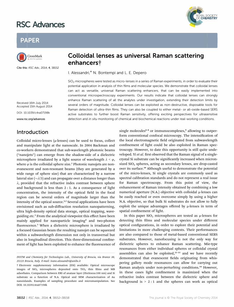
RSC Advances
PAPER
Colloidal lenses
INSTM and Chemistry for Technologies La
25123 Brescia, Italy. E-mail: ivano.alessand
† Electronic supplementary informationimages of SiO2 microspheres depositadsorbates. Comparison between ERI of asubstrate as a function of N.A. Opticnanoislands. Examples of sampling prDOI: 10.1039/c4ra07198k
Cite this: RSC Adv., 2014, 4, 38152
Received 16th July 2014Accepted 15th August 2014
DOI: 10.1039/c4ra07198k
www.rsc.org/advances
38152 | RSC Adv., 2014, 4, 38152–3815
as universal Raman scatteringenhancers†
I. Alessandri,* N. Bontempi and L. E. Depero
SiO2 microspheres were tested as micro-lenses in a series of Raman experiments, in order to evaluate their
potential application in analysis of thin films and molecular species. We demonstrate that colloidal lenses
can act as versatile, universal Raman scattering enhancers, that can be easily implemented into
conventional microspectroscopy experiments. Our results indicate that colloidal lenses can strongly
enhance Raman scattering of all the analytes under investigation, extending their detection limits by
several orders of magnitude. Colloidal lenses can be exploited as non-destructive, disposable tools for
Raman detection of ultra-thin films. They can also be coupled to either metal- or all-oxide-based SERS
active substrates to further boost Raman sensitivity, offering exciting perspectives for ultrasensitive
detection and in situ monitoring of chemical and biochemical reactions under real-working conditions.
Introduction
Colloidal micro-lenses (m-lenses) can be used to focus, collectand manipulate light at the nanoscale. In 2004 Backman andco-workers demonstrated that sub-wavelength photonic beams(“nanojets”) can emerge from the shadow-side of a dielectricmicrosphere irradiated by a light source of wavelength l < 4,where 4 is the colloidal sphere size.1 Photonic nanojets are non-evanescent and non-resonant beams (they are generated by awide range of sphere size) that are characterized by a narrowlateral size (�l/3) and can propagate over a distance longer thanl, provided that the refractive index contrast between sphereand background is less than 2 : 1. As a consequence of lightconcentration, the intensity of the optical eld in the focalregion can be several orders of magnitude larger than theintensity of the optical source.2,3 Several applications have beenenvisioned such as sub-diffraction resolution nanopatterning,ultra high-density optical data storage, optical trapping, wave-guiding etc.3 From the analytical viewpoint this effect have beenmainly applied for nanoparticle imaging4 and two-photonuorescence.5 When a dielectric microsphere is irradiated bya focused Gaussian beam the resulting nanojet can be squeezedwithin a subwavelength dimension not only in transversal butalso in longitudinal direction. This three-dimensional conne-ment of light has been exploited to enhance the uorescence of
b., University of Brescia, via Branze 38,
(ESI) available: Optical microscopeed onto TiO2 thin lms and MBnatase layer (thickness:100 nm) and Sial and SEM characterization of Auocedure and micromanipulation. See
8
single molecules6–8 or immunocomplexes,9 allowing to outper-form conventional confocal microscopy. The intensication ofthe local electromagnetic eld originated from subwavelengthconnement of light could be also exploited in Raman spec-troscopy. However, to date this opportunity is still quite unde-veloped. Yi et al. rst observed that the Raman signal of a single-crystal Si substrate can be signicantly increased when micron-sized SiO2 spheres, acting as secondary lenses, are drop-castedover its surface.10 Although useful to demonstrate the potentialof the micro-lenses, Si single crystals are commonly used asspectral calibration standards and do not represent a real issuefor Raman spectroscopy. From this point of view, theenhancement of Raman intensity obtained by combining a lownumerical aperture (N.A.) objective with colloidal m-lenses canbe easily reached or even overcome simply by using a higherN.A. objective, so that bulk Si substrates do not allow to fullyexploit the unique advantages offered by m-lenses in term ofspatial connement of light.
In this paper SiO2 microspheres are tested as m-lenses fordetecting thin lms and molecular species under differentoptical congurations, in order to explore their potential andlimitations in more challenging contexts. Their performancesare also compared to those of metal-based conventional SERSsubstrates. However, nanofocusing is not the only way fordielectric spheres to enhance Raman scattering. Mie-typeresonances from either individual spheres or colloidal crystalassemblies can also be exploited,11–13 and we have recentlydemonstrated that evanescent elds originating from whis-pering gallery mode resonances can allow for carrying outRaman analysis under non-perturbing conditions.14 However,in these cases light connement is maximized when therefractive index contrast between the dielectric sphere andbackground is > 2 : 1 and the spheres can work as optical
This journal is © The Royal Society of Chemistry 2014
Paper RSC Advances
cavities rather than lenses. In the last part of this paper we willdemonstrate that m-lenses can be synergistically coupled tocore/shell colloidal crystal resonators (optical cavities) and, ingeneral, to any kind of Raman-active substrates to enhance theiroverall sensitivity, playing as universal, disposable tools forRaman microspectroscopy.
Fig. 2 Normalized intensity of the anatase Eg(1) Raman mode as afunction of film thickness. Spectra were acquired over 30 different
Materials and methodsSiO2 microspheres
Commercial monodisperse SiO2 microspheres (Microparticle-GmbH, size: 2.06 � 0.05 mm, 50 mg mL�1) were used utilizedas colloidal micro-lenses, by directly dropping 1 mL of eitherwater of ethanol solution (dilution: 1/1000) on the surface of thetarget. Further details on individual experiments are given inResults and discussion section.
areas with or without single SiO2 spheres using a 0.50 N.A. microscopeobjective. If not indicated, error bars are included within the experi-mental point size.
MicroRaman analysisThe Raman measurements were carried out by means of a high-resolution Raman microscope (Labram HR-800, Horiba/Jobin-Yvon. Exciting source: He–Ne laser (l ¼ 633 nm), withdifferent numerical aperture objectives (0.50, 0.75, 0.9) inbackscattering conguration. A scheme of each individualexperimental conguration is given in the Results and discus-sion section. All data reported in Fig. 1, 2 and 3b results fromsampling over 30 different spheres. Each spectrum results fromthe average of three measurements and is automaticallygenerated by the acquisition soware (LabSpec). Error barsindicate the standard deviation.
The analysis of thin and ultra-thin lms was carried out onTiO2 layers deposited onto Si (100) single-crystal wafer by
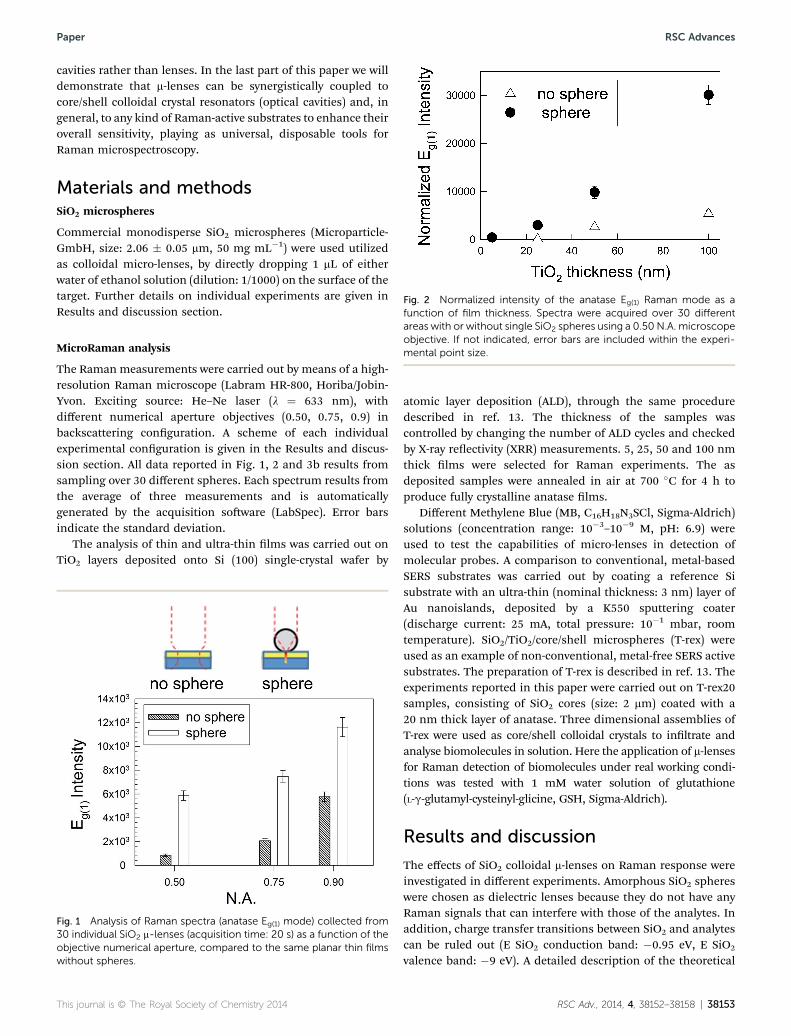
Fig. 1 Analysis of Raman spectra (anatase Eg(1) mode) collected from30 individual SiO2 m-lenses (acquisition time: 20 s) as a function of theobjective numerical aperture, compared to the same planar thin filmswithout spheres.
This journal is © The Royal Society of Chemistry 2014
atomic layer deposition (ALD), through the same proceduredescribed in ref. 13. The thickness of the samples wascontrolled by changing the number of ALD cycles and checkedby X-ray reectivity (XRR) measurements. 5, 25, 50 and 100 nmthick lms were selected for Raman experiments. The asdeposited samples were annealed in air at 700 �C for 4 h toproduce fully crystalline anatase lms.
Different Methylene Blue (MB, C16H18N3SCl, Sigma-Aldrich)solutions (concentration range: 10�3–10�9 M, pH: 6.9) wereused to test the capabilities of micro-lenses in detection ofmolecular probes. A comparison to conventional, metal-basedSERS substrates was carried out by coating a reference Sisubstrate with an ultra-thin (nominal thickness: 3 nm) layer ofAu nanoislands, deposited by a K550 sputtering coater(discharge current: 25 mA, total pressure: 10�1 mbar, roomtemperature). SiO2/TiO2/core/shell microspheres (T-rex) wereused as an example of non-conventional, metal-free SERS activesubstrates. The preparation of T-rex is described in ref. 13. Theexperiments reported in this paper were carried out on T-rex20samples, consisting of SiO2 cores (size: 2 mm) coated with a20 nm thick layer of anatase. Three dimensional assemblies ofT-rex were used as core/shell colloidal crystals to inltrate andanalyse biomolecules in solution. Here the application of m-lensesfor Raman detection of biomolecules under real working condi-tions was tested with 1 mM water solution of glutathione(L-g-glutamyl-cysteinyl-glicine, GSH, Sigma-Aldrich).
Results and discussion
The effects of SiO2 colloidal m-lenses on Raman response wereinvestigated in different experiments. Amorphous SiO2 sphereswere chosen as dielectric lenses because they do not have anyRaman signals that can interfere with those of the analytes. Inaddition, charge transfer transitions between SiO2 and analytescan be ruled out (E SiO2 conduction band: �0.95 eV, E SiO2
valence band: �9 eV). A detailed description of the theoretical
RSC Adv., 2014, 4, 38152–38158 | 38153
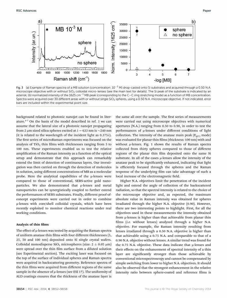
Fig. 3 (a) Example of Raman spectra of a MB solution (concentration: 10�3 M) drop-casted onto Si substrates and acquired through a 0.50 N.A.microscope objective with or without SiO2 colloidal micro-lenses (see the main text for details). The Si peak of the substrate is indicated by anasterisk; (b) normalized intensity of the 1625 cm�1 MB peak (corresponding to the C–C ring stretching mode) as a function of MB concentration.Spectra were acquired over 30 different areas with or without single SiO2 spheres, using a 0.50 N.A. microscope objective. If not indicated, errorbars are included within the experimental point size.
RSC Advances Paper
background related to photonic nanojet can be found in liter-ature.1–7 On the basis of the model described in ref. 3 we canassume that the lateral size of a photonic nanojet propagatingfrom 2 mm sized silica spheres excited at l¼ 633 nm is�240 nm(it is related to the wavelength of the incident light as 0.375l).The rst series of microRaman experiments was focused on theanalysis of TiO2 thin lms with thicknesses ranging from 5 to100 nm. These experiments enabled us to test the relativeamplication of the Raman intensity as a function of the opticalsetup and demonstrate that this approach can remarkablyextend the limit of detection of continuous layers. Our investi-gation was then carried on through the detection of moleculesin solution, using different concentrations of MB as a molecularprobe. Here the analytical capabilities of the m-lenses werecompared to those of conventional, SERS-active gold nano-particles. We also demonstrated that m-lenses and metalnanoparticles can be synergistically coupled to further extendthe performances of SERS substrates. Finally, different proof-of-concept experiments were carried out in order to combinem-lenses with core/shell colloidal crystals, which have beenrecently exploited to monitor chemical reactions under realworking conditions.
Analysis of thin lms
The effect of m-lenses was tested by acquiring the Raman spectraof uniform anatase thin lms with four different thicknesses (5,25, 50 and 100 nm) deposited onto Si single crystal wafers.Colloidal monodisperse SiO2 microspheres (size: 2 � 0.05 mm)were spread over the thin lm surface from a diluted solution(see Experimental section). The exciting laser was focused onthe top of the surface of individual spheres and Raman spectrawere acquired in backscattering geometry. Reference spectra ofthe thin lms were acquired from different regions of the samesample in the absence of m-lenses (see ESI 1†). The uniformity ofALD coatings ensures that the thickness of the anatase layer is
38154 | RSC Adv., 2014, 4, 38152–38158
the same all over the sample. The rst series of measurementswere carried out using microscope objectives with numericalapertures (N.A.) ranging from 0.50 to 0.90, in order to test theperformances of m-lenses under different conditions of lightcollection. The intensity of the anatase main peak (Eg(1) mode)was evaluated for planar thin lms (thickness: 100 nm) with andwithout m-lenses. Fig. 1 shows the results of Raman spectracollected from thirty spheres compared to those of differentregions of the planar thin lm deposited onto the same Sisubstrate. In all of the cases m-lenses allow the intensity of theanatase peak to be signicantly enhanced, indicating that lightis efficiently focused through the spheres and the Ramanresponse of the underlying lm can take advantage of such alocal increase of the electromagnetic eld.
Higher N.A. objectives limit the penetration of the incidentlight and extend the angle of collection of the backscatteredradiation, so that the spectral intensity is related to the choice ofthe microscope objective and, as expected, the maximumabsolute value in Raman intensity was obtained for spheresirradiated through the higher N.A. objective (0.90). However,there are two interesting points to highlight. First, for all theobjectives used in these measurements the intensity obtainedfrom m-lenses is higher than that achievable from planar thinlms (i.e. without lenses) analyzed through a higher N.A.objective. For example, the Raman intensity resulting fromlenses irradiated through a 0.50 N.A. objective is higher thanthat achievable using a 0.75 N.A. and comparable to that of a0.90 N.A. objective without lenses. A similar trend was found forthe 0.75 N.A. objective. These data indicate that m-lenses andtheir effects on the enhancement of spectral intensity of a thinlayer are signicantly stronger than those achievable byconventional microspectroscopy and cannot be compensated bysimple switching from lower to higher N.A. objectives. It shouldalso be observed that the strongest enhancement in the relativeintensity ratio between sphere-coated and reference lms is
This journal is © The Royal Society of Chemistry 2014
Paper RSC Advances
achieved by the lowest N.A. objective. This result is in qualita-tive agreement with the experiments reported by Yi et al. onbulk silicon.10 In that paper the authors found that themaximum enhancement of the Raman intensity betweensphere-coated and uncoated bulk Si substrates (ERI) is about 6.This value is achieved when the size of the sphere approachesthat of the incident laser beam waist (�2.5 mm). On the basis ofgeometrical considerations they also calculated that the Ramanenhancement factor (EF) achievable using SiO2 spheres with anoptimal size of 2.5 mm is around 104. In our case, a directcomparison of ERI between the main Raman modes of anatase(Eg(1)) and Si (TO) in anatase-coated Si substrates enabled us toelicit the specic sensitivity of m-lenses to the analysis of thinlayers, unravelling that using a 0.5 N.A. objective the relativeenhancement of Raman intensity of the anatase mode isremarkably stronger (�7 vs. 4) than that of the underlying Sisubstrate. (ESI 2†) This enhanced sensitivity might be explainedconsidering that photons coming from the Si substrate aregathered more efficiently as the solid angle of light collection isincreased using higher N.A. objectives. As a result, the sensi-tivity towards the thin lm is strongly reduced. Moreover,higher N.A. aperture objectives are more sensitive to the posi-tion of the focal point on the sphere, as demonstrated by theincrease of error bars on passing from 0.5 to 0.9 N.A. objectives.For all of these reasons, and since m-lenses could nd inter-esting applications in microuidic chambers and operando-likereactors, where 0.5 N.A. long-working distance objectivesrepresent a suitable solution for acquiring Raman spectra, all ofthe following experiments were carried out with 0.5 N.A.objectives.
Fig. 2 shows the results of measurements on anatase layersas a function of the lm thickness with or without addition ofcolloidal m-lenses over thirty different regions. These data showthat m-lenses allow for a general enhancement of Raman scat-tering. Themaximum ERI is achieved for thicker lms (100 nm),indicating that under these conditions the nanojet beampropagates across the coating layer for more than 50 nm beforebeing focused. However, although their absolute intensity isquite low, the Raman signals of the thinnest (20 and 5 nm thick)anatase layers were detected only by means of m-lenses.
These data show that even ultra-thin layers can signicantlyexperience the benecial effects of eld enhancement inducedby m-lenses. This allows for extending the detection limit to athickness level that cannot be usually reached by conventionalmicroRaman. Moreover, colloidal microspheres can be quicklydispersed on a thin lm surface, used as m-lenses for the Ramananalysis and then easily removed by sonication. This three-stepprocedure allows for probing and investigating thin layersthrough a non-destructive approach that does not alter theoriginal structure of the lm.
Analysis of molecular species and comparison to metal-basedSERS substrates
The potential of m-lenses in detection of molecular species wastested using MB solutions over a wide concentration range(from 10�3 to 10�9 M). The Si substrates were soaked in a vessel
This journal is © The Royal Society of Chemistry 2014
containing 4 mL of dye solution. Upon drying at roomtemperature, each sample was analyzed by measuring differentregions of the substrate. All the selected areas were at andclean; optically visible agglomerates of MB were not consideredin sampling (see ESI 3†). As a selected example, Fig. 3a showsthe Raman spectra of a 10�3 M MB solution acquired with andwithout colloidal m-lenses using a 0.50 N.A. objective. Theresults of measurements extended over the whole concentrationrange is shown in Fig. 3b, which refers to the intensity of the MBmain peak around 1625 cm�1 (C–C ring stretching mode).Again, m-lenses strongly enhance the MB Raman signal (theintensity ratio between the peak of a 10�3 M solution analyzedwith and without m-lenses is about 30) and extend sensitivity tovery low concentration (10�7 M), i.e. three orders of magnitudebeyond the detection limit of the analogous planar reference.
It is now interesting to compare the enhanced Raman scat-tering achieved by m-lenses to that resulting from moreconventional, metal-based SERS substrates. In general SERS-active materials consist of Ag or Au nanoparticles that aredeposited from solution giving rise to fractal-like aggregates.15
These aggregates are characterized by a dense distribution ofSERS “hot-spots”, which originate from junctions between twoor more individual NPs. Hot-spots can allow to reach the ulti-mate detection limits of this technique (e.g., in single moleculedetection experiments). However, the intensity of theenhancement strongly depends on hot-spots distribution,which is very hard to predict. In order to obtain a more homo-geneous signal we prepared our SERS active substrates througha sputter-coating deposition of Au nanoislands onto the surfaceof a Si wafer.16,17 Although not optimized for achieving thestrongest SERS effects (the limit of detection of MB is only 10�8
M for sputter-coated substrates, just one order of magnitudelower than that obtained by m-lenses without gold), thisapproach gives much more reproducible results than fractalassemblies, allowing to test m-lenses under reliable conditions.(ESI 4†) Fig. 4a shows as an example the C–C ring stretchingRaman mode at 1625 cm�1 of a 10�5 M MB solution adsorbedonto Au nanoisland thin lms and the same area upon focusingthrough SiO2 m-lenses. As predicted by nite-difference time-domain (FDTD) simulations, a metal layer is expected toreduce the penetration depth of a nanojet, leading to a moreeffective connement of light at the SiO2/metal interface.18,19
Colloidal lenses enhance the intensity by a �1.5 factor. Asreported in the inset of Fig. 4a, m-lenses extend the limit ofdetection to less than 10�9 M, i.e. one order of magnitude lowerthan that of Au nanoislands without lenses. Fig. 4b summarizesthe results of MB detection using different substrates. Colloidallens-assisted detection on Au nanoislands outperforms thesensitivity of simple planar substrates by 5 orders of magnitudeand even stronger effects can be expected by using more effi-cient metal-based SERS substrates.
These results indicate that the use of colloidal lenses is aconvenient and very practical means to extend the Ramansensitivity of any type of SERS active substrate when the analyteis uniformly distributed all over the substrate itself. On theother hand, the main concern about the use of m-lenses foranalysis of molecular probes or inhomogenous samples is
RSC Adv., 2014, 4, 38152–38158 | 38155
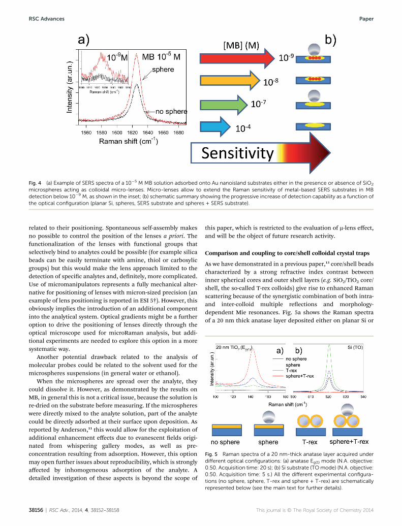
Fig. 4 (a) Example of SERS spectra of a 10�5 M MB solution adsorbed onto Au nanoisland substrates either in the presence or absence of SiO2
microspheres acting as colloidal micro-lenses. Micro-lenses allow to extend the Raman sensitivity of metal-based SERS substrates in MBdetection below 10�9 M, as shown in the inset; (b) schematic summary showing the progressive increase of detection capability as a function ofthe optical configuration (planar Si, spheres, SERS substrate and spheres + SERS substrate).
Fig. 5 Raman spectra of a 20 nm-thick anatase layer acquired underdifferent optical configurations: (a) anatase Eg(1) mode (N.A. objective:0.50. Acquisition time: 20 s); (b) Si substrate (TOmode) (N.A. objective:0.50. Acquisition time: 5 s.) All the different experimental configura-tions (no sphere, sphere, T-rex and sphere + T-rex) are schematically
RSC Advances Paper
related to their positioning. Spontaneous self-assembly makesno possible to control the position of the lenses a priori. Thefunctionalization of the lenses with functional groups thatselectively bind to analytes could be possible (for example silicabeads can be easily terminate with amine, thiol or carboxylicgroups) but this would make the lens approach limited to thedetection of specic analytes and, denitely, more complicated.Use of micromanipulators represents a fully mechanical alter-native for positioning of lenses with micron-sized precision (anexample of lens positioning is reported in ESI 5†). However, thisobviously implies the introduction of an additional componentinto the analytical system. Optical gradients might be a furtheroption to drive the positioning of lenses directly through theoptical microscope used for microRaman analysis, but addi-tional experiments are needed to explore this option in a moresystematic way.
Another potential drawback related to the analysis ofmolecular probes could be related to the solvent used for themicrospheres suspensions (in general water or ethanol).
When the microspheres are spread over the analyte, theycould dissolve it. However, as demonstrated by the results onMB, in general this is not a critical issue, because the solution isre-dried on the substrate before measuring. If the microsphereswere directly mixed to the analyte solution, part of the analytecould be directly adsorbed at their surface upon deposition. Asreported by Anderson,11 this would allow for the exploitation ofadditional enhancement effects due to evanescent elds origi-nated from whispering gallery modes, as well as pre-concentration resulting from adsorption. However, this optionmay open further issues about reproducibility, which is stronglyaffected by inhomogeneous adsorption of the analyte. Adetailed investigation of these aspects is beyond the scope of
38156 | RSC Adv., 2014, 4, 38152–38158
this paper, which is restricted to the evaluation of m-lens effect,and will be the object of future research activity.
Comparison and coupling to core/shell colloidal crystal traps
As we have demonstrated in a previous paper,13 core/shell beadscharacterized by a strong refractive index contrast betweeninner spherical cores and outer shell layers (e.g. SiO2/TiO2 core/shell, the so-called T-rex colloids) give rise to enhanced Ramanscattering because of the synergistic combination of both intra-and inter-colloid multiple reections and morphology-dependent Mie resonances. Fig. 5a shows the Raman spectraof a 20 nm thick anatase layer deposited either on planar Si or
represented below (see the main text for further details).
This journal is © The Royal Society of Chemistry 2014
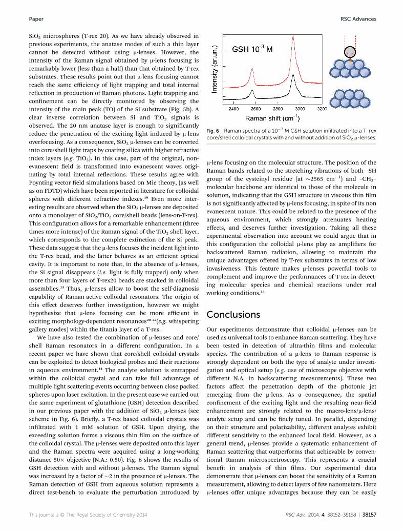
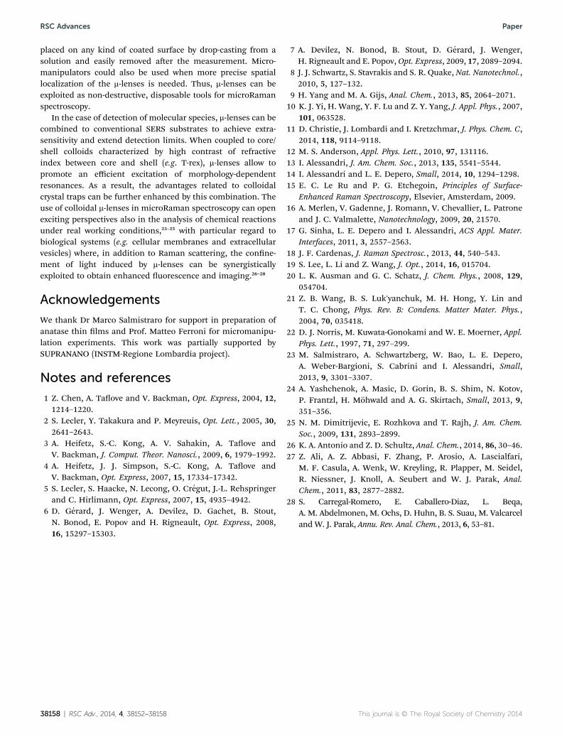
Fig. 6 Raman spectra of a 10�3 M GSH solution infiltrated into a T-rexcore/shell colloidal crystals with and without addition of SiO2 m-lenses.
Paper RSC Advances
SiO2 microspheres (T-rex 20). As we have already observed inprevious experiments, the anatase modes of such a thin layercannot be detected without using m-lenses. However, theintensity of the Raman signal obtained by m-lens focusing isremarkably lower (less than a half) than that obtained by T-rexsubstrates. These results point out that m-lens focusing cannotreach the same efficiency of light trapping and total internalreection in production of Raman photons. Light trapping andconnement can be directly monitored by observing theintensity of the main peak (TO) of the Si substrate (Fig. 5b). Aclear inverse correlation between Si and TiO2 signals isobserved. The 20 nm anatase layer is enough to signicantlyreduce the penetration of the exciting light induced by m-lensoverfocusing. As a consequence, SiO2 m-lenses can be convertedinto core/shell light traps by coating silica with higher refractiveindex layers (e.g. TiO2). In this case, part of the original, non-evanescent eld is transformed into evanescent waves origi-nating by total internal reections. These results agree withPoynting vector eld simulations based on Mie theory, (as wellas on FDTD) which have been reported in literature for colloidalspheres with different refractive indexes.19 Even more inter-esting results are observed when the SiO2 m-lenses are depositedonto a monolayer of SiO2/TiO2 core/shell beads (lens-on-T-rex).This conguration allows for a remarkable enhancement (threetimes more intense) of the Raman signal of the TiO2 shell layer,which corresponds to the complete extinction of the Si peak.These data suggest that the m-lens focuses the incident light intothe T-rex bead, and the latter behaves as an efficient opticalcavity. It is important to note that, in the absence of m-lenses,the Si signal disappears (i.e. light is fully trapped) only whenmore than four layers of T-rex20 beads are stacked in colloidalassemblies.13 Thus, m-lenses allow to boost the self-diagnosiscapability of Raman-active colloidal resonators. The origin ofthis effect deserves further investigation, however we mighthypothesize that m-lens focusing can be more efficient inexciting morphology-dependent resonances20–22(e.g. whisperinggallery modes) within the titania layer of a T-rex.
We have also tested the combination of m-lenses and core/shell Raman resonators in a different conguration. In arecent paper we have shown that core/shell colloidal crystalscan be exploited to detect biological probes and their reactionsin aqueous environment.14 The analyte solution is entrappedwithin the colloidal crystal and can take full advantage ofmultiple light scattering events occurring between close packedspheres upon laser excitation. In the present case we carried outthe same experiment of glutathione (GSH) detection describedin our previous paper with the addition of SiO2 m-lenses (seescheme in Fig. 6). Briey, a T-rex based colloidal crystals wasinltrated with 1 mM solution of GSH. Upon drying, theexceeding solution forms a viscous thin lm on the surface ofthe colloidal crystal. The m-lenses were deposited onto this layerand the Raman spectra were acquired using a long-workingdistance 50� objective (N.A.: 0.50). Fig. 6 shows the results ofGSH detection with and without m-lenses. The Raman signalwas increased by a factor of �2 in the presence of m-lenses. TheRaman detection of GSH from aqueous solution represents adirect test-bench to evaluate the perturbation introduced by
This journal is © The Royal Society of Chemistry 2014
m-lens focusing on the molecular structure. The position of theRaman bands related to the stretching vibrations of both –SHgroup of the cysteinyl residue (at �2565 cm�1) and –CH2–
molecular backbone are identical to those of the molecule insolution, indicating that the GSH structure in viscous thin lmis not signicantly affected by m-lens focusing, in spite of its nonevanescent nature. This could be related to the presence of theaqueous environment, which strongly attenuates heatingeffects, and deserves further investigation. Taking all theseexperimental observation into account we could argue that inthis conguration the colloidal m-lens play as ampliers forbackscattered Raman radiation, allowing to maintain theunique advantages offered by T-rex substrates in terms of lowinvasiveness. This feature makes m-lenses powerful tools tocomplement and improve the performances of T-rex in detect-ing molecular species and chemical reactions under realworking conditions.14
Conclusions
Our experiments demonstrate that colloidal m-lenses can beused as universal tools to enhance Raman scattering. They havebeen tested in detection of ultra-thin lms and molecularspecies. The contribution of a m-lens to Raman response isstrongly dependent on both the type of analyte under investi-gation and optical setup (e.g. use of microscope objective withdifferent N.A. in backscattering measurements). These twofactors affect the penetration depth of the photonic jetemerging from the m-lens. As a consequence, the spatialconnement of the exciting light and the resulting near-eldenhancement are strongly related to the macro-lens/m-lens/analyte setup and can be nely tuned. In parallel, dependingon their structure and polarizability, different analytes exhibitdifferent sensitivity to the enhanced local eld. However, as ageneral trend, m-lenses provide a systematic enhancement ofRaman scattering that outperforms that achievable by conven-tional Raman microspectroscopy. This represents a crucialbenet in analysis of thin lms. Our experimental datademonstrate that m-lenses can boost the sensitivity of a Ramanmeasurement, allowing to detect layers of few nanometers. Herem-lenses offer unique advantages because they can be easily
RSC Adv., 2014, 4, 38152–38158 | 38157
RSC Advances Paper
placed on any kind of coated surface by drop-casting from asolution and easily removed aer the measurement. Micro-manipulators could also be used when more precise spatiallocalization of the m-lenses is needed. Thus, m-lenses can beexploited as non-destructive, disposable tools for microRamanspectroscopy.
In the case of detection of molecular species, m-lenses can becombined to conventional SERS substrates to achieve extra-sensitivity and extend detection limits. When coupled to core/shell colloids characterized by high contrast of refractiveindex between core and shell (e.g. T-rex), m-lenses allow topromote an efficient excitation of morphology-dependentresonances. As a result, the advantages related to colloidalcrystal traps can be further enhanced by this combination. Theuse of colloidal m-lenses in microRaman spectroscopy can openexciting perspectives also in the analysis of chemical reactionsunder real working conditions,23–25 with particular regard tobiological systems (e.g. cellular membranes and extracellularvesicles) where, in addition to Raman scattering, the conne-ment of light induced by m-lenses can be synergisticallyexploited to obtain enhanced uorescence and imaging.26–28
Acknowledgements
We thank Dr Marco Salmistraro for support in preparation ofanatase thin lms and Prof. Matteo Ferroni for micromanipu-lation experiments. This work was partially supported bySUPRANANO (INSTM-Regione Lombardia project).
Notes and references
1 Z. Chen, A. Taove and V. Backman, Opt. Express, 2004, 12,1214–1220.
2 S. Lecler, Y. Takakura and P. Meyreuis, Opt. Lett., 2005, 30,2641–2643.
3 A. Heifetz, S.-C. Kong, A. V. Sahakin, A. Taove andV. Backman, J. Comput. Theor. Nanosci., 2009, 6, 1979–1992.
4 A. Heifetz, J. J. Simpson, S.-C. Kong, A. Taove andV. Backman, Opt. Express, 2007, 15, 17334–17342.
5 S. Lecler, S. Haacke, N. Lecong, O. Cregut, J.-L. Rehspringerand C. Hirlimann, Opt. Express, 2007, 15, 4935–4942.
6 D. Gerard, J. Wenger, A. Devilez, D. Gachet, B. Stout,N. Bonod, E. Popov and H. Rigneault, Opt. Express, 2008,16, 15297–15303.
38158 | RSC Adv., 2014, 4, 38152–38158
7 A. Devilez, N. Bonod, B. Stout, D. Gerard, J. Wenger,H. Rigneault and E. Popov, Opt. Express, 2009, 17, 2089–2094.
8 J. J. Schwartz, S. Stavrakis and S. R. Quake, Nat. Nanotechnol.,2010, 5, 127–132.
9 H. Yang and M. A. Gijs, Anal. Chem., 2013, 85, 2064–2071.10 K. J. Yi, H. Wang, Y. F. Lu and Z. Y. Yang, J. Appl. Phys., 2007,
101, 063528.11 D. Christie, J. Lombardi and I. Kretzchmar, J. Phys. Chem. C,
2014, 118, 9114–9118.12 M. S. Anderson, Appl. Phys. Lett., 2010, 97, 131116.13 I. Alessandri, J. Am. Chem. Soc., 2013, 135, 5541–5544.14 I. Alessandri and L. E. Depero, Small, 2014, 10, 1294–1298.15 E. C. Le Ru and P. G. Etchegoin, Principles of Surface-
Enhanced Raman Spectroscopy, Elsevier, Amsterdam, 2009.16 A. Merlen, V. Gadenne, J. Romann, V. Chevallier, L. Patrone
and J. C. Valmalette, Nanotechnology, 2009, 20, 21570.17 G. Sinha, L. E. Depero and I. Alessandri, ACS Appl. Mater.
Interfaces, 2011, 3, 2557–2563.18 J. F. Cardenas, J. Raman Spectrosc., 2013, 44, 540–543.19 S. Lee, L. Li and Z. Wang, J. Opt., 2014, 16, 015704.20 L. K. Ausman and G. C. Schatz, J. Chem. Phys., 2008, 129,
054704.21 Z. B. Wang, B. S. Luk'yanchuk, M. H. Hong, Y. Lin and
T. C. Chong, Phys. Rev. B: Condens. Matter Mater. Phys.,2004, 70, 035418.
22 D. J. Norris, M. Kuwata-Gonokami and W. E. Moerner, Appl.Phys. Lett., 1997, 71, 297–299.
23 M. Salmistraro, A. Schwartzberg, W. Bao, L. E. Depero,A. Weber-Bargioni, S. Cabrini and I. Alessandri, Small,2013, 9, 3301–3307.
24 A. Yashchenok, A. Masic, D. Gorin, B. S. Shim, N. Kotov,P. Frantzl, H. Mohwald and A. G. Skirtach, Small, 2013, 9,351–356.
25 N. M. Dimitrijevic, E. Rozhkova and T. Rajh, J. Am. Chem.Soc., 2009, 131, 2893–2899.
26 K. A. Antonio and Z. D. Schultz, Anal. Chem., 2014, 86, 30–46.27 Z. Ali, A. Z. Abbasi, F. Zhang, P. Arosio, A. Lascialfari,
M. F. Casula, A. Wenk, W. Kreyling, R. Plapper, M. Seidel,R. Niessner, J. Knoll, A. Seubert and W. J. Parak, Anal.Chem., 2011, 83, 2877–2882.
28 S. Carregal-Romero, E. Caballero-Diaz, L. Beqa,A. M. Abdelmonen, M. Ochs, D. Huhn, B. S. Suau, M. Valcarceland W. J. Parak, Annu. Rev. Anal. Chem., 2013, 6, 53–81.
This journal is © The Royal Society of Chemistry 2014
Copyright © 2022 FDOKUMEN