clear medical use of contact lenses - Aston Publications Explorer
165
CLEAR MEDICAL USE OF CONTACT LENSES Deborah S. Jacobs, MD, MSc, Massachusetts Eye & Ear, Cornea and Refractive Surgery Service, Harvard Medical School, Boston MA, USA Karen G. Carrasquillo, OD, PhD, FBCLA BostonSight, Needham, MA, USA Paul D. Cottrell, MCOptom, PGDip, FBCLA Birmingham & Midland Eye Centre, Birmingham, UK Fernando J. Fernández-Velázquez, OD Centro Fernandez-Velazquez, Madrid, Spain Raquel Gil-Cazorla, MCOptom, PhD, FBCLA Optometry and Vision Science, Aston University, Birmingham, UK Isabelle Jalbert, OD, PhD School of Optometry and Vision Science, UNSW Sydney, Australia Andrew D. Pucker, OD, PhD, University of Alabama at Birmingham, Birmingham, AL, USA Kellen Riccobono, OD EYESPOT, Boston, MA, USA Danielle M. Robertson, OD, PhD UT Southwestern Medical Center at Dallas, TX, USA Loretta Szczotka-Flynn OD, PhD Department of Ophthalmology & Visual Science, Case Western Reserve University, Cleveland, OH, USA Lynne Speedwell FCOptom, MSc Great Ormond Street Hospital for Children NHS Trust, Moorfields Eye Hospital, London, UK Fiona Stapleton FCOptom, PhD, FBCLA School of Optometry and Vision Science, UNSW Sydney, Australia Running title: CLEAR Medical Uses Report Keywords Therapeutic contact lens, bandage lens, scleral lens, irregular astigmatism, aphakia, ocular surface disease
-
Upload
khangminh22 -
Category
Documents
-
view
2 -
download
0
Transcript of clear medical use of contact lenses - Aston Publications Explorer

CLEAR MEDICAL USE OF CONTACT LENSES Deborah S. Jacobs, MD, MSc, Massachusetts Eye & Ear, Cornea and Refractive
Surgery Service, Harvard Medical School, Boston MA, USA
Karen G. Carrasquillo, OD, PhD, FBCLA BostonSight, Needham, MA, USA
Paul D. Cottrell, MCOptom, PGDip, FBCLA Birmingham & Midland Eye Centre,
Birmingham, UK
Fernando J. Fernández-Velázquez, OD Centro Fernandez-Velazquez, Madrid,
Spain
Raquel Gil-Cazorla, MCOptom, PhD, FBCLA Optometry and Vision Science,
Aston University, Birmingham, UK
Isabelle Jalbert, OD, PhD School of Optometry and Vision Science, UNSW
Sydney, Australia
Andrew D. Pucker, OD, PhD, University of Alabama at Birmingham, Birmingham,
AL, USA
Kellen Riccobono, OD EYESPOT, Boston, MA, USA
Danielle M. Robertson, OD, PhD UT Southwestern Medical Center at Dallas, TX,
USA
Loretta Szczotka-Flynn OD, PhD Department of Ophthalmology & Visual
Science, Case Western Reserve University, Cleveland, OH, USA
Lynne Speedwell FCOptom, MSc Great Ormond Street Hospital for Children
NHS Trust, Moorfields Eye Hospital, London, UK
Fiona Stapleton FCOptom, PhD, FBCLA School of Optometry and Vision
Science, UNSW Sydney, Australia
Running title: CLEAR Medical Uses Report
Keywords
Therapeutic contact lens, bandage lens, scleral lens, irregular astigmatism,
aphakia, ocular surface disease
Abbreviations
BCVA: Best-corrected visual acuity
BSCL: Bandage soft contact lens
cGVHD: Chronic Graft-versus-host Disease
CXL: Corneal Collagen Cross-linking
EDTA: Ethylenediamine-tetraacetic Acid
FDA: Food and Drug Administration (US)
IATS: Infant Aphakia Treatment Study
IOP: Intraocular pressure
ICRS: Intracorneal Ring Segments
LASEK: Laser Epithelial Keratomileusis
LASIK: Laser-assisted in situ Keratomileusis
LSCD: Limbal stem cell deficiency
MK: Microbial keratitis
NEI-VFQ-25: National Eye Institute Visual Function Questionnaire (25 item)
NSAID: Nonsteroidal anti-inflammatory drug
OSDI: Ocular surface disease index
PED: Persistent epithelial defect
PK: Penetrating keratoplasty
PMD: Pellucid marginal degeneration
PMMA: Polymethyl methacrylate
PRK: Photorefractive keratectomy
PROSE: Prosthetic Replacement of the Ocular Surface Ecosystem
QOL: Quality of Life
RCES: Recurrent Corneal Erosion Syndrome
SiHy: Silicone hydrogel
SLK: Superior limbic keratoconjunctivitis
SJS: Stevens-Johnson Syndrome
TFOS DEWS II: Tear Film and Ocular Surface Dry Eye Workshop II

Abstract
The medical use of contact lenses is a solution for many complex ocular
conditions, including high refractive error, irregular astigmatism, primary and
secondary corneal ectasia, disfiguring disease, and ocular surface disease. The
development of highly oxygen permeable soft and rigid materials has extended
the suitability of contact lenses for such applications. There is consistent
evidence that bandage soft contact lenses, particularly silicone hydrogel lenses,
improve epithelial healing and reduce pain in persistent epithelial defects, after
trauma or surgery, and in corneal dystrophies. Drug delivery applications of
contact lens hold promise for improving topical therapy. Modern scleral lens
practice has achieved great success for both visual rehabilitation and therapeutic
applications, including those requiring retention of a tear reservoir or protection
from an adverse environment. This report offers a practical and relevant
summary of the current evidence for the medical use of contact lenses for all eye
care professionals including optometrists, ophthalmologists, opticians, and
orthoptists. Topics covered include indications for use in both acute and chronic
conditions, lens selection, patient selection, wear and care regimens, and
recommended aftercare schedules. Prevention, presentation, and management
of complications of medical use are reviewed.

1 Introduction
Clinicians have long appreciated that contact lenses play a role in the care of
patients with ophthalmic disease. Contact lenses therefore have a medical use, in
addition to their use for correction of refractive error. This medical use has evolved
over decades alongside advances in contact lens materials and design.
Appreciation of the role a lens might play in stabilising the ocular surface,
neutralising refractive error, and improving visual function, combined with
awareness of potential complications and how to avoid them, has yielded vast
experience and a body of literature on the medical use of contact lenses.
Statements on the quality of evidence are based on the approach discussed in the
CLEAR Evidence-based Practice Report [1].
1.1 Definition of Medical Contact Lenses
The literature search conducted to create this report failed to find a field-wide,
accepted definition for medical contact lenses. After discussion and consensus,
the following definition for medical contact lenses has been adopted by the
subcommittee on Medical Use Contact Lenses:
Medical Contact Lenses are any type of contact lens that is worn for the primary
purpose of treating an underlying disease state or complicated refractive status.
Medical contact lenses may or may not correct refractive error. Medical contact
lenses are prescribed for reasons other than the cosmetic purpose of eliminating
the need for spectacles. There is no universally accepted definition for medical
contact lenses, but in some countries, some insurance companies set
requirements as to the condition or diagnosis (e.g., cornea ectasia, unilateral
aphakia) or degree of refractive error (e.g. high myopia) before a coverage or
reimbursement is granted. Because of this, requirements and definitions can vary
from nation to nation based on the structure of healthcare in the respective country.
1.1.1 Regulatory bodies, labelling, and “therapeutic” lenses
In the United States (US), mass produced contact lenses are regulated by the US
Food and Drug Administration (FDA). The first (hydrogel) soft CL was approved by
the US FDA in 1971 as a new drug [2,3]. Contact lenses were later reclassified by
the US FDA as Class III medical devices (high risk) in 1976 when the Medical
Device Amendment was passed. Later regulatory revisions have since reclassified
daily wear soft lenses and rigid corneal lenses as Class II medical devices
(moderate to high risk), while overnight and myopia management contact lenses,
are considered Class III medical devices [4]. The European Medicines Agency of
the European Union, the Australian Therapeutic Goods Administration in Australia,
the Drug Controller General of India, and the National Medical Products
Administration in China perform a similar function and have similar classification
systems for regulating medical devices [4].
National regulatory bodies take guidance from the International Organisation for
Standardization when creating their policies. This body has specifically set
international industry standards that are broadly recognised by regulatory
agencies throughout the world for lens and care product labelling [2]. They have
also set efficacy standards for the care systems needed to maintain contact lenses
[5]. Professional organisations likewise provide best practice guidance to
clinicians, and they provide feedback to the above regulatory bodies when new
policies are being set [6].
While any CL might be used medically or as a therapeutic or bandage CL, some
contact lenses are labelled for use as a bandage lens or with a therapeutic
indication for use.
1.1.2 Clinical definitions
Contact lenses are used for numerous medical purposes that include, but are not
limited to, treating patients with corneal ectasia, ocular surface disease, after
ocular surgery and in the setting of high refractive error [7]. The authors

recommend the following set of definitions to be adopted by the community for
medical use of contact lenses.
• Therapeutic or Bandage Contact Lenses are lenses that are used for the
treatment of ocular discomfort or to support the cornea during healing after
surgery or when the cornea is being treated for an underlying disease state
or to protect the cornea from the environment or mechanical interaction with
the lids.
• Rehabilitative Contact Lenses are lenses that are prescribed for
conditions that prevent a patient from achieving adequate visual function
with spectacles because of high, irregular, or asymmetric refractive error.
Partially or completely occlusive lenses that improve function or cosmesis
after trauma, surgery, or stroke also fall into this category.
1.2 Lens types used for medical purposes
It is widely accepted that Müller described the first CL for correcting refractive error
in the late 1880s [8], but concurrently, in 1888, Fick described what may be the
first medical use of contact lenses in his report of the improvement of vision in
patients with keratoconus [9]. Müller’s lenses, which were scleral lenses made of
glass and used to correct his own high myopia, unfortunately induced corneal
oedema and ocular pain because they were impermeable to oxygen. Because of
this, they could only be worn for short periods of time. Polymethyl methacrylate
(PMMA) corneal lenses were used on a therapeutic basis as early as the late
1950s and early 1960s [10,11]. Oxygen impermeable PMMA contact lenses have
largely been replaced by rigid lens materials, which are used on a therapeutic basis
in corneal and scleral lenses [12–15].
The soft contact lens market emerged in the early 1970s with the creation of cross-
linked hydrogel polymers and the introduction of hydrogel contact lenses [16–22].
Hydrogel contact lenses inherently lacked the ability to transmit enough oxygen to

the cornea in order to avoid corneal hypoxia during overnight wear [16], which
propelled the creation of the much more oxygen permeable silicone and then
silicone hydrogel (SiHy) contact lenses [23–25]. There was hope that SiHy contact
lenses would reduce the rate of microbial keratitis (MK) associated with overnight
night CL wear. Post-market experience reveals that infection rates are similar
between SiHy and hydrogel contact lenses [26,27], While hydrogel contact lenses
are still labelled and used in some countries for overnight wear and as therapeutic
or bandage lenses, it is generally only SiHy lenses that currently carry therapeutic
or bandage indications for use. The following is a discussion on how these material
properties, modes of wear, and innovation in design might affect the choice of lens
for medical use of contact lenses.
1.2.1 Hydrogel/Silicone Hydrogel
The first US FDA approved soft contact lens was a conventional hydrogel lens
(Soflens) developed by Bausch and Lomb [16]. Conventional hydrogel contact
lenses fail to meet the criteria (87.0 Dk/t) set forth by Holden and Mertz to provide
the cornea with enough oxygen to avoid excess corneal swelling during overnight
wear [28], a limitation to their use (see CLEAR Complications Report [30]).
Eventually, this challenge of hypoxia was circumvented by the release of the first
silicone hydrogel contact lens (PureVision) by Bausch & Lomb with reports of
improved performance compared to hydrogel lenses for both cosmetic [23] and
broad therapeutic use [31]. While silicone hydrogel contact lenses have a reduced
likelihood of corneal hypoxia [32–35], they have failed to address many of the
other issues that are problematic for CL wearers [36].
Furthermore, the literature currently lacks well-controlled studies related to these
materials being used specifically for medical purposes. One of the first reports on
therapeutic soft was published in 1971 [16]. That study described the use of the
Soflens for treating 45 patients with corneal disease (corneal oedema, dry eyes,
corneal ulcers, and advanced corneal conditions such as keratoconus). They
found hydrogel contact lenses were an effective bandage lens for reducing

external ocular discomfort. Early evidence in a study with 278 participants
demonstrated that hydrogel contact lenses were effective therapeutic lenses for
treating about half of participants with acute or chronic corneal diseases [20]. In a
prospective study, 91% of the adult participants (n = 70) found benefit from
wearing silicone hydrogel contact lenses for therapeutic purposes for conditions
such as bullous keratopathy and post-operative corneal epitheliopathy [37].
Similarly, silicone hydrogel contact lenses can be beneficial post-LASEK [38], and
a prospective study found that SiHy lenses (n = 29) are safe and effective for
children for therapeutic purposes for conditions such as corneal burns or wounds
[39]. Similarly, a multicentre study reported general success of a senofilcon A lens
as bandage lens for a range of therapeutic indications [40]. These data fail to
provide clear evidence with regards to selecting a hydrogel or silicone hydrogel
contact lens for medical use. Similarly, disposable soft contact lens that are
replaced on a daily, bi-weekly, or monthly basis are now readily available, though
there is no clear evidence for selecting one wear schedule over another for medical
purposes [41].
1.2.2 Specialty soft lenses
Standard, commercially available soft contact lenses are typically between 13.8
mm and 14.5 mm in diameter. There are two special types of soft lenses that have
medical use: large diameter hydrogel soft lenses and custom toric hydrogel or SiHy
lenses.
Large diameter hydrogel soft contact lenses are available for off the shelf use in
eyes with larger corneal diameters and can be used for medical purposes such as
bleb leak or when there is poor retention of typical diameter lenses due to
exposure, ocular surface disease or unusual corneal or scleral topography. The
sub-committee on Medical Use Contact Lenses acknowledges that there are off
the shelf large diameter contact lenses (e.g., Kontur, Hydrolens, Eye-58) and that
these are in widespread use in the locales in which they are marketed. However,
no primary references related to these contact lenses were identified during the

preparation of this report, and because of this, more investigation on this topic is
needed. A randomised, crossover study with 25 patients who wore a well-fitted
optimum diameter soft contact lenses or a soft lens that was 1.2 mm larger in
successive trials were examined to determine if excessively large lens would have
a negative impact on the ocular surface [42]. While the larger lens did not fit as
well, there was no difference in ocular comfort between the groups and there was
minimal difference in ocular signs. This work will help to inform futures studies as
to which parameters of custom soft contact lens fit are critical for success in
diseased eyes.
Current literature suggests good long-term tolerance of custom soft contact lenses.
A review of 11 charts from patients who began wearing their custom soft contact
lenses between 1982 and 1984 found that 7 out of these 11 patients were still
wearing their lenses in 1986 [43]. A study that enrolled 105 patients who had
astigmatism and who were empirically fitted with the Igel Rx toric soft lens, found
that 74% of the enrolled subjects were able to wear the contact lens full-time and
that 89% of enrolled subjects were able to achieve vision that was within one line
of their best-corrected visual acuity [44]. A more recent 12-month prospective
study with the Kerasoft® iC (custom soft) contact lens in a group of 43 patients
who had a variety of irregular corneal conditions found that, at 12 months, 84% of
the subjects were still wearing these lenses with the majority achieving good vision
and comfort [45].
1.2.3 Rigid corneal lenses
While rigid corneal lenses make poor choices as therapeutic contact lenses for
ocular surface disease because of their mobility, small diameter and associated
tear film instability, rigid corneal lenses have been used for decades for visual
rehabilitation [25,46–48]. Rigid corneal lenses may be particularly useful in patients
with high refractive error or aphakia because the lenses needed to correct these
conditions would be thick to meet power requirements, limiting oxygen
transmission through the lens [48]. Rigid corneal lenses allow for greater access

to atmospheric oxygen than any soft lens which covers the cornea beyond the
limbus. Additionally, rigid corneal lenses allow for much greater tear exchange
compared to scleral lenses, especially a sealed one, so atmospheric oxygen
should be more available to the cornea with this approach [49,50], just as a SiHy
lens is likely to allow for better tear exchange than a sealed scleral lens [51]. Rigid
corneal lenses are useful in the visual rehabilitation of corneal scars because their
capacity to neutralise any corneal irregularity outweighs any worsening of straylight
[52]. Nevertheless, the prescribing of rigid corneal lenses has lost some favour for
cosmetic purposes and for corneal disease cases in recent years because of the
challenges associated with fitting this modality and because many patients find
scleral lenses more comfortable [53]. Some wearers of rigid corneal lenses
however, report corneal lenses to be more comfortable than scleral lenses and
chose to continue with them, suggesting that this modality remains an option for
management of disease [54].
1.2.4 Hybrid lenses
A hybrid lens is a contact lens that has a central rigid corneal lens for providing
clear vision which is fused to a soft contact lens skirt intended to help with lens
stability and comfort. The utility of early hybrid lenses was limited due to low
oxygen transmission contributing to corneal neovascularisation and breakage at
the lens skirt junction [55]. Recently, a number of low quality evidence reports,
which are fully described in sections 4.1.1 (keratoconus) and 4.2.1 (penetrating
keratoplasty), suggest that hybrid lenses are useful in treating patients with
irregular corneas (e.g. keratoconus) and with history of surgical interventions (e.g.,
keratoplasty) [56–60].
1.2.5. Piggyback lenses
A piggyback lens system, which is a rigid lens worn on top of a soft lens can be
used for medical purposes when surgery, trauma, or disease results in irregular
astigmatism or high power requirements, such that a rigid corneal lens is not
tolerated [61]. Because of the combined thickness of two lenses on the eye, the

issue of adequate oxygen transmission is critical to consider in selection of each
lens [62]. Many parameters can be manipulated along with adoption of innovations
in lenses and materials, to improve comfort, vision, and physiological function [63–
65].
1.2.6 Scleral lenses
Scleral lenses were not only the first contact lens, but they were also the first
contact lenses used for therapeutic purposes [11,66,67]. Scleral lenses are large
diameter contact lenses, initially impression moulded [68,69] and then lathed, that
completely vault the cornea and land on the sclera/episclera and overlying
conjunctiva [15,70]. Scleral lenses enclose a fluid reservoir that is filled with
preservative free sterile saline, creating a tear lens between the ocular surface and
the contact lens. This feature neutralises the majority of anterior corneal
aberrations, provides lubrication and support of the ocular surface, as well as
protection from exposure and/or external mechanical irritation from the lids/lashes.
Scleral lenses can play a therapeutic role in ocular surface disease and in the
visual rehabilitation of corneal ectasia and irregular astigmatism [71] (see CLEAR
Sclerals report [72]).
Scleral lenses have important medical uses as bandage lenses for comfort, as
therapeutic lenses for supporting the surface, and in improvement in vision in the
setting of disease. There is a large body of evidence on the use of scleral lenses
for visual rehabilitation in conditions characterised by irregular astigmatism, to be
reviewed in section 4 below. Over 20 years ago, there emerged low quality
evidence studies that supported prescribing scleral lenses as therapeutic contact
lenses for patients who have severe dry eye. A study of 11 subjects found that
91% had better visual acuity and symptomatic improvement while wearing modern
scleral lenses [73]. A retrospective chart review including 49 patients who were
fitted with scleral lenses for ocular surface diseases, such as Stevens-Johnson
syndrome, with dry eye symptoms [74], found that fitting these patients with scleral
lenses resulted in 82% having substantial improvements in ocular surface

symptoms and 92% having improvements in visual function and quality of life.
Another retrospective chart review evaluated 48 patients who were fitted with
scleral lenses after failure with other modalities [75]. The authors found that, while
not all patients could wear scleral lenses (10.4%) due to mostly handling issues,
81% of their included subjects had improvements in ocular surface symptoms,
similar to the findings of Romero-Rangel et al. Similarly, Jacobs and Rosenthal
performed a chart review of 33 patients fitted with scleral lenses for ocular surface
disease who were subsequently contacted to complete a survey related to ocular
surface symptoms. They found that 97%, 94%, and 97% of their subjects had
improvements in pain, photophobia, and quality of life, respectively [76]. The same
group reported significant improvement in visual function as measured by NEI
VFQ-25 in a 2006 cohort of 100 patients with ectasia, irregular astigmatism, and
ocular surface disease [77]. Consistent with these reports of broad utility, a survey
of 292 practitioners revealed that scleral lens wear improves vision and reduces
corneal staining in their patients [78]. In a study of corneal nerve structure and
function in 20 patients fitted with a fluid filled scleral lens, patients with a distorted
cornea had significantly reduced basal tear production and increased corneal
sensation after long-term wear of the scleral lens, whereas patients with ocular
surface disease did not show any changes in tear production or corneal sensation
[79].
2 Bandage lenses in the acute setting
2.1 After keratorefractive procedures
Bandage soft contact lenses (BSCLs) were incorporated into the post-operative
regimen following surface ablation procedures to attenuate post-operative pain,
reduce the need for topical and/or systemic analgesia, to provide the regenerating
epithelium protection from shear stress induced by blinking, and promote re-
epithelialisation. SiHy lens resulted in faster corneal reepithelialisation and
reduced patient discomfort in comparison to a hydrogel lens [80]. As discussed in
the subsequent section, lens fitting has also been suggested to be important for
achieving optimal pain control and epithelial healing [81]. Drawbacks to the use of

BSCLs in the acute setting are the same as for other CL uses, specifically the
potential for infection. In a retrospective study across six military centres for kerato-
refractive surgery, 25,337 eyes that underwent photorefractive keratectomy
(PRK)were identified [82], in which BSCL is standard part of post-operative
regimen. Only five eyes in that cohort developed MK, four of which were culture
positive for Staphylococcus, a common inhabitant of the normal skin microbiota.
2.1.1 Photorefractive keratectomy (PRK)
Numerous prospective studies provide strong evidence to support the use of
BSCLs immediately following PRK. PRK is a procedure in which the corneal
epithelium is removed, followed by ablation of the anterior limiting membrane and
the stroma using the excimer laser, to alter corneal shape and thus refractive
power. In almost all studies, ocular discomfort is assessed using validated
subjective questionnaires. To mitigate pain and promote re-epithelialisation,
BSCLs are usually worn for three to five days [83,84]. Reports of the additive use
of BSCLs along with topical anaesthetic agents, topical nonsteroidal anti-
inflammatory (NSAID) eye drops, or both, after PRK [85,86]. Both studies found
that patients treated with both topical agents and BSCLs experienced less pain
than patients treated with both agents and no lens, supporting the practice of using
BSCLs following PRK. More recently, an evaluation of the use of BSCLs soaked
in 0.45% ketorolac [87] found that the use of the ketorolac-soaked lens significantly
reduced postoperative pain.
Despite the theoretical benefits of increased oxygen transmissibility with silicone
hydrogel lenses, medium quality evidence studies are equivocal as far as the
superiority of silicone hydrogels to hydrogels. In a prospective study of 100 patients
randomised to either a silicone hydrogel or hydrogel lens, the silicone hydrogel
lens outperformed its hydrogel competitor [80]. However, a subsequent study’s
retrospective analysis of epithelial healing after PRK in 206 patients did not find a
difference in the rate of re-epithelialisation [83]. While patient-reported pain levels

and the frequency of haze were higher with the hydrogel lens, more infiltrates were
seen with wear of the silicone hydrogel lens [83].
While more recent studies have primarily used silicone hydrogel lenses, there are
very few comparative controlled studies that support the use of a specific silicone
hydrogel lens after PRK. Available evidence suggests that second and third
generation silicone hydrogel lenses may confer some advantages in terms of pain
and discomfort; however, re-epithelialisation is unchanged. The data reporting and
comparing various SiHy lenses after PRK are summarised in Table 1.
Table 1: Summary of prospective published studies (all case control or cohort
studies) comparing the efficacy of various silicone hydrogel contact lenses after
PRK.
Author, Year Study
design
Number of
patients (n)
Materials
compared
Study
outcomes
Grentzelos et al., 2009
[88]
Randomised,
Contralateral
44 Lotrafilcon A
Lotrafilcon B
No differences in re-
epithelialisation
Razmjoo et al., 2012
[84]
Randomised,
Contralateral
44 Senofilcon A
Lotrafilcon A
Pain and discomfort
lowest with
senofilcon A, no
difference in
epithelial healing
Plaka et al., 2013
[89]
Non-randomised
Contralateral
47 Lotrafilcon B
Asmofilcon A
Faster epithelial
healing with
asmofilcon in first 3
days; no other
differences noted
Taylor et al., 2014
[90]
Randomised,
Contralateral
45 Senofilcon A
Balafilcon A
Lotrafilcon A
Pain levels highest
with balafilcon A >
lotrafilcon A >
senofilcon A
Mukherjee et al., 2015
[91]
Randomised,
Contralateral
24 Comfilcon A
Senofilcon A
A reduction in pain
with wear of the
senofilcon A lens; no

difference in
epithelial healing
Eliaçik et al., 2015
[92]
Randomised,
Contralateral
21 Lotrafilcon B
Comfilcon A
No difference in
overall healing rate,
size of epithelial
defect through
postop day 3 and
discomfort were both
reduced with
comfilcon A
Mohammadpour et al.
2015
[93]
Randomised,
Contralateral
60 Balafilcon A
Lotrafilcon A
Less pain with
lotrafilcon A;
Foreign body
sensation with
balafilcon A
Mohammadpour et.,
2018
[94]
Randomised,
Contralateral
60 Lotrafilcon B
Comfilcon A
No differences in
pain or ocular
discomfort
Yuksel et al., 2019
[95]
Randomised,
Contralateral
34 Samfilcon A
Lotrafilcon B
Some differences in
healing and pain on
postop day 2; no
differences between
lens types by postop
day 3
Duru et al., 2020
[96]
Randomised,
Contralateral
43 Senofilcon A
Lotrafilcon B
Less pain and tearing
over first 48 hours
postop with
senofilcon A, no
difference in
epithelial healing
Bagherian et al., 2020
[97]
Randomised,
Contralateral
45 Both
generations of
balafilcon A
No difference in
epithelial healing,
second generation
lenses tended to
have increased
deposits

Duru et al., 2020
[98]
Randomised,
Contralateral
37 Balafilcon A
Samfilcon A
Better comfort with
samfilcon A, no
differences in
epithelial healing
Two medium quality evidence studies have reported on the efficacy of
investigational silicone shields following PRK. The first study concluded that the
investigational device had a relatively good margin of safety, however no
comparator BSCL was used [99]. A subsequent study included the use of a
comparator lens and found an improvement in visual recovery with the silicone
shield; however, ocular discomfort was greater compared to the BSCL and no
statistical differences were noted in epithelial healing [100].
The efficacy of sutureless cryopreserved amniotic membrane placed at the time of
the procedure on epithelial healing compared to BSCL after PRK found that the
amniotic membrane was not superior to a BSCL in the promotion of epithelial
healing after PRK [101]. Similarly, a cohort study found that the use of sutureless
amniotic membrane was not more effective than BSCLs in preventing haze after
PRK [102]. In a similar study, the use of cultured human allogeneic epidermal
keratinocyte onlays as a method to promote healing and reduce pain was
compared to BSCL wear [103]. Patients were randomised into one of three
treatment groups with the cultured human allogeneic epidermal keratinocyte
onlays with BSCL, with amniotic membrane and a BSCL, or a group with BSCL
alone. Study findings demonstrated that a shorter duration of lens wear was
required for the group treated with the amniotic membrane, but no other
differences were noted.
Lens fitting is also an important determinant for managing postoperative pain in
PRK. In a prospective study, 140 patients were fitted with a silicone hydrogel lens
available in two base curves: 8.4 mm and 8.8 mm [104]. The relationship between
the lens base curve and anterior corneal curvature impacted comfort, with corneas
with a postoperative keratometry (K) value of less than 38 dioptres (8.8 mm) having
better pain outcomes when wearing the 8.8 mm base curve, whereas steeper
corneas with a postop K value greater than 42 dioptres (8.04 mm) did better with
the 8.4 mm lens [104]. Misalignment between the base curve of the contact lens
and corneal curvature was a factor driving premature lens loss. This study
highlights that lens fit, as opposed to lens material or Dk, warrants consideration
and study in the selection of BSCL after PRK.
To further investigate the parameters associated with BSCL wear and pain control,
a prospective study examined the timing of BSCL removal in 260 eyes of 130
patients, removing the lens either on day 4 or day 7 post-operatively [105]. They
found no differences in pain between the two groups, although interestingly, the
group in which the BSCL was removed on day 4 post-op had an increase in the
frequency of complications, including filamentary keratitis, corneal erosion, and
haze. This finding suggests that longer postoperative lens wear was associated
with fewer complications. Visual recovery at one month was also improved in the
seven-day BSCL group, although this change was not evident at three months.
A prospective study examined if the temperature of the irrigating solution and
BSCL had an effect on post-PRK pain, with one group receiving chilled (2-5°C)
balanced salt solution and a chilled BSCL, while the other group received the
solution and BSCL at room temperature (21-23°C) [106]. No difference in post-
operative pain measures were noted between the two groups.
2.1.2 Laser assisted in situ keratomileusis (LASIK)
LASIK is a procedure in which a corneal flap is cut using a blade or femto-laser
under which an excimer laser is used to ablate corneal stroma, reshaping the
cornea and altering its optical power. There is insufficient evidence to support the
routine use of BSCLs following LASIK. A controlled study of the use of hydrogel
BSCLs after LASIK in 200 myopic eye found that the majority of patients did not
experience better postoperative comfort from the application of a BSCL after
LASIK, nor did the BSCL protect from the occurrence of microstriae [107]. Another

study compared the use of BSCLs to no lens wear immediately following LASIK
and reported that 29% of patients were intolerant to BSCLs, necessitating removal
at one hour [108]. The authors concluded that BSCL wear was effective for up to
four hours after LASIK to decrease symptoms, while flap oedema and the
presence of mucoid secretions were limitations of overnight wear. In terms of pain
control, a prospective investigation of the efficacy of BSCLs after LASIK compared
to topical medications on the first postoperative day [109] concluded that the use
of topical medications was superior to the BSCL in terms of pain control and visual
acuity. Additionally, patients preferred not wearing a BSCL.
The use of BSCLs after LASIK can alter corneal topography and increase corneal
oedema, reducing uncorrected distance visual acuity [110]. The mechanism for
this was both an increase in corneal asphericity, likely due to the mechanical
pressure of the CL, and corneal flap oedema. The authors speculated that the
oedema in the flap was due to preservatives in the topical medications. The finding
of oedema in the flap was consistent with that reported by another group [108].
Some beneficial effects of BSCLs on wound healing after LASIK have been
reported. A prospective, randomised study evaluated the effect of a BSCL on
fibrosis at the flap margin with 41 patients (82 eyes) fitted with a BSCL in one eye
but not in the contralateral, control eye [111]. Pain and photophobia were milder in
the eyes fitted with a BSCL than those with no BSCL, however the BSCL was
associated with a foreign body sensation. While the BSCL did appear to improve
healing at the flap margin, there were no differences in any of the other outcome
measures at six months.
Evidence supporting the use of BSCLs for the prevention of epithelial ingrowth is
equivocal based on three retrospective reports in the literature. The largest of
these evaluated 16,702 eyes in 12,485 patients over a ten-year period who
underwent an enhancement [112]. No differences were found in visual acuity or
the rate of epithelial ingrowth between eyes that did or did not receive BSCL. The
second, much smaller study also looked at the rate of epithelial ingrowth following
an enhancement [113]. While the authors reported a potential trend towards an
increase in epithelial ingrowth in the BSCL group, the study groups were small and
there were no statistical differences reported. A third earlier study retrospectively
evaluated the rate of epithelial ingrowth after LASIK included 783 eyes over a
three-year study period, including eyes undergoing primary procedure and 108
undergoing enhancement [114]. All eyes were given a BSCL for the first day after
surgery. In this cohort, only three eyes developed epithelial ingrowth Due to the
small number of cases reported, the authors concluded that extensive cleaning of
the stromal interface during surgery combined with BSCLs may decrease the rate
of epithelial ingrowth, although there was no control group with no lens for
comparison.
Only one report was found on the use of BSCLs in epi-LASIK [115]. This
prospective study evaluated the effects of two different base curves (8.4 mm and
8.8 mm) on the corneal epithelium and postoperative pain. Twenty-seven patients
were fitted with both base curves in a contralateral fashion. There were no
differences in any of the outcome measures, except for uncorrected distance visual
acuity, with a statistically significant improvement with the 8.8 mm base curve on
day four postop visit that was no longer evident by day seven. Similarly, a
significantly significant improvement in uncorrected visual acuity was noted with
the 8.8 mm base curve lens in patients with higher levels of myopia preoperatively
and keratometry measurements of ≥ 43 dioptres at day four post op but was no
longer significant at day seven.
There is medium quality evidence supporting the use of BSCLs in the management
of post-LASIK complications. In a report of 5,896 eyes that had LASIK using a
microkeratome, 95 eyes had intra-operative epithelial damage [116]. BSCLs were
used to treat epithelial defects that were larger than three mm in size. All of these

eyes healed in 1-3 days; 3 eyes with 4-5mm defect were left with visually
symptomatic irregular or against-the-rule astigmatism that was successfully
treated with transepithelial phototherapeutic keratectomy. Another report of 5,566
eyes that underwent LASIK using a microkeratome evaluated over a one-year
period eight eyes that had recurrent epithelial loosening [117]. Lubrication and
BSCLs were used in 4 eyes with the remaining 4 requiring anterior stromal
puncture plus wear of BSCL for 1 month for resolution of symptoms. Additionally,
a case of blunt trauma leading to a corneal flap dehiscence in a 32-year-old man
with a history of LASIK was successfully treated with a BSCL [118].
No differences were found in the incidence of corneal epithelial ingrowth and in
visual outcomes after enhancement by flap lift when comparing those that were
fitted in a BSCL after the procedure to those who were not [112]. Additionally,
bacterial contaminants on used bandage lenses after LASIK or PRK have been
characterised, but have not necessarily been associated with complications as
usually only ocular commensal microorganisms are isolated [119–122]
2.1.3 Laser epithelial keratomileusis (LASEK)
LASEK is a procedure in which an “epithelium only” flap is created under which
the stromal ablation is performed. Only four studies to date have evaluated the
use of BSCLs following LASEK. One study demonstrated that SiHy BSCLs were
well tolerated and effective in patients undergoing LASEK, however it is
important to note that no comparator lens or control group were used [38]. Two
other research groups later compared the use of hydrogel and SiHy lenses after
LASEK. Both groups concluded that SiHy lenses were superior to hydrogels as
BSCLs after LASEK [123]. In the final study, the efficacy of two different
generations of SiHy lenses were evaluated [124]. Greater discomfort and an
increase in deposits on the lens surface were found with wear of a first-
generation silicone hydrogel compared to the second-generation lens material.
Whether the advantage is based on oxygen transmission, modulus, tendency to
deposits, or another material feature remains unknown.

2.1.4 Phototherapeutic keratectomy (PTK)
PTK is a procedure in which epithelial debridement and excimer laser ablation are
used to treat anterior corneal disease including corneal dystrophies and scarring.
Most evidence supporting the use of BSCLs after PTK ablation comes from prior
work evaluating their use after PRK. A small case series investigating the use of
BSCLs after PTK on patients with recurrent corneal erosions reported that eight
eyes presented with erosions secondary to bullous keratopathy and all patients
responded well to PTK with BSCLs [125] In a small case series on the use of PTK
in children, five eyes from five children (six to eight years of age) with superficial
anterior scarring were treated to prevent the development of amblyopia [126]. All
eyes were fitted with BSCLs postoperatively along with topical antibiotics, steroids,
and artificial tears. Four eyes showed an improvement in best corrected visual
acuity. There was no mention of the specific BSCL that was used.
2.2 Corneal Collagen Crosslinking (CXL)
BSCLs are standard practice in CXL with epithelium removal (epi-off) to promote
epithelial healing and reduce postoperative pain [127,128]. The only studies
addressing the effects of BSCLs following CXL are focused on the risk of infection,
which remains very low (0.0017%-0.71%) [129,130]. The use of steroids and a
BSCL was the largest risk factor identified for infection after surgery [129]. Rates
of infection were higher than in PRK for reasons unknown, however it is speculated
that the increased incidence of atopy among patients with keratoconus may be
contributory.
One of the few studies to evaluate the use of BSCLs on pain control and re-
epithelialisation after CXL examined the use of balafilcon A and hioxifilcon A lens
materials, and reported that the epithelium underwent complete healing by day
three with no differences in pain [131]. Collagen shields have also been tested
for their efficacy in managing epithelial defects after CXL [132]. In a prospective
study, all epithelial defects were fully healed by day four, with complete re-

epithelialisation at one month. While the authors demonstrated that the collagen
shield was effective, there was no comparator BSCL or non-treated control.
2.3 Ethylenediamine-tetraacetic acid (EDTA) chelation
For patients with band keratopathy, the corneal epithelium is removed by
mechanical debridement and the cornea is then treated with EDTA to remove
calcium deposits located within the anterior stroma and anterior limiting lamela
membrane. Historically, either a BSCL is inserted or the eye is patched after the
procedure. In recent years, there has been a shift towards increased BSCL use.
Evidence supporting the efficacy of BSCL after EDTA chelation is scant. In a small
cohort, 19% ethanol was used to detach the epithelium to prevent the
development of a post-procedure corneal abrasion and the clinical experience of
three patients fitted with silicone hydrogel BSCL for one to two weeks following the
procedure was described [133]. In this small cohort, the BSCL was very effective
in reducing postoperative pain. A retrospective study evaluated 89 cases that were
all fitted with a BSCL postop and focused on the recurrence of disease; however,
no mention of the BSCL type or impact of the BSCL on procedural outcomes or
ocular discomfort were noted [134].
2.4 Persistent epithelial defects (PED)
Corneal epithelial defects are classified as PEDs when they are non-responsive to
treatment after two weeks [135]. PEDs can occur from a myriad of aetiologies.
These include iatrogenic (e.g. toxic keratitis secondary to chronic use of
medications containing benzalkonium chloride), surgical complications,
neurotrophic keratopathy, ocular surface disease, infection, and trauma [136].
Importantly, if the epithelial defect is not properly treated, it can progress to
ulceration and/or scarring. Past treatments involved pressure patching along with
antibiotics and cycloplegics. BSCLs are now considered a standard therapy and
are used as part of a conservative management approach, along with lubrication,
punctal occlusion, and epithelial debridement [136,137]. The BSCL protects from
damage from the eyelid that may contribute to mechanical erosions, thus aiding in

epithelial healing. BSCLs are superior to pressure patching because the eye can
be monitored without removal. Soft contact lenses are routinely used in these
cases since standard rigid corneal lenses are small, move too much, and are too
abrasive for a PED. Drawbacks to using BSCLs for PEDs include lens
displacement and risk of microbial keratitis [46].
Medium quality evidence exists to support the use of BSCLs in the treatment of
PEDs. In a prospective study, all patients with PEDs following vitreoretinal surgery
that showed no improvement after patching for two weeks were fitted with BSCLs,
though the exact lens type not specified [138]. Patients were then treated with
topical 1% indomethacin or artificial tears. No differences in epithelial staining were
noted between the groups, but pain was reduced in patients that used both the
topical NSAID and BSCL. It was noted that NSAID use was associated with the
development of fine stromal opacities which resolved over time.
Early prospective study of a balafilcon A lens in an overnight wear modality of 3-
90 days for therapeutic indications including PEDs found that 15 of 19 patients
with PEDs demonstrated complete healing and BSCLs were well tolerated;
however, of note, two eyes did develop MK [139]. A similar study used the
lotrafilcon A lens, also in an overnight wear modality for 3 days to 13 months, as a
BSCL for a variety of ocular surface diseases, including four PEDs [140]. All PEDs
were successfully healed with good tolerance and comfort of the BSCL and without
sight-threatening complications or vision loss. Due to the successful study
outcome, the authors theorised that the benefit of the longer wear schedule was
advantageous as it reduced the risk of epithelial damage associated with more
frequent replacement.
A retrospective review compared the use of a porcine collagen shield (Bausch
&Lomb, Rochester, NY) worn for 24 hours to a standard hydrogel BSCL for PEDs
[141]. Patients that wore the BSCLs had complete re-epithelialisation, whereas no
healing was evident in patients wearing the collagen shield. In a small case series

examining the use of a topical regenerative agent and a BSCL for the treatment
of PEDs, all eyes and PEDs fully healed within three weeks with said treatment
[142]. Large diameter BSCLs have also been used following allogenic simple
limbal epithelial transplantation for the treatment of PEDs with successful re-
epithelialisation in 93% of eyes [143]. In this case series, all patients wore BSCLs,
thus no control group was included.
There is only weak evidence supports the successful use of BSCLs in conjunction
with autologous serum eye drops. The first evaluation of the therapeutic efficacy
of this treatment was in a small case series on six eyes with PEDs due to various
aetiologies [144]. All eyes resolved without complications and BSCLs were well
tolerated, despite reports of white protein deposition on the BSCL surface. More
recently, the efficacy of autologous serum and BSCLs for the therapeutic
management of PEDs was studied [145] and all twenty-one eyes that received
20% autologous serum along with a silicone hydrogel BSCL demonstrated
complete re-epithelialisation without recurrence during a 3 month follow-up period.
A retrospective evaluation of 12 eyes with PEDs due to previous infectious events
were similarly treated with complete resolution of the PED [146].
While soft contact lenses are mostly used as BSCLs for the treatment of PEDs,
published studies support the use of scleral lenses for the management of
longstanding cases, even those that have failed with prior treatment. The first
report regarding the use of scleral lenses for the treatment of PEDs found that
scleral lenses promoted re-epithelialisation of the defect in a majority of cases,
however four eyes (29%) developed microbial keratitis [147]. A later report by Lim
et al. evaluated clinical outcomes for PEDs treated with the Prosthetic
Replacement of the Ocular Surface Ecosystem (PROSE) device, with daily
cleaning and replenishment as well as the use of a prophylactic, non-preserved
antibiotic in the reservoir [148]. Unlike the prior study, most eyes had complete re-
epithelialisation with no adverse events with the prophylactic antibiotic treatment.
Other studies report success with the PROSE device for the treatment of PEDs

and recurrent corneal erosion syndrome secondary to multiple aetiologies [149–
151]. Interestingly, in one study, all were patients that had failed to heal with wear
of a silicone hydrogel BSCL [149].
2.5 Corneal abrasions and recurrent corneal erosion syndrome (RCES)
RCES is a painful condition that often occurs upon awakening as a result of
nocturnal drying. In addition to pain, hyperaemia, tearing, photophobia, and blurry
vision are often present. BSCLs are routinely used in patients with RCES to protect
the damaged epithelium and facilitate re-epithelialisation. They also help to
mitigate pain. A 2018 Cochrane review of interventions for RCES found benefit of
therapeutic contact lens over lubricant with no adverse effects, based on one
study, (detailed below), using a modern lens that met criteria for inclusion in the
review [152]. The ideal BSCL for treating RCES would have high oxygen
transmissibility and a flat base curve [153]. The preference towards the use of a
flat base curve suggests that the mechanical contact from the flat lens is superior
to a tight lens, although there is little evidence in the BSCL literature to support this
and thus represents an area where further studies are needed. The primary risk of
using BSCLs in patients with RCES remains infection, which is increased if
steroids are used to help suppress inflammation [153,154]. The largest
retrospective study to evaluate BSCLs compared to other treatment modalities for
RCES spanned an eight-year period [155]. During that time, 104 patients were
evaluated and full re-epithelialisation was seen in all eyes wearing BSCLs. A
randomized controlled trial (RCT) study also compared treatment with either ocular
lubricants or a BSCL for patients with RCES [156]. After three months, 71.4% and
73.3% of patients in the BSCL group and ocular lubricant group, respectively,
showed complete re-epithelialisation. They further reported that wear of the BSCL
enhanced the rate of re-epithelialisation. A report on the impact of long term
overnight wear of the BSCL in patients with RCES found that 75% of these patients
had no further recurrences when followed over one year [157].

A retrospective analysis of the efficacy of a silicone hydrogel BSCL across three
different clinical sites concluded that the lens was effective at managing patients
with RCES [40]. No controls or comparators were used in this study.
There is medium quality evidence to support the use of BSCLs for traumatic
corneal abrasions. The first report on this topic was in 1987, which compared the
use of BSCLs to patching in patients with abrasions greater than four mm2 in size
[158]. Overall, patients wearing BSCLs healed faster and experienced less pain.
In studies using BSCLs combined with a topical NSAID to treat RCES, successful
re-epithelialisation and a significant reduction in pain were reported [159,160]. In
addition, patients wearing a BSCL were able to resume normal activities earlier
than the patched group [160]. However, at the same time, evidence emerged to
suggest that pressure patch offers no advantage in the treatment of corneal
abrasion, suggesting that the CL studies did not use the appropriate control group
[161].
A retrospective examination of all patients that presented with traumatic, non-
infected corneal erosions over a five-year period found that, despite a large
variation in defect size, all eyes wearing a BSCL re-epithelialised between one and
three days [162]. In agreement with these studies, a smaller retrospective study
examined patients with traumatic corneal abrasions that presented to one of
several U.S. military battalion aid stations [163]. Successful re-epithelialisation
with the BSCL was achieved in 87% of patients.
A later report on the efficacy of the same silicone hydrogel BSCL compared to
patching for the treatment of traumatic erosions showed that patients wearing
BSCL had less pain than those that were patched [164]. The authors point out
that the pressure patch had to be removed for the instillation of topical medications
and was replaced at home by family members or the patient. Thus, the strength of
the pressure patch may have varied and contributed to the differences reported.
2.6 Over-tissue adhesive (glue) for corneal perforations and lacerations
Fibrin and cyanoacrylate tissue adhesive are sometimes used in patients with
corneal perforation. Depending on the size of the perforation and location within
the cornea, tissue adhesive along with a BSCL may be sufficient to avoid the need
for surgical intervention to stabilize the globe, with cyanoacrylate adhesive more
effective than fibrin for larger perforations. The application of a BSCL is essential,
as the surface of the adhesive is rough and may be dislodged by squeezing or
blink [165–167]. There are numerous case reports/series that reported on the use
of tissue glue with BSCLs [168–172]. Unfortunately, there is little detail or data as
far as the optimal lens materials or fitting parameters to use in such of cases.
2.7 Over-amniotic membrane – frozen, dried
BSCLs have been used over amniotic membranes to promote retention. Many
case studies and reviews report on the efficacy of amniotic membranes for the
treatment of severe corneal epithelial defects, with and without stromal ulceration,
and mention the use of BSCLs as part of their treatment protocols, but no studies
have examined the benefits of their inclusion [173–177].
Only two studies were identified that support the use of BSCLs for the retention of
amniotic membranes. An evaluation of the use of amniotic membranes for the
treatment of epithelial defects due to surgery or unresponsive PEDs found that
BSCLs were successfully used in 5 of 20 patients to maintain membrane position
when the membrane only covered the cornea [178]. The use of the BSCL
increased retention time of the membrane and four of the five eyes re-
epithelialised. A subsequent study in the UK investigated the efficacy of amniotic
membrane transplantation with and without a BSCL [179], and whilst there was a
fairly high failure rate with the amniotic membrane, the application of a BSCL
immediately post-operatively increased their likelihood of success.
2.8 Post-operative use for comfort and healing

There is moderate quality evidence for the use of BSCLs after
phacoemulsification, penetrating keratoplasty, ptosis repair, and pterygium
surgery.
2.8.1 Phacoemulsification
There are two recent cohort studies that report on the use of BSCLs after cataract
surgery. In a comparison of clinical signs and symptoms of dry eye after
phacoemulsification performed after retrobulbar block, those who wore BSCLs for
1 week had reduced signs and symptoms of dry eye when compared to those who
wore an eye pad on the first day, at all evaluated time points (1 week, 1 month, 3
months) [180]. Another study examined the effects of BSCL wear after
phacoemulsification on dry eye symptoms in patients with mild meibomian gland
dysfunction [181]. Dry eye symptoms were improved with BSCL use for 1 week
versus no lens, with a statistically significant decrease in OSDI and subjective
symptom scores at day 7 and day 14 post-op. No statistically significant difference
was seen at day 1, day 30, and day 90 post-op.
2.8.2 Penetrating keratoplasty
BSCLs have been used after penetrating keratoplasty to enhance re-
epithelialisation, prevent suture irritation, smooth irregularities in the wound
margin, and to act as a tamponade for wound leaks since the mid-1970’s [182–
185]. In a randomised prospective study, 14 of 26 patients that underwent either
deep anterior lamellar keratoplasty or penetrating keratoplasty were fitted with
BSCLs and compared to a no lens control group [186]. No differences in any of the
outcome measures were found between these two groups. The authors went on
to speculate that postoperative pain may not be a factor due to damaged nerve
endings as a result of keratoplasty, and thus BSCLs would not be beneficial for
pain management in this setting. Moreover, in healthy patients, epithelialisation is
usually complete approximately two days post-grafting. Unless the patient had an
abnormal wound healing response or impaired tear secretion, BSCLs may not
enhance healing. Other proposed factors that may influence the success of the
BSCL are the health status of the donor and recipient corneas, surgical
parameters, and postoperative therapeutic regimens used.
A retrospective study designed to examine the frequency of PEDs after penetrating
keratoplasty found that, of 11 eyes that were initially treated with a BCL (either a
silicone hydrogel or scleral lens), four eyes were successfully healed in the first
two weeks [187]. The remaining eyes needed additional treatment due to
lagophthalmos or mechanical restriction.
2.8.3 Ptosis repair
A prospective report of 30 patients undergoing bilateral ptosis repair using a
contralateral eye design found that the eye wearing the BSCL experienced less
pain and ocular discomfort [188]. A RCT of 30 eyes in 30 children undergoing
surgery for congenital blepharoptosis found better tear film breakup time, tear
meniscus height and less fluorescein staining, supporting a conclusion that BSCL
wear after surgery was beneficial to protect the ocular surface [189]. The use of a
BSCL was reported as beneficial in prevention of epithelial breakdown that
occurred in two cases where surgical intervention was required to treat exposure
keratopathy after blepharoplasty [190]
2.8.4 Pterygium surgery
The use of BSCLs following pterygium surgery is equivocal. A comparison of the
effects of BSCLs after pterygium surgery on re-epithelialisation and pain in 39 eyes
(20 eyes randomised to the BSCL group, 19 to the no lens control group) found
BSCL use significantly reduced pain and enhanced epithelial healing [191]. In a
prospective study, pain, discomfort, and sleep quality during the initial 24 hours
after pterygium surgery were compared between subjects using BSCLs versus
subjects who were patching [192]. Interestingly, pain levels were worse and sleep
quality was decreased in the BSCL group. Thus, the authors concluded that,
despite certain drawbacks, patching is superior to BSCLs after pterygium surgery.
Another comparison of the use of BSCL wear versus patching on pain levels after

pterygium removal reported an increase photophobia in the BSCL group on the
day of surgery, but no other differences were found for any of the remaining
outcome measures [193].
Another investigation of the efficacy of BSCLs compared to patching for pain
control and re-epithelialisation was performed in 30 eyes, and, in contrast to
previous studies, found that BSCL wear improved both pain and rate of re-
epithelialisation when compared to patching over the first three post-operative
days [194]. Similarly, a study of “scleral” design large diameter (18.0, 18.5, and
19.0mm) hydrogel lens after pterygium surgery reported that symptoms were less
intense for patients who wore a lens compared to patients who wore no lens [195],
with the larger diameter a plausible explanation for discordance with other studies.
2.8.5 Intracorneal ring segments.
There is limited evidence that BSCLs can be worn after implantation of intracorneal
ring segments to aid recovery and comfort [196–199].
2.9 Bleb leaks, perforations, and trauma
There is medium quality evidence, for the use of BSCLs for the treatment of bleb
leaks following trabeculectomy and in setting of wound leaks, trauma, and
perforations.
2.9.1 Bleb leaks
BSCLs can be used for both early and late post-trabeculectomy bleb leaks. It is
suggested that early bleb leaks occur within the first two months after surgery and
are seen in up to 30% of patients, whereas an older study suggests that the rate
may be even higher (up to 56%) in patients who undergo a fornix-based as
opposed to a limbal-based procedure [200,201]. Late bleb leaks occur more than
two months after surgery and are usually associated with the use of
antimetabolites [202,203]. For small bleb leaks located close to the limbus, a soft
contact lens can be used as a tamponade. A BSCL also facilitates re-
epithelialisation over the wound margin. For optimal results, BSCLs need to cover
the superior conjunctiva, with a minimum of two to three mm above the limbus.
The fit must be checked 30 minutes after insertion to confirm that no air bubble is
present and that there is adequate coverage and proper movement. The size of
the lens needed depends on corneal diameter, which may vary depending on race
[204]. Complications from BSCL use for bleb leaks include displacement of the
contact lens under the conjunctival incision or irritation of the wound margin, which
can further increase the leak and risk of endophthalmitis.
The earliest report on the use of BSCLs for complications after trabeculectomy
was in 1990 with subjects fitted with a 20.5 mm overall diameter Xylofilcon B lens
[205]. The BSCLs were effective for deepening postoperative shallow chambers
within 5 days in 5 of 5 patients; bleb leaks presenting days to months after surgery
closed in 8 of 10 patients after a mean of 2.2 months of wear. More recently, a
prospective evaluation of the safety and efficacy of BSCLs following
trabeculectomy was reported in 200 eyes, divided into two groups (one group that
wore a contact lens after surgery and a no lens control). At one year, success
rates, defined as achievement of optimal intraocular pressure (IOP), were higher
for patients that wore BSCLs [206].
The remaining studies are retrospective and case series. A retrospective analysis
of early bleb leaks following trabeculectomy using mitomycin C included a mix of
both fornix- and limbal-based procedures and found that 13 of the 27 bleb leaks
reported were treated with bandage lenses with resolution within 1 week [200].
However, no specific analysis was done to determine the efficacy of BSCLs. An
evaluation of 19 eyes with early bleb leaks after fornix-based trabeculectomy
reported that the mean CL wear time to heal bleb leaks was 24 days [207]. The
authors concluded that BSCLs for the use of bleb leaks were effective, although
no control group was included.
A study of 11 patients with early bleb leaks in a Chinese cohort fitted with SiHy
Balafilcon A BSCLs with 2 mm of coverage superior to the limbus concluded that
BSCLs were effective for treating bleb leaks after fornix-based procedures [208].
BSCLs of 18 mm diameter worn for 2 weeks were reported as useful to ensure
healing of residual bleb leaks in a report of sutureless revision of overhanging
filtering blebs [209].
The use of hyperdry amniotic membrane patching along with tissue adhesive for
bleb leaks and perforations was evaluated in a prospective study of five eyes, two
with bleb leaks and three with perforations due to diabetes, herpetic infection, and
trauma. Application of 2-octyl-cyanoacrylate glue to the epithelial side of the
amniotic membrane and a hydrogel BSCL were used. All five cases had complete
healing [210]. Finally, an unusual case reported ischaemic necrosis of the
conjunctiva and an early bleb leak in a 27-year-old male patient one week after
trabeculectomy. The leak was successfully treated with a 15.5 mm hydrogel BSCL
and IOP was stable throughout a two-year follow up period [211].
A small case series consisting of seven patients treated for late bleb leaks with
BSCL reported only one patient healed with the use of a BSCL [212].
2.9.2 Perforations
A series of corneal perforations and ulcerations in Sjøgren syndrome patients were
successfully treated with soft contact lenses [213]. There is a single report of
management of acute hydrops with perforation with BSCL [214].
2.9.3 Trauma
There is long history of use of bandage contact lens in penetrating wounds of the
cornea [215]. Additionally, topical pilocarpine can be used as an adjunct to free
incarcerated iris [216].

3 Contact lenses for chronic disease
3.1 Ocular surface disease
The history of use of contact lenses in the management of ocular surface disease
is described earlier in this report (see Section 1.2.1). There are several conditions
in which the experience and literature is substantial, warranting detailed
consideration.
3.1.1 Chronic Graft versus Host Disease
Ocular involvement in chronic graft versus host disease (cGVHD) after either bone
marrow transplantation and haematopoietic stem cell transplantation, has been
reported to be between 40-60%, with a higher incidence after bone marrow
transplantation (~60%) [217–219]. Ocular manifestations include
keratoconjunctivitis sicca (most prevalent), inflammatory signs in the conjunctiva
(chemosis, oedema, pseudo-membrane formation [acute GVHD only]), chronic
blepharitis, meibomian gland atrophy and dysfunction, and lid fibrosis and atrophy
with keratinisation sometimes leading to entropion or ectropion [218–221]. Corneal
epithelial manifestations include punctate erosions, filamentary keratitis, and
epithelial defects. At times, the immunocompromised state of these patients can
lead to ocular infections and ulcerations that result in corneal melts and visual loss
[218,220]. More recently, superior limbic keratoconjunctivitis (SLK) has also been
reported as a manifestation of ocular cGVHD [217,221–223]
The first reports on the use of contact lenses as a therapeutic option in the
management of ocular cGVHD were published in the 1970s and these continue to
be published to date. While all these reports focus on the therapeutic use of contact
lenses in the management of cGVHD, it is important to highlight that, despite the
reported benefits of contact lenses, the overall treatment approach for ocular
cGVHD is multimodal [217,218,221,223,224]. In addition to the therapeutic use of
contact lenses, treatment and management often include a combination of some
or most of the following: preservative free artificial tears, autologous serum tears,

punctal plugs, topical cyclosporine, topical steroids, topical tacrolimus, oral
tetracyclines, and moisture chamber goggles.
Although conventional wisdom is that soft contact lenses are “contra-indicated” or
problematic in eyes that are dry, an early study on the benefits of soft contact
lenses in the treatment and management of keratoconjunctivitis sicca reported
good tolerance and visual benefits [67]. This was later substantiated for cGVHD,
particularly in a well-designed prospective, interventional case series that studied
patients with moderate to severe dry eye signs and symptoms from cGVHD [225].
In this study, an inclusion criterion was a minimum score of 50 on the Ocular
Surface Disease Index (OSDI) survey, a widely accepted subjective evaluation of
symptoms. With planned overnight wear of a lotrafilcon A silicone hydrogel lens
(Focus NIGHT & DAY, CIBA Vision, GA), there was improvement in visual acuity
and OSDI scores at 1-week and 1-month. Dry eye signs (Schirmer’s, tear breakup
time, and corneal staining) remained unchanged compared to baseline and there
were no adverse effects. No patients were on topical steroid or cyclosporine A
and no prophylactic antibiotics were used.
In a substantially larger clinical trial of multiple soft lenses, outcomes of soft lens
wear in cGVHD were reported at 3 months in the 19 patients recruited. Patients
were prescribed prophylactic antibiotic drops (ofloxacin, 0.3% ophthalmic solution
or moxifloxacin, 0.5% ophthalmic solution, 4x daily) while wearing soft lenses.
Lenses were worn in a continuous manner for up to a month and replaced every
2-4 weeks. Disposable hydrogel or SiHy soft contact lenses ranging from 14-18
mm in size were used, including PureVision (Bausch & Lomb, Rochester, NY),
SofLens 38 (Bausch & Lomb), Flexlens (Ideal Optics, Duluth, GA) and Kontur
(Kontur Kontact Lens, Hercules, CA). No adverse events were noted, and there
was improvement in both visual acuity and objective measures in ~50% of the
patients [226]. Similarly, a retrospective, non-comparative study examined the
effect of 3 different types of silicone hydrogels in the treatment and management
of cGVHD and found similar outcomes, with resolution of objective clinical signs in

~55% of the subjects [227]. Minimal adverse events were reported other than 2
infectious events, a Pseudomonas keratitis and herpetic keratitis, the latter which
was not believed to be related to contact lens wear. Additional therapeutic benefits
of soft contact lenses in cGVHD include the ability to heal persistent epithelial
defects [223] and to manage superior limbic keratoconjunctivitis-like inflammation
[222].
Most of the studies reporting on the therapeutic use of contact lenses for the
treatment and management of ocular cGVHD focus on the use of scleral lenses
[14,76,221,222,227–234]. Although the therapeutic benefits of scleral lenses were
reported as early as the 1970s, a large retrospective case series in 2005[14,67]
analysed 875 eyes of 538 patients fitted with fluid-ventilated, gas-permeable
scleral lenses (PROSE, BostonSight, Needham, MA) during an 18-year period
[14]. In this cohort, ocular cGVHD was present in 50 eyes. Of the 50 eyes, four
presented with PEDs, which were all healed with overnight wear of scleral lenses
and were subsequently transitioned to daily wear, with one exception that failed
secondary to a leaky descemetocele. There was improvement in comfort and
vision in forty-nine eyes. Two years later, the impact of this fluid ventilated scleral
lens (PROSE, BostonSight, Needham, MA) in 9 patients with cGVHD was reported
[235]. Aside from improvement in symptoms, signs, and vision, there was a mean
improvement in OSDI scores from 81 to 12 (85% improvement; 1-23 months).
Similarly, there was a substantial positive impact on pain, photophobia, and quality
of life (QOL) in an interventional case series of 33 consecutive patients with
cGVHD using a ventilated scleral lens (PROSE, BostonSight, Needham, MA), with
97% of patients reporting improvement on QOL [76].
An improvement in vision and clinical signs was reported by others with other
scleral lenses soon after, including paediatric patients with ocular cGVHD
[228,234,236]. Table 2 summarises the literature on the effect of scleral lens
treatment and management on QOL, visual function, and dry eye symptoms in
patients with ocular cGVHD. Although only one study was prospective and the

remainder are retrospective case series, the weight of evidence suggests that
scleral lenses are beneficial as far as improving visual function, OSDI, and/or QOL
in cGVHD.
Table 2: Studies reporting on the results of validated QOL measures, visual
function measures, and dry eye symptom assessments when scleral lenses were
used in the treatment and management of ocular cGVHD.
Report Type of Study Validated PRO
measure Results
Takahide et. al.[235]
Prospective n=9
OSDI
Median OSDI improved from 81 to 21 in 2 weeks, improvement to median
of 12 at time of last contact (median 8
months later)
Jacobs and Rosenthal[76]
Retrospective, n=33 QOL
73% reported highest improvement level for
QOL
Theophanous et. al.[232]
Retrospective, n=40
OSDI
Of the remaining 29 patients (8 patients died
and 3 discontinued wear), average OSDI scores improved from 72.6 ± 20.1 to 21.1 ±
14.9 (P < .0001)
Rossi et. al.[231]
Retrospective, n=16
OSDI NEI VFQ-25
OSDI score improved from 92.1 ± 11.3 to 23.5
± 11.2 (P = 0.002) NEI VFQ-25 improved from 41.3 ± 7 to 83.1 ±
15.9 (P = 0.003), 6 months after scleral lens
fitting
Deloss et. al. [237]
Retrospective, n=18 (main PROSE
centre)
NEI VFQ-25 /VFQ-SRGH
Improvement of 30 points (P<0.001) in NEI
VFQ-25 and 13 (P=0.0456) in VFQ-SRGH, comparing
baseline to 6-month follow-up
n=6 (network PROSE sites)
Improvement of 41 points (P<0.001) in NEI

VFQ-25 and 20 (P=0.0041) in VFQ-SRGH, comparing
baseline to 6-month follow-up
La Porta Weber et. al.[238]
Retrospective n=2
OSDI/SF-36v2
Significant improvements in OSDI
and SF-36v2 at 12 months compared to
baseline (both P < .001)
Magro et. al.[239]
Retrospective, n=60 OSDI
Significant improvement in mean OSDI at 2
months compared to baseline (86 vs 30,
P<0.001),
n – number of patients; PRO – Patient reported outcome; OSDI – Ocular Surface Disease Index;
QOL – Quality of life; NEI VFQ-25 – National Eye Institute Visual Function Questionnaire; VFQ-
SRGH - Visual Function Questionnaire Self-Reported General Health; SF-36v2 – Short Form-36
Health Survey
Reports on the long-term benefit and effectiveness of scleral lenses in
management of ocular cGVHD found a 90% continuation rate of scleral lens wear
at 32 months and a 75% continuation rate at 5 years in the cGVHD subgroup, with
the latter being the only study reporting on outcomes more than 3 years after
initiation of scleral lens wear [240,241]. Benefits have also been reported in the
management of SLK secondary to ocular cGVHD [222], stabilisation of a
descemetocele [242], and management of cGVHD patients after cataract
extraction [243].
While there are reported improvements in clinical signs with the use of scleral
lenses, there were no changes in the expression of inflammatory biomarkers was
observed in a conjunctival impression cytology study, pre- and post-scleral lens
wear [233]. Additionally, no changes in tear production, corneal sensation, or the
corneal nerve plexus were noted after long-term wear [244].
There are reports of scleral lens dropout in a small minority of patients for various
reasons including accumulation of lens debris in the lens reservoir and/or on the
front surface of the lens [241,245]. A single case report describes a cGVHD patient
that required overnight wear of PROSE scleral lenses in order to heal bilateral
PEDs, with a wearing schedule that alternated between 2 pairs of lenses, each
worn for 12 hours and then disinfected prior to next use [230]. Patients wearing
PROSE medical contact lenses because of ocular surface disease were less likely
to continue wearing the device at 5 years than those wearing PROSE contact
lenses because of an irregular ocular surface (64% versus 84%, p=0.01) [241].
Subgroup analysis indicated that, out of the patients with ocular surface disease,
those with cGVHD had the highest rate of success (75%) and patients with
improved vision at 6 months were less likely to have discontinued wear of PROSE
lenses at 5 years [241].
3.1.2 Sjøgren syndrome
Sjøgren syndrome is an autoimmune condition characterised by lymphocytic
infiltration of the salivary and lacrimal glands, resulting in symptoms of dry eye and
dry mouth [246,247].
Similar to the TFOS International Dry Eye Workshop (TFOS DEWS II) report, a
consensus on clinical guidelines for the management of dry eye disease in
Sjogren’s syndrome was created in 2015 by a panel of experts who reviewed and
analysed contemporary published literature on the topic [248]. This review created
criteria for staging of the disease, along with including recommended treatment
options depending on staging of severity. When reviewing and discussing the role
of therapeutic contact lenses, it referred to the TFOS DEWS II report criteria,
ranking the use of lenses as a stage III treatment along with autologous serum
tears and permanent punctal occlusion, indicated for use if stage I and II
treatments were inadequate [248,249].

Evidence is largely limited to case seris regarding the therapeutic use of contact
lenses in Sjøgren syndrome. The first report found in the literature discussing the
benefits of contact lenses specifically to manage the ocular manifestations of
Sjøgren syndrome was published in 1948 [250]. In 1971, another report was
published on the successful use of soft contact lenses in Sjøgren syndrome
patients [18]. In 2011, a case report demonstrated the successful management of
a patient with Sjøgren syndrome for over 25 years with contact lenses [251]. The
patient initially started with rigid corneal lenses and later switched to soft lenses.
The use of hydroxypropyl cellulose ophthalmic inserts were later required for
continued success [251]. Another early report focused on the management of
sequalae from Sjøgren syndrome and reported on a series of corneal perforations
and ulcerations successfully treated with soft contact lenses in Sjøgren syndrome
[213].
A RCT compared the efficacy of silicone hydrogel contact lenses and autologous
serum tears in the management of dry eye associated with Sjøgren syndrome
[252]. Of the total of 36 eyes, 19 eyes were placed in the BSCLs group. The study
found that SiHy contact lenses were more effective than autologous serum tears,
as demonstrated by improvements in visual acuity, OSDI scores, and corneal
staining scores.
While there is evidence of management success with soft contact lenses, there
are various case series and cohort studies that report either complications from
BSCLs in Sjøgren syndrome patients or a failure of soft lenses to provide adequate
corneal protection [223,253–258]. A cohort study of 69 consecutive microbial
keratitis cases in various ocular surface diseases found 20 cases corresponding
to 17 Sjøgren syndrome patients, nine of which were associated with soft contact
lens use. Of note, most of the causative organisms responsible for microbial
keratitis in Sjøgren syndrome were Gram positive bacteria [254].

Most studies regarding scleral lenses and their potential benefits in the
management of the ocular sequalae of Sjøgren syndrome are case series or cohort
designs. Even though there are some reports from as early as the 1970’s reporting
on the benefit of scleral lenses for the treatment and management of dry eye
disease [67,259,260], none specifically report on the benefits of dry eye signs and
symptoms associated with Sjøgren syndrome. In 1992, a case series reported on
the use of scleral lenses for ocular surface disease and dry eye [73]. While most
patients could be fitted with good comfort, and wearing time of at least eight hours,
it is notable that the 2 patients (3 eyes) that failed scleral lens fitting, likely because
of suction complications, had Sjøgren syndrome. The lenses were 22 to 23.5 mm
in diameter and were fitted with minimal central and limbal clearance. A more
recent case series that reported on scleral lens fitting failure in this patient
population was part of a prospective interventional case series, where patients of
moderate to severe dry eye were fitted with mini-scleral lenses. The one patient
with Sjøgren syndrome in the cohort did not end up wearing the lenses for
unspecified reasons [261]. A prospective interventional case series of 41 eyes
fitted with a scleral lens (Esclera), 11 of which had dry eye associated with Sjøgren,
reported both improvement in symptoms and quality of life, based on OSDI and
Short-Form Health Survey (SF-36v2) scores, respectively; however, data for
Sjogren’s patients was not analysed separately [238].
There are numerous case series that describe the successful management of dry
eye patients, including those with Sjøgren syndrome, with fluid-ventilated scleral
lenses [14,74,147,262–264]. There is a single case report of polymicrobial and
microbial keratitis in a Sjøgren patient in association with scleral lens use [265].
The patient was using a topical steroid at the time and the authors did not share
any information about the cleaning and disinfection protocol used by the patient,
nor were care practices taken into consideration in the discussion as a potential
contributing factor [265].

Interestingly, even though there is medium quality evidence (from case series and
cohort studies) of improvement in symptoms and QOL with scleral lenses in
patients with Sjogren’s, a cohort based impression cytology study found that the
inflammatory response in a Sjøgren patient actually increased with scleral lens
wear. In this study, an increase in the HLA inflammatory marker was noted after
12 months of scleral lens wear, with the percentage of Sjogren’s patients exhibiting
the marker increasing from 11% to 67% [233]. These results warrant further study
into the role of inflammation in success or failure of scleral lenses in Sjøgren
syndrome.
3.1.3 Stevens-Johnson syndrome (SJS)
Early reports on the therapeutic use of soft contact lenses to treat SJS described
moderate to poor outcomes [20,253,266–268]. Of these, a report from 1971
discussed outcomes of more than 200 patients fitted in soft contact lenses,
including SJS patients, and reported that soft lenses were not indicated for SJS,
given the poor outcomes [253]. In a series of 278 patients with corneal disease,
18 of which fell into the dry eye disease category and 5 of which had SJS, 58% of
the patients were successful with soft lenses though one had a microbial keratitis
complication [20]. These findings were later supported by a cohort study from 1984
which found that more severe SJS cases were prone to microbial keratitis and poor
outcomes [269]. Another study showed that the use of BSCLs was prevalent in
SJS cases that developed MK and found Gram-positive cocci present in the
majority of cases, with Gram-negative bacilli less commonly noted [254].
With the advent of new soft contact lens materials, some case series report on
complications, e.g., tight lens syndrome [270], while other such studies have
reported on successful management outcomes [271–273]. It is worth mentioning
that most of the latter studies report the benefit of soft lenses in managing
secondary sequelae or complications that arise from SJS versus primary ocular
symptoms and signs.
Based on the evidence found in the literature, SJS patients benefit more from
scleral lenses than other contact lens options. These benefits have been reported
as early as the late 1960s in case reports [260,274,275]. A study from 1966
reported on the successful outcome of flush-fitting scleral lenses (moulded lenses
that conform to the cornea and sclera) [274], which was soon followed by reports
of SJS patients managed with this same type of scleral lens design and fitting
approach [67,275]. In a case series on moulded PMMA lenses used for a range of
indications including Stevens-Johnson syndrome, no serious complications were
reported [13].
With the advent of materials with increased oxygen permeability, an increase in
use of scleral lenses as a therapeutic option to treat and manage ocular
manifestations of SJS became evident, with an increase in studies published in
literature from the 1990s and into the 21st century. Most studies provide low quality
of evidence. Recent reviews of current ophthalmological treatment strategies for
SJS and Toxic Epidermal Necrolysis highlight scleral lenses as a treatment
modality to be considered in sub-acute and chronic disease in this patient
population [276,277].
One of the first reports on the benefits of fluid-ventilated scleral lenses in chronic
SJS was published in 1998 [12]. The same group, shortly thereafter, reported for
the first time on the impact these lenses can have on the QOL in this patient
population. While this study analysed various ocular surface diseases, SJS was
by far the predominant disease category, with 54 out of 76 eyes having SJS (71%)
[74]. A vast majority (92%) of patients reported an improvement in QOL secondary
to decreased pain and photophobia with scleral lens wear. The impact on QOL in
SJS has been studied with validated and standardised questionnaires. It is
interesting to note that OSDI outcomes were very similar for this patient population
in two different research groups. Each group found that OSDI scores improved
from ~70 to ~30 (76 to 37 and 70 to 37, respectively) in cohorts of 39 and 16
patients, using two different scleral lens designs [278,279]. Improvement in visual

function, as assessed by the NEI VFQ-25 questionnaire, was reported by one of
these groups [278] and several years later by another group reporting on a series
of 167 eyes with SJS fitted with a PROSE device [280]. In contrast, another report
found no improvement in visual function with a corneo-scleral lens in SJS,
indicating that a corneoscleral lens, as opposed to a scleral lens, may be
problematic for a fragile ocular surface [281]. Various other case series have been
published reporting on successful patient outcomes with scleral lenses [282–284].
One particularly beneficial application of scleral lenses in this patient population
(and other conditions) is in the treatment and management of persistent epithelial
defects [14,74,147,148,285].
There are several reports on the use of scleral lenses in children with chronic
sequelae of SJS [236,244,286]. Although fitting children and teens with pain and
photophobia can be challenging, Wang et al. reported success in fitting two thirds
of children with SJS, including children as young as 4 years of age [244]. A recent
cohort study, which included 568 eyes from 284 children with chronic sequelae of
SJS, compared outcomes between conservative therapy (lubricant and topical
agents) and definitive therapy (PROSE treatment, mucous membrane grafting,
limbal transplantation, and keratoprosthesis) [287]. At 5 years, in eyes with lid-
related keratopathy, mucous membrane grafting was significantly more effective
than PROSE, although both were significantly better than conservative therapy;
the combination of mucous membrane grafting followed by PROSE provided the
best results as far as preventing the development or progression of keratopathy.
A recent association between SJS and subsequent development of ectasia has
been made in various case series [288–291]. These studies highlight an important
consideration that the visual potential of patients with SJS may be significantly
impacted not only by superficial keratopathy and opacification but also by the
visual consequences of corneal ectasia. Clinicians should keep this association in
mind when addressing reduced visual acuity in SJS patients. SJS patients may

benefit from front surface eccentricity, and perhaps higher order aberration
correction, in scleral lenses as do other patients with ectasia [292].
3.1.4 Over keratoprosthesis
The role of a contact lens over a keratoprosthesis is to maintain hydration and to
protect the corneal tissue that surrounds the anterior plate of the keratoprosthesis,
which is prone to desiccation, epithelial breakdown, dellen formation, and corneal
melt [293]. A cohort study of a small series of patients (n=19) from 2002 found
that soft contact lenses were protective against desiccation [294]. In 2016, another
cohort study of the outcomes in 92 patients (103 eyes) at 12, 24 and 36 months,
demonstrated, via multivariate analysis, that eyes that had soft contact lenses
removed or lost and not replaced were more likely to develop complications [295]
A lstudy found that a standard 16 mm hydrogel soft lens yielded a satisfactory fit
in only 30% of a small series of 16 patients [296]
While soft contact lenses provide a therapeutic role, complications do arise related
to the overnight wear that is typically necessary in the setting of keratoprostheses.
These complications can include development of deposits on the front surface,
keratolysis [297], and microbial keratitis [298–303]. Several case series have
reported on surface deposits on soft contact lenses compromising visual acuity
[293,304–306]. Optic zones composed of rigid corneal lens materials may help
reduce the amount of surface deposits (hybrid or large diameter rigid lenses) [305].
In a prospective, non-randomised study, the use of topical N-acetylcysteine in
conjunction with soft lenses was evaluated and a 20% decrease in protein
deposition was reported when compared to using no agent [307].
A Lretrospective study found that 67% of eyes fitted with BSCLs over
keratoprostheses had positive surveillance cultures of their BSCLs. The growth of
15 or more colonies of coagulase-negative staphylococcus occurred only in
patients not on prophylactic vancomycin [308]. A surveillance study found that
eyes implanted with a keratoprosthesis, many of which were on prophylactic
antibiotics and wearing a BSCL, were more likely to have bacterial growth upon
culture of conjunctival swabs compared to the fellow eye [300]. A cohort study
from 2018 compared the effects of prophylactic use of vancomycin and linezolid in
the formation of biofilm on soft lenses worn by patients with the Boston Type I
keratoprosthesis for 1 month [303]. The study found that not only was there no
infection reported in any of the treated eyes, but also the rate of biofilm formation
decreased with the topical use of either vancomycin and linezolid.
The emergence of fungal infection or resistant bacteria is a concern with long term
prophylactic use of antibiotics with keratoprostheses. A study evaluated 182
patients (202 eyes) and reported 4 definite and 1 probable fungal infections (3
endophthalmitis and 2 keratitis) yielding a low incidence of 0.009 fungal infections
per patient-year in the setting of keratoprostheses with BSCL [302]. uthors Others
suggestthat risk may be higher in fungal endemic areas, warranting antifungal
prophylaxis [301]. A cohort study reported that 50% of the subjects with
keratoprostheses who were treated prophylactically developed resistance to
fourth-generation fluoroquinolones [299]. A single case series found that the
addition of 5% povidone-iodine to a daily prophylactic fluoroquinolone prevented
infectious complications and fungal colonisation in patients sampled at intervals of
several months [309].
3.1.5 Atopic keratoconjunctivitis / Allergy
Atopic keratoconjunctivitis and vernal keratoconjunctivitis are bilateral, chronic
allergic conditions affecting the eyelids and ocular surface. In its advanced form,
atopic keratoconjunctivitis has potentially blinding sequelae and is characterised
by decreased tear production, lid margin keratinisation and significantly scarred lid
surfaces that can easily and repeatedly damage the vulnerable corneal surface.
Vernal keratoconjunctivitis is characterised by tarsal papillae, Horner-Trantas dots,
and superficial punctate keratopathy.

The use of therapeutic scleral lenses for dermatological-associated disorders was
initially proposed in 2005 in a study which reported reduced pain and photophobia
and improved vision in four eyes fitted for indications of atopic keratoconjunctivitis
[14]. A case series of 6 patients (10 eyes) with advanced disease demonstrated
that atopic keratoconjunctivitis can be very successfully managed with therapeutic
wear of scleral lenses [310]. In that series, all patients were able to wear lenses
during daytime hours for more than one year and observed improvements in visual
acuity, conjunctival hyperaemia, chemosis, and corneal epithelial defects [310].
Therapeutic scleral lenses have also been successfully prescribed to manage four
patients with vernal keratoconjunctivitis and associated keratoconus (5 eyes) and
limbal stem cell deficiency (LSCD) (2 eyes) [311].
3.1.6 Superior limbic keratoconjunctivitis/filamentary keratitis
Filamentary keratitis is a chronic, recurrent disorder of the cornea (and rarely the
conjunctiva) characterised by the presence of fine strands of degenerated
epithelial cells and mucous attached at one or both ends to the ocular surface
[312–314]. Filamentary keratitis is frequently observed in severe diseases of the
ocular surface including aqueous deficient dry eye, allergic conjunctivitis, viral
keratitis, exposure keratitis, post ocular surgery or trauma, and in association with
autoimmune diseases [313]. Filamentary keratitis often does not respond well to
conventional lubricant therapy [315]. The management of filamentary keratitis can
involve medical contact lenses, typically in severely symptomatic refractory cases
where other treatments (e.g. mucolytics, topical steroids) have failed. In the case
of filamentary keratitis, the rationale for medical contact lenses would be to provide
pain relief and promote epithelial healing. Contact lenses protect the cornea from
frictional forces of the lids and prevent stimulation of exposed or vulnerable
epithelial nerve endings, providing immediate pain relief. At the same time, the
post-lens tear film provides a stable interface under which epithelial cells can safely
migrate and filamentous keratopathy can heal [314].

SLK has been described as a disease characterised by inflammation of the upper
palpebral and superior bulbar conjunctiva, staining of the superior conjunctiva,
keratinisation of the superior limbus, and corneal and conjunctival filaments
[222,316]. The pathogenesis of SLK remains unclear, however, it is known to be
associated with conditions such as thyroid eye disease and tight upper lids. SLK
may be caused by repeated microtrauma between the tarsal and superior
palpebral conjunctiva, and medical contact lenses would thus protect the limbus
from the action of the upper eyelid during blink [317]. Completed resolution of SLK
and prevention of recurrences have been reported with use of BSCLs, as long as
the lens remains in place [318,319]. In a report of two SLK cases, rapid resolution
of symptoms was noted with use of unilateral SiHy contact lenses in the worse eye
as quickly as one hour after lens instillation, although the mechanism of bilateral
symptomatic relief with unilateral lens wear remained unexplained [319]. SLK-like
inflammation has been described in ocular cGVHD in a case series of 13 patients
(26 eyes) [222]. All 26 eyes showed superior conjunctival injection and staining
and filamentary keratitis in 11 eyes. All were initially treated with conservative
treatment alone (lubrication, topical cyclosporine A, topical corticosteroids, punctal
occlusion, serum tears). This conservative treatment failed for 16 eyes; lenses
were used with success in 12 eyes, with the remaining 4 eyes requiring a scleral
lens to resolve signs and symptoms, suggesting that scleral lenses may provide
more significant protection of the superior cornea from the overriding palpebral
conjunctiva than BSCLs in cases of advanced disease [74,222].
The first few successful reports of the medical use of soft lenses occurred in cases
of filamentary keratitis secondary to brain stem injury [320]. In a series of seven
patients, bandage SiHy contact lenses (balafilcon A) were used for the acute
management of symptomatic filamentary keratitis associated with adenoviral
keratoconjunctivitis, with filaments disappearing by the next day [321]. The first
successful resolution of filamentary keratitis of all eyes fitted with therapeutic
scleral lenses was reported in 2005 [14]. A single case report of a patient with
filamentary keratitis successfully treated with scleral lenses later showed that

significant reduction of filaments was observed after only 3 hours of lens wear on
the first day, and complete absence of filaments was confirmed following 6 hours
of scleral lens wear on the second day [322]. Similarly, a patient with debilitating
filamentary keratitis secondary to cGVHD was successfully fitted with scleral
lenses and the filaments completely disappeared during the first day of wear with
no further episodes of filamentary keratitis developing over the next 6 months
[323].
3.1.7 Mucous membrane pemphigoid
Mucous membrane pemphigoid is a group of rare autoimmune disorders
characterised by blistering lesions that affect the mucous membranes of the body.
In the context of the eyes, this is known as ocular cicatricial pemphigoid. Clinical
manifestations of the disease can include nonspecific chronic conjunctivitis (in mild
disease), symblepharon, exposure keratopathy, and trichiasis. Treatment with
scleral lenses has been described in the literature for cases of mucous membrane
pemphigoid/ocular cicatricial pemphigoid, including those with persistent epithelial
defects where therapeutic soft lenses failed to fully heal the epithelium
[14,74,324,325]. One case was managed with overnight wear of scleral lenses,
with instructions to remove the lenses twice daily for instillation of topical
medication and for cleaning and disinfection [324]. In this case, the increased risk
for complications such as corneal oedema and microbial keratitis (see section
below) associated with overnight wear was weighed against the immense likely
benefit of providing the continuous protection of the ocular surface required for
adequate healing; ultimately the treatment was successful with healing of the
epithelial defect, improved vision, and stable management over a reported 17
month follow up period. Another case was successfully refitted with a mini-scleral
contact lens during waking hours and a soft lens of unspecified material at night
[325]. Such a strategy may be less successful in allowing persistent epithelial
defects to heal but may help to minimise the overnight corneal swelling.
3.1.8 Aniridia

Aniridia is a rare congenital abnormality characterised by the partial or complete
absence of the iris, caused principally by heterozygous mutation in the paired box
6 (PAX6) gene responsible for normal ocular development [326,327]. Associated
corneal abnormalities occur in up to 90% of patients with aniridia and include
progressive LSCD and conjunctivalisation of the cornea, causing recurrent
epithelial breakdown and stromal scarring [327]. A single case report was found
where long term use of a soft hydrogel therapeutic lens with concurrent topical
prophylactic antibiotic and topical corticosteroid drops helped to resolve a
persistent epithelial defect post penetrating keratoplasty [328]. Three eyes with
aniridia and associated LSCD and/or persistent epithelial defects were fitted with
daily or planned overnight wear (48- or 72-hour intervals) scleral lenses in a large
case series, although outcomes were not reported [14]. Subsequently, successful
long term (3 and 5 year) management of LSCD and persistent epithelial defects
associated with aniridic keratopathy was described in a case report where a 30-
year-old female was fitted sequentially with daily wear scleral lenses (PROSE,
BostonSight, Needham MA), minimizing scar formation and allowing for optimal
visual acuity to be maintained [329]. It remains unclear whether the use of a contact
lens alters the course of disease in aniridic keratopathy. It may be that support of
the surface with a therapeutic contact lens, before the onset of LSCD and epithelial
defects, may maximize visual acuity and delay sight threatening corneal
breakdown and opacification.
3.1.9 Limbal Stem Cell Deficiency (LSCD) – a paradox
LSCD is a hereditary (e.g. ectodermal dysplasia, aniridia) or acquired (e.g.
chemical injury, drugs, multiple surgeries at the limbus, contact lens, Stevens-
Johnson syndrome, mucous membrane pemphigoid) multifactorial disease of the
ocular surface [330]. Maintenance of a healthy cornea requires functioning limbal
stem cells which serve as a barrier to prevent encroachment of the transparent
cornea by the conjunctival epithelial cells [331]. LSCD can be partial (involving
some but not all of the limbus) or total. Clinically, LSCD manifests as an abnormally
staining corneal surface, whorl-like epitheliopathy, superficial vascularisation,

recurrent or persistent epithelial defects, scarring, ulceration, and opacification.
Symptoms are variable and progressive and can include decreased vision,
photophobia, tearing, redness, and pain [330].
Whilst therapeutic (soft and rigid corneal) lens wear plays a role in the
management of visual deficit and breakdown in LSCD, LSCD paradoxically can be
a result of soft lens wear, with up to 15% of LSCD cases attributed to contact lens
use. Soft lens wear is thought to induce LSCD either from chronic long term
mechanical friction on the limbus, from hypoxia, and through toxicity from use of
preserved storage and disinfection solutions, or through a combination of these
mechanisms [332–337]. Please refer to the CLEAR Complications Report [30] and
LSCD.
Conservative measures such as preservative-free ocular lubricants, topical steroid
and other anti-inflammatory treatments, and lens cessation will sometimes may fail
in the management of LSCD [338]. Scleral lenses warrant consideration as a
therapeutic option for patients with LSCD, including those with soft lens induced
disease. There is limited quality evidence that large diameter scleral lenses are
useful in the setting of LSCD related to cGVHD, and may be the treatment of choice
when there is longstanding superior limbic keratoconjunctivitis [222]. Use of
customized large diameter scleral lenses for treatment of LSCD including a case
associated with Stevens-Johnson syndrome has also been reported [284]. LSCD
has also been described in the setting of ocular cGVHD in two of 13 patients (3 of
26 eyes) where SLK was left untreated for some time [222]. These authors
recommend that large-diameter scleral lenses are the treatment of choice for
patients with advanced LSCD. Surgical intervention including limbal stem cell
transplantation may be required over the long term if conservative measures fail
[338,339].There is emerging consensus among cornea specialists that specialized
rigid scleral contact lenses can stabilize the ocular surface in patients with LSCD
and that such lenses play a role in medical management prior to consideration of
surgical intervention [340]. Best practices have yet to be established as to lens
diameter, central and peripheral clearance, optic and haptic thickness, width of
landing zone, and wear and care regimens for scleral lenses in the setting of LSCD,
so clinicians are advised to proceed with extreme caution given that hypoxia,
mechanical insult, and solution toxicity can occur with any lens type and contribute
to LSCD. We concur with the authors of the Global Consensus on the
Management of Limbal Stem Cell Deficiency [340], that close monitoring of
patients with LSCD during the use of therapeutic contact lenses is absolutely
essential.
See CLEAR Future Report [341] for the use of contact lenses in stem cell
transplantation.
3.2 Exposure keratopathy
Contact lenses have long been a treatment option for patients with exposure
keratopathy (also called exposure keratitis, although inflammation is not the
primary process), with the aim of providing a protective barrier and maintaining
continued lubrication at the cornea. The earliest recorded scleral shell, made in
1887 by an artificial eye manufacturer, was a protective device for a patient who
had eyelid damage which had left the cornea exposed [342]. The potential use of
BSCLs in such applications was identified soon after their introduction [17].
There is a large body of evidence supportive of the use of scleral lenses in the
management of exposure keratopathy, usually within larger case series reviewing
all scleral lens use in a specific institution. Exposure keratopathy cases were
among those described in early reports of scleral lens use for ocular surface
disease [14,15,73–75,240,262,343,344]. There is evidence that overnight scleral
lens wear can be a safe and effective treatment for exposure keratopathy [285]
and in the treatment of persistent epithelial defects in which exposure may be
contributory [148], although further research into appropriate wearing schedules is
required. The risks and benefits of overnight wear should be carefully considered
before recommendation as these products are labelled for daytime use only.

Where lens wear for a continuous period is indicated, consideration should be
given to appropriate wear and care regimens such as a two lens regimen with one
lens being worn for 12 hours while another is disinfected [324].
Case reports and small series regarding the use of scleral lenses for exposure, in
some instances associated with neurotrophic keratopathy, have emerged [345–
347]. Since that time, higher level evidence has emerged from a larger series (18
eyes in 29 patients) reporting the effectiveness of PROSE scleral lenses in patients
with exposure keratopathy who failed with conventional therapy, serving as an
alternative to lid surgery [348]. In an important cohort study of 53 eyes in 45
patients with postsurgical lagophthalmos and exposure keratopathy, treatment
with a scleral lens was compared to the usual standard of care, and a scleral lens
gave rapid, substantial and sustained visual improvement, often from a worse
starting point, than eyes in the standard of care group [349]. There are reports of
scleral lens use for exposure keratopathy of specific aetiology; thermal injury
[350,351], Graves’ disease [352], craniofacial trauma [353], acoustic neuroma
[354], facial palsy [284,355], post neurosurgery [356] and whilst awaiting eyelid
reconstruction [357]. All studies reported that the use of scleral lenses gave
positive results based on their outcomes, including improved visual acuity, ocular
comfort, and clinical findings. One report noted a patient preference for treatment
with a scleral lens over other strategies such as tarsorrhaphy [345].
Success with BSCL use is described, but it should be noted that patients with
exposure keratopathy, particularly when combined with neurotrophic keratopathy,
will be at greater risk of soft contact lens complications than healthy patients [358].
Soft lenses should be used with caution and after consideration of the risks and
benefits. Pain relief has been reported in three cases and soft contact lenses have
been used in one paediatric case [39,40]. Soft lenses have been used to aid
recovery from exposure related complications such as corneal perforation [359],
corneal ulceration [360], and where surgical intervention was required to treat
exposure keratopathy after blepharoplasty [190].

A randomised pilot trial in sedated, critically ill patients found that patients wearing
BSCLs replaced every four days showed no progression of exposure keratopathy
and healing of keratitis in patients with initial signs. This was found to be equivalent
to the use of punctal plugs and was favourable to the regular use of ocular
lubricants [361]
Of the many papers reporting on strategies for management of exposure, including
large reviews, relatively few mention contact lenses, suggesting that contact
lenses are an overlooked management option.
3.3 Neurotrophic keratopathy
Neurotrophic keratopathy (also called neurotrophic keratitis, although inflammation
is not the primary process) is a degenerative condition caused by impairment of
trigeminal nerve function characterised by reduced corneal sensation, persistent
corneal epithelial defects, ulcers and perforation. Contact lens therapies aim to
prevent progression of corneal damage and to promote epithelial healing [362].
Review articles tentatively propose the use of contact lenses in neurotrophic
keratopathy but warn of possible secondary infection or sterile hypopyon [362–
365]. A thorough review has suggested, that contact lenses should be used with
‘extreme caution’ as the reduced corneal sensation reduces ‘alarm’ signals for
infection [366], and Baenninger advocates the use of prophylactic non-preserved
antibiotic eye drops alongside contact lens use [367].
If used with appropriate caution, BSCLs [31,37,368,369] and scleral lenses worn
daily [14,284,345,347,370,371] or overnight with daily removal and disinfection
[148], have been used successfully to aid epithelial recovery where other
treatments had seen only partial success or failure, although the number of cases
presented is small.

Soft contact lenses significantly reduced the recovery time for “neurogenic” corneal
ulcers from an average of 47 days to 11 days in a cohort of 20 patients, 10 of whom
were fitted with a SiHy lens. No complications were reported in the contact lens
group, whereas patients in the non-contact lens group showed significant
complications associated with progression of their underlying condition [369].
The largest scleral lens case series presented 34 eyes that were fitted with the
Boston PROSE device after unsuccessful treatment with BSCLs. In all cases the
integrity of the cornea was restored and maintained [14]. The use of scleral lenses
in paediatric cases has also proven to be successful. In one study, 17 eyes of 12
patients fitted for corneal anaesthetic conditions over a ten-year period noted no
infections, though one patient developed transient corneal oedema that did not
affect long term lens wear [236]. In a smaller series of six eyes of five patients,
similar results were found [372].
It is appropriate for authors to advocate caution when fitting contact lenses in cases
of neurotrophic keratopathy as both soft [358] and scleral [373] lenses treated
without due care have led to unwanted complications. A safety driven rationale of
avoiding steroids or preservatives in the tear reservoir has been recommended, as
well as moving to daytime only wear and discontinuing prophylactic antibiotics as
soon as re-epithelialisation is complete [147,148].
The recent development of contact lens carriers for amniotic membrane has added
another treatment option [366].
3.4 Corneal Dystrophy – Reis-Buckler, Meesmann, Epithelial Basement
Membrane Dystrophy, and others
Most cases of contact lens fitting for corneal dystrophy aim to reduce symptoms of
discomfort or improve visual acuity in an irregular cornea. There are some cases
in which fitting a contact lens can slow the condition or aid resolution of
symptomatic episode. The current knowledge in the field reviewed providing

insight into each corneal dystrophy and the methods and rationale for contact lens
management [374]. Most evidence, such as for Meesmann corneal dystrophy,
[375] are only from case reports.
Corneal dystrophies are associated with recurrent corneal erosion syndrome
(RCES) [376]. Epithelial basement membrane dystrophy has been reported as the
leading non-traumatic cause of RCES [377,378]. The role of contact lenses in the
management of RCES is detailed in section 2.5 above.
Meesmann corneal dystrophy causes corneal microcysts that can lead to recurrent
erosions. In severe cases, corneal distortion and irregular astigmatism are thought
to be related to subepithelial scarring from persistent recurrent erosions [379].
Resolution of microcysts associated with Meesmann corneal dystrophy has been
reported in three patients using soft contact lenses, with a hypothesis that the
hypoxia induced by the presence of a CL caused a change in corneal metabolism
[380]. This is opposed to the usual understanding that corneal hypoxia induces
microcystic change [381]. A case report suggests that high modulus SiHy lenses
may offer advantage in the management of epithelial defects in the setting of
Messman dystrophy [382]. A reduction of corneal opacities caused by Lisch
corneal dystrophy has been reported in two patients who wore daily soft contact
lenses, and progression was noted on cessation of wear [383]. In a retrospective
cohort study of 40 eyes of 20 patients with gelatinous drop-like corneal dystrophy,
overnight wear soft contact lenses slowed the progression of corneal opacities and
delayed surgery [384].
Posterior polymorphous corneal dystrophy causes corneal distortion that is very
similar to that caused by keratoconus in 37% of cases. In 86% of cases, it is
associated with zinc finger e-box binding homeobox 1 (ZEB1) genetic mutation
[385]. Contact lens fitting for posterior polymorphous corneal dystrophy is only for
optical correction purposes, with one case of a successful fitting of a rigid corneal

lens reported in literature [386]. In this, as with most other corneal dystrophies,
the use of contact lenses is incidental for refractive purposes.
3.5 Bullous keratopathy
Bullous keratopathy is a result of corneal endothelial failure and corneal oedema
leading to epithelial bullae, which can cause pain when ruptured. Historically, flush
fitting scleral “shell” lenses have been reported to be useful for bullous keratopathy
[11,248,259,275]. Moreover, soft contact lenses emerged as a therapeutic option
for bullous keratopathy in the 1970s, with several large case series reporting
favourable outcomes [19,67,387–390]. Twenty four hour wear was established as
the preferred wear schedule in order to provide adequate protection whilst sleeping
[391]. A contact lens prevents the mechanical effects of eyelid trauma and bullae
rupture [392]. With modern cataract surgical technique and then advances in
lamellar keratoplasty, symptomatic bullous keratopathy is now rarely encountered.
A small number of bullous keratopathy cases can be found in reports of the
therapeutic use of SiHy lenses. Considerable or complete pain relief with no
adverse events has been reported in small case series (maximum of 7 eyes) when
using Balafilcon lenses [31,393] and in larger series (max 47 eyes) using
Lotrafilcon A lenses [37,140,394–396].
A well-constructed prospective comparative study of 22 patients that compared
two SiHy and one conventional hydrogel materials [397] concluded that SiHys are
a safe and effective alternative to conventional hydrogels in management of
bullous keratopathy. Silicone hydrogels were shown to outperform conventional
hydrogel lenses in patient comfort, with no significant differences in pain relief, lens
fitting or deposit build up (medium quality of evidence).
There was concern that wearing a BSCL would increase the risk of corneal
ulceration in cases of bullous keratopathy. A large early study of 278 patients
showed positive outcomes when using BSCL with concurrent antibiotic therapy to
mitigate this risk [20]. However, these results were not replicated, and in a series

of 918 patients the use of topical antibiotics increased the risk of ulceration in
patients who were concurrently treated with steroid and BSCL [398]. It is worth
noting this series covered therapeutic use prior to the introduction of SiHy lenses
and also included topical steroid use. Although the advent of SiHy lenses has not
reduced rates of microbial keratitis in general, there have been no reports to
explore this in the setting of therapeutic use for bullous keratopathy, in part
because prevalence of this condition decreased with improved cataract surgical
techniques. One experience with 74 patients (102 intervals of use) treated with
SiHy bandage SCL for ocular surface disease found no cases of MK in their ten
patients with bullous keratopathy [399].
There is a paucity of recent evidence regarding the use of antibiotics with BSCLs
for bullous keratopathy, though a survey of 52 consultant ophthalmologists found
that only 42.3% prescribe prophylactic antibiotics when using BSCLs, with the
most common indication noted as “pain relief (e.g. bullous keratopathy or current
erosion syndrome)” [400].
In a prospective case series of 47 patients with primary or secondary bullous
keratopathy fitted with SiHy therapeutic lenses, a decrease in limbal
neovascularisation was noted in 32 (68%) patients [401], although there was no
control group fitted with hydrogel lenses; thus, superiority to hydrogel lenses in that
regard has not been demonstrated.
4 Contact lenses for visual rehabilitation that cannot be achieved with
spectacles.
4.1 Due to disease
4.1.1 Keratoconus
Contact lenses have been considered as the primary mode of treatment for the
vast majority of patients with keratoconus (65-97% of patients) [402–406] and the
successful fitting and use of contact lenses in this patient group has been reported

to significantly lower the risk of undergoing keratoplasty [407]. There are four
groups of lenses which have been included in this review: rigid corneal, large-
diameter rigid or scleral contact lenses including PROSE, hybrid, and customised
SiHy lenses.
Rigid corneal lenses have been considered the gold-standard correction in patients
with keratoconus due to the superior visual correction provided by the rigid, regular
front surface of the lens (when compared to soft lenses that mould to the irregular
shape of keratoconic corneas) [408–413]. When fitted with rigid corneal lenses,
the number of eyes with 20/40 or better-corrected vision increased to 88-95%
[414,415]. This is particularly meaningful as a visual acuity of worse than 20/40
has been associated with reduced QOL [416]. Additionally, a significant difference
in best corrected vision with rigid corneal lenses versus spectacles has been noted
with a visual improvement of 0.3logMAR [417]. Rigid corneal lenses provide
superior visual performance and a more significant reduction of 3rd-order
aberrations compared to standard toric soft contact lenses [418]. Binocular
resolution and stereoacuity also improve from spectacles to rigid corneal lenses in
bilateral keratoconus [419]. Nonetheless, visual performance and optical quality
do not appear to improve commensurately with the sophistication of contact lens
design [420,421]. Furthermore, rigid corneal lens wear has been associated with
a 2-fold increase in the risk of scarring; additionally, among rigid corneal lens
wearers, the flatness of the fit was significantly associated with incident scarring
[422,423]. However for more advanced disease with steep keratometric values of
more than 52 dioptres (6.50 mm), rigid corneal lenses may not guarantee a
relatively good score in vision-related QOL [424].
Hybrid contact lenses may provide improvement in terms of vision and comfort
when compared to rigid corneal lenses [58,59]. In a retrospective chart review of
54 rigid corneal lens wearers with irregular corneas who were refitted into a hybrid
contact lens (SynergEyes, Carlsbad, CA), hybrid contact lenses improved visual
acuity on average four lines over the patients’ spectacles and 79.5% of patients

found their hybrid lenses more comfortable than their habitual rigid corneal lenses
[56]. Other studies comparing the ClearKone hybrid to rigid corneal lenses,
confirmed the superior comfort with the hybrid and similar acuity compared to the
rigid corneal lenses [57–59]. Nevertheless, in a randomised study of 50 patients
comparing hybrid (SynergEyes) to soft contact lenses found that soft lenses were
still more comfortable than hybrid lenses although visual acuity was better with the
hybrid lenses [425]. There is a single report of use of a hybrid lens in a 9 year old
with bilateral keratoconus to facilitate participation in sporting activities [426].
Piggyback lens systems offer lens stabilisation and centration, as well as reduction
of mechanical trauma from rigid lenses in keratoconic patients, while also
potentially increasing tolerance to contact lenses. A case series found
improvement in visual acuity with a piggyback system when compared to habitual
rigid corneal lens correction and that many patients could return to wear of the rigid
corneal lens alone after several months of piggyback wear [427]. Soft contact
lenses with power -1.50 and -3.00D provide a flatter anterior surface and thus,
might be more suitable for piggy-back lens fitting [428] Further, use of negative
powered soft lenses (-6.00 or -3.00) in piggyback fitting reduced rigid corneal lens
power without impacting VA in patients with keratoconus [429].
Customised SiHy contact lenses have been evaluated in several studies. There
was no statistically significant difference between best-corrected visual acuity
(BCVA) with these lenses compared to their rigid corneal counterparts with
different designs. In one study, two cohorts with similar pre-fitting characteristics
(mean keratometry 46.34 D (7.28 mm) vs 47.75 D (7.07 mm) and best spectacle-
corrected visual acuity [BSCVA] Log MAR 0.33 vs 0.4) were compared, and the
outcome BCVAs were similar [430]. Additionally, in studies comparing visual
outcomes between soft designs and rigid corneal lenses in mild to moderate
keratoconus, no statistically significant difference in vision was found when
comparing the two modalities [45,430,431]. In moderate to advanced keratoconus,
one study found a mean increase in visual acuity of 3.6 ± 1.8 Snellen lines between

BCVA with spectacles and BCVA with the Toris K (SwissLens, Prilly, Switzerland)
specialty soft contact lens [432]. Similar results were found in another study with
the Toris K lens [433]. Rigid corneal and SiHy contact lenses for keratoconus have
a similar impact on QOL [434,435]. One of the limitations of these studies is limited
representation of severely advanced KCN in the populations evaluated.
Scleral lenses form a heterogeneous group that encompasses corneo-scleral,
mini-scleral as well as full scleral designs. Compared with rigid corneal lenses,
scleral lenses do not provide any improvements in terms of BCVA but do provide
improved comfort in well-designed studies [54,436]. Most previous lcohort studies
assessed contact lens performance via BCVA and wear time in keratoconus
patients with good outcomes and safety profiles, regardless of diameter and fitting
approaches (Table 3) [434,437–443]. Scleral lenses can also provide satisfactory
visual acuity after corneal hydrops in keratoconus [444]. In a prospective crossover
study of rigid corneal, specialty rigid corneal, specialty soft and scleral contact
lenses in a small number of subjects with early, moderate and advanced
keratoconus, visual performance and optical quality did not improve
commensurately with the sophistication of lens design across disease severity.
The authors conclude that non-visual factors like quality of the lens fitting, wearing
comfort and cost may therefore drive the selection of lens type in keratoconus
more than the performance efficacy of these lenses [420].
Scleral corneal lenses have led to a decreased need for corneal transplants in
severe keratometry (defined as max keratometry ≥ 70 D [4.82mm]) [445]. This is
consistent with another report that patients successfully using rigid corneal lenses
have one-third the risk of undergoing keratoplasty, while those using scleral lenses
have one-fifth the risk [407]. In a study with 36 keratoconic patients fitted in PROSE
scleral lenses and 37 undergoing keratoplasty, those fitted in PROSE had faster
achievement of visual improvement, a better mean visual acuity, even amongst
the Amsler-Krumeich stage 4 keratoconus group [446]. However, the group that
underwent keratoplasty did have more severe ectasia than those in the PROSE
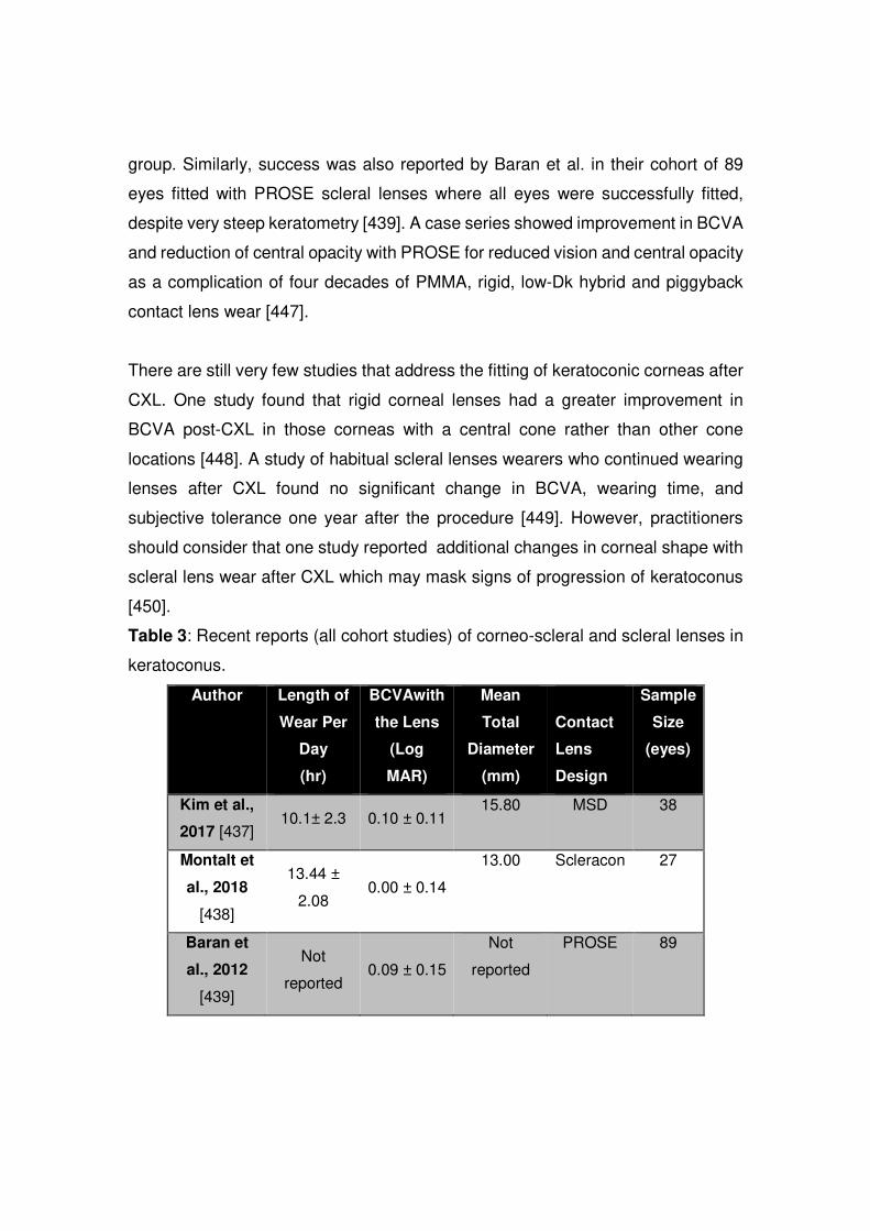
group. Similarly, success was also reported by Baran et al. in their cohort of 89
eyes fitted with PROSE scleral lenses where all eyes were successfully fitted,
despite very steep keratometry [439]. A case series showed improvement in BCVA
and reduction of central opacity with PROSE for reduced vision and central opacity
as a complication of four decades of PMMA, rigid, low-Dk hybrid and piggyback
contact lens wear [447].
There are still very few studies that address the fitting of keratoconic corneas after
CXL. One study found that rigid corneal lenses had a greater improvement in
BCVA post-CXL in those corneas with a central cone rather than other cone
locations [448]. A study of habitual scleral lenses wearers who continued wearing
lenses after CXL found no significant change in BCVA, wearing time, and
subjective tolerance one year after the procedure [449]. However, practitioners
should consider that one study reported additional changes in corneal shape with
scleral lens wear after CXL which may mask signs of progression of keratoconus
[450].
Table 3: Recent reports (all cohort studies) of corneo-scleral and scleral lenses in
keratoconus.
Author Length of
Wear Per
Day
(hr)
BCVAwith
the Lens
(Log
MAR)
Mean
Total
Diameter
(mm)
Contact
Lens
Design
Sample
Size
(eyes)
Kim et al.,
2017 [437] 10.1± 2.3 0.10 ± 0.11
15.80 MSD 38
Montalt et
al., 2018
[438]
13.44 ±
2.08 0.00 ± 0.14
13.00 Scleracon 27
Baran et
al., 2012
[439]
Not
reported 0.09 ± 0.15
Not
reported
PROSE 89

Arumugam
et al., 2014
[440]
Not
reported 0.23±0.30
19.15±0.56 PROSE 85
Ortenberg
et al., 2013
[441]
10.00 0.20±0.14
18.50 Microlens
Scleral
105
Lee et al.,
2013
[434]
Not
reported 0.079±0.10
Not
reported
PROSE 45
Suarez et
al., 2018
[442]
9.2±2.8 0.20±0.14
16.50 ICD 10
Fernández-
Velázquez,
2019
[443]
12.19±1.96 -0.02±0.10
15.00 ALEXA
ES
46
4.1.2 Pellucid Marginal Degeneration (PMD)
It is worth mentioning that the diagnostic differentiation of PMD from keratoconus
by topography is complex and clouds any analysis of treatment of either [451,452].
This might not be critical if dealing only with the overall refractive status of the eye
but might be more critical with the study of the role of intracorneal ring segments
(ICRS), and perhaps also the role of contact lenses.
Seventy-five eyes out of 85 eyes (88.2%) with PMD were managed non-surgically
with contact lenses (51.8%) or spectacles (36.4%) in a case series study [453].
These rates are comparable with a cohort study with 30 eyes, which found that
88.8% of the eyes were managed non-surgically [454]. A retrospective study of
patients fitted with a customised SiHy contact lens found comparable wearing time
and improvement in visual acuity with the lens when compared to the BSCVA in
patients with keratoconus and PMD [430].

Significant improvement in visual acuity was also seen with PROSE wear in a level
case series of 20 eyes in 12 of 19 patients with PMD [455]. In another case series
of 24 eyes in 12 patients with PMD in which half successfully wore the study lens,
concluded that a scleral contact lens with 16.5 or 17mm diameter can be used
successfully [456]. There is the conventional wisdom that a corneal rigid lens will
move to the steepest quadrant, and with inferior steepening characteristic of PMD.
Rigid corneal lenses will be unstable and fail. This recent evidence suggests that
scleral lenses are a useful option as alternative to penetrating keratoplasty which
may be considered higher risk due to likely need for a large, decentred graft.
4.1.3 Keratoglobus / Brittle Cornea Syndrome
Keratoglobus is a bilateral ectatic disorder of the cornea, principally characterised
by a globular protrusion of the cornea associated with diffuse thinning from limbus
to limbus [457]. Other ophthalmic features might include extreme corneal thinning,
irregular corneal astigmatism, high myopia, blue sclera, and retinal detachment
[458]. There are no specific studies or literature on contact lens fitting in cases of
keratoglobus. In one case series, two paediatric patients and two adults with
keratoglobus were fitted with PROSE scleral lenses with varying results, with one
patient developing hydrops and another patient having successful wear without
incident over a 3 year follow-up [459].
4.1.4 Irregular astigmatism from infection, trauma, dystrophy or
degeneration

Rigid corneal lenses can be considered for visual rehabilitation in scarred corneas
and with corneal opacities after trauma or infection [52,460–462]. More recently,
semi-scleral contact lenses provide excellent visual acuity and comfort in patients
with such irregular corneas [441,463]. Moreover, PROSE scleral lens treatment
had a positive impact on visual acuity for a wide range of corneal irregularities,
suggesting that PROSE lenses, and perhaps any scleral lens, may offer
improvements in visual acuity without surgical intervention [464]. However,
patients with lower endothelial cell counts or with Fuchs’ dystrophy were more
likely to fail treatment with PROSE [245].
4.2 Irregular astigmatism after to surgery
4.2.1 Penetrating keratoplasty (PK)
Refractive error that cannot be corrected via spectacles after PK is often
successfully corrected with contact lenses [465]. Indications for contact lens fitting
typically include visual rehabilitation for the correction of irregular astigmatism or
anisometropia following surgery [75,466,467]. In addition to improvement of visual
acuity, contrast sensitivity is also enhanced with use of rigid corneal lenses after
PK [468]. In patients with keratoconus that proceed to PK, between 31% and 56%
return to contact lens wear after surgery [469–471].
A variety of lens types have been fitted after PK for both visual rehabilitation and
therapeutic uses. There are many case series and case reports that document
successful visual and physiological results with all available lens types. These
reports are considered low level evidence, and many of them also appear to test
(and promote) a single lens design with no comparison group. Beginning with
hydrogels, these have been used for visual purposes [472] or as BSCLs [466,473].
Soft disposable contact lenses have been used to treat persistent epithelial
defects, wound leak, and dry eye after keratoplasty [473]. Hydrophilic lenses have
also been used as a corneal stent to realign the graft-host interface for cases of
partial wound dehiscence [474]. Rigid corneal lenses have a long track record of
use after PK for visual rehabilitation in traditional spherical or bitoric designs
[467,475], or larger diameter intralimbal lenses [469,476,477]. Success with hybrid
lenses has been described after PK with new [56,60] and older generation designs
[478–480]. Specifically, rigid corneal lens intolerant PK patients had an 80%
success when refitted with modern hybrid lens designs [56]. A case series with 20
patients who had undergone keratoplasty and were unable to achieve adequate
vision in spectacles were fitted with hybrid UltraHealth (SynergEyes, Inc.,
Carlsbad, CA, USA) contact lenses [60]. The authors found that all patients in their
study could be fitted with the hybrid contact lens, with the lens providing good
ocular comfort and BCVA of 0.05 logMAR or better in 80% of the subjects. Lastly,
scleral lens use as an option for visual rehabilitation after PK has dominated the
literature in the past 2 decades [481–488], with these studies documenting good
success after between 3 months and 9 years after fitting. Three of 31 patients
(~10%) fitted with scleral lenses post-PK discontinued due to discomfort over a
period of 3 years or more [486].
Prior to the widespread use of scleral lenses, various case series in the literature
documented fitting techniques and success rates specific to various rigid corneal
lens designs. These include the use of traditional spherical [489], intralimbal [477],
reverse geometry oblate [490,491], bispheric [492], keratoconic prolate, and bitoric
lenses [493,494]. Interestingly, very few fitting algorithms are found in the
literature. When photokeratoscopy and later videokeratography became available
in the late 1980s, several authors reported fitting success in post-PK eyes by
utilising specific topographic variables [495,496] or topographically-guided
software [497]. One paper described a tricurve intralimbal design fitting approach
to select the initial lens’ base curve by using the videokeratography values over
the elevated edges of the transplant wound [477].
Rigid corneal lenses generally do not disrupt corneal physiology, integrity or
topography even when worn for many years after PK. A comparison of the
endothelium of post-PK eyes fitted with rigid corneal lenses versus post-PK eyes
that never wore contact lenses did not demonstrate any significant decreases in

endothelial cell density of the corneal grafts in the contact lens wearing group three
years after fitting [498]. However, just as in CL wearers without a history of PK,
lenses made of oxygen impermeable materials (PMMA) induce polymegethism
and pleomorphism in PK eyes [499]. In a series of studies, corneal curvatures
derived from topography, and corneal indices such as symmetry, remained stable
after rigid corneal lens fitting [495,500,501]. In fact, well-centred rigid corneal
lenses can increase the regularity and decrease the asymmetry of grafts [496,502].
Scleral lens fitting is a popular modern form of visual correction after PK, although
scleral lenses have also been used to treat grafts with ocular surface disease such
as filamentary keratitis [322]. Just as with corneal lens designs, low level evidence
in the form of case series document successful lens fitting with mini-scleral designs
[481–485], sclerals [75,484,486,487,503], and custom designs with asymmetric
peripheries such as the PROSE lens [439].
Long term success with scleral lenses up to 9 years post-keratoplasty has been
documented [486], but when lens discontinuation occurs it is typically due to
difficulty with lens handling [484,485], economic considerations [485], or due to
endothelial dysfunction and/or decompensation and subsequent graft oedema
[245]. Interestingly, failure due to progression of ectasia at the graft-host interface
and discontinuation on that basis has not been reported; in a cohort of PROSE
patient fitted for ectasia including those with grafts, there was no eye that could not
be fit due to extent of ectasia [439]. Progression can be addressed with increase
of lens vault that is independent of haptic or optic base curves. Transient epithelial
macrocysts have been described secondary to graft oedema, anatomical
alterations within the cornea, and negative pressure behind the scleral lens;
however, the macrocysts do not lead to further complications or prevent successful
lens wear [284]. Scleral lens wear can induce graft oedema, perhaps related to
design and fitting parameters [504,505], particularly in eyes with low endothelial
cell count [245].

Microbial keratitis has been reported in contact lens users after PK but not at a
concerning rate, and there is no evidence that the risk of MK is higher in CL
wearers after PK than in other contact lens wearers. In fact, since most lenses
fitted after PK are manufactured in rigid materials, the rate of MK is expected to be
low, as rigid lenses have lower rate of infection compared to soft lenses [506,507].
Specific to post-PK scleral lens use, two case series reported instances of MK: 2
of 33 eyes in a series from Israel [486], and 1 of 27 eyes in a series from Brazil
[483]. Moreover, there are several singular case reports of rare and atypical
microbial infections in the post-PK cornea wearing a rigid lens, suggesting the lens
was the vector for transmission [508–510]. See section 2.8.2. for information on
PK eyes fitted with early generation BSCL in the early post-operative phase.
The older literature documents significant corneal neovascularisation with low Dk
hydrogel lenses for aphakia and/or overnight wear after PK [511–514] or in with
lower Dk rigid corneal lens wear [500]. Although graft rejection has been
documented during both corneal [471] and scleral lens wear [484,486], allograft
rejections are usually not attributed to the use of the contact lenses. Rejection and
loss of endothelial reserve may preclude future contact lens wear [471,515,516],
especially prior to the development of higher Dk materials. Rarely other adverse
events of contact lens use can occur in post-PK eyes, such as inadvertently
inducing complications such as wound dehiscence after rigid corneal lens removal
via plunger (reported in one series of 3 patients) [517].
Similar considerations for lens use are relevant in other forms of corneal
transplantation. Scleral lenses are also successfully fitted after deep anterior
lamellar keratoplasty [485] and typically follow the same successes and barriers,
with the exception of endothelial dysfunction in corneal transplant involving a donor
endothelium.

4.2.2 LASIK / PRK
Contact lens options after LASIK and PRK range from soft hydrophilic lenses to
corneoscleral lenses to highly customized scleral lenses [518,519]. Soft lenses
are typically used when residual ametropia remains after the surgical procedure
[520,521] or as BSCLs (see Sections 2.1.1 and 2.1.2).
Although rigid corneal lenses can be used to correct residual ametropia [521], the
most common indication for fitting rigid lenses after PRK or LASIK is for the
correction of optical aberrations and irregular astigmatism, which may or may not
be related to ectasia. Most studies in the literature contribute low level evidence
case reports and series. Although two case reports document successful use of a
soft spherical [522] or toric lens [523] for post-LASIK ectasia, rigid corneal lenses
are typically used since they decrease higher order aberrations in patients with
visual complaints and irregular astigmatism [524–529].
The types of rigid corneal lenses fitted after LASIK and PRK range from simple
tricurve designs to complex reverse geometry designs. Traditional and aspheric
lens designs [530–532] as well as specialty lenses, often reverse geometry
designs, are successful for ectasia and complications after LASIK, PRK and radial
keratotomy [490,533–537]. For myopic photoablative procedures, the post-
operative oblate shape produces a more difficult corneal contour to fit, since
traditional rigid corneal lens nomograms for base curve selection and peripheral
curve systems may not be appropriate. Some have advocated reverse geometry
lens designs [490,524,528,534] although use of although successful use of same
or only minimally flatter (0.1-0.2mm) aspheric rigid corneal lens than preoperatively
has been reported with good centration attributed to the negative pressures
created by the bearing of the periphery and excess central apical clearance [530].
Lathe cut SiHy lenses have been used in post-LASIK ectasia with good results in
a case report [538] and limited case series [539]. New generation hybrid lenses
have been successfully fitted after failed LASIK in case reports [540] and limited
case series [56]. Additionally, there are several case reports for successful

corneoscleral [541] and scleral [542,543] lens fitting for post-LASIK ectasia. In
larger case series, corneoscleral lenses have shown good success in fitting post-
LASIK ectasia after 1 year of follow-up [518,544] and one series demonstrated that
corneal biomechanical parameters increased after 1 year of corneoscleral lens use
without adverse clinical effects [545].
Rigid corneal lenses can be used to improve visual acuity and reduce higher order
aberrations after unsatisfactory refractive surgery outcomes, an effect that persists
after the lens is removed due to moulding effect [546]. The same centre found that
miniscleral lenses can reduce persistent optical symptoms in patients with good
uncorrected acuity after corneal refractive surgery by reducing post-operative
spherical aberration [547]. Full scleral lenses, including the custom PROSE
[464,519] and the EyePrint Pro [284], have also been effective to treat post-
refractive surgery complications including ectasia, optical aberrations, dry eye and
neuralgia.
Several papers have been published on base curve selection using topographic
data for fitting rigid corneal lens designs after LASIK and PRK [548–551]. For
example, the power on the transition zone of the corneal topographic map, 0.2 mm
outside the ablated refractive area, facilitated selection of the back-optic zone
radius of a well fitted rigid cornea lens after LASIK [549]. Additionally, comparing
axial and tangential anterior curvature maps, the average axial curvature 4.0 mm
from the vertex normal or 2.0 mm from the vertex normal on tangential maps were
best predictors of accurate spherical rigid corneal lens base curve selection after
LASIK [548].
There are two case reports describing adverse effects with contact lenses after
LASIK or PRK. Late-onset interface inflammation was reported after a patient slept
in a cosmetic soft lens 18 months after LASIK [552] and crystalline keratopathy
was reported in a corneoscleral lens wearer several years after LASIK [553].

4.2.3 Radial keratotomy
Radial keratotomy and astigmatic keratotomy are rarely performed today as stand-
alone procedures for myopia and astigmatism, but they are still used in conjunction
with other refractive and surgical techniques. Nonetheless, many patients that had
radial keratotomy performed in the past 30-40 years still exist in the contact lens
practice, as many of these patients are now presbyopic and suffer from over-
correction [554], hyperopic drift [555], and diurnal variations [556] since their
original procedure. From the Prospective Evaluation of Radial Keratotomy study,
58% of patients felt some type of optical correction was required 10 years
postoperatively; 23% of patients had overcorrections or induced hyperopia greater
than 1 dioptre, and 17% had under corrections or residual myopia greater than 1
dioptre [557].
Similar to the PK and LASIK/PRK fitting options above, the literature (mostly in the
form of case series) documents a wide range of CL fitting options after radial
keratotomy [558–560], which are considered more challenging to fit compared to
nonsurgical eyes [561,562]. Soft lenses range from traditional daily disposable
hydrogels [563] to conventional soft lenses [558,559]. Single case reports have
described a high Dk lens compensating for diurnal fluctuations [556] or a prosthetic
soft lens fitted after ruptured radial keratotomy incisions with iris damage [564].
Successful use of hybrid lenses has also been described in case series [559]
Rigid corneal lens use includes reverse geometry lenses [490,558,565], which are
often orthokeratology-designed lenses but used for daily wear to better match the
post-radial keratotomy oblate contour [546,566,567], or plateau designs fitted soon
after radial keratotomy to manipulate the healing process and influence the
refractive results [568]. Traditional prolate designs have also been used
extensively [525,559] with various fitting algorithms proposed. Some early case
series proposed fitting based on pre-operative keratometry readings [554]. In other
series, empirical lens fitting based on flat post-operative keratometry readings has
worked well, although the base curves of the final lenses were usually steeper than

the post-operative flat keratometry [569]. Using corneal topography, efficient base
curve selection has been reported by selection of the lens base curve equal to the
value 3.5-4.0 mm superior to the visual axis on an axial map [570]. When traditional
corneal lenses are fitted after RK, or any myopic photoablation procedure which
creates an oblate cornea, the contact lens vaults the flatter central cornea resulting
in a plus powered lacrimal lens which must be compensated for with additional
minus power in the lens [571]. Rigid corneal lenses also have a moulding effect
that alters corneal power [572,573] and can improve vision temporarily after lens
removal [546].
Successful scleral lenses use of various types has also been described [284,574–
576]. Scleral lenses are currently the most popular option in the recent literature.
Historical complications associated with contact lens use after radial keratotomy
that have been reported in the literature most often include superficial corneal
neovascularisation along the incisions with low Dk soft lens wear [554,577]
especially when the incisions reach the limbus [578].
4.2.4 Intra-corneal ring segments
Intra-corneal ring segments (ICRS) are flexible, crescent-shaped rings of
polymethyl methacrylate inserted intrastromally into the peripheral cornea. The
goal is to reduce refractive error by physically changing and flattening the shape
of the cornea, and reduce corneal astigmatism. The US FDA first approved
INTACS (Addition Technology Inc.) intracorneal rings in 1999 to offer an
alternative to contact lenses [579] and the use of ICRS to treat keratoconus and
contact lens intolerance was first reported in 2000 [580,581].
Since then, numerous reports confirm that intrastromal corneal rings improve the
tolerance of contact lenses in keratoconus [582–587], although one study found
only a minor improvement [588]. Contact lenses are more straightforward to fit to
a flatter and more regular cornea [198,588,589].

In 2007, the UK National Institute for Health and Clinical Excellence completed an
overview of the insertion of ICRS for corneal disease, in particular keratoconus
[198]. All patients who were contact lens-intolerant at baseline were able to use a
contact lens after surgery [585], a finding confirmed by the Medical Advisory
Secretariat [590].
Contact lenses are frequently required to achieve BCVA after the insertion of
ICRS, especially with advanced keratoconus [199,583,584,587,591–594]. Various
lens types and designs have been described for this indication, and various contact
lens modalities have been found to be well tolerated after surgery, including
disposable soft lenses, toric soft lenses [595], custom-made soft lenses
[583,592,593,596,597], rigid corneal lenses [584,596], piggy-back lenses
[587,591,592,596,598], corneo-scleral lenses [599,600] and scleral lenses
[542,594,601], which were reported to be useful after other lens types had failed
[601].
Contact lenses can improve visual function after implantation of ICRS by reducing
higher order corneal aberrations including piggyback [587]; soft [597]; corneo-
scleral [599]; and scleral designs [589,594].
Most reports of contact lens after ICRS are of a small number of cases with short
follow-up. Low rates of injection and of staining from lens wear over ICRS are
reported [593]; Montalt et al. report no adverse effects in 27 eyes of 27 patients
with keratoconus fitted with corneo-scleral lenses after ICRS implantation and
followed for 1 year [599]. Severe complications, including corneal
neovascularisation and fungal infection (Fusarium sp.) have been reported [199],
but there has been no systematic study or report on the issue. One study looked
at the biomechanical impact of 1 year of corneo-scleral lens wear in normal
patients, and in keratoconic patients with and without ICRS and found no
statistically significant effects on corneal viscoelastic properties in any of the
groups [600].
Scleral lenses were tolerated by patients who could not wear other types of contact
lenses after ICRS [601]. PRK can also be performed after INTACS to reduce the
residual refractive error or reduce the astigmatism enough to improve tolerance
[197].
4.2.5 High refractive error
The first reference to fitting contact lenses for high myopia was in 1889 by August
Müller of Kiel in Germany who corrected his own 14 D of myopia with a scleral
lens. Widespread adoption of corneal contact lenses yielded numerous reports of
correction of high ametropia [602–605]. Generally, papers referring to high
hypermetropia and astigmatism are generally incorporated into those for general
ametropia or aphakia [488,602,604,606,607].
In patients with high astigmatism, empirical fitting of both rigid and soft lenses can
provide equally good results which equate to the acuity of spectacles [608], with
more recent reports that rigid corneal lenses improve vision compared to
spectacles [609,610].
Fonda (1974) noted that Bennett (1963), quoting the calculations by J.L. Francis,
showed that a contact lens gives 24% more magnification than a -20D spectacle
lens at a back vertex distance of 12 mm [611]. Contact lenses therefore provide
better acuity than spectacles for highly myopic refractive errors [612]. The reverse
is true for high hypermetropes or aphakes. Also, more accommodation is required
by myopes and less by hypermetropes when they transfer from spectacles to
contact lenses [613]. Contact lenses also provide better contrast sensitivity than
spectacles in higher prescriptions [614]. A high success rate was reported for rigid
corneal lenses prescribed to be worn overnight for up to one month for high
refractive errors, including myopia, hypermetropia and astigmatism [606].
Good results including stereopsis can be achieved treating myopic anisometropic
amblyopia with contact lenses [604,605,615,616] with aniseikonia not a problem
[617–619]. One study reported amblyopia only improved in eyes with a refractive
error up to 9 dioptres [620].
Scleral lenses can be useful for high myopes and also high astigmats, in particular
those with high astigmatism resulting from tilted grafts [488].
Rigid corneal contact lens correction of myopic refractive error, particular higher
refractive error, may be associated with development of ptosis. The degree of
ptosis was found to increase with higher degrees of myopia and longer duration of
rigid corneal contact lens wear [621]. Ptosis may arise as an issue in unilateral
wear or when there is high anisometropia.
When manufacturing high minus rigid corneal lenses, thinning the lenticular
junction prevents excessive movement on blink [622]. The correction of soft CL
spherical aberration is not beneficial to the majority of patients in practice [623].
See CLEAR Orthokeratology Report regarding the use of orthokeratology to slow
myopia progression and reduce refractive error in high myopia [624].
4.2.6 Aphakia
Contact lenses have been the correction of choice for aphakia for many years.
Starting in the 1940s, there were trials to correct aphakia with contact lenses,
especially unilateral aphakia [625–628]. Continued reports have appeared in the
literature over the decades and a review of aphakic lens fitting was written in 1979
[629]. Since aphakia is much less common now that intraocular lenses are the
norm, most aphakic contact lenses required are for paediatric aphakia, aphakia
following trauma, or ectopia lentis.
Over the years, rigid [606,630–633], soft [629,634–639], and scleral [488] lenses
have been used successfully to correct aphakia.

Aphakic lens designs present certain challenges due to the large hyperopic
refractive error being corrected. High plus power mandates high central thickness,
which for any material, reduces oxygen transmission and increases theoretical risk
of hypoxic complications such as neovascularisation, endothelial decompensation,
and infection. High power lenses have mechanical issues related to their weight
and to lid interactions as well as optical issues related to spherical aberration. The
visual performance of two aspheric designs of soft aphakic lenses was compared
to a spherical design, and the better of the two aspheric designs was assessed for
mechanical and visual performance as well as physiological acceptance. Visual
performance of all three lens types showed no significant difference and the
mechanical performance of the aspheric lenses behaved more like low power
lenses due to the reduced bulk and smooth profile of the lenses [637].
In 1967, a large rigid lens, the Apex lens, was designed to provide mechanical
stability [630], and is the basis for more recent lens designs. More recently, a mini-
scleral lens design has been successfully fitted for severe corneal irregularity and
aphakia after trauma [640].
In the case of unilateral aphakia, aniseikonia can be a limitation to spectacle wear.
For example, +20.00 D spectacle lens produces approximately 30% more
magnification than a contact lens [613]. Dallos scleral lenses have been fitted in a
group of unilaterally aphakic patients in an attempt to achieve binocular vision
[626]. Unilateral aphakes fitted with PMMA contact lenses [641] and PMMA, soft
and overnight wear soft lenses [642] were found to have gross binocular vision
with some degree of aniseikonia.
Eighty percent of contact lens wearing unilateral aphakes demonstrated
suppression in the aphakic eye [643]. A group of 24 patients with at least 2 years
of disrupted binocular function after unilateral traumatic cataract or unilateral
traumatic cataract followed by uncorrected aphakia had intractable diplopia when
the cataract was removed and the aphakia corrected with a contact or intraocular
lens [644].
Hydrogel contact lenses have been fitted as an occlusive lens in the unoperated
eye of unilateral aphakes in an attempt to treat dense amblyopia, either using a
lens with a black opaque centre [645] or a high-plus lens [646]. Success was
measured as the time the lens was worn, and four of 13 patients (nine of whom
were aphakic) successfully wore the lens between 26 and 60 months, however,
visual acuity was not measured as an outcome. Good compliance was achieved
with 6/10. Additionally, diplopia was found to be a problem when only one eye was
aphakic [644,647].
Traumatic aphakia is frequently accompanied by corneal irregularity and scarring,
therefore rigid lenses provide the best chance of visual rehabilitation, despite
comfort sometimes being an issue with monocular use of rigid lenses [631].
However, soft lenses can be fitted initially followed by refitting with rigid corneal
lenses if adequate vision cannot be achieved with soft lenses [647]. Scleral lenses
were a safe and effective option to improve vision with reasonable comfort [640].
In cases of traumatic aphakia with additional complaints of photophobia from
traumatic aniridia, rigid corneal lenses made of fluorosilicone acrylate with a dark
brown tint have been fitted with success and decreased photophobia [632].
Although initial reports on contact lenses for traumatic aphakia in children reported
disappointing results [648], rigid lenses were used in unilateral traumatic aphakes
with good results in children over 7 years old [649]. Subsequent to that, intraocular
lenses produced better outcomes than contact lenses in traumatic aphakia [650].
Scleral lenses, bitoric corneal lenses, multifocal soft lenses, and hybrid lenses can
also be used [651,652].
In some instances of ocular trauma, a combined procedure of cataract extraction
and PK may be necessary. In these cases, the patients require a contact lens if

they do not have an intraocular lens inserted at the time of surgery. Overnight
wear aphakic soft lenses have been used, but it was noted that this modality was
frequently problematic and it was advised that sutures should be removed before
fitting the lenses [513]. However, another report found the same lens modality gave
successful results [512]. Rigid corneal lenses worn on an overnight wear basis
may be useful for patients who were unsuccessful with soft lenses [653].
Other conditions that result in aphakia include patients with ectopia lentis and
Marfan syndrome, which provide unique challenges when fitting contact lenses.
Corneas are much flatter than average [654–656] and a larger than average
horizontal visible iris diameter is common, along with a high level of myopia and
astigmatism [657]. In one study, seven different contact lens modalities were used
to fit patients with ectopia lentis; the authors noted that this group of patients
included those who had not had lensectomy [658], and that aphakic contact lenses
could be used if the native lens was sufficiently dislocated to create an aphakic
axis within the pupil.
Contact lenses can be used to correct aphakic refractive error in patients with
ectopia lentis, as modern methods of cataract extraction and IOL fixation in
absence of capsular support have yet to be demonstrated as safe over decades
of use and are associated with complications [659–661]. Improvement of BCVA
with a contact lens has been reported to improve over one or more years [662].
The issue of overnight wear versus daily wear of contact lenses for aphakia has
been given substantial consideration, including patient experience and risk of
complications, as many patients are elderly and face challenges with daily lens
insertion, removal, and care. Overnight wear was considered to give the best
experience for aphakic patients using both rigid [606,636,638,663–665] and
silicone elastomer lenses [666]. However, there was a high risk of problems and
adverse events including MK and vision loss [636,638,663,665]. Low numbers of
complications were found in daily wear aphakic rigid corneal lens wearers, which
included corneal punctate staining and contact lens-papillary conjunctivitis
[631,632].
Only half (21/40) of aphakes fitted with overnight wear lenses were successful,
with the risk of serious complication being six times greater compared to daily wear
lenses (55% compared to 8.8%) [667]. Serious complications included MK, tight
fit syndrome, corneal erosions, neovascularisation >2 mm, stromal oedema, and
blepharoconjunctivitis [667]. In one study, corneal oedema was a problem with
overnight wear, and three out of 150 patients lost their sight as a result of MK and
hypopyon [629]. In a review of 100 aphakes fitted with overnight wear soft lenses,
four patients developed major complications, three of whom were diabetic [668].
The authors advised that special consideration should be given when fitting
diabetics with overnight wear aphakic lenses. Because of the higher risks in
overnight wear, daily wear is preferable [669]. Higher rates of complications were
found in overnight wear hydrophilic lens wearers [638,670], while there were less
problems from daily wear rigid and soft lenses [647]. An examination of the
endothelium of a group of unilateral aphakes noted that overnight wear of both
rigid corneal and hydrogel lenses had a deleterious effect, presumably due to
chronic hypoxia [669]. However, silicone elastomer contact lenses produced no
effect on the corneal endothelium in a unilateral lens wearing group [666]. This
was similar to earlier findings by Schoessler et al. for patients wearing lenses in
both eyes [671].
4.2.7 Paediatric aphakia
Using hard lenses to fit babies after surgery for congenital cataracts was first
mentioned in 1959 [672], with numerous other reports to follow [673–676].
Numerous reports of the use of rigid corneal lenses in young aphakes followed
[677–681] as well as the use of scleral lenses [236,682]. Paediatric aphakic
patients have been successfully fitted with soft lenses, in overnight and daily wear
[679,683]. A silicone rubber material was reported to give sufficient oxygen for

overnight wear [684], and numerous reports of the use of Silsoft lenses for this
indication followed [651,677,681,685–689].
In a cohort of 240 eyes in 184 patients, 22% of the 112 eyes fit with Silsoft lenses
abandoned lens wear for a variety of reasons, with “dense amblyopia or
retinoblastoma” reported as the most common reason [685]. No patients had
contact lens complications with permanent visual sequelae. Recent evidence
shows no visual advantage of IOLs over contact lens correction for infants and
toddlers, with contact lenses yielding fewer adverse events such as visual axis
opacities [690–694]. A report of corneal changes in children after unilateral
cataract surgery in the Infant Aphakia Treatment Study (IATS) found endothelial
cell density and central corneal thickness were less favourable for infants treated
with aphakic contact lenses compared to those treated with intraocular lenses at
the 5-year outcome examination, [695] but the long term implications remain
unknown. In a secondary analysis, the IATS found that children who wore contact
lenses for a larger amount of their waking hours in a day throughout the study
period of 5 years had better visual outcomes, even after accounting for patching
adherence [696].
As far as choice of lens type, overnight wear silicone elastomer lenses have been
fitted up to ages 3-4 years and replaced with daily wear high water content lenses
thereafter [687] with report of15% of eyes with complications, all without sequelae.
Better vision with rigid corneal lenses compared to Silsoft lenses was reported in
a cohort of patients with unilateral infant aphakia followed to 5 years of age [697].
In a study comparing overnight wear of silicone elastomer lenses (mostly 7-21
nights) with daily use of rigid corneal lenses, visual acuity data at 1 and 5 years of
age failed to provide convincing evidence regarding which approach is the best
option for managing infantile aphakia [697]. There was a trend toward better BCVA
in the rigid corneal lens group, but this did not reach statistical significance,
possibly due to small sample size of 57 infants. There is medium quality evidence
that there are fewer adverse ocular events with the rigid corneal lenses, but more

lenses are lost. In one study, 22% had CL related adverse events with overnight
wear Silsoft lenses, which was supported by the same percentage of patients in
another study, who also developed adverse events, including corneal abrasion,
bacterial keratitis and corneal opacity [686,697]. All but one wore overnight wear
silicone elastomer lenses, and all resolved without sequelae. In the IATS group,
18% had adverse events, all of which resolved when the lenses were removed and
treated with topical antibiotics [692]. Silsoft lenses were fitted and replaced at 3-
monthly intervals in one study [687] that reported few complications below 3 years
old, although 4/26 eyes (15%) had adverse events. It is worthwhile to note that
approximately 6 lens changes were needed during the first year in a report on
contact lens correction of unilateral nontraumatic paediatric aphakes [679].
Another study reported an average of approximately 9 lens changes and 21 visits
in the first year in their practice, favouring the use of customisable soft hydrophilic
lenses. Frequent lens loss, frequent need for replacement, and frequent visits
represent a substantial commitment for all parties. An evidence based medicine
report by the American Academy of Ophthalmology [681] reported that there was
limited quality and amount of evidence that showed that silicone elastomer and
rigid corneal lenses were effective for treating aphakia in children. They reported
that data show that silicone elastomer lenses are easier to fit and have the wear
and care advantage that they may be worn on an overnight wear basis. Their data
found that although rigid corneal lenses must be removed every night and require
a more customised fit, they are associated with fewer adverse events. Others
report no cases of adverse events with daily wear rigid corneal lenses and
[677,698,699]. A report on 16 eyes wearing rigid corneal lenses on a 1-week
overnight wear basis found no observable increases in the rate of microbial
infection [700]. Silicone elastomer is used frequently in the US but less commonly
in other countries. Custom lathe cut SiHy lenses are an option elsewhere [651].
Overnight wear contact lenses were poorly tolerated in children with unilateral
aphakia after radiation and cataract surgery in the setting of orbital
rhabdomyosarcoma [701]. Special fitting and power considerations are needed
for the small eyes of infants following congenital cataract surgery as they have

small, steep corneas [702]. Because it is not always possible to take keratometry
measurements, the first lens used is fitted empirically according to expected
corneal radius for the patient’s age [703,704]; there is a rapid rate of corneal
flattening during the first 18 months of life [651]. Corneal radius of a neonate is
between 48.50 D (6.96 mm) and 47.00 D (7.18 mm) and corneal diameter is
approximately 10mm; although, in one study of congenital cataracts, 57% were
found to have microcornea [705–708]. Contact lenses were generally fitted within
one month of surgery for a wide range of ages [677,679,685,686,688,700,703].
Hydrogel lenses with radii of 7.00 to 7.60 mm in infants and of 7.80 to 8.10 mm in
children two years and older have been fitted [683]. Silsoft lenses were fitted with
a 7.50 mm back-optic zone radius in patients up to the age of 1.5 years old, and
by the age of 4 years, a back-optic zone radius of 7.90 mm was needed [687].
Lenses of diameter 7.50 mm or 7.70 mm have been fitted for the first 6 months,
followed by flatter lenses of 7.7m0 m from 6-18 months, flattening further to 7.70
or 7.90mm for children over 18 months old [709]. In the IATS, 84% of patients
were initially fitted with a 7.50 mm radius [697]. Rigid corneal lenses were fitted
steeper than flattest K readings to ‘give a grip’ on small corneas [678]. The average
radius was 46.75 D (7.20 mm). Saltarelli fitted Dyna Z Intralimbal lenses with an
average base curve of 7.9 to 7.5 mm [700]. In the IATS study, the mean radius of
rigid corneal lenses was 47.62 D (7.08 mm) up to the age of 12 months and
flattened to 44.31 D (7.60 mm) by age 5 years [697].
Silicone elastomer lenses (Silsoft, Bausch & Lomb) are available in an 11.3mm
diameter, used to fit young infants [710] and in 12.5mm for older children who need
lower powers. Rigid corneal lenses are fitted as an intralimbal or paralimbal fitting.
Complete corneal coverage is recommended to give better stability, with a
diameter of 10.8-11.2 mm depending on corneal diameter [651]. Larger diameters
can be fitted as can a range of small diameters, as small as 7.5-9.5 mm depending
on corneal diameter. The mean diameter fitted in the IATS cohort was 9.4 mm
[678,697,700].For soft and SiHy contact lenses, the total diameters should be 2.5-
3.0 mm larger than the horizontal visible iris diameter for normal size eyes

[651,704] and 11-12 mm for microphthalmic eyes. Some microphthalmic eyes
have been fitted with lenses of 10.5 mm or smaller diameter [651,704].
The power of aphakic lenses for infants and young children is much higher than in
adults because the eyes are smaller with steeper corneas and shorter axial
lengths. Mean lens powers used when fitting aphakic infants range from +21.36 to
+29.60 D [677,683,686–689,699,711]. Lens power is estimated as this cannot be
obtained directly from refraction in paediatric patients and can be significantly
miscalculated with aphakic spectacle correction [689]. Some literature presents
guidelines for calculating lens power either through using an equation or
referencing a table of expected values [712–714].
Because infants generally look at close objects such as faces, food, and toys, the
power is increased by 2-4 dioptres to provide a near focus to preclude refractive
amblyopia [651,679,686,709].
5 Tinted, opaque, or prosthetic lenses
Contact lenses can be used to modify or enhance the appearance of an eye and
improve vision in certain eye conditions such as in aniridia, trauma, diplopia,
albinism or retinal disease. Depending on the tint applied, a contact lens can be
translucent, semi opaque or completely opaque across all or part of the lens
diameter.
• A Cosmetic Tinted Lens can be defined as a lens that is designed to beautify
the appearance of a healthy eye, frequently used to create an effect of
enlarging the iris and changing iris colour. These lenses are considered a
fashion accessory [715].
• A Therapeutic Tinted Lens is used to treat an ocular disease or defect. They
can be prescribed to reduce glare, photophobia, enhance colour vision and
for occlusion therapy in amblyopia [715].

• A Prosthetic Tinted Lens is designed to improve cosmesis of an otherwise
cosmetically abnormal eye. These lenses are typically used in congenital
abnormalities, disfiguring disease, and penetrating trauma [715].
5.1 For photophobia (albinism, migraine, etc)
Centrally tinted soft contact lenses are available in a range of colours, tint densities
and pupil options (Ultravision, Cantor and Nissel) and can be used to reduce
photophobia in patients with various retinal conditions such as in retinitis
pigmentosa, cone dystrophies, and albinism. Case reports and case series show
a marked reduction in photophobia and improved daily QOL in adults and in
children [716–722].
5.2 For glare (mydriasis, traumatic aniridia, coloboma etc)
Therapeutic tinted lens can be used to resolve glare for patients with pupils that
are nonreactive or suffered traumatic injury [723]. In cases of traumatic aphakia
with additional complaints of photophobia from traumatic aniridia, rigid corneal
lenses made of fluorosilicone acrylate with a dark brown tint have been fitted [632].
Partially occlusive lenses with a clear pupil can offer relief to patients who complain
of glare after peripheral iridotomy.
5.3 Colour-blindness
The use of red tinted lenses to enhance colour vision in patients with colour-
blindness has been reported as early as 1988 [724]. Various contact lens systems
such as X-Chrom lens or ChromaGen are available. Subjects with congenital
colour vision deficiency wearing two types of tinted contact lenses (light red and
dark red tints) improved on Ishihara plates, although the Farnsworth 100 hue test
did not show any improvement with either lens [725]. An improvement on Ishihara
plates with the ChromaGen contact lens, especially in deutan subjects, has been
reported but again there was no significant effect on the Farnsworth lantern test
performance. Subjects also reported enhanced colour perception with ChromaGen
lenses, although significant difficulties with vision in dim light were apparent [726].

Despite widespread use of tinted contact lenses, there is still little evidence that
supports their efficacy in enhancing colour perception (see CLEAR Future Report)
[341].
5.4 Disfiguring disease
Visible abnormalities of the anterior ocular structures can be masked using printed,
dyed or iris-painted prosthetic contact lenses (Ultravision, Cantor and Nissel). A
retrospective analysis of 42 patients (48 eyes) for whom an iris-painted soft contact
lens was prescribed reported that three tint patterns were used depending on the
therapeutic purpose [727]. All tint patterns prescribed had the same tinted iris
structure but differed in pupil type, either containing a solid black pupil, no pupil, or
a clear pupil. The authors reported an improvement in BCVA in those patients
wearing a clear-pupil prosthetic soft CL. However, 40% of wearers experienced
complications, with the incidence higher in patients wearing the solid-pupil type.
This may be because such patients had greater disturbance to the corneal barrier
function and endothelial reserve at baseline and are thus more susceptible and/or
that they were less sensitive to early symptoms of hypoxia or infection because of
reduced vision. High satisfaction has been reported in 25 of 33 patients fitted with
hand-painted cosmetic lenses for cosmetic purposes, finding a 76% satisfaction
regarding lens comfort and 88% satisfaction regarding lens colour and appearance
[728]. The diameter of most soft lenses are approximately 15 mm up to 22 mm
(Cantor and Nissel). Hand painted scleral shells, made by taking ocular
impressions, can be used with good cosmetic success, where the eye has become
phthisical.
5.5 For amblyopia treatment
Opaque tinted contact lenses can be a potential alternative to amblyopia occlusive
treatment in those patients who were patch-intolerant and failed with conventional
therapies and in those with intractable diplopia. Several case series have reported
the efficacy of using overnight and daily wear opaque contact lenses to treat

amblyopia in children and adults, suggesting that this approach is a useful
alternative to improve compliance in occlusion therapy [645,729–733]. High-
positive contact lenses for optical penalisation of the unoperated eye have also
been used to treat strabismic amblyopia in unilateral aphakes [645,646]. One
study in normally sighted patients suggests that modifying the size of the opaque
pupil can allow different degrees of penalisation while potentially leaving peripheral
fusion intact [734].
6 Nystagmus
The use of rigid corneal and soft contact lenses has been recommended over
spectacles in nystagmus, however, published data is scarce and equivocal. Early
studies have reported that contact lenses dampen ocular oscillation in nystagmus
(presumably because they provide tactile feedback about the eye motion to the
ocular motor system) and provide better visual acuity than spectacles, particularly
in infantile nystagmus [735–738].
Some studies report improvements in visual acuity and eye movement and others
show no changes or variable results [739–743]. These studies are mainly
retrospective and small case series or reports. More recently, a randomised
controlled trial assessed the use of rigid corneal and soft contact lenses in infantile
nystagmus, reporting that neither lens type reduced nystagmus or improved best
corrected visual acuity as compared to wearing spectacles [744].
A short term rebound phenomenon was reported in a 20-year-old female
nystagmus patient who was fitted with a contact lens for a 90-minute trial session
[745]. Upon removal of the contact lens, the patient experienced a short-term
rebound phenomenon that included oscillopsia and dizziness. The authors
hypothesised that relieving the dampening effect of contact lenses on congenital
nystagmus might have induced the rebound phenomenon consisting of a transient
increase in nystagmus intensity and/or changing the foveation period.

There is a lack of good-quality evidence studies in this field. Well-designed trials
with standardised methodology are needed to establish the effectiveness of
contact lenses in infantile nystagmus.
6.5 Ptosis
There is low quality evidence that scleral lenses can be useful in the management
of ptosis of various aetiology, including ocular myopathy, complicated ptosis due
to long term rigid corneal lens wear, phthisis bulbi, and myopathy from Kearns-
Sayre syndrome [746,747]. The scleral lenses improved cosmesis, increased both
the palpebral aperture and marginal reflex distance, and provided visual correction
successfully for all patients [747].
7 Medical use of contact lenses with prophylactic antibiotics
Due to the potential risk of MK associated with overnight wear of therapeutic
contact lenses, many practitioners prefer disposable contact lenses with high
oxygen transmissibility materials, such as SiHy materials, along with prophylactic
antibiotics (preferably preservative free). However, there is not strong evidence
that directly addresses and supports this practice. A survey of 52 consultant
ophthalmologists found that only 42.3% prescribe prophylactic antibiotics when
they use bandage contact lenses [400]
While there are reports of MK despite the use of prophylactic antibiotics which
might in turn cause toxicity and other complications, some authors believe the
reported benefits surpass the reported complications
[20,79,147,152,293,301,302,308].
Some studies do not report any episodes of MK with therapeutic contact lenses
for various ocular surface disorders with or without prophylactic antibiotics
[31,140,225,226,321]. Other studies show that the use of prophylactic antibiotics
did not eliminate the risk of MK in patients treated with therapeutic SiHy contact
lenses [139,399]. One study found an increased risk of MK when antibiotics were
used in association with therapeutic hydrogel lens and steroid for bullous
keratopathy [398] (See section 3.5). It is conceivable that prolonged prophylactic
antibiotic use during bandage contact lens wear could potentially lead to increased
antibiotic resistance in pathogenic and changes to the microbiota of the eye, which
may be of importance in the protection of the ocular surface.
When antibiotic prophylaxis is used concurrently with therapeutic lenses, the agent
is sometimes rotated regularly to decrease the risk for development of microbial
resistance, e.g. moxifloxacin 0.5%, followed by tobramycin 0.3%, followed by
polymyxin B-trimethoprim for 1 month or ofloxacin 0.3% to sodium sulfacetamide
10% to levofloxacin 0.5% to polymyxin/trimethoprim [328,748].
There is substantial literature on the role of prophylactic antibiotics with therapeutic
contact lens with keratoprostheses (see section 3.1.4), a clinical situation that
warrants special consideration.
In a retrospective case study of patients with refractory persistent epithelial defects
fitted with overnight wear scleral lenses with various prophylactic antibiotic
regimens or none, MK occurred in 4 of 14 eyes analysed [147]. A more recent
retrospective case series reported no cases of microbial keratitis in 20 eyes with
persistent epithelial defects with a regimen incorporating overnight wear of scleral
lenses with daily removal for cleaning and the addition of a preservative free fourth-
generation fluoroquinolone antibiotic drop to the lens reservoir at time of insertion
[148].
Most studies are inadequately powered and of insufficient quality to provide firm
evidence to inform the development of management guidelines. Well-designed,
masked, randomised, controlled trials are needed to establish the benefits of the
use of prophylactic antibiotics treatment regimens along with therapeutic contact
lenses.
8 Medical use of contact lens during therapy with other agents
This section summarises the in vivo reports describing the concurrent use of
topical therapeutic agents with contact lenses. Contact lenses for drug delivery are
under development and this is covered in the CLEAR Future Report [341].
8.1 Corticosteroids and NSAIDs
There is some concern about concurrent use of contact lenses with topical
corticosteroids or NSAIDs because in vitro data suggest that topical corticosteroids
and NSAIDs cause corneal toxicity [749], which could exacerbated by contact
lenses because lenses may increase the retention time of the drugs. Only one
study has investigated the combination of 1% prednisolone acetate and hydrogel
contact lenses in rabbits [750]. In this study, lenses were soaked in 1%
prednisolone acetate for 2 minutes prior to application. Eyes of the treated animals
were compared to animals that received 1% prednisolone acetate drops without
contact lenses. The cornea and aqueous humour were sampled, and data suggest
that using these drugs concurrently with contact lenses enables more efficient drug
delivery over a sustained amount of time to the ocular surface in an animal model.
No reports were found at the time of this report related to the use of corticosteroids
and NSAIDs, for treating animal or human disease, though these investigations
would be welcomed because they have the potential to increase treatment efficacy
and compliance. These data would likewise better help the community determine
the safety of this practice.
8.2 Glaucoma medications
Glaucoma medications were among the first medications that were prescribed in
combination with contact lenses. The active ingredients and preservatives within
glaucoma medications are known to induce or exacerbate existing ocular surface
disease [751]; therefore, concurrent use of glaucoma medications with lenses
should be used with caution. Early work related to concurrent use of glaucoma
medications with contact lense focused on using pilocarpine for the treatment of
angle closure or primary open angle glaucoma. The studies found that contact

lenes enhanced reduction in the IOP with pilocarpine [752,753]. Hillman et al.
likewise found that contact lenses soaked in pilocarpine were associated with
reduced side effects seen with the systemic absorption of the drug compared to
topically applied drops [753]. The use of pilocarpine however has diminished with
the advent of more effective medications such as topical prostaglandin analogues,
beta-adrenergic antagonists, alpha-adrenergic agonists, and carbonic anhydrase
inhibitors [751]. The literature search performed for this report did not uncover any
literature related to the concurrent use of these medications with contact lenses.
See CLEAR Future Report for glaucoma drug delivery applications [341].
8.3 Autologous serum tears / other blood products
There are a growing number of reports within the literature that have used 20% to
50% autologous serum tears in combination with contact lenses for treating
persistent epithelial defects (PEDs). A retrospective case review of five patients
who had PEDs and were treated with 20% autologous serum tears and ocufilcon
D contact lenses found that five of the six treated eyes resolved after a mean of
14.2 days; however, it took one patient about 90 days to fully recover [144]. This
study reported no adverse events other than deposits on the lenses. In a
prospective study of eight patients who were treated with 50% autologous serum
tears and senofilcon A contact lenses, PEDs resolved in an average of 11.9 days
[223]. In 12 patients who had a PED secondary to infectious keratitis, treated with
balafilcon A contact lenses and 20% autologous serum tears there was a similar
outcome [146]. In a prospective study of 21 patients, recalcitrant PEDs were
successfully healed with BSCLs and 20% autologous serum tears [145]. A
subgroup of subjects was randomised to continue the use of 20% autologous
serum tears for an additional two weeks after PED resolution and BSCL removal
while the other subjects discontinued autologous serum tear use after BSCL
removal. Recurrence rates in PED were significantly decreased in those subjects
who continued serum tears after BSCL removal [145]. In a prospective study of 40
Sjøgren Syndrome patients who were randomised to either 50% autologous serum
tears or balafilcon A BSCLs, both groups had a significant improvement in OSDI

scores [252]. Of note, the BSCL group had better OSDI scores than the autologous
serum tears group, likely because of the protection that the BSCLs offer from the
external environment. While these results are promising, there is still a need for a
randomised study that compares patients who are treated with BSCLs alone,
autologous serum tears alone, or the combination of the two treatments to
determine this treatment strategy’s true effectiveness and safety.
8.4 Antibiotics
Antibiotics have been used in combination with contact lenses for prophylactic
purposes after a corneal injury or in conjunction with surgical interventions
[20,139,155]. When used for these purposes, practitioners typically directly apply
the drops to a contact lens wearing eye [20,139,155]. While this practice is
common, no well-controlled human studies have been reported in corneal disease.
Nevertheless, lenses pre-soaked in gentamicin were able to provide an inhibitory
concentration of gentamicin in the tear film of healthy human adults for up to three
days [754]. Senofilcon A contact lenses soaked in gentamicin, kanamycin,
tobramycin, ciprofloxacin, or ofloxacin were able to deliver antibiotics to the
anterior chamber [755]. The aqueous fluid was sampled during surgery and in all
cases contact lenses enhanced antibiotic concentrations within the anterior
chamber. A similar conclusion was found in a rabbit study of tobramycin
penetration of the cornea [756]. A rabbit animal model of P. aeruginosa corneal
infection demonstrated that molecularly imprinted contact lenses delivered
ciprofloxacin and produced a similar amount of corneal resolution during eight
hours of treatment, comparable to topical eye drops [757]. While the above work
is promising, well-controlled human studies still need to be conducted to determine
if these therapeutic options are safe and effective when used for either
preventative procedures such as cataract surgery or for treating bacterial keratitis.
9 Complications of medical use of contact lenses
For many of the indications in which medical contact lenses are used, the ocular
surface is compromised and thus more vulnerable or prone to complications

including infection. Complications may in some instances be precipitated by the
disease for which the therapeutic lenses were prescribed (see section 9.3 of
CLEAR Complications Report) [30].
9.1. Microbial keratitis
The incidence and prevalence of MK in contact lenses worn for medical use is
largely unknown. As with cosmetic wear, it is likely that soft medical lenses carry a
higher risk, particularly when worn overnight [507,758]. These risks may also be
reduced but not eliminated with concurrent use of prophylactic antibiotics, see
section 7, above. Many patients with autoimmune and inflammatory diseases
where medical contact lenses are indicated may also be receiving corticosteroid
or immunosuppressive treatments locally or systematically (e.g. methotrexate,
cyclosporine), potentially putting them at higher risk of developing an infection.
9.1.1 Microbial keratitis and BSCLs
Case reports of MK appeared in the early use of BSCLs in the setting of chronic
ocular surface disease; it is notable that both of these cases were in teens with
cGVHD, a 13-year-old girl with Staphylococcus aureus keratitis and a 14-year-
old boy with Pseudomonas aeruginosa keratitis with cGVHD [219,759]. Microbial
keratitis is sporadically reported when high oxygen transmissible SiHy lenses are
worn therapeutically [227,760,761].
In a large retrospective review at a Chinese University Hospital, 6,188 patients
(6,385 eyes) were fitted with SiHy contact lenses whilst using antibiotic prophylaxis
over a 42-month period. Within this cohort, eight patients (0.13%) developed MK
for an annualised incidence of 3.7 cases per 10,000 per year [762]. In a case series
of 74 patients using medical SiHy contact lenses for 102 intervals of use, two
patients (3 eyes) (2.7%) developed MK whilst on antibiotic prophylaxis [399]. Older
age and keratoplasty were positive risk factors for MK in a large retrospective case
review of patients fitted with medical contact lenses [762]. In a case series of 3
patients with epidermolysis bullosa treated with overnight wear BSCLs, several

episodes of MK occurred during the next 1 to 4 years, particularly when
prophylaxis was not used [748]. The underlying disease and predisposition to
infection might influence the decision for and choice of antibiotic prophylaxis.
Therapeutic soft lenses are routinely applied after refractive surgical procedures
involving epithelial debridement or the creation of a corneal flap (e.g. PRK, LASIK,
LASEK) to promote epithelial healing, preserve flap integrity, and relieve pain. Soft
lenses are typically fitted immediately after surgery, for 3 to 4 days of continuous
wear. Severe complications following refractive surgery are rarely reported,
however, numerous low quality evidence publications involving single case report
or case series following PRK, LASIK, and LASEK have been reported [256,763–
768]. It can, however, be difficult to ascertain whether these complications are
caused by wear of the therapeutic lens, by the surgical procedure, or by a
combination of both. Severe complications of refractive surgery in young, healthy
individuals can be particularly distressing when they occur in both eyes and have
blinding consequences [769].
Concomitant use of topical antibiotics, ocular lubricants, and topical steroid or
NSAID eyedrops is common in the setting of refractive surgery. Cases of MK after
PRK where BSCLs were used without appropriate prophylactic antibiotics or with
insufficient dosage, highlighted the risks involved in this practice [770,771]. A
prospective randomised trial investigating standard postoperative PRK procedures
with and without use of a BSCL demonstrated the effectiveness of these lenses in
managing postoperative pain, but complications were observed [772]. One of 47
patients (2.1%) developed bacterial keratitis [773]. A large retrospective review of
MK cases among 107,613 patients (204,586 eyes) who had undertaken LASIK at
a single clinic in Spain suggested therapeutic lens usage is a significant risk factor
for MK in those with intraoperative epithelial defects [774]. In another large
retrospective review of infectious keratitis after PRK at six United States Army and
Navy refractive surgery centres, five eyes from 25,337 PRK procedures developed
MK during therapeutic lens wear in the first postoperative week [82]. Risk factors

of contact lens manipulation and working in a medical environment were identified
[82].
Bacterial contaminants of worn bandage lenses after LASIK or PRK have been
characterised, but these have not necessarily been associated with complications
as usually only ocular commensal microorganisms have been isolated [119–122].
Microbial keratitis caused by unusual organisms are nevertheless occasionally
reported. A case of bilateral fungal keratitis in a wearer of BSCLs after bilateral
penetrating keratoplasty has been reported [256]. A case of severe unilateral
methicillin-resistant Staphylococcus aureus keratitis 3 days after PRK has been
reported [775]. Microbial keratitis after LASIK can be complicated by the location
of the infection at the interface, making it more difficult to culture the organisms
and preventing adequate penetration of the topical antibiotics [776].
9.1.2 Microbial keratitis and scleral lenses
A review of the literature in 2016 [777] described 11 cases of microbial keratitis
associated with scleral lens wear published as retrospective case reports or case
series over the previous 15 years [147,227,265,351,373,486,778]. For most of
these, risk factors for infection were present including overnight wear, ocular
surface disease, epithelial compromise, use of oral or topical corticosteroids, or
poor compliance. Four cases of MK occurred in 13 complicated cases (14 eyes)
with persistent epithelial defects fitted with scleral lenses on an overnight wear
basis; as a result, it was suggested that conversion to daily wear of scleral lenses
should occur as soon as the epithelial defects have resolved [147]. An explanation
for this apparent high rate of infection was proposed [324]: patients were
instructed to place one or two drops of medications in the scleral lens reservoir
before topping off with sterile, non-preserved saline before lens insertion and the
concentration of preservative or corticosteroids in the bowl of the scleral lens may
have contributed to epithelial toxicity and local immunosuppression, leading to a
higher rate of MK. A subsequent and larger series reported no cases of MK in an
updated regimen that included daily lens removal and cleaning and the use of 1

drop of preservative-free moxifloxacin in the reservoir daily, with all other
medications applied directly to the ocular surface prior to lens insertion or at the
time of daily lens removal [779]. See CLEAR Sclerals Report for general
discussion of scleral lenses [72].
9.2 Corneal Inflammatory Events
Episodes of acute red eye were first described by Zantos and Holden in overnight
wearers of hydrogel lenses for the correction of low refractive errors [780], with
similar complications reported during therapeutic lens wear [31,395,773]. A patient
with ocular burns and a corneal epithelial defect treated with a therapeutic SiHy,
worn on an overnight wear basis, developed sterile peripheral corneal infiltrates
two weeks after initiation of treatment [31]. Cultures from corneal scrapings were
negative but those from the bandage lens were positive for Staphylococcus
aureus. Such inflammatory reactions associated with bacterial colonisation of the
contact lens by S. aureus have previously been reported during overnight wear of
soft lenses [781].
In a prospective randomised post-PRK trial, two of 47 patients (4.3%) developed
corneal infiltrative events during therapeutic lens wear [773]. In a large
retrospective review of infectious keratitis after PRK at six United States Army and
Navy refractive surgery centres, among 25,337 PRK procedures there were 5 eyes
in 5 patients with culture proven or clinically suspected microbial keratitis and 26
eyes developed corneal infiltrates (presumed to be sterile) in the first
postoperative week during the period of therapeutic lens wear [82]. These latter
cases improved with removal of the bandage lens and increasing of antibiotic
coverage.
There is a single case report of recurrent acute red eye reaction in a keratoconic
patient fitted with mini-scleral contact lenses [782]. Unilateral severe bulbar
conjunctival and limbal hyperaemia, slightly swollen superior eyelid, diffuse
infiltrates in the mid-peripheral cornea and minimal punctate corneal fluorescein

staining were noted with no corneal ulceration or other signs of corneal infection.
The episodes were successfully managed with temporary cessation of contact lens
wear, cold compresses, topical lubricants, and topical corticosteroids four times
daily, followed by attention to improved lens hygiene.
9.3 Hypoxia with scleral lens wear
Complications commonly observed with traditional non-oxygen permeable scleral
lenses included corneal oedema and neovascularisation [783]. Whilst hypoxia was
a common complication of traditional scleral lenses made of glass and PMMA, the
advent of modern rigid scleral lenses significantly shifted the complications profile.
This shift was clearly illustrated in a retrospective analysis of 343 patients fitted
with PMMA scleral lenses [344] and 85 patients who were refitted or newly fitted
with rigid scleral lenses [783].
There is, however, little evidence that clinically significant levels of corneal oedema
above 4.5% are induced by daily wear of well fitted modern scleral lenses [784].
Statistically but non-clinically significant corneal swelling of less than 2% following
8 hours of daily wear of scleral lenses was demonstrated in healthy young patients
with no prior corneal transplantation [784]. Transient corneal oedema has been
reported with modern rigid scleral lenses [73,486,504,785]. There is more scleral
lens induced central corneal oedema in eyes after penetrating keratoplasty than in
control eyes [505] with overnight wear [786] and during closed eye conditions
[787]. Scleral lens wear is reported to alter curvature and pachymetry in patients
with keratoconus [788].
Clinically significant corneal oedema in the setting of scleral lens wear has been
successfully managed using a combination of lens refit, temporary lens
discontinuation, and topical instillation of hypertonic saline [486,504]. Patients
fitted with scleral lenses post-penetrating keratoplasty may have reduced
endothelial counts and function and may require closer monitoring and attention to
aspects of fit to improve oxygen availability [486,504,505]. Finally, modelling of

oedema levels suggest that overnight wear of scleral lenses may more easily yield
clinically significant levels of oedema and should therefore be avoided or
minimised as much as possible [789].
In a study evaluating the long-term success of the PROSE medical devices,
ophthalmic complications (redness, corneal graft failure, elevated pressure, and
opacification) were uncommon, accounting for 2.5% (3 of 121) of discontinuation
at 5 years [241]. A 6.4% rate of failure among 125 patients that received PROSE
medical devices over a 4-year period has been reported [245]. Patients with lower
endothelial cell counts after penetrating keratoplasty or with Fuchs’ dystrophy were
more likely to fail treatment.
In a large retrospective study, patients who removed lenses during the day were
reported to have better success as measured by total wearing time with scleral
lenses (96% versus 52% success rate, respectively) [441]. For many patients,
removal of the lenses during the day provides an opportunity to clear accumulated
debris in the post-lens fluid reservoir and restore vision. Increased post-lens tear
turbidity during scleral lens wear was demonstrated in a recent investigation of 26
keratoconic patients over eight hours of wear [790]. The composition of the
particulates in the stagnant post lens tear film has not been fully characterised,
though inflammatory leukocytes have recently been identified [791].
9.3.1 Epithelial bullae with medical use of scleral lens
Epithelial bullae are described as “oval shaped, fairly large (40 microns or larger)
epithelial formations with indistinct borders that can coalesce into clusters and tend
to be associated with chronic corneal oedema” [792]. It has been postulated that
in the setting of scleral lenses, these epithelial bullae may form due to weakened
connections in the basal corneal epithelium rather than a purely hypoxic
mechanism [792]. In support of this, peripheral epithelial bullae described in a case
series of 15 patients after only six hours of wear of small diameter scleral lenses
were attributed to lens bearing and the resulting interaction between the lens and
the cornea [793]. In another case series, three patients fitted with scleral lenses
for different indications (cGVHD, aniridia, and LSCD) each developed small
transient epithelial bullae [792]. In all cases, these appearances were
asymptomatic and transient; they flattened and disappeared immediately following
scleral lens removal, although persistence of epithelial bullae for up to a week has
been reported [792,793]. Conversely an epithelial bulla has been reported in a
patient fitted post-penetrating keratoplasty during an episode of significant corneal
oedema [504]. Practitioners should be alert to the possible development of
epithelial bullae during scleral lens wear and note that these will on occasion best
be detected while the scleral lens is still on the eye due to their possible transient
nature.
9.4 Lens retention
There are many case reports of contact lenses retained asymptomatically in the
upper fornix, but only one related to the use of a BSCL that was retained folded for
six and a half years in the superior subtarsal space of an elderly patient with dry
eye disease [794].
9.5 The challenge of concurrent vs. intercurrent disease
An intercurrent disease is one that has an impact on or is related to the safety,
treatment efficacy or other relevant conditions but is not directly caused by the
condition. For example, a patient fitted with a therapeutic lens for severe ocular
surface disease may have an episode of MK or a patient fitted with a therapeutic
lens for a PED may develop neovascularisation. In such cases it may be unclear
whether the keratitis is an intercurrent event related to the ocular surface disease
or the PED and unrelated to the use of therapeutic lenses, or whether therapeutic
lens wear precipitated these complications. Complications may in some instances
be precipitated by the disease for which the therapeutic lenses were prescribed.
For example, corneal melt and perforation has been reported in patients with
cGVHD wearing BSCLs, however, this complication is likely due to the immune-
mediated disease rather than to therapeutic lens wear [795,796].

In the setting of therapeutic lens use, the risk of complications may therefore be
high because of the abnormal ocular environment rather than because of the risks
inherent with lens wear. In such instances, it may be tempting for practitioners
and/or patients to attribute any or all complications to lens wear and thus
temporarily or permanently end their use. Any complication, no matter its
presumed cause, must of course be managed appropriately and this will often
require use of topical antibiotic and/or anti-inflammatory medication; however, it
must be remembered that therapeutic lenses often significantly improve the quality
of life for those where their use is indicated. Practitioners should be encouraged to
consider whether certain aspects of lens type, design, material, and wear and care
regimen may be modified to mitigate the future risk of complications. For example,
a switch to a higher oxygen permeable material or improvement in fluid ventilation
in the case of a scleral lens, or reduction in wear time might help reduce the risk
of progression of neovascularisation. Extra precautions may be warranted in order
to minimise the risks such as more regular follow-up visits or use of topical
prophylactic agents.
10 Patient instructions and education in wear and care
10.1 Insertion and removal
Many of the patient instructions that apply to refractive and cosmetic use of contact
lenses are relevant in the context of medical use. For example, thorough
handwashing prior to any insertion, removal, or manipulation of any contact lens is
indicated. Handling of large diameter scleral lenses often poses significant
challenges for patients. Insertion can be particularly difficult where the diameter of
some scleral lenses is larger than the interpalpebral fissure. Patients should be
instructed to fill the scleral reservoir with sterile, non-preserved saline before lens
insertion, to avoid the formation of air bubbles. Family members may need to be
taught insertion and removal [285], for example, when children or patients with
poor acuity bilaterally are fitted. In a retrospective study of 49 patients fitted with

scleral lenses for chronic ocular surface disease, nine (18%) reported insertion
difficulties and 16 reported (33%) difficulties during removal [74]. In another large
retrospective study of 178 patients fitted with scleral lenses for a range of
indications, 36% reported routinely needing more than one attempt to correctly
insert their lenses [797]. In a one-year prospective study of scleral lens wear
success, there was a reduction in number of attempts to achieve successful
insertion and removal over 1 year of follow-up in the 73% who were wearing lenses
at one year [798]. In a study evaluating the long-term success of the PROSE
medical device, difficulty with insertion/removal was a reason for discontinuation
of wear in the first year of wear for 3% of patients (accounting for 12.5% of
discontinued patients at 5 years) [241]. The most frequently reported difficulty
during lens insertion in successful wearers was a trapped air bubble behind the
lens [797]. The need to establish a good tear fluid reservoir without significant
spillage of saline during insertion also presents challenges for some patients.
Scleral lenses can be inserted by hand or with the help of rubber devices such as
plungers (ventilated and non-ventilated), scleral cups, or rings Which stabilise the
lens during preparation and insertion [799,800]. As with storage cases, plungers
should be cleaned and disinfected after each use [800]. Stands that hold the
plunger are also available for patients with motor deficits. A no-touch technique for
insertion of a BSCL using a sterile Minims dropperette has also been described
[801].
10.2 Wearing time
Many patients wearing therapeutic lenses are dependent on them for functional
vision and/or comfort. Because of this, patients may choose to wear them all
waking hours, potentially increasing risk of complications.
Several large retrospective reviews indicate that many patients fitted with daily
wear scleral lenses for long term use are able to successfully wear these for 12 to
16 hours (range 3 to 19 hours) [74,228,486,488,797,802]. A large retrospective
study found an average of 10 hours or more wearing time in 59% (n=538) of
patients [488]. In line with these findings, 650 practitioners in the Scleral Lenses in
Current Ophthalmic Evaluation survey reported recommending wearing of scleral
lenses for approximately 12 hours (range 2 to 19) [802]. In one retrospective large
case series, patients fitted with scleral lenses for indications related to dry eye
disease showed a shorter median wearing time (14 hours versus 16 hours per day)
than those fitted for other indications such as keratoconus or penetrating
keratoplasty [797]. Similarly, those wearing scleral lenses for keraconus had better
“success,” defined as wear of more than 10 hours per day, than those fitted after
PK or for other reasons (72% of patients versus 50%, respectively) [441].
Overnight wear is sometimes indicated for successful medical use of contact
lenses because the symptoms or complications that occur during or following sleep
may be as bad as or worse than during the day [285,324]. Therapeutic hydrogel
or SiHy are routinely used on an overnight wear basis without removal, knowing
that oxygen requirements of the cornea are met and that the need for removal and
reinsertion of the contact lenses is minimised. As discussed above, overnight wear
of scleral lenses is made difficult by the likelihood of clinically significant corneal
oedema and unacceptable accumulation of debris in the post lens tear film
developing. Nevertheless, instances where the specific indication (e.g. persistent
corneal erosions, trichiasis) make overnight use of scleral lenses a reasonable
option for ocular surface disease have been reported [148,285]. In such cases,
daily removal for cleaning and replenishment of the tear reservoir is advised.
10.3 Disinfection regimen
Traditional rigid lens care products including multipurpose solutions are sometimes
recommended, but there is concern that use of multipurpose solution in hydrophilic
materials or in the scleral lens reservoir might increase the risk of toxicity to the
ocular surface [803]. This would be particularly problematic in patients that use
lenses on a therapeutic basis for ocular surface disease or those that are prone to
allergic and hypersensitivity reactions such as individuals with keratoconus. Also

of concern is the fact that a substantial percentage of eyecare practitioners (38%)
reported that they recommended tap water for rinsing of scleral lenses. This is of
concern, because the use of tap water to rinse contact lenses, and tap water
exposure, have been associated with an increased risk of Acanthamoeba keratitis
[802,804,805] and increased risk of any MK or infiltrative event.
10.4 Compliance
Poor compliance has consistently been shown to increase the risk of complications
in soft contact lens wearers using lenses for the correction of low refractive errors
[806]. It has been suggested that patients fitted with medical contact lenses may
be more diligent with compliance, as these patients often have careful ocular
hygiene needs related to their underlying condition [777]. Nevertheless,
noncompliance with eyedrop usage (3 of 8 patients) and extending wearing time
past 30 days (2 of 8 patients) were reported in cases of MK in wearers of BSCLs
[762]. In a large retrospective review of 97 patients fitted with scleral lenses, 23%
of patients who were strongly recommended to use unpreserved saline solution to
fill the scleral still opted for preserved saline to decrease expenses [441].
Good compliance is particularly important for scleral lenses because of their sealed
fit; any contamination of scleral lenses may expose the eye for the duration of wear
time, with little opportunity for replenishment or tear exchange behind the lens.
Compliance in the setting of medical contact lenses can be complicated by the
complexity of the management needs of the underlying conditions and the lens
regimen prescribed. For example, patients may often have to instill multiple topical
agents (e.g. lubricants, prophylactic antibiotics, etc.). Those with prescribed topical
medications should be told to remove lenses for cleaning and disinfection, and for
drop instillation as required, appreciating that drops applied over the surface of a
scleral lens will not have access to the cornea and anterior chamber as they would
over a hydrophilic soft contact lens. Poor compliance due to misunderstanding
practitioner instructions has been reported in a case of MK in a wearer of scleral
lenses for neurotrophic keratopathy [373].

10.5 Follow-up schedule
In a large retrospective review of 6,188 patients (6,385 eyes) fitted with SiHy
contact lenses for a broad range of medical purposes with antibiotic prophylaxis at
a Chinese University Hospital, weekly follow-ups were scheduled [762]. Infection
was identified in eight patients (0.13%) and they reported that three were non-
compliant with their eye drops and two prolonged wear past 30 days. The authors
advised close follow-up attention, although it is not clear if this is to monitor for
infection or reinforce good compliance with prescribed medications and wear and
care regimen. When contact lenses were used for the management of
symptomatic filamentary keratitis in the setting of adenoviral keratoconjunctivitis
(infection), follow ups were scheduled next day and every 3 days thereafter for as
long as the therapeutic lens was in place [321].
When medical contact lenses are considered for use on an overnight wear
schedule, patient education and follow-up are critical. The risk for the development
of MK, possible perforation and vision loss should be explicitly discussed and
patients should be advised to return immediately for early signs of infection.
With medical use of scleral lenses, follow-up visits should be scheduled after at
least 5 to 6 hours of wear as it is important to assess the lens fit following complete
lens settling and to examine the ocular physiology at maximum wear time.
10.6 Patient experience of medical use of contact lenses
In a retrospective review of 49 patients fitted with scleral lenses, 92% of patients
reported improvement in their QOL related to daily activities such as reading,
driving, watching television and working [74]. In a large retrospective study, 178
patients fitted with scleral lenses for a range of indications were on average
satisfied with their correction (mean 4.3 on a 1 to 5 overall satisfaction scale).
Ninety nine percent of wearers described their satisfaction as average or above
with only 1% of wearers ranking it as very poor or poor [797]. There is evidence of

improvement in QOL measured with the NEI VFQ-25 when PROSE type medical
contact lenses were worn across all diagnoses and in a study of scleral lenses in
patients with Steven Johnson syndrome [77,278]. These studies found improved
quality of life measured at 6 months. A related study by Shepard et al. found that
the PROSE device was cost-effective and cost beneficial [807]. There was a
sustained similar level of improvement in visual function at 5-years in a second
PROSE cohort [241]. The change in NEI VFQ-25 score in this cohort was +19
points with 8-10 points generally considered to be clinically significant. Dramatic
improvements in activities of daily living facilitated by use of medical lenses include
the ability to drive once again [373].
In a case series of three patients with epidermolysis bullosa treated with overnight
wear BSCLs, the families of two patients who experienced a severe complication
secondary to medical contact lens usage (i.e. MK) stated without hesitation that
“their child’s QOL was substantially improved with BSCLs, and that the risk of
vision loss from infection was outweighed by the significant improvement in
perceived quality of life” [748].
11 Summary
The medical use of contact lenses is well documented from initial reports of contact
lenses in the late 19th century to the 21st century. The development of rigid gas
permeable materials improved physiologic compatibility for the management of
ectasia and astigmatism, aphakia, and high refractive error with corneal lenses.
Further advances in materials and manufacturing have allowed for the use of
scleral lenses in the management of these conditions and in the management of
ocular surface disease. Meanwhile, soft contact lenses were recognised early for
their potential as bandage lenses, relieving symptoms and promoting healing; they
are used in this capacity to this day. Contact lenses are not simply cosmetic
alternative to spectacles. They play an important role in the management of
disease, particularly for less common conditions for which there are no good
medical or surgical options. All eye care providers: ophthalmologists, optometrists,

orthoptists and opticians alike, should be aware of advances in the medical use of
contact lenses. Although this work aims to be an exhaustive review of the evidence
in literature regarding the medical use of contact lenses, it is likely that much
information, such as complications, are under reported. Continued investigation
and innovation to increase awareness and utilisation as well as to reduce
complications is warranted.
12. Acknowledgement
The CLEAR initiative was facilitated by the BCLA, with financial support by way of
Educational Grants for collaboration, publication and dissemination provided by
Alcon and CooperVision.
13 References
[1] Wolffsohn JS, Dumbleton K, Huntjens B, Kandel H, Koh S, Kunnen CME, Nagra M, Pult H,
Sulley AL, Vianya-Estopa M, Walsh K, Wong S, Stapleton F. CLEAR - evidence based
contact lens practice. Contact Lens and Anterior Eye 2021;44:In press.
[2] Hampton D, Tarver ME, Eydelman MB. Latest food and drug administration’s efforts to
improve safe contact lens use. Eye Contact Lens 2015;41:1–2.
https://doi.org/10.1097/ICL.0000000000000117.
[3] Saviola JF. Contact lens safety and the FDA: 1976 to the present. Eye Contact Lens
2007;33:404–9. https://doi.org/10.1097/ICL.0b013e318157e82a.
[4] Zaki M, Pardo J, Carracedo G. A review of international medical device regulations: Contact
lenses and lens care solutions. Contact Lens Anterior Eye 2019;42:136–46.
https://doi.org/10.1016/j.clae.2018.11.001.
[5] Kramer A, Rudolph P, Werner HP. Antimicrobial efficacy of contact lens care products and
critical comment on ISO/FDIS 14729. Dev Ophthalmol 2002;33:343–61.
https://doi.org/10.1159/000065913.
[6] Steinemann TL. Microbiological Testing for Contact Lens Care Products-Public Workshop:
FDA white oak conference center, Silver Spring Maryland. Eye Contact Lens Sci Clin Pract
2009;35:115–6. https://doi.org/10.1097/icl.0b013e3181a2c9ad.
[7] Nau CB, Harthan J, Shorter E, Barr J, Nau A, Chimato NT, et al. Demographic
Characteristics and Prescribing Patterns of Scleral Lens Fitters: The SCOPE Study. Eye
Contact Lens 2018;44:S265–72. https://doi.org/10.1097/ICL.0000000000000399.
[8] Pearson RM, Efron N. Hundredth anniversary of August Müller’s inaugural dissertation on
contact lenses. Surv Ophthalmol 1989;34:133–41. https://doi.org/10.1016/0039-

6257(89)90041-6.
[9] Fick AE. A contact-lens. 1888 (translation). Arch Ophtalmol 1888;17:215–6.
[10] Ridley F. Contact lenses in treatment of keratoconus. Br J Ophthalmol 1956;40:295‐304.
[11] Ridley F. Therapeutic uses of scleral contact lenses. Int Ophthalmol Clin 1962;2:687–716.
https://doi.org/10.1097/00004397-196210000-00007.
[12] Cotter JM, Rosenthal P. Scleral contact lenses. J Am Optom Assoc 1998;69:33–40.
[13] Foss AJE, Trodd TC, Dart JKG. Current indications for scleral contact lenses. CLAO J
1994;20:115–8.
[14] Rosenthal P, Croteau A. Fluid-ventilated, gas-permeable scleral contact lens is an effective
option for managing severe ocular surface disease and many corneal disorders that would
otherwise require penetrating keratoplasty. Eye Contact Lens 2005;31:130–4.
https://doi.org/10.1097/01.ICL.0000152492.98553.8D.
[15] Schein O, Rosenthal P, Ducharme C. A gas-permeable scleral contact lens for visual
rehabilitation. Am J Ophthalmol 1990;109:318–22. https://doi.org/10.1016/S0002-
9394(14)74558-1.
[16] Buxton JN, Locke CR. A therapeutic evaluation of hydrophilic contact lenses. Am J
Ophthalmol 1971;72:532–5. https://doi.org/10.1016/0002-9394(71)90847-6.
[17] Gasset AR, Kaufman HE. Therapeutic uses of hydrophilic contact lenses. Am J Ophthalmol
1970;69:252–9. https://doi.org/10.1016/0002-9394(70)91287-0.
[18] Kaufman H, Uotila M, Gasset A, Wood T, Ellison E. The Medical Uses of Soft Contact
Lenses. Trans - Am Acad Ophthalmol Otolaryngol 1971;75:361–73.
[19] Amos DM. The use of soft bandage lenses in corneal disease. Optom Vis Sci 1975;52:524–
32. https://doi.org/10.1097/00006324-197508000-00003.
[20] Dohlman C, Boruchoff SA, Mobilia EF. Complications in Use of Soft Contact Lenses in
Corneal Disease. Arch Ophthalmol 1973;90:367–71.
https://doi.org/10.1001/archopht.1973.01000050369006.
[21] Lindahl KJ, DePaolis MD, Aquavella J V., Temnycky GO, Erdey RA. Applications of
hydrophilic disposable contact lenses as therapeutic bandages. CLAO J 1991;17:241–3.
[22] Mobila E, Dohlman C, Holly F. A comparison of various soft contact lenses for therapeutic
purposes. Contact Intraoc Lens Med J 1977;3:9–16.
[23] Nilsson SEG. Seven-day extended wear and 30-day continuous wear of high oxygen
transmissibility soft silicone hydrogel contact lenses: A randomized 1-year study of 504
patients. CLAO J 2001;27:125–36.
[24] Schafer J, Mitchell GL, Chalmers RL, Long B, Dillehay S, Barr J, et al. The stability of
dryness symptoms after refitting with silicone hydrogel contact lenses over 3 years. Eye
Contact Lens 2007;33:247–52. https://doi.org/10.1097/ICL.0b013e3180587e21.
[25] Musgrave CSA, Fang F. Contact lens materials: A materials science perspective. Materials

(Basel) 2019;12:261. https://doi.org/10.3390/ma12020261.
[26] Stapleton F, Keay L, Edwards K, Holden B. The epidemiology of microbial keratitis with
silicone hydrogel contact lenses. Eye Contact Lens 2013;39:79–85.
https://doi.org/10.1097/ICL.0b013e3182713919.
[27] Holden BA, Sweeney DF, Sankaridurg PR, Carnt N, Edwards K, Stretton S, et al. Microbial
keratitis and vision loss with contact lenses. Eye Contact Lens 2003;29:S131-4; discussion
S143-4, S192-4. https://doi.org/10.1097/00140068-200301001-00035.
[28] Holden BA, Mertz GW. Critical oxygen levels to avoid corneal edema for daily and extended
wear contact lenses. Investig Ophthalmol Vis Sci 1984;25:1161–7.
https://doi.org/10.1016/0039-6257(85)90097-9.
[29] Sweeney DF. Corneal exhaustion syndrome with long-term wear of contact lenses. Optom
Vis Sci 1992;69:601–8. https://doi.org/10.1097/00006324-199208000-00002.
[30] Stapleton F, Bakkar M, Carnt N, Chalmers R, Kumar A, Marasini S, et al. CLEAR: Contact
Lens Complications. Contact Lens Anterior Eye 2021;44:In Press.
[31] Lim L, Tan D, Chan W. Therapeutic Use of Bausch & Lomb PureVision Contact Lenses.
CLAO J 2001;27:179–85.
[32] Dumbleton K. Noninflammatory silicone hydrogel contact lens complications. Eye Contact
Lensntact Lens 2003;29:S186-9; Discussion S190-1, S192-4.
https://doi.org/10.1097/00140068-200301001-00051.
[33] Brennan NA, Coles MLC, Comstock TL, Levy B. A 1-year prospective clinical trial of
balafilcon A (PureVision) silicone-hydrogel contact lenses used on a 30-day continuous
wear schedule. Ophthalmology 2002;109:1172–7. https://doi.org/10.1016/S0161-
6420(02)01045-X.
[34] Chalmers RL, Dillehay S, Long B, Barr JT, Bergenske P, Donshik P, et al. Impact of previous
extended and daily wear schedules on signs and symptoms with high Dk lotrafilcon A
lenses. Optom Vis Sci 2005;82:549–54. https://doi.org/10.1097/00006324-200506000-
00019.
[35] Sweeney DF. Have silicone hydrogel lenses eliminated hypoxia? Eye Contact Lens
2013;39:53–60. https://doi.org/10.1097/ICL.0b013e31827c7899.
[36] Stapleton F, Tan J. Impact of contact lens material, design, and fitting on discomfort. Eye
Contact Lens 2017;43:32–9. https://doi.org/10.1097/ICL.0000000000000318.
[37] Ambroziak AM, Szaflik JP, Szaflik J. Therapeutic use of a silicone hydrogel contact lens in
selected clinical cases. Eye Contact Lens 2004;30:63–7.
https://doi.org/10.1097/01.ICL.0000105563.54932.44.
[38] Szaflik JP, Ambroziak AM, Szaflik J. Therapeutic use of a lotrafilcon A silicone hydrogel soft
contact lens as a bandage after LASEK surgery. Eye Contact Lens 2004;30:59–62.
https://doi.org/10.1097/01.ICL.0000107181.42704.D8.

[39] Bendoriene J, Vogt U. Therapeutic use of silicone hydrogel contact lenses in children. Eye
Contact Lens 2006;32:104–8. https://doi.org/10.1097/01.icl.0000174755.50802.15.
[40] Shafran T, Gleason W, Osborn Lorenz K, Szczotka-Flynn LB. Application of senofilcon A
contact lenses for therapeutic bandage lens indications. Eye Contact Lens 2013;39:315–
23. https://doi.org/10.1097/ICL.0b013e3182993921.
[41] Sankaridurg P, Lazon De La Jara P, Holden B. The future of silicone hydrogels. Eye Contact
Lens 2013;39:125–9. https://doi.org/10.1097/ICL.0b013e31827d1297.
[42] Wolffsohn J, Hall L, Young G. Clinical comparison of optimum and large diameter soft
contact lenses. Contact Lens Anterior Eye 2018;41:405–11.
https://doi.org/10.1016/j.clae.2018.03.004.
[43] Molinari JF, Ebert CA, Caplan L. Clinical evaluation of a custom toric hydrogel contact lens.
J Am Optom Assoc 1987;58:173–6.
[44] De Brabander J, Brinkman CJJ, Nuyts RMMA, Van Mil C, Sax T, Brookman E. Clinical
evaluation of a custom-made toric soft lens. Contact Lens Anterior Eye 2000;23:22–8.
https://doi.org/10.1016/S1367-0484(00)80037-5.
[45] Su S, Johns L, Rah MJ, Ryan R, Barr J. Clinical performance of KeraSoft(®) IC in irregular
corneas. Clin Ophthalmol 2015;9:1953–64. https://doi.org/10.2147/OPTH.S87176.
[46] Blackmore SJ. The use of contact lenses in the treatment of persistent epithelial defects.
Cont Lens Ant Eye 2010;33:239–44. https://doi.org/10.1016/j.clae.2010.06.004.
[47] Mannis MJ, Zadnik K. Fitting gas-permeable contact lenses after penetrating keratoplasty.
Am J Ophthalmol 1985;100:491–2. https://doi.org/10.1016/0002-9394(85)90530-6.
[48] Espy JW. An extended wear hard contact lens in aphakia. Ann Ophthalmol 1979;11:323–7.
[49] Michaud L, van der Worp E, Brazeau D, Warde R, Giasson CJ. Predicting estimates of
oxygen transmissibility for scleral lenses. Contact Lens Anterior Eye 2012.
https://doi.org/10.1016/j.clae.2012.07.004.
[50] Skidmore K V., Walker MK, Marsack JD, Bergmanson JPG, Miller WL. A measure of tear
inflow in habitual scleral lens wearers with and without midday fogging. Contact Lens
Anterior Eye 2019;42:36–42. https://doi.org/10.1016/j.clae.2018.10.009.
[51] Paugh JR, Chen E, Heinrich C, Miller H, Gates T, Nguyen AL, et al. Silicone Hydrogel and
Rigid Gas-Permeable Scleral Lens Tear Exchange. Eye Contact Lens 2018;44:97–101.
https://doi.org/10.1097/ICL.0000000000000400.
[52] de Jong B, van der Meulen IJE, van Vliet JMJ, Lapid-Gortzak R, Nieuwendaal CP, van den
Berg TJTP. Effects of Corneal Scars and Their Treatment With Rigid Contact Lenses on
Quality of Vision. Eye Contact Lens 2018;44 Suppl 1:S216–20.
https://doi.org/10.1097/ICL.0000000000000384.
[53] Efron N. Obituary—Rigid contact lenses. Cont Lens Anterior Eye 2010;33:245–52.
https://doi.org/10.1016/j.clae.2010.06.009.

[54] Levit A, Benwell M, Evans BJW. Randomised controlled trial of corneal vs. scleral rigid gas
permeable contact lenses for keratoconus and other ectatic corneal disorders. Cont Lens
Ant Eye 2020:S1367-0484(19)30268-1. https://doi.org/10.1016/j.clae.2019.12.007.
[55] Abdalla YF, Elsahn AF, Hammersmith KM, Cohen EJ. Synergeyes lenses for keratoconus.
Cornea 2010;29:5–8. https://doi.org/10.1097/ICO.0b013e3181a9d090.
[56] Nau AC. A comparison of synergeyes versus traditional rigid gas permeable lens designs
for patients with irregular corneas. Eye Contact Lens 2008;34:198–200.
https://doi.org/10.1097/ICL.0b013e31815c859b.
[57] Hashemi H, Shaygan N, Asgari S, Rezvan F, Asgari S. ClearKone-synergeyes or rigid gas-
permeable contact lens in keratoconic patients: A clinical decision. Eye Contact Lens
2014;40:95–8. https://doi.org/10.1097/ICL.0000000000000016.
[58] Hassani M, Jafarzadehpur E, Mirzajani A, Yekta A, Khabazkhoob M. A comparison of the
visual acuity outcome between Clearkone and RGP lenses. J Curr Ophthalmol 2018;30:85–
6. https://doi.org/10.1016/j.joco.2017.08.006.
[59] Carracedo G, González-Méijome JM, Lopes-Ferreira D, Carballo J, Batres L. Clinical
performance of a new hybrid contact lens for keratoconus. Eye Contact Lens 2014;40:2–6.
https://doi.org/10.1097/ICL.0b013e3182a70ff2.
[60] Altay Y, Balta O, Burcu A, Ornek F. Hybrid contact lenses for visual management of patients
after keratoplasty. Niger J Clin Pr 2018;21:451–5.
https://doi.org/10.4103/njcp.njcp_103_17.
[61] Kok JH, van Mil C. Piggyback lenses in keratoconus. Cornea 1993;12:60–4.
https://doi.org/10.1097/00003226-199301000-00010.
[62] Weissman BA, Ye P. Calculated tear oxygen tension under contact lenses offering
resistance in series: piggyback and scleral lenses. Contact Lens Anterior Eye 2006;29:231–
7. https://doi.org/10.1016/j.clae.2006.09.001.
[63] Giasson CJ, Perreault N, Brazeau D. Oxygen tension beneath piggyback contact lenses
and clinical outcomes of users. CLAO J 2001;27:144–50.
[64] López-Alemany A, González-Méijome JM, Almeida JB, Parafita MA, Refojo MF. Oxygen
transmissibility of piggyback systems with conventional soft and silicone hydrogel contact
lenses. Cornea 2006;25:214–9. https://doi.org/10.1097/01.ico.0000178276.90892.ac.
[65] Florkey LN, Fink BA, Mitchell GL, Hill RM. Corneal oxygen uptake associated with
piggyback contact lens systems. Cornea 2007;26:324–35.
https://doi.org/10.1097/ICO.0b013e31802cd8dc.
[66] van der Worp E, Barnett M, Johns L. Scleral lenses: History & future. Contact Lens Anterior
Eye 2018;41:243–4. https://doi.org/10.1016/j.clae.2018.04.201.
[67] Gould HL. Therapeutic effect of flush fitting scleral lenses and hydrogel bandage lenses. Int
Surg 1973;58:469‐72.

[68] Ezekiel D. Gas permeable haptic lenses. J Br Contact Lens Assoc 1983;6:158–61.
https://doi.org/https://doi.org/10.1016/S0141-7037(83)80064-0.
[69] Lyons CJ, Buckley RJ, Pullum K, Sapp N. Development of the gas-permeable impression-
moulded scleral contact lens. A preliminary report. Acta Ophthalmol Suppl (Oxf )
1989;192:162–4. https://doi.org/10.1111/j.1755-3768.1989.tb07108.x.
[70] Visser R. An innovative therapeutic scleral lens for various pathologies [in Dutch]. Ned
Tijdschr Voor Optom En Contactologie 1990;3:10–4.
[71] Jacobs DS. Update on scleral lenses. Curr Opin Ophthalmol 2008.
https://doi.org/10.1097/ICU.0b013e328302cc4f.
[72] Barnett M, Courey C, Fadel D, Lee K, Michaud L, Montani G, et al. CLEAR: Scleral Lenses.
Contact Lens Anterior Eye 2021;44:In Press.
[73] Kok JHC, Visser R. Treatment of ocular surface disorders and dry eyes with high gas-
permeable scleral lenses. Cornea 1992;11:518–22. https://doi.org/10.1097/00003226-
199211000-00006.
[74] Romero-Rangel T, Stavrou P, Cotter J, Rosenthal P, Baltatzis S, Foster CS. Gas permeable
scleral contact lens therapy in ocular surface disease. Am J Ophthalmol 2000;130:25–32.
https://doi.org/10.1016/S0002-9394(00)00378-0.
[75] Segal O, Barkana Y, Hourovitz D, Behrman S, Kamun Y, Avni I, et al. Scleral contact lenses
may help where other modalities fail. Cornea 2003;22:308–10.
https://doi.org/10.1097/00003226-200305000-00006.
[76] Jacobs DS, Rosenthal P. Boston scleral lens prosthetic device for treatment of severe dry
eye in chronic graft-versus-host disease. Cornea 2007.
https://doi.org/10.1097/ICO.0b013e318155743d.
[77] Stason WB, Razavi M, Jacobs DS, Shepard DS, Suaya JA, Johns L, et al. Clinical benefits
of the Boston Ocular Surface Prosthesis. Am J Ophthalmol 2010;149:54–61.
https://doi.org/10.1016/j.ajo.2009.07.037.
[78] Schornack M, Nau C, Nau A, Harthan J, Fogt J, Shorter E. Visual and physiological
outcomes of scleral lens wear. Contact Lens Anterior Eye 2019;42:3–8.
https://doi.org/10.1016/j.clae.2018.07.007.
[79] Wang Y, Kornberg DL, St Clair RM, Lee M, Muhic I, Ciralsky JB, et al. Corneal nerve
structure and function after long-term wear of fluid-filled scleral lens. Cornea 2015;34:427–
32. https://doi.org/10.1097/ICO.0000000000000381.
[80] Engle AT, Laurent JM, Schallhorn SC, Toman SD, Newacheck JS, Tanzer DJ, et al. Masked
comparison of silicone hydrogel lotrafilcon A and etafilcon A extended-wear bandage
contact lenses after photorefractive keratectomy. J Cataract Refract Surg 2005;31:681–6.
https://doi.org/10.1016/j.jcrs.2004.09.022.
[81] Fay J, Juthani V. Current Trends in Pain Management After Photorefractive and

Phototherapeutic Keratectomy. Curr Opin Ophthalmol 2015;26:255–9.
https://doi.org/10.1097/ICU.0000000000000170.
[82] Wroblewski K, Pasternak J, Bower K, Schallhorn S, Hubickey W, Harrison C, et al. Infectious
Keratitis After Photorefractive Keratectomy in the United States Army and Navy.
Ophthalmology 2006;113:520–5. https://doi.org/10.1016/J.OPHTHA.2005.09.038.
[83] Edwards J, Bower K, Sediq D, Burka J, Stutzman R, Vanroekel C, et al. Effects of Lotrafilcon
A and Omafilcon A Bandage Contact Lenses on Visual Outcomes After Photorefractive
Keratectomy. J Cataract Refract Surg 2008;34:1288–94.
https://doi.org/10.1016/J.JCRS.2008.04.024.
[84] Razmjoo H, Abdi E, Atashkadi S, Reza A, Reza P, Akbari M. Comparative Study of Two
Silicone Hydrogel Contact Lenses Used as Bandage Contact Lenses After Photorefractive
Keratectomy - PubMed. Int J Prev Med 2012;3:718–22.
[85] Cherry P, Tutton M, Adhikary H, Banerjee D, Garston B, Hayward J, et al. The Treatment
of Pain Following Photorefractive Keratectomy. J Refract Corneal Surg 1994;10:S222–5.
[86] Cherry PMH. The treatment of pain following excimer laser photorefractive keratectomy:
Additive effect of local anesthetic drops, topical diclofenac, and bandage soft contact.
Ophthalmic Surg Lasers 1996;27:S477-80. https://doi.org/10.1016/S0002-9394(14)70611-
7.
[87] Shetty R, Dalal R, Nair A, Khamar P, D’Souza S, Vaishnav R. Pain Management After
Photorefractive Keratectomy. J Cataract Refract Surg 2019;45:972–6.
https://doi.org/10.1016/J.JCRS.2019.01.032.
[88] Grentzelos MA, Plainis S, Astyrakakis NI, Diakonis VF, Kymionis GD, Kallinikos P, et al.
Efficacy of 2 types of silicone hydrogel bandage contact lenses after photorefractive
keratectomy. J Cataract Refract Surg 2009;35:2103–8.
https://doi.org/10.1016/j.jcrs.2009.07.015.
[89] Plaka A, Grentzelos MA, Astyrakakis NI, Kymionis GD, Pallikaris IG, Plainis S. Efficacy of
two silicone-hydrogel contact lenses for bandage use after photorefractive keratectomy.
Contact Lens Anterior Eye 2013;36:243–6. https://doi.org/10.1016/j.clae.2013.02.015.
[90] Taylor K, Caldwell M, Payne A, Apsey D, Townley J, Reilly C, et al. Comparison of 3 Silicone
Hydrogel Bandage Soft Contact Lenses for Pain Control After Photorefractive Keratectomy.
J Cataract Refract Surg 2014;40:1798–804. https://doi.org/10.1016/J.JCRS.2014.02.040.
[91] Mukherjee A, Ioannides A, Aslanides I. Comparative evaluation of Comfilcon A and
Senofilcon A bandage contact lenses after transepithelial photorefractive keratectomy. J
Optom 2015;8:27–32. https://doi.org/10.1016/j.optom.2014.02.002.
[92] Eliaçik M, Erdur SK, Gülkilik G, Özsütçü M, Karabela Y. Compare the effects of two silicone-
hydrogel bandage contact lenses on epithelial healing after photorefractive keratectomy
with anterior segment optical coherence tomography. Cont Lens Anterior Eye 2015;38:215–

9. https://doi.org/10.1016/j.clae.2015.01.017.
[93] Mohammadpour M, Amouzegar A, Hashemi H, Jabbarvand M, Kordbacheh H, Rahimi F, et
al. Comparison of Lotrafilcon B and Balafilcon A Silicone Hydrogel Bandage Contact Lenses
in Reducing Pain and Discomfort After Photorefractive Keratectomy: A Contralateral Eye
Study. Contact Lens Anterior Eye 2015;38:211–4.
https://doi.org/10.1016/J.CLAE.2015.01.014.
[94] Mohammadpour M, Heidari Z, Hashemi H, Asgari S. Comparison of the Lotrafilcon B and
Comfilcon A Silicone Hydrogel Bandage Contact Lens on Postoperative Ocular Discomfort
After Photorefractive Keratectomy. Eye Contact Lens 2018;44:S273-6.
https://doi.org/10.1097/ICL.0000000000000471.
[95] Yuksel E, Ozulken K, Uzel M, Taslipinar Uzel A, Aydoğan S. Comparison of Samfilcon A
and Lotrafilcon B Silicone Hydrogel Bandage Contact Lenses in Reducing Postoperative
Pain and Accelerating Re-Epithelialization After Photorefractive Keratectomy. Int
Ophthalmol 2019;39:2569–74. https://doi.org/10.1007/S10792-019-01105-9.
[96] Duru Z, Duru N, Ulusoy D. Effects of Senofilcon A and Lotrafilcon B Bandage Contact
Lenses on Epithelial Healing and Pain Management After Bilateral Photorefractive
Keratectomy. Contact Lens Anterior Eye 2020;43:169–72.
https://doi.org/10.1016/J.CLAE.2019.08.008.
[97] Bagherian H, Zarei-Ghanavatim S, Momeni-Moghaddam H, Wolffsohn J, Sedaghat M,
Naroo S, et al. Masked Comparison of Two Silicone Hydrogel Bandage Contact Lenses
After Photorefractive Keratectomy. Cont Lens Ant Eye 2020;43:244–9.
https://doi.org/10.1016/J.CLAE.2020.02.005.
[98] Duru N, Altunel O, Sırakaya E, Küçük B. Comparison of the Balafilcon A and Samfilcon A
Lenses on Postoperative Pain Control and Epithelial Healing Time After Photorefractive
Keratectomy: A Contralateral Eye Study. Lasers Med Sci 2020;35:1955–60.
https://doi.org/10.1007/S10103-020-02985-5.
[99] Sáles C, Manche E. Prospective Evaluation of a Novel Silicone Corneal Shield After PRK:
6-month Efficacy, Safety, and Predictability Outcomes. Clin Ophthalmol 2019;13:115–21.
https://doi.org/10.2147/OPTH.S183120.
[100] Hirabayashi K, Sáles C, Slade S, Manche E. Prospective, Randomized, Eye-To-Eye
Comparison of a New Silicone Corneal Shield Versus Conventional Bandage Contact Lens
After Photorefractive Keratectomy. J Cataract Refract Surg 2019;45:1782–8.
https://doi.org/10.1016/J.JCRS.2019.08.008.
[101] Vlasov A, Sia R, Ryan D, Mines M, Stutzman R, Rivers B, et al. Sutureless Cryopreserved
Amniotic Membrane Graft and Wound Healing After Photorefractive Keratectomy. J
Cataract Refract Surg 2016;42:435–43. https://doi.org/10.1016/J.JCRS.2015.11.045.
[102] Cox A, Sia R, Purt B, Ryan D, Beydoun H, Colyer M, et al. Assessment of Corneal Haze
After PRK and the Effect of Sutureless Amniotic Membrane Graft by Corneal Densitometry.
J Refract Surg 2020;36:293–9. https://doi.org/10.3928/1081597X-20200406-01.
[103] Chung S, Kim E, Ryu I, Kim J, Lee H. Effectiveness of Cultured Human Keratinocyte Onlays
on Epithelial Healing and Clinical Outcome After Photorefractive Keratectomy. J Refract
Surg 2008;24:826–32. https://doi.org/10.3928/1081597X-20081001-10.
[104] Taylor K, Molchan R, Townley J, Caldwell M, Panday V. The Effect of Silicone Hydrogel
Bandage Soft Contact Lens Base Curvature on Comfort and Outcomes After
Photorefractive Keratectomy. Eye Contact Lens 2015;41:77–83.
https://doi.org/10.1097/ICL.0000000000000067.
[105] Mohammadpour M, Shakoor D, Hashemi H, Aghaie Meybodi M, Rajabi F, Hosseini P.
Comparison of Bandage Contact Lens Removal on the Fourth Versus Seventh
Postoperative Day After Photorefractive Keratectomy: A Randomized Clinical Trial. J Curr
Ophthalmol 2016;29:103–7. https://doi.org/10.1016/J.JOCO.2016.08.008.
[106] Zarei-Ghanavati S, Nosrat N, Morovatdar N, Abrishami M, Eghbali P. Efficacy of Corneal
Cooling on Postoperative Pain Management After Photorefractive Keratectomy: A
Contralateral Eye Randomized Clinical Trial. J Curr Ophthalmol 2017;29:264–9.
https://doi.org/10.1016/J.JOCO.2017.04.004.
[107] Sekundo W, Dick H, Meyer C. Benefits and Side Effects of Bandage Soft Contact Lens
Application After LASIK: A Prospective Randomized Study. Ophthalmology
2005;112:2180–3. https://doi.org/10.1016/J.OPHTHA.2005.06.032.
[108] Orucov F, Frucht-Pery J, Raiskup F, Strasman E, Landau D, Solomon A. Quantitative
Assessment of Bandage Soft Contact Lens Wear Immediately After LASIK. J Refract Surg
2010;26:744–8. https://doi.org/10.3928/1081597X-20091209-04.
[109] Ahmed IIK, Breslin CW. Role of the bandage soft contact lens in the postoperative laser in
situ keratomileusis patient. J Cataract Refract Surg 2001;27:1932–6.
https://doi.org/10.1016/S0886-3350(01)01183-X.
[110] Seguí-Crespo M, Parra Picó J, Ruíz Fortes P, Artola Reig A, Blanes-Mompó FJ, Pérez-
Cambrodí RJ. Usefulness of bandage contact lenses in the immediate postoperative period
after uneventful myopic LASIK. Contact Lens Anterior Eye 2018;41:187–92.
https://doi.org/10.1016/j.clae.2017.11.006.
[111] Zhao L, Li L, Liu J, Li P. Bandage Contact Lens Application Reduces Fibrotic Wound Healing
of Flap Margins After FS-LASIK: A Prospective Randomized Clinical Trial. J Ophthalmol
2019;2019. https://doi.org/10.1155/2019/3074659.
[112] Ortega-Usobiaga J, Baviera-Sabater J, Llovet-Osuna F, González-López F, Bilbao-
Calabuig R, Llovet-Rausell A, et al. Outcomes of Flap Lift Laser In Situ Keratomileusis
Enhancements in a Large Patient Population: Does Application of a Bandage Contact Lens
Affect Incidence of Epithelial Ingrowth? Cornea 2019;38:1531–5.

https://doi.org/10.1097/ICO.0000000000002132.
[113] Chan C, Boxer Wachler B. Comparison of the Effects of LASIK Retreatment Techniques on
Epithelial Ingrowth Rates. Ophthalmology 2007;114:640–2.
https://doi.org/10.1016/J.OPHTHA.2006.06.062.
[114] Walker M, Wilson S. Incidence and Prevention of Epithelial Growth Within the Interface After
Laser in Situ Keratomileusis. Cornea 2000;19:170–3. https://doi.org/10.1097/00003226-
200003000-00009.
[115] Kim JS, Na KS, Joo CK. Base curves of therapeutic lenses and their effects on post Epi-
LASIK vision and pain: A prospective randomized clinical trial. Jpn J Ophthalmol
2009;53:368–73. https://doi.org/10.1007/s10384-009-0672-9.
[116] Smirennaia E, Sheludchenko V, Kourenkova N, Kashnikova O. Management of corneal
epithelial defects following laser in situ keratomileusis. J Refract Surg 2001;17:S196-9.
[117] Oruçoğlu F, Kenduşim M, Ayoglu B, Toksu B, Goker S. Incidence and Management of
Epithelial Loosening After LASIK. Int Ophthalmol 2012;32:225–8.
https://doi.org/10.1007/S10792-012-9557-X.
[118] Iskeleli G, Ozkok A, Cicik E. Traumatic Corneal Flap Dehiscence 6 Years After LASIK. J
Refract Surg 2009;25:787–91. https://doi.org/10.3928/1081597X-20090515-04.
[119] Detorakis ET, Siganos DS, Houlakis VM, Kozobolis VP, Pallikaris LG. Microbiological
examination of bandage soft contact lenses used in laser refractive surgery. J Refract Surg
1998;14:631–5. https://doi.org/10.3928/1081-597X-19981101-10.
[120] Dantas PEC, Nishiwaki-Dantas MC, Ojeda VHO, Holzchuh N, Mimica LJ. Microbiological
study of disposable soft contact lenses after photorefractive keratectomy. CLAO J
2000;26:26–9.
[121] Pereira CEG, Hida RY, Silva CB, De Andrade MR, Fioravanti-Lui GA, Lui-Netto A. Post-
photorefractive keratectomy contact lens microbiological findings of individuals who work in
a hospital environment. Eye Contact Lens 2015;41:167–70.
https://doi.org/10.1097/ICL.0000000000000102.
[122] Liu X, Wang P, Kao AA, Jiang Y, Li Y, Long Q. Bacterial contaminants of bandage contact
lenses used after laser subepithelial or photorefractive keratectomy. Eye Contact Lens
2012;38:227–30. https://doi.org/10.1097/ICL.0b013e31824f19dd.
[123] Xie W, Zeng J, Cui Y, Li J, Li Z, Liao W, et al. Comparation of Effectiveness of Silicone
Hydrogel Contact Lens and Hydrogel Contact Lens in Patients After LASEK. Int J
Ophthalmol 2015;8:1131–5. https://doi.org/10.3980/J.ISSN.2222-3959.2015.06.09.
[124] Qu X, Dai J, Jiang Z, Qian Y. Clinic Study on Silicone Hydrogel Contact Lenses Used as
Bandage Contact Lenses After LASEK Surgery. Int J Ophthalmol 2011;4:314–8.
https://doi.org/10.3980/J.ISSN.2222-3959.2011.03.22.
[125] Lin PY, Wu CC, Lee SM. Combined phototherapeutic keratectomy and therapeutic contact
lens for recurrent erosions in bullous keratopathy. Br J Ophthalmol 2001;85:908–11.
https://doi.org/10.1136/bjo.85.8.908.
[126] Kollias A, Spitzlberger G, Thurau S, Gruterich M, Lackerbauer C. Phototherapeutic
Keratectomy in Children. J Refract Surg 2007;23:703–8.
[127] Pron G, Ieraci L, Kaulback K, Secretariat MA, Ontario HQ. Collagen Cross-Linking Using
Riboflavin and Ultraviolet-A for Corneal Thinning Disorders: An Evidence-Based Analysis.
Ont Health Technol Assess Ser 2011;11:1.
[128] Wollensak G. Crosslinking Treatment of Progressive Keratoconus: New Hope. Curr Opin
Ophthalmol 2006;17:356–60. https://doi.org/10.1097/01.ICU.0000233954.86723.25.
[129] Tzamalis A, Romano V, Cheeseman R, Vinciguerra R, Batterbury M, Willoughby C, et al.
Bandage Contact Lens and Topical Steroids Are Risk Factors for the Development of
Microbial Keratitis After Epithelium-Off CXL. BMJ Open Ophthalmol 2019;4:e000231.
https://doi.org/10.1136/BMJOPHTH-2018-000231.
[130] Shetty R, Kaweri L, Nuijts R, Nagaraja H, Arora V, Kumar R. Profile of Microbial Keratitis
After Corneal Collagen Cross-Linking. Biomed Res Int 2014;2014:340509.
https://doi.org/10.1155/2014/340509.
[131] Kocluk Y, Cetinkaya S, Sukgen E, Günay M, Mete A. Comparing the Effects of Two Different
Contact Lenses on Corneal Re-Epithelialization After Corneal Collagen Cross-Linking.
Pakistan J Med Sci 2017;33:680–5. https://doi.org/10.12669/PJMS.333.12241.
[132] Guber I, Bergin C, Malde S, Guber J, Hamada S, Lake D. First Experience With Oasis
Collagen SOFT SHIELD ® for Epithelial Defect After Corneal Cross-Linking. Int Ophthalmol
2019;39:2149–51. https://doi.org/10.1007/S10792-018-01070-9.
[133] De Ortueta D, Schreyger F, Baatz H. Band Keratopathy: A Modified Treatment. Eur J
Ophthalmol 2006;16:618–20. https://doi.org/10.1177/112067210601600420.
[134] Al-Hity A, Ramaesh K, Lockington D. EDTA Chelation for Symptomatic Band Keratopathy:
Results and Recurrence. Eye (Lond) 2018;32:26–31.
https://doi.org/10.1038/EYE.2017.264.
[135] McCulley J, Horowitz B, Husseini Z, Horowitz M. Topical Fibronectin Therapy of Persistent
Corneal Epithelial Defects. Fibronectin Study Group. Trans Am Ophthalmol Soc
1993;91:367–86; Discussion 386-90.
[136] Vaidyanathan U, Hopping GC, Liu HY, Somani AN, Ronquillo YC, Hoopes PC, et al.
Persistent Corneal Epithelial Defects: A Review Article. Med Hypothesis, Discov Innov
Ophthalmol J 2019;8:163–76.
[137] Katzman L, Jeng B. Management Strategies for Persistent Epithelial Defects of the Cornea.
Saudi J Ophthalmol 2014;28:168–72. https://doi.org/10.1016/J.SJOPT.2014.06.011.
[138] Oskouee S, Amuzadeh J, Rajabi M. Bandage Contact Lens and Topical Indomethacin for
Treating Persistent Corneal Epithelial Defects After Vitreoretinal Surgery. Cornea

2007;26:1178–81. https://doi.org/10.1097/ICO.0B013E318151F811.
[139] Arora R, Jain S, Monga S, Narayanan R, Raina U, Mehta D. Efficacy of Continuous Wear
PureVision Contact Lenses for Therapeutic Use. Cont Lens Ant Eye 2004;27:39–43.
https://doi.org/10.1016/J.CLAE.2003.09.004.
[140] Kanpolat A, Uçakhan ÖÖ. Therapeutic Use of Focus® Night and DayTM Contact Lenses.
Cornea 2003;22:726–34. https://doi.org/10.1097/00003226-200311000-00004.
[141] Groden L, White W. Porcine Collagen Corneal Shield Treatment of Persistent Epithelial
Defects Following Penetrating Keratoplasty. CLAO J 1990;16:95–7.
[142] Kymionis G, Liakopoulos D, Grentzelos M, Diakonis V, Klados N, Tsoulnaras K, et al.
Combined Topical Application of a Regenerative Agent With a Bandage Contact Lens for
the Treatment of Persistent Epithelial Defects. Cornea 2014;33:868–72.
https://doi.org/10.1097/ICO.0000000000000169.
[143] Riedl JC, Musayeva A, Wasielica-Poslednik J, Pfeiffer N, Gericke A. Allogenic simple limbal
epithelial transplantation (alloSLET) from cadaveric donor eyes in patients with persistent
corneal epithelial defects. Br J Ophthalmol 2020. https://doi.org/10.1136/bjophthalmol-
2019-315176.
[144] Schrader S, Wedel T, Moll R, Geerling G. Combination of serum eye drops with hydrogel
bandage contact lenses in the treatment of persistent epithelial defects. Graefe’s Arch Clin
Exp Ophthalmol 2006;244:1345–9. https://doi.org/10.1007/s00417-006-0257-y.
[145] Lee YK, Lin YC, Tsai SH, Chen WL, Chen YM. Therapeutic outcomes of combined topical
autologous serum eye drops with silicone–hydrogel soft contact lenses in the treatment of
corneal persistent epithelial defects: A preliminary study. Contact Lens Anterior Eye
2016;39:425–30. https://doi.org/10.1016/j.clae.2016.06.003.
[146] Wang W, Lee Y, Tsai S, Lin Y, Chen Y. Autologous Serum Eye Drops Combined With
Silicone Hydrogen Lenses for the Treatment of Postinfectious Corneal Persistent Epithelial
Defects. Eye Contact Lens 2017;43:225–9.
https://doi.org/10.1097/ICL.0000000000000261.
[147] Rosenthal P, Cotter J, Baum J. Treatment of Persistent Corneal Epithelial Defect With
Extended Wear of a Fluid-Ventilated Gas-Permeable Scleral Contact Lens. Am J
Ophthalmol 2000;130:33–41. https://doi.org/10.1016/S0002-9394(00)00379-2.
[148] Lim P, Ridges R, Jacobs D, Rosenthal P. Treatment of Persistent Corneal Epithelial Defect
With Overnight Wear of a Prosthetic Device for the Ocular Surface. Am J Ophthalmol
2013;156:1095–101. https://doi.org/10.1016/J.AJO.2013.06.006.
[149] Ling J, Gire A, Pflugfelder S. PROSE Therapy Used to Minimize Corneal Trauma in Patients
With Corneal Epithelial Defects. Am J Ophthalmol 2013;155:615–9, 619,e1-2.
https://doi.org/10.1016/J.AJO.2012.09.033.
[150] He X, Donaldson KE, Perez VL, Sotomayor P. Case Series: Overnight Wear of Scleral Lens

for Persistent Epithelial Defects. Optom Vis Sci 2018.
https://doi.org/10.1097/OPX.0000000000001162.
[151] Khan M, Manuel K, Vegas B, Yadav S, Hemmati R, Al-Mohtaseb Z. Case series: Extended
wear of rigid gas permeable scleral contact lenses for the treatment of persistent corneal
epithelial defects. Contact Lens Anterior Eye 2019.
https://doi.org/10.1016/j.clae.2018.09.004.
[152] Watson SL, Leung V. Interventions for recurrent corneal erosions. Cochrane Database Syst
Rev 2018;7:CD001861. https://doi.org/10.1002/14651858.CD001861.pub4.
[153] Miller DD, Hasan SA, Simmons NL, Stewart MW. Recurrent corneal erosion: A
comprehensive review. Clin Ophthalmol 2019;13:325–35.
https://doi.org/10.2147/OPTH.S157430.
[154] Liu C, Buckley R. The Role of the Therapeutic Contact Lens in the Management of
Recurrent Corneal Erosions: A Review of Treatment Strategies - PubMed. Contact Lens
Assoc Ophthalmol 1996;22:79–82.
[155] Reidy J, Paulus M, Gona S. Recurrent Erosions of the Cornea: Epidemiology and
Treatment. Cornea 2000;19:767–71. https://doi.org/10.1097/00003226-200011000-00001.
[156] Ahad M, Anandan M, Tah V, Dhingra S, Leyland M. Randomized Controlled Study of Ocular
Lubrication Versus Bandage Contact Lens in the Primary Treatment of Recurrent Corneal
Erosion Syndrome. Cornea 2013;32:1311–4.
https://doi.org/10.1097/ICO.0B013E31829DEC39.
[157] Fraunfelder FW, Cabezas M. Treatment of recurrent corneal erosion by extended-wear
bandage contact lens. Cornea 2011;30:164–6.
https://doi.org/10.1097/ICO.0b013e3181e84689.
[158] Acheson J, Joseph J, Spalton D. Use of Soft Contact Lenses in an Eye Casualty Department
for the Primary Treatment of Traumatic Corneal Abrasions. Br J Ophthalmol 1987;71:285–
9. https://doi.org/10.1136/BJO.71.4.285.
[159] Salz J, Reader A, Schwartz L, Van Le K. Treatment of Corneal Abrasions With Soft Contact
Lenses and Topical Diclofenac. J Refract Corneal Surg 1994;10:640–6.
[160] Donnenfeld E, Selkin B, Perry H, Moadel K, Selkin G, Cohen A, et al. Controlled Evaluation
of a Bandage Contact Lens and a Topical Nonsteroidal Anti-inflammatory Drug in Treating
Traumatic Corneal Abrasions. Ophthalmology 1995;102:979–84.
https://doi.org/10.1016/S0161-6420(95)30926-8.
[161] Kaiser PK. A Comparison of Pressure Patching versus No Patching for Corneal Abrasions
due to Trauma or Foreign Body Removal. Ophthalmology 1995;102:1936–42.
https://doi.org/10.1016/S0161-6420(95)30772-5.
[162] Gilad E, Bahar I, Rotberg B, Weinberger D. Therapeutic Contact Lens as the Primary
Treatment for Traumatic Corneal Erosions. Isr Med Assoc J 2004;6:28–9.

[163] Buglisi J, Knoop K, Levsky M, Euwema M. Experience With Bandage Contact Lenses for
the Treatment of Corneal Abrasions in a Combat Environment. Mil Med 2007;172:411–3.
https://doi.org/10.7205/MILMED.172.4.411.
[164] Triharpini N, Gede Jayanegara I, Handayani A, Widiana I. Comparison Between Bandage
Contact Lenses and Pressure Patching on the Erosion Area and Pain Scale in Patients With
Corneal Erosion. Asia-Pacific J Ophthalmol 2015;4:97–100.
https://doi.org/10.1097/APO.0000000000000010.
[165] Sharma A, Kaur R, Kumar S, Gupta P, Pandav S, Patnaik B, et al. Fibrin Glue Versus N-
butyl-2-cyanoacrylate in Corneal Perforations. Ophthalmology 2003;110:291–8.
https://doi.org/10.1016/S0161-6420(02)01558-0.
[166] Setlik D, Seldomridge D, Adelman R, Semchyshyn T, Afshari N. The Effectiveness of
Isobutyl Cyanoacrylate Tissue Adhesive for the Treatment of Corneal Perforations. Am J
Ophthalmol 2005;140:920–1. https://doi.org/10.1016/J.AJO.2005.04.062.
[167] Jhanji V, Young AL, Mehta JS, Sharma N, Agarwal T, Vajpayee RB. Management of corneal
perforation. Surv Ophthalmol 2011;56:522–38.
https://doi.org/10.1016/j.survophthal.2011.06.003.
[168] Okabe M, Kitagawa K, Yoshida T, Koike C, Katsumoto T, Fujihara E, et al. Application of 2-
octyl-cyanoacrylate for corneal perforation and glaucoma filtering bleb leak. Clin Ophthalmol
2013;7:649–53. https://doi.org/10.2147/OPTH.S43106.
[169] Siatiri H, Moghimi S, Malihi M, Khodabande A. Use of Sealant (HFG) in Corneal
Perforations. Cornea 2008;27:988–91. https://doi.org/10.1097/ICO.0B013E31817780E6.
[170] Moorthy S, Jhanji V, Constantinou M, Beltz J, Graue-Hernandez E, Vajpayee R. Clinical
Experience With N-butyl Cyanoacrylate Tissue Adhesive in Corneal Perforations Secondary
to Herpetic Keratitis. Cornea 2010;29:971–5.
https://doi.org/10.1097/ICO.0B013E3181CBFA13.
[171] Timlin HM, Hall HN, Foot B, Koay P. Corneal perforation from peripheral ulcerative
keratopathy in patients with rheumatoid arthritis: Epidemiological findings of the British
Ophthalmological Surveillance Unit. Br J Ophthalmol 2018;102:1298–302.
https://doi.org/10.1136/bjophthalmol-2017-310671.
[172] Lee WB, O’Halloran HS, Grossniklaus HE. Pellucid Marginal Degeneration and Bilateral
Corneal Perforation: Case Report and Review of the Literature. Eye Contact Lens
2008;34:229. https://doi.org/10.1097/ICL.0B013E318164771B.
[173] Borroni D, Wowra B, Romano V, Boyadzhieva M, Ponzin D, Ferrari S, et al. Simple Limbal
Epithelial Transplantation: A Review on Current Approach and Future Directions. Surv
Ophthalmol 2018;63:869–74. https://doi.org/10.1016/J.SURVOPHTHAL.2018.05.003.
[174] Shalabi N, Karp C, Aziz H, Jeng B, Galor A. Superficial Epithelial Keratectomy, Cautery,
and Amniotic Membrane Transplant for the Treatment of Painful Bullous Keratopathy in
Eyes With Poor Visual Potential. Cornea 2014;33:755–9.
https://doi.org/10.1097/ICO.0000000000000137.
[175] Gregory M, Spiteri-Cornish K, Hegarty B, Mantry S, Ramaesh K. Combined Amniotic
Membrane Transplant and Anterior Stromal Puncture in Painful Bullous Keratopathy:
Clinical Outcome and Confocal Microscopy. Can J Ophthalmol Can d’ophtalmologie
2011;46:169–74. https://doi.org/10.3129/I10-116.
[176] Letko E, Stechschulte S, Kenyon K, Sadeq N, Romero T, Samson C, et al. Amniotic
Membrane Inlay and Overlay Grafting for Corneal Epithelial Defects and Stromal Ulcers.
Arch Ophthalmol 2001;119:659–63. https://doi.org/10.1001/ARCHOPHT.119.5.659.
[177] Sultana N, Chaurasia S, Ramappa M. High-resolution optical coherence tomography in a
case of descemetocele managed with amniotic membrane transplantation. Indian J
Ophthalmol 2018;66:315–7. https://doi.org/10.4103/ijo.IJO_697_17.
[178] Gris O, Del Campo Z, Wolley-Dod C, Güell J, Bruix A, Calatayud M, et al. Amniotic
Membrane Implantation as a Therapeutic Contact Lens for the Treatment of Epithelial
Disorders. Cornea 2002;21:22–7. https://doi.org/10.1097/00003226-200201000-00006.
[179] Saw V, Minassian D, Dart J, Ramsay A, Henderson H, Poniatowski S, et al. Amniotic
Membrane Transplantation for Ocular Disease: A Review of the First 233 Cases From the
UK User Group. Br J Ophthalmol 2007;91:1042–7.
https://doi.org/10.1136/BJO.2006.098525.
[180] Shi D, Song H, Ding T, Qiu W, Wang W. Evaluation of the Safety and Efficacy of Therapeutic
Bandage Contact Lenses on Post-Cataract Surgery Patients. Int J Ophthalmol
2018;11:230–4.
[181] Chen X, Yuan R, Sun M, Chen X, Lin S, Ye J, et al. Efficacy of an Ocular Bandage Contact
Lens for the Treatment of Dry Eye After Phacoemulsification. BMC Ophthalmol 2019;19:13.
https://doi.org/10.1186/S12886-018-1023-8.
[182] Aquavella J V. New Aspects of Contact Lenses in Ophthalmology. Adv Ophthalmol
1976;32:2–34.
[183] Aquavella J V, Shaw EL. Hydrophilic Bandages in Penetrating Keratoplasty. Ann
Ophthalmol 1976;8:1207–19.
[184] Ramjiani V, Fearnley T, Tan J. A Bandage Contact Lens Prevents Extrusion of Ocular
Contents. Contact Lens Anterior Eye 2016;39:78–9.
https://doi.org/10.1016/J.CLAE.2015.09.006.
[185] Van den Heurck J, Boven K, Anthonissen L, Van Hoey M, Koppen C. A Case of Late
Spontaneous Post-Radial Keratotomy Corneal Perforation Managed With Specialty Lenses.
Eye Contact Lens 2018;44:S341–4. https://doi.org/10.1097/ICL.0000000000000353.
[186] Shimazaki J, Shigeyasu C, Saijo-Ban Y, Dogru M, Den S. Effectiveness of bandage contact
lens application in corneal epithelialization and pain alleviation following corneal

transplantation; prospective, randomized clinical trial. BMC Ophthalmol 2016;16:174.
https://doi.org/10.1186/s12886-016-0346-6.
[187] Fu Y, Liu J, Tseng S. Ocular Surface Deficits Contributing to Persistent Epithelial Defect
After Penetrating Keratoplasty. Cornea 2012;31:723–9.
https://doi.org/10.1097/ICO.0B013E31821142EE.
[188] Adam R, Harvey J, Gould J, Suntheralingam S, Farrokhyar F. The Role of Postoperative
Bandage Contact Lens in Patients Undergoing Fasanella-Servat Ptosis Repair. Ophthalmic
Plast Reconstr Surg 2020;Epub ahead. https://doi.org/10.1097/IOP.0000000000001690.
[189] Chen L, Pi L, Ke N, Chen X, Liu Q. The Protective Efficacy and Safety of Bandage Contact
Lenses in Children Aged 5 to 11 After Frontalis Muscle Flap Suspension for Congenital
Blepharoptosis: A Single-Center Randomized Controlled Trial. Med 2017;96:e8003.
https://doi.org/10.1097/MD.0000000000008003.
[190] Patel BCK, Patipa M, Anderson RL, McLeish W. Management of postblepharoplasty lower
eyelid retraction with hard palate grafts and lateral tarsal strip. Plast Reconstr Surg
1997;99:1251–60. https://doi.org/10.1097/00006534-199704001-00007.
[191] Chen D, Lian Y, Li J, Ma Y, Shen M, Lu F. Monitor Corneal Epithelial Healing Under
Bandage Contact Lens Using Ultrahigh-Resolution Optical Coherence Tomography After
Pterygium Surgery. Eye Contact Lens 2014;40:175–80.
https://doi.org/10.1097/ICL.0000000000000027.
[192] Prat D, Zloto O, Ben Artsi E, Ben Simon G. Therapeutic Contact Lenses vs. Tight Bandage
Patching and Pain Following Pterygium Excision: A Prospective Randomized Controlled
Study. Graefe’s Arch Clin Exp Ophthalmol 2018;256:2143–8.
https://doi.org/10.1007/S00417-018-4118-2.
[193] Yeung S, Lichtinger A, Kim P, Elbaz U, Ku J, Teichman J, et al. Efficacy and Safety of
Patching vs Bandage Lens on Postoperative Pain Following Pterygium Surgery. Eye (Lond)
2015;29:295–6. https://doi.org/10.1038/EYE.2014.286.
[194] Daglioglu M, Coskun M, Ilhan N, Tuzcu E, Ilhan O, Keskin U, et al. The Effects of Soft
Contact Lens Use on Cornea and Patient’s Recovery After Autograft Pterygium Surgery.
Contact Lens Anterior Eye 2014;37:175–7. https://doi.org/10.1016/J.CLAE.2013.09.012.
[195] Arenas E, Garcia S. A Scleral Soft Contact Lens Designed for the Postoperative
Management of Pterygium Surgery. Eye Contact Lens 2007;33:9–12.
https://doi.org/10.1097/01.ICL.0000226947.66077.37.
[196] Miranda D, Sartori M, Francesconi C, Allemann N, Ferrara P, Campos M. Ferrara
Intrastromal Corneal Ring Segments for Severe Keratoconus. J Refract Surg 2003;19:645–
53. https://doi.org/10.3928/1081-597X-20031101-06.
[197] Rabinowitz YS. INTACS for keratoconus. Int Ophthalmol Clin 2006;46:91–103.
https://doi.org/10.1097/00004397-200604630-00009.

[198] National Institute for Health and Excellence. Interventional procedure overview of corneal
implants for keratoconus 2007:1–25.
[199] Carrasquillo KG, Rand J, Talamo JH. Intacs for keratoconus and post-LASIK ectasia:
Mechanical versus femtosecond laser-assisted channel creation. Cornea 2007;26:956–62.
https://doi.org/10.1097/ICO.0b013e31811dfa66.
[200] Alwitry A, Rotchford A, Patel V, Abedin A, Moodie J, King A. Early Bleb Leak After
Trabeculectomy and Prognosis for Bleb Failure. Eye 2009;23:858–63.
https://doi.org/10.1038/EYE.2008.130.
[201] Batterbury M, Wishart P. Is High Initial Aqueous Outflow of Benefit in Trabeculectomy? Eye
1993;7 (Pt 1):109–12. https://doi.org/10.1038/EYE.1993.23.
[202] Greenfield DS, Liebmann JM, Jee J, Ritch R. Late-onset bleb leaks after glaucoma filtering
surgery. Arch Ophthalmol 1998;116:443–7. https://doi.org/10.1001/archopht.116.4.443.
[203] Wilkins M, Indar A, Wormald R. Intra-operative Mitomycin C for glaucoma surgery.
Cochrane Database Syst Rev 2005:CD002897.
https://doi.org/10.1002/14651858.cd002897.
[204] Qin B, Tang M, Li Y, Zhang X, Chu R, Huang D. Anterior Segment Dimensions in Asian and
Caucasian Eyes Measured by Optical Coherence Tomography. Ophthalmic Surgery,
Lasers & Imaging 2012;43:135. https://doi.org/10.3928/15428877-20120102-03.
[205] Blok M, Kok J, Van Mil C, Greve E, Kijlstra A. Use of the Megasoft Bandage Lens for
Treatment of Complications After Trabeculectomy. Am J Ophthalmol 1990;110:264–8.
https://doi.org/10.1016/S0002-9394(14)76342-1.
[206] Li B, Zhang M, Yang Z. Study of the Efficacy and Safety of Contact Lens Used in
Trabeculectomy. J Ophthalmol 2019;2019:1839712. https://doi.org/10.1155/2019/1839712.
[207] Gollakota SR, Garudadri CS, Mohamed A, Senthil S. Intermediate Term Outcomes of Early
Posttrabeculectomy Bleb Leaks Managed by Large Diameter Soft Bandage Contact Lens.
J Glaucoma 2017;26:816–21. https://doi.org/10.1097/IJG.0000000000000726.
[208] Wu Z, Huang C, Huang Y, Zhang W, Ma D. Soft Bandage Contact Lenses in Management
of Early Bleb Leak Following Trabeculectomy. Eye Sci 2015;30:13–7.
[209] Anis S, Ritch R, Shihadeh W, Liebmann J. Sutureless Revision of Overhanging Filtering
Blebs. Arch Ophthalmol 2006;124:1317–20.
https://doi.org/10.1001/ARCHOPHT.124.9.1317.
[210] Kitagawa K, Yanagisawa S, Watanabe K, Yunoki T, Hayashi A, Okabe M, et al. A Hyperdry
Amniotic Membrane Patch Using a Tissue Adhesive for Corneal Perforations and Bleb
Leaks. Am J Ophthalmol 2009;148:383–9. https://doi.org/10.1016/J.AJO.2009.03.030.
[211] Kiranmaye T, Garudadri CS, Senthil S. Role of oral doxycycline and large diameter bandage
contact lens in the management of early post-trabeculectomy bleb leak. BMJ Case Rep
2014;2014:bcr2014208008. https://doi.org/10.1136/bcr-2014-208008.

[212] Mandal A. Management of the Late Leaking Filtration Blebs. A Report of Seven Cases and
a Selective Review of the Literature. Indian J Ophthalmol 2001;49:247–54.
[213] Pfister RR, Murphy GE. Corneal Ulceration and Perforation Associated With Sjögren’s
Syndrome. Arch Ophthalmol 1980;98:89–94.
https://doi.org/10.1001/archopht.1980.01020030091006.
[214] Yeh S, Smith J. Management of Acute Hydrops With Perforation in a Patient With
Keratoconus and Cone Dystrophy: Case Report and Literature Review. Cornea
2008;27:1062–5. https://doi.org/10.1097/ICO.0B013E31817618C2.
[215] Leibowitz HM. Hydrophilic contact lenses in corneal disease. IV. Penetrating corneal
wounds. Arch Ophthalmol 1972;88:602–6.
https://doi.org/10.1001/archopht.1972.01000030604005.
[216] Hugkulstone CE. Use of a bandage contact lens in perforating injuries of the cornea. J R
Soc Med 1992;85:322–3.
[217] Balasubramaniam SC, Raja H, Nau CB, Shen JF, Schornack MM. Ocular graft-versus-host
disease: A review. Eye Contact Lens 2015;41:256–61.
https://doi.org/10.1097/ICL.0000000000000150.
[218] Riemens A, Te Boome L, Imhof S, Kuball J, Rothova A. Current insights into ocular graft-
versus-host disease. Curr Opin Ophthalmol 2010;21:485–94.
https://doi.org/10.1097/ICU.0b013e32833eab64.
[219] Franklin RM, Kenyon KR, Tutschka PJ, Saral R, Richard Green W, Santos GW. Ocular
Manifestations of Graft-vs-Host Disease. Ophthalmology 1983;90:4–13.
https://doi.org/10.1016/S0161-6420(83)34604-2.
[220] Nassar A, Tabbara KF, Aljurf M. Ocular manifestations of graft-versus-host disease. Saudi
J Ophthalmol 2013;27:215–22. https://doi.org/10.1016/j.sjopt.2013.06.007.
[221] Espana EM, Shah S, Santhiago MR, Singh AD. Graft versus host disease: Clinical
evaluation, diagnosis and management. Graefe’s Arch Clin Exp Ophthalmol
2013;251:1257–66. https://doi.org/10.1007/s00417-013-2301-z.
[222] Sivaraman KR, Jivrajka R V., Soin K, Bouchard CS, Movahedan A, Shorter E, et al. Superior
Limbic Keratoconjunctivitis-like Inflammation in Patients with Chronic Graft-Versus-Host
Disease. Ocul Surf 2016;14:393–400. https://doi.org/10.1016/j.jtos.2016.04.003.
[223] Choi JA, Chung S-H. Combined Application of Autologous Serum Eye Drops and Silicone
Hydrogel Lenses for the Treatment of Persistent Epithelial Defects. Eye Contact Lens
2011;37:370–3. https://doi.org/10.1097/ICL.0b013e318233c9bb.
[224] Tung CI. Current approaches to treatment of ocular graft-versus-host disease. Int
Ophthalmol Clin 2017;57:65–88. https://doi.org/10.1097/IIO.0000000000000167.
[225] Russo PA, Bouchard CS, Galasso JM. Extended-wear silicone hydrogel soft contact lenses
in the management of moderate to severe dry eye signs and symptoms secondary to graft-

versus-host disease. Eye Contact Lens 2007;33:144–7.
https://doi.org/10.1097/01.icl.0000244154.76214.2d.
[226] Inamoto Y, Sun YC, Flowers MED, Carpenter PA, Martin PJ, Li P, et al. Bandage Soft
Contact Lenses for Ocular Graft-versus-Host Disease. Biol Blood Marrow Transplant
2015;21:2002–7. https://doi.org/10.1016/j.bbmt.2015.07.013.
[227] Stoyanova EI, Otten HM, Wisse R, Rothova A, Riemens A. Bandage and scleral contact
lenses for ocular graft-versus-host disease after allogeneic haematopoietic stem cell
transplantation. Acta Ophthalmol 2015;93:e604. https://doi.org/10.1111/aos.12711.
[228] Severinsky B, Millodot M. Current applications and efficacy of scleral contact lenses — a
retrospective study. J Optom 2010;3:158–63. https://doi.org/10.1016/S1888-
4296(10)70022-4.
[229] Shahnazi KC, Isozaki VL, Chiu GB. Effect of Scleral Lens Wear on Central Corneal
Thickness and Intraocular Pressure in Patients With Ocular Surface Disease. Eye Contact
Lens Sci Clin Pract 2020;46:341–7. https://doi.org/10.1097/icl.0000000000000670.
[230] Chiu GB, Theophanous C, Irvine JA. PROSE Treatment in Atypical Ocular Graft-Versus-
Host Disease. Optom Vis Sci 2016;93:1444–8.
https://doi.org/10.1097/OPX.0000000000001003.
[231] Rossi P, Delcampe A, Gueudry J, Duncombe A, Gabison E, Doan S, et al. Gas-permeable
scleral lens for management of severe keratoconjunctivitis sicca secondary to chronic graft-
versus-host disease. J Fr Ophtalmol 2015;38:793–9.
https://doi.org/10.1016/j.jfo.2015.04.012.
[232] Theophanous C, Irvine JA, Parker P, Chiu GB. Use of Prosthetic Replacement of the Ocular
Surface Ecosystem Scleral Lenses in Patients with Ocular Chronic Graft-versus-Host
Disease. Biol Blood Marrow Transplant 2015;21:2180–4.
https://doi.org/10.1016/j.bbmt.2015.07.027.
[233] Weber SLP, Hazarbassanov RM, Nasaré A, Gomes JÁP, Hofling-Lima AL. Conjunctival
impression cytology evaluation of patients with dry eye disease using scleral contact lenses.
Contact Lens Anterior Eye 2017;40:151–6. https://doi.org/10.1016/j.clae.2016.12.008.
[234] Schornack MM, Baratz KH, Patel S V., Maguire LJ. Jupiter scleral lenses in the
management of chronic graft versus host disease. Eye Contact Lens 2008;34:302–5.
https://doi.org/10.1097/ICL.0b013e318188e205.
[235] Takahide K, Parker PM, Wu M, Hwang WYK, Carpenter PA, Moravec C, et al. Use of Fluid-
Ventilated, Gas-Permeable Scleral Lens for Management of Severe Keratoconjunctivitis
Sicca Secondary to Chronic Graft-versus-Host Disease. Biol Blood Marrow Transplant
2007;13:1016–21. https://doi.org/10.1016/j.bbmt.2007.05.006.
[236] Gungor I, Schor K, Rosenthal P, Jacobs DS. The Boston Scleral Lens in the treatment of
pediatric patients. J AAPOS 2008;12:263–7. https://doi.org/10.1016/j.jaapos.2007.11.008.

[237] Deloss KS, Le HG, Gire A, Chiu GB, Jacobs DS, Carrasquillo KG. PROSE Treatment for
Ocular Chronic Graft-Versus-Host Disease as a Clinical Network Expands. Eye Contact
Lens 2016;42:262–6. https://doi.org/10.1097/ICL.0000000000000186.
[238] La Porta Weber S, Becco De Souza R, Gomes JÁP, Hofling-Lima AL. The Use of the
Esclera Scleral Contact Lens in the Treatment of Moderate to Severe Dry Eye Disease. Am
J Ophthalmol 2016;163:167-173.e1. https://doi.org/10.1016/j.ajo.2015.11.034.
[239] Magro L, Gauthier J, Richet M, Robin M, Nguyen S, Suarez F, et al. Scleral lenses for severe
chronic GvHD-related keratoconjunctivitis sicca: A retrospective study by the SFGM-TC.
Bone Marrow Transplant., vol. 52, Nature Publishing Group; 2017, p. 878–82.
https://doi.org/10.1038/bmt.2017.9.
[240] Schornack MM, Pyle J, Patel S V. Scleral lenses in the management of ocular surface
disease. Ophthalmology 2014;121:1398–405.
https://doi.org/10.1016/j.ophtha.2014.01.028.
[241] Agranat JS, Kitos NR, Jacobs DS. Prosthetic replacement of the ocular surface ecosystem:
Impact at 5 years. Br J Ophthalmol 2016;100:1171–5. https://doi.org/10.1136/bjophthalmol-
2015-307483.
[242] Xu M, Randleman JB, Chiu GB. Long-Term Descemetocele Management With Prosthetic
Replacement of the Ocular Surface Ecosystem (PROSE) Treatment. Eye Contact Lens
2020. https://doi.org/10.1097/ICL.0000000000000602.
[243] Franco RDM, Kron-Gray MM, Parra-Colin PD La, He Y, Musch DC, Mian SI, et al. Outcomes
of cataract surgery in graft-versus-host disease. Cornea 2015;34:506–11.
https://doi.org/10.1097/ICO.0000000000000395.
[244] Wang Y, Rao R, Jacobs DS, Saeed HN. Prosthetic Replacement of the Ocular Surface
Ecosystem Treatment for Ocular Surface Disease in Pediatric Patients With Stevens-
Johnson Syndrome. Am J Ophthalmol 2019;201:1–8.
https://doi.org/10.1016/j.ajo.2019.01.006.
[245] Schear MJ, Ibrahim K, Winokur J, Busuioc C, Udell I, Steiner A. Treatment Limitations With
PROSE (Prosthetic Replacement of the Ocular Surface Ecosystem): One Centers
Experience. Eye Contact Lens 2019;45:315–7.
https://doi.org/10.1097/ICL.0000000000000610.
[246] Shiboski CH, Shiboski SC, Seror R, Criswell LA, Labetoulle M, Lietman TM, et al. 2016
American College of Rheumatology/European League Against Rheumatism Classification
Criteria for Primary Sjögren’s Syndrome: A Consensus and Data-Driven Methodology
Involving Three International Patient Cohorts. Arthritis Rheumatol 2017;69:35–45.
https://doi.org/10.1002/art.39859.
[247] Caffery B, Harthan J, Srinivasan S, Acs M, Barnett M, Edmonds C, et al. Sjogren’s
syndrome in optometric practices in North America. Contact Lens Anterior Eye
2018;41:518–26. https://doi.org/10.1016/j.clae.2018.08.006.
[248] Foulks GN, Forstot SL, Donshik PC, Forstot JZ, Goldstein MH, Lemp MA, et al. Clinical
guidelines for management of dry eye associated with Sjögren disease. Ocul Surf
2015;13:118–32. https://doi.org/10.1016/j.jtos.2014.12.001.
[249] Lemp MA, Baudouin C, Baum J, Dogru M, Foulks GN, Kinoshita S, et al. The definition and
classification of dry eye disease: Report of the definition and classification subcommittee of
the international Dry Eye WorkShop (2007). Ocul Surf 2007;5:75–92.
https://doi.org/10.1016/s1542-0124(12)70081-2.
[250] Campbell DR. Sjogren’s disease: case fitted with contact lenses. Proc R Soc Med
1948;41:722.
[251] Wander AH. Long-term use of hydroxypropyl cellulose ophthalmic insert to relieve
symptoms of dry eye in a contact lens wearer: Case-based experience. Eye Contact Lens
2011;37:39–44. https://doi.org/10.1097/ICL.0b013e3181f84f92.
[252] Li J, Zhang X, Zheng Q, Zhu Y, Wang H, Ma H, et al. Comparative Evaluation of Silicone
Hydrogel Contact Lenses and Autologous Serum for Management of Sjögren Syndrome-
Associated Dry Eye. Cornea 2015;34:1072–8.
https://doi.org/10.1097/ICO.0000000000000515.
[253] Ruben M. Soft lenses. Trans Ophthalmol Soc U K 1971;91:59–74.
[254] Ormerod LD, Fong LP, Foster CS. Corneal infection in mucosal scarring disorders and
Sjögren’s syndrome. Am J Ophthalmol 1988;105:512–8. https://doi.org/10.1016/0002-
9394(88)90243-7.
[255] Tzamalis A, Matsou A, Anastasopoulos E, Ziakas N. Treatment of spontaneous corneal
perforation secondary to undiagnosed Sjögren’s syndrome using regenerating agent and
autologous serum eye drops. Eur J Ophthalmol 2019:1120672119.
https://doi.org/10.1177/1120672119853106.
[256] Höflin-Lima AL, Roizenblatt R. Therapeutic contact lens-related bilateral fungal keratitis.
CLAO J 2002;28:149–50. https://doi.org/10.1097/01.ICL.0000019778.71413.20.
[257] Bhamra J, Wong J, Gohill J. Oral pilocarpine for the treatment of keratoconjunctivitis sicca
with central corneal irregularity. J Cataract Refract Surg 2003;29:2236–8.
https://doi.org/10.1016/S0886-3350(03)00471-1.
[258] Asbell P, Torres M. Therapeutic Dilemmas in External Ocular Diseases. Drugs
1991;42:606–15. https://doi.org/10.2165/00003495-199142040-00004.
[259] Gould HL. The dry eye and scleral contact lenses. Am J Ophthalmol 1970;70:37–41.
https://doi.org/10.1016/0002-9394(70)90666-5.
[260] Krejci L. Scleral gel contact lenses in treatment of dry eyes. Br J Ophthalmol 1972;56:425–
8. https://doi.org/10.1136/bjo.56.5.425.
[261] Alipour F, Kheirkhah A, Jabarvand Behrouz M. Use of mini scleral contact lenses in

moderate to severe dry eye. Cont Lens Ant Eye 2012;35:272–6.
https://doi.org/10.1016/j.clae.2012.07.006.
[262] Rosenthal P, Cotter J. The Boston scleral lens in the management of severe ocular surface
disease. Ophthalmol Clin North Am 2003;16:89–93. https://doi.org/10.1016/S0896-
1549(02)00067-6.
[263] Bavinger JC, DeLoss K, Mian SI. Scleral lens use in dry eye syndrome. Curr Opin
Ophthalmol 2015;26:319–24. https://doi.org/10.1097/ICU.0000000000000171.
[264] Chahal HS, Estrada M, Sindt CW, Boehme JA, Greiner MA, Nerad JA, et al. Scleral Contact
Lenses in an Academic Oculoplastics Clinic: Epidemiology and Emerging Considerations.
Ophthal Plast Reconstr Surg 2017;34:231–6.
https://doi.org/10.1097/IOP.0000000000000929.
[265] Fernandes M, Sharma S. Polymicrobial and microsporidial keratitis in a patient using Boston
scleral contact lens for Sjogren’s syndrome and ocular cicatricial pemphigoid. Contact Lens
Anterior Eye 2012;36:95–7. https://doi.org/10.1016/j.clae.2012.10.082.
[266] Espy JW. Management of Corneal Problems by Hydrophilic Contact Lenses. Am J
Ophthalmol 1971;72:521–6. https://doi.org/10.1016/0002-9394(71)90845-2.
[267] Gasset A, Lobo L. Simplified Soft Contact Lens Treatment in Corneal Diseases. Ann
Ophthalmol 1977;9:843–8.
[268] Dohlman C, Ahmad B, Carroll JM, Refojo MF. Contact lens glued to Bowman’s membrane:
A review. Am J Optom Arch Am Acad Optom 1969;46:434–9.
[269] Dada V, Kalra VK, Angra SK. Role of soft contact lens in ocular surface problems. Indian J
Ophthalmol 1984;32:519–21.
[270] Bouchard CS, Lemp MA. Tight lens syndrome associated with a 24-hour disposable
collagen lens: A case report. CLAO J 1991;17:141–2.
[271] Schulz E. [The application of highly hydrophilic contact lenses in the Fuchs-Steven-Johnson
syndrome (author’s transl)]. Klin Monbl Augenheilkd 1979;174:113–8.
[272] Isawi H, Dhaliwal DK. Corneal melting and perforation in Stevens Johnson syndrome
following topical bromfenac use. J Cataract Refract Surg 2007;33:1644–6.
https://doi.org/10.1016/j.jcrs.2007.04.041.
[273] Haefliger IO, Vysniauskiene I, Pimentel AR, Soares EJC, Piffaretti JM. Free autologous
buccal mucosal graft transplantation to treat ocular complications after toxic epidermal
necrolysis: Case report. Klin. Monbl. Augenheilkd., vol. 221, Klin Monbl Augenheilkd; 2004,
p. 395–7. https://doi.org/10.1055/s-2004-812852.
[274] Girard LJ, Soper JW. Flush-fitting scleral contact lenses. Am J Ophthalmol 1966;61:1109–
11, 1113–23. https://doi.org/10.1016/0002-9394(66)90232-7.
[275] Gould HL. Therapeutic Contact Lenses. Int Ophthalmol Clin 1970;10:131–41.
[276] Ciralsky JB, Sippel KC, Gregory DG. Current ophthalmologic treatment strategies for acute

and chronic Stevens-Johnson syndrome and toxic epidermal necrolysis. Curr Opin
Ophthalmol 2013;24:321–8. https://doi.org/10.1097/ICU.0b013e3283622718.
[277] Kohanim S, Palioura S, Saeed HN, Akpek EK, Amescua G, Basu S, et al. Acute and Chronic
Ophthalmic Involvement in Stevens-Johnson Syndrome/Toxic Epidermal Necrolysis - A
Comprehensive Review and Guide to Therapy. II. Ophthalmic Disease. Ocul Surf
2016;14:168–88. https://doi.org/10.1016/j.jtos.2016.02.001.
[278] Tougeron-Brousseau B, Delcampe A, Gueudry J, Vera L, Doan S, Hoang-Xuan T, et al.
Vision-Related Function After Scleral Lens Fitting in Ocular Complications of Stevens-
Johnson Syndrome and Toxic Epidermal Necrolysis. Am J Ophthalmol 2009;148:852-9.e2.
https://doi.org/10.1016/j.ajo.2009.07.006.
[279] Heur M, Bach D, Theophanous C, Chiu GB. Prosthetic replacement of the ocular surface
ecosystem scleral lens therapy for patients with ocular symptoms of chronic Stevens-
Johnson syndrome. Am J Ophthalmol 2014;158:49–54.
https://doi.org/10.1016/j.ajo.2014.03.012.
[280] Papakostas TD, Le HG, Chodosh J, Jacobs DS. Prosthetic replacement of the ocular
surface ecosystem as treatment for ocular surface disease in patients with a history of
stevens-johnson syndrome/toxic epidermal necrolysis. Ophthalmology 2015.
https://doi.org/10.1016/j.ophtha.2014.08.015.
[281] Lee SM, Kim YJ, Choi SH, Oh JY, Kim MK. Long-term effect of corneoscleral contact lenses
on refractory ocular surface diseases. Contact Lens Anterior Eye 2019;42:399–405.
https://doi.org/10.1016/j.clae.2018.10.011.
[282] Fine P, Savrinski B, Millodot M. Contact lens management of a case of Stevens-Johnson
syndrome: A case report. Optometry 2003;74:659–64.
[283] Laroche J, Baëchelé F, Delcampe A, Drouin M, Ortega M, Hoang-Xuan T. Bringing back
scleral contact lenses. J Fr Ophtalmol 2004;27:877–82. https://doi.org/MDOI-JFO-10-2004-
27-8-0181-5512-101019-ART03 [pii].
[284] Nguyen MTB, Thakrar V, Chan CC. EyePrintPRO therapeutic scleral contact lens:
indications and outcomes. Can J Ophthalmol 2018;53:66–70.
https://doi.org/10.1016/j.jcjo.2017.07.026.
[285] Tappin MJ, Pullum KW, Buckley RJ. Scleral contact lenses for overnight wear in the
management of ocular surface disorders. Eye 2001;15:168–72.
https://doi.org/10.1038/eye.2001.54.
[286] Rathi V, Mandathara PS, Vaddavalli PK, Srikanth D, Sangwan VS. Fluid filled scleral contact
lens in pediatric patients: Challenges and outcome. Contact Lens Anterior Eye
2012;35:189–92. https://doi.org/10.1016/j.clae.2012.03.001.
[287] Basu S, Shanbhag SS, Gokani A, Kedar R, Bahuguna C, Sangwan VS. Chronic Ocular
Sequelae of Stevens-Johnson Syndrome in Children: Long-term Impact of Appropriate

Therapy on Natural History of Disease. Am J Ophthalmol 2018;189:17–28.
https://doi.org/10.1016/j.ajo.2018.01.028.
[288] Saeed HN, Kohanim S, Le HG, Chodosh J, Jacobs DS. Stevens-Johnson Syndrome and
Corneal Ectasia: Management and a Case for Association. Am J Ophthalmol
2016;169:276–81. https://doi.org/10.1016/j.ajo.2016.06.039.
[289] Rathi V, Taneja M, Dumpati S, Mandathara PS, Sangwan VS. Role of Scleral Contact
Lenses in Management of Coexisting Keratoconus and Stevens-Johnson Syndrome.
Cornea 2017;36:1267–9. https://doi.org/10.1097/ICO.0000000000001310.
[290] Ibrahim OMA, Yagi-Yaguchi Y, Noma H, Tsubota K, Shimazaki J, Yamaguchi T. Corneal
higher-order aberrations in Stevens-Johnson syndrome and toxic epidermal necrolysis.
Ocul Surf 2019;17:722–8. https://doi.org/10.1016/j.jtos.2019.07.006.
[291] Maharana PK, Sahay P, Sen S, Venugopal R, Titiyal JS, Sharma N. Corneal Ectasia in
Stevens-Johnson Syndrome: A Sequela of Chronic Disease. Am J Ophthalmol 2018;193:1–
9. https://doi.org/10.1016/j.ajo.2018.05.030.
[292] Hussoin T, Le HG, Carrasquillo KG, Johns L, Rosenthal P, Jacobs DS. The effect of optic
asphericity on visual rehabilitation of corneal ectasia with a prosthetic device. Eye Contact
Lens 2012. https://doi.org/10.1097/ICL.0b013e3182657da5.
[293] Thomas M, Shorter E, Joslin C, Mcmahon T, Cortina M. Contact Lens Use in Patients With
Boston Keratoprosthesis Type 1. Eye Contact Lens 2015;41:334–40.
https://doi.org/10.1097/ICL.0000000000000154.
[294] Dohlman CH, Dudenhoefer EJ, Khan BF, Morneault S. Protection of the ocular surface after
keratoprosthesis surgery: The role of soft contact lenses. CLAO J 2002;28:72–4.
[295] Kammerdiener LL, Speiser JL yn., Aquavella J V., Harissi-Dagher M, Dohlman CH,
Chodosh J, et al. Protective effect of soft contact lenses after Boston keratoprosthesis. Br J
Ophthalmol 2016;100:549–52. https://doi.org/10.1136/bjophthalmol-2014-306396.
[296] Theodossiades J, Shah S, Wilkins MR. Improving Bandage Contact Lens Fits in Boston
Keratoprosthesis Patients. Invest Ophthalmol Vis Sci 2014;55:3739.
[297] Adesina OO, Vickery JA, Ferguson CL. Stromal Melting Associated With a Cosmetic
Contact Lens Over a Boston Keratoprosthesis: Treatment With a Conjunctival Flap. Eye
Contact Lens 2013;39:4–6. https://doi.org/10.1097/ICL.0b013e31824daad2.
[298] Keating A, Pineda R. Trichosporon asahii Keratitis in a Patient With a Type I Boston
Keratoprosthesis and Contact Lens. Eye Contact Lens 2012;38:130–2.
https://doi.org/10.1097/ICL.0b013e31822c3703.
[299] Torres-Netto EA, Silva LD, Bordon Riveros MA, Santos A, Sousa LB, Oliveira LA. Boston
Type I Keratoprosthesis: Antibacterial Resistance and Microbiota Evaluation of Soft Contact
Lenses. Am J Ophthalmol 2018;192:178–83. https://doi.org/10.1016/j.ajo.2018.05.021.
[300] Lee SH, Mannis MJ, Shapiro B, Li JY, Polage C, Smith W. Evaluation of microbial flora in

eyes with a Boston type 1 Keratoprosthesis. Cornea 2013;32:1537–9.
https://doi.org/10.1097/ICO.0b013e3182a81992.
[301] Jain V, Mhatre K, Shome D, Pineda R. Fungal keratitis with the type 1 Boston
keratoprosthesis: Early Indian experience. Cornea 2012;31:841–3.
https://doi.org/10.1097/ICO.0b013e3182068614.
[302] Barnes SD, Dohlman CH, Durand ML. Fungal colonization and infection in Boston
keratoprosthesis. Cornea 2007;26:9–15.
https://doi.org/10.1097/01.ico.0000224650.19837.25.
[303] Farooq A V., Hou JH, Jassim S, Haq Z, Tu EY, De La Cruz J, et al. Biofilm formation on
bandage contact lenses worn by patients with the Boston type 1 keratoprosthesis: A pilot
comparison study of prophylactic topical vancomycin 15 mg/mL and linezolid 0.2%. Eye
Contact Lens 2018;44:S106–9. https://doi.org/10.1097/ICL.0000000000000337.
[304] Nau AC, Drexler S, Dhaliwal DK, Mah F, Deschler E, Raju L. Contact Lens Fitting and Long-
Term Management for the Boston Keratoprothesis. Eye Contact Lens 2014;40:185–9.
https://doi.org/10.1097/ICL.0000000000000021.
[305] Beyer J, Todani A, Dohlman C. Prevention of visually debilitating deposits on soft contact
lenses in keratoprosthesis patients. Cornea 2011;30:1419–22.
https://doi.org/10.1097/ICO.0b013e31821f183a.
[306] Shorter E, Joslin C, McMahon T, De la Cruz J, Cortina M. Bandage CL fitting characteristics
and complications in patients with Boston Type I keratoprosthesis surgery. Invest
Ophthalmol Vis Sci 2013;54:3464.
[307] Kruh JN, Kruh-Garcia NA, Foster CS. Evaluation of the effect of N-acetylcysteine on protein
deposition on contact lenses in patients with the boston keratoprosthesis type i. J Ocul
Pharmacol Ther 2015;31:314–22. https://doi.org/10.1089/jop.2015.0010.
[308] Rai R, Shorter E, Cortina MS, McMahon T, Cruz JD La. Contact lens surveillance cultures
in boston type 1 keratoprosthesis patients. Eye Contact Lens 2013;39:175–8.
https://doi.org/10.1097/ICL.0b013e31827aff8f.
[309] Magalhães FP, Nascimento HM Do, Ecker DJ, A. Sannes-Lowery K, Sampath R,
Rosenblatt MI, et al. Microbiota evaluation of patients with a Boston Type I keratoprosthesis
treated with topical 0.5% moxifloxacin and 5% povidone-iodine. Cornea 2013;32:407–11.
https://doi.org/10.1097/ICO.0b013e31824a8b9b.
[310] Margolis R, Thakrar V, Perez VL. Role of rigid gas-permeable scleral contact lenses in the
management of advanced atopic keratoconjunctivitis. Cornea 2007;26:1032–4.
https://doi.org/10.1097/ICO.0b013e3181245172.
[311] Rathi VM, Mandathara PS, Vaddavalli PK, Srikanth D, Sangwan VS. Fluid filled scleral
contact lens in pediatric patients: Challenges and outcome. Contact Lens Anterior Eye
2012;35:189–92. https://doi.org/10.1016/j.clae.2012.03.001.

[312] Diller R, Sant S. A case report and review of filamentary keratitis. Optometry 2005;76:30–
6. https://doi.org/10.1016/S1529-1839(05)70252-9.
[313] Chen S, Ruan Y, Jin X. Investigation of the clinical features in filamentary keratitis in
Hangzhou, east of China. Med 2016;95:e4623.
https://doi.org/10.1097/MD.0000000000004623.
[314] Albietz J, Sanfilippo P, Troutbeck R, Lenton LM. Management of filamentary keratitis
associated with aqueous-deficient dry eye. Optom Vis Sci 2003;80:420–30.
https://doi.org/10.1097/00006324-200306000-00007.
[315] Albietz J, Sanfilippo P, Troutbeck R, Lenton LM. Management of filamentary keratitis
associated with aqueous-deficient dry eye. Optom Vis Sci 2003;80:420–30.
https://doi.org/10.1097/00006324-200306000-00007.
[316] Nelson JD. Superior Limbic Keratoconjunctivitis (SLK). Eye 1989;3:180–9.
[317] Cher I. Superior limbic keratoconjunctivitis: Multifactorial mechanical pathogenesis. Clin
Exp Ophthalmol 2000;28:181–4.
[318] Mondino BJ, Zaidman GW, Salamon SW. Use of Pressure Patching and Soft Contact
Lenses in Superior Limbic Keratoconjunctivitis. Arch Ophthalmol 1982;100:1932–4.
https://doi.org/10.1001/archopht.1982.01030040912008.
[319] Watson S, Tullo AB, Carley F. Treatment of superior limbic keratoconjunctivitis with a
unilateral bandage contact lens. Br J Ophthalmol 2002;86:485–6.
https://doi.org/10.1136/bjo.86.4.485.
[320] Zaidman GW, Geeraets R, Paylor RR, Ferry AP. The Histopathology of Filamentary
Keratitis. Arch Ophthalmol 1985;103:1178–81.
https://doi.org/10.1001/archopht.1985.01050080090028.
[321] Uçakhan Ö, Yanik Ö. The use of bandage contact lenses in adenoviral keratoconjunctivitis.
Eye Contact Lens 2016;42:388–91. https://doi.org/10.1097/ICL.0000000000000206.
[322] Bhattacharya P, Mahadevan R. Case Report: Post-keratoplasty Filamentary Keratitis
Managed with Scleral Lens. Optom Vis Sci 2018;95:682–6.
https://doi.org/10.1097/OPX.0000000000001252.
[323] Iacobbo AM. Managing Filamentary Keratitis With Mini Scleral Contact Lenses Managing
Filamentary Keratitis With Mini Scleral Contact Lenses Custom-designed scleral lenses
helped relieve a patient ’ s ocular symptoms of graft versus host disease . Contact Lens
Spectr 2010;August:1–7.
[324] Schornack MM, Baratz KH. Ocular cicatricial pemphigoid: the role of scleral lenses in
disease management. Cornea 2009;28:1170–2.
https://doi.org/10.1097/ICO.0b013e318199fa56.
[325] Kumar M, Shetty R, Jayadev C. Role of mini-scleral lens in mucous membrane pemphigoid.
Ind J Ophthalmol 2017;65:320–2.

[326] Gehring WJ. The master control gene for morphogenesis and evolution of the eye. Genes
to Cells 1996;1:11–5. https://doi.org/10.1046/j.1365-2443.1996.11011.x.
[327] Nelson LB, Spaeth GL, Margo CE, Jackson L, Nowinski S. Aniridia: A review. Surv
Ophthalmol 1984;28:621–42.
[328] Ozbek Z, Raber IM. Successful management of aniridic ocular surface disease with long-
term bandage contact lens wear. Cornea 2006;25:245–7.
https://doi.org/10.1097/01.ico.0000176602.49258.ea.
[329] Kojima T, Hasegawa A, Nakamura T, Isogai N, Kataoka T, Ichikawa K. Five-year PROSE
treatment for aniridic keratopathy. Optom Vis Sci 2016.
https://doi.org/10.1097/OPX.0000000000000942.
[330] Deng SX, Borderie V, Chan CC, Dana R, Figueiredo FC, Gomes JAP, et al. Global
Consensus on Definition, Classification, Diagnosis, and Staging of Limbal Stem Cell
Deficiency. Cornea 2019;38:364–75. https://doi.org/10.1097/ICO.0000000000001820.
[331] Tseng SCG. Concept and application of limbal stem cells. Eye 1989;3:141–57.
https://doi.org/10.1038/eye.1989.22.
[332] Jeng BH, Halfpenny CP, Meisler DM, Stock EL. Management of Focal Limbal Stem Cell
Deficiency Associated With Soft Contact Lens Wear. Cornea 2011;30:18–23.
https://doi.org/10.1016/j.jcjo.2017.03.017.
[333] Nguyen DQ, Srinivasan S, Hiscott P, Kaye SB. Thimerosal-induced limbal stem cell failure:
Report of a case and review of the literature. Eye Contact Lens 2007;33:196–8.
https://doi.org/10.1097/01.icl.0000247636.10720.19.
[334] Chan CC, Holland EJ. Severe limbal stem cell deficiency from contact lens wear: Patient
clinical features. Am J Ophthalmol 2013;155:544-549.e2.
https://doi.org/10.1016/j.ajo.2012.09.013.
[335] Rossen J, Amram A, Milani B, Park D, Harthan J, Joslin C, et al. Contact Lens-induced
Limbal Stem Cell Deficiency. Ocul Surf 2016;14:419–34.
https://doi.org/10.1016/j.jtos.2016.06.003.
[336] Donisi PM, Rama P, Fasolo A, Ponzin D. Analysis of limbal stem cell deficiency by corneal
impression cytology. Cornea 2003;22:533–8. https://doi.org/10.1097/00003226-
200308000-00009.
[337] Martin R. Corneal conjunctivalisation in long-standing contact lens wearers. Clin Exp Optom
2007;90:26–30. https://doi.org/10.1111/j.1444-0938.2006.00083.x.
[338] Rossen J, Amram A, Milani B, Park D, Harthan J, Joslin C, et al. Contact Lens-induced
Limbal Stem Cell Deficiency. Ocul Surf 2016;14:419–34.
https://doi.org/10.1016/j.jtos.2016.06.003.
[339] Shen C, Chan CC, Holland EJ. Limbal Stem Cell Transplantation for Soft Contact Lens
Wear-Related Limbal Stem Cell Deficiency. Am J Ophthalmol 2015;160:1142-1149.e1.

https://doi.org/10.1016/j.ajo.2015.07.038.
[340] Deng SX, Kruse F, Gomes JAP, Chan CC, Daya S, Dana R, et al. Global Consensus on
the Management of Limbal Stem Cell Deficiency. Cornea 2020;39:1291–302.
https://doi.org/10.1097/ICO.0000000000002358.
[341] Jones L, Hui A, Phan C-M, Read M, Azar D, Buch J, et al. CLEAR: Contact Lens
Technologies of the Future. Contact Lens Anterior Eye 2021;44:In Press.
[342] Nissel G. The müllers of wiesbaden. Optician 1965;150:591–4.
[343] Pullum KW, Buckley RJ. A study of 530 patients referred for rigid gas permeable scleral
contact lens assessment. Cornea 1997;16:612–22. https://doi.org/10.1097/00003226-
199711000-00003.
[344] Tan DTH, Pullum KW, Buckley RJ. Medical applications of scleral contact lenses: 1. A
retrospective analysis of 343 cases. Cornea 1995;14:121–9.
https://doi.org/10.1097/00003226-199503000-00001.
[345] Weyns M, Koppen C, Tassignon MJ. Scleral contact lenses as an alternative to tarsorrhaphy
for the long-term management of combined exposure and neurotrophic keratopathy. Cornea
2013;32:359–61. https://doi.org/10.1097/ICO.0b013e31825fed01.
[346] Gire A, Kwok A, Marx D. PROSE treatment for lagophthalmos and exposure keratopathy.
Ophthal Plast Reconstr Surg 2013;29:e38-40.
https://doi.org/10.1097/IOP.0b013e3182674069.
[347] Grey F, Carley F, Biswas S, Tromans C. Scleral contact lens management of bilateral
exposure and neurotrophic keratopathy. Cont Lens Anterior Eye 2012;35:288–91.
https://doi.org/10.1016/j.clae.2012.07.009.
[348] Chahal JS, Heur M, Chiu GB. Prosthetic Replacement of the Ocular Surface Ecosystem
Scleral Lens Therapy for Exposure Keratopathy. Eye Contact Lens 2017;43:240–4.
https://doi.org/10.1097/ICL.0000000000000265.
[349] Gervasio KA, Godfrey KJ, Marlow ED, Lee MN, Lelli GJ. Prosthetic Replacement of the
Ocular Surface Ecosystem (PROSE) Versus Standard of Care for Postsurgical
Lagophthalmos and Exposure Keratopathy: Trends in Visual Outcomes. Ophthal Plast
Reconstr Surg 2019;35:281–5. https://doi.org/10.1097/IOP.0000000000001233.
[350] Burns CL, Chylack LT. Thermal burns: The management of thermal burns of the lids and
globes. Ann Ophthalmol Ophthalmol 1979;11:1358–68. https://doi.org/10.1097/00006534-
198006000-00044.
[351] Kalwerisky K, Davies B, Mihora L, Czyz CN, Foster JA, Demartelaere S. Use of the Boston
ocular surface prosthesis in the management of severe periorbital thermal injuries: A case
series of 10 patients. Ophthalmology 2012;119:516–21.
https://doi.org/10.1016/j.ophtha.2011.08.027.
[352] Harthan JS. Therapeutic use of mini-scleral lenses in a patient with Graves’

ophthalmopathy. J Optom 2014;7:62–6. https://doi.org/10.1016/j.optom.2012.11.002.
[353] Williams ZR, Aquavella J V. Management of exposure keratopathy associated with severe
craniofacial trauma. J Cataract Refract Surg 2007;33:1647–50.
https://doi.org/10.1016/j.jcrs.2007.04.035.
[354] Goren SB, Shoch D. The use of the flush-fitting scleral contact shell after surgical
intervention on acoustic neuroma. Trans Am Ophthalmol Soc 1970;68:277–91.
[355] Zaki V. A non-surgical approach to the management of exposure keratitis due to facial palsy
by using mini-scleral lenses. Med (United States) 2017;96:e6020.
https://doi.org/10.1097/MD.0000000000006020.
[356] Kloek CE, Jeng-Miller KW, Jacobs DS, Dunn IF. Prosthetic Replacement of the Ocular
Surface Ecosystem Treatment of Ocular Surface Disease After Skull Base Tumor
Resection. World Neurosurg 2018;110:e124-8. https://doi.org/10.1016/j.wneu.2017.10.111.
[357] Samimi DB, Chiu GB, Burnstine MA. PROSE scleral lens: A novel aid for staged eyelid
reconstruction. Ophthal Plast Reconstr Surg 2014;30:e119-21.
https://doi.org/10.1097/IOP.0b013e3182a64fc9.
[358] Kent HD, Cohen EJ, Laibson PR, Arentsen JJ. Microbial keratitis and corneal ulceration
associated with therapeutic soft contact lenses. CLAO J 1990;16:49–52.
[359] Yamaguchi K, Okabe H, Tamai M. Corneal perforation in a patient with Cockayne’s
syndrome. Cornea 1991;10:79–80. https://doi.org/10.1097/00003226-199101000-00017.
[360] Galvis V, Niño CA, Tello A, Grice JM, Gómez MA. Topical insulin in neurotrophic
keratopathy after resection of acoustic neuroma. Arch Soc Esp Oftalmol 2019;94:100–4.
https://doi.org/10.1016/j.oftal.2018.06.003.
[361] Bendavid I, Avisar I, Serov Volach I, Sternfeld A, Dan Brazis I, Umar L, et al. Prevention of
Exposure Keratopathy in Critically Ill Patients: A Single-Center Randomized, Pilot Trial
Comparing Ocular Lubrication with Bandage Contact Lenses and Punctal Plugs. Crit Care
Med 2017;45:1880–6. https://doi.org/10.1097/ccm.0000000000002681.
[362] Bonini S, Rama P, Olzi D, Lambiase A. Neurotrophic keratitis. Eye 2003;17:989–95.
https://doi.org/10.1038/sj.eye.6700616.
[363] Lambiase A, Rama P, Aloe L, Bonini S. Management of neurotrophic keratopathy. Curr
Opin Ophthalmol 1999;10:270–6. https://doi.org/10.1097/00055735-199908000-00009.
[364] Sacchetti M, Lambiase A. Diagnosis and management of neurotrophic keratitis. Clin
Ophthalmol 2014;8:571–9. https://doi.org/10.2147/OPTH.S45921.
[365] Pushker N, Dada T, Vajpayee RB, Gupta V, Aggrawal T, Titiyal JS. Neurotrophic
keratopathy. CLAO J 2001;27:100–7. https://doi.org/10.1016/j.yaoo.2019.05.003.
[366] Dua HS, Said DG, Messmer EM, Rolando M, Benitez-del-Castillo JM, Hossain PN, et al.
Neurotrophic keratopathy. Prog Retin Eye Res 2018;66:107–31.
https://doi.org/10.1016/j.preteyeres.2018.04.003.

[367] Baenninger PB. Survey on Bandage Contact Lens Practice in the United Kingdom. J Clin
Exp Ophthalmol 2014;5. https://doi.org/10.4172/2155-9570.1000325.
[368] Reynolds SA, Kabat AG. Therapeutic options for the management of early neurotrophic
keratopathy: A case report and review. Optometry 2006;77:503–7.
https://doi.org/10.1016/j.optm.2006.05.001.
[369] Sun YZ, Guo L, Zhang FS. Curative effect assessment of bandage contact lens in
neurogenic keratitis. Int J Ophthalmol 2014;7:980–3. https://doi.org/10.3980/j.issn.2222-
3959.2014.06.12.
[370] Gould HL. Treatment of neurotrophic keratitis with scleral contact lenses. Eye Ear Nose
Throat Mon 1967;46:1406–14.
[371] Gumus K, Gire A, Pflugfelder SC. The successful use of boston ocular surface prosthesis
in the treatment of persistent corneal epithelial defect after herpes zoster ophthalmicus.
Cornea 2010;29:1465–8. https://doi.org/10.1097/ICO.0b013e3181da58b9.
[372] Remington CD, Jacobs DS. PROSE treatment for pediatric patients with neurotrophic
keratitis. Investig Ophthalmol Vis Sci 2015;56:6076.
[373] Zimmerman AB, Marks A. Microbial keratitis secondary to unintended poor compliance with
scleral gas-permeable contact lenses. Eye Contact Lens 2014.
https://doi.org/10.1097/ICL.0b013e318273420f.
[374] Kwan JT, Dalton K, Weissman BA. Contact lens applications and the corneal dystrophies:
A review. Eye Contact Lens 2016;42:177–84.
https://doi.org/10.1097/ICL.0000000000000170.
[375] Jalbert I, Stapleton F. Management of Symptomatic Meesmann Dystrophy. Optom Vis Sci
2009;86:e1202-6. https://doi.org/10.1097/OPX.0b013e3181baad27.
[376] Lisch W. Corneal dystrophies as a cause of recurrent erosions. Acta Ophthalmol 2019;97.
https://doi.org/10.1111/j.1755-3768.2019.8251.
[377] Laibson PR. Recurrent corneal erosions and epithelial basement membrane dystrophy. Eye
Contact Lens 2010;36:315–7. https://doi.org/10.1097/ICL.0b013e3181f18ff7.
[378] Williams R, Buckley RJ. Pathogenesis and treatment of recurrent erosion. Br J Ophthalmol
1985;69:435–7. https://doi.org/10.1136/bjo.69.6.435.
[379] Sullivan LS, Baylin EB, Font R, Daiger SP, Pepose JS, Clinch TE, et al. A novel mutation
of the Keratin 12 gene responsible for a severe phenotype of Meesmann’s corneal
dystrophy. Mol Vis 2007;13:975–80.
[380] Bourne WM. Soft contact lens wear decreases epithelial microcysts in Meesmann’s corneal
dystrophy. Trans Am Ophthalmol Soc 1986;84:170–82.
[381] Keay L, Jalbert I, Sweeney DF, Holden BA. Microcysts: clinical significance and differential
diagnosis. Optometry 2001;72:452–60.
[382] Simard P, Bitton E. The use of high modulus silicone hydrogel (SiHy) lens in the

management of epithelial defects. Contact Lens Anterior Eye 2008;31:154–7.
https://doi.org/10.1016/j.clae.2008.03.003.
[383] Lisch W, Wasielica-Poslednik J, Lisch C, Saikia P, Pitz S. Contact lens-induced regression
of lisch epithelial corneal dystrophy. Cornea 2010;29:342–5.
https://doi.org/10.1097/ICO.0b013e3181aabefe.
[384] Maeno S, Soma T, Tsujikawa M, Shigeta R, Kawasaki R, Oie Y, et al. Efficacy of therapeutic
soft contact lens in the management of gelatinous drop-like corneal dystrophy. Br J
Opthalmol 2020;104:241–6. https://doi.org/10.1136/bjophthalmol-2018-313809.
[385] Aldave AJ, Ann LB, Frausto RF, Nguyen CK, Yu F, Raber IM. Classification of posterior
polymorphous corneal dystrophy as a corneal ectatic disorder following confirmation of
associated significant corneal steepening. JAMA Ophthalm 2013;131:1583–90.
https://doi.org/10.1001/jamaophthalmol.2013.5036.
[386] Rosa JM da S, Sobrinho MV de A, Lipener C. Contact lens fitting in a patient with Alport
syndrome and posterior polymorphous corneal dystrophy: A case report. Arq Bras Oftalmol
2016;79:42–3. https://doi.org/10.5935/0004-2749.20160012.
[387] Hull DS, Hyndiuk RA, Chin GN, Schultz RO. Clinical experience with the therapeutic
hydrophilic contact lens. Ann Ophthalmol 1975;7:555–9, 561–2.
[388] Leibowitz HM, Rosenthal P. Hydrophilic Contact Lenses in Corneal Disease: II. Bullous
Keratopathy. Arch Ophthalmol 1971;85:283–5.
https://doi.org/10.1001/archopht.1971.00990050285006.
[389] Levinson A, Weissman BA, Sachs U. Use of the bausch & lomb soflensTM plano T contact
lens as a bandage. Optom Vis Sci 1977;54:97–103. https://doi.org/10.1097/00006324-
197702000-00005.
[390] Ruben M. Soft contact lens treatment of bullous keratopathy. Trans Ophthalmol Soc U K
1975;95:75–8.
[391] Gasset AR, Kaufman HE. Bandage lenses in the treatment of bullous keratopathy. Am J
Ophthalmol 1971;72:376–80. https://doi.org/10.1016/0002-9394(71)91308-0.
[392] Levenson JE. Corneal edema: Cause and treatment. Surv Ophthalmol 1975;20:190–204.
https://doi.org/https://doi.org/10.1016/0039-6257(75)90002-8.
[393] Gong X, Zhong X, Yang X, Wang M. The study of the therapeutic application of PV contact
lens. Yan Ke Xue Bao = Eye Sci 2005;21:67–9, 81.
[394] Özkurt Y, Rodop Ö, Oral Y, Çömez A, Kandemir B, Doǧan ÖK. Therapeutic applications of
lotrafilcon A silicone hydrogel soft contact lenses. Eye Contact Lens 2005;31:268–9.
https://doi.org/10.1097/01.ICL.0000163449.92288.71.
[395] Montero J, Sparholt J, Mély R, Long B. Retrospective case series of therapeutic applications
of lotrafilcon A silicone hydrogel soft contact lenses. Eye Contact Lens 2003;29:S54-6;
Discussion S57-9, S192-4. https://doi.org/10.1097/01.ICL.0000056622.11813.DA.

[396] Bayraktutar B, Tuntas Bilen F. Therapeutic use of air optix night and day (ALCON, USA)
contact lenses. Acta Clin Croat Suppl 2014.
[397] Lim N, Vogt U. Comparison of conventional and silicone hydrogel contact lenses for bullous
keratoplasty. Eye Contact Lens 2006;32:250–3.
https://doi.org/10.1097/01.icl.0000219499.24304.d3.
[398] Luchs JL, Cohen EJ, Rapuano CJ, Laibson PR. Ulcerative keratitis in bullous keratopathy.
Ophthalmology 1997;104:816–22. https://doi.org/10.1016/S0161-6420(97)30228-0.
[399] Saini A, Rapuano CJ, Laibson PR, Cohen EJ, Hammersmith KM. Episodes of microbial
keratitis with therapeutic silicone hydrogel bandage soft contact lenses. Eye Contact Lens
2013;39:324–8. https://doi.org/10.1097/ICL.0b013e31829fadde.
[400] Baenninger PB, Dinah C, Figueiredo FC. Opinions on Bandage Contact Lens Practice in
the UK. Invest Ophthalmol Vis Sci 2012;53:4702.
[401] Ambroziak AM, Szaflik JP, Szaflik J. Therapeutic use of a silicone hydrogel contact lens in
selected clinical cases. Eye Contact Lens 2004.
https://doi.org/10.1097/01.ICL.0000105563.54932.44.
[402] Wei RH, Khor W-B, Lim L, Tan DT. Contact lens characteristics and contrast sensitivity of
patients with keratoconus. Eye Contact Lens 2011;37:307–11.
https://doi.org/10.1097/ICL.0b013e3182254e7d.
[403] Sharma R, Titiyal JS, Prakash G, Sharma N, Tandon R, Vajpayee RB. Clinical Profile and
Risk Factors for Keratoplasty and Development of Hydrops in North Indian Patients With
Keratoconus. Cornea 2009;28:367–70. https://doi.org/10.1097/ICO.0b013e31818cd077.
[404] Zadnik K, Barr JT, Edrington TB, Everett DF, Jameson M, McMahon TT, et al. Baseline
findings in the collaborative longitudinal evaluation of keratoconus (CLEK) study. Invest
Ophthalmol Vis Sci 1998;39:2537–46.
[405] Weed KH, MacEwen CJ, McGhee CNJ. The Dundee University Scottish Keratoconus Study
II: A prospective study of optical and surgical correction. Ophthalmic Physiol Opt
2007;27:561–7. https://doi.org/10.1111/j.1475-1313.2007.00524.x.
[406] Lim N, Vogt U. Characteristics and functional outcomes of 130 patients with keratoconus
attending a specialist contact lens clinic. Eye 2002;16:54–9.
https://doi.org/10.1038/sj.eye.6700061.
[407] Ling JJ, Mian SI, Stein JD, Rahman M, Poliskey J, Woodward MA. Impact of Scleral Contact
Lens Use on the Rate of Corneal Transplantation for Keratoconus. Cornea 2020:Volume
Publish Ahead of Print. https://doi.org/10.1097/ICO.0000000000002388.
[408] Rathi VM, Mandathara PS, Dumpati S. Contact lens in keratoconus. Indian J Ophthalmol
2013;61:410–5. https://doi.org/10.4103/0301-4738.116066.
[409] Mandell RB. Contemporary management of keratoconus. Int Contact Lens Clin 1997;24:43–
58.

[410] Davis LJ, Schechtman KB, Wilson BS, Rosenstiel CE, Riley CH, Libassi DP, et al.
Longitudinal changes in visual acuity in keratoconus. Invest Ophthalmol Vis Sci
2006;47:489–500. https://doi.org/10.1167/iovs.05-0381.
[411] Edrington TB, Szczotka LB, Barr JT, Achtenberg JF, Burger DS, Janoff AM et al. Rigid
contact lens fitting relationships in keratoconus. Optom Vis Sci 1999;76:692–9.
[412] Mandathara Sudharman P, Rathi V, Dumapati S. Rose K lenses for keratoconus--an Indian
experience. Eye Contact Lens 2010;36:220–2.
https://doi.org/10.1097/ICL.0b013e3181e5cd0b.
[413] Downie LE, Lindsay RG. Contact lens management of keratoconus. Clin Exp Optom
2015;98:299–311. https://doi.org/10.1111/cxo.12300.
[414] Zadnik K, Barr JT, Gordon MO ET. Collaborative Longitudinal Evaluation of Keratoconus
(CLEK) Study Group. Biomicroscopic signs and disease severity in keratoconus. Cornea
1996;15:139–46.
[415] Kang YS, Park YK, Lee JS, Lee SU, Shin JH, Han YS, et al. The effect of the YK lens in
keratoconus. Ophthal Physiol Opt 2010;30:267–73. https://doi.org/10.1111/j.1475-
1313.2010.00724.x.
[416] Kymes SM, Walline JJ, Zadnik K, Gordon MO. Quality of life in keratoconus. Am J
Ophthalmol 2004;138:527–35. https://doi.org/10.1016/j.ajo.2004.04.031.
[417] Nejabat M, Khalili MR, Dehghani C. Cone location and correction of keratoconus with rigid
gas-permeable contact lenses. Cont Lens Ant Eye 2012;35:17–21.
https://doi.org/10.1016/j.clae.2011.08.007.
[418] Jinabhai A, Radhakrishnan H, Tromans C, O’Donnell C. Visual performance and optical
quality with soft lenses in keratoconus patients. Ophthal Physiol Opt 2012;32:100–16.
https://doi.org/10.1111/j.1475-1313.2011.00889.x.
[419] Nilagiri VK, Metlapally S, Kalaiselvan P, Schor CM, Bharadwaj SR. LogMAR and
Stereoacuity in Keratoconus Corrected with Spectacles and Rigid Gas-permeable Contact
Lenses. Optom Vis Sci 2018;95:391–8. https://doi.org/10.1097/OPX.0000000000001205.
[420] Kumar P, Bandela PK, Bharadwaj SR. Do visual performance and optical quality vary
across different contact lens correction modalities in keratoconus? Cont Lens Ant Eye 2020.
https://doi.org/10.1016/j.clae.2020.03.009.
[421] Gupta R, Sinha R, Singh P, Sharma N, Tandon R, Titiyal JS. Rose-K versus Soper contact
lens in keratoconus: a randomized comparative trial. Middle East Afr J Ophthalmolalmol
2014;21:50–5. https://doi.org/10.4103/0974-9233.124095.
[422] Barr JT, Wilson BS, Gordon MO, Rah MJ, Riley C, Kollbaum PS, et al. Estimation of the
incidence and factors predictive of corneal scarring in the Collaborative Longitudinal
Evaluation of Keratoconus (CLEK) study. Cornea 2006;25:16–25.
https://doi.org/10.1097/01.ico.0000164831.87593.08.

[423] Barr JT, Zadnik K, Wilson BS, Edrington TB, Everett DF, Fink BA, et al. Factors associated
with corneal scarring in the Collaborative Longitudinal Evaluation of Keratoconus (CLEK)
Study. Cornea 2000;19:501–7. https://doi.org/10.1097/00003226-200007000-00020.
[424] Wu Y, Tan Q, Zhang W, Wang J, Yang B, Ma W, et al. Rigid gas-permeable contact lens
related life quality in keratoconic patients with different grades of severity. Clin Exp Optom
2015;98:150–4. https://doi.org/10.1111/cxo.12237.
[425] Lipson MJ, Musch DC. Synergeyes versus soft toric lenses: Vision-related quality of life.
Optom Vis Sci 2007;84:593–7. https://doi.org/10.1097/OPX.0b013e31811ece4a.
[426] Downie LE. The necessity for ocular assessment in atopic children: bilateral corneal
hydrops in an 8 year old. Pediatrics 2014;134:e596-601.
https://doi.org/10.1542/peds.2013-3750.
[427] Sengor T, Kurna SA, Aki S, Özkurt Y. High Dk piggyback contact lens system for contact
lens-intolerant keratoconus patients. Clin Ophthalmol 2011;5:331–5.
https://doi.org/10.2147/OPTH.S16727.
[428] Romero-Jiménez M, Santodomingo-Rubido J, Flores-Rodríguez P, González-Méijome JM.
Which soft contact lens power is better for piggyback fitting in keratoconus? Cont Lens Ant
Eye 2013;36:45–8. https://doi.org/10.1016/j.clae.2012.10.070.
[429] Romero-Jiménez M, Santodomingo-Rubido J, González-Meijóme J-M, Flores-Rodriguez P,
Villa-Collar C. Which soft lens power is better for piggyback in keratoconus? Part II. Cont
Lens Ant Eye 2015;38:48–53. https://doi.org/10.1016/j.clae.2014.09.012.
[430] Fernandez-Velazquez FJ. Kerasoft IC compared to Rose-K in the management of corneal
ectasias. Cont Lens Ant Eye 2012;35:175–9. https://doi.org/10.1016/j.clae.2012.02.005.
[431] Sultan P, Dogan C, Iskeleli G. A retrospective analysis of vision correction and safety in
keratoconus patients wearing Toris K soft contact lenses. Int Ophthalmol 2016;36:799–805.
https://doi.org/10.1007/s10792-016-0200-0.
[432] Yilmaz I, Ozcelik F, Basarir B, Demir G, Durusoy G, Taskapili M. Clinical Performance of
Toris K Contact Lens in Patients with Moderate to Advanced Keratoconus: A Real Life
Retrospective Analysis. J Ophthalmol 2016;2016:1–6.
https://doi.org/10.1155/2016/2358901.
[433] Gumus K, Kahraman N. A New Fitting Approach for Providing Adequate Comfort and Visual
Performance in Keratoconus. Eye Contact Lens 2016;42:225–30.
https://doi.org/10.1097/ICL.0000000000000183.
[434] Lee JC, Chiu GB, Bach D, Bababeygy SR, Irvine J, Heur M. Functional and visual
improvement with prosthetic replacement of the ocular surface ecosystem scleral lenses for
irregular corneas. Cornea 2013. https://doi.org/10.1097/ICO.0b013e3182a73802.
[435] Yildiz EH, Erdurmus M, Elibol ES, Acar B, Vural ET. Contact lens impact on quality of life in
keratoconus patients: rigid gas permeable versus soft silicone-hydrogel keratoconus

lenses. Int J Ophthalmol 2015;8:1074–7. https://doi.org/10.3980/j.issn.2222-
3959.2015.05.38.
[436] Bergmanson JPG, Walker MK, Johnson LA. Assessing Scleral Contact Lens Satisfaction in
a Keratoconus Population. Optom Vis Sci 2016;93:855–60.
https://doi.org/10.1097/OPX.0000000000000882.
[437] Kim S, Lee JS, Park YK, Lee SU, Park YM, Lee JH, et al. Fitting miniscleral contact lenses
in Korean patients with keratoconus. Clin Exp Optom 2017;100:375–9.
https://doi.org/10.1111/cxo.12424.
[438] Montalt JC, Porcar E, España-Gregori E, Peris-Martínez C. Visual quality with corneo-
scleral contact lenses for keratoconus management. Cont Lens Ant Eye 2018;41:351–6.
https://doi.org/10.1016/j.clae.2018.01.002.
[439] Baran I, Bradley JA, Alipour F, Rosenthal P, Le HG, Jacobs DS. PROSE treatment of
corneal ectasia. Cont Lens Ant Eye 2012;35:222–7.
https://doi.org/10.1016/j.clae.2012.04.003.
[440] Arumugam AO, Rajan R, Subramanian M, Mahadevan R. PROSE for irregular corneas at
a tertiary eye care center. Eye Contact Lens 2014;40:71–3. https://doi.org/10.1111.
[441] Ortenberg I, Behrman S, Geraisy W, Barequet IS. Wearing time as a measure of success
of scleral lenses for patients with irregular astigmatism. Eye Contact Lens 2013;39:381–4.
https://doi.org/10.1097/ICL.0b013e31829e8faa.
[442] Suarez C, Madariaga V, Lepage B, Malecaze M, Fournié P, Soler V, et al. First Experience
With the ICD 16.5 Mini-Scleral Lens for Optic and Therapeutic Purposes. Eye Contact Lens
2018;44:44–9. https://doi.org/10.1097/ICL.0000000000000293.
[443] Fernández-Velázquez FJ. Performance and predictability of a new large diameter contact
lens design in keratoconic corneae. Cont Lens Ant Eye 2019;42:289–94.
https://doi.org/10.1016/j.clae.2019.02.014.
[444] Kreps EO, Claerhout I, Koppen C. The Outcome of Scleral Lens Fitting for Keratoconus
With Resolved Corneal Hydrops. Cornea 2019;38:855–8.
https://doi.org/10.1097/ICO.0000000000001946.
[445] Koppen C, Kreps EO, Anthonissen L, Van Hoey M, Dhubhghaill SN, Vermeulen L. Scleral
Lenses Reduce the Need for Corneal Transplants in Severe Keratoconus. Am J Ophthalmol
2018. https://doi.org/10.1016/j.ajo.2017.10.022.
[446] Deloss KS, Fatteh NH, Hood CT. Prosthetic Replacement of the Ocular Surface Ecosystem
(PROSE) scleral device compared to keratoplasty for the treatment of corneal ectasia. Am
J Ophthalmol 2014. https://doi.org/10.1016/j.ajo.2014.07.016.
[447] Cressey A, Jacobs DS, Carrasquillo KG. Management of vascularized limbal keratitis with
prosthetic replacement of the ocular surface system. Eye Contact Lens 2012;38:137–40.
https://doi.org/10.1097/ICL.0b013e31823bafbc.

[448] Saraç Ö, Kars ME, Temel B, Çağıl N. Clinical evaluation of different types of contact lenses
in keratoconus management. Cont Lens Ant Eye 2019;42:482–6.
https://doi.org/10.1016/j.clae.2019.02.013.
[449] Visser ES, Soeters N, Tahzib NG. Scleral lens tolerance after corneal cross-linking for
keratoconus. Optom Vis Sci 2015;92:318–23.
https://doi.org/10.1097/OPX.0000000000000515.
[450] Severinsky B, Fadel D, Davelman J, Moulton E. Effect of Scleral Lenses on Corneal
Topography in Keratoconus: A Case Series of Cross-Linked Versus Non-Cross-Linked
Eyes. Cornea 2019;38:986–91. https://doi.org/10.1097/ICO.0000000000002008.
[451] Belin MW, Asota IM, Ambrosio RJ, Khachikian SS. What’s in a name: keratoconus, pellucid
marginal degeneration, and related thinning disorders. Am J Ophthalmol 2011;152:157-
62.e1. https://doi.org/10.1016/j.ajo.2011.03.028.
[452] Koc M, Tekin K, Inanc M, Kosekahya P, Yilmazbas P. Crab claw pattern on corneal
topography: pellucid marginal degeneration or inferior keratoconus? Eye 2018;32:11–8.
https://doi.org/10.1038/eye.2017.198.
[453] Tzelikis PF, Cohen EJ, Rapuano CJ, Hammersmith KM, Laibson PR. Management of
pellucid marginal corneal degeneration. Cornea 2005;24:555–60.
https://doi.org/10.1097/01.ico.0000153555.82278.5b.
[454] Kompella VB, Aasuri MK, Rao GN. Management of pellucid marginal corneal degeneration
with rigid gas permeable contact lenses. CLAO J 2002;28:140–5.
[455] Rathi VM, Dumpati S, Mandathara PS, Taneja MM, Sangwan VS. Scleral contact lenses in
the management of pellucid marginal degeneration. Contact Lens Anterior Eye 2016.
https://doi.org/10.1016/j.clae.2015.11.005.
[456] Asena L, Altınörs DD. Clinical outcomes of scleral Misa lenses for visual rehabilitation in
patients with pellucid marginal degeneration. Cont Lens Ant Eye 2016;39:420–4.
https://doi.org/10.1016/j.clae.2016.06.010.
[457] Wallang BS, Das S. Keratoglobus. Eye 2013;27:1004–12.
https://doi.org/10.1038/eye.2013.130.
[458] Joshi SA, Uppapalli S, More P, Deshpande M. Unusual case of globe perforation: the brittle
cornea without systemic manifestations. BMJ Case Rep 2016;13:1511–6.
https://doi.org/10.1136/bcr-2016-215722.
[459] Rathi VM, Murthy SI, Bagga B, Taneja M, Chaurasia S, Sangwan VS. Keratoglobus: An
experience at a tertiary eye care center in India. Indian J Ophthalmol 2015;63:233–8.
https://doi.org/10.4103/0301-4738.156927.
[460] Dada VK, Agarwal LP, Martin S, Harris RL. Visual Acuity Improvement in Eyes With Corneal
Scars Fitted With Contact Lenses. Am J Optom Physiol Opt 1975;52:211–5.
https://doi.org/10.1097/00006324-197503000-00005.

[461] Ozkan B, Elibol O, Yuksel N, Altintas O, Karabas L, Caglar Y. Why do patients with improved
visual acuity drop out of RGP contact lens use? Ten-year follow-up results in patients with
scarred corneas. Eur J Ophtahmol 2009;19:343–7.
[462] Titiyal JS, Das A, Dada VK, Tandon R, Ray M, Vajpayee RB. Visual performance of rigid
gas permeable contact lenses in patients with corneal opacity. CLAO J 2001;27:163–5.
[463] Romero-Jiménez M, Flores-Rodríguez P. Utility of a semi-scleral contact lens design in the
management of the irregular cornea. Cont Lens Ant Eye 2013;36:146–50.
https://doi.org/10.1016/j.clae.2012.12.006.
[464] Parra AS, Roth BM, Nguyen TM, Wang L, Pflugfelder SC, Al-Mohtaseb Z. Assessment of
the Prosthetic Replacement of Ocular Surface Ecosystem (PROSE) scleral lens on visual
acuity for corneal irregularity and ocular surface disease. Ocul Surf 2018;16:254–8.
https://doi.org/10.1016/j.jtos.2018.01.003.
[465] Maeyens E, Houttequiet I, Missotten L. Corneal grafts and contact lens fitting. [German] TT
- Kontaktlinsenanpassung bei hornhauttransplantaten. Contactologia 1994.
[466] Beekhuis WH, van Rij G, Eggink FA, Vreugdenhil W, Schoevaart CE. Contact lenses
following keratoplasty. CLAO J 1991;17:27–9.
[467] Wietharn BE, Driebe Jr. WT. Fitting contact lenses for visual rehabilitation after penetrating
keratoplasty. Eye Contact Lens 2004;30:31–3.
https://doi.org/10.1097/01.ICL.0000101488.84455.E6.
[468] Wicker D, Sanislo S, Green DG. Effect of contact lens correction of sine wave contrast
sensitivity in keratoconus patients after penetrating keratoplasty. Optom Vis Sci
1992;69:342–6. https://doi.org/10.1097/00006324-199205000-00002.
[469] Geerards AJ, Vreugdenhil W, Khazen A. Incidence of rigid gas-permeable contact lens wear
after keratoplasty for keratoconus. Eye Contact Lens 2006;32:207–10.
https://doi.org/10.1097/01.icl.0000191953.84449.d6.
[470] Lass JH, Lembach RG, Park SB, Hom DL, Fritz ME, Svilar GM, et al. Clinical management
of keratoconus. A multicenter analysis. Ophthalmology 1990;97:433–45.
https://doi.org/10.1016/s0161-6420(90)32569-1.
[471] Silbiger JS, Cohen EJ, Laibson PR. The rate of visual recovery after penetrating
keratoplasty for keratoconus. CLAO J 1996;22:266–9.
[472] Katsoulos C, Nick V, Lefteris K, Theodore M. Fitting the post-keratoplasty cornea with
hydrogel lenses. Cont Lens Anterior Eye 2009;32:22–6.
https://doi.org/10.1016/j.clae.2008.07.002.
[473] Arora R, Gupta S, Taneja M, Raina UK, Mehta DK. Disposable contact lenses in penetrating
keratoplasty. CLAO J 2000;26:127–9.
[474] Mannis MJ, Zadnik K. Hydrophilic contact lenses for wound stabilization in keratoplasty.
CLAO J 1988;14:199–202.

[475] Phan VA, Kim YH, Yang C, Weissman BA. Bitoric rigid gas permeable contact lenses in the
optical management of penetrating keratoplasty. Cont Lens Anterior Eye 2014;37:16–9.
https://doi.org/10.1016/j.clae.2013.07.015.
[476] Ozbek Z, Cohen EJ. Use of intralimbal rigid gas-permeable lenses for pellucid marginal
degeneration, keratoconus, and after penetrating keratoplasty. Eye Contact Lens
2006;32:33–6. https://doi.org/10.1097/01.icl.0000174759.91179.30.
[477] Eggink FA, Nuijts RM. A new technique for rigid gas permeable contact lens fitting following
penetrating keratoplasty. Acta Ophthalmol Scand 2001;79:245–50.
https://doi.org/10.1034/j.1600-0420.2001.790307.x.
[478] Chung CW, Santim R, Heng WJ, Cohen EJ. Use of SoftPerm contact lenses when rigid gas
permeable lenses fail. CLAO J 2001;27:202–8.
[479] Maguen E, Caroline P, Rosner IR, Macy JI, Nesburn AB. The use of the SoftPerm lens for
the correction of irregular astigmatism. CLAO J 1992;18:173–6.
[480] Binder PS, Kopecky L. Fitting the SoftPerm contact lens after keratoplasty. CLAO J
1992;18:170–2.
[481] Otten HM, van der Linden B, Visser ES. Clinical Performance of a New Bitangential Mini-
scleral Lens. Optom Vis Sci 2018;95:515–22.
https://doi.org/10.1097/OPX.0000000000001228.
[482] Yan P, Kapasi M, Conlon R, Teichman JC, Yeung S, Yang Y, et al. Patient comfort and
visual outcomes of mini-scleral contact lenses. Can J Ophthalmol 2017;52:69–73.
https://doi.org/10.1016/j.jcjo.2016.07.008.
[483] Rocha GA, Miziara PO, Castro AC, Rocha AA. Visual rehabilitation using mini-scleral
contact lenses after penetrating keratoplasty. Arq Bras Oftalmol 2017;80:17–20.
https://doi.org/10.5935/0004-2749.20170006.
[484] Barnett M, Lien V, Li JY, Durbin-Johnson B, Mannis MJ. Use of Scleral Lenses and
Miniscleral Lenses After Penetrating Keratoplasty. Eye Contact Lens 2016;42:185–9.
https://doi.org/10.1097/ICL.0000000000000163.
[485] Alipour F, Jabarvand Behrouz M, Samet B. Mini-scleral lenses in the visual rehabilitation of
patients after penetrating keratoplasty and deep lamellar anterior keratoplasty. Cont Lens
Ant Eye 2015;38:54–8. https://doi.org/10.1016/j.clae.2014.10.001.
[486] Severinsky B, Behrman S, Frucht-Pery J, Solomon A. Scleral contact lenses for visual
rehabilitation after penetrating keratoplasty: long term outcomes. Cont Lens Anterior Eye
2014;37:196–202. https://doi.org/10.1016/j.clae.2013.11.001.
[487] Visser ES, Visser R, van Lier HJ, Otten HM. Modern scleral lenses part I: clinical features.
Eye Contact Lens 2007;33:13–20. https://doi.org/10.1097/01.icl.0000233217.68379.d5.
[488] Pullum KW, Whiting MA, Buckley RJ. Scleral contact lenses: The expanding role. Cornea
2005;24:269–77. https://doi.org/10.1097/01.ico.0000148311.94180.6b.

[489] Genvert GI, Cohen EJ, Arentsen JJ, Laibson PR. Fitting gas-permeable contact lenses after
penetrating keratoplasty. Am J Ophthalmol 1985;99:511–4. https://doi.org/10.1016/s0002-
9394(14)77947-4.
[490] Lim L, Siow KL, Sakamoto R, Chong JS, Tan DT. Reverse geometry contact lens wear after
photorefractive keratectomy, radial keratotomy, or penetrating keratoplasty. Cornea
2000;19:320–4. https://doi.org/10.1097/00003226-200005000-00012.
[491] Koffler BH, Clements LD, Litteral GL, Smith VM. A new contact lens design for post-
keratoplasty patients. CLAO J 1994;20:170–5. https://doi.org/10.1097/00140068-
199407000-00007.
[492] Weiner BM, Nirankari VS. A new biaspheric contact lens for severe astigmatism following
penetrating keratoplasty. CLAO J 1992;18:29–33.
[493] Gruenauer-Kloevekorn C, Kloevekorn-Fischer U, Duncker GI. Contact lenses and special
back surface design after penetrating keratoplasty to improve contact lens fit and visual
outcome. Br J Ophthalmol 2005;89:1601–8. https://doi.org/10.1136/bjo.2005.069500.
[494] Lin JC, Cohen EJ, Rapuano CJ, Laibson PR. RK4 (reverse-geometry) contact lens fitting
after penetrating keratoplasty. Eye Contact Lens 2003;29:44–7.
https://doi.org/10.1097/00140068-200301000-00011.
[495] Lopatynsky M, Cohen EJ, Leavitt KG, Laibson PR. Corneal topography for rigid gas
permeable lens fitting after penetrating keratoplasty. CLAO J 1993;19:41–4.
https://doi.org/10.1097/00140068-199301000-00007.
[496] Manabe R, Matsuda M, Suda T. Photokeratoscopy in fitting contact lens after penetrating
keratoplasty. Br J Ophthalmol 1986;70:55–9. https://doi.org/10.1136/bjo.70.1.55.
[497] Szczotka LB, Reinhart W. Computerized videokeratoscopy contact lens software for RGP
fitting in a bilateral postkeratoplasty patient: a clinical case report. CLAO J 1995;21:52–6.
[498] Bourne WM, Shearer DR. Effects of long-term rigid contact lens wear on the endothelium
of corneal transplants for keratoconus 10 years after penetrating keratoplasty. CLAO J
1995;21:265–7.
[499] Matsuda M, MacRae SM, Inaba M, Manabe R. The effect of hard contact lens wear on the
keratoconic corneal endothelium after penetrating keratoplasty. Am J Ophthalmol
1989;107:246–51. https://doi.org/10.1016/0002-9394(89)90307-3.
[500] Gomes JA, Rapuano CJ, Cohen EJ. Topographic stability and safety of contact lens use
after penetrating keratoplasty. CLAO J 1996;22:64–9.
[501] Sperber LT, Lopatynsky MO, Cohen EJ. Corneal topography in contact lens wearers
following penetrating keratoplasty. CLAO J 1995;21:183–90.
[502] Wilson SE, Friedman RS, Klyce SD. Contact lens manipulation of corneal topography after
penetrating keratoplasty: a preliminary study. CLAO J 1992;18:177–82.
[503] Visser ES, Van der Linden BJ, Otten HM, Van der Lelij A, Visser R. Medical applications
and outcomes of bitangential scleral lenses. Optom Vis Sci 2013;90:1078–85.
https://doi.org/10.1097/OPX.0000000000000018.
[504] Guillon NC, Godfrey A, Hammond DS. Corneal oedema in a unilateral corneal graft patient
induced by high Dk mini-scleral contact lens. Cont Lens Anterior Eye 2018;41:458–62.
https://doi.org/10.1016/j.clae.2018.05.004.
[505] Kumar M, Shetty R, Khamar P, Vincent SJ. Scleral Lens-Induced Corneal Edema after
Penetrating Keratoplasty. Optom Vis Sci 2020;97:697–702.
https://doi.org/10.1097/OPX.0000000000001571.
[506] Dart JK, Radford CF, Minassian D, Verma S, Stapleton F. Risk factors for microbial keratitis
with contemporary contact lenses: a case-control study. Ophthalmology 2008;115:1647–
54, 1654 e1-3. https://doi.org/10.1016/j.ophtha.2008.05.003.
[507] Stapleton F, Keay L, Edwards K, Naduvilath T, Dart JKG, Brian G, et al. The Incidence of
Contact Lens-Related Microbial Keratitis in Australia. Ophthalmology 2008;115:1655–62.
https://doi.org/10.1016/j.ophtha.2008.04.002.
[508] Lin A, Driebe Jr. WT, Polack P. Alcaligenes xylosoxidans keratitis post penetrating
keratoplasty in a rigid gas permeable lens wearer. CLAO J 1998;24:239–41.
[509] Ritterband DC, Seedor JA, Shah MK, Waheed S, Schorr I. A unique case of Cryptococcus
laurentii keratitis spread by a rigid gas permeable contact lens in a patient with
onychomycosis. Cornea 1998;17:115–8. https://doi.org/10.1097/00003226-199801000-
00017.
[510] Kremer I, Goldenfeld M, Shmueli D. Fungal keratitis associated with contact lens wear after
penetrating keratoplasty. Ann Ophthalmol 1991;23:342–5.
[511] Lemp MA. The effect of extended-wear aphakic hydrophilic contact lenses after penetrating
keratoplasty. Am J Ophthalmol 1980;90:331–5. https://doi.org/10.1016/s0002-
9394(14)74912-8.
[512] Dangel ME, Kracher GP, Stark WJ, Maumenee AE, Martin NF. Aphakic extended-wear
contact lenses after penetrating keratoplasty. Am J Ophthalmol 1983;95:156–60.
https://doi.org/10.1016/0002-9394(83)90008-9.
[513] Mannis MJ, Matsumoto ER. Extended-wear aphakic soft contact lenses after penetrating
keratoplasty. Arch Ophthalmol 1983;101:1225–8.
https://doi.org/10.1001/archopht.1983.01040020227013.
[514] Mackman G, Polack FM, Sidrys L. Fluorescein angiography of soft contact lens induced
vascularization in penetrating keratoplasty. Ophthalmic Surg 1985;16:157–61.
[515] Vail A, Gore SM, Bradley BA, Easty DL, Rogers CA, Armitate WJ. Clinical and surgical
factors influencing corneal graft survival, visual acuity, and astigmatism. Corneal Transplant
Follow-up Study Collaborators. Ophthalmology 1996;103:41–9.
https://doi.org/10.1016/s0161-6420(96)30734-3.

[516] Smiddy WE, Hamburg TR, Kracher GP, Stark WJ. Visual correction following penetrating
keratoplasty. Ophthalmic Surg 1992;23:90–3.
[517] Ingraham HJ, Perry HD, Epstein AB, Donnenfeld ED, Gwin TD, Carlson AN, et al. Suction
cup/contact lens complications following penetrating keratoplasty. CLAO J 1998;24:59–62.
[518] Montalt JC, Porcar E, España-Gregori E, Peris-Martínez C. Corneoscleral contact lenses
fitting on irregular corneas after laser-assisted in situ keratomileusis. Arq Bras Oftalmol
2018;81:310–5. https://doi.org/10.5935/0004-2749.20180061.
[519] Mian SZ, Agranat JS, Jacobs DS. Prosthetic Replacement of the Ocular Surface Ecosystem
(PROSE) treatment for complications after LASIK. Eye Contact Lens 2016.
https://doi.org/10.1097/ICL.0000000000000303.
[520] Bufidis T, Konstas AGP, Pallikaris IG, Siganos DS, Georgiadis N. Contact lens fitting
difficulties following refractive surgery for high myopia. CLAO J 2000;26:106–10.
[521] Lim L, Siow KL, Chong JS, Tan DT. Contact lens wear after photorefractive keratectomy:
comparison between rigid gas permeable and soft contact lenses. CLAO J 1999;25:222–7.
[522] Chou B, Wachler BSB. Soft contact lenses for irregular astigmatism after laser in situ
keratomileusis. J Refract Surg 2001;17:692–5. https://doi.org/10.3928/1081-597X-
20011101-10.
[523] Roncone DP. Toric soft contact lens fit in a postoperative LASIK keratoectasia patient with
high and irregular astigmatism. Optometry 2011;82:751–6.
https://doi.org/10.1016/j.optm.2011.06.011.
[524] Villa-Collar C, González-Méijome JM, Gutiérrez-Ortega R. Objective evaluation of the visual
benefit in contact lens fitting after complicated LASIK. J Refract Surg 2009;25:591–8.
https://doi.org/10.3928/1081597X-20090610-04.
[525] Renesto A da C, Lipener C. [Contact lens fitting after refractive surgery]. Arq Bras Oftalmol
2005;68:93–7. https://doi.org/10.1590/s0004-27492005000100017.
[526] Ward MA. Contact lens management following corneal refractive surgery. Ophthalmol Clin
North Am 2003;16:395–403. https://doi.org/10.1016/S0896-1549(03)00059-2.
[527] Astin CLK, Gartry DS, Steele ADMG. Contact lens fitting after photorefractive keratectomy.
Br J Ophthalmol 1996;80:597–603. https://doi.org/10.1136/bjo.80.7.597.
[528] Tan G, Chen X, Xie RZ, He H, Liu Q, Guo Y, et al. Reverse geometry rigid gas permeable
contact lens wear reduces high-order aberrations and the associated symptoms in post-
LASIK patients RGP reduces HOAs in post-LASIK patients. Curr Eye Res 2010;35:9–16.
https://doi.org/10.3109/02713680903421186.
[529] Tan G, Yang J, Chen X, He H, Zhong X. Changes in wave-front aberrations after rigid gas
permeable contact lens fitting in post-laser in situ keratomileusis patients with visual
complaints. Can J Ophthalmol 2010;45:264–8. https://doi.org/10.3129/i09-268.
[530] Schipper I, Businger U, Pfarrer R. Fitting contact lenses after excimer laser photorefractive

keratectomy for myopia. CLAO J 1995;21:281–4.
[531] Astin CLK. Contact lens fitting after photorefractive keratectomy: A comparison of two
groups of patients. Ophthalmic Physiol OptPhysiological Opt 1995;15:371–4.
https://doi.org/10.1016/0275-5408(95)00048-I.
[532] Ward MA. Visual rehabilitation with contact lenses after laser in situ keratomileusis. J
Refract Surg 2001;17:433–40. https://doi.org/10.3928/1081-597X-20010701-05.
[533] Wang D, Xie PY, Zhou JL. [Clinical study on treatment of secondary keratoconus with
special designed rigid gas permeable contact lens]. Chinese J Ophthalmol 2013;49:327–
33. https://doi.org/10.3760/cma.j.issn.04124081.2013.04.008.
[534] Steele C, Davidson J. Contact lens fitting post-laser-in situ keratomileusis (LASIK). Contact
Lens Anterior Eye 2007;30:84–93. https://doi.org/10.1016/j.clae.2006.12.005.
[535] Gruenauer-Kloevekorn C, Fischer U, Kloevekorn-Norgall K, Duncker GIW. Varieties of
contact lens fittings after complicated hyperopic and myopic laser in situ keratomileusis. Eye
Contact Lens 2006;32:233–9. https://doi.org/10.1097/01.icl.0000208985.64397.ed.
[536] O’Donnell C, Welham L, Doyle S. Contact lens management of keratectasia after laser in
situ keratomileusis for myopia. Eye Contact Lens 2004;30:144–6.
https://doi.org/10.1097/01.ICL.0000138716.11297.7f.
[537] Lin JC, Rapuano CJ, Cohen EJ. RK4 lens fitting for a flap striae in a LASIK patient. Eye
Contact Lens 2003;29:76–8. https://doi.org/10.1097/01.ICL.0000056623.43897.EF.
[538] Lagina AL. Soft contact lens optimizes visual goals for a patient with keratoectasia. Optom
Vis Sci 2015;92:e409-13. https://doi.org/10.1097/OPX.0000000000000703.
[539] Uçakhan ÖO, Bayraktutar BI. KeraSoft 3 contact lenses in corneal ectasia. Eye Contact
Lens 2014;40:390–4. https://doi.org/10.1097/ICL.0000000000000092.
[540] Piñero DP, Pérez-Cambrodí RJ, Ruiz-Fortes P, Blanes-Mompó FJ. New-generation hybrid
contact lens for the management of extreme irregularity in a thin cornea after unsuccessful
excimer laser refractive surgery. Eye Contact Lens 2014;40:e16-20.
https://doi.org/10.1097/ICL.0b013e31829e8f90.
[541] Evans J, Hau S. The therapeutic and optical application of a rigid gas permeable semi-
limbal diameter contact lens. Cont Lens Anterior Eye 2009;32:165–9.
https://doi.org/10.1016/j.clae.2009.04.006.
[542] Kramer EG, Boshnick EL. Scleral lenses in the treatment of post-LASIK ectasia and
superficial neovascularization of intrastromal corneal ring segments. Cont Lens Anterior Eye
2015;38:298–303. https://doi.org/10.1016/j.clae.2015.02.003.
[543] Mahadevan R, Jagadeesh D, Rajan R, Arumugam AO. Unique hard scleral lens post-LASIK
ectasia fitting. Optom Vis Sci 2014;91:S30–3.
https://doi.org/10.1097/OPX.0000000000000170.
[544] Porcar E, España E, Montalt JC, Benlloch-Fornés JI, Peris-Martínez C. Post-LASIK visual

quality with a corneoscleral contact lens to treat irregular corneas. Eye Contact Lens
2017;43:46–50. https://doi.org/10.1097/ICL.0000000000000231.
[545] Montalt JC, Porcar E, España-Gregori E, Peris-Martínez C. Corneal Biomechanical
Parameters With Corneoscleral Contact Lenses in Post-Laser In Situ Keratomileusis Eyes.
Eye Contact Lens 2018;44:S65-9. https://doi.org/10.1097/ICL.0000000000000420.
[546] Gemoules G. Therapeutic effects of contact lenses after refractive surgery. Eye Contact
Lens 2005;31:12–22. https://doi.org/10.1097/01.ICL.0000141922.70452.6B.
[547] Gemoules G, Morris KM. Rigid gas-permeable contact lenses and severe higher-order
aberrations in postsurgical corneas. Eye Contact Lens 2007;33:304–7.
https://doi.org/10.1097/ICL.0b013e318033edde.
[548] Szczotka-Flynn L, Jani BR. Comparison of axial and tangential topographic algorithms for
contact lens fitting after LASIK. Eye Contact Lens 2005;31:257–62.
https://doi.org/10.1097/01.ICL.0000161707.83797.87.
[549] Eggink FAGJ, Beekhuis WH, Nuijts RMMA. Rigid gas-permeable contact lens fitting in
LASIK patients for the correction of multifocal corneas. Graefe’s Arch Clin Exp Ophthalmol
2001;239:361–6. https://doi.org/10.1007/s004170100282.
[550] Szczotka LB, Aronsky M. Contact lenses after LASIK. J Am Optom Assoc 1998;69:775–84.
[551] Choi HJ, Kim MK, Lee JL. Optimization of contact lens fitting in keratectasia patients after
laser in situ keratomileusis. J Cataract Refract Surg 2004;30:1057–66.
https://doi.org/10.1016/j.jcrs.2003.10.013.
[552] Choi HW, Moon SW, Nam KH, Chung SH. Late-onset interface inflammation associated
with wearing cosmetic lenses 18 months after laser in situ keratomileusis. Cornea
2008;27:252–4. https://doi.org/10.1097/ICO.0b013e31815bcd9c.
[553] Modabber M, Darvish-Zargar M, Breton L, Chung DD, Duong H, Aldave AJ, et al. Crystalline
Keratopathy in Post-LASIK Ectasia: A Case Report. Cornea 2019;38:635–8.
https://doi.org/10.1097/ICO.0000000000001849.
[554] Shivitz IA, Arrowsmith PN, Russell BM. Contact Lenses in the Treatment of Patients with
Overcorrected Radial Keratotomy. Ophthalmology 1987;94:899–903.
https://doi.org/10.1016/S0161-6420(87)33344-5.
[555] Thornton S. Management of hyperopic shift after radial keratotomy. Refract Corneal Surg
1992;8:325–30.
[556] Inoue T, Maeda N, Inoue Y, Shimomura Y, Tano Y. Minimizing radial-keratotomy-induced
diurnal variation in vision using contact lenses. J Cataract Refract Surg 2000;26:1680–3.
https://doi.org/10.1016/S0886-3350(00)00718-5.
[557] Waring GO, Lynn MJ, McDonnell PJ. Results of the Prospective Evaluation of Radial
Keratotomy (PERK) Study 10 Years After Surgery. Arch Ophthalmol 1994;112:1298–308.
https://doi.org/10.1001/archopht.1994.01090220048022.

[558] Hau SCH, Ehrlich DP. Contact lens fitting following unsuccessful refractive surgery.
Ophthalmic Physiol Opt 2003;23:329–40. https://doi.org/10.1046/j.1475-
1313.2003.00125.x.
[559] Alió JL, Belda JI, Artola A, García-Lledó M, Osman A. Contact lens fitting to correct irregular
astigmatism after corneal refractive surgery. J Cataract Refract Surg 2002;28:1750–7.
https://doi.org/10.1016/S0886-3350(02)01489-X.
[560] DePaolis MD. The role of contact lenses in the management of the radial keratotomy patient.
Optom Clin 1994;4:25–34.
[561] Yeung KK, Olson MD, Weissman BA. Complexity of contact lens fitting after refractive
surgery. Am J Ophthalmol 2002;133:607–12. https://doi.org/10.1016/S0002-
9394(02)01349-1.
[562] Zadnik K. Contact lens management of patients who have had unsuccessful refractive
surgery. Curr Opin Ophthalmol 1999;10:260–3. https://doi.org/10.1097/00055735-
199908000-00007.
[563] Chen YW, Lee JS, Hou CH, Lin KK. Correction of hyperopia with astigmatism following
radial keratotomy with daily disposable plus spherical contact lens: a case report. Int
Ophthalmol 2018;38:2199–204. https://doi.org/10.1007/s10792-017-0702-4.
[564] Estrada LN, Rosenstiel CE. Prosthetic contact lenses: a role in the treatment of ruptured
RK incision with iris damage. CLAO J 2002;28:107–8.
[565] Lindsay RG. Contact lens fitting after radial keratotomy. Clin Exp Optom 2002;85:198–202.
https://doi.org/10.1111/j.1444-0938.2002.tb03035.x.
[566] Burns-LeGros D, Wagner H. Paragon corneal refractive therapy lens prescribed for daily
wear in a post-radial keratometry patient. Eye Contact Lens 2007;33:50–3.
https://doi.org/10.1097/01.icl.0000233220.73699.ba.
[567] Forister JFY, Sun A, Weissman BA. Progress report on a post-radial keratotomy patient 20
years after surgery. Eye Contact Lens 2007;33:334–7.
https://doi.org/10.1097/ICL.0b013e318030f1b6.
[568] Koffler BH, Smith VM, Clements LD. Achieving additional myopic correction in
undercorrected radial keratotomy eyes using the Lexington RK splint design. CLAO J
1999;25:21–7.
[569] Lee AM, Kastl PR. Rigid gas permeable contact lens fitting after radial keratotomy. CLAO J
1998;24:33–5.
[570] McDonnell PJ, Garbus JJ, Caroline P, Yoshinaga PD. Computerized analysis of corneal
topography as an aid in fitting contact lenses after radial keratotomy. Ophthalmic Surg
1992;23:55–9. https://doi.org/10.3928/1542-8877-19920101-16.
[571] Titiyal JS, Dutta R, Sinha R, Sharma N, Dada VK, Vajpayee RB. Contact lens fitting for post-
radial-keratotomy residual myopia. Clin Exp Ophthalmol 2003;31:48–51.

https://doi.org/10.1046/j.1442-9071.2003.00599.x.
[572] Astin CL. Keratoreformation by contact lenses after radial keratotomy. Ophthalmic Physiol
Opt 1991;11:156–62. https://doi.org/10.1111/j.1475-1313.1991.tb00215.x.
[573] Harris WF, Malan DJ, Astin CLK. Keratoreformation by contact lenses after radial
keratotomy: a re‐analysis. Ophthalmic Physiol Opt 1992;12:376–80.
https://doi.org/10.1111/j.1475-1313.1992.tb00412.x.
[574] Chu HS, Wang IJ, Tseng GA, Chen WL, Hou YC, Hu FR. Mini-Scleral Lenses for Correction
of Refractive Errors After Radial Keratotomy. Eye Contact Lens 2018;44:S164-8.
https://doi.org/10.1097/ICL.0000000000000437.
[575] Parminder A, Jacobs DS. Advances in scleral lenses for refractive surgery complications.
Curr Opin Ophthalmol 2015;26:243–8. https://doi.org/10.1097/ICU.0000000000000173.
[576] Rathi VM, Mandathara PS, Dumpati S, Vaddavalli PK, Sangwan VS. Boston ocular surface
prosthesis: An Indian experience. Indian J Ophthalmol 2011;59:279–81.
https://doi.org/10.4103/0301-4738.81994.
[577] Katz HR, Duffin RM, Glasser DB, Pettit TH. Complications of contact lens wear after radial
keratotomy in an animal model. Am J Ophthalmol 1982;94:377–82.
https://doi.org/10.1016/0002-9394(82)90364-6.
[578] Kałuzny JJ, Kałuzny J, Mierzejewski A, Kropińska E. Personal observations with the use of
soft contact lenses after radial keratotomy. Klin Oczna 1997;99:51–4.
[579] Addition Technology Inc. Summary of safety and probable benefit for H040002 INTACS®
Prescription Inserts for Keratoconus 2004:9–20.
[580] Colin J, Cochener B, Savary G, Malet F. Correcting keratoconus with intracorneal rings. J
Cataract Refract Surg 2000;26:1117–22. https://doi.org/10.1016/S0886-3350(00)00451-X.
[581] Colin J, Cochener B, Savary G, Malet F, Holmes-Higgin D. INTACS inserts for treating
keratoconus: one-year results. Ophthalmology 2001;108:1409–14.
https://doi.org/10.1016/s0161-6420(01)00646-7.
[582] Colin J. European clinical evaluation: Use of Intacs for the treatment of keratoconus. J
Cataract Refract Surg 2006;32:747–55. https://doi.org/10.1016/j.jcrs.2006.01.064.
[583] Colin J, Malet FJ. Intacs for the correction of keratoconus: Two-year follow-up. J Cataract
Refract Surg 2007;33:69–74. https://doi.org/10.1016/j.jcrs.2006.08.057.
[584] Shetty R, Kurian M, Anand D, Mhaske P, Narayana KM, Shetty BK. Intacs in advanced
keratoconus. Cornea 2008;27:1022–9. https://doi.org/10.1097/ICO.0b013e318172fc54.
[585] Alió JL, Shabayek MH, Artola A. Intracorneal ring segments for keratoconus correction:
Long-term follow-up. J Cataract Refract Surg 2006;32:978–85.
https://doi.org/10.1016/j.jcrs.2006.02.044.
[586] Hashemian MN, Zare MA, Mohammadpour M, Rahimi F, Fallah MR, Panah FK. Outcomes
of Single Segment Implantation of Conventional Intacs versus Intacs SK for Keratoconus. J
Ophthalmic Vis Res 2014;9:305–9. https://doi.org/10.4103/2008-322X.143359.
[587] Kumar M, Shetty R, Kumar RS, Nagaraj S, Shetty B. Use of wavefront imaging technology
to demonstrate improvement in corneal aberrations using piggyback contact lens in a
keratoconus eye with intrastromal corneal ring segment implantation: A case report. Eye
Contact Lens 2016;42:e12-6. https://doi.org/10.1097/ICL.0000000000000159.
[588] Kymionis GD, Siganos CS, Tsiklis NS, Anastasakis A, Yoo SH, Pallikaris AI, et al. Long-
term Follow-up of Intacs in Keratoconus. Am J Ophthalmol 2007;143:236–44.
https://doi.org/10.1016/j.ajo.2006.10.041.
[589] Serramito-Blanco M, Carpena-Torres C, Carballo J, Piñero D, Lipson M, Carracedo G.
Anterior Corneal Curvature and Aberration Changes after Scleral Lens Wear in Keratoconus
Patients with and Without Ring Segments. Eye Contact Lens 2019;45:141–8.
https://doi.org/10.1097/ICL.0000000000000534.
[590] Medical Advisory Secretariat. Intrastromal corneal ring implants for corneal thinning
disorders: an evidence-based analysis. Ont Health Technol Assess Ser 2009;9:1–90.
[591] Hladun L, Harris M. Contact lens fitting over intrastromal corneal rings in a keratoconic
patient. Optometry 2004;75:48–54. https://doi.org/10.1016/S1529-1839(04)70010-X.
[592] Carballo-Alvarez J, Puell MC, Cuiña R, Diaz-Valle D, Vazquez JM, Benitez-del-Castillo JM.
Soft contact lens fitting after intrastromal corneal ring segment implantation to treat
keratoconus. Cont Lens Ant Eye 2014;37:377–81.
https://doi.org/10.1016/j.clae.2014.06.001.
[593] Fernández-Velázquez FJ, Fernández-Fidalgo MJ. Feasibility of custom-made hydrogel
contact lenses in keratoconus with previous implantation of intracorneal ring segments.
Cont Lens Ant Eye 2015;38:351–6. https://doi.org/10.1016/j.clae.2015.03.016.
[594] Alipour F, Rahimi F, Hashemian MN, Ajdarkosh Z, Roohipoor R, Mohebi M. Mini-scleral
contact lens for management of poor visual outcomes after intrastromal corneal ring
segments implantation in keratoconus. J Ophthalmic Vis Res 2016;11:252–7.
https://doi.org/10.4103/2008-322X.188400.
[595] Nepomuceno RL, Boxer Wachler BS, Weissman BA. Feasibility of contact lens fitting on
keratoconus patients with INTACS inserts. Contact Lens Anterior Eye 2003;26:175–80.
https://doi.org/10.1016/S1367-0484(03)00049-3.
[596] Moreira LB, Bardal RAC, Crisigiovanni LR. Contact lenses fitting after intracorneal ring
segments implantation in keratoconus. Arq Bras Oftalmol 2013;76:215–7.
https://doi.org/10.1590/S0004-27492013000400004.
[597] Carracedo G, Canales J, Gonzalez P, Recchioni A, Carpena-Torres C, Carballo-Álvarez J.
The effect of soft contact lens thickness in visual function after intracorneal ring segments
surgery. Cont Lens Ant Eye 2018;41:180–6. https://doi.org/10.1016/j.clae.2017.09.020.
[598] Smith KA, Carrell JD. High-Dk piggyback contact lenses over intacs for keratoconus: A case

report. Eye Contact Lens 2008;34:238–41.
https://doi.org/10.1097/ICL.0b013e31815b7d91.
[599] Montalt JC, Porcar E, España-Gregori E, Peris-Martínez C. Visual quality with corneo-
scleral contact lenses after intracorneal ring segment (ICRS) implantation for keratoconus
management. Contact Lens Anterior Eye 2019;42:111–6.
https://doi.org/10.1016/j.clae.2018.07.006.
[600] Porcar E, Montalt JC, España-Gregori E, Peris-Martínez C. Impact of Corneoscleral Contact
Lens Usage on Corneal Biomechanical Parameters in Keratoconic Eyes. Eye Contact Lens
2019;45:318–23. https://doi.org/10.1097/ICL.0000000000000579.
[601] Rathi VM, Mandathara PS, Dumpati S, Sangwan VS. Scleral lens after intracorneal ring
segments in patients with keratoconus. Contact Lens Anterior Eye 2018;41:234–7.
https://doi.org/10.1016/j.clae.2017.10.013.
[602] Braff SM. The use of corneal contact lenses in high hyperopia--a case report. Am J Optom
Arch Am Acad Optom 1951;28:369–71. https://doi.org/10.1097/00006324-195107000-
00007.
[603] Berens C, Girard LJ, Foree K. Corneal contact lenses: a clinical investigation. Trans Am
Ophthalmol Soc 1952;50:55–75.
[604] Steiner AA. Corneal Contact Lenses - Special Value in Severe Anisometropia in Children.
Calif Med 1960;92:348–9.
[605] Ruben M. The Use of Corneal Lenses in Developmental Anisometropia. Proc R Soc Med
1965;58:112–5.
[606] Kastl PR, Johnson WC. Fluoroperm® extended wear RGP contact lenses for myopia,
hyperopia, aphakia, astigmatism, and keratoconus. CLAO J 1989;15:61–3.
[607] Jurkus JM. Contact lenses for children. Optom Clin 1996;5:91–104.
[608] Michaud L, Barriault C, Dionne A, Karwatsky P. Empirical fitting of soft or rigid gas-
permeable contact lenses for the correction of moderate to severe refractive astigmatism:
A comparative study. Optometry 2009;80:375–83.
https://doi.org/10.1016/j.optm.2008.11.008.
[609] Tragakis MP, Brown SI. Hydrophilic Contact Lenses For Correcting Irregular and High
Astigmatism. Arch Ophthalmol 1972;88:596–601.
https://doi.org/10.1001/archopht.1972.01000030598004.
[610] Opačić D, Miljak S, Ćuruvija-Opačić K. The level of improvement of visual acuity in high
corneal astigmatism with rigid gas permeable contact lenses. Coll Antropol 2015;39:229–
32.
[611] Fonda G. Evaluation of contact lenses for central vision in high myopia. Br J Ophthalmol
1974;58:141–7. https://doi.org/10.1136/bjo.58.2.141.
[612] Astin CLK. Contact lens fitting in high degree myopia. Cont Lens Ant Eye 1999;22:S14-19.

https://doi.org/10.1016/S1367-0484(99)80038-1.
[613] Ford M, Stone J, Rabbetts R. 7 - Optics and Lens Design. In: Phillips AJ, Speedwell LBT-
CL (Sixth E, editors. Contact Lenses, London: Elsevier; 2019, p. 132-157.e33.
https://doi.org/https://doi.org/10.1016/B978-0-7020-7168-3.00007-6.
[614] Collins JW, Carney LG. Visual performance in high myopia. Curr Eye Res 1990;9:217–23.
https://doi.org/10.3109/02713689009044516.
[615] Mets M, Price RL. Contact lenses in the management of myopic anisometropic amblyopia.
Am J Ophthalmol 1981;91:484–9. https://doi.org/10.1016/0002-9394(81)90237-3.
[616] Gianoli F, Klainguti G. [Use of contact lenses for correction of amblyopia in high unilateral
myopia in children]. J Fr Ophtalmol 2003;26:483–8.
[617] Flick H. [Unilateral myopia in children (author’s transl)]. Klin Monatsblatter Fur Augenheilkd
1979;175:27–31.
[618] van der Torren K. Treatment of amblyopia in strongly anisometropic eyes. Doc Ophthalmol
1985;59:99–104. https://doi.org/10.1007/BF00162017.
[619] Winn B, Ackerley RG, Brown CA, Murray FK, Paris J, John MFS. Reduced aniseikonia in
axial anisometropia with contact lens correction. Ophthalmic Physiol Opt 1988;8:341–4.
https://doi.org/10.1111/j.1475-1313.1988.tb01064.x.
[620] Roberts CJ, Adams GGW. Contact lenses in the management of high anisometropic
amblyopia. Eye 2002;16:577–9. https://doi.org/10.1038/sj.eye.6700159.
[621] Watanabe A, Imai K, Kinoshita S. Impact of high myopia and duration of hard contact lens
wear on the progression of ptosis. Jpn J Ophthalmol 2013;57:206–10.
https://doi.org/10.1007/s10384-012-0222-8.
[622] Moore CF, Mandell RB. The design of high-minus contact lenses. Contact Lens Spectr
1989;4:43–7.
[623] De Brabander J, Chateau N, Bouchard F, Guidollet S. Contrast sensitivity with soft contact
lenses compensated for spherical aberration in high ametropia. Optom Vis Sci 1998;75:37–
43. https://doi.org/10.1097/00006324-199801000-00023.
[624] Vincent S, Cho P, Chan K, Fadel D, Ghorbani-Mojarrad N, Gonzalez-Meijome J, et al.
CLEAR: Orthokeratology. Contact Lens Anterior Eye 2021;44:In Press.
[625] Gettes BC, Ravdin EM. Monocular aphakia and exotropia corrected by contact lenses. Am
J Ophthalmol 1949;32:850–1. https://doi.org/10.1016/s0002-9394(49)90015-x.
[626] Hirtenstein A. Contact lens in unilateral aphakia. Br J Ophthalmol 1950;34:668–74.
https://doi.org/10.1136/bjo.34.11.668.
[627] Rosenbloom AA. The correction of unilateral aphakia with corneal contact lenses. Am J
Optom Arch Am Acad Optom 1953;30:536–42. https://doi.org/10.1097/00006324-
195310000-00009.
[628] Ogle KN, Burian HM, Bannon RE. On the correction of unilateral aphakia with contact
lenses. AMA Arch Ophthalmol 1958;59:639–52.
https://doi.org/10.1001/archopht.1958.00940060023001.
[629] Ruben M. Fitting of hard and soft contact lenses for aphakia - a review. Aust J Ophthalmol
1979;7:117–28. https://doi.org/10.1111/j.1442-9071.1979.tb01483.x.
[630] Fraser JP, Gordon SP. The “apex” lens for uniocular aphakia. Ophthal Opt 1967;7:1190–4,
1247–53.
[631] Kanpolat A, Ciftçi OU. The use of rigid gas permeable contact lenses in scarred corneas.
CLAO J 1995;21:64–6.
[632] Luo WL, Tong JP, Shen Y. Rigid gas-permeable contact lens for visual rehabilitation in
aphakia following trauma. Clin Exp Optom 2012;95:499–505.
https://doi.org/10.1111/j.1444-0938.2012.00764.x.
[633] Chung MY, Miller KM, Weissman BA. Morcher iris reconstruction lens and rigid contact lens
for traumatic aniridia. Eye Contact Lens 2009;35:108–10.
https://doi.org/10.1097/ICL.0b013e318199b00b.
[634] Lobascher D, Chaston J, Morris J, Ruben M. Soft contact lenses in cases of aphakia. Br J
Ophthalmol 1974;58:1009–15. https://doi.org/10.1136/bjo.58.12.1009.
[635] Clements DB. Continuous wear soft lenses in the treatment of aphakia. Trans Ophthalmol
Soc U K 1977;97:145–7.
[636] Kersley HJ, Kerr C, Pierse D. Hydrophilic lenses for “continuous” wear in aphakia: Definitive
fitting and the problems that occur. Br J Ophthalmol 1977;61:38–42.
https://doi.org/10.1136/bjo.61.1.38.
[637] Bleshoy H, Guillon M. Aspheric aphakic soft lens design -Clinical results. J Br Contact Lens
Assoc 1984;7:41–4, 46–7. https://doi.org/10.1016/S0141-7037(84)80008-7.
[638] Carpel EF, Parker PJ. Extended wear aphakic contact lens fitting in high-risk patients. CLAO
J 1985;11:231–3.
[639] Lemp MA, Gold JB. The effects of extended-wear hydrophilic contact lenses on the human
corneal epithelium. Am J Ophthalmol 1986;101:274–7. https://doi.org/10.1016/0002-
9394(86)90818-4.
[640] Alipur F, Hosseini SS. Visual management of aphakia with concomitant severe corneal
irregularity by mini-scleral design contact lenses. J Curr Ophthalmol 2016;28:27–31.
https://doi.org/10.1016/j.joco.2016.01.004.
[641] Dyer JA, Ogle KN. Correction of unilateral aphakia with contact lenses. Report of seven
cases. Am J Ophthalmol 1960;50:11–7. https://doi.org/10.1016/0002-9394(60)90834-5.
[642] Highman VN. Stereopsis and aniseikonia in uniocular aphakia. Br J Ophthalmol
1977;61:30–3. https://doi.org/10.1136/bjo.61.1.30.
[643] Guillon M, Warland J. Aniseikonia and binocular function inunilateral aphakes wearing
contact lenses. J Br Contact Lens Assoc 1980;3:36–8. https://doi.org/10.1016/S0141-

7037(80)80020-6.
[644] Pratt-Johnson JA, Tillson G. Intractable diplopia after vision restoration in unilateral cataract.
Am J Ophthalmol 1989;107:23–6. https://doi.org/10.1016/0002-9394(89)90809-X.
[645] Joslin CE, McMahon TT, Kaufman LM. The effectiveness of occluder contact lenses in
improving occlusion compliance in patients that have failed traditional occlusion therapy.
Optom Vis Sci 2002;79:376–80. https://doi.org/10.1097/00006324-200206000-00011.
[646] Saltarelli DP, Walker Motley W. Optical penalization with contact lenses for children with
unilateral aphakia: An alternative to patching. Eye Contact Lens 2013;39:405–9.
https://doi.org/10.1097/ICL.0b013e3182a27798.
[647] Boghani S, Cohen EJ, Jones-Marioneaux S. Contact lenses after corneal lacerations. CLAO
J 1991;17:155–8.
[648] Daniel R. An Evaluation of Contact lenses in unilateral post-traumatic aphakic children.
Contact Lens 1974;4:16–24.
[649] Jain IS, Mohan K, Gupta A. Unilateral traumatic aphakia in children: Role of corneal contact
lenses. J Pediatr Ophthalmol Strabismus 1985;22:137–9. https://doi.org/10.3928/0191-
3913-19850701-06.
[650] Benezra D, Cohen E, Rose L. Traumatic cataract in children: Correction of aphakia by
contact lens or intraocular lens. Am J Ophthalmol 1997;123:773–82.
https://doi.org/10.1016/S0002-9394(14)71126-2.
[651] Lindsay RG, Chi JT. Contact lens management of infantile aphakia. Clin Exp Optom
2010;93:3–14. https://doi.org/10.1111/j.1444-0938.2009.00447.x.
[652] Aung YY, McLeod A. Contact lens management of irregular corneas after traumatic
aphakia: A pediatric case series. Contact Lens Anterior Eye 2015.
https://doi.org/10.1016/j.clae.2015.03.015.
[653] Davis LJ. Rigid gas permeable extended wear (RGPEW) for the postoperative patient: a
review and clinical observations. J Am Optom Assoc 1994;65:179–86.
[654] Maumenee IH. The eye in the Marfan syndrome. Trans Am Ophthalmol Soc 1981;79:684–
733.
[655] Konradsen TR, Zetterström C. A descriptive study of ocular characteristics in Marfan
syndrome. Acta Ophthalmol 2013;91:751–5. https://doi.org/10.1111/aos.12068.
[656] Kinori M, Wehrli S, Kassem IS, Azar NF, Maumenee IH, Mets MB. Biometry Characteristics
in Adults and Children With Marfan Syndrome: From the Marfan Eye Consortium of
Chicago. Am J Ophthalmol 2017;177:144–9. https://doi.org/10.1016/j.ajo.2017.02.022.
[657] Rezar-Dreindl S, Stifter E, Neumayer T, Papp A, Gschliesser A, Schmidt-Erfurth U. Visual
outcome and surgical results in children with Marfan syndrome. Clin Exp Ophthalmol
2019;47:1138–45. https://doi.org/10.1111/ceo.13596.
[658] Yeung KK, Weissman BA. Contact lens correction of patients with Marfan syndrome. J Am

Optom Assoc 1997;68:367–72.
[659] Morrison D, Sternberg P, Donahue S. Anterior chamber intraocular lens (ACIOL) placement
after pars plana lensectomy in pediatric marfan syndrome. J AAPOS 2005;9:240–2.
https://doi.org/10.1016/j.jaapos.2005.02.004.
[660] Zheng D, Wan P, Liang J, Song T, Liu Y. Comparison of clinical outcomes between iris-
fixated anterior chamber intraocular lenses and scleral-fixated posterior chamber intraocular
lenses in Marfan syndrome with lens subluxation. Clin Exp Ophthalmol 2012.
https://doi.org/10.1111/j.1442-9071.2011.02612.x.
[661] Sen P, Attiku Y, Bhende P, Rishi E, Ratra D, Sreelakshmi K. Outcome of sutured scleral
fixated intraocular lens in Marfan syndrome in pediatric eyes. Int Ophthalmol 2020;40:1531–
8. https://doi.org/10.1007/s10792-020-01322-7.
[662] Speedwell L, Russell-Eggitt I. Improvement in visual acuity in children with ectopia lentis. J
Pediatr Ophthalmol Strabismus 1995;32:94–7. https://doi.org/10.3928/0191-3913-
19950301-08.
[663] Oxford Cataract Treatment and Evaluation Team. The use of contact lenses to correct
aphakia in a clinical trial of cataract management. Eye 1990;4:138–44.
https://doi.org/10.1038/eye.1990.18.
[664] Jackson GK, Aquavella J V. Clinical experience with hydrophilic lenses in monocular
aphakia. Ann Ophthalmol 1976;8:156–63.
[665] Jong KY, Kastl PR. Bausch and Lomb CW 79 aphakic extended wear contact lens: Long-
term follow-up. CLAO J 1997;23:78–80.
[666] Carlson KH, Ilstrup DM, Bourne WM, Dyer JA. Effect of silicone elastomer contact lens wear
on endothelial cell morphology in aphakic eyes. Cornea 1990;9:45–7.
https://doi.org/10.1097/00003226-199001000-00009.
[667] Graham CM, Dart JKG, Wilson-Holt NW, Buckley RJ. Prospects for contact lens wear in
aphakia. Eye 1988;2:48–55. https://doi.org/10.1038/eye.1988.12.
[668] Eichenbaum JW, Feldstein M, Podos SM. Extended-wear aphakic soft contact lenses and
corneal ulcers. Br J Ophthalmol 1982;66:663–6. https://doi.org/10.1136/bjo.66.10.663.
[669] Dada VK, Mehta MR, Jain AK. Pitfalls in aphakic contact lens fitting. Indian J Ophthalmol
1990;38:27–9.
[670] Barner S, Marner K, Fahmy JA. Clinical experience with continuous-wear hydrophilic
contact lenses in aphakia. Acta Ophthalmol 1980;58:83–9. https://doi.org/10.1111/j.1755-
3768.1980.tb04568.x.
[671] Schoessler JP, Barr JT, Fresen DR. Corneal endothelial observations of silicone elastomer
contact lens wearers. Int Contact Lens Clin 1984;11:337–40.
[672] Sato T, Saito N. Contact lenses for babies and children. Contacto 1959;3:419–24.
[673] Cassady JR. Contact lenses for an infant, after bilateral cataract surgery. Am J Ophthalmol

1963;56:305—307. https://doi.org/10.1016/0002-9394(63)91869-5.
[674] Lake L, Manson N. The treatment of infantile aphakia with contact lenses. Trans Ophthalmol
Soc U K 1964;84:687–92.
[675] Gould HL. Visual rehabilitation of aphakic infants with contact lenses. J Pediatr Ophthalmol
Strabismus 1969;6:203–6.
[676] Pratt-Johnson JA, Tillson G. Hard contact lenses in the management of congenital
cataracts. J Pediatr Ophthalmol Strabismus 1985;22:94–6. https://doi.org/10.3928/0191-
3913-19850501-05.
[677] Amos CF, Lambert SR, Ward MA. Rigid gas permeable contact lens correction of aphakia
following congenital cataract removal during infancy. J Pediatr Ophthalmol Strabismus
1992;29:243–5. https://doi.org/10.3928/0191-3913-19920701-13.
[678] McQuaid K, Young T. Rigid Gas Permeable Contact Lens Changes in the Aphakic Infant.
CLAO J 1998;24:36–40.
[679] Chen YCE, Hu AC, Rosenbaum A, Spooner S, Weissman BA. Long-term results of early
contact lens use in pediatric unilateral aphakia. Eye Contact Lens 2010;36:19–25.
https://doi.org/10.1097/ICL.0b013e3181c6dfdc.
[680] Loudot C, Jourdan F, Benso C, Denis D. [Aphakia correction with rigid contact lenses in
congenital cataract]. J Fr Ophtalmol 2012;35:599–605.
https://doi.org/10.1016/j.jfo.2012.04.003.
[681] Lambert SR, Kraker RT, Pineles SL, Hutchinson AK, Wilson LB, Galvin JA, et al. Contact
Lens Correction of Aphakia in Children: A Report by the American Academy of
Ophthalmology. Ophthalmology 2018;125:1452–8.
https://doi.org/10.1016/j.ophtha.2018.03.014.
[682] Ezekiel DF. A gas-permeable paediatric aphakic scleral contact lens. Optician 1995;35:25–
7.
[683] Morris JA, Taylor D, Rogers JE, Warland VJ. Contact lens treatment of aphakic infants and
children. J Br Contact Lens Assoc 1979;2:22–30.
https://doi.org/https://doi.org/10.1016/S0141-7037(79)80006-3.
[684] Gurland JE. Use of Silicone Lenses in Infants and Children. Ophthalmology 1979;86:1599–
604. https://doi.org/10.1016/S0161-6420(79)35359-3.
[685] Levin A V., Edmonds SA, Nelson LB, Calhoun JH, Harley RD. Extended-wear Contact
Lenses for the Treatment of Pediatric Aphakia. Ophthalmology 1988;95:1107–13.
https://doi.org/10.1016/S0161-6420(88)33052-6.
[686] Aasuri MK, Venkata N, Preetam P, Rao NT. Management of pediatric aphakia with Silsoft
contact lenses. CLAO J 1999;25:209–12.
[687] De Brabander J, Kok JHC, Nuijts RMMA, Wenniger-Prick LJJM. A practical approach to and
long-term results of fitting silicone contact lenses in aphakic children after congenital

cataract. CLAO J 2002;28:31–5.
[688] Özbek Z, Durak I, Berk TA. Contact lenses in the correction of childhood aphakia. CLAO J
2002;28:28–30.
[689] Trivedi RH, Wilson ME. Selection of an initial contact lens power for infantile cataract
surgery without primary intraocular lens implantation. Ophthalmology 2013;120:1973–6.
https://doi.org/10.1016/j.ophtha.2013.03.013.
[690] Vasavada AR, Trivedi RH, Nath VC. Visual axis opacification after AcrySof intraocular lens
implantation in children. J Cataract Refract Surg 2004;30:1073–81.
https://doi.org/10.1016/j.jcrs.2003.08.020.
[691] Solebo AL, Cumberland P, Rahi JS. 5-year outcomes after primary intraocular lens
implantation in children aged 2 years or younger with congenital or infantile cataract:
findings from the IoLunder2 prospective inception cohort study. Lancet Child Adolesc Heal
2018;2:863–71. https://doi.org/10.1016/S2352-4642(18)30317-1.
[692] Lambert SR, Lynn MJ, Hartmann EE, DuBois L, Drews-Botsch C, Freedman SF, et al.
Comparison of contact lens and intraocular lens correction of monocular aphakia during
infancy: A randomized clinical trial of HOTV optotype acuity at age 4.5 years and clinical
findings at age 5 years. JAMA Ophthalmol 2014;132:676–82.
https://doi.org/10.1001/jamaophthalmol.2014.531.
[693] Koo EB, VanderVeen DK, Lambert SR. Global Practice Patterns in the Management of
Infantile Cataracts. Eye Contact Lens 2018;44:S292-296.
https://doi.org/10.1097/ICL.0000000000000461.
[694] Lambert SR, Aakalu VK, Hutchinson AK, Pineles SL, Galvin JA, Heidary G, et al. Intraocular
Lens Implantation during Early Childhood: A Report by the American Academy of
Ophthalmology. Ophthalmology 2019;126:1454–61.
https://doi.org/10.1016/j.ophtha.2019.05.009.
[695] Morrison DG, Lynn MJ, Freedman SF, Orge FH, Lambert SR. Corneal Changes in Children
after Unilateral Cataract Surgery in the Infant Aphakia Treatment Study. Ophthalmology
2015;122:2186–92. https://doi.org/10.1016/j.ophtha.2015.07.011.
[696] Cromelin CH, Drews-Botsch C, Russell B, Lambert SR, Infant Aphakia Treatment Study
Group. Association of Contact Lens Adherence With Visual Outcome in the Infant Aphakia
Treatment Study: A Secondary Analysis of a Randomized Clinical Trial. JAMA Ophthalmol
2018;136:279–85. https://doi.org/10.1001/jamaophthalmol.2017.6691.
[697] Russell B, DuBois L, Lynn M, Ward MA, Lambert SR. The Infant Aphakia Treatment study
contact lens experience to age 5 years. Eye Contact Lens 2017;43:352–7.
https://doi.org/10.1097/ICL.0000000000000291.
[698] Shaughnessy MP, Ellis FJ, Jeffery AR, Szczotka L. Rigid gas-permeable contact lenses are
a safe and effective means of treating refractive abnormalities in the pediatric population.

CLAO J 2001;27:195–201.
[699] Zhang X, Zeng J, Cui D, Li Z, Hu Y, Long W, et al. Rigid gas permeable contact lenses for
visual rehabilitation of unilateral aphakic children in China. Contact Lens Anterior Eye
2019;42:502–5. https://doi.org/10.1016/j.clae.2018.12.009.
[700] Saltarelli DP. Hyper oxygen-permeable rigid contact lenses as an alternative for the
treatment of pediatric aphakia. Eye Contact Lens 2008;34:84–93.
https://doi.org/10.1097/ICL.0b013e31811eadaa.
[701] Morgan KS, Braverman DE, Baker JD. The correction of unilateral aphakia in children
treated for orbital rhabdomyosarcoma. J Pediatr Ophthalmol Strabismus 1990;27:70–2;
discussion 73.
[702] Baradaran-Rafii A, Shirzadeh E, Eslani M, Akbari M. Optical correction of aphakia in
children. J Ophthalmic Vis Res 2014;9:71–82.
[703] Amaya LG, Speedwell L, Taylor D. Contact lenses for infant aphakia. Br J Ophthalmol
1990;74:150–4. https://doi.org/10.1136/bjo.74.3.150.
[704] Speedwell L. Paediatric contact lenses. Contact Lenses. 6th ed., Elsevier; 2019.
https://doi.org/10.1016/B978-0-7020-7168-3.00024-6.
[705] Inagaki Y. The Rapid Change of Corneal Curvature in the Neonatal Period and Infancy.
Arch Ophthalmol 1986;104:1026–7.
https://doi.org/10.1001/archopht.1986.01050190084044.
[706] Moore BD. Mensuration data in infant eyes with unilateral congenital cataracts. Optom Vis
Sci 1987;64:204–10. https://doi.org/10.1097/00006324-198703000-00007.
[707] Asbell PA, Chiang B, Somers ME, Morgan KS. Keratometry in children. CLAO J
1990;16:99–102.
[708] Sorsby A, Sheridan M. The eye at birth: measurement of the principal diameters in forty-
eight cadavers. J Anat 1960;94:192–7.
[709] Lightman JM, Marshall DJ. Clinical Evaluation of Back Optic Radius and Power
Determination by Age in Pediatric Aphakia Due to Congenital Cataract Fitted with a Silicone
Elaster Contact Lens. Optom Vis Sci 1996;73:22–7.
[710] Lomb B and. SilSoft Package Insert. Webpage 2015. https://www.bausch.com/Portals/77/-
/m/BL/United States/Files/Package Inserts/Vision Care/lenses/Silsoft-PIFG.pdf (accessed
June 4, 2020).
[711] Nelson LB, Cutler SI, Calhoun JH, Wilson TW, Harley RD. Silsoft Extended Wear Contact
Lenses in Pediatric Aphakia. Ophthalmology 1985;92:1529–31.
https://doi.org/10.1016/S0161-6420(85)33825-3.
[712] Nyström A, Lundqvist K, Sjöstrand J. Longitudinal change in aphakic refraction after early
surgery for congenital cataract. J AAPOS 2010;14:522–6.
https://doi.org/10.1016/j.jaapos.2010.09.018.

[713] Martin NF, Kracher GP, Stark WJ, Maumenee AE. Extended-Wear Soft Contact Lenses for
Aphakic Correction. Arch Ophthalmol 1983;101:39–41.
https://doi.org/10.1001/archopht.1983.01040010041003.
[714] Lambert SR, Buckley EG, Drews-Botsch C, DuBois L, Hartmann E, Lynn MJ, et al. The
infant aphakia treatment study: Design and clinical measures at enrollment. Arch
Ophthalmol 2010;128:21–7. https://doi.org/10.1001/archophthalmol.2009.350.
[715] Efron N. Contact Lens Practice. 3rd ed. Elsevier; 2017.
[716] Park WL, Sunness JS. Red contact lenses for alleviation of photophobia in patients with
cone disorders. Am J Ophthalmol 2004;137:774–5.
https://doi.org/10.1016/j.ajo.2003.09.061.
[717] Rajak SN, Currie ADM, Dubois VJP, Morris M, Vickers S. Tinted Contact Lenses as an
Alternative Management for Photophobia in Stationary Cone Dystrophies in Children. J
AAPOS 2006;10:336–9. https://doi.org/10.1016/j.jaapos.2006.04.001.
[718] Jonsson ÅC, Burstedt MSI, Golovleva I, Sandgren O. Tinted contact lenses in Bothnia
dystrophy. Acta Ophthalmol Scand 2007;85:534–9. https://doi.org/10.1111/j.1600-
0420.2007.00894.x.
[719] Schornack MM, Brown WL, Siemsen DW. The use of tinted contact lenses in the
management of achromatopsia. Optometry 2007;78:17–22.
https://doi.org/10.1016/j.optm.2006.07.012.
[720] Severinsky B, Yahalom C, Sebok TF, Tzur V, Dotan S, Moulton EA. Red-tinted contact
lenses may improve quality of life in retinal diseases. Optom Vis Sci 2016;93:445–50.
https://doi.org/10.1097/OPX.0000000000000761.
[721] Omar R, Idris SS, Meng CK, Knight VF. Management of visual disturbances in albinism: A
case report. J Med Case Rep 2012;6:316. https://doi.org/10.1186/1752-1947-6-316.
[722] Carracedo G, Carballo J, Loma E, Felipe G, Cacho I. Contrast sensitivity evaluation with
filter contact lenses in patients with retinitis pigmentosa: A pilot study. J Optom 2011;4:134–
9. https://doi.org/10.1016/S1888-4296(11)70055-3.
[723] Olali C, Mohammed M, Ahmed S, Gupta M. Contact lens for failed pupilloplasty. J Cataract
Refract Surg 2008;34:1995–6. https://doi.org/10.1016/j.jcrs.2008.06.040.
[724] Terry RL. The use of tinted contact lenses in a case of congenital rod monochromatism.
Clin Exp Optom 1988;71:188–90. https://doi.org/10.1111/j.1444-0938.1988.tb03851.x.
[725] Mutalib HA, Sharanjeet-Kaur, Keu LK, Choo PF. Special tinted contact lens on colour-
defects. Clin Ter 2012;163:199–204.
[726] Swarbrick HA, Nguyen P, Nguyen T, Pham P. The ChromaGen contact lens system: colour
vision test results and subjective responses. Ophthalmic Physiol Opt 2001;21:182–96.
https://doi.org/10.1046/j.1475-1313.2001.00583.x.
[727] Kanemoto M, Toshida H, Takahiro I, Murakami A. Prosthetic soft contact lenses in Japan.

Eye Contact Lens 2007;33:300–3. https://doi.org/10.1097/ICL.0b013e3180319ce9.
[728] Cole CJ, Vogt U. Medical uses of cosmetic colored contact lenses. Eye Contact Lens
2006;32:203–6. https://doi.org/10.1097/01.icl.0000219747.08551.60.
[729] Tsubota K, Yamada M. Treatment of amblyopia by extended-wear occlusion soft contact
lenses. Ophthalmologica 1994;208:214–5. https://doi.org/10.1159/000310491.
[730] Eustis HS, Chamberlain D. Treatment for amblyopia: Results using occlusive contact lens.
J Pediatr Ophthalmol Strabismus 1996;33:319–22. https://doi.org/10.3928/0191-3913-
19961101-09.
[731] Anderson JE, Brown SM, Mathews TA, Mathews SM. Opaque contact lens treatment for
older children with amblyopia. Eye Contact Lens 2006;32:84–7.
https://doi.org/10.1097/01.icl.0000174758.03018.a9.
[732] Garcia-Romo E, Perez-Rico C, Roldán-Díaz I, Arévalo-Serrano J, Blanco R. Treating
amblyopia in adults with prosthetic occluding contact lenses. Acta Ophthalmol
2018;96:e347–54. https://doi.org/10.1111/aos.13585.
[733] Mintz-Hittner HA, Fernandez KM. Successful amblyopia therapy initiated after age 7 years:
Compliance cures. Arch Ophthalmol 2000;118:1535–41.
https://doi.org/10.1001/archopht.118.11.1535.
[734] Collins RS, McChesney ME, McCluer CA, Schatz MP. Occlusion properties of prosthetic
contact lenses for the treatment of amblyopia. J AAPOS 2008;12:565–8.
https://doi.org/10.1016/j.jaapos.2008.04.008.
[735] Enoch JM, Windsor CE. Remission of nystagmus following fitting contact lenses to an infant
with aniridia. Am J Ophthalmol 1968;66:333–5. https://doi.org/10.1016/0002-
9394(68)92084-9.
[736] Allen ED, Davies PD. Role of contact lenses in the management of congenital nystagmus.
Br J Ophthalmol 1983;67:834–6. https://doi.org/10.1136/bjo.67.12.834.
[737] Dell’Osso LF, Traccis S, Abel LA, Erzurum SI. Contact lenses and congenital nystagmus.
Clin Vis Sci 1988;3:229–32.
[738] Golubovic S, Marjanovic S, Cvetkovic D, Manic S. The application of hard contact lenses in
patients with congenital nystagmus. Fortschritte Der Ophthalmol 1989;86:535–9.
[739] Stevenson G, Gardner L. Progressive cone dystrophy, nystagmus and contact lenses.
Contact Lens Anterior Eye 2010;33:228–30. https://doi.org/10.1016/j.clae.2010.02.006.
[740] Matsubayashi K, Fukushima M, Tabuchi A. Application of soft contact lenses for children
with congenital nystagmus. Neuro-Ophthalmology 1992;12:47–52.
https://doi.org/10.1080/01658107.1992.11978667.
[741] Rutner D, Ciuffreda KJ. Soft Contact Lenses to Improve Motor & Sensory Function in
Congential Nystagmus-A Case Study. J Behav Optom 2005;16:17–9.
[742] Theodorou M, Quartilho A, Xing W, Bunce C, Rubin G, Adams G, et al. Soft Contact Lenses

to Optimize Vision in Adults with Idiopathic Infantile Nystagmus: A Pilot Parallel Randomized
Controlled Trial. Strabismus 2018;26:11–21.
https://doi.org/10.1080/09273972.2017.1418394.
[743] Biousse V, Tusa RJ, Russell B, Azran MS, Das V, Schubert MS, et al. The use of contact
lenses to treat visually symptomatic congenital nystagmus. J Neurol Neurosurg Psychiatry
2004;75:314–6. https://doi.org/10.1136/jnnp.2003.010678.
[744] Jayaramachandran P, Proudlock FA, Odedra N, Gottlob I, McLean RJ. A randomized
controlled trial comparing soft contact lens and rigid gas-permeable lens wearing in infantile
nystagmus. Ophthalmology 2014;121:1827–36.
https://doi.org/10.1016/j.ophtha.2014.03.007.
[745] Safran AB, Gambazzi Y. Congenital nystagmus: Rebound phenomenon following removal
of contact lenses. Br J Ophthalmol 1992;76:497–8. https://doi.org/10.1136/bjo.76.8.497.
[746] Lindsay RG, Ezekiel DF. Ptosis prop gas permeable scleral lens fitting for a patient with
ocular myopathy. Clin Exp Optom 1997;80:123–6. https://doi.org/10.1111/j.1444-
0938.1997.tb04866.x.
[747] Katsoulos K, Rallatos GL, Mavrikakis I. Scleral contact lenses for the management of
complicated ptosis. Orbit 2018;37:201–7. https://doi.org/10.1080/01676830.2017.1383475.
[748] Rashad R, Weed MC, Quinn N, Chen VM. Extended Wear Bandage Contact Lenses
Decrease Pain and Preserve Vision in Patients with Epidermolysis Bullosa: Case Series
and Review of Literature. Ocul Immunol Inflamm 2020;28:279–83.
https://doi.org/10.1080/09273948.2019.1587472.
[749] Lee JS, Kim YH, Park YM. The toxicity of nonsteroidal anti-inflammatory eye drops against
human corneal epithelial cells in vitro. J Korean Med Sci 2015;30:1856–54.
https://doi.org/10.3346/jkms.2015.30.12.1856.
[750] Hull DS, Edelhauser HF, Hyndiuk RA. Ocular Penetration of Prednisolone and the
Hydrophilic Contact Lens. Arch Ophthalmol 1974;92:413–6.
https://doi.org/10.1001/archopht.1974.01010010425011.
[751] Zhang X, Vadoothker S, Munir WM, Saeedi O. Ocular surface disease and glaucoma
medications: A clinical approach. Eye Contact Lens 2019;45:11–8.
https://doi.org/10.1097/ICL.0000000000000544.
[752] Podos SM, Becker B, Asseff C, Hartstein J. Pilocarpine therapy with soft contact lenses.
Am J Ophthalmol 1972;73:336–41. https://doi.org/10.1016/0002-9394(72)90062-1.
[753] Hillman JS. Management of acute glaucoma with Pilocarpine soaked hydrophilic lens. Br J
Ophthalmol 1974;58:674–9. https://doi.org/10.1136/bjo.58.7.674.
[754] Busin M, Spitznas M. Sustained Gentamicin Release by Presoaked Medicated Bandage
Contact Lenses. Ophthalmology 1988;95:796–8. https://doi.org/10.1016/S0161-
6420(88)33106-4.

[755] Hehl EM, Beck R, Luthard K, Guthoff R, Drewelow B. Improved penetration of
aminoglycosides and fluoroquinolones into the aqueous humour of patients by means of
Acuvue contact lenses. Eur J Clin Pharmacol 1999;55:317–23.
https://doi.org/10.1007/s002280050635.
[756] Matoba AY, McCulley JP. The Effect of Therapeutic Soft Contact Lenses on Antibiotic
Delivery to the Cornea. Ophthalmology 1985;92:97–9. https://doi.org/10.1016/S0161-
6420(85)34062-9.
[757] Hui A, Willcox M, Jones L. In vitro and in vivo evaluation of novel ciprofloxacin-releasing
silicone hydrogel contact lenses. Investig Ophthalmol Vis Sci 2014;55:4896–904.
https://doi.org/10.1167/iovs.14-14855.
[758] Poggio EC, Glynn RJ, Schein OD, Seddon JM, Shannon MJ, Scardino VA, et al. The
Incidence of Ulcerative Keratitis among Users of Daily-Wear and Extended-Wear Soft
Contact Lenses. N Engl J Med 1989;321:779–83.
https://doi.org/10.1056/NEJM198909213211202.
[759] Insler MS, Tauber S. Microbial keratitis in graft vs. host disease. CLAO J 1987;13:171–3.
[760] Montero J, Sparholt J, Mély R, Long B. Retrospective case series of therapeutic applications
of lotrafilcon A silicone hydrogel soft contact lenses. Eye Contact Lens 2003.
https://doi.org/10.1097/01.ICL.0000056622.11813.DA.
[761] Koh S, Maeda N, Soma T, Hori Y, Tsujikawa M, Watanabe H, et al. Development of
methicillin-resistant staphylococcus aureus keratitis in a dry eye patient with a therapeutic
contact lens. Eye Contact Lens 2012;38:200–2.
https://doi.org/10.1097/ICL.0b013e31823ff1f4.
[762] Zhu B, Liu Y, Lin L, Huang X, Zhang Y, Zheng J, et al. Characteristics of Infectious Keratitis
in Bandage Contact Lens Wear Patients. Eye Contact Lens 2019;45:356–9.
https://doi.org/10.1097/ICL.0000000000000593.
[763] Szaflik JP, Ambroziak AM, Szaflik J. Therapeutic use of a lotrafilcon A silicone hydrogel soft
contact lens as a bandage after LASEK surgery. Eye Contact Lens 2004.
https://doi.org/10.1097/01.ICL.0000107181.42704.D8.
[764] Förster W, Becker K, Hungermann D, Busse H. Methicillin-resistant Staphylococcus aureus
keratitis after excimer laser photorefractive keratectomy. J Cataract Refract Surg
2002;28:722–4. https://doi.org/10.1016/S0886-3350(01)01076-8.
[765] Hill VE, Brownstein S, Jackson WB, Mintsioulis G. Infectious Keratopathy Complicating
Photorefractive Keratectomy. Arch Ophthalmol 1998;116:1382–4.
[766] Sampath R, Ridgway A, Leatherbarrow B. Bacterial Keratitis Following Excimer Laser
Photorefractive Keratectomy: A Case Report. Eye 1994;8:481–2.
[767] Amayem A, Ali AT, Waring GO, Ibrahim O. Bacterial keratitis after photorefractive
keratectomy. J Refract Surg 1996;12:642–4. https://doi.org/10.3928/1081-597X-19960701-

19.
[768] Karp CL, Tuli SS, Yoo SH, Vroman DT, Alfonso EC, Huang AH, et al. Infectious keratitis
after LASIK. Ophthalmology 2003;110:503–10. https://doi.org/10.1016/S0161-
6420(02)01760-8.
[769] Taylan Sekeroglu H, Erdem E, Yar K, Yağmur M, Ersoz TR, Uguz A. A Rare Devastating
Complication of Lasik: Bilateral Fungal Keratitis. J Ophthalmol 2010;2010:1–4.
https://doi.org/10.1155/2010/450230.
[770] Malling S. Keratitis with loss of useful vision after photorefractive keratectomy. J Cataract
Refract Surg 1999;25:137–9. https://doi.org/10.1016/S0886-3350(99)80023-6.
[771] Won Ryang Wee, Ji Young Kim, Yong Suk Choi, Jin Hak Lee. Bacterial keratitis after
photorefractive keratectomy in a young, healthy man. J Cataract Refract Surg 1997;23:954–
6. https://doi.org/10.1016/s0886-3350(97)80260-x.
[772] Lim-Bon-Siong R, Valluri S, Gordon ME, Pepose JS. Efficacy and safety of the ProTek
(Vifilcon A) therapeutic soft contact lens after photorefractive keratectomy. Am J Ophthalmol
1998;125:169–76. https://doi.org/10.1016/S0002-9394(99)80087-7.
[773] Lim-Bon-Siong R, Valluri S, Gordon ME, Pepose JS. Efficacy and safety of the ProTek
(Vifilcon A) therapeutic soft contact lens after photorefractive keratectomy. Am J Ophthalmol
1998;125:169–76. https://doi.org/10.1016/S0002-9394(99)80087-7.
[774] Llovet F, de Rojas V, Interlandi E, Martín C, Cobo-Soriano R, Ortega-Usobiaga J, et al.
Infectious Keratitis in 204 586 LASIK Procedures. Ophthalmology 2010;117:232-238.e4.
https://doi.org/10.1016/j.ophtha.2009.07.011.
[775] Förster W, Becker K, Hungermann D, Busse H. Methicillin-resistant Staphylococcus aureus
keratitis after excimer laser photorefractive keratectomy. J Cataract Refract Surg
2002;28:722–4. https://doi.org/10.1016/S0886-3350(01)01076-8.
[776] Karp CL, Tuli SS, Yoo SH, Vroman DT, Alfonso EC, Huang AH, et al. Infectious keratitis
after LASIK. Ophthalmology 2003;110:503–10. https://doi.org/10.1016/S0161-
6420(02)01760-8.
[777] Walker MK, Bergmanson JP, Miller WL, Marsack JD, Johnson LA. Complications and fitting
challenges associated with scleral contact lenses: A review. Contact Lens Anterior Eye
2016. https://doi.org/10.1016/j.clae.2015.08.003.
[778] Farhat B, Sutphin JE. Deep anterior lamellar keratoplasty for acanthamoeba keratitis
complicating the use of boston scleral lens. Eye Contact Lens 2014;40:2013–5.
https://doi.org/10.1097/ICL.0b013e3182997c4c.
[779] Lim P, Ridges R, Jacobs DS, Rosenthal P. Treatment of persistent corneal epithelial defect
with overnight wear of a prosthetic device for the ocular surface. Am J Ophthalmol 2013.
https://doi.org/10.1016/j.ajo.2013.06.006.
[780] Zantos SG, Holden BA. Ocular Chances Associated with Continuous Wear of Contact

Lenses. Aust J Optom 1978;61:418–26. https://doi.org/10.1111/j.1444-
0938.1978.tb01482.x.
[781] Jalbert I, Willcox MD, Sweeney DF. Isolation of Staphylococcus aureus from a contact lens
at the time of a contact lens-induced peripheral ulcer: case report. Cornea 2000;19:116–20.
[782] Bruce AS, Nguyen LM. Acute red eye associated with contact lens wear for keratoconus.
Clin Exp Optom 2013;96:245–8. https://doi.org/10.1111/cxo.12033.
[783] Tan D, Pullum K, Buckley R. Medical applications of scleral contact lenses: 2. Gas-
permeable scleral contact lenses. Cornea 1995;14:130–7.
https://doi.org/10.1097/00003226-199503000-00002.
[784] Vincent SJ, Alonso-Caneiro D, Collins MJ, Beanland A, Lam L, Lim CC, et al. Hypoxic
corneal changes following eight hours of scleral contact lens wear. Optom Vis Sci
2016;93:293–9. https://doi.org/10.1097/OPX.0000000000000803.
[785] Tan B, Zhou Y, Yuen TL, Lin K, Michaud L, Lin MC. Effects of Scleral-lens Tear Clearance
on Corneal Edema and Post-lens Tear Dynamics: A Pilot Study. Optom Vis Sci
2018;95:481–90. https://doi.org/10.1097/OPX.0000000000001220.
[786] Smith GT, Mireskandari K, Pullum KW. Corneal Swelling with Overnight Wear of Scleral
Contact Lenses. Cornea 2004;23:29–34. https://doi.org/10.1097/00003226-200401000-
00005.
[787] Fisher D, Collins MJ, Vincent SJ. Fluid reservoir thickness and corneal oedema during
closed eye scleral lens wear. Cont Lens Ant Eye 2020:S1367-0484(20)30175-2.
https://doi.org/10.1016/j.clae.2020.08.002.
[788] Soeters N, Visser ES, Imhof SM, Tahzib NG. Scleral lens influence on corneal curvature
and pachymetry in keratoconus patients. Contact Lens Anterior Eye 2015;38:294–7.
https://doi.org/10.1016/j.clae.2015.03.006.
[789] Kim YH, Tan B, Lin MC, Radke CJ. Central Corneal Edema with Scleral-Lens Wear. Curr
Eye Res 2018;43:1305–15. https://doi.org/10.1080/02713683.2018.1500610.
[790] Carracedo G, Serramito-Blanco M, Martin-Gil A, Wang Z, Rodriguez-Pomar C, Pintor J.
Post-lens tear turbidity and visual quality after scleral lens wear. Clin Exp Optom
2017;100:577–82. https://doi.org/10.1111/cxo.12512.
[791] Postnikoff CK, Pucker AD, Laurent J, Huisingh C, McGwin G, Nichols JJ. Identification of
leukocytes associated with midday fogging in the post-lens tear film of scleral contact lens
wearers. Investig Ophthalmol Vis Sci 2019;60:226–33. https://doi.org/10.1167/iovs.18-
24664.
[792] Isozaki VL, Chiu GB. Transient corneal epithelial bullae associated with large diameter
scleral lens wear: A case series. Contact Lens Anterior Eye 2018.
https://doi.org/10.1016/j.clae.2018.05.002.
[793] Nixon AD, Barr JT, VanNasdale DA. Corneal epithelial bullae after short-term wear of small

diameter scleral lenses. Contact Lens Anterior Eye 2017;40:116–26.
https://doi.org/10.1016/j.clae.2016.11.007.
[794] Ho DKH, Mathews JP. Folded bandage contact lens retention in a patient with bilateral dry
eye symptoms: A case report. BMC Ophthalmol 2017;17:1–3.
https://doi.org/10.1186/s12886-017-0505-4.
[795] Kim M-H, Chung S-H, Seung Kim H, Na K-S. The Use of Conjunctival Pedicle Flaps to
Prevent Corneal Perforation in Graft-Versus-Host Disease. Semin Ophthalmol
2017;32:462–5. https://doi.org/10.3109/08820538.2015.1119860.
[796] Yoshida A, Kawano Y-I, Kato K, Yoshida S, Yoshikawa H, Muta T, et al. Apoptosis in
perforated cornea of a patient with graft-versus-host disease. Can J Ophthalmol
2006;41:472–5.
[797] Visser ES, Visser R, Van Lier HJJ, Otten HM. Modern scleral lenses part II: Patient
satisfaction. Eye Contact Lens 2007;33:21–5.
https://doi.org/10.1097/01.icl.0000228964.74647.25.
[798] Macedo-de-Araújo RJ, van der Worp E, González-Méijome JM. A one-year prospective
study on scleral lens wear success. Contact Lens Anterior Eye 2019:S1367-
0484(19)30298-X. https://doi.org/10.1016/j.clae.2019.10.140.
[799] Barnett M, Toabe M. Scleral Lens Care and Handling. Contact Lens Spectr 2016;31:26–7.
[800] Fadel D, Toabe M. Compliance Using Scleral Lenses. J Contact Lens Res Sci 2018;2:e22–
9. https://doi.org/10.22374/jclrs.v2i1.21.
[801] Ahmad R, Manjunatha NP, Nag P, Desai SP. Insertion of a bandage contact lens with
Minims. Eye Contact Lens 2007;33:89–90.
https://doi.org/10.1097/01.icl.0000237788.91658.0f.
[802] Harthan J, Nau CB, Barr J, Nau A, Shorter E, Chimato NT, et al. Scleral Lens Prescription
and Management Practices: The SCOPE Study. Eye Contact Lens 2017;44:Suppl 1: S228-
232. https://doi.org/10.1097/ICL.0000000000000387.
[803] Fadel D, Toabe M. Scleral Lens Hygiene and Care. J Contact Lens Res Sci 2018;2:e30–7.
https://doi.org/10.22374/jclrs.v2i1.20.
[804] Legarreta JE, Nau AC, Dhaliwal DK. Acanthamoeba keratitis associated with tap water use
during contact lens cleaning: Manufacturer guidelines need to change. Eye Contact Lens
2013;39:158–61. https://doi.org/10.1097/ICL.0b013e31827a79ee.
[805] Arshad M, Carnt N, Tan J, Ekkeshis I, Stapleton F. Water Exposure and the Risk of Contact
Lens-Related Disease. Cornea 2019. https://doi.org/10.1097/ICO.0000000000001898.
[806] Stapleton F, Keay L, Jalbert I, Cole N. The epidemiology of contact lens related infiltrates.
Optom Vis Sci 2007;84:257–72. https://doi.org/10.1097/OPX.0b013e3180485d5f.
[807] Shepard DS, Razavi M, Stason WB, Jacobs DS, Suaya JA, Cohen M, et al. Economic
Appraisal of the Boston Ocular Surface Prosthesis. Am J Ophthalmol 2009.

https://doi.org/10.1016/j.ajo.2009.07.012.